ascofrance.frascofrance.fr/uploads/forum_file/1975-n139-francis-0001.pdf · 16/06/1975 · issued...
TRANSCRIPT
Issued 16th June, 1975 Mycological Papers No. 139
ANTHOSTOMELLA SACC. (Part I). By
SHEILA M. FRANCIS
Thirty species, one variety and one forma are described, including two new species, Anthostomella caricis sp. nov. and A. sabiniana sp. no v. and two new combinations, A. formosa Kirschst. var. taxi (Grove) comb. nov. and A. leptospora (Sacc.) comb. nov.
CONTENTS P A G E
I. Introduction 2 II. M o r p h o l o g y . 3
III. T a x o n o m y • • 6 IV. Anthostomella Sacc. . . . 8 V . Identification and K e y t o Species 9
VI . T h e Species . . . 11
O n Angiospermae (excluding Palmae)
1. A. appendiculosa . . 1 1 2. A. arenaria 13 3. A. caricis ; 15
.4. A. chionostoma . 1 6 5. A. clypeata f. rubi-ulmifolii . 19 6. A. clypeoides . . 21 7. A. fuegiana , . . . . . . . . . . . 22 8. A. leptospora . . . 2 4 9. A. limitata . . . . 26
10. A. lugubris . . . . . . . . . . . . 2 9 11. A. phaeosticta . . . . 3 1 12; A. punctulata . ; . . . . . . . . . 35 13. A. rubicola . . . . 38 14. A. scotina . . . ........ 40 15; A. sepelibilis . . . . . . . . . . . 4 4 16. A. smilacis ...........46 17. A. spartii ...........49 18. A. sphaeroidea . . . . . . . . . . . 5 0 19. A, tomicoides . . . . 52 20. A. tomicum . . . . . 55 21. A. tumulosa. . . . . . . . . . . . 5 8 22. A. unguiculata . . . . . . < . . . . 6 0
O n Pa lmae
23. A. consanguinea . . . 6 2 24. A. contaminans 64 25. A. palmicola . . . . . 6 6 26 . A. phoenicicola . 70
O n Gymnospermae
27. A. conorum 73 28. A. formosa 75 29 . A. formosa var. taxi . . . . . . . . . ' . 77 30. A. pedemontana 79 31 . A. rehmii 81 32. A. sabiniana 83
P A G E VII . Genera and Spec ie s—Doubt fu l and Exc luded
VIII . Acknowledgments I X . References . . . . . . X . Spore D i a g r a m . . . . .
X I . H o s t Index X I I . F u n g u s Index
85 88 89 92 93 96
I. INTRODUCTION
The genus Anthostomella was defined by Saccardo (1875, p. 84) as ' Perithecia epidermide adhaefente et circa ostiolum vix enimpens nigrificata tecta' and included in the section ' Phaeosporae, simplices v. caespitose, tectae' . Three species were listed, but not described—A. limitata Sacc., A. tomicoides Sacc, A. perfidiosa (De Not.) Sacc. By 1882, the genus included 68 species which Saccardo (1882, p. 278) divided into three subgenera, 1. Euan-thostomella: ' sporidiis muticis ' (34 sp.); 2. Entosordaria: ' sporidiis hinc v. utrinque hyalino appendicular ' (15 sp.); 3. Desciscentes: ' macula stromatica circa ostiolum nulla v. saltern non indicata ' (19 sp.). The third subgenus contained many species which later authors have transferred to other genera, and is not considered further in this paper. In 1891 a second list of 36 species was divided between the first two of the subgenera (Saccardo, 1891, p. 505) but in the six subsequent lists in Sylloge Fungorum (1895-1928) the species, now totalling 184, were not divided.
Much importance has been attached to the first two subgenera that Saccardo used in these lists, and Hohnel (1920, p. 165) raised Entosordaria to generic level and transferred 20 species from Anthostomella, mostly without examination. Hohnel also included in his genus several species in which the brown cell became septate, i.e., E. altipeta (Peck) Hohnel and E. apiculata (Curr.) Hohnel. The name had already been used at generic level by Spegazzini (1910, p. 40) in describing Entosordaria perseicola, but it was not validly published. Later authors have varied in their acceptance of Entosordaria as a separate genus; Petrak (1924, p. 74) and Arx & Muller (1954, p. 308) maintained it, while others preferred the earlier concept of the genus Anthostomella, i.e., Munk (1957, p. 119), Eriksson (1966, p. 317), Martin (1969, p. 393). Eriksson (1966, p. 319) showed that the spores of E. perfidiosa (De Not.) Hohnel, the species selected by Hohnel as the type of the genus, possessed a 'unique germ apparatus of radiating slits' and referred the genus with this single species to the Amphisphaeriaceae Wint. sensu Muller & Arx (1962, p. 688).
Petrak (1923, p. 253) and Miller (1928, p. 305) have pointed out that it is often difficult to separate Anthostomella (with distinct perithecia immersed beneath a clypeus) from Anthostoma Nits, (where the perithecia are aggregated in a stroma). The two genera were united, as Anthostoma, by Arx & Muller (1954, p. 313); but in their most recent classification (Muller & Arx, 1974) they separate them again, retaining Anthostomella and Entosordaria in the Xylariaceae while Anthostoma is grouped with the Sphaeriaceae.
Most mycologists would probably agree that the genus Anthostomella as defined in this paper is not sufficiently precise, but while attention has been paid to the classification of these fungi at generic level and above, little is known of the species. In Anthostomella, where about 250 species have now been described, it is usually impossible to name a collection with any confidence from the original descriptions and there is little later work to consult. Traverse (1907, p. 475) described the Italian species but based his work firmly on that of Saccardo although he often added valuable observations of his own. Hohnel (1920a) made critical studies of a small number of species and these are referred to in the species descriptions that follow. Munk (1957, p. 119) in 'Danish Pyrenomycetes' described five species and Dennis (1968, p. 274) included four in the ' British Ascomycetes '. The key to species by Martin (1969, p. 393), based on his examination of the material at NY, does not form an adequate guide to the European species of the genus.
The selection of the species included in this paper is nominally limited to those collected in Europe, except that all collections of a species in IMI are listed wherever they originated and, in addition, all species recorded on Conifers have been investigated. Apart from the material on Conifers, the species were limited to those found on the stems and leaves of herbaceous plants; very few on wood are included as these need to be studied in conjunction with species described in Anthostoma, Coniochaeta, Leptomassaria and possibly other genera and, it is hoped, will form the subject of a later paper. Not all the species recorded could be traced, the material of Crouan and Fabre cannot be loaned, and Petrak's herbarium is not at present available.
II. MORPHOLOGY
Saccardo's definition of Anthostomella has been given in the Introduction. These notes record the similarities and differences from this concept found in the species examined for this paper.
The arrangement of perithecia, while fairly constant for any one species, shows many variations between species. They may be single and widely scattered or pressed tightly together in groups of varying size. They are always immersed, sometimes raising the epidermis in conspicuous bumps; in a few examples of A. limitata the perithecia have appeared to be partially erumpent, but in section the epidermis is seen fused to the outer stromatic covering of the perithecium and appears to replace the usual clypeus. Weathering of the substratum can also leave perithecia looking almost superficial but the reason for this appearance is usually obvious from an examination of the material.
The clypeus consists of dark thick walled hyphae covering one or more perithecia, usually confined to the epidermal cells, but also filling the subepidermal cells immediately surrounding the papilla. The extent of the clypeus varies considerably both between species and also, to a certain

extent, in different samples of the same species. As already mentioned, no true clypeus has been seen in A. limitata and none at all was found in A. formosa, A. formosa var. taxi and A. pedemontana, the last three species all occurring on needles of Pinus sylvestris. The only other species known to occur on pine needles, A. sabiniana on Pinus sabiniana however, has a well-developed clypeus. A. appendiculosa, on Rubus, and A. lugubris on Ammophila are examples of species in which the clypeal hyphae are heavily developed and obscure all the plant cells above each perithecium. A secondary clypeus occurs in species such as A. juegiana where the perithecia form in the thin leaves of Luzula sylvatica; they occupy the whole depth of the leaf and clypeal hyphae develop in the epidermal cells both immediately above and below the fruit bodies. The clypeus in the upper epidermis is always formed around the papilla and, even in the early stages of development, never completely covers the perithecium. In very young material, where the papilla has not yet broken through the epidermis, a pale central spot is seen in the centre of the clypeus and is often remarked upon by collectors in their descriptions. In section, this area is seen to 'be packed with very small cells that stain deeply in cotton blue and these are presumably the meristematic cells of the developing papilla.
The perithecium pierces the epidermal layer by means of a papillate ostiole. I have referred to this structure as a papilla in the species descriptions, partly for brevity and also because the word aptly describes its appearance. The papilla usually protrudes slightly and is conical or truncate. The wall is formed from two layers which, in appearance at least, correspond to those of the peridium, details of which are given below. The inner wall of the papilla is lined with periphyses which project into the central cavity.
The marginal line, a narrow black line, confined to the epidermal cells, which surround small groups of perithecia, has been seen in some collections of A. phoeniciola and A. contaminans. The lines are not invariably present and were seen in less than half the specimens examined of A. phoenicicola. The lines in this species are blacker and more sharply defined than in A. contaminans where they tend to be rather diffuse and greyish black. The clearest example of a marginal line was seen oh a leaf of Chamaerops humilis evenly covered with perithecia of A. palmicola amongst which were occasional small groups of A. phoenicicola all neatly fenced off by a narrow black line. Hohnel's suggestion (1918, p. 70) that the line was a stromatic feature relating these fungi to Anthostoma is mentioned in the notes on A. phoenicicola. I do not think it is very similar to an Anthostoma-type stroma (although I have not studied many of these species in detail) but rather resembles a boundary or line of interaction produced by some species of fungi in the presence of another.
The perithecia are usually spherical to subglobose apart from variations in shape dictated by the anatomy of the host plant. I have used the word 'peridium' to describe the entire wall of the fruitbody. As Holm (1958, p. 780) and Lundqvist (1972, p. 17) have pointed out it is often difficult to know which portion of the wall is stromatic; in section, two
layers are seen, a dark outer one and a hyaline inner one. The outer layer is formed from thick walled cells brownish black in colour; to the outside this wall is clearly defined but to the inside it merges rather imperceptibly with the inner hyaline layer. This is formed from cells with much thinner walls which are strongly compressed. In very thin sections this layer may sometimes part slightly from the outer layer and be seen as a separate entity but more often, apart from the change in colour, it is difficult to decide where one begins and the other finishes. The ascogenous hyphae, from which the asci develop, are found on the inner side of the hyaline layer, over the basal and lower portion of the walls.
The asci are unitunicate, cylindrical, with a rounded apex and a short stalk. A. tomicum is an exception here with its long narrow tapering stalk. The apical ring referred to in the descriptions is that part of the apical apparatus which stains blue in iodine. It has a characteristic shape and size for each species and.has been measured for each description. The following species had no visible ring, A. formosa, A. formosa var. taxi, A. pedemontana, A. palmicola, A. smilacis and A. sphaeroidea. There is little correlation between the absence of a ring and the absence of a clypeus. The first three species do not possess either; in A. palmicola and A. smilacis the clypeus is sparse (or often absent in A. palmicola); while in A. sphaeroidea a dense, if somewhat atypical clypeus is present.
The ascospores have either a single brown cell or are two celled with a large brown cell and a much smaller hyaline cell. The latter I have called a dwarf cell, a phrase used by Eriksson (1966, p. 318) in translating a description by Chadefaud (1953, p. 513) of the spores of Hypocopra amphisphaeroides (Ellis & Everh.) Griffiths. I think it a more accurate description of the structure than the words ' appendage ' or ' pedicel' which have also been used. An appendage is defined in Ainsworth & Bisby's Dictionary as a " process (outgrowth) of any sort". Pedicel was used by Lundqvist (1972) in his study of the Sordariaceae for ' any cellular hyaline appendage to the spore'; but this word, with its connotation of ' attachment ' cannot be used very suitably to describe the small cell of an Anthostomella spore which has little connection, to my mind at least, either with stalks or foot cells. The formation of the dwarf cell is described in the notes on A. scotina.
To define spore shapes I have used the terms in Ainsworth & Bisby (1971, PI. xvi). Oval to ellipsoidal is the most usual shape and the spores are often curved or inequilateral. Two of the species on Palms have more rounded spores which are laterally compressed. The walls are smooth in all species except in A. consanguinea where the spores have a wall that is echinulate to verrucose, this is especially obvious in immature spores. A gelatinous sheath surrounds the spores of many species varying in thickness from a barely discernible covering to an envelope several microns wide. This may form a thickened cap at one end of the spore as in A.t phaeosticta or be extended in tongue-like flanges as in A. chionostoma. In most species there is a longitudinal germ slit which is the same length as the spore

although in a few species such as A. appendiculosa and A. formosa it is much shorter. A. limitata has a very characteristic diagonal or crossed diagonal slit, while in A. chionostoma it forms an undulating line across the spore. In spores which are laterally compressed the germ slit is equatorial.
Culture. Muller & Ahmad (1963, p. 30) have reported the development of perithecia in artificial culture of Anthostomella [as Anthostoma] mindorensis Rehm. At this Institute, a culture of A. formosa has produced perithecia freely on sterilized wheat straw. The culture was sent by Dr. C. S. Millar of Aberdeen University and was obtained from samples of 1st year needles of Pinus nigra var. mariiima.
I have made many attempts to germinate the ascospores of species of Anthostomella using material of different ages, on a variety of the media used at the G.M.I.; on fresh and sterilized leaves of the different host plants over a range of temperatures. None have been successful and it is obvious that the problem requires a more detailed study.
III. TAXONOMY
T H E T Y P E S P E C I E S
Saccardo did not designate a type for the genus, and in 1931 Clements & Shear selected A. phaeosticta. Eriksson (1966, p. 317) rejected this choice and substituted A. limitata. He argued that A. phaeosticta possessed appendiculate spores (i.e., with a dwarf cell) whereas Saccardo considered species with non-appendiculate spores to be most typical of his genus. It is clear both from an examination of Plate 10, fig. 5, in 'The Genera of Fungi ' and the exsiccata quoted (Rehm, Ascomyceten no. 2106) that the species referred to by Clements & Shear was not A. phaeosticta but A. punctulata, a species without a dwarf cell. Eriksson's argument is not affected however as A. limitata was the only one of these three species to have been listed by Saccardo when he defined the genus in 1875; A. limitata therefore has priority over A. phaeosticta and A. punctulata. The choice of A. limitata as the type of genus does however raise other problems which were not discussed by Eriksson. I have been unable to trace any of the original collections of this species. No true clypeus was present in the material that I have seen, and the germ slit, of crossed diagonal lines, on the ascospores was unlike that seen in other species examined. There is also the possibility that Saccardo confused the species with A. clypeata (De Not.) Sacc.
When Saccardo (1882, p. 278) subdivided the genus he called the group with single celled spores Euanthostomella while species with appendiculate, or two celled spores, were placed in the section Entosordaria. Eriksson considered this arrangement to be proof that Saccardo thought of the single celled spores as most typical of the genus. This may be so; but the
generic concept included both types of spore and two out of the three species (i.e., A. tomicoides and A. perfidiosa) that originally comprised the genus had appendiculate spores. An essential feature of the genus is however the blackened epidermis above each perithecium. The term 'clypeus ' which was first used by Fuckel in 1869 (Symb. my col. : 117) in describing the genus Clypeosphaeria was not used by Saccardo in Anthostomella. Later authors, however, have used the word to describe the blackened epidermis in both genera. A. limltata, without a clypeus, does not correctly typify Anthostomella. The choice therefore lies between A. tomicoides and A. perfidiosa. As already discussed, Eriksson has transferred the latter, as Entosordaria perfidiosa, to the Amphisphaeriaceae Wint. A. tomicoides is therefore selected as the type of the genus.
R E L A T E D G E N E R A
Hohnel (1920a, p. 174) and Arx & Miiller (1954, p. 313) have discussed the genera which are closely related to Anthostomella. Their synonomy, where it relates to Anthostomella and not to Anthostoma, has in general been followed. Astrocystis Berk. & Br. and Phaeaspis Kirschst. are however excluded (see section VII) while Paranthostomella Speg. has been retypified and retained in synonomy.
Maurinia was erected by Niessl (1876, p. 198) to include those species of Anthostomella in which the inner wall of the ascus was thickened and perforated. He cited, but did not validly transfer, Sphaeria lugubris as an example of the genus Maurinia and directed that the reader should apportion the other species mentioned in the paper between these two genera.
Spegazzini described Phaeophomatospora, in 1909, with the character "Es t Phomatospora [as Phomastospord] sporiis fuligineis praedita". The type, and only species, P. argentinensis is synonomous with A. limitata.
Paranthostomella was erected by Spegazzini in 1910 for fungi similar to Anthostomella but lacking a clypeus; three species were described, but no type cited. Hohnel (1920a, p. 175) pointed out that the three species were not congeneric. The first, P. eryngiicola had one celled spores which were biseriate within small clavate, thick walled asci, while P. unciniicola and P. valdiviana had two celled spores and were probably species of Entosordaria. Hohnel chose the first species to be described, P. eryngiicola as the type of the genus. Later additions of species by other authors, notably Savulescu (1934, p. 7) have contributed further divergent elements to the genus. I think that Hohnel's choice, as type, of the first species to be described was arbitrary, it does not correctly represent the genus as defined by Spegazzini and should be rejected. The second species, P. unciniicola, is selected as the type; it is a species of Anthostomella (type material seen from L P S ) and its selection enables Paranthostomella to be quoted in synonomy with Anthostomella.

In 1935 Kirschstein established Neesiella and in 1936, Myconeesia, with A. formosa as the type for both genera. He was following Spegazzini in segregating species without a clypeus into a separate genus. Kirschstein's genera are discussed in more detail in the notes after A. formosa on p. 75.
IV. ANTHOSTOMELLA Sacc.
Anthostomella Sacc, Atti Accad. scient. veneto-trent.-istriana 4: 84 (1875).
Maurinia Niessl, Verh. naturf. Ver. Briinn 14: 198, 1875 (1876). Myconeesia Kirschst., Annls mycol. 34: 200 (1936). Neesiella sensu Kirschst., Annls mycol. 33: 217 (1935). Non Schiffn.
in Engler A. & Prantl K., Die naturlichen Pfianzenfamilien 1 (91-92): 32 (1893).
Par'anthostomella Speg., Fungi Cbilenses: 42 (1910). Phaeophomatospora Speg., An. Mus. nac. Hist. nat. B. Aires, ser. 3, 12:
339 (1909).
Type species: A. tomicoides Sacc, designated S. Francis.
Perithecia separate, scattered or grouped together, immersed beneath a clypeus which is usually well defined but occasionally reduced or absent. The clypeus is formed from dark thick walled hyphae in the epidermal cells above each perithecium and, in some species, the subepidermal cells as well. The epidermis is pierced by a papilla which may or may not project, its central canal is thickly lined with short periphyses. The peridium consists of an outer layer of dark, small thick walled cells forming a pseudoparenchyma and an inner layer of hyaline thin walled highly compressed cells, the inner layer of which gives rise to the asci. Asci numerous, developing from ascogenous hyphae over the base and sides of the inner wall, unitunicate, cylindrical, an apical apparatus which stains blue in iodine is usually, but not invariably, present, eight ascospores are uniseriate, or less frequently, partially biseriate, especially when young. Ascospores brown, one celled or two celled with a large brown cell and a small hyaline dwarf cell, ellipsoidal, inequilateral, or more rounded and then usually laterally compressed; often surrounded by a mucilaginous sheath. Germ slit longitudinal, usually straight or sometimes curving diagonally across the spore. Paraphyses present and often numerous, persisting in some species, but usually deliquescing as the asci mature.
Conidial stages not known.
On stems and leaves of many plants, often host specific and probably weakly parasitic although fruiting (and therefore collected) on dead tissue.
Widely distributed in both temperate and tropical regions.

V. IDENTIFICATION AND KEY TO SPECIES
I D E N T I F I C A T I O N
Three methods for naming a species are provided, a Key to Species; a Host Index with additional keys for species on Carex (p. 16), Palmae (p. 62), Rubus (p. 40) and Smilax (p. 48); and a Spore Diagram which enables species with large, small or unusual shaped spores to be recognised quickly. Old material is difficult to identify as sheaths disappear, dwarf cells drop off and the size and shape of the brown cell may alter appreciably.
Measurements for all structures, including the apical ring, are given with the height, or length, first and the width second.
For most species, the number of the description is the same as that of the figure; where it differs the figure number is given below the synonomy.
K E Y T O S P E C I E S
T h e spore measurement for length of two celled spores includes the dwarf cell. If the spores are old, this cell may have disappeared leaving a h i lum, or flattened area, at one end of the brown cell. T h e length measurement in the key should then be reduced by 2 yx for spores less than 20 n long and by 4 /* for spores over 20 n in length.
1. On Angiospermae 2 On Gymnospermae see p. 72
2. Spores one celled, brown 3 Spores two celled, with a large brown cell and a small hyaline dwarf cell. (If spores appear mature and are hyaline see A: rubicola, p. 38) 17
Spores one celled 3. Spores 18 A* or longer 4
Spores 12-18> 6 Spores less than 12 At 13
Spores 18 ii or longer 4. On Ammophila, spores 18-22 X 8-11 u- (10) A. lugubris
Not on Ammophila 5 5. Germ slit a conspicuous undulating line, spores
18-25 X 7-11 AS one end apiculate (4) A. chionostoma Germ slit not conspicuous, spores 15-23 X 7-9 A*, ends rounded, not apiculate (21 A. tumulosa
Spores 12-18 A* long 6. Spore width 8 A* or greater 7
Spore width less than 8 v 9 7. On Carex, Gramineae & Juncus,
spores 15-23 X 7-9 A* (21) A. tumulosa On Smilax and stems of Dicotyledons 8

10.
11.
12.
Spores 14-18 X 8-10 n, sheath 2-3 n wide, on Smilax Spores 12-15 X 8-9 M , sheath less than 2 /*, not always visible, on stems of Dicotyledons Perithecial width 600 M or greater, spores 11-14 (16) X 5-7 ^ Perithecial width 300 n or less On Cladium, spores 12-16 X 5-6 M Not on Cladium Spore wall roughened, especially when young, spores inequilateral, 13-16 X 5-6 ju., known only on Palms Spore wall always smooth, spores equal sided, on Palms and other Monocotyledons Spores 14-18 X 5-7 /*, on Palms and (rarely) Smilax Spores 12—15 X 6-7 M , on Car ex and Glyceria
(16) A. smilacis
(17) A. spartii
(20) A. tomicum 10
(8) A. leptospora 11
(23) A. consanguinea
12
(24) A. contaminans (3) A. caricis
Spores less than 12 v- long 13. Spores compressed laterally, usually on Palms
Spores not compressed, not on Palms 14. Spores 9-12 X 6-8 X 3-4 spherical, narrow
sheath, ascus tip not blue in iodine Spores 9-12 X 5-6 X 2-3 ellipsoidal, no sheath, ascus tip blue in iodine
15. Ascus tip not blue in iodine, spores 8-11 X 3-5/* Ascus tip blue in iodine
16. Spores 6-9 X 3-4 M , oval, reniform, ends rounded (12) A. punctulata Spores 8-12 X 4-5 ti, oval, not reniform, ends tapered (9) A. limitata
14 15
(25) A. palmicola
(26) A. phoenicicola
(18) A. sphaeroidea 16
Spores two celled
17. Spores 20 M or longer Spores 14-20 u- long Spores less than 14 A
18 22 24
Spores 20 p or longer 18. On Rubus ' ' 19
Not on Rubus 20 19. Spores 28-36 X 8-10 p., large cell soon brown,
dwarf cell cordate (1) A. appendiculosa Spores 23-30 X 5-6 large cell long remaining hyaline, dwarf cell rostrate (13) A. rubicola
20. Spores 8 M or more wide, on Ammophila Spores 7 A* or less in width, not on Ammophila
21. Spores 20-22 X 3-5 AS brown cell 10-12 X 3-5 AS with hyaline end of 3-4 A* Spores 18-28 X 5-7 AS brown cell 16-25 X 5-7 without a hyaline end
Spores 14-20 v- long 22. On Smilax, spores 14-18 X 5-6 /*
On Gramineae or stems of Dicotyledons 23. Spores 12-16 X 6-8 AS equal sided or weakly
inequilateral, on Gramineae Spores 14-19 X 5-8 AS strongly inequilateral, rarely on Gramineae
(10) A, lugubris 21
(14) A. scotina
(7) A. fuegiana
(15) A. sepelibilis 23
(11) A. phaeosticta
(19) A. tomicoides
Spores less than 14 u long 24. Spores 8-10 X 4-5 AS on leaves of Dicotyledons (22) A. unguiculata
Spores 10 M or longer, not on leaves of Dicotyledons 25 25. On Gramineae 26
On Rubus or Epilobium 27 26. Spores 10-12 X 4-5 A*, on Elymus arenarius (2) A. arenaria
Spores 12-16 X 6-8 At, not recorded on Elymus (11) A. phaeosticta 27. Spores 10-15 X 4-5 AS equal sided, on Rubus, (5) A. clypeata f.
known only from Portugal rubi-ulmifolii Spores 10-14-5 X 3-5 At, inequilateral, on Rubus and Epilobium from England and Switzerland (6) A. clypeoides
VI. THE SPECIES
SPECIES ON ANGIOSPERMAE (excluding Palmae)
1. Anthostomella appendiculosa (Berk. & Br.) Sacc, Michelia 1: 244 (1878).
Sphaeria appendiculosa Berk. & Br., Ann. Mag. nat. Hist. ser. 2, 7: 189 (1851).
Non. A. appendiculosa sensu Sacc, Michelia 1: 244 (1878) = A. rubicola.
Rebentischia appendiculosa (Berk. & Br.) Sacc. Nuovo G. bot. ital. 8: 177 (1876).
Anthostoma appendiculosa (Berk. & Br.) Cooke, Grevillea 17: 90 (1889). Entosordaria appendiculosa (Berk. & Br.) Hohnel, Sber. Akad.
Wiss. Wien Math.-nat. 129: 166 (1920).
On stems of Rubus fruticosus in Britain.
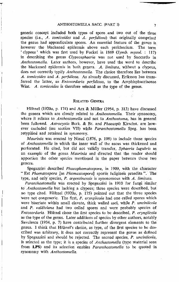
Perithecia large and prominent, either single, or more often in groups, immersed beneath blackened, shining epidermis which is pierced by a stout conical papilla. The torn host tissue usually forms a greyish-white circlet around the base of the papilla, described by Berkeley as ' a little white meal ' . The clypeus is circular and about 1 mm. wide when covering a single perithecium, but when covering several growing close together, may be up to 1 cm. long and several mm. wide. It consists of a dense mass of dark thick walled hyphae which completely obscures the epidermal and cortical cells around the papilla. Perithecia, 363-518 A high and 487-684 /* wide. The peridium is 36 v- wide with an outer layer, 23 M , of dark thick walled cells, and an inner layer, 13 M , of hyaline, thin walled compressed cells. Asci, 133-184 X 10-13 M , cylindrical, apex rounded, with a prominent apical ring, 5 X 3 M ; a short slightly tapered stalk; eight ascospores
F I G . 1. A. appendiculosa from lectotype.

are biseriate at first, uniseriate when mature and colour brown in the ascus. Ascospores, 28-36 X 8-10 p-, two celled, with a small hyaline, cordate, dwarf cell, 4-6 X 3 p- and a large brown cell, 24-30 X 8-10 p, oval-ellipsoidal with the free end rounded or tapered, but not sharply pointed; the longitudinal germ slit is short, 8-10 A , and easily seen. Paraphyses numerous^ 4 P- wide.
When Berkeley & Broome described Sphaeria appendiculosa in 1851 no locality was given. In Broome's herbarium in K a collection he made at Batheaston in March 1850 is the only one prior to this date. This has been selected as the lectotype. There are five other collections of this fungus by Broome from Batheaston between 1851 and 1871 and one from Mossburnford nr. Jedburgh in Scotland. For the next hundred years the species does not seem to have been found. European mycologists confused it with another species, curiously similar and much more common, also found on stems of Rubus. This latter species was first recognised by Spegazzini (1910, p. 40) and described by him as Entosordaria rubicola. He pointed out that while his fungus was very similar to Berkeley & Broome's it differed in having smaller spores. It was this smaller spored species that Saccardo described when he transferred Sphaeria appendiculosa to Anthostomella and the one he issued as Mycotheca veneta no. 1190.
In 1970 I found one small group of perithecia resembling Broome's species in a collection of A. rubicola (IMI 82751) and a year later found a bramble bush on the W. Coast of Scotland which provided plentiful fresh material and convinced me that there were indeed two distinct species. A. appendiculosa seems to be rare and I have found it only at*this one site, although I have examined many hundreds of brambles on the West Coast. The differences between the two species are listed after the description of A. rubicola.
S P E C I M E N S E X A M I N E D
Sphaeria appendiculosa, o n stems of Rubus, England: Batheaston, Somerset , iii. 1850, Herb. C. E. Broome no . 92 (K) Lectotype .
On stems of Rubus, Batheaston, iii. 1850, C. E . B r o o m e , e x Herb. Bloxam (probably part of lectotype); Batheaston, C. E . Broome , iii. 1851; i. 1859; iii. 1864; i. 1871 (K). Rabenhorst , Fungi europaei ser. 2 , no . 52 , Batheaston , i. 1859, leg. C. E . B r o o m e (K); Herb. Berkeley, Mossburnford (nr. Jedburgh, Scotland) n o date, A . Jerdon (K); England: Boscastle, Cornwall , ix. 1960, B. Sutton, with A. rubicola (82751); Scot land: Crinan, Argyl l , v. 1971, S.M.F. (180618).
2. Anthostomella arenaria O. Eriksson, Ark. Bot., ser. 2, 6: 443 (1967).
On Elymus arenarius in Finland, Norway and Sweden.
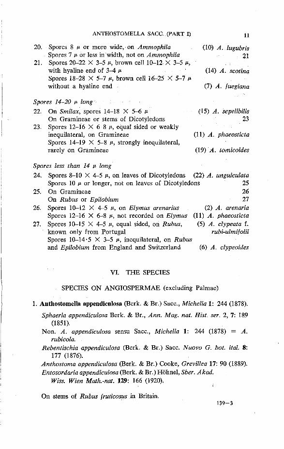
Perithecia scattered, usually single beneath the blackened epidermis which is pierced by a short wide papilla. The clypeus is not dense and is formed from dark thick walled hyphae which occur, rather sparsely, in the epidermal cells above each perithecium. Perithecia, 259-311 p high
and 220-301 v- wide. The peridium is 23 M wide, with an outer layer, 17 A , of dark thick walled cells and an inner layer, 6 M , of hyaline compressed cells. Asci, 79-91 X 6-7 u, cylindrical, apex rounded with an apical ring 2-5 X 2 /i; the stalk is short and tapered; eight ascospores are uniseriate. Ascospores, 10-12 X 4-5 At, two celled, with a hyaline dwarf cell, 2 X 1 -5 A* and a brown cell, 8-10 X 4-5 At, broadly ellipsoidal, inequilateral; the longitudinal germ line is indistinct. Paraphyses deliquesce early and are not usually seen in mature perithecia.
F I G . 2. A. arenaria from holotype.
Eriksson notes that this species has never been found on bouldery sea shores while it is common on sandy beaches where the tufts of Elymus are usually larger.
S P E C I M E N S E X A M I N E D
O n Elymus arenarius, S w e d e n : Vasterbotten, Nysatra par. Vannskaren, 14. viii. 1964. O. Eriksson, 2423a ( U P S ) Holotype .

3. Anthostomella caricis S. Francis sp. nov.
Holotypus: In foliis vetustis Caricis pendulae, England: Axminster, Somerset, 12. iv. 1973, S. M. Francis (IMI 183760).
Perithecia sparsa, substrato immersa, epidermide atrata tecta, globosa, 238-300 fi diam., papillata. Peridium, 18 u- crassum, strato externo 12 n praecipue ex cellulis fuscis crassoparietalibus composito, strato interno, 6 yx ex cellulis hyalinis compressis tenuiparietalibus. Asci, 88-91 X 8-10 n unitunicati, cylindracei, apice rotundato, iodo caerulescente, breviter stipi-tati, 8-spori. Ascosporae, 12-15 X 6-7 unicellularae, ellipsoideae, fuscae; strato gelatinoso tenui indutae; rima germinativa longitudinalis. Paraphyses, 1-5-2 M latae, mucilaginescentes.
F I G . 3. A. caricis f rom holotype.
On fallen leaves of Carex paniculata, C. pendula and Glyceria maxima in Britain and Guernsey (Channel Islands).
Perithecia scattered, usually single, immersed beneath the blackened epidermis which is pierced by a small blunt papilla. The clypeus varies

in size from a barely visible blackening around the papilla, to an area 150-270 p in diameter. It is formed from small wedges of dark thick walled hyphae which develop between the cuticle and upper epidermis. Perithecia, 300 p high and 238-300 p wide. The peridium is 18 p wide, with an outer layer, 12 p, of dark thick walled cells, and an inner layer, 6 p, of hyaline compressed cells. The wall seems to be more brittle than that of other species studied and breaks with a characteristic ' pop ' when tapped under a coverslip. Asci, 88-91 X 8-10 P, unitunicate cylindrical, apex rounded with an apical ring, 2 X 3 p ; a short tapered stalk; eight ascospores are uniseriate, often overlapping, and sometimes partially biseriate. Ascospores, 12-15 X 6-7 P, one celled, ellipsoidal; a narrow sheath surrounds each spore; the longitudinal germ slit is clearly visible. Paraphyses, 1-5-2 P wide, becoming mucilaginous.
A. caricis has been collected on several occasions in this country but has always been confused with other species. It is near A. leptospora, but differs in having wider spores which are equal sided, and in the shorter, wider, asci. On Car ex pendula it is often associated with A. punctulata from which it is easily distinguished by the size of the spores. The spores of A. punctulata are 6-9 X 3-4 P.
S P E C I M E N S E X A M I N E D
O n Carex pendula, England: Axminster , Somerset , 12. iv. 1973, S .M.F. ( I M I 183760) Holotype; Batheaston, Somerset , x. 1861, [as Sphaeria phaeosticta] e x Herb. Currey ( K ) ; Abbotsbury, Dorset , iii. 1974, S .M.F. (183759). O n Carex paniculata, Channel Islands: Guernsey, M . B. Ellis [as A. tomicoides] vi. 1947 (56817a) , vii. 1947 (56816). O n Glyceria maxima, England: W h e a t f e n Broad, Norfo lk , E . A . Ellis [as A. tomicum var. leptospora] (42035). Hos t indet. iii. 1859, vi. 1867, [as Sphaeria tomicum] ex Herb. C. E. Broome OX).
Key to species of Anthostomella recorded on Carex spp.
1. Spores two celled Spores one celled
2. Spores 12 p or longer Spores less than 12 p
3. Spores 15-23 X 7-9 P, inequilateral Spores 12-15 X 6-7 p, equal sided
4. Spores 6-9 X 3-4 p oval-reniform, ends rounded Spores 8-12 X 4-5 p oval, not reniform, ends tapered
(19) A. tomicoides 2 3 4
(21) A. tumulosa (3) A. caricis
(12) A. punctulata
(9) A. limitata
4. Anthostomella chionostoma (Dur. & Mont.) Sacc, Sylloge Fungorum 1: 285 (1882).
Sphaeria chionostoma Dur. & Mont., in Montagne, Syll. Gen. Sp. crypt.: 237 (1856),* see footnote, p. 17.
Anthostomella secalis Karst., Revue mycol. 12: 128(1890).

Anthostomella helichrysi Fabre f. solidaginis Rehm, Hedwigia 34: Rept. (163), (1895).
A nthostoma italicum Sacc. & Speg., Michelia 1: 326 (1878). Xylosphaeria italicum (Sacc. & Speg.) Cooke, Grevillea 17: 85 (1889)
On Andropogon virginicum, Agropyron pungens, Elymus arenarius, lmperata arundinacea, Secale cereale, Solidago canadensis, Spartium junceum in Algeria, Britain, France, India, Italy, Switzerland and New Jersey, U.S.A.
Perithecia single, or several together, immersed but raising the blackened epidermis is noticeable bumps. The clypeus is the same diameter as the perithecium it covers. On grass leaves where the perithecium fills the whole depth of the leaf, a smaller, secondary clypeus may form in the epidermal cells beneath each fruit body. The papilla is wide, conical and surrounded at the base by a conspicuous white circlet of torn host tissue. Perithecia, 286-350 M (549 At) high and 407-622 A wide (heights up to 549 At are found on grasses where the perithecium tends to be almost spherical). The peridium is 35 M wide, with an outer layer, 20 At, of dark thick walled cells, and an inner layer, 15 / A , of hyaline thin walled, compressed cells. Asci, 143-171 X 10-13 / A , cylindrical, apex rounded with an apical ring, 5 - 1 0 X 6 - 7 ft; little or no stalk; eight ascospores are obliquely uniserate. Ascospores, 18-25 X 7-11 n, one celled, ellipsoidal, inequilateral, one end somewhat rounded, the other apiculate; a hyaline sheath surrounds each spore and extends, as a tongue, 5-6 A long, at each end, these tongues are visible both in the ascus and in free mature spores but disappear from very old spores, they are most easily seen in water or erythrosin mounts; the germ slit is conspicuous and forms an undulating line down the length of the spore. Paraphyses numerous and thread-like.
A. chionostoma although widely distributed, is rarely collected, and whenever found has usually been described as a new species. It is present, but was not recognised, on the type material of Anthostoma mortuosum (Ellis) Sacc. at K. This collection is on the stems of two different plants, Eupatorium purpureum and Andropogon virginicum. The fungus on the Eupatorium agrees with the description given by Ellis (1882, p. 73) and is A. tomicoides, but on the stems of Andropogon the species present is A. chionostoma. The material distributed as Anthostoma italicum by Roumeguere in F. gall, exsicc. no. 4772, and by Thiimen in Mycotheca
* T h e original description of this species was published in Exploration scientifique de I'Algerie pendant les annees 1840-1842. Botanique. Paris 1846-1868 [ 1 8 6 9 ] . T h e work was published in parts and Stafleu (1967, p. 122) records the date of publication of Botanique I, Cryptogamie, livr. 18 -20 (pp. 411-631) as October 1869. The^ description of S. chionostoma is o n p . 521; it is referred t o by M o n t a g n e in Syll. Gen. Sp. crypt. (1856), on p. 237 and p. xvi , as if the publication date was ' 1 8 4 6 ' . T h e description therefore appeared 23 years later than M o n t a g n e had expected. See Mycol. Pap. 135: 12 (1974).

universalis no. 2264 came from a collection by J. Therry on Lavandula from Isere. The fungus is not conspecific with Anthostoma italicum Sacc. & Speg., and is near Anthostomella delitescens (De Not.) Sacc. (not described in this paper).
F I G . 4 . A. chionostoma A , from (Sheffield 1 8 5 0 ) ; B , from holotype of Anthostoma italicum Sacc. & Speg.; C, f rom ho lo type Of A. chionostoma.
The single British collection of A. chionostoma was made by Webster, in Norfolk, on the two sand dune grasses Agropyron pungens and Elymus arenarius. The large perithecia, 0*5 mm, diam., resemble those of A, tomicum but this latter species has much smaller spores, 11-14 X 5-7 p.. In spore size A. chionostoma is near A. lugubris and A. tumulosa but can be distinguished by the conspicuous undulating germ slit.
S P E C I M E N S E X A M I N E D
Sphaeria chionostoma Dur. & Mont . , ex Herb. Durieu de Maisonneuve, bases de tiges herbacees non determiners, Alger , i .1839, dedit. L . Mote lay ( P C ) Holotype . Anthostomella secalis, sur l e chaume Seigle (Secale cereale) pr£s St. Geonard, Fevrier, no . 179, co l lected b y Hariot [probably part o f the type described b y Karsten] ( P C ) . Anthostomella helichrysi f. solidaginis in R e h m , Ascomyceten no . 1132, an durren Stengeln, Solidago canadensis, Frauenfeld, Schweiz , x i .1892, leg. Wege l in ( K ) Isotype. Anthostoma italicum, ex Herb. Saccardo, no details on packet , ( P A D ) Holotype . Ell is , N. Am. Fungi, no . 897, in type col lect ion of Sphaeria mortuosa, only on stems of Andropogon virginicum, (not on Eupatorium), Newfield, N J , U . S . A . ( K ) .
O n Agropyron pungens and Elymus arenarius, E n g l a n d : Norfo lk , ix.1956, J. Webster (Sheffield 1851). On Imperata arundinacea, India, iii. 1971, A . P. Misra (155934). On Spartium junceum, Italy, vi i .1972, R. W. G. Dennis ( K ) .
5. Anthostomella clypeata (De Not.) Sacc. f. rubi-ulmifolii Gonz. Frag., Broteria, ser. Bot. 21: 131(1924).
Anthostomella appendiculosa (Berk. & Br.) Sacc. var. lusitanica Da Camara, Agronomia lusit. 11: 44(1949).
On stems of Rubus ulmifoliis and Rubus sp. in Portugal.
Perithecia separate or several together, immersed, but slightly raising the blackened epidermis. The papilla is small and conical with a flattened tip which just protrudes. The clypeus is not dense and is formed from dark thick walled hypae in the epidermal cells which surround the papilla. Perithecia, 209-253 P high and 230-330 P wide. The peridium is 17 P wide with an outer layer, 11 M of thick walled dark cells, and an inner layer, 6 p of hyaline thin walled cells. Asci, 91-110 X 6-9 p, cylindrical, apex rounded with an apical ring 3 X 3 M ; the stalk is short and tapered; eight ascospores are obliquely uniserate. Ascospores, 10-15 X 4-5 p, two celled, with a small hyaline dwarf cell 1-2 X 2 c, and a brown cell 9-13 X 4-5 p, oval to ellipsoidal, equal sided, the free end rounded; germ slit, longitudinal, the same length as the spore, very fine. Paraphyses numerous and conspicuous, 2-3 P wide.
A. clypeata f. rubi-ulmifolii is known from only two collections, both from Portugal. They were given two different names and both are inappropriate. The fungus has no similarity either with A. clypeata (which is a dubious species) or A. appendiculosa. It is near A. clypeoides from which it differs in having slightly larger spores which are equal sided, with the free end of the brown cell rounded rather than pointed. The asci are longer and have more persistent walls than those of A. clypeoides.

Although the nomenclature, as a forma of a dubious species, is unsatisfactory I have not changed the name of the taxon as further investigation of the tropical species in IMI may alter the concept both of this taxon and also A. clypeoides.
F I G . 5. A. clypeata f. rubi-ulmifolii f rom holotype.
S P E C I M E N S E X A M I N E D
On stems of Rubus ulmifoliis, Portugal , nr. P o v o a Lanhoso , S. Gens , vi i i .1924, leg. G. Sampaio , det. G. Fragoso , [as A. clypeata], ex Herb. Mus. Nac. Cienc. Nat. Madrid. Fungi , no . 7062 ( M A ) Holotype .
O B Rubus sp . , Portugal , Algarve pr. Caldas de Monchique , 9. i i i .1948, leg. M.R. de Sousa D ias , no . 409, type of A appendiculosa var. lusitanica, (LISE 23954).

6. Anthostomella clypeoides Rehm, Annls mycol. 7: 406 (1904). Entosordaria clypeoides (Rehm) Hohnel, Sber. Akad. Wiss. Wien
Math.-nat. 129: 166 (1920).
On stems of Epilobium angustifolium and Rubus sp. in Britain and Switzerland.
Perithecia scattered or, on Rubus, often in small groups, immersed beneath the blackened epidermis which is slightly raised. The clypeus is dense, with dark thick walled hyphae filling the plant cells surrounding the
20/i 10/x I : 1 I - J
F I G . 6. A. clypeoides a, from Kunze , Fungi selecti no . 329 ( K ) ; b , on Epilobium, 30.xii.1967, R . W . G . D . ( K ) .
papilla. Papilla small and conical, with a white circlet of torn host tissue at the base. Perithecia, 190-262 P- high, 217-228 p wide. The peridium is 26 p. wide with an outer layer, 18 p, of dense thick walled cells, and an inner
layer, 8 p, of thin walled compressed cells. Asci, 6 5 - 8 1 X 6 - 8 P, cylindrical, apex rounded, with an apical ring 2 X 2 / i ; the stalk is very short; eight ascospores are obliquely uniseriate. The ascus walls are thin and often disappear before the spores are fully mature. Ascospores, 1 0 - 1 4 - 5 X 3 - 5 p, two celled, with a small hyaline dwarf cell, 1—1-5 X 2 / J , and a brown cell, 9 - 1 3 X 3 - 5 P, oval to ellipsoidal, inequilateral, free end tapered; no germ slit seen. Paraphyses present in young perithecia, becoming mucilaginous.
This species is based on a collection by Winter on Rubus which was distributed by Kunze in his Fungi selecti, no. 329 , as A. clypeata. Kunze was commended (Revue mycol. 2: 56 , 1880) on the excellence of his exsiccatae which provided ample material of each fungus in varying stages of development; the packet of A. clypeata at K is no exception. Rehm created the new species A. clypeoides, as Winter (1886, p. 5 5 9 ) had noted that the spores in his collection had a distinct hyaline apiculus at the lower end and therefore differed from the spores of A. clypeata which De Notaris had described as one celled. (A. clypeata is discussed in Section VII).
I have seen no other collections of A. clypeoides on Rubus* There is a single specimen on Epilobium in K which consists of only a few perithecia. The spores are a little larger with the brown cell measuring 1 0 - 1 3 X 4 - 5 , while on Rubus it is 9 - 1 1 X 3 - 4 A .
There is also material in IMI on tropical grasses which can be broadly grouped with A. miscanthea Sacc. (Saccardo, 1917, p. 75) , a species not described here, but which is close to A. clypeoides. It seems likely that further work may show that the collections on Rubus and Epilobium form part of a variable species which should be defined more widely.
S P E C I M E N S E X A M I N E D
Joannes Kunze, Fungi selecti exsiccati, Fungi helvetici, no . 329, Anthostomella clypeata, ad Ruborum sarmenta arida, in pinetis Sihwald pr. Zurich, Helvet iae , vi i i .1878, G. Winter (K, P A D ) Isotypes.
On Epilobium angustifolium, Wakehurst P lace , Ardingly, E . Sussex, England, x i i .1967, R. W . G. D e n n i s (K).
7. Anthostomella fuegiana Speg., Boln Acad. nac. Cienc. Cordoba 11 (2): 194 (1888) .
Entosordaria fuegiana (Speg.) Hohnel, Sber. Akad. Wiss. Wien Math.-nat. 129: 166 (1920) .
Non Entosordaria fuegiana Speg., Boln Acad. nac. Cienc. Cordoba 27: 3 5 8 (1924). A. phaeosticta (Berk.) Sacc]
On Cladium mariscus, Eriophorum vaginatum, Luzula sylvatica and Rostkovia grandiflora in Britain, Denmark, Germany and Tierra del Fuego.
Perithecia separate but in groups, immersed beneath the blackened epidermis which is slightly raised and pierced by a small conical papilla. The leaf surface in the perithecial areas often has a pale, bleached appearance.
* F o o t n o t e in proof : T h e species has recently been found in England o n fa l len leaves of Rubus b y M. C. Clark; Winter's col lect ion w a s on stems.

The clypeus is densely black with an irregular edge and is formed from a network of thick dark walled hyphae in the epidermal cells above each perithecium. In Luzula sylvatica, where the perithecia occupy the whole depth of the thin leaf, a second clypeus often forms in the epidermal cells beneath each fruitbody. Perithecia, 250-385 P high and 242-308 P wide. The peridium is 23 p wide and consists of an outer layer, 17 p, of dark thick
F I G . 7 . A. fuegiana A , on Cladium ( 6 1 8 1 6 ) ; B , o n Rostkovia, holotype; C , o n Luzula ( 1 4 3 7 0 1 ) .
walled cells, and an inner layer, 6 p, of hyaline thin walled, compressed cells. Asci, 100-143 X 8-10 p, cylindrical to elliptical, apex rounded or slightly narrowed, with an apical ring, 3 X 3 p; the stalk is short, abruptly tapered and often curved; eight ascospores are partially biseriate. Ascospores, 18-28

S P E C I M E N S E X A M I N E D
On Rostkovia [as Rhosthkovia] grandiflora, Canal de Beagle (Fuego) , v. 1882, Spegazzini , (LPS 7114) Holotype .
On Cladium mariscus, England; Wheat fen Broad, Norfo lk , E . A . o r M . B. & J. P . Ell is , iv .1940 (21401 c); xi i .1946 (10889); i .1947 (10173, 10322); iv.1947 (14875 a, 16534 f); v .1947 (15338, 15393 a); iii .1948 (27771); v .1948 (34589); iv .1949 (34923); iv .1954 (56711); iv.1963 (100222); R. W . G. Denn i s , 22.X.1944 [as A. tomicum v . leptospora] (K); Fi lby Broad, Norfo lk , E . A . E . , iv .1950 (61816).
O n Eriophorum vaginatum, Denmark: Lyngby M o s e , vi i .1889, O. Rostrup (CP). O n Luzula sylvatica, England: Axminster , Somerset , iv .1973, S .M.F. (185000). Scotland: R h u m , iv.1961 [as A. tumulosa], R . W . G . D . (K); Soay, Skye, vi i .1968, S .M.F. (143701); vi i .1969, S .M.F. (151860); Crinan, Argyl l , v .1970, S .M.F. (151861); vii i .1970, S .M.F . (151862); L o c h Bharabhat, Lewis , vi i i .1973, R . W . G . D . (K).
8. Anthostomella leptospora S. Francis comb. nov. Anthostomella tomicum (Lev.) Sacc. var. leptospora Sacc, Sylloge
Fungorum 1: 282 (1882).
On dead leaves of Cladium mariscus in Britain and France.
Perithecia widely scattered, each immersed beneath a small clypeus, 200 p diameter, which is pierced by a short, blunt papilla. The clypeus is formed from thick walled brown hyphae which develop in the cells of the epidermis and parenchyma which surround the papilla. Perithecia, 231-264 p high and 187-231 p wide. The peridium is 26 P wide, with an outer layer, 19 of thick walled cells, and an inner layer, 7 P, of hyaline,
X 5-7 n, two celled, with a hyaline dwarf cell, 2-3 X 2 M , and a brown cell, 16-25 X 5-7 P, fusiform-oval, slightly inequilateral and often collapsing laterally, acutely pointed at the free end on Eriophorum, Luzula and Rostkovia, often more rounded on Cladium; the longitudinal germ slit is prominent. Pafaphyses numerous, 2 p wide, becoming mucilaginous.
Collections of this species in Europe have always been referred to A. tumulosa (see Petrak, 1931, p. 157; Munk, 1957, p. 121; Dennis, 1964, p. 119) and Spegazzini's description of A. fuegiana was overlooked. The external appearance of the two fungi is similar but the asci and spores are quite different. A. fuegiana is common in this country on the dry, dead, leaves of Luzula sylvatica.
Ascospores and fruitbodies of a fungus that appears to be identical with A. fuegiana have been found in the Netherlands in samples of peat formed in Atlantic, Sub-boreal and Sub-Atlantic periods (approximately 4000 B . C . to 1000 A . D . ) . The spores are perfectly preserved together with the outer wall of the fruitbody and a fragment of clypeus around the papilla. The peats contain Eriophorum, a present day host of the species, and it would be interesting to know whether the fungus can be found on living plants in the area. The material was sent to this Institute by Bas van Geel of the Universiteit van Amsterdam; the results are not yet published and I am grateful to Mr. van Geel for his permission to include this data.
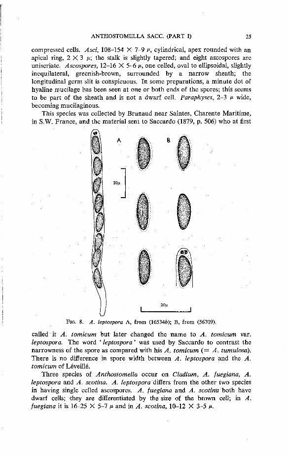
compressed cells. Asci, 108-154 X 7-9 v, cylindrical, apex rounded with an apical ring, 2 X 3 ju.; the stalk is slightly tapered; and eight ascospores are uniseriate. Ascospores, 12-16 X 5-6 v, one celled, oval to ellipsoidal, slightly inequilateral, greenish-brown, surrounded by a narrow sheath; the longitudinal germ slit is conspicuous. In some preparations, a minute dot of hyaline mucilage has been seen at one or both ends of the spores; this seems to be part of the sheath and is not a dwarf cell. Paraphyses, 2-3 p- wide, becoming mucilaginous.
This species was collected by Brunaud near Saintes, Charente Maritime, in S.W. France, and the material sent to Saccardo (1879, p. 506) who at first
called it A. tomicum but later changed the name to A. tomicum var. leptospora. The word ' leptospora' was used by Saccardo to contrast the narrowness of the spore as compared with his A. tomicum (= A. tumulosa). There is no difference in spore width between A. leptospora and the A. tomicum of Leveille.
Three species of Anthostomella occur on Cladium, A. fuegiana, A. leptospora and A. scotina. A. leptospora differs from the other two species in having single celled ascospores. A. fuegiana and A. scotina both have dwarf cells; they are differentiated by the size of the brown cell; in A. fuegiana it is 16-25 X 5-7 P and in A. scotina, 10-12 X 3-5 ti.
F I G . 8 . A. leptospora A , from ( 1 6 5 3 4 6 ) ; B , from ( 5 6 7 0 9 ) .
S P E C I M E N S E X A M I N E D
Herb. P.A. Saccardo, on Cladium mariscus [as A. tomicum], ( P A D ) Holotype . O n Cladium mariscus, England: Wheat fen Broad, Norfo lk , M . B. Ell is , x i i .1946
(10890); i.1947 (10324); iv.1947 (14875 b, 16534 g ) : v.1947 (15279 b, 15413 g); v.1948 (34590); iv .1954 (56709). Chippenham F e n , Cambs. v .1963, J. Webster (152291). Wales: Anglesey , iv .1958, J.W. (152290).
9. Anthostomella limitata Sacc, Atti Accad. scient. veneto-trent.-istriana 4: 101 (1875).
Anthostoma limitata (Sacc.) Cooke, Grevillea 17: 90(1889). Anthostomella gracilis Tassi, Bull. Lab. Orto Bot. Reale Univ. Siena
3: 53 (1900). Anthostomella melanoderma Rehm, Ost. bot. Z. 54: 82 (1904). Anthostomella argentinensis (Speg.) Petrak & Syd., Annls mycol. 23:
213 (1925). Phaeophomatospora argentinensis Speg., An. Mus. nac. Hist. nat. B.
Aires, ser. 3,12 : 339 (1909).
On Callistemon sp., Carex acutiformis, C. paniculata, C. riparia, Chamaerops humilis, Conium maculatum, Euphorbia cyparissias, Galium mollugo, Iris pseudacorus, Oenanthe crocata, Rosa sp., Rubus sp., Typha latifolia, Vitis vinifera, Umbelliferae indet., in Argentina, Britain, Channel Islands, Germany and Italy.
Perithecia small and inconspicuous, closely grouped together, immersed but usually raising the epidermis which is pierced by a small conical papilla. There is little or no clypeus but the epidermis may appear blackened around the papilla. Sections of perithecia show that the papilla has a thick wall, the outer surface of which fuses with the epidermal cells giving the appearance of a clypeus. A more general blackening of the epidermis appears to occur when the host tissue is old and decayed, a thin network of dark thick walled hyphae is then found in the epidermal and cortical cells surrounding the perithecia. Perithecia, 125-207 At high and 104-220 A* wide. The peridium is 16 M wide with an outer layer, 10 At, of dark thick walled cells, and an inner layer, 6 At, of hyaline compressed cells. Asci, 66-95 X 6-8 At, cylindrical, apex rounded with an apical ring 1 X 3 At; the slender tapered stalks often remain joined at the base giving characteristic bunches of asci in slide preparations; eight (occasionally six) ascospores are obliquely uniseriate. Ascospores, 8-12 X 4 - 5 At, one celled, oval with tapered ends, slightly inequilateral, a central guttule may partially obscure the faint germ slit which forms either a single diagonal or crossed diagonals. Paraphyses, 2 At wide, numerous at first but not persisting long.
A. limitata has been recorded on a wide range of plants and yet today it is scarcely known. Traverso (1907, p. 480) made a similar comment some sixty years ago suggesting that perhaps it was confused with other species. This is possible, and the similarity between A. limitata and'A clypeata is discussed in the notes on the latter species in section VII.
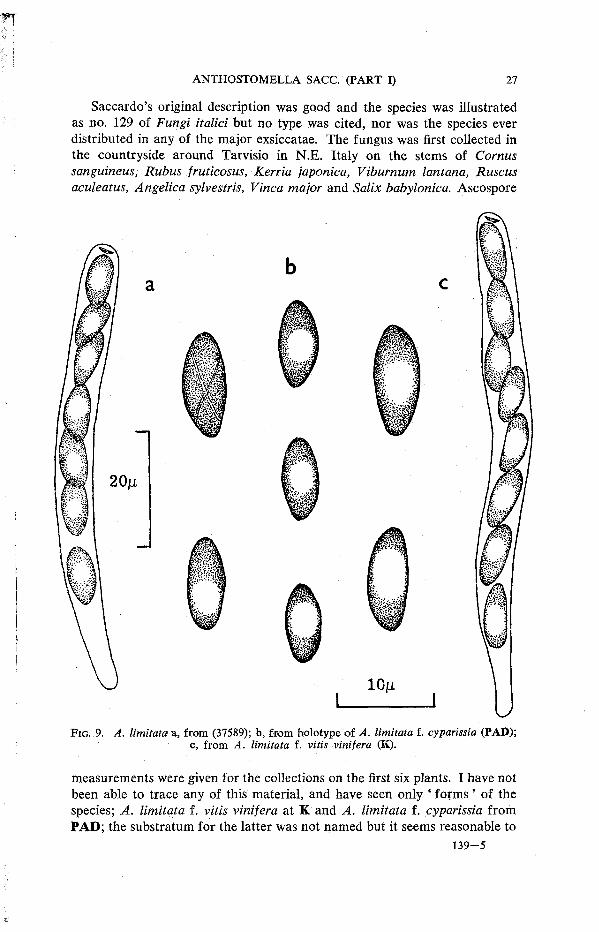
Saccardo's original description was good and the species was illustrated as no. 129 of Fungi italici but no type was cited, nor was the species ever distributed in any of the major exsiccatae. The fungus was first collected in the countryside around Tarvisio in N.E. Italy on the stems of Cornus sanguineus, Rubus fruticosus, Kerria japonica, Viburnum lantana, Ruscus aculeatus, Angelica sylvestris, Vinca major and Salix babylonica. Ascospore
F I G . 9 . A. limitata a, from ( 3 7 5 8 9 ) ; b, f rom ho lo type of A. limitata f. cyparissia ( P A D ) ; c, from A. limitata f. vitis vinifera (K).
measurements were given for the collections on the first six plants. I have not been able to trace any of this material, and have seen only ' forms' of the species; A. limitata f. vitis vinifera at K and A. limitata f. cyparissia from PAD; the substratum for the latter was not named but it seems reasonable to
assume it was Euphorbia cyparissias, especially as Traverse- (1907, p. 479) lists Euphorbia as a host plant for A. limitata. I have found no reasons in the literature for the designation of these collections as ' forms' of A. limitata, but presumably it was intended to indicate a different substratum or a slight difference in the fungus. The former seems unlikely as Saccardo, by listing eight host plants, had indicated a wide range for the species in his description. The only difference I have found between these two ' forms ' and A. limitata, as defined by Saccardo, was in the size of the spores, but it was very slight and not sufficient to maintain these two collections as distinct from the species. The table below compares the spore measurements of the two ' forms ' with the range of measurement found in other collections.
T A B L E I. Comparison of Ascus and Ascospore measurements in A. limitata
Asci Ascospores Saceardo's original diagnosis 7 0 - 9 0 X 4 - 5 M 10-12 X 4 - 5 At f. cyparissia ( P A D ) 6 4 - 8 5 X 6-8 A* 8 - 9 ( 1 1 ) X 3 - 5 At f. vitis vinifera (K) 8 0 - 9 0 X 6-8 M 10-11 (13) X 4 - 5 At on Galium (Herb. R e h m , S) 84-91 X 6-7 At 11 X 4 At on Galium mollugo (K) 66 X 6 H 9--11 X 3 - 5 At o n Carex acutiformis (17393c) 73 X 7 ft 10--11 X 4 At o n C. paniculata (56816) not seen 10-- 1 1 X 4 - 5 At on C. riparia (37589) 7 1 - 8 2 X 6-7 A 1 9--11 X 4 - 5 At o n Chamaerops sp. ( P A D ) not seen 10--12 X 4 At o n Iris pseudacorus (70198) 7 7 - 8 2 X 6-7 A* 10--12 X 4 - 5 At o n Oenanthe crocata (78035) 6 8 - 7 3 X 6 n 9--11 X 4 - 5 At o n Typha latifolia (34510) 6 6 - 7 7 X 6-8 At 8--12 X 4 - 5 At as A. gracilis ( S I E N A ) 95 X 5 p. 10 X 4 - 5 At as A. melanoderma (S) 69 X 7 - 8 At 11 X 4 - 5 At
The crossed diagonal germ slit found in the ascospore of this species is a very characteristic feature, but not always easily seen. It shows most clearly in a preparation which has been stained in cotton blue for several weeks—or months. If time presses, an iodine mount has been found helpful.
The possibility that A. limitata and A. clypeata may be the same species has already been mentioned. If this can ever be proved, then A. clypeata is the earlier, and therefore correct, name for the species. Because of this uncertainty and as I have so far been unable to trace any of the collections listed by Saccardo in his description, I have not designated a lectotype.
S P E C I M E N S E X A M I N E D
A. limitata f. cyparissia, n o details, Herb. Saccardo, ( P A D ) . A. limitata f. vitis vinifera, o n Vitis, Conegl iano, Aut . 1876, ex Herb. M. C. Cooke (K). A. gracilis, o n Callistemon sp. Ort. Bot . Siena, vi. 1900 ( S I E N A ) Holotype . A. melanoderma, on ? Umbel l i fer s tems, A n d e c h s a m A m m e r s e e , Oberbayern (W. Germany) H . R e h m (S) Holotype . Phaeophomatospora argentinensis, on Conium maculatum, Santa Catalina, Buenos Aires , x i .1905 , C. Spegazzini (LPS 1216) Holotype . Saccardo, Mycotheca veneta no . 1444, A. clypeata, o n Rubus & Rosa, Be l luno , Aut . 1879, C. Spegazzini (RO) . Herb. R Horti Romani, on Chamaerops humilis, nr. R o m e , i i .1884, Baccarini & A ve t t a ( P A D ) . Herb. H. Rehm, A. ? limitata, on Galium, locality i l legible, xi .1910, H . R e h m (S).
On Carex acutiformis, England: Wheat fen Broad, Norfo lk , E . A . Ell is , v i .1944, (17394c); iv.1945 (17393c). On Carex paniculata, Channel Islands: Guernsey, vi .1947,

M . B. Ellis (56816). On Carex riparia, England: W y t h a m Park, Oxford, ix .1949, M. B . & J. P. Ellis (37589). On Galium mollugo, England: Polperro, Cornwall , vi i .1927, F . Ri l s tone [as A. tomicoides], ex Herb. W. B. Grove (K). On Iris pseudacorus, England: Wheat fen Broad, Norfo lk , viii .1957, J. Webster (70198). O n Oenanthe crocata, Channel Islands: Jersey, xi i .1959, J.W. (78035). O n Typha latifolia, England: Wheat fen Broad, Norfo lk , v .1948, M.B.E. (34510).
10. Anthostomella lugubris (Rob. in Desm.) Sacc., Sylloge Fungorum 1: 278 (1882).
Sphaeria lugubris Rob. in Desm., Annls Sci. nat. (Bot.) ser. 3, 8: 172 (1847).
Sordaria lugubris (Rob. in Desm.) Ces. & De Not., Comment. Soc. critt. Ital. 1 (4): 226 (1863).
Anthostoma lugubris (Rob. in Desm.) Niessl, Verh. naturf. Ver. Briinn 10: 208, 1871 (1872).
On Ammophila arenaria in Britain, Belgium, Channel Islands, Denmark, Eire, France, Norway, Sweden.
Perithecia usually scattered, immersed beneath the blackened epidermis which is penetrated by a short wide papilla. The clypeus which measures 500-800 X 230-360 P is formed from dark thick walled hyphae which fill the epidermal cells above each perithecium. Perithecia, 250-331 P high and 300-400 p wide. The peridium is 25 P wide, with an outer layer, 13 p, of dark thick walled cells and an inner layer, 12 P of hyaline, thin walled, compressed cells. Asci, 126-161 X 11-14 P, cylindrical, apex rounded with an apical ring, 5-6 X 3-4 p; the stalk is very short and barely tapered; eight ascospores are uniseriate or partially biseriate when young. Ascospores, 18-22 (24) X 8-11 p, one celled, oval to ellipsoidal with tapered ends; a conspicuous sheath surrounds each spore and is particularly noticeable on young hyaline spores; longitudinal germ slit 10-12 P, not easily seen. Paraphyses numerous, 4-5 p wide.
' Cette jolie Spherie', a phrase used in the original description of A. lugubris, was first collected by Roberge on Ammophila from the dunes of Lyon-sur-Mer (now Lion), Calvados in N.W. France. All the material I have seen has been on Ammophila apart from one rather dubious specimen in K (ex Herb. Sir H. C. Hawley) labelled ' on Elymus, Blakeney '. This collection was very poor and it was impossible to be certain whether the species was A. lugubris or A. chionostoma which has been recorded on Elymus from the same area of Norfolk.
Sixteen of the nineteen collections examined agreed with the type material of A. lugubris, but three differed in having spores with a dwarf cell. A comparison of the perithecia and asci with those of the type showed no significant differences and, apart from the presence of the dwarf cell, the size and general shape of the spores was also similar. Each collection, from three different localities, consisted of only a few perithecia and although I have

searched extensively at the Rhum site I have not yet been able to find more material. For the present, therefore, these collections are considered to be an atypical form of A. lugubris and are listed separately in the ' Specimens examined'.
F I G . 10. A. lugubris A , from (143700); B , on Ammophila, R h u m , 4 . i x . l 9 6 2 , R . W . G . D . ( K ) .
Chitonospora ammophilae has been present either with A. lugubris or replacing it in some of the collections at K. The fungus was first described by Bommer & Rousseau (1890, p. 270) as Chitonospora ammophila Sacc,

Bomm. & Rouss., whereas the reference usually cited for this species, Saccardo (1891, p. 797) was published a year later with a different author citation, i.e., C. ammophila Bomm., Rouss. & Sacc. Miiller (1950, p. 188) included the genus with Leptosphaeria. The external appearance of C. ammophilae somewhat resembles A. lugubris, and the spores, when young, are of similar size and shape. The mature spores of Chitonospora, however, have three very clear and definite septa. When collecting on Ammophila the two fungi can be distinguished by looking for A. lugubris on white bleached leaves where the black clypeus and wide papilla are prominent, while C. ammophilae, which has a pointed papilla and no clypeus, occurs on leaves with a greyish-black stain.
A. lugubris differs from A. phaeosticta, the only other species of Anthostomella recorded on Ammophila, by its large single celled spores. The spores of A. phaeosticta are two celled and the brown cell measures 10-14 X 6-8/t.
S P E C I M E N S E X A M I N E D
Desmazieres , Plantes Crypt. France, Ed. I, Ser. I (1825-1851) no . 1792, Sphaeria lugubris R o b . in herb. , on Ammophila arenaria [as Calamagrostis] (K) Isotype. Ibid., Ed. II , Ser. I (1836-1851) no . 1442 (K). Westend. & Wallr. , Herb. Crypt. Belg., no . 1219, on A mmophila arenaria, d'Ostende (K).
O n Ammophila arenaria, Eng land: Perranporth, Cornwall , ix .1929, F . Ri ls tone = Chitonospora ammophilae (K); Scot land: Cape Wrath, Sutherland, ix .1954, R. W . G. D e n n i s (K); L o c h Boisdale, S. Uis t , vi i .1969, S .M.F. (143700); Ki lpheder S. Uis t , vi i i .1973, R . W . G . D . (K); Liskintyre dunes, S. Harris, vi i i .1973, R . W . G . D . (K); Sa lum Bay, Tiree, vii i .1973, R . W . G . D . (K); W a l e s : N e w b o r o u g h , Anglesey , vii .1927, P. G. M . R h o d e s , 3006, = Chitonospora ammophilae (K); St. David's Pembs . , vi i .1928, P .G.M.R. , 3649c (K); Freshwater Bay, Pembs. , vii i .1928, P .G.M.R. , 3748 (K); nr. Mochras , viii. 1929, P . G . M . R . 4322 , = Chitonospora ammophilae (K). Channel Islands: H e r m , ix .1931, P . G . M . R . 4990 (K); vi .1947, M . B . Ellis (36170). Eire: Crookhaven, Co. Cork, vi i .1964, M. Scarrell (K). O n Elymus arenarius, England: B lakeney , Norfo lk , Herb. Sir H. C. Hawley, doubtful record, old material , possibly A. chionostoma. (K).
Material wi th Dwarf Cell on Ascospore; E n g l a n d : Perranporth, Cornwall , vii i .1940, F . Ri ls tone (50712); Scot land: Ki lnwry D u n e , R h u m , ix.1962, R . W . G . D . (K); W a l e s : Tenby , Pembs. , vii .1928, P .G.M.R. 3662A (K).
11. Anthostomella phaeosticta (Berk.) Sacc, Michelia 1: 374 (1878). Sphaeria phaeosticta Berk., in Hooker, The Botany of the Antarctic
Voyage* 1 (9): 171 (1845). Non A. phaeosticta sensu Sacc, Michelia 1: 374 (1878); Fungi italici
no. 374; nec. S. phaeosticta sensu Berk., Ann. Mag. nat. Hist. ser. 2, 9: 383 (1852) [ = A. punctulata].
Anthostomella ammophilae (Phill. & Plowr.) Sacc, Sylloge Fungorum 1: 763 (1882) [as ' ammophila '].
Sphaeria ammophilae Phill. & Plowr., Grevillea 10: 73 (1881) [as ' ammophila'].
Entosordaria ammophilae (Phill. & Plowr.) Hohnel, Sber. Akad. Wiss. Wien Math.-nat. 129: 166(1920).
Anthostomella punctulata (Rob. in Desm.) Sacc. var. nardi Rehm, Annls mycol. 7: 408 (1909).
* See Stafleu, 1967, p. 207.

Entosordaria fuegiana Speg., Boln Acad. nac. Cienc. Cordoba 27: 358 (1924). Nom. lillegit. Non E. fuegiana (Speg.) Hohnel, Sber. Akad. Wiss. Wien Math.-nat. 129: 166 (1920) [ = A. fuegiana Speg.].
On Ammophila arenaria, Hierochloe antarctica, H. brunonis, Nardus stricta in Auckland Island and Campbell Island (New Zealand), Britain, Denmark, Germany, Norway and Tierra del Fuego.
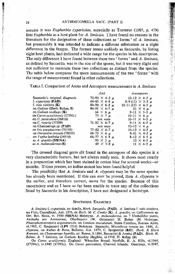
Perithecia scattered, separate or several close together, beneath conspicuous violet-black spots which are usually elongated in the longitudinal axis of the leaf. The clypeus is formed from a wedge of dark thick walled hyphae between the cuticle and epidermal cells, these may also fill the epidermal cells above each perithecium. The papilla is 100 v- wide, flat topped, with the central canal clearly visible. Perithecia, 250-394 ju. high and 228-291 M wide. The peridium is 18 v- wide, with an outer layer, 13 M , of dark thick walled cells and an inner layer, 6 M , of hyaline, thin walled, compressed cells. Asci, 88-106 X 9 M (X 13 M if spores biseriate) cylindrical, apex rounded with an apical ring 3 X 3 M ; stalk short and tapered; eight ascospores uniseriate or partially biseriate especially when young. The biseriate arrangement is very noticeable in young asci of the type material of A. phaeosticta. In his description Berkeley commented on the marked change in appearance between the short, wide immature ascus with biseriate spores and the mature ascus where the spores are uniseriate. This feature is much less marked in the collections on Ammophila and also in Spegazzini's collection on Hierochloe from Tierra del Fuego. Ascospores, 12-16 X 6-8 M , two celled, with a hyaline dwarf cell, 2 X 2 n and a brown cell 10-14 X 6-8 M , ellipsoidal, slightly inequilateral, the free end rounded. A narrow sheath surrounds the spore initially, it is slightly thickened at the end of the brown cell forming a small cap which remains visible in older spores when the rest of the sheath has disappeared. It was incorrectly described by Phillips & Plowright (for A. ammophilae) as a hyaline appendage. Germ slit longitudinal, 7-8 M . Paraphyses present in young perithecia, soon becoming mucilaginous.
The type material of Sphaeria phaeosticta is on Hierochloe brunonis from Auckland Island and Campbell Island. These islands, which lie about 400 miles to the south of New Zealand were visited on the Antarctic voyage (1839-1843) of H.M. discovery ships Erebus and Terror. (The islands were then called Lord Auckland's group and Campbell's island). J. D. Hooker was botanist to the expedition and he sent any fungi that were collected to Berkeley. Berkeley's descriptions were published, in 1845, under his own name, in The Botany of the Antarctic Voyage.
In 1852, Berkeley & Broome in 'Notices of British Fungi', no. 651, named a collection from Thornhaugh, Northants., on Carex pendula, Sphaeria phaeosticta. The illustration shows this fungus to be A. punctulata. This particular collection is not preserved at K, but there are many others
A. phaeosticta f rom holotype a, mature and b , immature asci and ascospores.
including four in the type folder of S. phaeosticta in Berkeley's herbarium in K of A. punctulata leaving no doubt that Berkeley, having described the Antarctic collection on Hierochloe as 5*. phaeosticta, then used the name for another species described by Desmazieres in 1851, i.e., S. punctulata. Saccardo in 1878 followed Berkeley, and his transfer of S. phaeosticta to Anthostomella is based on a collection of A. punctulata by Spegazzini on Arundo donax. Such was Berkeley's reputation that all collections of A. punctulata made in this country were referred to A. phaeosticta and the name ' punctulata' is not found in the British collections at K until the 1930s when Rhodes and Grove started to use it for their collections of an Anthostomella on Carex pendula. European mycologists expressed doubts from time to time as to whether there was any real difference between the exsiccatae issued as A. phaeosticta and A. punctulata (Niessl 1876, p. 198). Petrak (1940a, p. 340) considered the European forms of the two fungi to be the same (as indeed they were) but suggested that the type of A. phaeosticta might be different. Arx & Muller (1954, p. 316) combined the two species as Anthostoma punctulatum [as punctulata].
Phillips & Plowright, not knowing of Berkeley's confusion of the two species, redescribed a collection of A. phaeosticta on leaves of Ammophila as Sphaeria ammophilae. This is the specific epithet by which this widespread species has been known for the past 80 years.
A. cymbisperma Wint. (1887, p. 17) was described from a collection made by Hariot (no. 19) from Cape Horn on ' graminacearum ma jorum'. From Winter's description of the species it seems to be near A. phaeosticta. The material that Winter examined is not at B; there is a fragment of grass leaf which is probably an isotype collection at PC but I could find no fungus on the material. Spegazzini (1888, p. 194) in 'Fungi Fuegiani' lists the species, but it is not clear whether he is referring to the type material or to other collections considered to be the same species.
Petrak & Sydow (1924, p. 328) considered Coniothyrium ammophilae Oud. to be an overripe collection of A. phaeosticta [as A. ammophilae] in which the asci had dissolved. I have not seen material of this species. Martin (1969, p. 398) cited A. ammophilae [as A. ammophila Phill. & Plowr.] as a synonym of A. lugubris with a spore size of 20-5-23 X 9-5-10 M The two species are quite distinct and A. phaeosticta is easily distinguished by its smaller, two celled spores.
S P E C I M E N S E X A M I N E D
Sphaeria phaeosticta Berk. , o n Hierochloe brunonis, Auck land Group & Campbell Islands, Herb. Berkeley (K) Holotype. Sph. o n Ammophila, H o l m (England, Norfo lk ) vi i i .1880, ex Herb. W. B. Grove (K) probably Holotype as Herb. Plowright (K) has only a sketch of the species and no material. Anthostomella punctulata var. nardi, on Nardus stricta, Bayerischen Wald , 9 . i . l885, H. R e h m (S) Holo type . Entosordaria fuegiana on Hierochloe antarctica, Sholl Bay (Tierra del F u e g o ) (LPS) Holotype . Sydow, Mycotheca germanica, no . 980, A. ammophilae (60798).
On Ammophila arenaria, Scotland: Cumbrae, vii i .1914, D . A . B o y d (K); Ardnamur-chan, vi i i .1968, R . W . G . D . (K); Mul l , viii .1968 (K); Bute , v .1968, S .M.F. (143704);
v.1969 (143698); Coll , vii i .1973, R . W . G . D . (K); Islay, vi i i .1974, R . W . G . D . (K). Wales: Anglesey , vii .1927, P. G. M . Rhodes , 3006J, = Chitonospora ammophilae (K); ix .1950, J. Webster (56957); Tenby Burrows, Pembs. , vi i .1928, P . G . M . R . , det W. B . Grove (with A. lugubris) (K).
T h e col lect ions labelled A. phaeosticta which were found to be A. punctulata are listed under the latter name.
12. Anthostomella punctulata (Rob. in Desm.) Sacc, Sylloge Fungorum 1: 278 (1882).
Sphaeria punctulata Rob. in Desm., Annls Sci. nat. {Bot.) ser. 3, 16: 314 (1851).
Leptosphaeria phaeosticta Auersw., in Gonnerman & Rabenhorst's Mycologia Europaea Heft V & VI, tab. 11, fig. 154 (1869).
On dead leaves of Arundo donax, Carex pendula, Luzula pilosa, Phragmites communis, in Austria, Britain, France and Italy.
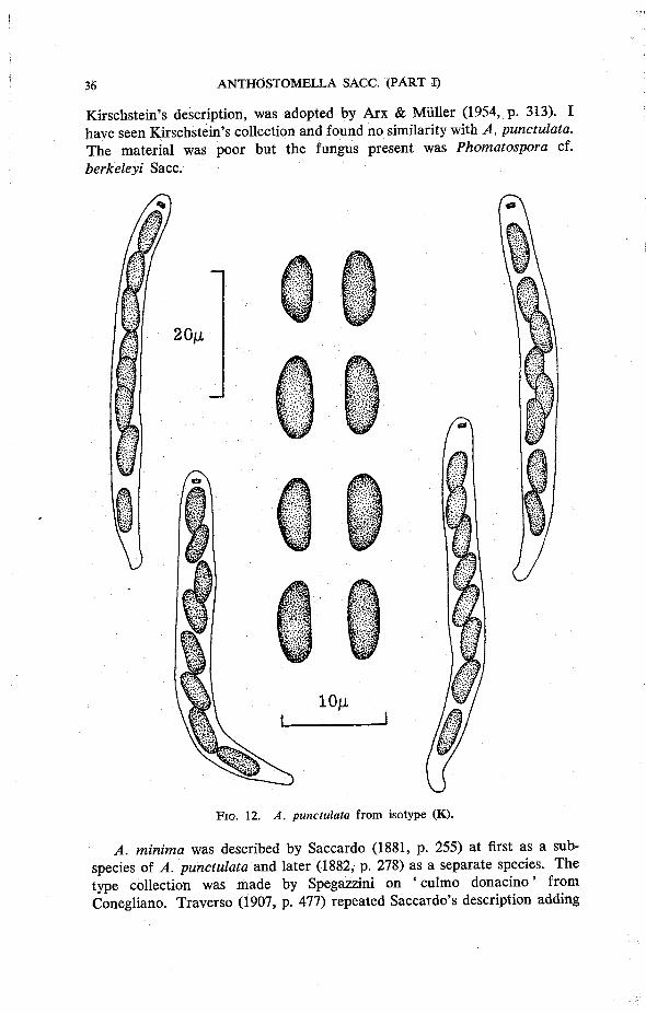
Perithecia numerous, in groups but usually separate, immersed beneath a small clypeus which is black in the centre but shades to a brownish-violet at the somewhat diffuse edge. Papilla small and conical. The clypeus consists of dark thick walled hyphae which form wedges between the epidermis and cuticle and also fill the epidermal cells above each perithecium. Perithecia, 170-192 P high, and 154-177 p wide. The peridium is 17 p wide with an outer layer, 12 P, of dark thick walled cells, and an inner layer, 5 P, of hyaline thin walled cells. Asci, 49-66 X 5-7 p, cylindrical, apex rounded with an apical ring, 1 X 2 P; a short, slightly tapered stalk; eight ascospores are uniseriate or partially biseriate. Ascospores, 6-9 X 3-4 P, one celled, reniform-oval with rounded ends, pale greenish brown; germ slit longitudinal, indistinct. Paraphyses numerous, 2 P wide.
The similarity between the exsiccatae issued as A. punctulata and A. phaeosticta had always puzzled European mycologists, with some justification. The type of A. phaeosticta was collected in the antarctic on Hierochloe brunonis and was described by Berkeley in 1845. Seven years later, in 1852, the same name was used by Berkeley & Broome to misidentify a collection of A. punctulata on Carex pendula. Examination of the type collections has shown A. phaeosticta to be quite distinct from A. punctulata but the two species have been confused from the date of Berkeley's error. A detailed account is given in the notes on A. phaeosticta.
A. punctulata is a common species in this country and can usually be found on the fallen leaves of a well established clump of Carex pendula. It was first collected in France, on this host, by Roberge; the description published by Desmazieres in his Notice 19 (not 20 as stated on the label of the type material) gives no locality for the collection. In Berkeley's herbarium at K ,' Caen ' is pencilled on a specimen from Roberge; Niessl (1872, p. 209) and Saccardo (1882, p. 278) also give Caen as the collection site,
Petrak (1940, p. 198) considered Phaeaspis calamophila Kirschst. (1939, p. 112) to be a form of A. punctulata. His opinion, based on a study of
Kirschstein's description, was adopted by Arx & Muller (1954, p. 313). I have seen Kirschstein's collection and found no similarity with A. punctulata. The material was poor but the fungus present was Phomatospora cf. berkeleyi Sacc.
F I G . 12. A. punctulata f rom isotype ( K ) .
A. minima was described by Saccardo (1881, p. 255) at first as a subspecies of A. punctulata and later (1882, p. 278) as a separate species. The type collection was made by Spegazzini on ' culmo donacino' from Conegliano. Traverso (1907, p. 477) repeated Saccardo's description adding

a footnote that the species differed from A. punctulata, which it was near, by the smaller spores and different host. There is no material of A. minima at PAD. A. phaeosticta sensu Saccardo ( = A. punctulata) was also a collection on Arundo donax from Conegliano by Spegazzini. I have seen this material and the spores were typical of A. punctulata and measured 6-9 X 3-4 ft. Owing to the confusion of A. phaeosticta and A. punctulata, the latter was not recognised in Italy and was not given in the lists of species published by Saccardo & Berlese (1885, p. 307), Bizzozero (1885, p. 194) or Traverso (1907, p. 476); whereas these authors all included A. minima. In the description of A. punctulata that Saccardo gave in the Sylloge Fungorum (1 :278), the measurements of asci, 60 X 5 ft and ascospores 10 ft, are larger than is usual for this species. He commented on this and noted the smaller measurements given by Niessl (1872, p. 209) from Desmazieres type material. Unfortunately Saccardo did not give the source of his material; the example of A. punctulata I received from PAD was of type material with spores measuring 6-9 X 3-4 ft. It is tempting to think that A. minima was a collection of A. punctulata which was thought to be a distinct species owing to the confusion and misinterpretation of A. punctulata and A. phaeosticta.
Having written this, I was scanning stems of Phragmites from the Slapton Ley Nature Reserve, Devon (IMI 183869) and found a small number of perithecia on a withered leaf tip which seem very close to A. minima. The measurements are: —perithecia, 143-165 ft high and 143-154 ft wide with a peridium 15 ft wide, the dark outer layer 10 ft and the hyaline inner layer 5 ft; asci, 48-53 X 4ft; ascospores, 5-7 X 2-5-3 ft. These measurements are certainly smaller than those of A. punctulata; more material is needed to determine whether the collection on Phragmites is a distinct species.
The name A. minima was used to describe a collection on Saccharum officinarum by Hudson (1962, p. 421). This collection (IMI 84472) is not similar to A. punctulata, the spores are rounded and laterally compressed; it is near A. ananassicola Bat. & Maia. The determination could not be verified as the type material of this collection could not be borrowed from Brazil.
S P E C I M E N S E X A M I N E D
Desmazieres , Plantes Crypt. France, Ed. I , Ser. I (1825-1851) no . 2080, Sphaeria punctulata R o b . in herb., Carex pendula (K) Isotype. Ibid., Ed. II, Ser. I (1836-1851) no . 1780. (K).
O n Carex pendula, England: Fairlight Glen , E . Sussex, iv .1934, P. G. M . R h o d e s , 5371 (K); M o c c a s Park, Hereford, ix .1951, M . B. Ellis (47039); Exeter , D e v o n , iv .1973, S .M.F. (185003); Scot land: Arduaine, Argyl l , v. 1971, S .M.F. (185002). O n Luzula pilosa, Worcs . , i i i .1928, P .G.M.R. (K). O n Phragmites communis, S lapton, D e v o n , iv.1974, M. C. Clark [spores smaller than t y p e ] (183869). Col lect ions labelled A. phaeosticta.
Herb. Berkeley, the second sheet of the type folder has 4 specimens , no host g iven, locality for two only, Bedford and Dale , Pembs . , no dates, all are A. punctulata (K) . Herb. P. A. Saccardo, Arundo donax, viii .1878 ( P A D ) . Rabenhorst , Fungi europaei Ed. nova, series secunda, no . 142, on Carex pendula, Batheaston, leg. C. E . B r o o m e (K). R e h m , Ascomyceten, no . 2106, on Carex pendula, Sonntagberg, i i .1914, P. Strasser (K).
O n Carex pendula, England: Spye Park, Wilts . , C. E . B r o o m e , i i .1851, i i .1859; Batheaston , Somerset , iv .1859, i .1860, x .1861 , i i i .1863, v .1869 (K). Scotland: W . Kilbride, Ayrshire, x .1896, D . A . B o y d (K). Austria: Wien , Purkensdorf, i i i .1939, F . Petrak (K).
13. Anthostomella rubicola (Speg.) Sacc. & Trott., Sylloge Fungorum 22: 100 (1913).
Entosordaria rubicola Speg., Fungi Chilenses : 40 (1910). Anthostomella appendiculosa sensu Sacc, Michelia 1: 244 (1878), non
(Berk. & Br.) Sacc.
On stems of Rubus fruticosus and i?. sanctus in Britain, Chile, Crete (Greece), Eire, France, Italy, Portugal and South Africa.
Perithecia immersed, single or in small groups beneath the blackened epidermis which is pierced by a narrow conical papilla. When covering a single perithecium the clypeus measures 400-570 M , while over a group of perithecia the blackened area may be 1-2 mm. in length. In section, the clypeus is dense, a tangle of dark thick walled hyphae forms beneath the cuticle and totally obscures all the plant cells around the papilla. Perithecia, 300-320 p. high and 330-374 P wide. The peridium is 18 /* wide consisting of an outer layer, 11 p of dark thick walled cells, and an inner layer, 7 p, of hyaline, thin walled, compressed cells. Asci, 91-151 X 7-11 P, cylindrical, apex rounded, with an apical ring, 3-4 X 2 P; little or no stalk; eight ascospores are obliquely uniseriate, or in young asci, partially biseriate, Ascospores, 23-30 X 5-6 P, two celled with a small hyaline rostrate dwarf cell, 5-6 X 3-4 p and a larger cell, also hyaline, 18-24 X 5-6 p , ellipsoidal with 3-4 guttules which give the spore a characteristic gleaming appearance, the free end is sharply pointed. The ' secondary appendage ' i n Spegazzini's description refers to this sharp point which becomes empty as the spore enlarges and protoplasm recedes slightly from the tip. The spores are unusual in remaining hyaline while in the ascus. In examining preparations from many perithecia an occasional ascus is seen containing brown spores, but more usually, the only brown spores seen are either lying on the surface of the Rubus stem, or inside old perithecia from which all other contents have disappeared. Paraphyses numerous in young perithecia, becoming mucilaginous later.
This is the species of Anthostomella most commonly found on dead stems of Rubus. It has always been confused with Berkeley & Broome's species, A. appendiculosa which is rare and has been collected from only three localities in the past hundred years. A. rubicola, on the other hand is well represented in herbaria [as A. appendiculosa] with collections from most parts of the world.
The main differences between the two species are listed in Table II.

20M
F I G . 1 3 . A. rubicola from ( 7 0 7 8 9 ) .
T A B L E II. Characters distinguishing A. appendiculosa from A. rubicola on Rubus
Clypeus
Perithecia
A s c i
Apica l ring
Ascospores
A. appendiculosa
conspicuous, black and shining
3 6 3 - 5 1 8 p. h igh 4 8 7 - 6 8 4 p wide
1 3 3 - 1 8 4 X 1 0 - 1 3 p.
5 X 3 p.
2 8 - 3 6 X 8 - 1 0 p.
spores b e c o m e brown in ascus; dwarf cell cordate
A. rubicola
m u c h less striking with a matt black appearance
3 0 0 - 3 2 0 p. high 3 3 0 - 3 7 4 p. wide
9 1 - 1 5 1 X 7 - 1 1 p
3 - 4 X 2p
2 3 - 3 0 X 5 - 6 p
spores hyaline in ascus, brown spores rarely seen; dwarf cel l rostrate
The following key separates the 1. Spores one celled, 8-12 X 4-5 M
Spores two celled 2. Spores 20 M or longer
Spores less than 20 n 3. Spores 23-30 X 5-6 M
Spores 28-36 X 8-10 M 4. Spores equal sided, 10-15 X 4-5 M
Spores inequilateral 5. Spores 14-5-19 X 5-8 n
Spores 10-14-5 X 3-5 /*
S P E C I M E N S E X A M I N E D
Entosordaria rubicola Speg. , on Rubus sanctus, Valdivia, Chile , i .1909, C. Spegazzini (LPS 7099) Holotype . M o u g e o t & Nest ler , Stirpes Crypt. Voges. no . 1448, Sphaeria clypeata N e e s , on Rubus, i .1860, de Lacro ix (Clypeosphaeria notarisii folder) (K) . Rabenhorst , Fungi europaei no . 331 , Sphaeria clypeiformis de Lcrx . , o n Rubus, St. Romani -ad-Vigennan (France) iii. 1859 (C. notarisii folder) (K). Saccardo, D . , Mycotheca italica, no . 76 , A. appendiculosa [as ' appendiculata'] o n Rubus, Padova , vi .1897 (K). Saccardo, P. A . , Mycotheca veneta, no . 1190, A. appendiculosa, on R. fruticosus, Conegl iano, aestate, 1876 (K). de Thi imen, Mycotheca universalis, no . 1749, on Rubus fruticosus, Lusitania, v .1879, A . F . Moller (K).
On stems of Rubus fruticosus, Eng land: Cornwall , Bosca&tle, iv .1971, B. Sutton (156689); Perranzebuloe, F . Ri ls tone , xi i .1934 (K) , v .1946 (5468), vii. 1946 (6147, 6148) , vii i .1946 (6146) , ix .1946 (6972), iv.1949 (34876); Perranporth, M . B. Ell is , v .1952 (49857); Tintagel , iv .1971, B. Sutton (156688). D e v o n , Beesands, vi i .1954, C. B o o t h (62086); B o v e y Tracey, ix .1969, S .M.F. (143705); Slapton, vii i .1974, D . L . Hawksworth (186862). Somerset , Charmey D o w n , x .1864, Herb. F. Currey (K). Surrey, R a n m o r e C o m m o n , iv.1947 (14496); Shere, x i .1872, Dr . Capron (K); Weybridge, x .1857, Herb. F. Currey (K) . Sussex, Ardingly, i i .1968, R. W . G. Denni s (K); Seaford, i i i .1956, S .M.F. (70789). Warwicks , i i i .1971, M . Clark (K). Isle of M a n : Bal ladoole , ix .1970, R . W . G . D . (K) . Eire: Co. Cork, vi i i .1966, M . Jones (122850). Crete, vi .1966, D . Re id (119912). S. Afr ica: Transvaal, Crocodile Val ley , ix .1959, H . Schuepp (K).
14. Anthostomella scotina (Dur. & Mont.) Sacc, Sylloge Fungorum 1: 288 (1882).
Sphaeria scotina Dur. & Mont., in Montagne, Syll. Gen. Sp. crypt.: 239 (1856).*
Entosordaria scotina (Dur. & Mont.) Schrantz [as (Dur. & Mont.) v. H.] . Bull. Soc, mycol. Fr. 76: 343 & 380 (1960). (Nom. inval., Art. 33.)
Anthostomella rostrispora (Ger.) Sacc, sensu Sacc, Michelia 1: 25 (1877).
Non Sphaeria rostraspora Gerard, [as rostrospora] Bull. Torrey bot. Club 5: 2.6 (1874).
Anthostomella rostrispora (Ger.) Sacc. var. foliicola Sacc, Sylloge Fungorum 1: 287 (1882).
Anthostomella foliicola (Sacc) Trav., Fl. ital. crypt., Pars. 1, 2 (2): 489 (1907).
* See footnote to A. chionostoma.
species recorded on Rubus. (9) A. limitata
2 3 4
(13) A. rubicola (1) A. appendiculosa
(5) A. clypeata f. rubi-ulmifolii
5 (19) A. tomicoides
(6) A. clypeoides

Heptameria foliicola (Sacc.) Hohnel, Sber. Akad. Wiss. Wien Math.-nat. 129: 177 (1920).
Anthostomella perseicola (Speg.) Sacc. & Trott., Sylloge Fungorum 22: 101 (1913).
Entosordaria perseicola Speg., Fungi Chilenses : 40 (1910).
On leaves of Betula pendula, Castanea sativa, Cladium mariscus, Persea lingue and Schoenoplectus lacustris in Algeria, Belgium, Britain, Chile and Italy.
Perithecia scattered, or in groups, immersed beneath a small black clypeus of diameter, 120-414 At, and consisting of dark thick walled cells which form thin wedges between the cuticle and epidemis and partially fill the epidermal cells above each perithecium. The papilla is conical, approximately 80 v- wide where it pierces the leaf surface. Perithecia, 180-264 At high and 165-242 At wide. The peridium is 18 At wide, with an outer layer, 11 M , of dark thick walled cells and an inner layer, 7 At, of hyaline compressed cells. Asci, 98-124 X 4-5 n (on Betula, 68-91 X 5-7 /*) cylindrical, apex rounded, with an apical ring, 2 X 1 A ; the stalk is slightly tapered; eight uniseriate ascospores appear to be widely spaced as the brown cells are separated by the hyaline end cells. The asci do not separate easily when making a preparation and appear to be stuck together with mucilage. Ascospores total length, 20-22 X 3-5 y.; brown cell, 10-12 X 3-5 fi (on Betula, t.l. 17-22 X 3-4 At; b .c , 8-10 X 3^4 /*). The spore is two celled (it appears to have three cells) and consists of a central brown cell, 10-12 X 3-5 At, fusiform, with one end flattened and the other slightly rounded, a dwarf cell, 5-6 X 2-3 At is attached at the flattened end and there is a gelatinous ' appendage ' or ' end cell ' at the rounded end; both are hyaline, strongly tapered and usually curved; one or both may have disappeared in old spores; longitudinal germ slit indistinct. Paraphyses, 2 At wide, soon becoming mucilaginous.
The structure of these curious spores is seen more easily if their development is studied. It was possible to do this with the excellent material on Cladium available in IMI. Hyaline immature spores are narrowly fusiform with both ends tapering to a sharp point. At first the whole length is filled with glistening protoplasmic contents which do not stain readily (Fig. 14 a). Later, when the contents do stain (in erythrosin) the protoplasm has already withdrawn from one end and now fills only two-thirds of the spore length (Fig. 14 b). This end remains empty, the wall seems sticky, and in the description above it is called the gelatinous ' appendage '. A faint septum then divides the protoplasm, cutting off a dwarf cell about 5 At long, at the other end of the spore (Fig. 14 c). The protoplasm remains in the dwarf cell for a short time, usually receding slightly from the pointed apex before finally disappearing (Fig. 14 d). The remaining central portion of protoplasm continues to develop to form the spore, increasing a little in width,
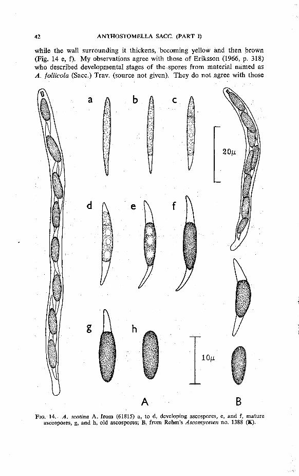
while the wall surrounding it thickens, becoming yellow and then brown (Fig. 14 e, f). My observations agree with those of Eriksson (1966, p. 318) who described developmental stages of the spores from material named as A. foliicola (Sacc.) Trav. (source not given). They do not agree with those
g
10M
A B F I G . 14. A. scotina A , from (61815) a, to d, developing ascospores, e, and f, mature
ascospores , g, and h, o ld ascospores; B, from Rehm's Ascomyceten no . 1388 ( K ) .

of Hohnel (1920 a, p. 177) who examined spores from a sample of Rehm's Ascomyceten no. 1388 (as A. rostrispora var. foliicola), and described the formation of 2-4 very thin transverse walls which cut off a central section while the end cells remained hyaline, thin walled and simulated slime appendages. On the basis of this spore septation he classified the species as a small-spored Heptameria, i.e., H. foliicola.
A. scotina is rare, and in spite of its characteristic spores has been described three times as a new species. The earliest collection was made before 1845 by Durieu in La Galle, Algeria and described as Sphaeria scotina. The type material of this collection, at PC, is very poor and consists of a few old perithecia and mature spores. Schrantz, who examined it in 1960, has given a brief description and illustrated a spore. The first record of this species for Europe was Spegazzini's collection on leaves of Castanea saliva from Conegliano. Saccardo (1877, p. 25) at first referred, this to A. rostrispora but later, when the species was illustrated as no. 177 of Fungi italici, he renamed it A. rostrispora var. foliicola* The name was based on Sphaeria rostraspora Gerard, a N. American collection on Inula helenium. Saccardo had not seen this material but thought the description agreed with Spegazzini's collection.
S. rostraspora is not an Anthostomella, it has large, superficial, perithecia and was considered by Martin (1967, p. 326) to be a species of Hypoxylon. On the herbarium label of the type material, the name is S. rostraspora, it was spelled 'rostrospora ' in the species description and as 'rostrispora' by Saccardo, Traverso and Rehm.
Traverso (1907, p. 489) separated the Italian collection, as A. foliicola, from the American S. rostraspora. He had seen the type material on Castanea (not now available at PAD) and considered it to be the same as Rehm, Ascomyceten no. 1388. This latter exsiccata is the only example of the species available for study in most herbaria. The material came from a collection by Mouton on leaves of Betula pendula [as B. alba] near Liege, Belgium, in 1900. In the two examples of the exsiccata I have seen (K & PAD) the ascospores and asci were slightly smaller than in all other collections, including the measurements Saccardo gave for the Spegazzini material on Castanea. I have noted the difference in the species description; if the fungus can be found again on Betula more material will show whether or not this is a constant difference.
The species on Canna glauca, from Argentina, named by Spegazzini (1881, p. 107) A. rostrispora * achira is an Anthostomella, but it differs from A. scotina in having wider asci, 93-116 X 8-10 M , while the spores, although of similar total length, 21-24 X 5 M , have a central brown cell, 15—16 X 5 n, which is larger in relation to the hyaline end cells.
S P E C I M E N S E X A M I N E D
Sphaeria scotina Dur . & Mont . , no details given, ex Herb. Durieu de Maisonneuve, ded. L. Mote lay (?C) probable Holotype. R e h m , Ascomyceten no . 1388, 4- rostrispora
* This is so in the C.M.I, copy of F u n g i italici; in the bound edit ion at R .B .G. K e w , the fungus is n a m e d A. rostispofa (Ger.) Sacc.

var, foliicola, o n Betula alba (— B. pendula), nr. Liege , Be lg ium, 1900, M o u t o n (K, P A D ) . Entosordaria perseicola Speg., on Persea lingue, Concepcion, Chile (LPS) Holotype .
O n Cladium mariscus, England: Wheat fen Broad, Norfo lk , ix .1945, E . A . Ellis (61815); xi i .1946, E . A . E . (10891); iv .1949, M . B . & J. P. Ellis (34926); iv .1954, M . B . E . (56710).
15. Anthostomella sepelibilis (Berk. & Curt.) Sacc., Sylloge Fungorum 1: 281 (1882).
Sphaeria sepelibilis Berk. & Curt., Grevillea 4: 146 (1876). Anthostomella eliminata (Berk. & Curt.) Sacc, Sylloge Fungorum 1: 281
(1882). Sphaeria eliminata Berk. & Curt., Grevillea 4: 148 (1876). Anthostomella constipata (Mont.) Sacc. var. diminuta Rehm, Mycotheca
rossica fasc. 2, no. 73 (1910). Anthostomella diminuta Hohnel, Sber. Akad. Wiss. Wien Math.-nat. 129:
180 (1920).
On stems of Smilax campestris, S. excelsa and S. laurifolia in Argentina, U.S.A., U.S.S.R.
Perithecia immersed, each beneath a round black clypeus pierced by a papilla which is small and barely projects but is made more conspicuous by a narrow white circlet around the base formed by the torn host tissue. The clypeus, 400-600 p diam., consists of dark brown hyphae which fill all the plant cells above each perithecium. Perithecia, 300 p high and 360 p wide. The peridium is 18 p wide with an outer layer, 10 p, of dark thick walled cells, and an inner layer, 8 p of hyaline, thin walled, compressed cells. Asci, 86-103 X 9 p (or X 13 p when spores are biseriate) cylindrical, apex rounded with an apical ring, 2 X 3 p; little or no stalk; eight ascospores are uniseriate or partially biseriate. The walls of the asci break down early and are seldom seen containing mature brown spores. Ascospores, 14-18 X 5-6 p, two celled, with a small hyaline dwarf cell, 2 X 2 P and a brown cell, 12-16 X 5-6 p, ellipsoidal or slightly inequilateral. The dwarf cell tends to disappear early and the brown cell then appears flattened at that end; a hyaline sheath of irregular shape surrounds each sppre, it is visible at both ends but only on one (the concave) side; an indistinct longitudinal germ slit is seen after several weeks staining in cotton blue. Paraphyses numerous, 2 P wide, mucilaginous and sticking together in a convoluted mass.
Sphaeria eliminata and S. sepelibilis were collected by Curtis in N. America and sent to Berkeley for determination. Berkeley published them, with the briefest of descriptions, as nos. 943 and 928 in his Notices of North American Fungi. This reversed the order of the original collection numbers and has given to S. sepelibilis a slightly unjustified priority.
In 1910, Tranzschel and Serebrianikow issued as no. 73 of their Mycotheca rossica a fungus which had been named by Rehm, A. constipata
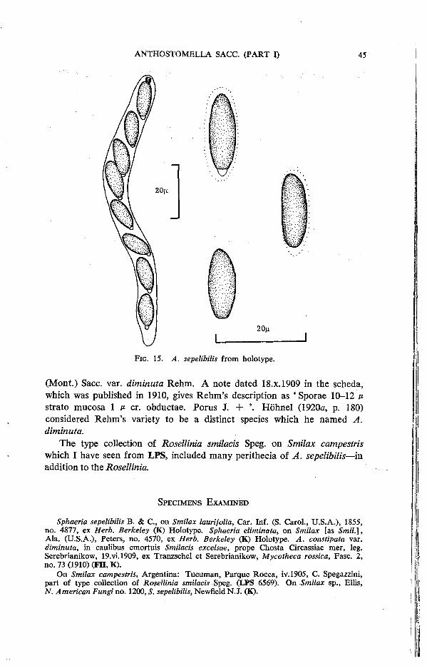
F I G . 15. A. sepelibilis f rom holotype.
(Mont.) Sacc. var. diminuta Rehm. A note dated 18.x. 1909 in the scheda, which was published in 1910, gives Rehm's description as ' Sporae 10-12 p. strato mucosa 1 p cr. obductae. Porus J. + '. Hohnel (1920a, p. 180) considered Rehm's variety to be a distinct species which he named A. diminuta.
The type collection of Rosellinia smilacis Speg. on Smilax campestris which I have seen from LPS, included many perithecia of A. sepelibilis—in addition to the Rosellinia.
S P E C I M E N S E X A M I N E D
Sphaeria sepelibilis B. & C , o n Smilax laurifolia, Car. Inf. (S. Carol. , U . S . A . ) , 1855, no . 4877 , ex Herb. Berkeley (K) Holotype . Sphaeria eliminata, o n Smilax [as Smil.\, A l a . ( U . S . A . ) , Peters , no . 4570, ex Herb. Berkeley (K) Holotype . A. constipata var. diminuta, in caulibus emortuis Smilacis excelsae, prope Chosta Circassiae mer , leg. Serebrianikow, 19.vi .1909, ex Tranzschel et Serebrianikow, Mycotheca rossica, Fasc . 2 , no . 73 (1910) ( F H , K) .
On Smilax campestris, Argentina: T u c u m a n , Parque R o c c a , iv .1905, C. Spegazzini , part of type col lect ion of Rosellinia smilacis Speg. (LPS 6569). On Smilax sp . , Ell is , N. American Fungi no . 1200, S. sepelibilis, Newfie ld N.J . (K).
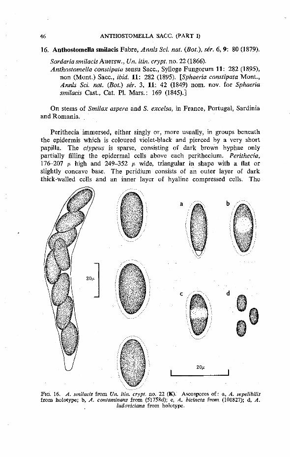
16. Anthostomella smilacis Fabre, Annls Sci. nat. {Bot.), ser. 6,9: 80 (1879).
Sordaria smilacis Auersw., Un. itin. crypt, no. 22 (1866). Anthostomella constipata sensu Sacc, Sylloge Fungorum 11: 282 (1895),
non (Mont.) Sacc, ibid. 11: 282 (1895). [Sphaeria constipata Mont., Annls Sci. nat. (Bot.) ser. 3, 11: 42 (1849) nom. nov. for Sphaeria smilacis Cast., Cat. PI. Mars.: 169 (1845).]
On stems of Smilax aspera and S. excelsa, in France, Portugal, Sardinia and Romania.
Perithecia immersed, either singly or, more usually, in groups beneath the epidermis which is coloured violet-black and pierced by a very short papilla. The clypeus is sparse, consisting of dark brown hyphae only partially filling the epidermal cells above each perithecium. Perithecia, 176-207 ju, high and 249-352 p wide, triangular in shape with a flat or slightly concave base. The peridium consists of an outer layer of dark thick-walled cells and an inner layer of hyaline compressed cells. The
F I G . 16. A. smilacis f rom Un. itin. crypt, no . 22 ( K ) . Ascospores of : a, A. sepelibilis f r o m holotype; b, A. contaminans f rom (51758d); c, A. bicincta f rom (101827); d, A.
ludoviciana f rom holotype .

width of this inner layer is a fairly uniform 3-5 M , while the outer layer measures 10-11 n at the sides but only 5 A* along the flattened base. Asci, 111-130 X 11-14 M , cylindrical or clavate, the apex is somewhat flattened, there is no apical ring; the stalk is abruptly tapered and very short; eight ascospores are obliquely uniseriate or partially biseriate. Ascospores, 14-18 X 8-10 ii, one celled, broadly ellipsoidal with rounded ends; surrounded by a conspicuous hyaline sheath 2-3 p wide; the longitudinal germ line is very distinct. Paraphyses in young perithecia are 4-5 M wide, but soon become mucilaginous and disappear.
The earliest authenticated collection of this fungus was made by Marcucci on Smilax aspera in Sardinia and was distributed as Sordaria smilacis Auersw., no. 22 of the Unio itin. crypt. Niessl (1876, p. 198), discussing the genus Anthostomella Sacc, noted that Sordaria smilacis should be included in the genus. Unfortunately, this new combination was not effectively published according to Article 34 of the Rules of Nomenclature. In 1879 the name Anthostomella smilacis was used by Fabre to describe the same fungus that he had recently collected in Vaucluse. Thus although Sordaria smilacis Auersw. is the earliest available legitimate epithet, a new combination in Anthostomella, based on Auerswald's type, cannot be made.
Twenty-one years earlier, in 1845, Castagne found an ascomycete on stems of Smilax which he published in his ' Catalogue des Plantes de Marseille ' a s Sphaeria smilacis Cast. He described the spores as ' oviformi-bus saepe nigris 2 septatis'. Desmazieres (1846, p. 73), to whom Castagne sent ' echantillons assez nombreux pour lui faire prendre place dans notre collection cryptogamique', also described the asci and spores as 'ascis clavatis, sporidiis ovoideis, 1-2 septatis saepe semiopacis' and added that the fungus appeared to be ' une Dothidea' when seen in transverse section and should perhaps be classified with these fungi. Saccardo (1883, p. 69) transferred the species to Leptosphaeria, as L. smilacis (Cast.) Sacc, apparently without seeing any material. Montagne, in 1849, changed the name to S. constipata as S. smilacis had already been used by Schweinitz. Saccardo later examined a portion of Castagne's material from Bruxelles (Fungi Herb. Brux. no. 13) and found a fungus with aseptate spores which were surrounded by a large sheath; he transferred the species to Anthostomella, as A. constipata. He noted that these observations differed somewhat from those of Castagne and Desmazieres, but decided that these authors had been in error in describing the spores as septate, and remiss in not mentioning the conspicuous sheath. I have seen this material from Bruxelles and found there were two species of fungi present. The species of Anthostomella described by Saccardo, which was identical with the fungus of Auerswald and Fabre, and also an immature bitunicate ascomycete which approximated more closely to the description given by Castagne and Desmazieres. I have found this bitunicate ascomycete (and no Anthostomella) in the Castagne collections (Leptosphaeria smilacis folder) at K. The bitunicate asci measured 68-81 X 17 M and the spores, 18-20 X 6-7 ii, were hyaline and aseptate and resembled a species of Botryo-

sphaeria. Hohnel (1929, p. 113) described an immature dothideaceous fungus from Castagne's material in a sample of Desmazieres, Plantes crypt. France, Ed. I, Ser. I, no. 1770, Sphaeria smilacis. The measurements Hohnel gives for asci, 90 X 16 p, and spores, 16-20 X 4-5-5 f-, are close to those I found in the K samples; but whereas I saw only immature, one celled spores, Hohnel found the spores to be 4-5 celled. He named his fungus Phragmocauma smilacis (Cast.) Hohnel. While these results do not completely solve the riddle of the identity of S. smilacis, I think it fair to say that the descriptions of Castagne and Desmazieres do not refer, as Saccardo believed, to the Anthostomella in the Bruxelles material. This cannot therefore be accepted as the first collection of A. smilacis.
Savulescu & Sandu, (1935, p. 166) decided that A. smilacis [as A. constipata] did not have a sufficiently well developed clypeus to remain in Anthostomella, and made the new combination Paranthostomella constipata.
A. smilacis Fabre , spec imen orig. e x Fabre , dedit Berlese, ( P A D ) Isotype. Unio itin. crypt, no . 22 , Sordaria smilacis Auersw. , o n Smilax aspera, Tacquitara, leg Marcucci , v .1866 (K, U P S ) . Fungi Herb. Brux. no . 13, Sphaeria constipata M o n t . , M o n t a u d les Miramas , France (A. smilacis f o u n d a n d also ? P h r a g m o c a u m a smilacis), ( B R ) . Herb. Myc. Romanicum Fasc . 2 3 , no . 1132 [as Paranthostomella constipata], o n Smilax excelsa, Dobrogea , iv .1934, Tr. Savulescu (K) , the only record of this species that is n o t o n S. aspera; the fungus in this exsiccata i n I M I (15220) is A. contaminans.
O n Smilax aspera var. nigra, Portugal. Estremadura, Queluz , v. 1950 (LISE 32025); Estremaduira, Terra d e Arrabida, vi i i .1952 (LISE 38539); Estremadura, pr. Oeiras, v .1955 (LISE 50103); Estremadura D o i s Portos Pos to Agrar io , v.1965 (LISE 65671); Beira Litoral, pr. Maiorca , Figuiera da Foz . , xi i .1951 (LISE 34221).
Leptosphaeria smilacis folder at K, Castagne's col lect ions o n Smilax aspera, M o n t a u d les Miramas, France . Destnazieres , Plantes Crypt. France, Ed . I. Ser. I (1825-1851) no . 1770, Sphaeria smilacis Cast. Ibid., Ed. II, Ser. I, no . 1420, 5. smilacis Herb. Bloxam and Herb. M. C. Cooke both have two specimens of Sph. smilacis Cast. T h e fungus in all these col lect ions w a s IPhragmocauma smilacis (Cast) H o h n e l . A. smilacis w a s not found; one spec imen in Herb. Bloxam had a small group of perithecia of A. contaminans.
Nine species, including A. sepelibilis and A. smilacis, have been recorded on Smilax. Four of these names are considered to be synonyms and are listed as such. The three remaining species are A. contaminans which is rare on Smilax, more usualon Palms, and is described in that section; A. bicincta Syd. recorded on Caryota sp. and Smilax in India, W. Pakistan and the Philippines; and A. ludoviciana Ellis & Langl. collected on Smilax in Louisiana, U.S.A. The last two species are not described in this paper but are included in the key which follows.
S P E C I M E N S E X A M I N E D
Key to species of Anthostomella on Smilax 1. Spores two celled (1:
Spores one celled 2. Spores brown with a central hyaline band
Spores uniformly brown
(15) A. sepelibilis 2
(A. bicincta) 3

3. Spores 5-6 M long Spores 12 M or longer
(A. ludoviciana) 4
4. Spores 8-10 M wide, ascus tip not blue in iodine Spores 5-7 M wide, ascus tip blue in iodine
(16) A. smilacis 5
5. Spores 14-18 X 5-7 n, both ends rounded (2 Spores 12-16 X 5-6 one end rounded, the other flattened, i.e., spores old and without a dwarf cell
(24) A. contaminans
(15) A. sepelibilis
17. Anthostomella spartii Berl. & Vogl., Atti Accad. sclent, veneto-trent.-istriana 10: 212 (1889).
Anthostomella mesembryanthemi Da Camara & de Vasconcelos, Agro-nomia lusit., 17: 93 (1955).
On dead stems of Coronilla glauca, Mesembryanthemum edule, Spartium junceum in Italy and Portugal.
Perithecia scattered and single, immersed beneath an ill-defined clypeus which appears externally as a narrow black circle surrounding the small, barely erumpent, papilla and in section as a thin network of brown, thick walled hyphae in the epidermal cells. Perithecia, 242-260 v- high and 200-300 p wide. The peridium is 18-23 M wide, with an outer layer, 13-17 v, of thick walled cells which are black in the upper half of the fruit body and yellowish-brown below and along the base. The inner layer, 5-6 /*, is hyaline with thin walled much compressed cells. Asci, 98-121 X 10-11 A , cylindrical, apex rounded, with an apical ring, 1'5 X 5 c; a short tapered stalk; eight ascospores are uniseriate. Ascospores, 12-15 X 8-9 M , one celled, broadly ellipsoidal with ends rounded or slightly tapered; a narrow sheath surrounds each spore but is not always easily seen; the longitudinal germ slit is very distinct and old spores usually divide along this line when gently squashed. Paraphyses numerous, 4-5 M wide.
The first collection of this species was made on Spartium junceum near Ancona, in Italy. I have not been able to trace this type material and my identification of the species is based on the original description and figures given by Berlese & Voglino. Two excellent collections from Portugal on Coronilla and Mesembryanthemum provided material for description. The latter was described as a new species, A. mesembryanthemi, but I do not think that it differs from A. spartii. A recent collection made in 1972 by Dennis provides assurance that the same fungus occurs on Spartium in Italy.
I have not selected a neotype as further searches may yet produce an early Italian collection.
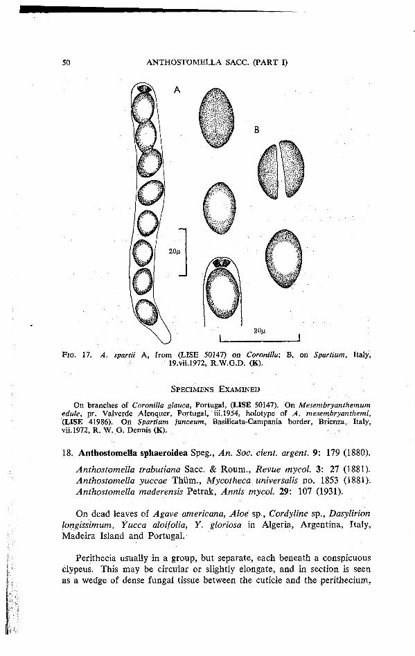
F I G . 17. A. spartii A , from (LISE 50147) on Coronilla; B , on Spartium, Italy, 19.vii .1972, R . W . G . D . (K) .
S P E C I M E N S E X A M I N E D
On branches of Coronilla glauca, Portugal , (LISE 50147). O n Mesembryanthemum edule, pr. Valverde Alenquer , Portugal , i i i .1954, holotype of A. mesembryanthemi, (LISE 41986) . O n Spartium junceum, Basil icata-Campania border, Brienza, Italy, vi i .1972, R. W . G. D e n n i s (K).
18. Anthostomella sphaeroidea Speg., An. Soc. cient. argent. 9: 179 (1880).
Anthostomella trabutiana Sacc. & Roum., Revue mycol. 3: 27 (1881). Anthostomella yuccae Thirm., Mycotheca universalis no. 1853 (1881). Anthostomella maderensis Petrak, Annls mycol. 29: 107 (1931).
On dead leaves of Agave americana, Aloe sp., Cordyline sp., Dasylirion longissimum, Yucca aloifolia, Y. gloriosa in Algeria, Argentina, Italy, Madeira Island and Portugal.
Perithecia usually in a group, but separate, each beneath a conspicuous clypeus. This may be circular or slightly elongate, and in section is seen as a wedge of dense fungal tissue between the cuticle and the perithecium,
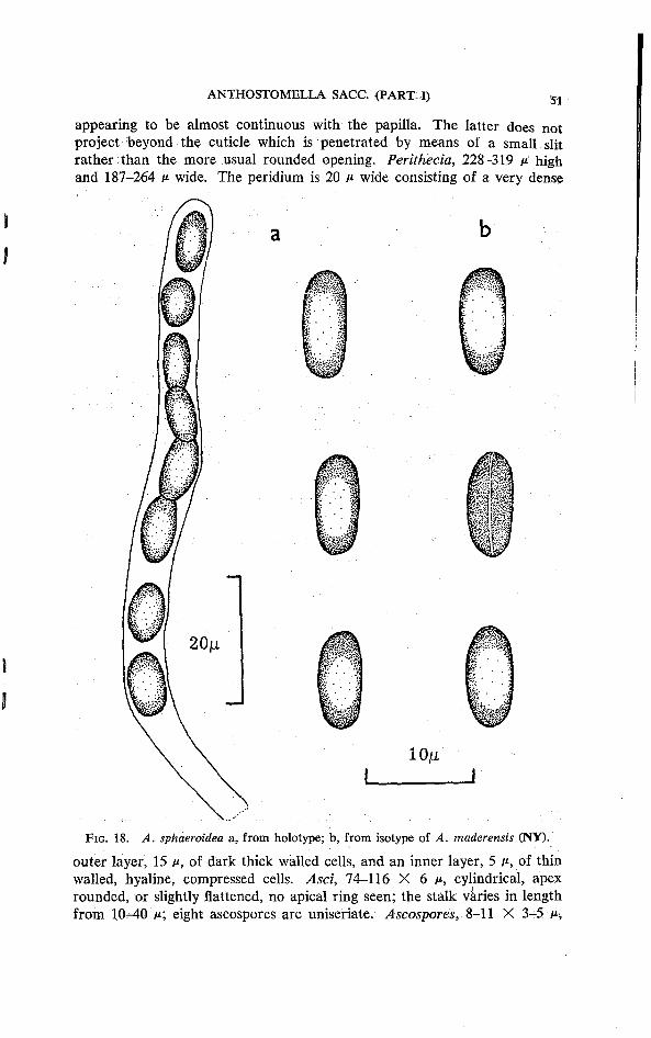
appearing to be almost continuous with the papilla. The latter does not project beyond the cuticle which is penetrated by means of a small slit rather than the more usual rounded opening. Perithecia, 228-319 y. high and 187-264 p wide. The peridium is 20 A* wide consisting of a very dense
F I G . 1 8 . A. sphaeroidea a, from holotype; b , from isotype of A. maderensis ( N Y ) .
outer layer, 15 P, of dark thick walled cells, and an inner layer, 5 p, of thin walled, hyaline, compressed cells. Asci, 74-116 X 6 p, cylindrical, apex rounded, or slightly flattened, no apical ring seen; the stalk varies in length from 10-40 M ; eight ascospores are uniseriate. Ascospores, 8-11 X 3-5 AH
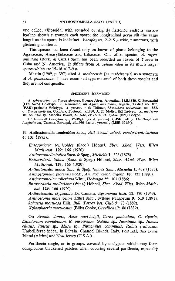
one celled, ellipsoidal with rounded or slightly flattened ends; a narrow hyaline sheath surrounds each spore; the longitudinal germ slit the same length as the spore, is indistinct. Paraphyses, 2-2-5 P wide, numerous, with glistening contents.
This species has been found only on leaves of plants belonging to the Agavaceae, Amaryllidaceae and Liliaceae. One other species, A. nigro-annulata (Berk. & Curt.) Sacc. has been recorded on leaves of Yucca in Cuba and N. America. It differs from A. sphaeroidea in its much larger spores which are 15-18 X 7-9 p.
Martin (1969, p. 397) cited A. maderensis [as madeirensis] as a synonym of A. phaeosticta. I have examined type material of both these species and they are not conspecific.
S P E C I M E N S E X A M I N E D
A. sphaeroidea, o n Yucca gloriosa, Buenos Aires , Argent ina, 18. i i . lS80, C. Spegazzini ( L P S 6765) Holotype . A. trabutiana, o n Agave americana, Algeria , Trabut no . 107, ( P A D ) probable Holotype . A. yuccae, in de Thi imen, Mycotheca universalis, no . 1853, o n Yucca aloifolia, Coimbra, Portugal , ix.1880, A . F . Mol ler , (K) Isotype. A. maderensis, o n Aloe sp. Madeira Island, A . A d e , e x Herb. H. Sydow ( N Y ) Isotype.
O n leaves of Cordyline sp., Portugal [as A. yuccae], (LISE 32410). O n Dasylirion longissimum, Cascais, Portugal, xi i .1950 [as A. yuccae], (LISE 32154).
19. Anthostomella tomicoides Sacc, Atti Accad. scient. veneto-trent.-istriana 4: 101 (1875).
Entosordaria tomicoides (Sacc.) Hohnel, Sber. Akad. Wiss. Wien Math.-nat. 129: 166 (1920).
Anthostomella italica Sacc. & Speg., Michelia 1: 328 (1878). Entosordaria italica (Sacc. & Speg.) Hohnel, Sber. Akad. Wiss. Wien
Math.-nat. 129: 166 (1920). Anthostomella italica Sacc. & Speg. *affinis Sacc, Michelia 1: 439 (1878). Anthostomella platensis Speg., An. Soc. cient. argent. 10: 135 (1880). Anthostomella mollerianaWint., Hedwigia25: 101 (1886). Entosordaria molleriana (Wint.) Hohnel, Sber. Akad. Wiss. Wien Math.-
nat. 129: 166 (1920). Anthostomella clypeatula Da Camara, Agronomia lusit. 11: 170 (1949). Anthostoma mortuosum (Ellis) Sacc, Sylloge Fungorum 9: 519 (1891). Sphaeria mortuosa Ellis, Bull. Torrey bot. Club 9: 73 (1882). Xylosphaeria mortuosum (Ellis) Cooke, Grevillea 17: 86 (1889).
On Arundo donax, Aster novi-belgii, Carex paniculata, C. riparia, Eupatorium cannabinum, E. purpureum, Galium sp., Jasminum sp., Juncus ejjusus, Juncus sp., Musa sp., Phragmites communis, Rubus fruticosus, Umbelliferae indet., in Britain, Channel Islands, Italy, Portugal, Sao Tome Island (Africa) and New Jersey (U.S.A.).
Perithecia single, or in groups, covered by a clypeus which may form conspicuous blackened patches when covering several perithecia, especially

F I G . 1 9 . A. tomicoides A , from A. italica in Spegazzini, Decades Myc. ital. n o . 1 3 ( K ) ; B , from (Sheffield B) o n Carex riparia; C, f r o m Herb. Sir H. C. Hawley, on Umbel l i fer
s t em ( K ) ; D , from (4654) o n Aster; E , f rom isotype of A. molleriana Wint . ( K ) .
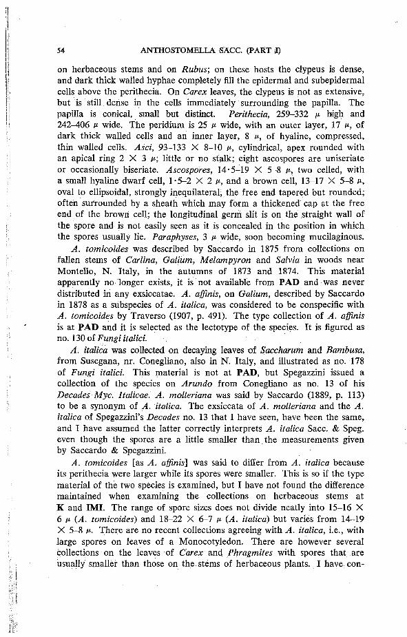
on herbaceous stems and on Rubus; on these hosts the clypeus is dense, and dark thick walled hyphae completely fill the epidermal and subepidermal cells above the perithecia. On Carex leaves, the clypeus is not as extensive, but is still dense in the cells immediately surrounding the papilla. The papilla is conical, small but distinct. Perithecia, 259-332 M high and 242-406 p wide. The peridium is 25 p wide, with an outer layer, 17 u, of dark thick walled cells and an inner layer, 8 P, of hyaline, compressed, thin walled cells. Asci, 93-133 X 8-10 p, cylindrical, apex rounded with an apical ring 2 X 3 A ; little or no stalk; eight ascospores are uniseriate or occasionally biseriate. Ascospores, 14-5-19 X 5-8 p, two celled, with a small hyaline dwarf cell, 1-5-2 X 2 p, and a brown cell, 13-17 X 5-8 p, oval to ellipsoidal, strongly inequilateral, the free end tapered but rounded; often surrounded by a sheath which may form a thickened cap at the free end of the brown cell; the longitudinal germ slit is on the straight wall of the spore and is not easily seen as it is concealed in the position in which the spores usually lie. Paraphyses, 3 p wide, soon becoming mucilaginous.
A. tomicoides was described by Saccardo in 1875 from collections on fallen stems of Carlina, Galium, Melampyron and Salvia in woods near Montello, N. Italy, in the autumns of 1873 and 1874. This material apparently no longer exists, it is not available from PAD and was never distributed in any exsiccatae. A. affinis, on Galium, described by Saccardo in 1878 as a subspecies of A. italica, was considered to be conspecific with A. tomicoides by Traverso (1907, p. 491). The type collection of A. affinis is at PAD and it is selected as the lectotype of the species. It is figured as no. 130 of Fungi italici.
A. italica was collected on decaying leaves of Saccharum and Bambusa, from Susegana, nr. Conegliano, also in N. Italy, and illustrated as no. 178 of Fungi italici. This material is not at PAD, but Spegazzini issued a collection of the species on Arundo from Conegliano as no. 13 of his Decades Myc. Italicae. A. molleriana was said by Saccardo (1889, p. 113) to be a synonym of A. italica. The exsiccata of A. molleriana and the A. italica of Spegazzini's Decades no. 13 that I have seen, have been the same, and I have assumed the latter correctly interprets A. italica Sacc. & Speg. even though the spores are a little smaller than the measurements given by Saccardo & Spegazzini.
A. tomicoides [as A. affinis'] was said to differ from A. italica because its perithecia were larger while its spores were smaller. This is so if the type material of the two species is examined, but I have not found the difference maintained when examining the collections on herbaceous stems at K and IMI. The range of spore sizes does not divide neatly into 15-16 X 6 p (A. tomicoides) and 18-22 X 6-7 P (A. italica) but varies from 14-19 X 5-8 p. There are no recent collections agreeing with A. italica, i.e., with large spores on leaves of a Monocotyledon. There are however several collections on the leaves of Carex and Phragmites with spores that are usually smaller than those on the stems of herbaceous plants. I have con-

sidered segregating these as a separate species, but, as in the case of A. tomicoides and A. italica, the measurements of spores from the Carex group overlap with those of species considered to be A. tomicoides. A. tomicoides is therefore treated as a species with a wide host range and a considerable variation in spore size.
S P E C I M E N S E X A M I N E D
A. italica subsp. A. affiinis, in Galii sylv. (Isylvestre) ex Herb. P. A. Saccardo ( P A D ) Lectotype , (K) Isolectotype. Spegazzini, Decades Myc. Italicae, no . 13, A. italica, ad folia Arundinis donacis, Conegl iano, xii .1878 (LPS, K) . A. platensis, in cu lmis ernortuis (1 Juncus), Buenos Aires , R i o d e la Plata, i i i .1880 (LPS 6763), H o l o type. A. molleriana, foliis Musarum, Insule St. T h o m e , ix .1895, A . M o Her (K, P A D , P C ) , Isotypes. A. clypeatula D a Camara, on stems of Jasminum, pr. Sintra, Estremadura, Portugal , i i i .1947, R. de S. Dias , no . 540 (LISE 23911) Holotype . Ell is , N. American Fungi, no. 897, Sphaeria mortuosa, only on Eupatorium purpureum stems (A. chionostoma o n Andropogon stems) (K).
On Aster novi-belgii, England: Perranzebuloe, Cornwall , iv .1946, F . Ri l s tone (4654). O n Carex paniculata, England: Spye Park, Wilts . , ex Herb. C. E. Broome, s ix col lect ions, 1850-1859 (K); Wales: Gann Estuary, Pembs . , vi. 1955, J. Webster (Sheffield 1716); Channe l Islands: Guernsey, M . B. Ellis, vi .1947 (56815); vii .1947 (56817); iii.1948 (269846) . O n Carex riparia, England: Esthwaite F e n , Westmorland, xi .1958 (Sheffield B) . O n Eupatorium cannabinum, Scotland: W. Kilbride, Ayrshire , v i .1914, xi. 1918, D . A . B o y d (K); Wales: Maenc lochog , Pembs . , vi i i .1928, P . G. M . R h o d e s , e x Herb. W. B. Grove (K). On Juncus effusus, England: Perranzebuloe, Cornwall , v. 1952, M . B . E . (50128); Channel Islands: H e r m , ix .1948, M . B . E . (34464). On Juncus spp., England: Lychett (?Dorset ) [as A. tomicum], x i i .1922, e x Herb. Sir H. C. Hawley ( K ) ; Draycott (?Somerset) [as Sphaeria tomicum], 1851, e x Herb. C. E. Broome (K); Ardingly , Sussex, xi i .1967, R. W. G. Denni s (K) . O n Phragmites communis, Castle H o w a r d , Yorks . , ix .1950, W . G. Bramley (Sheffield 836). On Rubus fruticosus, England: Cornwal l , B o c h y m , iv.1932, P. G. M . R h o d e s (K); Boscast le , x .1970, G. C. Ainsworth (184999); Perranzebuloe, v .1952; M . B . E . (50134); Surrey, Weybridge , i i .1854 [as Sphaeria appendiculosa var. m i n o r ] , ex Herb. C. E. Broome (K). On stems of Umbel l i fer indet. , England: L y m e Regis , Dorset [as Anthostoma], ex Herb. Sir H. C. Hawley (K).
20. Anthostomella tomicum (Lev.) Sacc, Atti Accad. sclent, veneto-trent.-istriana 4: 101 (1875).
Sphaeria tomicum Lev., Annls Sci. nat. (Bot.) ser 3, 9: 143 (1848). Non A. tomicum sensu Sacc, Michelia 1: 374 (1878) [—A. tumulosa]. Anthostomella megaclypeata Rehm, Annls mycol. 5: 537 (1907). Anthostomella subconica Rehm, Annls mycol. 5: 537 (1907). Anthostomella aruncz Hohnel, Annls mycol. 16: 71 (1918).
On dead leaves and stems of Aruncus Sylvester, Brachypodium pinnatum, Deschampsia caespitosa, D. flexuosa, Scirpus holoschoenus, Compositae indet., Labiatae indet., in Austria, Britain, France and Germany.
Perithecia single or two or three together, large, immersed but usually raising the blackened epidermis. Papilla sturdy, conical, with the central canal clearly visible. Clypeus strongly developed with dark thick walled hyphae completely filling the epidermal cells above each perithecium. Perithecia, 616-770 p. high and 655-800 p. wide. The peridium is 53 p. wide, with
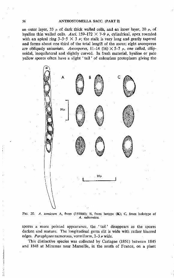
an outer layer, 33 A of dark thick walled cells, and an inner layer, 20 A , of hyaline thin walled cells. Asci, 159-172 X 7-9 c, cylindrical, apex rounded with an apical ring 3-3-5 X 3 c; the stalk is very long and gently tapered and forms about one third of the total length of the ascus; eight ascospores are obliquely uniseriate. Ascospores, 11-14 (16) X 5-7 /x, one celled, ellipsoidal, inequilateral and slightly curved. In fresh material, hyaline or pale yellow spores often have a slight ' ta i l ' of colourless protoplasm giving the
F I G . 20 . A. tomicum A, f rom (151860); B , f rom isotype ( K ) ; C, from holotype of A. subconica.
spores a more pointed appearance, the ' tai l ' disappears as the spores darken and mature. The longitudinal germ slit is wide with rather blurred edges. Paraphyses numerous, vermiform, 2-3 c wide.
This distinctive species was collected by Castagne (1851) between 1845 and 1848 at Miramas near Marseille, in the south of France, on a plant
he named as J uncus acutus. Sections cut at the Jodrell Laboratory, R. B. G., Kew have shown, however, that the plant is a member of the Cyperaceae, probably Scirpus holoschoenus. Castagne sent material for identification to Leveille and Desmazieres, both of whom prepared descriptions. Leveille published first and named the fungus Sphaeria tomicum; and Desmazieres withdrew his name merely mentioning it on the label of his Plantes Crypt, de France Ed I, Ser. I, no. 1776. This exsiccata is now the type material as Leveille's herbarium was destroyed in the Franco-Prussian war. Saccardo, in 1875, transferred S. tomicum to Anthostomella without comment in a footnote entitled ' Obs.' which followed the description of A. tomicoides Sacc. Three years later, in 1878, he published a description (illustrated as no. 373 of Fungi italici) of his A. tomicum, this referred to a species on Typha in Italy. It is A. tumulosa and is quite different from Leveille's fungus.
Although there are many specimens labelled A. tomicum in the British collection at K, none agree with the type material. The species was confused with A. tomicoides, A. tumulosa and A. caricis. The A. tomicum of Leveille was first found in Britain by Webster who made three collections between 1954-1957. I have found the species once, in Scotland in 1969, but although I have visited this site every year since then, I have never found the fungus again. Castagne (1851, p. 47) observed that it was rare. He found the species once on Ruscus aculeatus; it is possible that A. rusci which Fabre collected and described from the same host in Vaucluse, also in the south of France, is the same species.
A. arunci, A. megaclypeata and A. subconica were collected on herbaceous stems in Austria and Germany. They differ slightly from the type, and the collections on grasses, by having wider spores. The latter have a spore width of 5-6 c (occasionally 7 c) while in the collections on herbaceous stems the spores are 6-7 P wide.
A. tomicum is easily distinguished from the other species of Anthostomella by its large perithecia and asci with long tapering stalks.
S P E C I M E N S E X A M I N E D
Desmazieres , Plantes Crypt. France, Ed . I. Ser. I (1825-1851) Sphaeria tomicum Lev., o n (probably) Scirpus holoschoenus [ a s Juncus acutus~\, (PC) Isotype. Ibid., Ed. II , Ser. I (1836-1851) , no . 1426 (K). Herbs. Berkeley, Bloxam & Currey all have col lect ions labelled ' Sphaeria tomicum Lev. , Montaud , or M o n t a u d les Miramas , C a s t a g n e ' (? ex Castagne) (K). A. megaclypeata on stem of Labiatae, indet. Kastelruth, Tirol , 8 . ix . l903 , H . R e h m , (S) Holotype . A. subconica, s tem of Composi tae indet. , K a m p e n w a n d , Bavaria, ix .1904, H . R e h m (S) Holotype . A. arunci, on stems of Aruncus Sylvester, nr. Konigste in , Sachsen (Germany) , v : 1913, W. Krieger ( F H ) Holotype .
O n Brachypodium pinnatum, England: Worksop , N o t t s . , vi i .1954, J. Webster (56959). O n Deschampsia caespitosa, England: Fu lwood , nr. Sheffield, i i .1957, J,W. (68381); D. flexuosa, Scot land: Dunke ld , Perths., ix .1954, J.W. (6960). O n Gramineae indet., Scotland: M o r v e n , Argyl l , vi i .1969, S .M.F. (151860).
21. Anthostomella tumulosa (Rob. in Desm.) Sacc, Sylloge Fungorum 1: 282 (1882).
Sphaeria tumulosa Rob. in Desm., Annls Sci. nat. (Bot.) sir. 3, 16: 309 (1851).
Anthostomella tomicum sensu Sacc, Michelia 1: 374 (1878), non A. tomicum (Lev.) Sacc, Atti Accad. sclent, veneto-trent.-istriana 4: 101 (1875).
On Carex pendula, Glyceria sp, J uncus articulatus, Typha sp. and Gramineae indet. in Britain, France, Inaccessible Island nr. Tristan da Cunha and Italy.
Perithecia scattered, immersed beneath a conspicuous black clypeus which is penetrated by a small conical papilla. In young material the position where the papilla will eventually break through is marked by a small uncoloured spot in the clypeus. The clypeus is formed from dark thick walled hyphae in the upper and side walls of the epidermal cells. Perithecia, 275-308 p high and 220-301 P wide. The peridium is 15 P wide* with an outer layer, 10 p, of dark thick walled cells and an inner layer, 5 p, of hyaline, thin walled, compressed cells. Asci, 90-150 X 9-13 p (X 18 p when spores are biseriate) cylindrical, apex rounded, with an apical ring 4 X 3 p; little or no stalk; eight ascospores are uniseriate or partially biseriate. Ascospores, 15-23 X 7-9 p, one celled, ellipsoidal, inequilateral and slightly curved, ends rounded; a conspicuous sheath surrounds each spore; in some preparations, made from the type material, a very small hyaline dot of mucilage has been seen at one end of a few of the spores, this is not a dwarf cell and has not been found in any other collection; germ slit not seen. Paraphyses thread-like and numerous.
A. tumulosa is another of the species found by Roberge on the dunes of Calvados in N.W. France. The collection, on the dry stems of Juncus articulatas was plentiful and was included in both editions of Desmazieres, Plantes Crypt, de France and therefore widely distributed. The species is, however, not well known and has been confused with A. tomicum by Saccardo, while other workers have referred collections of A. fuegiana to A. tumulosa.
The relatively large, one celled spores of A. tumulosa distinguish it from all species except A. lugubris and A. chionostoma. It differs from A. lugubris in having spores which are narrower (7-9 p) and inequilateral. In A. lugubris the spores are 8-11 p wide and equal sided. A. chionostoma differs from both species in having a conspicuous undulating germ slit.
S P E C I M E N S E X A M I N E D
Desmazieres , Plantes Crypt. France, Ed. I, Ser. I (1825-1851) no . 2075, Sphaeria tumulosa R o b . in herb. , o n Juncus articulatus (K) Isotype. Ibid., Ed . II, Ser. I ( 1 8 3 6 -1851) no . 1775 (K) . Herb. Saccardo, o n Typha, i i i .1878 [as A. tomicum], ( P A D ) .
O n Carex pendula, England: Batheaston, Somerset , i .1850 OX); host unnamed , probably Carex, Batheaston , i i .1860 (K). O n Glyceria sp. , Inaccessible Island, i i .1938, E . Christopherson, no . 2465 (K). On Gramineae indet. , Scotland: L o c h H o u r n , Inverness , vi i i .1970, S .M.F. (185001).
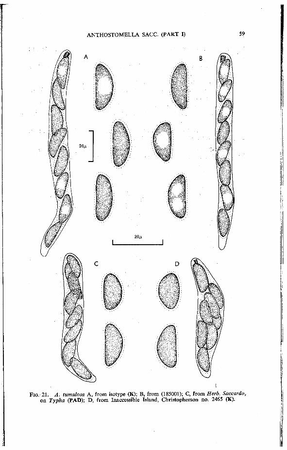
F I G . 2 1 . A. tumulosa A , from isotype (K); B , from ( 1 8 5 0 0 1 ) ; C, f rom Herb. Saccardo, o n Typha ( P A D ) ; D , f rom Inaccessible Island, Christopherson no . 2 4 6 5 (K).

22. Anthostomella unguiculata (Mont.) Sacc, Sylloge Fungorum 1: 288 (1882).
Sphaeria unguiculata Mont., in Gay, Historia fisica y politico de Chile (Bot.) 7: 464(1854).
Anthostomella magnoliae Ellis & Everh., / . Mycol. 4: 122 (1888). Entosordaria magnoliae (Ellis & Everh.) Hohnel, Sber. Akad. Wiss. Wien
Math.-nat. 129: 166 (1920). Anthostomella dryina Mouton, Bull. Soc. r. Bot. Belg. 39: 38 (1900). Entosordaria dryina (Mouton) Hohnel, Sber. Akad. Wiss. Wien
Math.-nat. 129: 166 (1920). On fallen leaves of Desfontainia sp., Magnolia sp. and Quercus sp. in
Belgium, Chile and Louisiana, U.S.A. ; , .
Perithecia on lower surface of leaf, separate and widely scattered, each immersed beneath a small, well defined, black clypeus. Papilla small, but
F I G . 22 . A. unguiculata a, from holotype of A. dryina; b , f rom holotype of A. magnoliae.

distinct. Material of all the type specimens examined was very sparse and no sections were cut. The diameter of the perithecia was approximately 187-207 p. Asci, 63-80 X 4-6 p, cylindrical, apex rounded with an apical ring, 2 X 2 P; the stalk is very short and tapered; eight ascospores are uniseriate. Ascospores, 8-5—10-5 X 3-5 P, two celled, with a small hyaline dwarf cell, 1 • 5 X 2 p and a brown cell 7-9 X 4-5 p, ellipsoidal, inequilateral, the free end bluntly pointed; a narrow sheath surrounds the spore, often forming a cap at the end of the brown cell, this was especially noticeable in A. magnoliae; germ slit inconspicuous and difficult to see. Paraphyses present in young perithecia, becoming mucilaginous.
The three type specimens each consist of only one leaf. What appears to be the same, species was found on an unnamed plant (described as ' stringlike ') on the Oxford University expedition to Sarawak in 1932.
A. unguiculata is near A. arenaria, a species found on Elymus arenarius, but has smaller spores. The spores of A. arenaria measure 10-12 X 4-5 p.
S P E C I M E N S E X A M I N E D
Sphaeria unguiculata, Herbier du Chili austral., envoye par M . Gay (3me envoi ) , n o further details. There were two packets at P C , one contained o n e leaf without the fungus , the other had three leaves only one of which showed perithecia (PC) Holotype .
A. magnoliae, Flora Ludoviciana, no. 1480, o n fallen leaves of Magnolia, Po inte a la H a c h e , P, O. , L a , (U .S .A . ) , vii i .1888, A . B. Langlois ( N Y ) probable Holotype .
A. dryina, feuille de Chene, Gonuse , (nr. Liege , Be lg ium) , n o date, V. M o u t o n , no . 38 ( B R ) Holo type .
O n indet. leaf, Sarawak (Malaysia) x .1932, P . M . Synge's col lector (K, I M I 62441).
SPECIES ON PALMAE
Four species of Anthostomella have been recorded on Palms in Europe; most of the collections coming from the Botanic Gardens of Italy, Spain and Portugal in the first half of this century. This figure is only a small proportion of the thirty-six species already recorded on Palms; a number which seems likely to increase as collections received at this Institute from countries where Palms are indigenous have already yielded new species.
There are three reports of diseases to Palms caused by species of Anthostomella but, in each case, it is difficult to identify the species concerned with certainty. Baccarini (1895, p. 196) recorded a disease of Date Palms (Phoenix dactylifera) from Sicily. Spots were found on the leaves, rachis and flowering shoots. An Anthostomella was isolated from the edge of the spots and tentatively named as A. contaminans. The spore measurements of 12 X 6 p that Baccarini gave are much smaller than those found by other workers for this species and are nearer to those of A. phoenicicola. Another record of an Anthostomella causing spots and streaks of the Date Palm came in a survey by Fawcett (1931, p. 23) of the diseases of this plant in N. Africa. In this case the species was said to be ' A. affinis molleriana ',*
* Joly (1961) considered the fungus to be Sphaerodothis phoenicis (Rol l . ) Joly [=Serenomyces phoenicis (Roll.) E . Muller & A h m a d ] .

Although this identification uses two specific epithets, making precise determination difficult, they are of species that I consider to be conspecific [as A. tomicoides]. The spores are two celled which make it reasonably certain that a species other than Baccarini's A. contaminans was concerned. The third record concerns A. palmicola [as A. pisana] which Briosi & Cavara (1890) found to be associated with yellow spots on the green living leaves of Chamaerops humilis in the Botanic Gardens at Pisa. These leaf spotting diseases are of little, or no, importance to commercial growers. They could however have a considerable ' nuisance value' to ornamental Palms in conservatories or Botanic Gardens, but I have found no recent references to this effect.
In the key to species given below, A. limitata is included as I have seen one collection on leaves of Chamaerops humilis. The species is usually found on the stems and leaves of herbaceous plants.
Key to the species of Anthostomella recorded on Palms in Europe.
1. Spores 13 p or longer 2 Spores less than 13 p 3
2. Spore wall roughened especially when young, germ slit not easily seen, spores 13—16 X 5-6 p (23) A. consanguinea Spore wall smooth, germ slit conspicuous, spores 14-18 X 5-7 p (24) A. contaminans
3. Spores rounded 9-12 X 6-8 X 3-4 P, with sheath, ascus tip not blue in iodine (25) A. palmicola Spores ellipsoidal, often inequilateral, no sheath, ascus tip blue in iodine 4
4- Spores laterally compressed, 9-12 X 5-6 X 2-3 p (26) A. phoenicicola Spores not laterally compressed, 8-12 X 4-5 p (9) A. limitata
23. Anthostomella consanguinea (Ces.) Sacc, Sylloge Fungorum 1: 282 (1882).
Sordaria consanguinea Ces., in Rabenhorst, Fungi europaei no. 2661 (1881); and Hedwigia 21: 10(1882).
Hypocopra consanguinea (Ces.) Sacc, Sylloge Fungorum 9: 492 (1891).
On leaves of Chamaerops humilis and Sabal minor and petioles of Serenoa repens in Italy, Portugal and Florida, U.S.A.
Perithecia immersed, but raising the epidermis slightly. The papilla, which does not project, is surrounded by a small, ill-defined clypeus which is formed from dark thick walled hyphae in the cells immediately adjacent to the papilla. Perithecia, 242 P high and 220-240 P wide. The peridium is 13 P wide and consists of an outer layer, 8 p, of dark thick walled cells and an inner layer, 5 P, of hyaline compressed cells. Asci, 95-116 X 8-11 P
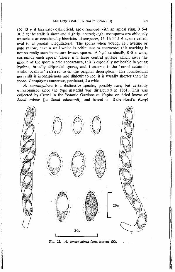
(X 13 v- if biseriate) cylindrical, apex rounded with an apical ring, 0-5-1 X 3 M ; the stalk is short and slightly tapered; eight ascospores are obliquely uniseriate or occasionally biseriate. Ascospores, 13-16 X 5-6 p, one celled, oval to ellipsoidal, inequilateral. The spores when young, i.e., hyaline or pale yellow, have a wall which is echinulate to verrucose; this marking is not so easily seen in mature brown spores. A hyaline sheath, 0-5 p wide, surrounds each spore. There is a large central guttule which gives the middle of the spore a pale appearance, this is especially noticeable in young hyaline, broadly ellipsoidal spores, and I assume is the ' omni aetate in medio ocellata' referred to in the original description. The longitudinal germ slit is inconspicuous and difficult to see, it is usually shorter than the spore. Paraphyses numerous, persistent, 3 P wide.
A. consanguinea is a distinctive species, possibly rare, but certainly unrecognised since the type material was distributed in 1861., This was collected by Cesati in the Botanic Gardens at Naples on dried leaves of Sabal minor [as Sabal adansonii] and issued in Rabenhorst's Fungi
F I G . 2 3 . A. consanguinea f rom isotype (K).
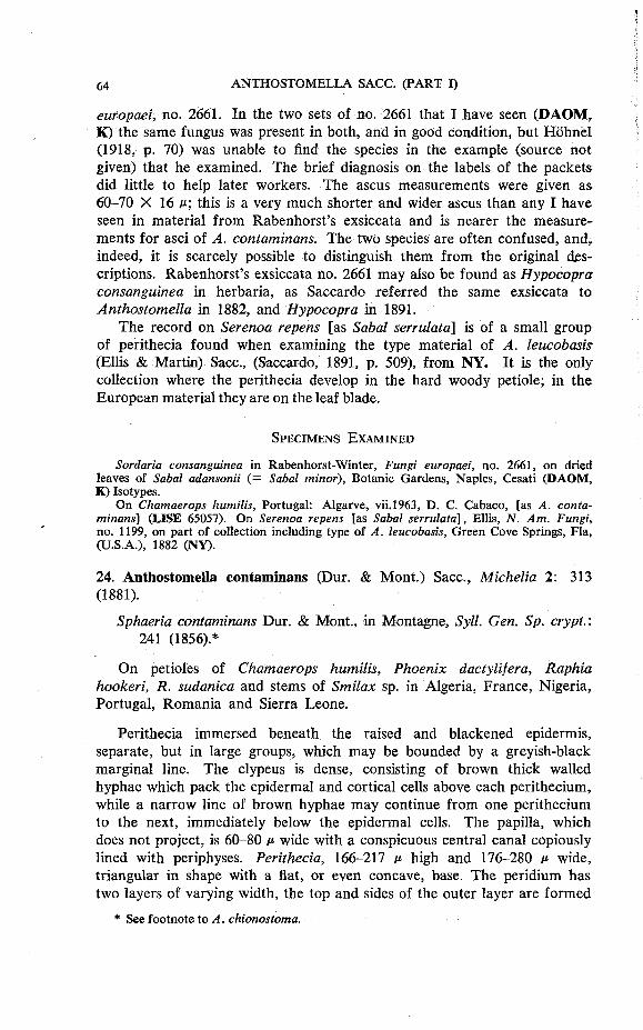
europaei, no. 2661. In the two sets of no. 2661 that I have seen (DAOM, K) the same fungus was present in both, and in good condition, but Hohnel (1918, p. 70) was unable to find the species in the example (source not given) that he examined. The brief diagnosis on the labels of the packets did little to help later workers. The ascus measurements were given as 60-70 X 16 p; this is a very much shorter and wider ascus than any I have seen in material from Rabenhorst's exsiccata and is nearer the measurements for asci of A. contaminans. The two species are often confused, and, indeed, it is scarcely possible to distinguish them from the original descriptions. Rabenhorst's exsiccata no. 2661 may also be found as Hypocopra consanguinea in herbaria, as Saccardo referred the same exsiccata to Anthostomella in 1882, and Hypocopra in 1891.
The record on Serenoa repens [as Sabal serrulata] is of a small group of perithecia found when examining the type material of A. leucobasis (Ellis & Martin) Sacc, (Saccardo, 1891, p. 509), from NY. It is the only collection where the perithecia develop in the hard woody petiole; in the European material they are on the leaf blade.
S P E C I M E N S E X A M I N E D
Sordaria consanguinea in Rabenhorst-Winter , Fungi europaei, no . 2661 , o n dried leaves of Sabal adansonii (= Sabal minor), Botanic Gardens, Naples , Cesati ( D A O M , K ) Isotypes.
On Chamaerops humilis, Portugal: Algarve , vi i .1963, D . C. Cabaco, [as A. contaminans] (LISE 65057). On Serenoa repens [as Sabal serrulata], El l is , N. Am. Fungi, no. 1199, on part of col lect ion including type of A. leucobasis, Green Cove Springs, F la , ( U . S . A . ) , 1882 ( N Y ) .
24. Anthostomella contaminans (Dur. & Mont.) Sacc, Michelia 2: 313 (1881).
Sphaeria contaminans Dur. & Mont., in Montagne, Syll. Gen. Sp. crypt.: 241 (1856).*
On petioles of Chamaerops humilis, Phoenix dactylifera, Raphia hookeri, R. sudanica and stems of Smilax sp. in Algeria, France, Nigeria, Portugal, Romania and Sierra Leone.
Perithecia immersed beneath the raised and blackened epidermis, separate, but in large groups, which may be bounded by a greyish-black marginal line. The clypeus is dense, consisting of brown thick walled hyphae which pack the epidermal and cortical cells above each perithecium, while a narrow line of brown hyphae may continue from one perithecium to the next, immediately below the epidermal cells. The papilla, which does not project, is 60-80 p wide with a conspicuous central canal copiously lined with periphyses. Perithecia, 166-217 P high and 176-280 p wide, triangular in shape with a flat, or even concave, base. The peridium has two layers of varying width, the top and sides of the outer layer are formed
* See footnote to A. chionostoma.

from dark thick walled cells, while along the base, the walls of the cells are yellow and much thinner. The inner layer is more uniform and formed from hyaline, strongly compressed, thin walled cells. Asci, 100-110 X 11-15 M , cylindrical, apex rounded or slightly flattened, with an apical ring, 1-1-5 X 4 c; the stalk is short and wide; eight ascospores are obliquely uniseriate or partially biseriate, especially when young. Ascospores, 14-18 X 5-7 ii, one celled, ellipsoidal to oval with tapered ends, not laterally compressed; surrounded by a hyaline sheath, 1-2 ii wide; longitudinal germ slit conspicuous and the same length as the spore. Paraphyses numerous, 2-2-5 ii wide, conspicuous, with glistening contents.
F I G . 24 . A- contaminans A , from holotype, a, immature ascospores; B , from (51758d).
The type material of this species in Montagne's herbarium at PC is on Chamaerops humilis. The small piece of petiole that I examined yielded three species of Anthostomella: A. phoenicicola; a species so far unnamed with rounded, laterally compressed spores which measured 12-14 X 9-10 X 3-4 M ; and a third species that agreed well with Montagne's description of A. contaminans and was the only species in the isotype material in Berkeley's herbarium at K.
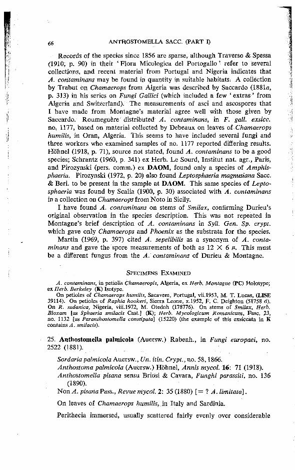
Records of the species since 1856 are sparse, although Traverso & Spessa (1910, p. 90) in their ' Flora Micologica del Portogallo ' refer to several collections, and recent material from Portugal and Nigeria indicates that A. contaminans may be found in quantity in suitable habitats. A collection by Trabut on Chamaerops from Algeria was described by Saccardo (1881a, p. 313) in his series on Fungi Gallici (which included a few ' extras ' from Algeria and Switzerland). The measurements of asci and ascospores that I have made from Montagne's material agree well with those given by Saccardo. Roumeguere distributed A. contaminans, in F. gall, exsicc. no. 1177, based on material collected by Debeaux on leaves of Chamaerops humilis, in Oran, Algeria. This seems to have included several fungi and three workers who examined samples of no. 1177 reported differing results. Hohnel (1918, p. 71), source not stated, found A. contaminans to be a good species; Schrantz (1960, p. 341) ex Herb. Le Sourd, Institut nat. agr., Paris, and Pirozynski (pers. comm.) ex DAOM, found only a species of Amphis-phaeria. Pirozynski (1972, p. 20) also found Leptosphaeria magnusiana Sacc. & Berl. to be present in the sample at DAOM. This same species of Leptosphaeria was found by Scalia (1900, p. 30) associated with A. contaminans in a collection on Chamaerops from Noto in Sicily.
I have found A. contaminans on stems of Smilax, confirming Durieu's original observation in the species description. This was not repeated in Montagne's brief description of A. contaminans in Syll. Gen. Sp. crypt. which gave only Chamaerops and Phoenix as the substrata for the species.
Martin (1969, p. 397) cited A. sepelibilis as a synonym of A. contaminans and gave the spore measurements of both as 12 X 6 p. This must be a different fungus from the A. contaminans of Durieu & Montagne.
S P E C I M E N S E X A M I N E D
A. contaminans, in petiolis Chamaeropis, Algeria , ex Herb. Montagne 0?C) Holotype; e x Herb. Berkeley (K) Isotype.
O n petioles of Chamaerops humilis, Sacavem, Portugal , vi i .1953, M . T. Lucas , (LISE 39114). O n petioles of Raphia hookeri, Sierra L e o n e , x .1952, F . C. D e i g h t o n (51758 d) . O n R. sudanica, Nigeria , vi i i .1972, M . Otedoh (178776). On stems of Smilax, Herb. Bloxam [as Sphaeria smilacis Cast.] (K); Herb. Mycologicum Romanicum, Fasc . 2 3 , no . 1132 [as Paranthostomella constipata] (15220) (the example of this exsiccata in K contains A. smilacis).
25. Anthostomella palmicola (Auersw.) Rabenh., in Fungi europaei, no. 2522 (1881).
Sordaria palmicola Auersw., Un. itin. Crypt., no. 58,1866. Anthostoma palmicola (Auersw.) Hohnel, Annls mycol. 16: 71 (1918). Anthostomella pisana sensu Briosi & Cavara, Funghi parassiti, no. 136
(1890).
Non A. pisana Pass., Revue mycol. 2: 35 (1880) [ = ? A. limitata].
On leaves of Chamaerops humilis, in Italy and Sardinia.
Perithecia immersed, usually scattered fairly evenly over considerable

areas. Clypeus either absent or sparsely developed as a few dark brown hyphae in the epidermal cells immediately surrounding the papilla. The papilla does not project beyond the leaf surface although the periphyses, which line its central canal may do so. Perithecia, 166-220 A high and 143-165 u- wide; when dry the base is strongly inverted. The peridium is 15 M wide, with an outer layer, 10 c, of dark thick walled cells, and an inner layer, 5 c, of hyaline compressed cells. Asci, 66-82 X 8-10 M , clavate at first, becoming cylindrical, with a rounded or slightly flattened apex, no apical ring visible; the stalk varies in length from 5-20 /*; eight ascospores are uniseriate at all stages of development. Ascospores, 9-12 X 6-8 X 3-4 c, one celled, rounded, slightly laterally compressed; surrounded by a narrow, but clearly visible, hyaline sheath; an equatorial germ slit is present, the edges appear slightly blurred, older spores tend to split along the line when crushed. Paraphyses numerous, 2-3 deliquescing early.
This species formed part of a large collection of cryptogams made by Dr. E. Marcucci on behalf of the Unio itineraria Cryptogamica, a society founded by Rabenhorst and Schimper in May 1863 (7. Bot., Lond. 2: 359, 1864). Marcucci was commissioned to collect in Sardinia and his material was sent out to subscribing members in 1866 (subscription, 12/ - a year). A. palmicola which was briefly described and named, as Sordaria palmicola, by Auerswald was no. 58 in this distribution. Unfortunately no. 58 was a mixed collection and also contained the fungus now known as Phaeochora steinheilii (Mont.) E. Muller. Both fungi were present in the isotype material that I saw from UPS. (Auerswald's material is not at present available from B). Marcucci's collection was used for three further exisiccatae—Rabenhorst, Fungi europaei, nos. 1446 and 2522, also Thumen's Mycotheca universalis, no. 2168; all three have varying mixtures of the same two fungi.
The exsiccatae that I examined at K, and reports in the literature (Traverso 1907, p. 486; Hohnel 1918, p. 70; Pirozynski 1972, p. 4) indicate that Fungi europaei 1446 and Myc. univ. 2168 contained Phaeochora, while Fungi europaei 2522 (where Rabenhorst transferred the species from Sordaria to Anthostomella) contained only material of the Anthostomella. In view of the large quantity of material which must have been involved in, each of Marcucci's collections, it is perhaps not surprising that more than one fungus was present in sample no. 58. What is curious is that the Phaeochora, much the more obvious of the two fungi, was not recorded for nearly twenty years. Recognition came in 1884 when in Barbey's ' Flora Sardoae' the two fungi present in Marcucci's material were separately recorded, A. palmicola on p. 205 and Phaeochora steinheilii [as Auerswaldia chamaeropis] on p. 247, the latter in a supplement contributed by Saccardo. It is sad to record that they lost their separate identity again in 1954 (Arx & Muller, 1954, p. 245) [as Sphaerodothis chamaeropis (Cooke) Shear]. '
The name, as Anthostoma palmicola (Auersw.) Hohnel, was however used by Muller & Dennis (1965, p. 370) for a collection (Dennis 12616)


made in Venezuela on Heliconia. The species in this collection is not conspecific with Anthostomella palmicola, it is near A. sequoiae Rehm. Hdhnel's Anthostoma palmicola is discussed further in the notes on A. phoenicicola.
A collection by Pirozynski on Elaeis guineensis, from Tanzania, was described by him (1972, p. 4) as A: palmicola but differing from the type in having smaller spores (6-5-8-5 X 5-5-6-5 X 3-3-5 P). The collection also differs from the European material in having a well developed clypeus and an ascus wall that deliquesces before the spores have matured. Further material of both taxa is needed to determine whether the Tanzanian material is distinct from A. palmicola.
A. pisana was collected in 1874 on arid petioles of Chamaerops humilis in the Botanic Gardens at Pisa (Passerini, 1880, p. 35). I have not been able to trace this material. Two exsiccata of the species were issued, Erb. Critt. Ital., ser. 2, no. 1169 in 1882, and Briosi & Cavara, Funghi parassiti, no. 136 in 1890; both are collections on leaves of Chamaerops from Pisa. Traverso (1907, p. 484) maintained A. pisana as a separate species but pointed out that the spore measurements that he found in an example of Erb. Critt. (10-12 X 7-8 p) were larger than those quoted by Passerini. Hohnel (1918, p. 70 & 1918a, p. 223) found the A. pisana of Briosi & Cavara to be identical with A. palmicola. I have seen specimens of both exsiccata at K and the fungus present in both was A. palmicola. However I do not think that they truly represent Passerini's species, as the diagnosis he gives, with measurements of asci, 45 X 5-5 M and spores, 10 X 5 p is quite unlike A. palmicola, but it is, however, very close to A. limitata. The material I received from PAD (when requesting the type of A. pisana) was a collection by Baccarini & Avetta on Chamaerops from a villa near Rome; the fungus present was A. limitata. I suggest that it was this species, and not A. palmicola, that Passerini described as A. pisana.
Martin (1969, p. 396) maintained A. palmicola and A. pisana [as A. pisana (Pers.) Sacc] as two species. The spore measurements of 11 X 5 p that he gives for A. palmicola and the description of ' stromata gregarious with shiny black ostioles' suggest that the species he examined was A. phoenicicola.
S P E C I M E N S E X A M I N E D
Unio itin. crypt., no . 58, Sordaria palmicola A w d . mspt. , Chamaerops humilis, A l g h e r o (Sardinia) vi .1866, Marcucci (Phaeochora steinheilii a lso present) (UPS) Isotype. Rabenhorst , Fungi europaei, no . 2522 , A. palmicola, o n Chamaerops humilis, Sardinia, Marcucc i (K). Erb. Critt. Ital., ser. II , no . 1169, on Chamaerops humilis, Orto Botanico , Pisa, x .1881 , A . Mori [as A. pisana] (K, P A R M A ) . Briosi & Cavara, Funghi parassiti, no. 136, o n Chamaerops humilis, Orto Botanico , Pisa [as A. pisana] (K, P A R M A , I M I 15147).
O n dead rachides of Elaeis guineensis, K a k o m b e , Tanzania , i .1964, K. A . Pirozynski (spores smaller than type) (106158 f).
Exsiccatae in which the fungus was not A. palmicola. Rabenhorst , Fungi europaei, no . 1446, Sordaria palmicola = Phaeochora steinheilii
( K) . Thi imen, Mycotheca universalis, no . 2168, Sordaria palmicola = Phaeochora steinheilii (K). Herb. R. Horti Romani, i i . l 884, Baccarini & Avet ta [as A. pisana] = A. limitata ( P A D ) .
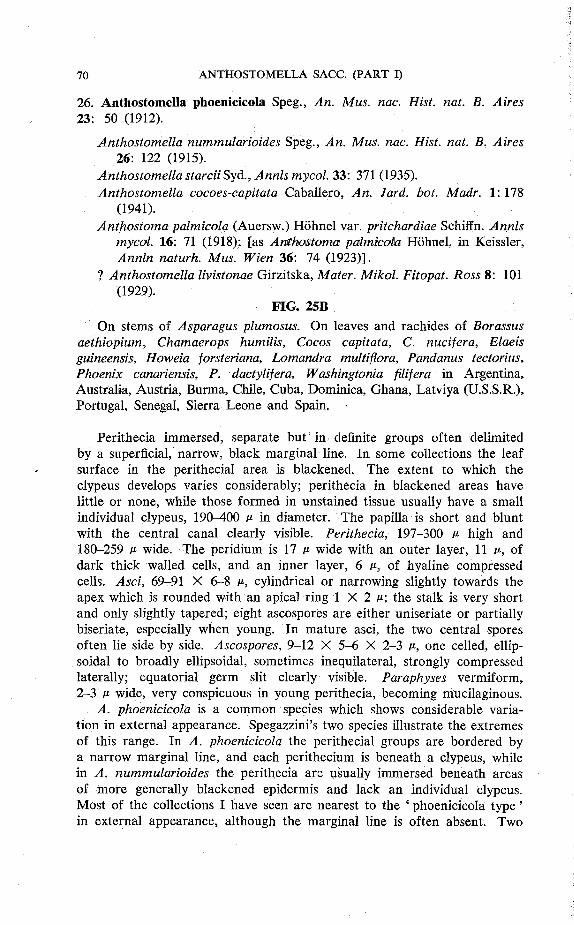
26. Anthostomella phoenicicola Speg., An. Mus. nac. Hist. nat. B. Aires 23: 50 (1912).
Anthostomella nummularioides Speg., An. Mus. nac. Hist. nat. B. Aires 26: 122 (1915).
Anthostomella starciiSyd., Annls mycol. 33: 371 (1935). Anthostomella cocoes-capitata Caballero, An. Jard. hot. Madr. 1: 178
(1941). Anthostoma palmicola (Auersw.) Hohnel var. pritchardiae Schiffn. Annls
mycol. 16: 71 (1918); [as Anthostoma palmicola Hohnel, in Keissler, Annln naturh. Mus. Wien 36: 74 (1923)].
? Anthostomella livistonae Girzitska, Mater. Mikol. Fitopat. Ross 8: 101 (1929).
FIG. 25B On stems of Asparagus plumosus. On leaves and rachides of Borassus
aethiopium, Chamaerops humilis, Cocos capitata, C. nucifera, Elaeis guineensis, Howeia forsteriana, Lomandra multiflora, Pandanus tectorius, Phoenix canariensis, P. dactylifera, Washingtonia filifera in Argentina, Australia, Austria, Burma, Chile, Cuba, Dominica, Ghana, Latviya (U.S.S.R.), Portugal, Senegal, Sierra Leone and Spain.
Perithecia immersed, separate but in definite groups often delimited by a Superficial, narrow, black marginal line. In some collections the leaf surface in the perithecial area is blackened. The extent to which the clypeus develops varies considerably; perithecia in blackened areas have little or none, while those formed in unstained tissue usually have a small individual clypeus, 190-400 p in diameter. The papilla is short and blunt with the central canal clearly visible. Perithecia, 197-300 p high and 180-259 p wide. The peridium is 17 P wide with an outer layer, 11 p, of dark thick walled cells, and an inner layer, 6 p, of hyaline compressed cells. Asci, 69-91 X 6-8 p, cylindrical or narrowing slightly towards the apex which is rounded with an apical ring 1 X 2 p; the stalk is very short and only slightly tapered; eight ascospores are either uniseriate or partially biseriate, especially when young. In mature asci, the two central spores often lie side by side. Ascospores, 9-12 X 5-6 X 2-3 P, one celled, ellipsoidal to broadly ellipsoidal, sometimes inequilateral, strongly compressed laterally; equatorial germ slit clearly visible. Paraphyses vermiform, 2-3 p wide, very conspicuous in young perithecia, becoming mucilaginous.
A. phoenicicola is a common species which shows considerable variation in external appearance. Spegazzini's two species illustrate the extremes of this range. In A. phoenicicola the perithecial groups are bordered by a narrow marginal line, and each perithecium is beneath a clypeus, while in A. nummularioides the perithecia are usually immersed beneath areas of more generally blackened epidermis and lack an individual clypeus. Most of the collections I have seen are nearest to the 'phoenicicola type ' in external appearance, although the marginal line is often absent. Two
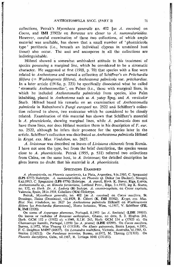
collections, Petrak's Mycotheca generalis no. 402 [as A. cocoina] on Cocos, and IMI 37922e on Borassus are closer to A. nummularioides. However, careful examination of these two collections, of which ample material was available, has shown that a small number of ' phoenicicola type ' perithecia (i.e., beneath an individual clypeus in unstained host tissue) also occur. The asci and ascospores in all the collections are indistinguishable.
Hohnel showed a somewhat ambivalent attitude in his treatment of species possessing a marginal line, which he considered to be a stromatic character. He suggested at first (1918, p. 70) that species with a line were related to Anthostoma and named a collection of Schiffner's on Pritchardia filifera (= Washingtonia filifera), Anthostoma palmicola var. pritchardiae. In a later article (1918a, p. 223) he specifically dissociated what he called * stromatic Anthostomellas', on Palms (i.e., those with marginal lines, in which he included Anthostomella palmicola) from species, also Palm inhabiting, placed in Anthostoma such as A. yatay Speg. and A. versicolor Starb. Hohnel based his remarks on an examination of Anthostomella palmicola in Rabenhorst's Fungi europaei no. 2522 and Schiffner's collection referred to above, two exsiccatae which he considered to be closely related. Examination of this material has shown that Schiffner's material is A. phoenicicola, showing marginal lines, while A. palmicola does not have these lines, nor does Hohnel mention them in his description of F. eur. no. 2522, although he infers their presence for the species later in the article. Schiffner's collection was distributed as Anthostoma palmicola Hohnel in Krypt. exs. Mus. Vindobon, no. 2627.
A. livistonae was described on leaves of Livistona chinensis from Russia. I have not seen the type, but from the brief description, the species seems close to A. phoenicicola. Petrak (1955, p. 531) referred two collections from China, on the same host, to A. livistonae; the detailed description he gives leaves no doubt that his material is A. phoenicicola.
S P E C I M E N S E X A M I N E D
A. phoenicicola, on Phoenix canadensis, L a Plata, Argent ina , 9 . i x . l 910 , C. Spegazzini ( L P S 6757) Ho lo type . A. nummularioides, o n Phoenix sp. Dakar [as D a c k a r ] , Senegal , 8 .x i i . l913 , C. Spegazzini (LPS 6776) Holo type . A. starcii, Herb. K. Stares, Riga , Latvia, Anthostomella sp . , on Howeia forsteriana, Lett land Prov. , Riga , 3 .V.1933 , leg K. Stares, no . 132, ex Herb. Dr. A. Ludwig ( B ) Isotype. A. cocoes-capitata, on Cocos capitata, Valenc ia , Spain, 28. iv.1938, Caballero ( M A ) Holotype .
Petrak, Mycotheca generalis, no . 402 [as A. cocoina] o n Cocos nucifera, St. D o m i n g o , Haina (Dominica) , vii i .1929, R. Ciferri (K, I M I 30302). Krypt.- exs. Mus. Hist. Nat. Vindobon, no. 2627 [as Anthostoma palmicola H o h n e l ] o n Washingtonia filifera [as Pritchardia filamentosa], Horto botanico , W i e n , vi .1917, V . Schiffner ( F H , K, I M I 12320).
O n stems of Asparagus plumosus, Portugal , ii. 1953 [as A. limitata] (LISE 39101). O n leaves or rachides of Borassus aethiopium, Ghana , no date, S. J. H u g h e s 241 , Herb . G C M 152 c (37922 e); v .1949, S.J.H. 243, Herb. G C M 154 a (37925 a) . On Chamaerops humilis, Portugal, i .1950 [as A. pisana] (LISE 65099). On Cocos nucifera, Burma, i i .1972, M y a Thaung 13 (171318). O n Elaeis guineensis, Sierra L e o n e , v .1951 , F . C. De ighton M 4 0 9 7 (46637). On Lomandra multiflora, Victoria, Austral ia , xi . 1965, G. Beaton (116212). On Pandanus tectorius, Burma, xi .1972, M . T h a u n g (171323). On Phoenix dactylifera, Cuba, xii. 1967, R. Urt iaga 1048 (131151).
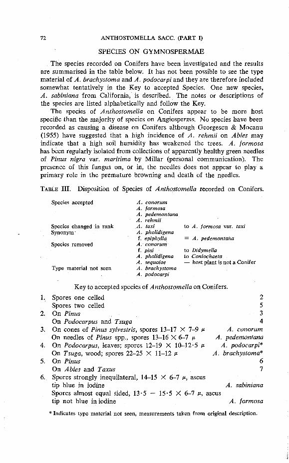
SPECIES ON GYMNOSPERMAE
The species recorded on Conifers have been investigated and the results are summarised in the table below. It has not been possible to see the type material of A. brachystoma and A. podocarpi and they are therefore included somewhat tentatively in the Key to accepted Species. One new species, A. sabiniana from California, is described. The notes or descriptions of the species are listed alphabetically and follow the Key.
The species of Anthostomella on Conifers appear to be more host specific than the majority of species on Angiosperms. No species have been recorded as causing a disease on Conifers although Georgescu & Mocanu (1955) have suggested that a high incidence of A. rehmii on Abies may indicate that a high soil humidity has weakened the trees. A. formosa has been regularly isolated from collections of apparently healthy green needles of Pinus nigra var. maritima by Millar (personal communication). The presence of this fungus on, or in, the needles does not appear to play a primary role in the premature browning and death of the needles.
T A B L E III. Disposition of Species of Anthostomella recorded on Conifers.
Species accepted
Species changed in rank Synonym
Species removed
Type material not seen
A. conorum A. formosa A. pedemontana A. rehmii A. taxi A. pholidigena f. epiphylla
A. conorum f. pint
A. pholidigena A. sequoiae A. brachystoma A. podocarpi
to A. formosa var. taxi
= A. pedemontana
to Didymella to Coniochaeta — host plant is not a Conifer
Key to accepted species of Anthostomella on Conifers 1. Spores one celled
Spores two celled 2. On Pinus
On Podocarpus and Tsuga 3. On cones of Pinus sylvestris, spores 13-17 X 7-9 P
On needles of Pinus spp., spores 13-16 X 6-7 P 4. On Podocarpus, leaves; spores 12-19 X 10-12-5 p
On Tsuga, wood; spores 22-25 X 11-12 P 5. On Pinus
On Abies and Taxus 6. Spores strongly inequilateral, 14-15 X 6-7 p, ascus
tip blue in iodine Spores almost equal sided, 13-5 — 15-5 X 6-7 p, ascus tip not blue in iodine
2 5 3 4
A. conorum A. pedemontana
A. podocarpi* A. brachystoma*
6 7
A. sabiniana
A. formosa
* Indicates type material not seen, measurements taken from original description.
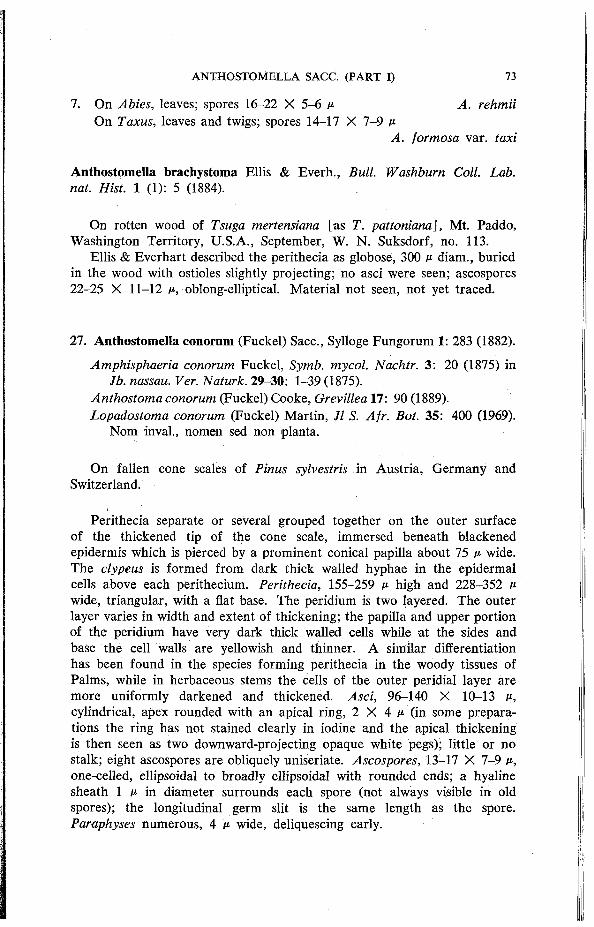
7. On Abies, leaves; spores 16-22 X 5-6 p A. rehmii On Taxus, leaves and twigs; spores 14-17 X 7-9 p
A. formosa var. taxi
Anthostomella brachystoma Ellis & Everh., Bull. Washburn Coll. Lab. nat. Hist. 1 (1): 5 (1884).
On rotten wood of Tsuga mertensiana [as T. pattoniand], Mt. Paddo, Washington Territory, U.S.A., September, W. N. Suksdorf, no. 113.
Ellis & Everhart described the perithecia as globose, 300 p diam., buried in the wood with ostioles slightly projecting; no asci were seen; ascospores 22-25 X 11-12 p, oblong-elliptical. Material not seen, not yet traced.
27. Anthostomella conorum (Fuckel) Sacc, Sylloge Fungorum 1: 283 (1882).
Amphisphaeria conorum Fuckel, Symb. mycol. Nachtr. 3: 20 (1875) in Jb. nassau. Ver. Naturk. 29-30: 1-39 (1875).
Anthostoma conorum (Fuckel) Cooke, Grevillea 17: 90(1889). Lopadostoma conorum (Fuckel) Martin, Jl S. Afr. Bot. 35: 400 (1969).
Nom inval., nomen sed non planta.
On fallen cone scales of Pinus sylvestris in Austria, Germany and Switzerland.
Perithecia separate or several grouped together on the outer surface of the thickened tip of the cone scale, immersed beneath blackened epidermis which is pierced by a prominent conical papilla about 75 p wide. The clypeus is formed from dark thick walled hyphae in the epidermal cells above each perithecium. Perithecia, 155-259 p high and 228-352 p wide, triangular, with a flat base. The peridium is two layered. The outer layer varies in width and extent of thickening; the papilla and upper portion of the peridium have very dark thick walled cells while at the sides and base the cell walls are yellowish and thinner. A similar differentiation has been found in the species forming perithecia in the woody tissues of Palms, while in herbaceous stems the cells of the outer peridial layer are more uniformly darkened and thickened. Asci, 96-140 X 10-13 p, cylindrical, apex rounded with an apical ring, 2 X 4 p (in some preparations the ring has not stained clearly in iodine and the apical thickening is then seen as two downward-projecting opaque white pegs); little or no stalk; eight ascospores are obliquely uniseriate. Ascospores, 13—17 X 7-9 p, one-celled, ellipsoidal to broadly ellipsoidal with rounded ends; a hyaline sheath 1 P in diameter surrounds each spore (not always visible in old spores); the longitudinal germ slit is the same length as the spore. Paraphyses numerous, 4 p wide, deliquescing early.
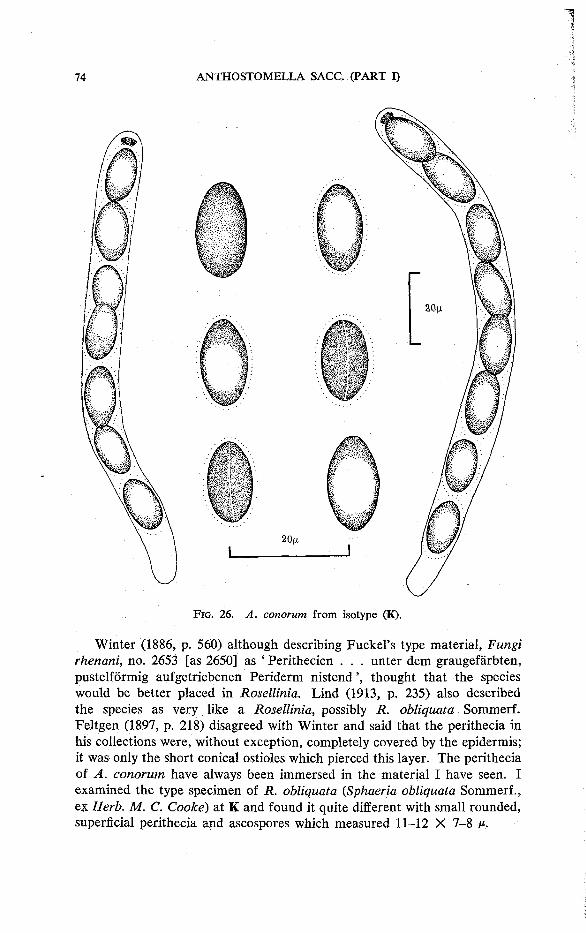
F I G . 2 6 . A. conorum f rom isotype ( K ) .
Winter (1886, p. 560) although describing FuckePs type material, Fungi rhenani, no. 2653 [as 2650] as ' Perithecien . . . unter dem graugefarbten, pustelformig aufgetriebenen Periderm nistend', thought that the species would be better placed in Rosellinia. Lind (1913, p. 235) also described the species as very like a Rosellinia, possibly JR. obliquata Sommerf. Feltgen (1897, p. 218) disagreed with Winter and said that the perithecia in his collections were, without exception, completely covered by the epidermis; it was only the short conical ostioles which pierced this layer. The perithecia of A. conorum have always been immersed in the material I have seen. I examined the type specimen of R. obliquata (Sphaeria obliquata Sommerf., ex Herb. M. C. Cooke) at K and found it quite different with small rounded, superficial perithecia and ascospores which measured 11-12 X 7-8 v.

The description given by Martin (1969, p. 400) of his Lopadostoma conorum as " stromata 1-2 peritheciate . . . spores broad oval, equilateral . . . 6 - 0 X 9 - 0 / * " does not agree with the type material of Amphisphaeria conorum at K.
Petrak & Sydow (1924, p. 333) considered Sphaeropsis acicola Pass, to be a synonym of A. conorum. They had seen a specimen and found asci present. Passerini (1889, p. 464) collected this fungus on leaves of Pinus nigra var. nigra [as P. austriaca] in the Botanic Garden at Parma. The collection is not available from PARMA, but from the habit and description I think the species is probably A. pedemontana rather than A. conorum.
S P E C I M E N S E X A M I N E D
Amphisphaeria conorum nov. sp., Fucke l Fungi rhenani no . 2653 , ad Pinus sylvestris conorum aridorum squamas. H i e m e , ca Vollrads, ex Herb. C. E. Broome (K) Isotype. Amphisphaeria conorum, Zurich, Helvetia, viii. 1876, G. Winter (K). Anthostomella conorum, F . Petrak, Mycotheca generalis no . 502, on Pinus sylvestris, Bayern, Kalmut bei Marktheidenfeld, i i i .1926, leg. A . A d e (E , K, I M I 30402); Kalmut , 17.vi.1926 (K, I M I 21408) .
A. conorum (Fuckel) Sacc. f. pini Fautrey, Revue mycol. 16: 165 (1894). R o u m e -guere's F. gall, exsicc. no . 6604 from M P U contains only a species of Didymella.
28. Anthostomella formosa Kirschst., Verh. hot. Ver. Prov. Brandenb. 66: 29 (1924).
Neesiella formosa (Kirschst.) Kirschst., Annls mycol. 33: 217 (1935). Myconeesia formosa (Kirschst.) Kirschst., Annls mycol. 34: 200 (1936).
FIG. 27A On fallen needles of Pinus nigra var. maritima, P. nigra var. nigra and
P. sylvestris in Austria, Britain, Germany and the Netherlands.
Perithecia immersed beneath the epidermis on both surfac'es of the needle. There is no clypeus and the papilla, which is conical, projects only slightly. The position of a perithecium is often indicated by the spores which lie in a sticky mass around the base of the papilla. Perithecia, 207-311 p high and 238-301 p wide. The peridium is 18 p wide with an outer layer, 10 p, of dark thick walled cells and an inner layer, 8 p of hyaline thin walled cells. Asci, 120-135 X 8 p, cylindrical with a rounded apex which appears to be thickened but no apical ring is visible in iodine; the stalk is short and tapered; eight ascospores are uniseriate and slightly oblique. Ascospores, 13-5-15-5 X 6-7 p, two celled, with a small hyaline dwarf cell, 1-5 X 2 P, and a brown cell, 12-14 X 6-7 P, ellipsoidal with the free end bluntly pointed; a narrow hyaline sheath surrounds each spore; the longitudinal germ slit is 7-10 p. Paraphyses numerous, 3 P wide.
Kirschstein himself, ten years after he had named A. formosa transferred the fungus, as type species, to the genus Neesiella Kirschsfe This was because it lacked a clypeus. A year later the name was changed again to Myconeesia as Neesiella had already been used by Schiffner for a genus in the

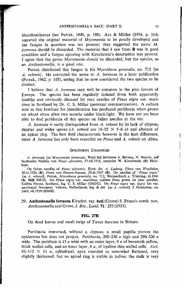
Marchantiaceae (see Petrak, 1940, p. 188). Arx & Muller (1954 , p. 314) reported the original material of Myconeesia to be poorly developed and the fungus in question was not present; they suggested the name M. formosa should be discarded. The material that I saw from B was in good condition and a fungus agreeing with Kirschstein's description was present. I agree that the genus Myconeesia should be discarded, but the species, as an Anthostomella, is a good one.
Petrak distributed this fungus in his Mycotheca generalis, no. 7 1 2 [as A. rehmii]. He corrected the name to A. formosa in a later publication (Petrak, 1962, p. 165), saying that he now considered the two species to be distinct.
I believe that A. formosa may well be common in the pine forests of Europe. The species has been regularly isolated from both apparently healthy and obviously diseased 1st year needles of Pinus nigra var. mari-tima in Scotland by Dr. C. S. Millar (personal communication). A culture sent to this Institute for identification has produced perithecia when grown on wheat straw after two months under black-light. We have not yet been able to find perithecia of this species on fallen needles at the site.
A. formosa is easily distinguished from A. rehmii by its lack of clypeus, shorter and wider spores (A. rehmii are 1 6 - 2 2 X 5 - 6 p) and absence of an apical ring. The best field characteristic however is the host difference, since A. formosa has only been recorded on Pinus and A. rehmii on Abies.
S P E C I M E N S E X A M I N E D
A. formosa [as Myconeesia formosa], Wald bei Schonow b. Bernau, N . Barnin , auf fau lenden Nade ln v o n Pinus sylvestris, 17.vii .1916, sammler W . Kirschstein (B) Holo type .
O n fallen needles of Pinus sylvestris, Herb. Dr. A. Ludwig, Flora von Westfalen, 20.x i .1926 (B); Flora von Hessen-Nassau, 20.ii i .1927 (B) . O n needles of 'Pinus nigra' [as A. rehmii], Petrak, Mycotheca generalis, no . 712 , Weissenbach a. Triesting, iii.1941
(K, I M I 30612). On Pinus nigra var. maritima, culture from green 1st year needles, Culb in Forest , Scotland, leg. C. S. Millar (156242). O n Pinus nigra var. nigra [as var. austriaca] Nunspeet , Ve luwe, Netherlands, leg. & det. [as A. rehmii] J. G r e m m e n , no . 1665, vii. 1959 (80668).
2 9 . Anthostomella formosa Kirschst. var. taxi (Grove) S. Francis comb. nov. Anthostomella taxi Grove, J. Bot., Lond. 71: 2 5 3 (1933) .
FIG. 27B On dead leaves and small twigs of Taxus baccata in Britain.
Perithecia immersed, without a clypeus, a small papilla pierces the epidermus but does not project. Perithecia, 2 0 0 - 2 3 0 p high and 2 9 0 - 3 2 0 p wide. The peridium is 15 P wide with an outer layer, 9 p of brownish yellow, thick walled cells, and an inner layer, 6 P, of hyaline thin walled cells. Asci, 9 3 - 1 1 2 X 1 1 M , cylindrical, apex rounded or somewhat flattened, very slightly thickened, but no apical ring is visible in iodine; the stalk is very
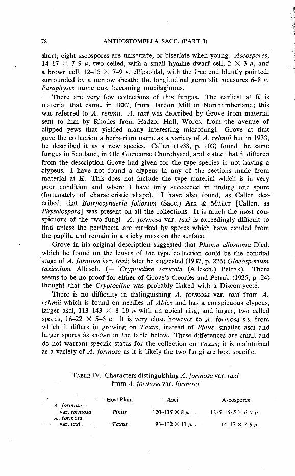
short; eight ascospores are uniseriate, or biseriate when young. Ascospores, 14-17 X 7-9 ft, two celled, with a small hyaline dwarf cell, 2 X 3 c , and a brown cell, 12-15 X 7-9 i*, ellipsoidal, with the free end bluntly pointed; surrounded by a narrow sheath; the longitudinal germ slit measures 6-8 p. Paraphyses numerous, becoming mucilaginous.
There are very few collections of this fungus. The earliest at K is material that came, in 1887, from Bardon Mill in Northumberland; this was referred to A. rehmii. A. taxi was described by Grove from material sent to him by Rhodes from Hadzor Hall, Worcs. from the avenue of clipped yews that yielded many interesting microfungi. Grove at first gave the collection a herbarium name as a variety of A. rehmii but in 1933, he described it as a new species. Callen (1938, p. 103) found the same fungus in Scotland, in Old Glencorse Churchyard, and stated that it differed from the description Grove had given for the type species in not having a clypeus. I have not found a clypeus in any of the sections made from material at K. This does not include the type material which is in very poor condition and where I have only succeeded in finding one spore (fortunately of characteristic shape). I have also found, as Callen described, that Botryosphaeria foliorum (Sacc.) Arx & Muller [Callen, as Physalospora] was present on all the collections. It is much the most conspicuous of the two fungi. A. formosa var. taxi is exceedingly difficult to find unless the perithecia are marked by spores which have exuded from the papilla and remain in a sticky mass on the surface.
Grove in his original description suggested that Phoma allostoma Died, which he found on the leaves of the type collection could be the conidial stage of A. formosa var. taxi; later he suggested (1937, p. 226) Gloeosporium taxicolum Allesch. ( = Cryptocline taxicola (Allesch.) Petrak). There seems to be no proof for either of Grove's theories and Petrak (1925, p. 24) thought that the Cryptocline was probably linked with a Discomycete.
There is no difficulty in distinguishing A. formosa var. taxi from A. rehmii which is found on needles of Abies and has a conspicuous clypeus, larger asci, 113—143 X 8—10 it with an apical ring, and larger, two celled spores, 16-22 X 5-6 it. It is very close however to A. formosa s.s. from which it differs in growing on Taxus, instead of Pinus, smaller asci and larger spores as shown in the table below. These differences are small and do not warrant specific status for the collection on Taxus; it is maintained as a variety of A. formosa as it is likely the two fungi are host specific.
T A B L E IV. Characters distinguishing A. formosa var. taxi from A. formosa var. formosa
Host Plant Asc i Ascospores A. formosa
var. formosa Pinus 120-135 X 8 ii 1 3 - 5 - 1 5 - 5 X 6-7 it A. formosa
var. taxi Taxus 9 3 - 1 1 2 X 11M 14-17 X 7-9 it

S P E C I M E N S E X A M I N E D
On leaves and thin stems of Taxus baccata, England: Hadzor Hal l , Worcs . , P . G. M . R h o d e s , 4399, 12.xi.1929 (K) Holotype; Hadzor Hall , Worcs . , 17.X.1930 (K); Old Glen-corse Churchyard, nr. Edinburgh, E . O. Callen, x .1934 (E); Carstairs, Lanark, R. W. G. Denn i s , i .1940 (K); [as A. rehmii] Bardon Mill , Northumber land, D . Carlyle, v .1887 (K); C. Farquarson, no locality, iii.1911 (K).
30. Anthostomella pedemontana Ferr. & Sacc, Atti del Congr. bot. di Palermo : 50 (1902).
Entosordaria pedemontana (Ferr. & Sacc.) Hohnel, Sber. Akad. Wiss. WienMath.-nat. 129: 166(1920).
A. pholidigena (Ellis) Berl. & Vogl. a epiphylla Massal., Mem. Accad. Agric. Sci. Verona ser. 3,65: 69 (1889); nomen nudum.
A. pholidigena (Ellis) Berl. & Vogl. f. epiphylla Massal. ex D. Sacc, in Bizzozero, G., Fl. VenetaCritt. Suppl: 46 (1899).
A. pholidigena (Ellis) Berl. & Vogl. var. epiphylla (Massal. ex. D. Sacc.) Trav. Fl. ital. crypt. Pars 1, 2 (2): 483 (1907).
F I G . 28. A. pedemontana f rom lectotype.
On fallen needles of Pinus sylvestris and P. wallichiana in Italy and West Pakistan.

Perithecia sparsely scattered, immersed. There is no clypeus and the epidermis is penetrated by a small, slightly protruding papilla. Perithecia, 220-250 u- high and 250-330 u- wide. The peridium is 13 v wide with an outer layer, 8 of dark thick walled cells, and an inner layer, 5 u-, of thin walled hyaline cells. Asci, 110-113 X 9-12 n, cylindrical with a rounded apex, no apical ring is visible in iodine; a very short stalk; eight (occasionally only four or six) ascospores are obliquely uniseriate. The wall is thin and often disappears before the spores mature. Ascospores, 13-16 X 6-7 v, one celled, oval to ellipsoidal with tapered ends, surrounded by a conspicuous hyaline sheath, 1-2 p wide; the longitudinal germ slit measures only 6 p and is easily seen. Paraphyses numerous, 3 p wide.
A. pedemontana was collected by Ferraris near Crescentino, Italy, in August 1900, on dead leaves of Pinus sylvestris. Only a slide, that he prepared and sent to Saccardo, is preserved at PAD. It shows a transverse section of a pine needle with perithecia, asci and ascospores. This slide must be taken as the lectotype unless the material that Ferraris collected can be located. He published a good illustration of the asci and spores in Malpighia 16: 13, Tav. 1, (1902).
Massalongo had collected the same fungus thirteen years earlier, in Verona, also on needles of Pinus sylvestris. He named the collection A. phalidigena (Ell.) Berl. & Vogl. a epiphylla in a list of the fungi of Verona, without a description and indicating, only in the choice of epithet, that he considered it to differ from A. pholidigena by growing on leaves rather than cone scales. D. Saccardo published a short description in 1899 giving the measurements of the asci as 114 X In and ascospores, 7-10 X 5-6 p. These figures are the same as those given by Ellis (1883, p. 54) in the original description of A. pholidigena and do not agree with Massalongo's fungus which has spores that measure 13-16 X 6-7 M-.
The only other collection that I have seen (IMI 92061a) was on the needles of Pinus watNohiawa [as P. excelsa] from West Pakistan. This was published as Anthostomella rehmii by Ahmad (1962, p. 129) and later (Ahmad, 1969, p. 23) transferred to Anthostoma. This transfer was also made by Cooke in 1889. Hohnel (1920, p. 166) suggested by means of a ' ? ' that A. pedemontana was synonymous with A. rehmii and included both species in Entosordaria. The two species are quite distinct and A. pedemontana has a single celled spore while in A. rehmii the spore is two celled.
The ascospores of A. pedemontana, on the needles, and A. conorum on the cones of Pinus sylvestris are somewhat similar in shape and size. Those of A. pedemontana however are narrower, with a width of 6-7 u-and have a short conspicuous germ slit 6 P long, while A. conorum has spores 7-9 p wide and the germ slit is the same length as the spore.
S P E C I M E N S E X A M I N E D
Anthostomella pedemontana Sacc. & Ferr. (published as Ferr. & Sacc. ) Vert, preparat ion microscopic , C. T . Ferraris. In fs. seche Pinus sylvestris [as silvestris], Sulpiano di
Verrua, pr. Crescentinc* (Italy), 29.vii i .1900 ( P A D ) Leototype. A. pholidigena a epiphylla [as A. pholidigenum], in foliis siccis Pini sylvestris, Tregnago , ix .1887, C M . (Massa-
longo) ( V E R ) . O n leaves of Pinus wallichiana [as P. excelsa], Chinari, Muzaffarabad, W. Pakistan,
c o m m . 1962, S. A h m a d (92061 a).
Anthostomella pholidigena (Ellis) Berl. & Vogl., Sylloge Fungorum Addit. 1-4: 44 (1886) = Sphaeria {Anfhmtomelld) pholidigena Ellis, Bull. Torrey bot. Club 10: 54 (1883) = Anthostoma pholidigena (Ellis) Cooke, Grevillea 17: 90 (1889).
The type material of this species at K in Ellis & Everhart, N. American Fungi, 2nd. ser., no. 1664, is a species of Coniochaeta. The material agrees with the description given by Ellis and has scattered, usually single perithecia which become superficial.
Martin (1969, p. 400) has cited A. pholidigena as a synonym of A. conorum [as Anthostoma conorum (Fckl.) Sacc] and transferred both to Lopadostoma conorum (Fckl.) Martin (non rite publ.).
Anthostomella pholidigena f. epiphylla see A. pedemontana.
Anthostomella podocarpi Sydow, Annls mycol. 28: 94 (1930). I have not been able to trace the type material of this species which was
collected in Venezuela on dead, but still attached, leaves of Podocarpus coriaceus. Sydow described the perithecia as 280-350 p in diameter; asci p. sp. 85-100 X 13-16 P,; ascospores 12-19 X 10-12-5 p., one celled.
31. Anthostomella rehmii (Thum.) Rehm, Ber. naturhist. Ver. Augsburg 26: 95 (1881).
Amphisphaeria rehmii Tniim., Mycotheca universalis no. 464 (1876), and Flora, Jena 60 (13): 204(1877).
Anthostoma rehmii (Tniim.) Cooke, Grevillea 17: 90(1889). Entosordaria rehmii (Thum.) Hohnel, Sber. Akad. Wiss. Wien Math.-nat.
Kl.,Abt. 1,129: 166(1920).
FIG. 27C On needles of Abies alba, West Germany and Romania.
Perithecia sparsely scattered on the upper surface of yellowed needles which are still attached to living branches, immersed beneath a circular, black and shining clypeus 130-238 P in diameter. There is a narrow collar of non-blackened leaf tissue at the base of the small conical papilla separating it from the clypeus. Sections show that the clypeus is formed from what appears to be an extension, in the shape of a wedge of the dark outer peridial wall. At the top of the perithecium, beneath the cuticle, there is

a small area of meristematic fungal tissue. The development of these cells tears the cuticle and the papilla then grows upwards to the exterior. A narrow ring of the hyaline meristematic cells remains around the base of the papilla and gives the ' white collar' appearance externally. Perithecia, 320-363 t1 high and 270-320 v- wide. The peridium is 25 » wide with an outer layer, 17 v-, of dark thick walled cells, and an inner layer, 8 p, of hyaline compressed : cells. Asci, 113-143 X 8-10 v-, cylindrical, apex rounded with an apical ring, 3 X 3 M ; a short wide stalk; eight ascospores are either obliquely uniseriate or partially biseriate. Ascospores, 16-22 X 5-6 M , two celled, with a hyaline dwarf cell, 2-3 X 1 • 5-2 M and a brown cell, 14-25 X 5-6 oval to ellipsoidal, slightly inequilateral, with the free end tapered and pointed; a narrow hyaline sheath surrounds each spore and is most easily seen on the concave side of the spore; no germ slit has been seen. Paraphyses present in young perithecia, becoming mucilaginous later.
The first collection of this fungus was made on leaves of Abies alba [as A. pectinata] in Bayreuth and issued as no. 464 of Thumen's Mycotheca universalis. Rehm's Ascomyceten no. 387 (1876) was material from the same collection. In later determinations the fungus has been confused, on the continent with A. formosa, and in this country with A. formosa var. taxi and I have seen no other collection of A. rehmii that agreed with the type material until Sandu-Ville (1971) recorded the fungus on Abies alba in Romania. The collection he cited, Georgescu & Mocanu (1955) is not available, but Dr. O. Constantinescu, to whom I am indebted for this information, traced an earlier collection of the species in the herbarium of the Institute of Forestry, Bucarest. This collection, which was loaned to me, came from one of the sites quoted by Georgescu & Mocanu and agrees exactly with Thumen's type.
The perithecia form on needles still attached to living branches. In the type material the yellowed, infected needles are scattered amongst green apparently healthy ones. Petrak (1962, p. 165) notes that perithecia are only found in good condition on needles still attached to the branches. Georgescu & Mocanu describe perithecia forming on needles that have turned red on dry branches due to the absence of light or other causes. In the material I saw from Romania all the needles were brown and dried but infected ones were easily spotted by their whitish and slightly shrivelled appearance. Georgescu & Mocanu suggest that the presence of A. rehmii in any quantity may indicate a weakening of the fir as a result of high soil humidity and that the soil should therefore be drained.
S P E C I M E N S E X A M I N E D
Amphisphaeria rehmii Thi im. , Mycotheca universalis no . 464, in Abietis pectinatae foliis emortuis ad ramos adhuc vivos, Bavaria, Bayreuth, Raro , Ineunte vere 1876, leg. de T h u m e n ( K ) Isotype. R e h m , Ascomyceten no . 387, exs . Th i imen Mycoth. univ. no . 4 6 4 , Bayreuth, ii i .1876 ( K ) .
On leaves of Abies alba, Piatra Mare , Predeal , R o m a n i a , 21.xi i .1939, leg. M . Badea, det. V . Tutunaru, e x Inst, of Forestry, Bucarest, (177570).
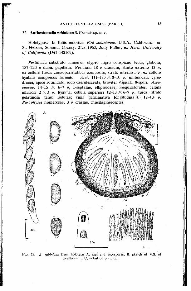
32. Anthostomella sabiniana S. Francis sp. nov.
Holotypus: In foliis emortuis Pini sabinianae, U.S.A., California: nr. St. Helena, Sonoma County, 21.xi.1963, Judy Fuller, ex Herb. University of California (IMI 142169).
Perithecia substrato immersa, clypeo nigro conspicuo tecta, globosa, 187-220 fi diam. papillata. Peridium 18 A* crassum, strato externo 13 A*, ex cellulis fuscis crassoparietalibus composito, strato interno 5 n, ex cellulis hyalinis compressis formato. Asci, 111-133 X 8-10 AI, unitunicati, cylin-dracei, apice rotundato, iodo caerulescente, breviter stipitati, 8-spori. Asco-sporae, 14-15 X 6-7 At, 1-septatae, ellipsoideae, inequilaterales, cellula inferiori 2 X 3 A 1 , hyalina, cellula superiori 12-13 X 6-7 y, fusca; strato gelatinoso tenui indutae; rima germinativa longitudinalis, 12-13 At. Paraphyses numerosae, 3 At crassae, mucilaginescentes.
2 0 j i
F I G . 2 9 . A. sabiniana f rom holotype A , asci and ascospores; B , sketch of V . S . of perithecium; C , detail o f peridium.
On needles of Pinus sabiniana in California, U.S.A.
Perithecia single, scattered, on both surfaces of the leaf, immersed beneath a conspicuous black clypeus which is 330-430 p in diameter. The clypeus is formed from dark thick walled hyphae inside the cells and between the walls of the epidermal cells surrounding the papilla. Papilla, 38 wide, short and barely projecting. Perithecia, 220-259 p high and 187-220 M wide. The peridium is 18 M wide with an outer layer, 13 p, of dark thick walled cells, and an inner layer, 5 v, of hyaline thin walled, compressed cells. Asci, 111-133 X 8-10 v, unitunicate, cylindrical, apex rounded with an apical ring 4 X 3 A; the stalk is short and slightly tapered; eight ascospores are obliquely uniseriate. Ascospores, 14-15 X 6-7 v-, two celled, with a small hyaline dwarf cell, 2 X 3 in placed asymmetrically, and a brown cell, 12-13 X 6-7 p, ellipsoidal, inequilateral, the free end rounded; a narrow sheath surrounds each spore and may form a small thickened cap at the free end of the brown cell; the longitudinal germ slit is the same length as the spore. Paraphyses numerous, 3 n wide, becoming mucilaginous.
A. sabiniana is known only from the original collection, part of which was sent [as A. conorum] to this Institute in August, 1969. It is near the European species, A. formosa which is found on needles of varieties of Pinus nigra and on P. sylvestris. The spores of the two species are similar in size, and both have a dwarf cell, but they are quite different in appearance. The brown cell of A. sabiniana is inequilateral with the dwarf cell perched asymmetrically at one end while the other end is rounded; in A. formosa, the brown cell is equal sided, the dwarf cell is positioned symmetrically and the opposite end of the brown cell is bluntly pointed. The asci of the two species are also of similar size, but in A. sabiniana the apex is rounded and has an apical ring which stains blue in iodine, and in A. formosa the tip of the ascus is more flattened and no apical ring is visible in iodine. The key to species on Conifers differentiates A. sabiniana from A. rehmii, the only other species with which it might be confused.
S P E C I M E N S E X A M I N E D
O n needles of Pinus sabiniana, U . S . A . , California: nr. St. H e l e n a , S o n o m a County , 21 .x i .1963, Judy Ful ler , ex Herb. University of California ( IMI 142169) Holotype .
Anthostomella sequoiae Rehm, Annls mycol. 5: 525 (1907).
On dried leaves of Sequoia, Sao Leopoldo, Rio grande do Sul, Brasil, 1906, leg. S. J. Rick (S) Holotype.
The substratum, in the type material from S, is not the leaf of a Conifer. The Jodrell Laboratory, Royal Botanic Gardens, Kew, has identified it as a Monocotyledon belonging to the Agavaceae. The fungus on this material agrees with Rehm's description.
VII. GENERA AND SPECIES—DOUBTFUL AND EXCLUDED
E X C L U D E D G E N E R A
Astrocystis Berk. & Br., J. Linn Soc. (Bot.) 14: 123 (1873).
Type Species: A. mirabilis Berk. & Br.
Hohnel (1909, p. 328) transferred A. mirabilis to Anthostomella and suggested that Astrocystis should form a section within the genus; apparently because the fruit body of A. mirabilis was immersed during its early development. Diehl (1925, p. 185) maintained Astrocystis as a distinct genus as he considered that the fruit body was immature while immersed, only becoming fully mature when erumpent. Hughes (1953, p. 9) transferred Rosellinia suMimbata (Dur. & Mont.) Pass, to Astrocystis: The perithecia are never immersed in this species and develop entirely on the surface, thus strengthening Diehl's argument that Astrocystis was not suitably classified with Anthostomella. Martin (1967, p. 322) transferred both species to Hy poxy Ion (non rite publ.).
Phaeaspis Kirschst., Annls mycol. 37: 112(1939).
Type Species: P. calamophila Kirschst.
Petrak (1940, p. 198) considered the species to be a poorly developed specimen of A. punctulata. I have seen the type material from B and it is Phomatospora cf. berkeleyi Sacc.
D O U B T F U L A N D E X C L U D E D S P E C I E S
A. aucubae Santor & Da Camara, Agronomia lusit. 17: 139 (1955).
Type species not available from LISFA; not possible to name from description.
A. berberidis Richon, Bull. Soc. mycol. Fr. 4: L l l l (1888).
Type material not traced; not possible to name from description.
A. bromi Richon, Cat. Champ. Marne: 259 (1889).
Type not traced; described as differing from A. tomicum by its curved boat-shaped spores, possibly A. chionostoma.
A. calamagrostidis Brun.
Type material not traced; probably A. lugubris. Brunaud (1887, p. 89) first published the species as A. elymi on Elymus europaeus (= Horde-lymus europaeus); but in a later note (1887a, p. 175) he described this as an error and changed the names to A. calamagrostidis on Calamagrostis arenaria (= Ammophilia arenaria).
A. clypeata (De Not.) Sacc, Sylloge Fungorum 1: 283 (1882).
Sordaria clypeata De Not., Sferiacei italici: 24 (1863).
The original collection described by De Notaris on Rubus is not available from RO. Traverso (1907, p. 481), who had seen the material,, cited Saccardo's Mycotheca veneta, no. 1444, A. clypeata, as a synonym. Hohnel (1920a, p. 178) thought that while Myc. ven. no. 1444 agreed almost exactly with A. limitata, the A. clypeata of De Notaris, according to the description given by Traverso, was another species. I have seen three specimens of Myc. ven. no. 1444; the material in each one was very sparse and in poor condition. In the two samples at K, the fungus was immature and the spores, which were barely delimited in the asci, were a pale grey-blue and agreed well with Hohnel's description. They did not resemble the young stages of A. limitata very closely and I also compared them with immature material of Clypeosphaeria notarisii Fuckel on Rubus; again there were similarities but the two were not identical. The material from RO (the third of the specimens of no. 1444 examined) was old, and the mature spores were unmistakably those of A. limitata. There were two plants, Rosa and Rubus, in Saccardo's Myc. ven. no. 1444, I think there may also have been two> fungi.
A. clypeata (De Not.) Sacc. var. macrospora Da Camara, Agronomia lusit. 11: 170 (1949).
Type material at LISE is Clypeosphaeria sp.
A. cocoes Da Camara, Anais Inst. sup. Agron. Univ. tec. Lisb. 3: 73 (1929).
Type material not available from LISI; probably A. phoenicicola Speg.
A. conorum (Fuckel) Sacc. f. pini Fautrey, Revue mycol. 16: 165 (1894).
Roumeguere's F. gall exsicc no. 6604 from MPU is Didymella sp.
A. cytisi (Fuckel) Sacc, Sylloge Fungorum 1: 290 (1882).
=Guignardia cytisi (Fuckel) Arx & Miiller, Beitr. Krypt.-Fl. Schweiz II (1): 49 (1954). See also Munk (1957, p. 472).
A. gynerii Tassi, Bull. Lab. Orto Bot. Reale Univ. Siena 2: 231 (1899).
Type material not available from SIENA; not possible to name from description.
A. lambottiana Fautrey, Revue mycol. 17: 167 (1895).
Roumeguere's F. gall, exsicc. no. 6803 from MPU is Coniochaeta cfr. leucoplaca (Berk. & Rav.) Cain.
A. molleriana Trav. & Spessa, Bolm Soc. broteriana 25: 169 (1910). A later homonym of A. molleriana Wint. (=A. tomicoides). Type
material not traced. The species was cited by Joly (1961) as a synonym of Sphaerodothis phoenicis (Roll.) Joly. Muller & Ahmad {Biologia 8, 1962) transferred this taxon to Serenomyces Petr. without citing A. molleriana in the synonomy.
A. myricae Grove, 7. Bot., Lond. 71: 252 (1933). Type material (very poor) at K is Coniochaeta pulveracea (Ehrh.) Munk.
A. palmacea (Cooke) Sacc, Sylloge Fungorum 1: 291 (1882). Type material at K is Didymosphaeria palmacea (Cooke) Ellis & Everh.
A. phaeosticta (Berk.) Sacc. subsp. iridis Fautrey, Revue mycol. 18: 68 (1896). Type material not available at MPU; possibly A. limitata.
A. pholidigena (Ellis) Berl. & Vogl., Sylloge Fungorum Addit. 1-4, : 44 (1886). The type material at K in Ellis & Everh., N. Am. Fungi, 2nd. series, no. 1664 is Coniochaeta sp.
A. pullulans (De Bary) Bennett, Ann. appl. Biol. 15: 390 (1928). Described by Bennett as the ascigerous stage of Aureobasidium pullulans (De Bary) Arnaud [as Dematium pullulans]. Nannfeldt (Melin & Nannfeldt, 1935, p. 444) and Hudson (1965, p. 325) have pointed out that, according to the description and illustrations given by Bennett, the fungus bore no relation either to Anthostomella, or the Xylariaceae, but was a member of the Pseudosphaeriales. A. pullulans was listed by Ainsworth (1937, p. 23) as a cereal disease in Britain, and was the name accepted by Viennot-Bourgin (1949, p. 609) for the fungus causing grape leaf scald. For an extensive review of Aureobasidium pullulans see Cooke (1959).
A. punicae Lucas & Da Camara, Agronomia lusit. 14: 197 (1952). Type material at LISE is Clypeosphaeria sp.
A. smilacinina (Peck) Sacc, Sylloge Fungorum 1: 281 (1882). Type material at NYS, examined by Barr, is Botryosphaeria smilacinina (Peck) Barr, Contr. Univ. Mich. Herb. 9 (8): 560 (1972).
A. sorbina Rehm, Annls mycol. 10: 390 (1912). 1
Type material at S is Camarographium sp.

A. ? steinheilii (Mont.) Sacc, Sylloge Fungorum 1: 293 (1882).
Type material at PC is Phaeochora steinheilii (Mont.) E. Muller, (Sydowia 18: 88, 1965). See also Joly (1961).
A. tomicoides Sacc. var. crassispora Bat. Fischman & Matta in Bat. et al., Atas Inst. Micol. 4: 224 (1967).
Type material at URM is Starbaeckiella sp.
Sphaeria tomicum Lev var. minor Berk. & Br., Ann. Mag. nat. Hist. ser. 2, 9: 324 (1852). On Deschampsia caespitosa [as Air a caespitosa], Bath-easton, Jan. 1850; on Juncus, Draycott. The variety was said to be similar to S. tomicum but far smaller; a figure shows one celled spores. In Herb. Berkeley at K a collection labelled ' S. tomicum var.' (on Aira caespitosa) has two species of Anthostomella, both are old but are probably A. tomicoides and A. tumulosa. The collection labelled ' S. tomicum (on Juncus), Draycott,' has only A. tomicoides. The presence of this species in both collections suggests it is the one referred to by Berkeley & Broome. A. tomicoides has a two celled spore. The single celled spore of the illustration could refer either to A. tomicoides without a dwarf cell, or to A. tumulosa. This difficulty, together with the fact that the British collections at K show that Berkeley & Broome consistently misidentified 5. tomicum Lev., make it impossible to determine the variety accurately. The name S. tomicum var. minor is therefore rejected (Article 70).
A. vaga Niessl, Hedwigia 26: 33 (1887) = Amphisphaerella vaga (Niessl) O. Erdks., Svensk bot. Tidskr. 60: 321 (1966).
A. xylostei (Pers. ex Fr.) Guyot, Annls Serv. bot. agron. Tunis 28: 72 (1958) ^Amphisphaerella xylostei (Pers. ex Fr.) Munk, Dansk bot. Ark. 15: 89 (1953).
VIII. ACKNOWLEDGMENTS
I wish to thank the Director of the Commonwealth Mycological Institute for allowing me facilities for carrying out this work, and all members of the staff for their unfailing assistance in many different ways.
I am grateful to Dr. C. Booth who gave me notes he had made on the British collections in IMI and has encouraged the growth of the work well beyond its original scope; Dr. R. W. G. Dennis for many helpful discussions; Dr. E. A. Ellis, Dr. K. A. Pirozynski and Prof. J. Webster for the loan of material; Dr. M. T. Lucas for her help in tracing collections recorded from Portugal.
I am indebted to the Directors of the following herbaria for lending, or

searching for, material: B, BR, BUCM, CP, DAOM, DAR, E, FH, K, LE, LISE, LPS, MA, MPU, NY, PAD, PARMA, PC, RO, S, SIENA, UPS, URM, VER.
My thanks are especially due to Mrs. G. B. Butterfill for her skilled technical assistance, and Mr. P. Basu for his help in preparing the Latin diagnoses.
IX. REFERENCES A h m a d , S. (1962). Further Contributions to the Fungi of West Pakistan II. Biologia,
Lahore 8: 123-150. A h m a d , S. (1969). Fung i o f Wes t Pakistan. Supplement I. Monogr. biol. Soc. Pakist.
5: 1-110. Ainsworth , G. C. (1937). The Plant Diseases of Great Britain. London: Chapman & Hall . A insworth , G. C. (1971). Ainsworth & Bisby's Dictionary of the Fungi. 6th. ed. Kew:
C o m m o n w e a l t h Mycologica l Institute. A r x , J. A . v o n & Muller, E . (1954). D i e Gattungen der amerosporen Pyrenomyceten .
Beitr. Krypt.-Fl. Schweiz. 11 (1): 1^434. Baccarini, P. (1895). Intorno ad una malatt ia del la pa lma da datteri. Boll. Soc. hot.
Hal.: 196-203 . Barbey, W. (1884). Florae Sardoae Compendium. Lausanne: Georges Bridel. Bizzozero, G. (1882-1885) . Flora Veneta Crittogamica. Parte I, I Funghi . Padova . B o m m e r , E . & Rousseau , M. (1890). Contributions a la Flore Myco log ique de Belg ique.
Bull. Soc. r. Bot. Belg. 29: 205-302 . Briosi , G. & Cavara, F . (1890). I funghi parassiti delle Piante colt ivate od utile essiccati
delineati e descritti. Fasc . 6, nos . 126-150. Brunaud, P. (1887). Champignons nouvel lement observes a u x environs de Saintes.
5 sine. J. Hist. nat. Bordeaux, ser. 2, 6 annee, no . 8 : 88 -90 . Brunaud, P. (1887a). Materiaux pour la Flore Myco log ique des environs Saintes. Act.
Soc. linn. Bordeaux, ser. 5, 41: 158-189; Callen, E. O. (1938). S o m e Fungi o n the Y e w . Trans. Br. mycol. Soc. 22: 94 -106 . Castagne, L. (1845). Catalogue des Plantes qui croissent naturellement aux environs
de Marseille. A i x : N i c o t & Pardigon. Castagne, L. (1851). Supplement au Catalogue des Plantes aux environs de Marseille.
A i x : N i c o t & Pardigon. Cesati , V . de & D e Notar is , G. (1863). Schema di classificazione degli Sferiacei italici
aschigeri. Comment. Soc. crittogam. Ital. 1 (4): 177-240 . Chadefaud, M . (1953). Sur un Hypocopra, sa posit ion systematique, ses spores et ses
asques. C. r. hebd. Seanc. Acad. Sci., Paris 236: 513 -514 . Clements , F . E . & Shear, C. L. (1931). The Genera of Fungi. N e w York: H . W.
Wi l son Co. Cooke , W. B. (1959). A n Ecologica l Life History of Aureobasidium pullulans ( D e Bary)
Arnaud. Mycopath. Mycol. appl. 12: 1-45. D e n n i s , R. W . G. (1964). T h e Fungi of the Isle of R h u m . Kew Bull. 19 (1): 7 7 - 1 3 1 . D e n n i s , R. W. G. (1968). British Ascomycetes. Lehre: J. Cramer. Desmazieres , J. B , H . J. (1846). Treiz ieme Not i ce . Annls. Sci. nat. (Bot.) ser. 3, 6: 62 -84 . Dieh l , W . W. (1925). T h e genus Astrocystis. Mycologia 17: 185-190 . Ell is , J. B . (1882). N e w Species of N o r t h Amer ican Fungi . Bull. Torrey bot. Club 9:
7 3 - 7 4 . Ell is , J. B. (1883). N e w North Amer ican Fungi . Bull. Torrey bot. Club 10: 5 2 - 5 4 . Eriksson, O. (1966). On Anthostomella Sacc., Entosordaria (Sacc.) H o h n . a n d some
related genera (Pyrenomycetes) . Svensk. bot. Tidskr. 60: 315-324 . Fabre , J. H . (1883). Essai sur les Spheriacees du Department de Vaucluse . ( D e u x i e m e
partie). Annls Sci. nat. (Bot.) sir. 6, 1 5 : 31-69 . Fawcet t , H . S. (1931). Observations on the Culture and Diseases of D a t e Pa lms in
N o r t h Africa. Eighth Ann. Rept. Date Growers' Inst., Apri l , 1931. Fel tgen, J. (1897-1899) . Vorstudien z u einer Pi lz -Flora des Grossherzogthums Luxem
burg. I Theil , Ascomycetes , Nacht . I. Reel. Mim. Trav. Soc. bot. iLuxemb. 14: 1-41 2T
Georgescu , C. C. & M o c a n u , V . V . (1955). Contribute la cunoasterea boli lor din culturile forestiere. Revta Padur. 70 (1): 2 3 - 2 6 .
G r e m m e n , J. (1960). A Contribution to the Mycofiora of P ine Forests in the Netherlands . Nova Hedwigia 1: 2 5 1 - 2 8 8 .
G r o v e , W. B . (1937). British Stem- and Leaf-Fungi (Coelomycetes) . Cambridge: University Press.
H a w k s w o r t h , D . L. (1974). Mycologist's Handbook. Kew: Commonwea l th Mycolog ica l Institute.
H o h n e l , F . v o n (1909). Fragmente zur Mykolog ie . 225. U b e r Melanomma trochus Penz . et Sacc. , und Astrocystis mirabilis Berk & Br. Sber. Akad. Wiss. Wien Math.-nat. 118: 326-328 .
H o h n e l , F . v o n (1918). Myco log i sche Fragmente . C C X I I . Uber Anthostomella palmicola (Auerswald) Rabh . Annls mycol. 16: 7 0 - 7 1 .
H o h n e l , F . v o n (1918a). R e h m : A s c o m y c e t e s exs. Fasc . 56 and 57. Kritisch bearbeitet von Prof. Dr . Franz v. H o h n e l Wien . Annls mycol. 16: 209 -224 .
H o h n e l , F . v o n (1920). Fragmente zur Myko log i e 1205. U b e r die Gat tung RhynchostomaKarsten. Sber. Akad. Wiss. Wien Math.-nat. 129: 164-167.
H o h n e l . F . v o n (1920a). Fragmente zur Mykolog ie 1212. Uber die Gattung Anthostomella Sacc. Sber. Akad. Wiss. Wien Math.-nat. 129: 174-181 .
H o h n e l , F . von (1929). U b e r Sphaeria smilacis Castagne. Mitt. bot. Inst. tech. Hochsch. Wien 6: 113-115 .
H o l m , L . (1958). S o m e Comments o n the Ascocarps of the Pyrenomycetes . Mycologia 50: 7 7 7 - 7 8 8 .
Hudson , H . J. (1962). Success ion of Micro-fungi on ageing leaves of Saccharum officinarum. Trans. Br. mycol. Soc. 45: 395-423 .
H u d s o n , H . J. (1966). A n ascomycete wi th Aureobasidium pullulans-iype conidia. Nova Hedwigia 10: 319-328 .
H u g h e s , S. J. (1953). F u n g i f rom the G o l d Coast II. Mycol. Pap. 50: 1-104. Joly , P . (1961). L e Genre Sphaerodothis Shear. Bull. Res. Coun. Israel, sect. D. (Botany)
10: 187-193 . Kirschstein, W . (1939). Uber neue seltene und kritische A s c o m y c e t e n und F u n g i
Imperfecti . II . Annls mycol. 37: 88-140 . Lind, J. (1913). Danish Fungi. Copenhagen. Lundqvist , N . (1972). Nordic Sordariaceae s. lat. Symb. bot. upsal. 20 (1): 1-374. Martin, P . (1967). Studies in the Xylariaceae: II. Rosellinia and the Pr imocinerea
sect ion of Hypoxylon. Jl S. Afr. Bot. 33 : 315-328 . Mart in , P. (1969). Studies in the Xylariaceae: VII . Anthostomella and Lopadostoma.
Jl S. Afr. Bot. 35: 393-410 . Mel in , E . & Nannfe ldt , J. A . (1935). Researches into the Blueing of Ground Woodpulp .
Svensk SkogsvFor. Tidskr. 32: 397-616 . Miller, J. H . (1928). Bio log ic Studies i n the Sphaeriales. II. Mycologia 20: 305 -339 . Miiller, E . (1950). D i e schweizerischen Arten der Gattung Leptosphaerid u n d ibrer
Verwandten. Sydowia 4: 185-319. Miil ler, E . & A h m a d , S. (1963). U e b e r drei A s c o m y c e t e n von Phoenix dactylifera L .
Biologia, Lahore 9: 2 9 - 3 2 . Miil ler, E . & A r x , J. A . v o n (1962). D i e Gat tungen der didymosporen Pyrenomyceten .
Beitr. Krypt.-Fl. Schweiz 11 (2): 1-922. Miil ler, E . & A r x , J. A . von (1974). Pyrenomycetes, Meliolales, Coronophorales,
Sphaeriales. In A insworth , G. C , Sparrow, F . K., Sussman, A . S. (ed.) The Fungi, an advanced Treatise 4 A : 87 -132 . A c a d e m i c Press: N e w Y o r k & L o n d o n .
Miiller, E . & Denn i s , R. W. G. (1965). Fungi venezuel iania: VIII . Kew Bull. 1 9 ( 3 ) : 357-386 .
M u n k , A . (1957). Danish Pyrenomycetes . Dansk Bot. Ark. 17 (1): 1 -491 . Niess l , G. v o n (1871-1872) . Beitrage zur Kenntniss der Pilze. Verh. naturf. Ver. Briinn
10: 153-217. Niess l , G. von (1875-1876) . N o t i z e n uber neue und kritische Pyrenomyceten . Verh.
naturf. Ver. Briinn 14: 165-217. Passerini, G. (1880). Microcyce tum ital icorum diagnoses. Revue mycol. 2: 3 3 - 3 6 . Passerini, G. (1889-1890) . Diagnos i di funghi nuovi , nota IV. Atti Accad. naz. Lincei
Memorie ser. 4 , 6: 4 5 7 - 4 7 0 . Petrak, F . (1923). Mykolog i sche Not i zen . VI . 245. Anthostoma moravicum n. spec.
Annls mycol. 2 1 : 2 5 2 - 2 5 5 .

Petrak, F . (1924). Mykologische Not izen . VII . 343. U b e r die Gattung Entosordaria (Sacc.) v. H o h n . Annls mycol. 22: 7 4 - 7 6 .
Petrak, F . (1925). Mykologische Not izen . VIII . 409 . U b e r Gloeosporium taxicolum Al lesch . Annls mycol. 23: 2 3 - 2 4 .
Petrak, F . (1931). Fung i Adeani . Ein Beitrag zur Pilzflora Bayerns und der angren-zenden Lander. Kryptog. Forsch. 2: 155-194.
Petrak, F . (1940). Mykolog ische Not izen . XII I . Annls mycol. 38: 181-267. Petrak, F . (1940a). Beitrage zur Pilzflora der U m g e b u n g v o n Wien . Annls mycol.
38: 339-386 . Petrak, F . (1955). Beitrage zur Pilzflora von Sud- und Ostasien. Sydowia 9: 5 3 0 - 5 3 8 . Petrak, F . (1962-1963) . Mykolog ische Beitrage zur osterreichischen Flora. Sydowia.
16: 155-198. Petrak, F . & Sydow, H . (1924). Kritisch-systematische Originaluntersuchungen uber
Pyrenomyzeten , Sphaeropsideen und Melanconieen . Annls mycol. 22: 318-386 . Pirozynski , K. A . (1972). Microfungi of Tanzania . Mycol. Pap. 129: 1-64. R e h m , H. (1909). D i e Clypeosphaeriaceae der deutschen Flora. Annls mycol. 7: 4 0 6 -
412 . Saccardo, P. A . (1875). Conspectus Generum Pyrenomycetum Ital icorum. Atti Accad.
scient. veneto-trent.-istriana 4: 77 -100 . Saccardo, P. A . (1875c) . Fungi Venet i novi vel critici, series IV , Atti Accad. scient.
veneto-trent.-istriana 4: 101-141 . Saccardo, P. A . (1877). Fungi Venet i novi vel critici, series VI . Michelia 1: 1-72. Saccardo, P . A . (1879). Fungi Gallici. Michelia 1: 500 -538 . Saccardo, P . A . (1881). Fung i Venet i novi vel critici, series X I I . Michelia 2: 2 4 1 - 3 0 1 . Saccardo, P . A . (1881a). Fung i Gallici , series III. Michelia 2: 3 0 2 - 3 7 1 . Saocairdo, P. A . (1882). Sylloge Fungorum omnium hucusque cognitorum, vol . I. Padua . Saccardo, P. A . (1889). Mycetes aliquot Guineensis . Bolm Soc. broteriana 7: 110-114 . Saccardo, P. A . (1891). Sylloge Fungorum omnium hucusque cognitorum, vo l . I X .
Padua. Sacoardo, P. A . (1895). Sylloge Fungorum omnium hucusque cognitorum, vol . X I . Padua. Saccardo, P . A . (1917). N o t a e Mycolog icae , series X X I I I . Fungi Phil ippinenses a cl.
C. F . Baker collecti et communicat i . Atti Accad. scient. veneto-trent.-istriana 10: 5 7 - 9 4 .
Saccardo, P . A . & Berlese , A . N . (1884-1885) . Catalogo dei Funghi Italiani. Atti Soc. Crittogam. Ital. Anno 28 , ser. 2 , 3 (4): 261 -368 .
Sandu-Vil le , C. (1971). Ciuperci Pyrenomycetes—Sphaeriales din Romania. Ed . A c a d . Republ . Social . Romania .
Savulescu, T . (1934). U n e nouvel le espece du genre Par anthostomella et considerat ions systematiques sur les Sphaeriales pseudostromatae. Archs roum. Path. exp. Microbiol. 7: 7 - 3 2 .
Savulescu, T. & Sandu-Ville , C. (1935). Beitrag zur Kenntnis der M i c r o m y c e t e n Rumaniens . Hedwigia 75 (3 & 4): 159-233 .
Scalia, G. (1900). I Funghi della Sicilia Orientale. Atti Accad. gioenia Sci. nat. 13 (4): 1-55.
Schrantz, J. P. (1960). Recherches sur les Pyrenomycetes de l'Ordre des Diatrypales, sepsu M . Chadefaud, 1957. Bull. Soc. mycol. Fr. 76: 305-407 .
Spegazzini , C. (1881). Fungi Argentini . Pugil lus quartus. An. Soc. cient. argent. 12: 97-117 .
Spegazzini, C. (1888). Fungi Fuegiani . Boln Acad. nac. Cienc. Cordoba 11 (2): 135-308. Spegazzini , C. (1910). Fungi Chilenses. B u e n o s Aires . Stafleu, F . A . (1967). Taxonomic Literature. Regnum Vegetabile 5 2 : i -xx , 1-556. Traverso, G. B . (1907). Flora Italica Cryptogama. Pars. I Fungi , Pyrenomycetae 2 (2):-
353 -492 . Traverso, G. B. & Spessa, C. (1910). L a Flora Mico log ica del Portogallo. Bolm Soc.
broteriana 25: 26 -187 . Viennot -Bourgin , G. (1949). Les Champignons parasites des plantes cultivees. 2 vo ls .
Paris: Masson & Cie. , Editeurs. • Winter , G. (1884-1887) . Ascomyceten: G y m n o a s c e e n und Pyrenomyceten . Rabenh.
Krypt. Fl. 1 (2): i-viii, .1-928, 67-112. Winter , G. (1887). Exot ische Pilze IV. Hedwigia 26: 6 -18 .
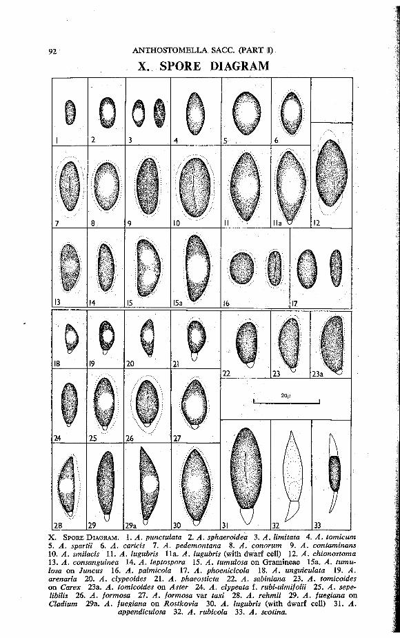
X. S P O R E D I A G R A M
X . S P O R E D I A G R A M . 1. A. punctulata 2. A. sphaeroidea 3 . A. limitata 4. A. tomicum 5. A. spartii 6. A. caricis 7. A. pedemontana 8. A. conorum 9. A. contaminans 10. A. smilacis 11. A. lugubris 11a. A. lugubris (with dwarf cell) }2 . A. chionostoma 13. A. consanguinea 14. ^4. leptospora 15. ^4. tumulosa on Gramineae 15a. X . tumulosa on Juncus 16. . 4 . palmicola 17. . 4 . phoenicicola 18. . 4 . unguiculata 19. . 4 . arenaria 20 . . 4 . clypeoides 21 . A . phaeosticta 22. A. sabiniana 23 . . 4 . tomicoides o n Carex 23a. . 4 . tomicoides o n Aster 24. /4 . clypeata f. rubi-ulmifolii 25 . . 4 . sepe-/fti/fo 26. . 4 . formosa 27. ^4. formosa var tan 28. ^4. rehmii 29 . ^4. fuegiana o n Cladium 29a. ^4. fuegiana o n Rostkovia 30. . 4 . lugubris (with dwarf cell) 31 . . 4 .
appendiculosa 32. . 4 . rubicola 33 . . 4 . scotina.
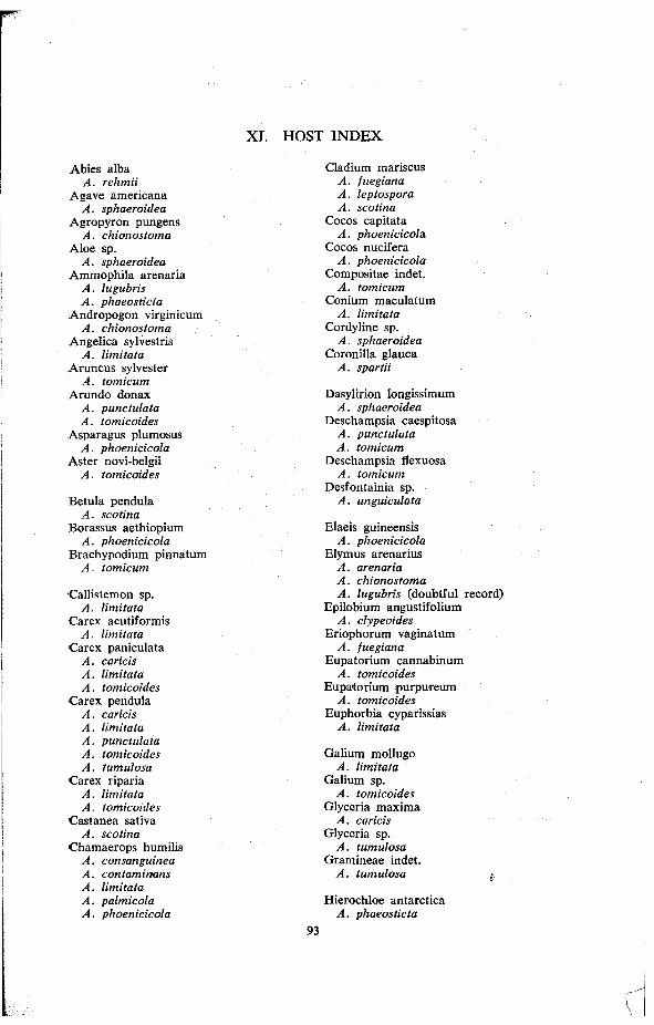
XI. HOST INDEX
A b i e s alba A. rehmii
A g a v e americana A. sphaeroidea
A g r o p y r o n pungens A. chionostoma
A l o e sp. A. sphaeroidea
A m m o p h i l a arenaria A. lugubris A. phaeosticta
A n d r o p o g o n virginicum A. chionostoma
A n g e l i c a sylvestris A. limitata
A r u n c u s Sylvester A. tomicum
A r u n d o donax A. punctulata A. tomicoides
Asparagus plumosus A. phoenicicola
A s t e r novi-belgii A. tomicoides
B e t u l a pendula A. scotina
Borassus aethiopium A. phoenicicola
Brachypodium pinnatum A. tomicum
Cal l i s temon sp. A. limitata
Carex acutiformis A. limitata
C a r e x paniculata A. caricis A. limitata A. tomicoides
C a r e x pendula A. caricis A. limitata A. punctulata A. tomicoides A. tumulosa
Carex riparia A. limitata A. tomicoides
Castanea sativa A. scotina
Chamaerops humil is A. consanguinea A. contaminans A. limitata A. palmicola A. phoenicicola
Cladium mariscus A. fuegiana A. leptospora A. scotina
Cocos capitata A. phoenicicola.
Cocos nucifera A. phoenicicola
Composi tae indet. A. tomicum
Conium maculatum A. limitata
Cordyline sp. A. sphaeroidea
Coronil la g lauca A. spartii
Dasyl ir ion longiss imum A. sphaeroidea
Deschampsia caespitosa A. punctulata A. tomicum
Deschampsia f lexuosa A. tomicum
Desfontainia sp. A. unguiculata
Elaeis guineensis A. phoenicicola
Elymus arenarius A. arenaria A. chionostoma A. lugubris (doubtful record)
Epi lobium angust i fol ium A. clypeoides
Eriophorum vaginatum A. fuegiana
Eupator ium cannabinum A. tomicoides
Eupator ium purpureum A. tomicoides
Euphorbia cyparissias A. limitata
Gal ium m o l l u g o A. limitata
Gal ium sp. A. tomicoides
Glyceria m a x i m a A. caricis
Glyceria sp. A. tumulosa
Gramineae indet. A. tumulosa i
Hierochloe antarctica A. phaeosticta
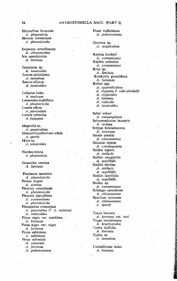
Hieroch loe brunonis A. phaeosticta
H o w e i a forsteriana A. phoenicicola
Imperata arundinacea A. chionostoma
Iris pseudacorus A. limitata
Jasminum sp. A. tomicoides
Juncus articulatus A. tumulosa
Juncus effusus A. tomicoides
Labiatae indet. A. tomicum
Lomandra multiflora A. phoenicicola
Luzula pilosa A. punctulata
Luzula sylvatica A. fuegiana
Magnol ia sp. A. unguiculata
M e s e m b r y a n t h e m u m edule A. spartii
M u s a sp. A. tomicoides
Nardus stricta A. phaeosticta
Oenanthe crocata A. limitata
Pandanus tectorius A. phoenicicola
Persea l ingue A. scotina
Phoen ix canariensis A. phoenicicola
P h o e n i x dactylifera A. contaminans A. phoenicicola
Phragmites communi s A. punctulata (? A. minima) A. tomicoides
Pinus nigra var. marit ima A. formosa.
Pinus nigra var. nigra A. formosa
Pinus sabiniana A. sabiniana
Pinus sylvestris A. conorum A. formosa A. pedemontana
Pinus wall ichiana A. pedemontana
Quercus sp. A. unguiculata
Raphia hookeri A. contaminans
Raphia sudanica A. contaminans
R o s a sp. A. limitata
Rostkov ia grandiflora A. fuegiana
Rubus spp. A. appendiculosa A. clypeata f. rubi-ulmifolii A. clypeoides A. limitata A. rubicola A. tomicoides
Sabal minor A. consanguinea
Schoenoplectus lacustris A. scotina
Scirpus ho loschoenus A. tomicum
Secale cereale A. chionostoma
Serenoa repens A. consanguinea
Smilax aspera A. smilacis
Smilax campestris A. sepelibilis
Smilax excelsa A. smilacis A. sepelibilis
Smilax laurifolia A. sepelibilis
Smilax sp. A. contaminans
Solidago canadensis A. chionostoma
Spartium junceum A. chionostoma A. spartii
Taxus baccata A. formosa var. taxi
Tsuga mertensiana A. brachystoma
Typha latifolia A. limitata
Typha sp. A. tumulosa
Umbel l i ferae indet. A. limitata

Vit is vinifera Y u c c a aloifolia A. limitata A. sphaeroidea
Y u c c a gloriosa "Washingtonia filifera A. sphaeroidea
A. phoenicicola
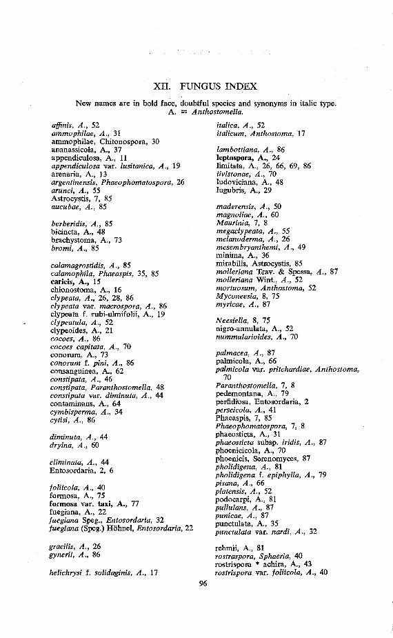
XII. FUNGUS INDEX
N e w names are in bold face , doubtful species and synonyms in italic type. A . = Anthostomella.
affinis, A., 52 ammophilae, A., 31 ammophi lae , Chitonospora, 30 ananassicola, A . , 37 appendiculosa, A . , 11 appendiculosa var. lusitanica, A., 19 arenaria, A . , 13 argentinensis, Phaeophomatospora, 26 arunci, A., 55 Astrocyst is , 7, 85 aucubae, A., 85
berberidis, A., 85 bicincta, A. , 48 brachystoma, A. , 73 bromi, A., 85
calamagrostidis, A., 85 calamophila, Phaeaspis, 35, 85 caricis, A , , 15 ch ionostoma, A. , 16 clypeata, A., 26, 28, 86 clypeata var. macrospora, A., 86 c lypeata f. rubi-ulmifol i i , A . , 19 clypeatula, A., 52 c lypeoides , A . , 21 cocoes, A., 86 cocoes capitata, A., 70 conorum, A. , 73 conorum f. pini, A., 86 consanguinea, A. , 62 constipata, A., 46 constipata, Paranthostomella, 48 constipata var. diminuta, A., 44 contaminans , A. , 6 4 cymbisperma, A., 34 cytisi, A., 86
diminuta, A., 44 dryina, A., 60
eliminata, A., 44 Entosordaria, 2, 6
foliicola, A., 40 formosa, A . , 75 formosa var. taxi , A . , 77 fuegiana, A. , 22 fuegiana Speg., Entosordaria, 32 fuegiana (Speg.) H o h n e l , Entosordaria, 22
italica, A., 52 italicum, Anthostoma, 17
lambottiana, A., 86 leptospora, A . , 24 limitata, A. , 26, 66, 69, 86 livistonae, A., 70 ludoviciana, A. , 48 lugubris, A . , 29
maderensis, A., 50 magnoliae, A., 60 Maurinia, 7, 8 megaclypeata, A., 55 melanoderma, A., 26 mesembryanthemi, A., 49 minima, A . , 36 mirabilis , Astrocystis , 85 molleriana Trav. & Spessa, A., 87 molleriana W i n t , A., 52 mortuosum, Anthostoma, 52 Myconeesia, 8, 75 myricae, A., 87
Neesiella, 8, 75 nigro-annulata, A. , 52 nummularioides, A., 70
palmacea, A., 87 palmicola , A . , 66 palmicola var. pritchardiae, Anthostoma,
70 Paranthostomella, 1, 8 pedemontana, A. , 79 perfidiosa, Entosordaria, 2 perseicola, A., 41 Phaeaspis , 7, 85 Phaeophomatospora, 1, 8 phaeosticta, A. , 31 phaeosticta subsp. iridis, A., 87 phoenic icola , A. , 70 phoenicis , Serenomyces , 87 pholidigena, A., 81 pholidigena f. epiphylla, A., 79 pisana, A., 66 platensis, A., 52 podocarpi , A. , 81 pullulans, A., 87 punicae, A., 87 punctulata, A. , 35 punctulata var. noraV, /4 . , 32
grarita , A , 26 rehmii, A. , 81 gynerii, A., 86 rostraspora, Sphaeria, 40
rostrispora * achira, A. , 43 helichrysi i. solidaginis, A., 17 rostrispora var. foliicola, A., 40
rubicola, A. , 38 rusci, A. , 57
sabiniana, A . , 83 secalis, A., 16 sepelibil is , A . , 44 sequoiae , A. , 84 scotina, A. , 4 0 smilacis , A . , 46 smilacis , Phragmocauma, 48 smilacinina, A., 87 sorbina, A., 87 spartii, A. , 49 Sphaerodothis , 61 , 67, 87 sphaeroidea, A . , 50 starcii, A., 70 steinheilii, A-., 88 steinheili i , Phaeochora , 88 subconica, A., 55
taxi, A., 11 tomicoides, A. , 8, 52 tomicoides var. crassispora, A., 88 tomicum, A . , 55 tomicum var. leptospora, A., 24 tomicum var. minor, Sphaeria, 88 trabutiana, A., 50 tumulosa , A. , 58
unguiculata, A. , 60
vaga, A., 88
Xylosphaeria, 17, 52 xylostei, A., 88
yuccae, A., 50