6.02 parasites - internal. parasitology the study of organisms that live on or in other organisms...
TRANSCRIPT

S
6.02 Parasites - Internal

Parasitology
The study of organisms that live on or in other organisms to survive
Parasites – may invade internal or external parts of the animal
Most have one location they live in, and feed off the host Host – animal that is infected
Go through a life cycle: born as larvae grow and reproduce
Can invade digestive tract, skin, or muscles

Parasites
Endoparasites – live within the host Whipworms, hookworms, heartworms, coccidian,
tapeworms, and strogyles
Ectoparasites – live on the host Ticks, fleas, mites

Modes of Transmission
Direct – transmission occur when the parasite is passed directly from one host to another Animals ingest infected feces or vomit, parasites
enter skin through penetration, mothers pass it to offspring through placenta or milk
Indirect – transmission occurs when an animal ingests a paratenic host Paratenic host – animal that can harbor the
parasite without becoming infected. Parasite remains inactive until appropriate host ingests it Rodents, rabbits, birds, even flies

Damage Caused by Parasites
Ectoparasite
Damage and irritate skin
Cause restlessness, nervousness
Transmit infectious organisms
Draw blood
Endoparasite
Absorb food intended for host
Draw blood and lymph fluid
Damage tissue and cause internal bleeding
Create obstructions in intestines and circulatory system
Produce toxins

Internal Parasites
Occur in small and large animals
Many internal parasites invade the intestinal system causing vomiting and diarrhea
Diagnosed through fecal flotation

Roundworms (Ascarids)
Most common intestinal parasite in small and large animals
Most common internal parasite in puppies and kittens/often occur in young animals
Live in small intestines
Can lay up to 200,000 eggs a day
Diagnosed by finding eggs in feces Eggs are circular with dark circular centers

Roundworms – Life Cycle
Parasite enters body by direct or indirect means
Migrates through various organs to the lungs, and into trachea
From trachea it is swallowed and larva travel to small intestines where they mature to adults
Eggs are shed in the feces
Cycle begins again

Roundworms - Types
Toxascaris leonina Dog and cat Simplest life cycle Eggs hatch after ingested Eggs passed in feces After 3-6 days, they are infected in the environment Animals become infected if they eat something
contaminated with infected feces
Roundworms - Types
Toxocara canis Dog Complicated life cycle Hardy and resistant Animal can become infected by ingestion of eggs,
transport host, or by larvae entering animal while in uterus
Migrate through circulatory system Can encyst (become walled off or inactive) if enter
body tissues

Roundworm - Types
Toxocara cati Cat Similar to T. canis Infective eggs are swallowed; larvae hatch;
penetrate stomach wall Larvae migrate to liver, other tissues, and lungs
Roundworm - Types
Parascaris equorum In large animals
Swallowed in contaminated hay or water Young worms burrow through intestinal wall Mature in intestine and lay eggs that are passed in
feces
In small animals Infections will show signs of vomiting, diarrhea, and
bloated stomach Visible in feces

Roundworms – Clinical Signs
Small animal symptoms Vomiting, diarrhea, bloated stomach, visible
roundworms in feces, potbelly, coughing, anemia (in severe cases), dull coat
Large animal symptoms Abdominal pain (common cause of obstruction colic
in foals), coughing, diarrhea, visible roundworm in feces, potbelly, anemia (severe cases)

Roundworms - Zoonosis
Roundworms cause visceral larval migraines in humans

Tapeworms - Cestode
Common in both dogs and cats
Has a long, flat, segmented body
Head is called the scolex
Segments called proglottids Contain eggs and look like grains of rice when shed in
feces Can be seen around the anus of dogs or cats
Adults attach to wall of small intestines where they absorb nutrients
Feas are vectors for tapeworms so animals with a heavy flea infestation should be tested for tapeworm

Tapeworm - Types
Dipylidium caninum – dog and cat Can be up to 20” long Lives in small intestines
Taenia pisiformis – dog
Taenia taeniaeformis – cat

Tapeworm – Modes of Transmission/Clinical Signs
Modes of Transmission Indirect
Animals ingest a flea or other intermediate hosts such as rabbits, mice, or lice
Clinical Signs Poor hair coat, abdominal discomfort, visible
segments in feces

Tapeworm – Life Cycle
Intermediate host eats the eggs
Dog or cat eats intermediate host
Parasite larvae migrate to small intestines to mature
Ptoglottids are shed in feces
Life cycle begins again

Hookworms
Ancylostoma caninum
Destructive parasite of dogs and cats that feeds on wall of small intestines
Teeth-like structures or cutting plates – attach to wall of intestine and feed on animal blood
Parasite changes feeding sites frequently causing hemorrhaging that leads to anemia and protein loss
Death is common in untreated puppies

Hookworm – Life Cycle
4 – 6 weeks
Larvae are ingested, absorbed, or passed to the host by indirect means and migrate to lungs via bloodstream
From the lungs, larvae pass into trachea where they partially develop, coughed up and then swallowed to the small intestines
Larvae mature to adults, begin to feed, shed eggs in feces
Cycle starts again

Hookworm – Types/Modes of Transmission
Types Ancylostoma – common in warm climates Uncineria – common in northern U.S. & Canada
Modes of Transmission Direct
Ingestion, skin penetration, transmammary

Hookworm – Clinical Signs/Zoonosis
Clinical Signs Vomiting, diarrhea, anemia, weakness, black
darkened feces or blood in stool, dull coat, pale mucus membranes, anorexia
Cause cutaneous larval migraines in humans where larvae burrow through humans skin, most common in young children

Whipworm
Trichuris vulpis
Infect animals through ingesting contaminated food or water
Adults have whip-like shape
Attach to large intestines and cecum by threading their “whip” through intestinal lining
Very rare in cats
Adult whipworms are not visible in feces
Whipworm – Life Cycle
4 – 7 weeks
Eggs eaten by a host
Larvae hatch and begin their development in small and large intestines
Larvae mature to adults in cecum or large intestines
Eggs containing infective larvae are passed in feces
Cycle begins again

Whipworm – Types/Modes of Transmission
Types Trichuris vulpis
Modes of Transmission Direct
Ingestion of eggs

Whipworm – Clinical Signs
Diarrhea
Weight loss/emaciation
Possible blood in feces
Anemia
Dehydration
Death may occur in severe cases

Protozoa
Several different species commonly known as coccidian
Infection of protozoa is called coccidiosis
Coccidia infest walls of the intestines

Protozoa – Types/Modes of Transmission
Types Isospera – dog and cat Sarcocystis – dog and cat Toxoplasma gondii – cat
Modes of Transmission: Direct
Ingestion of eggs Indirect
Ingestion of infected animals

Protozoa – Life Cycle
Eggs or an infected animal ingested by host
Parasite’s cell wall is digested and enters epithelial cells of intestines where it begins to mature
During maturation, the eggs divide and develop into male and female
Fertilized female (oocyst) ruptures out of epithelial cell, passed in feces
Cycle begins again

Protozoa – Clinical Signs/Zoonosis
Clinical Signs: Diarrhea in puppies and kittens
Zoonosis Some species will cause disease in humans:
Toxoplasma gondii – causes abortions Giardia – “Beaver Fever” is transmitted in
contaminated food and water

Heartworm
Dirofilaria immitis
Affects heart and circulatory system of infected animals
Can affect dog, cats, ferrets, and even humans; although in humans the parasite is eliminated in the lungs
Adult heartworms live in the right side of the heart and in pulmonary artery
Worms are free living meaning they do not attach to the host’s body

Heartworm – Mode of Transmission
Mosquitoes are the vector
Heartworm is most prevalent in the southern states with warmer climates (larvae need warm temps to mature) and greater number of mosquitoes.

Heartworm – Life Cycle
Mosquito bites infected animal and picks up D. immitis microfilaria
Mosquito takes its next meal and microfilaria (larvae) passed to that animal
Microfilaria take 3 months to migrate to right side of heart During the 3 month migration, an infected animal shows no
clinical signs
Migration in the heart takes another 3 months
After 6-month migration and maturation period, adult heartworms begin producing microfilaria that are released into the bloodstream

Effects of Heartworm Disease
Damage to the pulmonary artery lining
Compromised blood flow
Fluid leaks from the lungs
Life span: Dogs – adults live about 5 years Cats – adults live only 1-3 years

Heartworms – Clinical Signs
Symptoms in dogs Dogs with a light infection may be asymptomatic,
but with increasing worms there will be exercise intolerance, difficulty breathing, deep cough, decreased appetite, weight loss
Symptoms in cats Typically show no signs, but may die suddenly Or may show signs similar to dogs

Heartworms - Diagnosis
Methods will be unsuccessful unless the disease has progressed at least 6 months since no adults are present until that time
Methods: Blood smear to look for microfilaria Antigen test – test for antigens produced by adult
females; there are many commercial tests available X-ray – look for enlarged heart and pulmonary artery

Heartworms - Treatment
Often dangerous
Possibility that the dog won’t survive treatment or adult worms will form a clot in arteries
Surgery can be done to remove adult worms
Two non-surgical methods often used in combination Using an arsenic compound to kill adults, however
arsenic could also kill the dog Use ivermectin to kill microfilaria
Prevention with monthly treatments is better way to manage heartworms

Strongyles
Common parasite of large animals
Bloodworms or redworms
Eggs in manure hatch into larvae
Larvae mature in the intestinal tract and can cause extensive damage to lining of blood vessels
Strongyles
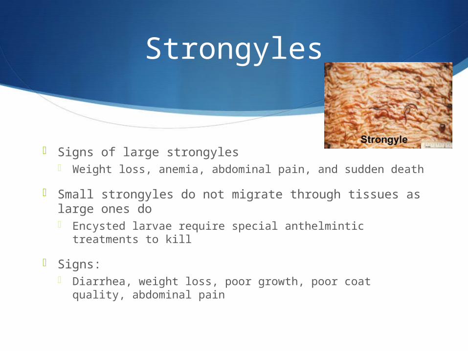
Signs of large strongyles Weight loss, anemia, abdominal pain, and sudden death
Small strongyles do not migrate through tissues as large ones do Encysted larvae require special anthelmintic treatments to
kill
Signs: Diarrhea, weight loss, poor growth, poor coat quality,
abdominal pain