> 80 million total ct scans / year in usa in 2010 (25% ed) · `early trauma ct `the...
TRANSCRIPT

Martin GunnDepartment of RadiologyUniversity of Washington
I have no actual or potential conflict of interest in relationship to this presentation.
I do have the following declarations:Grant support:
National Electrical Manufacturers’ Association (via subcontract).Philips Healthcare.NIH/NBIB (1-R21-EB016872-01A1)
Medical Advisor:TransformativeMed.
Book royalties:Cambridge University Press.Wolters-Kluwer.
Other financial or material support:Spouse is a contractor to Wolters-Kluwer health.
Early trauma CTThe “Pan-Scan”: Whole body CT in TraumaBlunt splenic trauma and angioembolizationImpact of CT’s increasing sensitivity:
Detection of early & minor disease.The “incidentaloma”
Radiation dose considerations
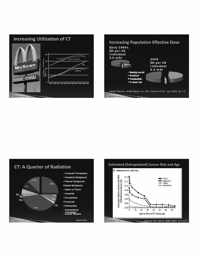
> 80 million total CT scans / year in USA in 2010 (25% ED)
2000 2001 2002 2003 2004 2005 2006 2007 2008 2009 2010
50
100
150
200
0
ED
Inpatient Hosp Outpt
Office
Image adapted from Levin D, Rao VM, Parker L, JACR 2013 9(11): 795
Proc
edur
es /
1,00
0 M
edic
are
bene
ficia
ries

[email protected] [email protected]
1. Salim A. et al Arch Surg. 2006 May;141(5):468-732. Reiger J Trauma. 2009 Mar;66(3):648-57.3. Leidner B, Beckman MO Emerg Rad 2001; 8(1):20-28
High accuracy for a wide range of injuries1.Low missed injury rate1,2.Can be performed rapidly2,3.
0 min 9min 79 min 99 min
Harborview Medical Center, July-August 20051.
Trauma patients ISS > 15.
Time to get a CT.
Some other locations have had similar results2.
1. Fung et al, Eur J Radiology 2009; 72 134.2. Easton et al, ANZ J Surg 82 (2012) 644
Report

The longer the hemodynamically unstable patient stays in the ED, the higher the risk of death1,2.
Mortality rose to 8.3% if patient in ED for 4-5 hours1.Outcome better if primary diagnosis < 40 mins of arrival vs. > 50 mins.
Causes of long stays:MRI for spine fractures.Multiple consulting services: “Consultarrhea.”
1. Mowery NT et al, J Trauma 2011 70(6)2. Chalfini D et al, Crit Care Med. 2007 Jun;35(6):1477-83
Earlier identification of: Surgical head injury.Primary bleeding site.• i.e. pelvis vs. intra-peritoneal, vs. chest.Non-surgical causes of hypotension.
Best way of controlling circulation is to stop the bleeding.
Pelvic fractures requiring angioembolization.• Time to angioembolization affects survival1.
1. Awasthi S, et al, Acad Emerg Med. 2008 Oct;15(10):895-9
1. Weninger et al, J Trauma. 2007;62:584–591.2. Wurmb TE et al, J Trauma 2009;66(3):658-665.3. Fanucci E et al, Emerg Radiol 2007;13(5):251-257.4. Sedlic A, et al, Emerg Radiol 2013;20(5):401-408.
Author Patients Standard Protocol (mins) Early WBCT (mins)
Weninger et al1 185 old185 ED-CT
104 +/- 21 70 +/- 17
Wurmb et al2 79 std82 resus CT
70 23
Fanucci et al3 20 segmented26 single-pass
32 22 mins
Sedlic et al4 34 Segmented33 single pass
18 10
Prospective study in 14 French hospitalsWBCT (N = 1,696) vs selective (N = 254).
Crude mortality rate @ 30 daysWBCT (16%) vs. selective CT (22%)OR for death to 0.58 with injury adjustment.
Yeguiayan J et al Critical Care 2012, 16:R101
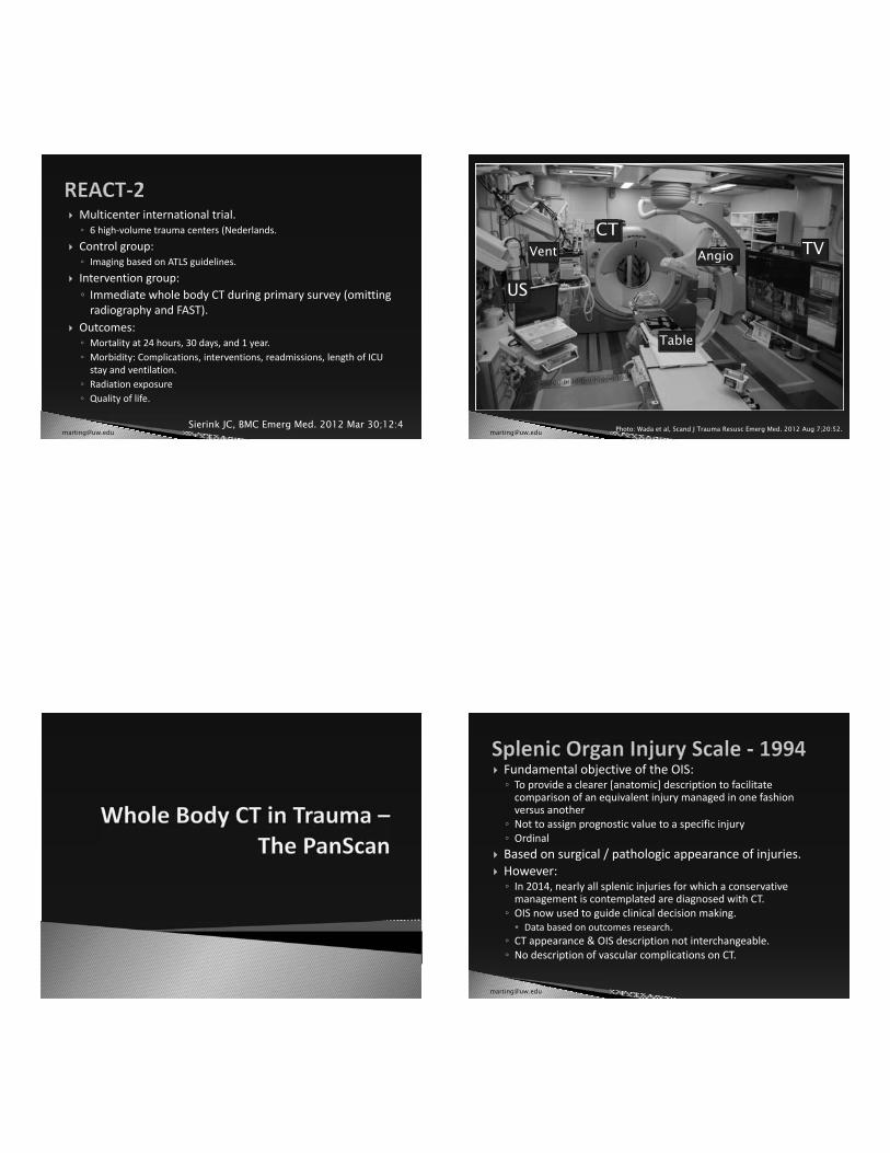
Multicenter international trial.6 high-volume trauma centers (Nederlands.
Control group:Imaging based on ATLS guidelines.
Intervention group: Immediate whole body CT during primary survey (omitting radiography and FAST).
Outcomes: Mortality at 24 hours, 30 days, and 1 year.Morbidity: Complications, interventions, readmissions, length of ICU stay and ventilation.Radiation exposureQuality of life.
Sierink JC, BMC Emerg Med. 2012 Mar 30;12:[email protected] Photo: Wada et al, Scand J Trauma Resusc Emerg Med. 2012 Aug 7;20:52.
US
Angio
CTVent TV
Table
[email protected] [email protected]
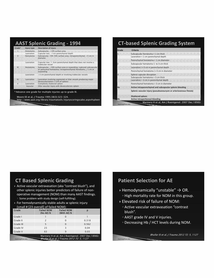
Fundamental objective of the OIS:To provide a clearer [anatomic] description to facilitate comparison of an equivalent injury managed in one fashion versus anotherNot to assign prognostic value to a specific injuryOrdinal
Based on surgical / pathologic appearance of injuries.However:
In 2014, nearly all splenic injuries for which a conservative management is contemplated are diagnosed with CT.OIS now used to guide clinical decision making. • Data based on outcomes research.CT appearance & OIS description not interchangeable.No description of vascular complications on CT.
A Advance one grade for multiple injuries up to grade III.
Grade* Injury type Description of injuryI Hematoma Subcapsular, <10% surface area
Laceration Capsular tear, <1cm parenchymal depthII Hematoma Subcapsular, 10%-50% surface area, intraparenchymal, <5 cm in
diameter
Laceration Capsular tear, 1-3cm parenchymal depth that does not involve a trabecular vessel
III Hematoma Subcapsular, >50% surface area or expanding; ruptured subcapsular or parecymal hematoma; intraparenchymal hematoma > 5 cm or expanding
Laceration >3 cm parenchymal depth or involving trabecular vessels
IV Laceration Laceration involving segmental or hilar vessels producing major devascularization (>25% of spleen)
V Laceration Completely shattered spleenVascular Hilar vascular injury with devascularizes spleen
Moore EE et al, J Trauma 1995;38(3):323-324.http://www.aast.org/library/traumatools/injuryscoringscales.aspx#spleen
Grade Criteria
1 Subcapsular hematoma < 1 cm thickLaceration < 1 cm parenchymal depthParenchymal hematoma < 1 cm diameter
2 Subcapsular hematoma 1- to 3-cm thickLaceration 1–3 cm in parenchymal depthParenchymal hematoma 1–3 cm in diameter
3 Splenic capsular disruptionSubcapsular hematoma > 3 cm thickLaceration > 3 cm in parenchymal depthParenchymal hematoma > 3 cm in diameter
4a Active intraparenchymal and subcapsular splenic bleedingSplenic vascular injury (pseudoaneurysm or arteriovenous fistula)
Shattered spleen4b Active intraperitoneal bleeding
Marmery H et al, Am J Roentgenol. 2007 Dec;189(6):
Active vascular extravasation (aka “contrast blush”), and other splenic injuries better predictors of failure of non-operative management (NOM) than many AAST findings.
Some problem with study design (self-fulfilling).For hemodynamically stable adults w splenic injury (small # [23 overall] of failed NOM):
Marmery H et al, Am J Roentgenol. 2007 Dec;189(6):Bhullar IS et al, J Trauma 2012 72: 5, 1127
Grade Failed NOM (No AE) %
Failed NOM(With AE) %
p
Grade I 1 0 1Grade II 2 0 0.318Grade III 5 0 0.562Grade IV 23 3 0.04Grade V 63 9 0.03
Hemodynamically “unstable” OR.High mortality rate for NOM in this group.
Elevated risk of failure of NOM:Active vascular extravasation “contrast blush”.AAST grade IV and V injuries.Decreasing Hb / HCT levels during NOM.
Bhullar IS et al, J Trauma 2012 72: 5, 1127
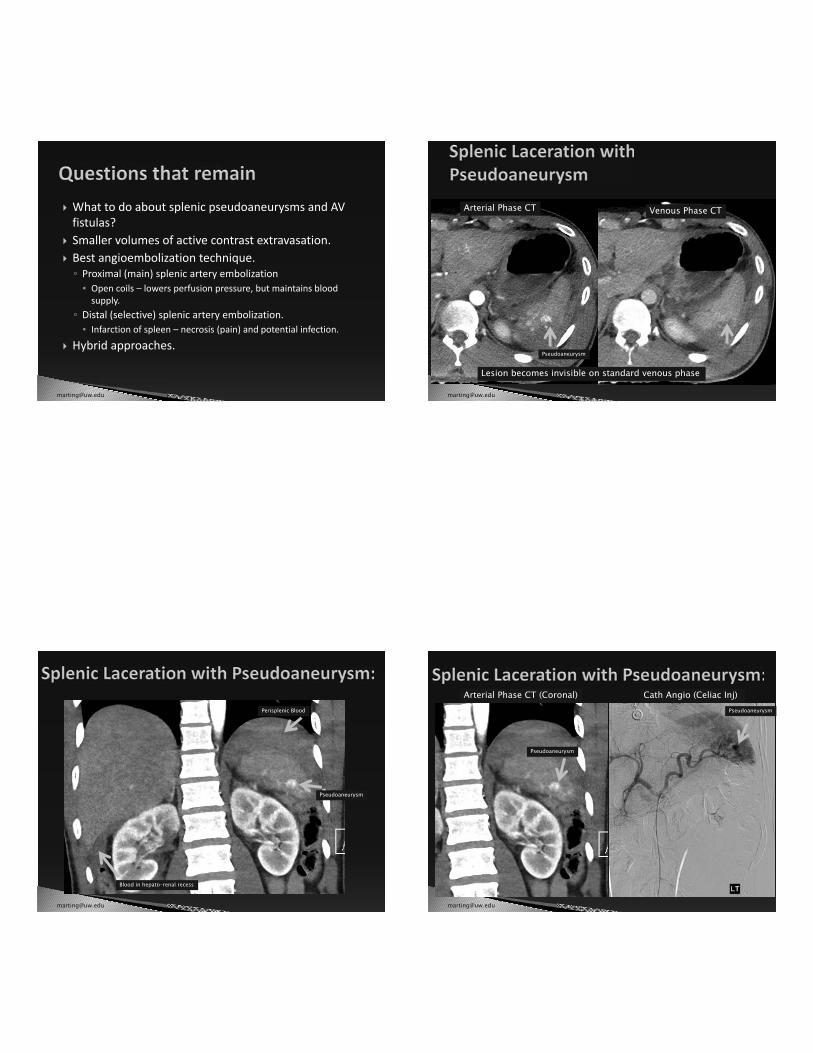
What to do about splenic pseudoaneurysms and AV fistulas?Smaller volumes of active contrast extravasation.Best angioembolization technique.
Proximal (main) splenic artery embolization• Open coils – lowers perfusion pressure, but maintains blood
supply.Distal (selective) splenic artery embolization.• Infarction of spleen – necrosis (pain) and potential infection.
Hybrid approaches.
Arterial Phase CT Venous Phase CT
Lesion becomes invisible on standard venous phase
Pseudoaneurysm
Pseudoaneurysm
Perisplenic Blood
Blood in hepato-renal recess
Arterial Phase CT (Coronal)
Pseudoaneurysm
Pseudoaneurysm
Cath Angio (Celiac Inj)

[email protected] [email protected]
Arterial Phase CT “Portal” Venous Phase CT
Note: Lesion is invisible on standard venous phase
PseudoaneurysmLaceration, but pseudoaneurysm not visible
Pseudoaneurysm(Enlarging)
Pseudoaneurysm)Pseudoaneurysm)
Cath Angio (Splenic art. Inj) Cath Angio (Splenic art. segmental Inj)

[email protected] [email protected]
Intra-splenic.Appears as rounded dense puddles on CT.Density mirrors vascular phase on CT.
Density on arterial phase = density of aorta / splenic artery.Density on portal venous phase = density of portal / splenic veins.Density on delayed phase = density of IVC.
Tend to be invisible on portal venous phase and delayed phase CT.Dense area does NOT increase in size with longer duration.Density of lesion drops with progressively duration after contrast injection.
Hemoperitoneum
Contrast Extravasation
Peri-spenichemoperitoneum
Laceration
Contr. Extrav. Contr. Extrav.
Contr. Extrav.
Art. Phase (~30 sec after contrast)
Venous Phase ~70 sec after contrast)
Delayed Phase 300 sec after contrast)

Increases in quantity/size with increasing time following contrast injection.Volume visible on CT:
Arterial phase < portal venous phase < delayed phase.Increases in density during phases:
Arterial phase < portal venous phase < delayed phase.
May appear to arise or “squirt” from laceration.Spreads in peritoneal space / Irregularly shaped.
Trauma Surgeon:“CT’s are too sensitive: you guys report too many things!”
Radiologist:“Don’t blame the scanner, it’s just that you (and we) don’t have a clue what to do with all these findings.”
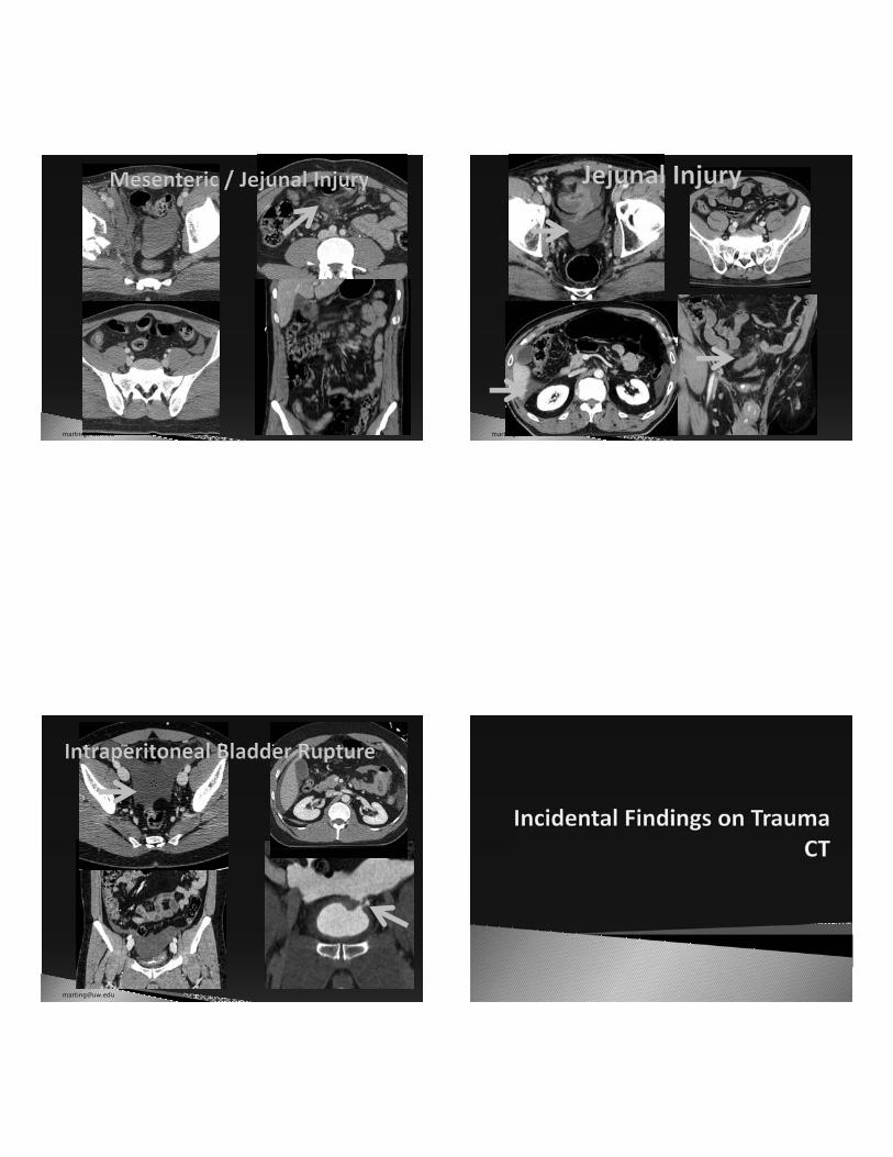
Detection of early disease.e.g.
“Occult” pneumothoraxFree intraperitoneal fluidPulmonary embolismActive vascular extravasation
Detection of diseases unrelated to trauma.The “incidentaloma.”
Rib Fx
[email protected] [email protected]
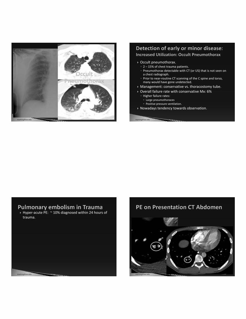
Occult pneumothorax.2 – 15% of chest trauma patients.Pneumothorax detectable with CT (or US) that is not seen on a chest radiograph.Prior to near-routine CT scanning of the C spine and torso, many would have gone undetected.
Management: conservative vs. thoracostomy tube.Overall failure rate with conservative Mx: 6%
Higher failure rates:• Large pneumothoraces• Positive pressure ventilation.
Nowadays tendency towards observation.
Hyper-acute PE: ~ 10% diagnosed within 24 hours of trauma.

[email protected] Rodriguez et al J Trauma. 2002 Jul;53(1):79-85. [email protected]
Small quantities of physiologic intraperitoneal fluid seen in both males and females.Free fluid is the most sensitive sign of bowel injury.Predictors of a “benign” cause for “free’ fluid:
Isolated to pelvis (pelvic, below 3rd sacral segment).Low density (< 15 HU).Small quantity ( 2.5cm in cranio-caudal extent.)
Isolated fluid outside the pelvis, especially between the mesenteric leaves, bowel loops, or in the paracolic gutters should be treated with suspicion.
For isolated small volumes of free “benign” pelvic fluid, observation appears safe.
Drasin TE, et al. AJR. 2008;191:1821-1826.

12 – 45% of abdomino-pelvic CT scans will have incidental findings.
Some have imaging features diagnostic of benign disease.Lower rates with head and neck.Higher rates (up to 45% for WBCT, most from abdomen).Some have potential serious morbidity.
Increasing incidence with increasing age.Majority of patients with incidental findings (75%) have no traumatic injury in abdomen or pelvis.
Higher chance in some studies of identifying an incidental findings (30%) vs. a traumatic abnormality (15%).
Munk MD, et al, J Emerg Med 2010;38(3):346-350. van Vugt R et al, J Trauma 2011, Apr 29.
R² = 0.8658
0
0.1
0.2
0.3
0.4
0.5
0.6
0.7
20 30 40 50 60 70 80 90 100
Rate
0%
10%
20%
30%
40%
50%
60%
70%
80%
90%
100%
Incidental No IncidentalAge (years)
Perc
enta
ge o
f CT
Exam
inat
ions
Age – upper bound of decade (years)
“Maj
or” I
ncid
enta
l Rat
e
Strong correlation between subject age and the major incidental findings rate (p< 0.0001).
Potential pitfalls:AnxietyIncreased healthcare costs for follow-up examinations.Coordination of care issues.Potential medico-legal liabilities (“lost to follow-up”): • Focused on the primary disease process, forget about the
incidental finding.Potential morbidly for unnecessary biopsy or surgery.Downstream healthcare costs.
Incidentals need to be included in comparative effectiveness studies.
2000 2001 2002 2003 2004 2005 2006 2007 2008 2009 2010
50
100
150
200
0
ED
Inpatient Hosp Outpt
Office
Proc
edur
es /
1,00
0 M
edic
are
bene
ficia
ries
Early 1980s.ED per US Individual3.6 mSv 2006
ED per US Individual6.2 mSv
Health Physics, NCRP Report no. 160, Volume 97(1), July 2009, pp 1-5
[email protected] NCRP 2006 [email protected] Brenner DF, Hall EJ, NEJM 3007; 357:2277

0 50 100 250150 200
1.01
1.02
1.03
1.04
1.05
1.06
1.00
1.07
BIER VII ReportEffective Dose (mSv)
Life
tim
e at
trib
uta
ble
ris
k (L
AR)
Linear No Threshold (LNT)Linear ThresholdSupra-LinearHormesis
Although low-dose techniques may be used to reduce the dose of any single scan, repeat scanning increases radiation dose for major-trauma patients.Correlates with ISS and LOS1.
CT accounts for 93% of radiation exposure1.
ISS Radiation exposureISS < 9 25 mSvISS 9+ 14 mSvMean 15.51 - 1062 mSv
1. Sharma et al, J Emerg Med. 2011 Dec;41(6):6402. Kim PK et al, J Trauma. 2004 Sep;57(3):510-4
[email protected] Psoter KJ, et al JACR 2013 [email protected] Psoter KJ, Eur J Radiol. 2013 Jun;82(6):969.
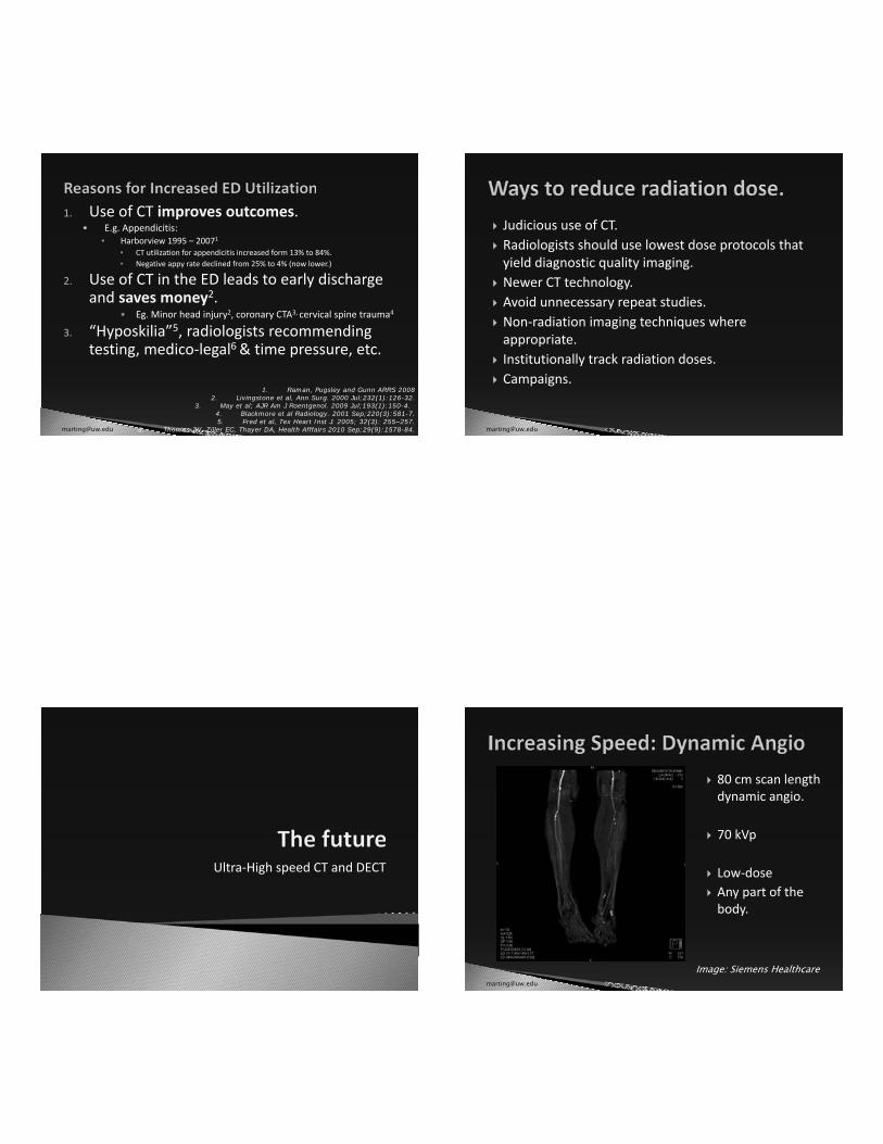
1. Use of CT improves outcomes.• E.g. Appendicitis:
• Harborview 1995 – 20071
• CT utilization for appendicitis increased form 13% to 84%.• Negative appy rate declined from 25% to 4% (now lower.)
2. Use of CT in the ED leads to early discharge and saves money2.
• Eg. Minor head injury2, coronary CTA3, cervical spine trauma4
3. “Hyposkilia”5, radiologists recommending testing, medico-legal6 & time pressure, etc.
1. Raman, Pugsley and Gunn ARRS 20082. Livingstone et al, Ann Surg. 2000 Jul;232(1):126-32.
3. May et al; AJR Am J Roentgenol. 2009 Jul;193(1):150-4. 4. Blackmore et al Radiology. 2001 Sep;220(3):581-7.5. Fred et al, Tex Heart Inst J. 2005; 32(3): 255–257.
6. Thomas JW, Ziller EC, Thayer DA, Health Afffairs 2010 Sep;29(9):1578-84. [email protected]
Judicious use of CT.Radiologists should use lowest dose protocols that yield diagnostic quality imaging.Newer CT technology.Avoid unnecessary repeat studies.Non-radiation imaging techniques where appropriate.Institutionally track radiation doses.Campaigns.
Ultra-High speed CT and DECT
80 cm scan length dynamic angio.
70 kVp
Low-doseAny part of the body.
Image: Siemens Healthcare

Future CT in Trauma:Potentially earlier, integrated with resuscitation.Whole body CT is now the mainstay in BAT, but outcomes are not well established.Newer: High speed CT and dual energy CT.
Trend towards endovascular therapy for selected subgroups of patients.Need to tame the information explosion with detection of more subtle disease and incidental findings.Increasing concerns about radiation exposure, but there is still potential to improve appropriateness and reduce radiation dose.