:: slide 1creyes/classes/ch.04_test1_notes_images.pdf · the nervous system to integrate complex...
TRANSCRIPT

:: Slide 1 ::
What Is Biological Psychology?
How Do Neurons Communicate?
How Is the Nervous System Organized?
Central Nervous System
Peripheral Nervous System and Endocrine System
Note about the image on this slide: Perceived social isolation or connectedness can produce changes in the cells of our immune system.
:: Slide 2 ::
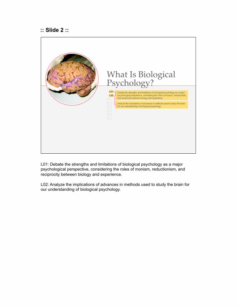
L01: Debate the strengths and limitations of biological psychology as a major psychological perspective, considering the roles of monism, reductionism, and reciprocity between biology and experience.
L02: Analyze the implications of advances in methods used to study the brain for our understanding of biological psychology.

:: Slide 3 ::
• Biological psychology is the branch of psychology that seeks to explain human experience and behavior in terms of processes in the physical body.
• The renaissance philosopher René Descartes proposed the duality of body and mind - that mind is separate from the physical body, and is not subject to the laws of matter.
• Biological Psychology instead presumes the competing philosophy of monism – that mind and body are one.

:: Slide 4 ::
• This emphasis on the connection between the body and mind leads biological psychologists to combine the theories and research methods used in psychology with those from other disciplines, such as biology, physiology, biochemistry, and neuroscience.
• Biological psychologists are especially interested in the activity of the nervous system, which acquires sensory information, processes that information, and sends instructions to the body for behavior.
:: Slide 5 ::
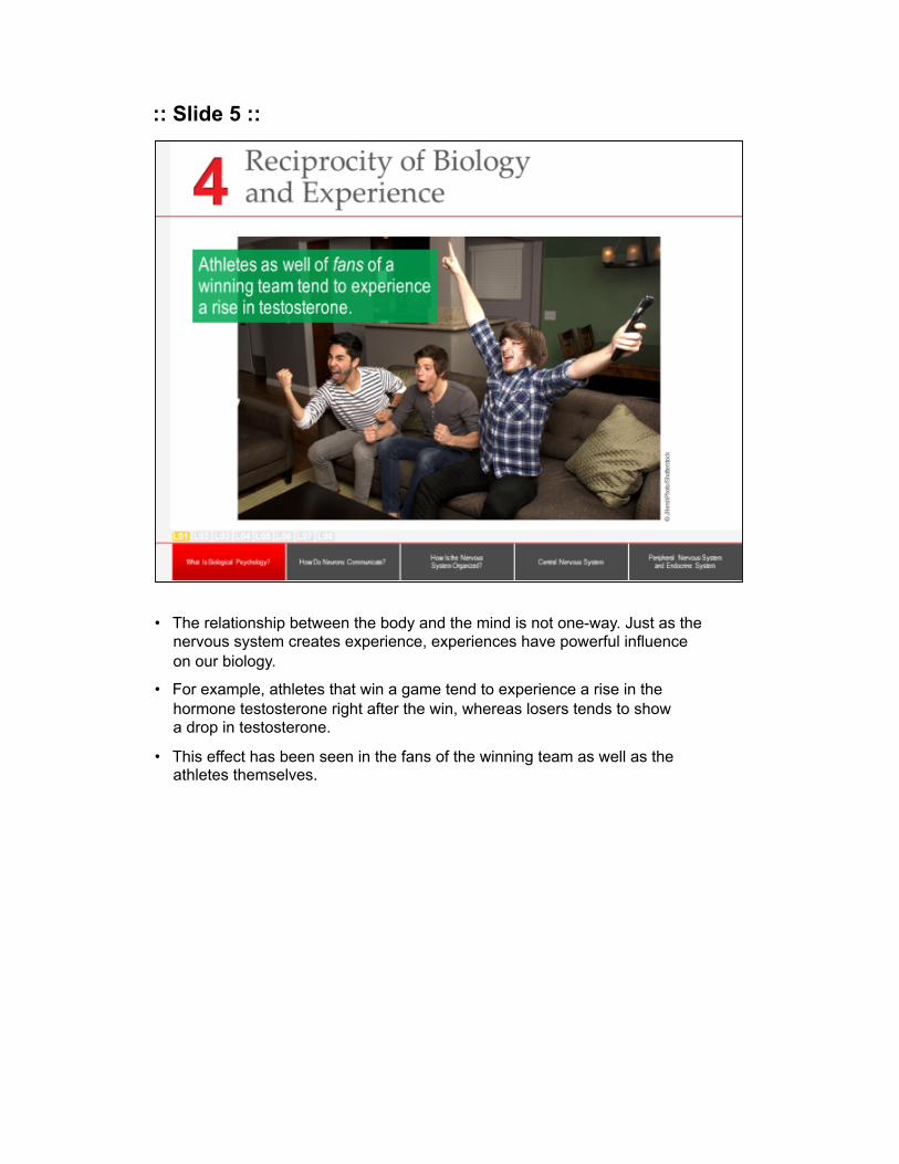
• The relationship between the body and the mind is not one-way. Just as the nervous system creates experience, experiences have powerful influence on our biology.
• For example, athletes that win a game tend to experience a rise in the hormone testosterone right after the win, whereas losers tends to show a drop in testosterone.
• This effect has been seen in the fans of the winning team as well as the athletes themselves.
:: Slide 6 ::
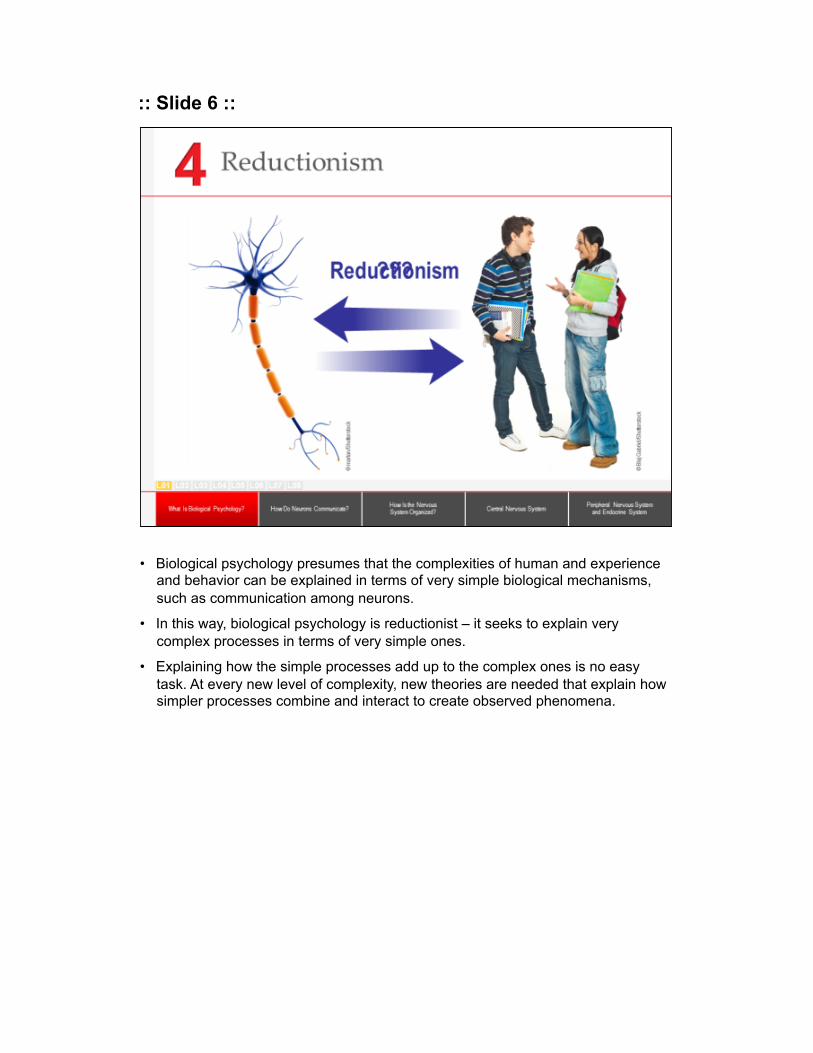
• Biological psychology presumes that the complexities of human and experience and behavior can be explained in terms of very simple biological mechanisms, such as communication among neurons.
• In this way, biological psychology is reductionist – it seeks to explain very complex processes in terms of very simple ones.
• Explaining how the simple processes add up to the complex ones is no easy task. At every new level of complexity, new theories are needed that explain how simpler processes combine and interact to create observed phenomena.

:: Slide 7 ::
• Biological psychology has benefitted greatly from recent advances in technologies for studying the human brain.
• Before these advances researchers were limited to studying human corpses, people with traumatic brain injury, or laboratory animals.
• In the last 20 years, researchers have made major advances in our ability to study brain activity associated with experience and behavior in healthy, living people.

:: Slide 8 ::
• In electroencephalography, or EEG, researchers used sensors placed on the scalp to measure electrical activity in the brain.
• Researchers can measure resting brain activity, or changes in activity associated with specific stimuli or events, called “event-related potentials.”

:: Slide 9 ::
• In functional magnetic resonance imaging, or fMRI, researchers measure the brain’s use of oxygen while participants complete cognitive or emotional tasks.
• When an area of the brain has been active, it needs more oxygen; fMRI techniques measure delivery of oxygen by the blood.
:: Slide 10 ::
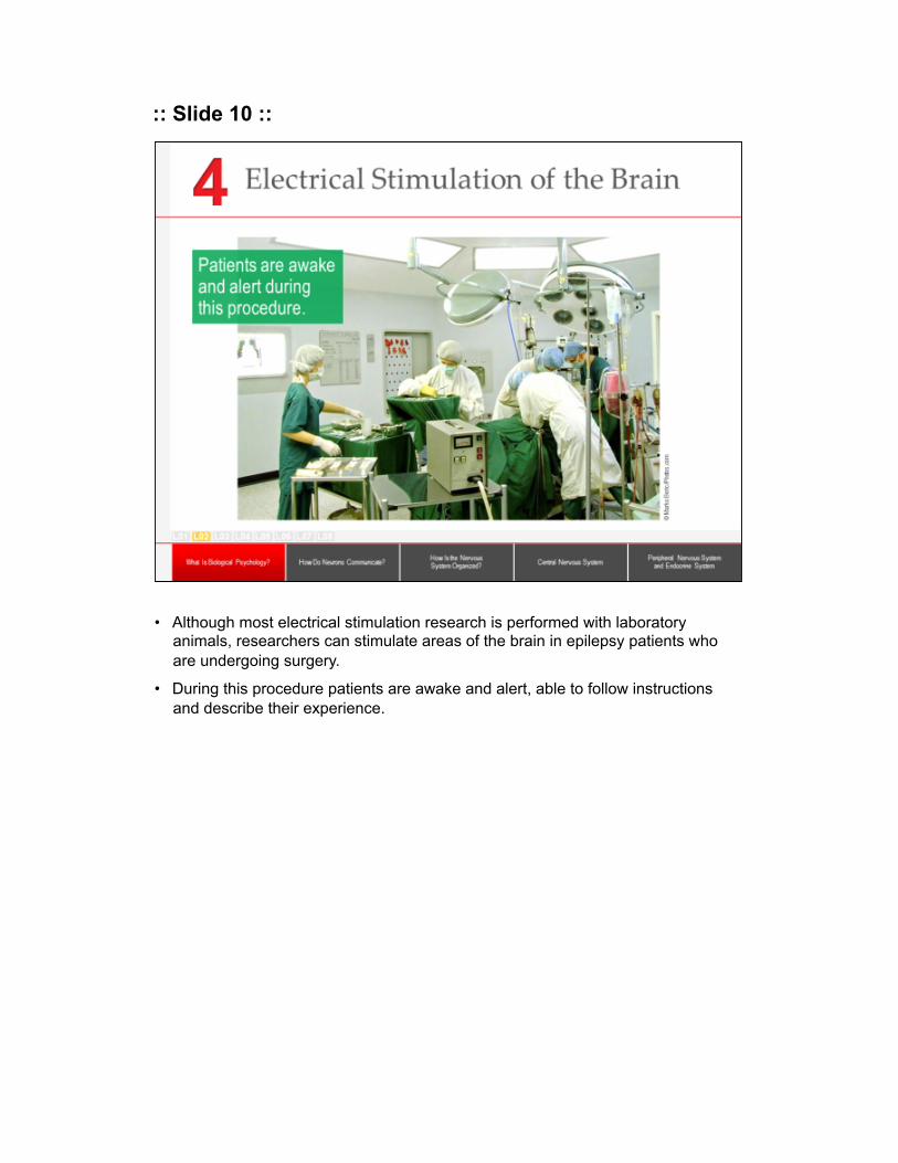
• Although most electrical stimulation research is performed with laboratory animals, researchers can stimulate areas of the brain in epilepsy patients who are undergoing surgery.
• During this procedure patients are awake and alert, able to follow instructions and describe their experience.

:: Slide 11 ::
L03: Explain what it means for a neuron to “fire” an action potential, describing how the neuron’s structure makes this possible.
L04: Explain the process by which neurons communicate with each other, allowing the nervous system to integrate complex information.
L05: Differentiate the roles played by major neurotransmitters in supporting physical functioning and psychological experience.
Note about the image on this slide: This neuron has been grown on a silicon chip, a line of research that might lead to better treatment for people with amputated limbs. Prosthetic devices, like an artificial leg or hand, could possibly communicate directly with the person’s nervous system.
:: Slide 12 ::
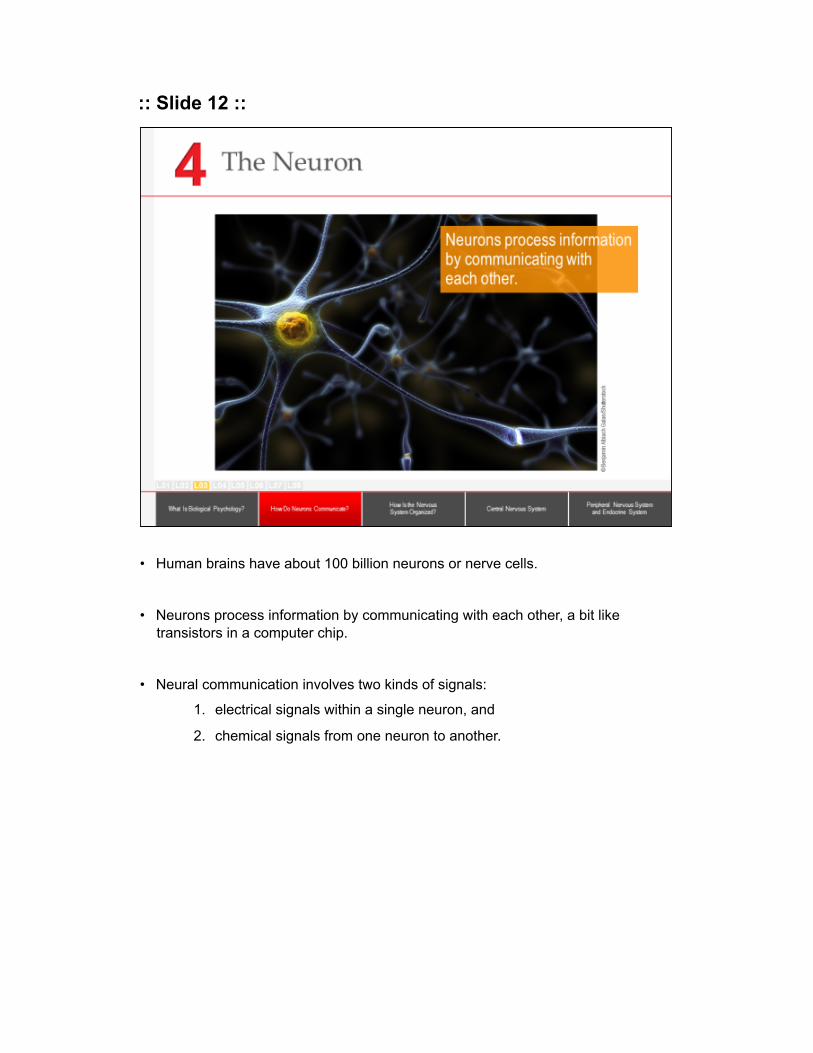
• Human brains have about 100 billion neurons or nerve cells.
• Neurons process information by communicating with each other, a bit like transistors in a computer chip.
• Neural communication involves two kinds of signals:
1. electrical signals within a single neuron, and
2. chemical signals from one neuron to another.
:: Slide 13 ::
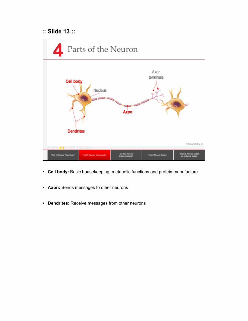
• Cell body: Basic housekeeping, metabolic functions and protein manufacture
• Axon: Sends messages to other neurons
• Dendrites: Receive messages from other neurons
:: Slide 14 ::
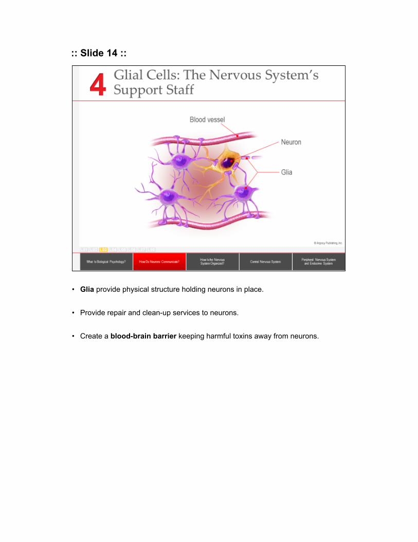
• Glia provide physical structure holding neurons in place.
• Provide repair and clean-up services to neurons.
• Create a blood-brain barrier keeping harmful toxins away from neurons.
:: Slide 15 ::
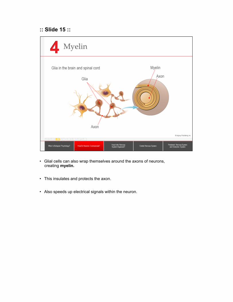
• Glial cells can also wrap themselves around the axons of neurons, creating myelin.
• This insulates and protects the axon.
• Also speeds up electrical signals within the neuron.

:: Slide 16 ::
• At rest, neurons build up an electrical potential or charge across the cell membrane.
• The neuron “fires” when gates in the axon membrane open, allowing electrically charged ions to move freely.
:: Slide 17 ::
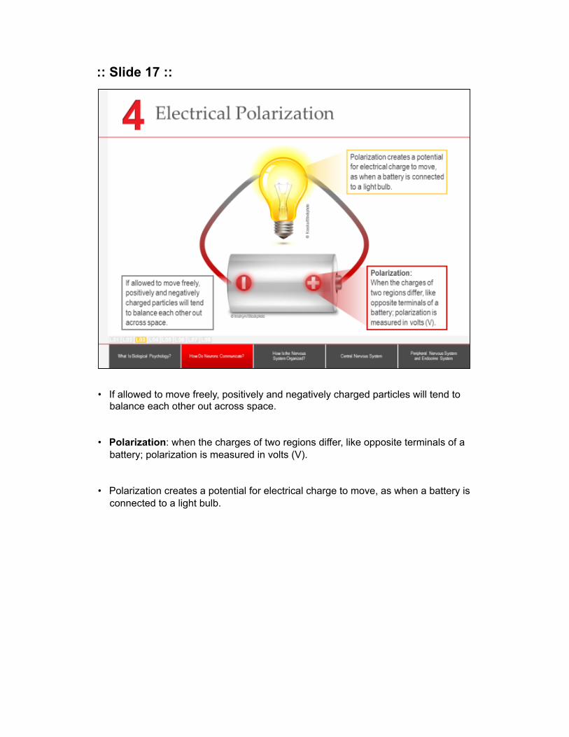
• If allowed to move freely, positively and negatively charged particles will tend to balance each other out across space.
• Polarization: when the charges of two regions differ, like opposite terminals of a battery; polarization is measured in volts (V).
• Polarization creates a potential for electrical charge to move, as when a battery is connected to a light bulb.

:: Slide 18 ::
• At rest, ion pumps and gates in the cell membrane push sodium (Na++) ions outside the cell, and pull Potassium (K+) ions inside.
• Potassium ions leak back out easily, but sodium ions are trapped outside. Several large, negatively charged molecules are trapped inside the cell.
• This creates an electrical polarization, called the resting potential, with a relative positive charge outside the cell membrane, and a negative charge inside (about -70 mV).

:: Slide 19 ::
• Events in the cell body and dendrites can cause the axon to depolarize slightly.
• If depolarization reaches a threshold of about -65 mV, the axon generates an action potential.
• During an action potential, gates open in the membrane allowing Na++ ions to enter the cell, then other gates allow K+ ions to exit.
• The polarization across the neural membrane briefly reverses (positive charge inside) and then gates close to re-establish the resting potential.

:: Slide 20 ::
• Action potentials do not affect the whole axon at once; they begin near the cell body and move toward the axon terminal as depolarization spreads.
• If the axon is myelinated, the action potential will “skip” from one Node of Ranvier to the next, speeding up propagation.
• During an action potential, gates open in the membrane allowing Na++ ions to enter the cell, then other gates allow K+ ions to exit.
• After an action potential, the axon enters a brief refractory period where the resting potential is re-established, and the neuron cannot fire.
:: Slide 21 ::
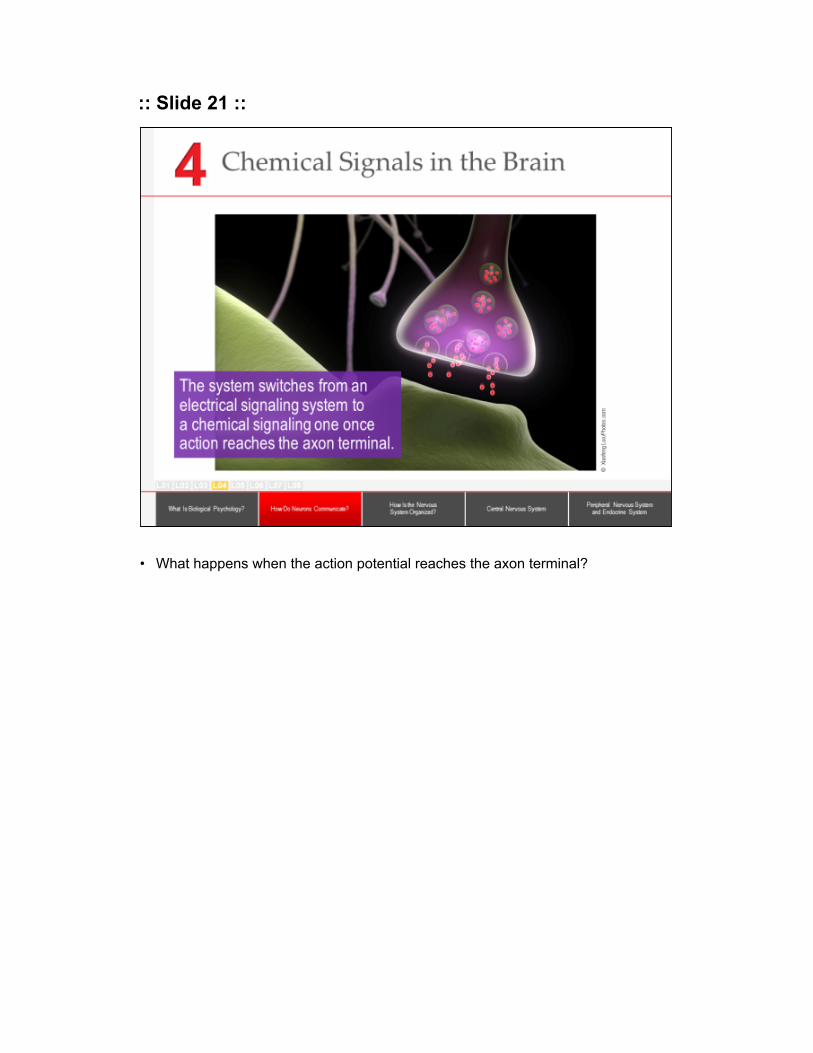
• What happens when the action potential reaches the axon terminal?
:: Slide 22 ::
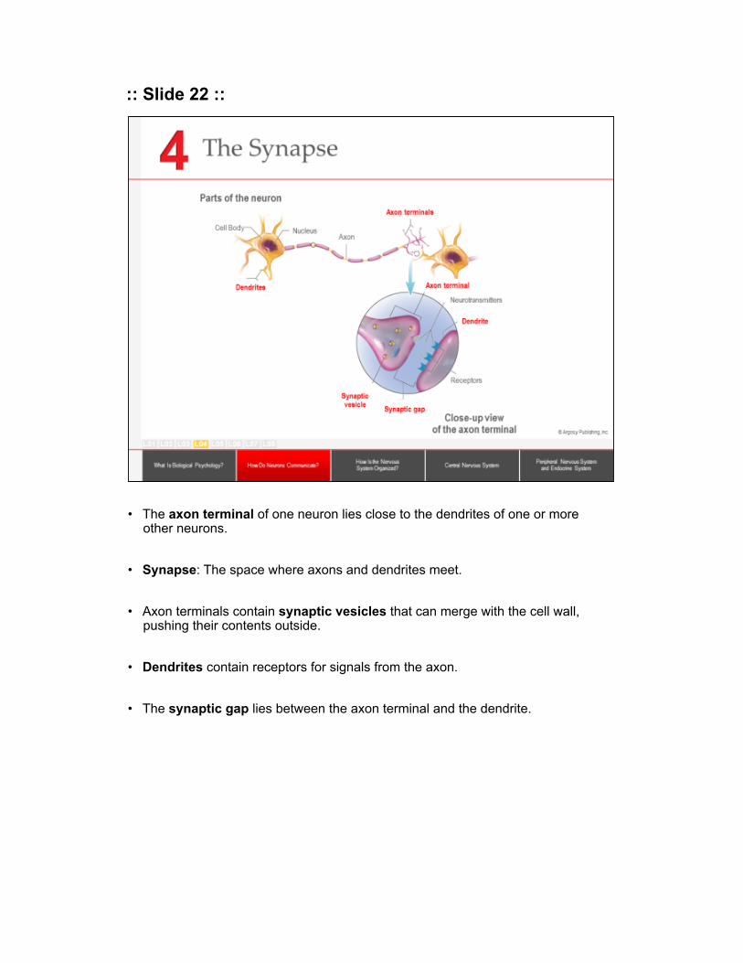
• The axon terminal of one neuron lies close to the dendrites of one or more other neurons.
• Synapse: The space where axons and dendrites meet.
• Axon terminals contain synaptic vesicles that can merge with the cell wall, pushing their contents outside.
• Dendrites contain receptors for signals from the axon.
• The synaptic gap lies between the axon terminal and the dendrite.

:: Slide 23 ::
• When the action potential reaches the axon terminal, synaptic vesicles fuse with the axon membrane.
• Neurotransmitters, or chemical signals, are pushed into the synaptic gap.
• The neurotransmitters cross the gap and connect with receptors on dendrites on the other side of the gap.

:: Slide 24 ::
• Some receptors act to depolarize the nearby cell membrane.
• This increases the probability of an action potential.
• This excitatory input serves to “pass the message along” to the next neuron.

:: Slide 25 ::
• Other receptors act to increase the polarization of the nearby cell.
• This decreases the probability of an action potential.
• Inhibitory input helps prevent the receiving cell from firing.

:: Slide 26 ::
• A single neuron may receive input from many synapses.
• Neurons “decide” whether to generate an action potential by adding up excitatory and inhibitory input.
:: Slide 27 ::
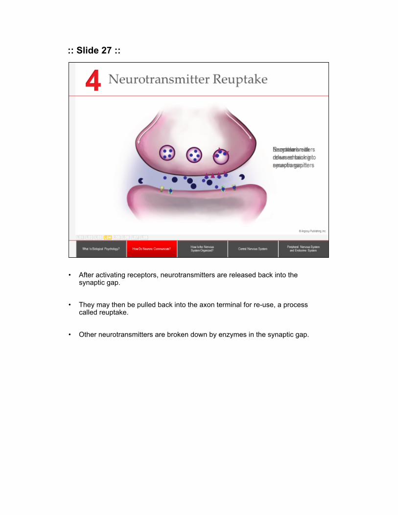
• After activating receptors, neurotransmitters are released back into the synaptic gap.
• They may then be pulled back into the axon terminal for re-use, a process called reuptake.
• Other neurotransmitters are broken down by enzymes in the synaptic gap.

:: Slide 28 ::
• Researchers have identified more than 50 different chemicals that act as neurotransmitters.
• Different neurotransmitters are found in neural systems with different functions, and may activate excitatory or inhibitory receptors.

:: Slide 29 ::
• Main neurotransmitter at the neuromuscular junction, where neurons instruct muscles to contract.
• Important neurotransmitter in the autonomic nervous system, carrying messages from the brain to the glands and visceral organs.
• Used in brain circuits supporting learning and memory.

:: Slide 30 ::
• Increases activity in brain areas supporting alertness and vigilance.
• Primary neurotransmitter in the “fight/flight” sympathetic branch of the autonomic nervous system, increasing heart rate, blood pressure, and breathing.
• Abnormalities in norepinephrine systems have been implicated in post-traumatic stress disorder (PTSD).

:: Slide 31 ::
• Primary neurotransmitter in brain circuits supporting anticipation of rewards, such as food, sex, and many addictive drugs.
• Used in brain areas supporting fine motor control; Parkinson’s disease is caused by deterioration of dopamine-producing cells in these areas.
• Important in some areas supporting planning and controlled cognition; abnormalities implicated in ADHD and schizophrenia.

:: Slide 32 ::
• Used in brain areas that regulate cycles of sleep and appetite.
• Important for neural structures supporting some aspects of memory and learning.
• Used in brain circuits that regulate mood.

:: Slide 33 ::
• Used by neural systems that modulate the experience of pain.
• Also released during strenuous physical exercise, after eating spicy foods, and after orgasm.
• Involved in some feelings of pleasure and enjoyment, such as eating.

:: Slide 35 ::
• Most addictive drugs, including alcohol, cocaine, and methamphetamine, activate dopamine receptors in the brain’s reward circuits. Alcohol also primarily activates GABA.
• Morphine and other opiate painkillers mimic the activity of endorphins.
• Acetylcholine receptor agonists can be used to treat Alzheimer’s disease.
• Dopamine agonists can be used to help treat Parkinson’s disease; however, side effects may include gambling and other addictive behaviors, and hallucinations typical of schizophrenia.

:: Slide 36 ::
• “Alpha blockers” and “beta blockers” are both norepinephrine receptor antagonists, used to treat problems with heart rate and blood pressure.
• The poison curare blocks acetylcholine receptors, preventing contraction of most muscles Including the diaphragm), leading to paralysis and death.
• Antipsychotic medications used to treat schizophrenia block dopamine receptors in the brain; however, side effects include depressed mood and Parkinson’s-like motor tremors.

:: Slide 37 ::
• Selective serotonin reuptake inhibitors (SSRIs) such as Prozac are used to treat depression and some other mood disorders.
• Some pesticides and nerve gasses interfere with acetylcholine breakdown enzymes, causing permanent muscle spasms, paralysis, and death.
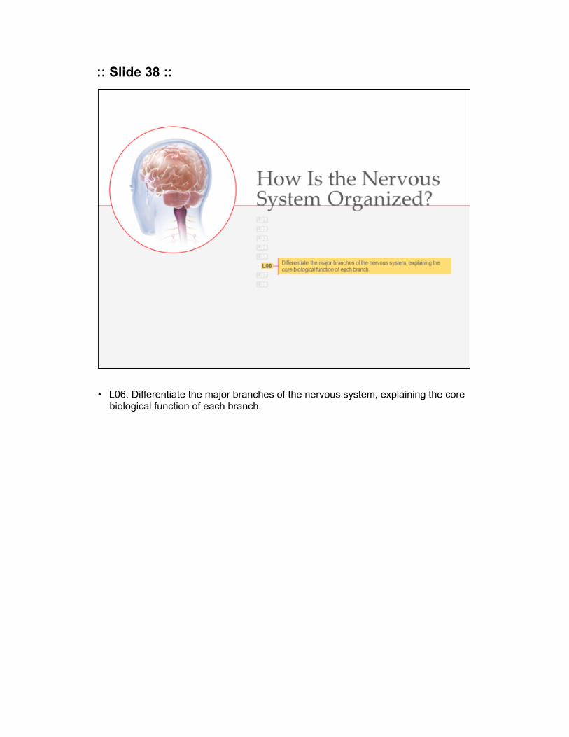
:: Slide 38 ::
• L06: Differentiate the major branches of the nervous system, explaining the core biological function of each branch.

:: Slide 39 ::
• The nervous system is organized into several major branches, each of which serves a somewhat different function. This chart shows the overall structure of the nervous system.

:: Slide 40 ::
• At the highest level of organization, the nervous system is divided into the central nervous system and the peripheral nervous system. The function of the central nervous system is to process incoming information, and send commands to the rest of the body. Unlike the peripheral nervous system, the central nervous system is encased in bone – the skull and vertebrae.
• The peripheral nervous system consists of nerves, or fibers made of multiple neurons, that extend from the central nervous system to the rest of the body.
• The peripheral nervous system can be divided into two smaller branches, which sometimes run along the same nerve fibers. The somatic nervous system brings in information from the senses about the state of the body and the environment.
• The somatic nervous system also sends commands to voluntary muscles, like the skeletal muscles of the head, neck, arms, and legs.
• The other branch, the autonomic nervous system, sends commands to visceral organs such as the heart, lungs, and stomach.

:: Slide 43 ::
• The spinal cord is a long tract of nerve tissue that carries information between the brain and body. The spinal cord also controls a number of reflexes that do not involve the brain at all, such as the knee-jerk reflex.
• The spinal cord is carefully encased in the bones of the vertebrae, which provide protection while still allowing flexible movement.
• The inner part of the spinal cord consists of neuron cell bodies, which have a grayish color.
• The outer sheath of the spinal cord is made up of bundles of axons. Because these axons are myelinated, the outer tissue has a white color.
• Nerves enter and exit the spinal cord between the vertebrae.
:: Slide 48 ::
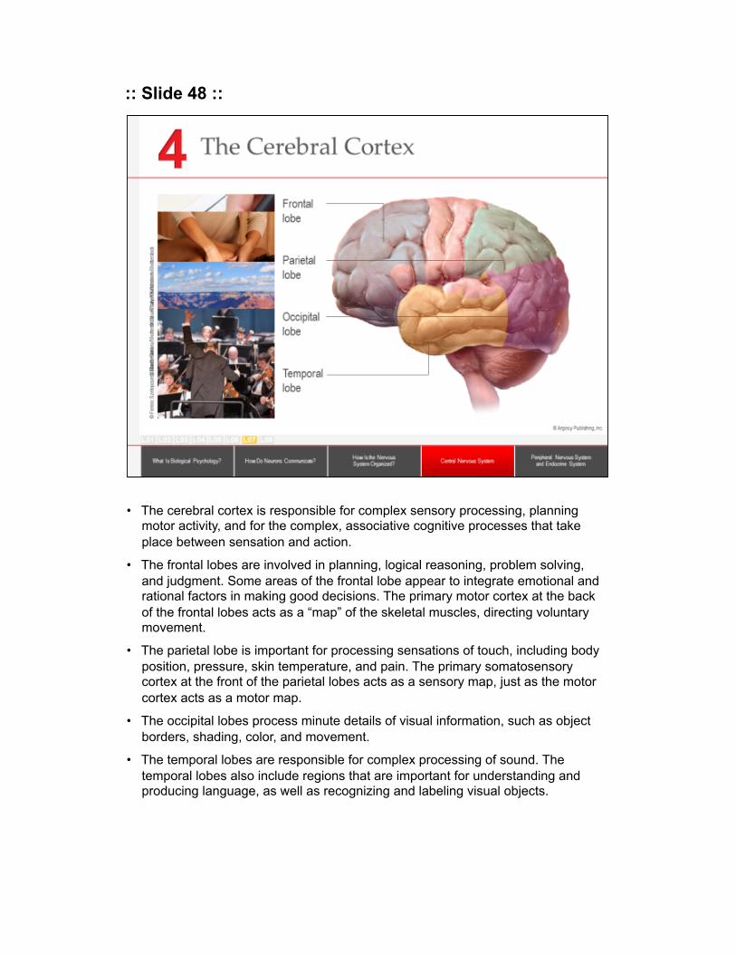
• The cerebral cortex is responsible for complex sensory processing, planning motor activity, and for the complex, associative cognitive processes that take place between sensation and action.
• The frontal lobes are involved in planning, logical reasoning, problem solving, and judgment. Some areas of the frontal lobe appear to integrate emotional and rational factors in making good decisions. The primary motor cortex at the back of the frontal lobes acts as a “map” of the skeletal muscles, directing voluntary movement.
• The parietal lobe is important for processing sensations of touch, including body position, pressure, skin temperature, and pain. The primary somatosensory cortex at the front of the parietal lobes acts as a sensory map, just as the motor cortex acts as a motor map.
• The occipital lobes process minute details of visual information, such as object borders, shading, color, and movement.
• The temporal lobes are responsible for complex processing of sound. The temporal lobes also include regions that are important for understanding and producing language, as well as recognizing and labeling visual objects.

:: Slide 52 ::
• The other branch, the autonomic nervous system, carries commands from the brain (especially the hypothalamus) to visceral organs such as the heart, lungs, blood vessels, digestive system, and genitalia.
• The ANS is divided into two further branches, which have largely opposing effects although they are independent systems. The sympathetic division is often called the “fight-flight” system, as it supports the body in physical exertion. When sympathetic activation is dominant the body is focused on delivering energy to the skeletal muscles.
• The parasympathetic division is sometimes called the “rest and repair” system. When parasympathetic activation is dominant the body is focused on digesting food, storing energy for the future, and directing energy toward growth and reproduction.