ß-tcp bone substitutes in tibial plateau depression fractures · ß-tcp bone substitutes in tibial...
TRANSCRIPT
The Knee 24 (2017) 1138–1145
Contents lists available at ScienceDirect
The Knee
ß-TCP bone substitutes in tibial plateau depression fractures
Tim Rolvien a, Florian Barvencik a, Till Orla Klatte b, Björn Busse a, Michael Hahna,Johannes Maria Rueger b, Martin Rupprecht b,c,⁎a Department of Osteology and Biomechanics, University Medical Center Hamburg-Eppendorf, Lottestrasse 59, 22529 Hamburg, Germanyb Department of Trauma, Hand and Reconstructive Surgery, University Medical Center Hamburg-Eppendorf, Martinistrasse 52, 20246 Hamburg, Germanyc Department of Pediatric Orthopaedics, Altonaer Children's Hospital, Hamburg, Germany
a r t i c l e i n f o
⁎ Corresponding author at: Department of Pediatric OE-mail address: [email protected] (M. Rupprecht).
http://dx.doi.org/10.1016/j.knee.2017.06.0100968-0160/© 2017 Elsevier B.V. All rights reserved.
a b s t r a c t
Article history:Received 16 December 2016Received in revised form 20 March 2017Accepted 27 June 2017
Background: The use of beta-tricalciumphospate (ß-TCP, Cerasorb®) ceramics as an alternativefor autologous bone-grafting has been outlined previously, however with no study focusing onboth clinical and histological outcomes of ß-TCP application in patients with multi-fragmenttibial plateau fractures. The aim of this study was to analyze the long-term results of ß-TCPin patients with tibial plateau fractures.Methods: 52 patients were included in this study. All patients underwent open surgery withß-TCP block or granulate application. After a mean follow-up of 36 months (14–64 months),the patients were reviewed. Radiography and computed-tomography were performed, whilethe Rasmussen score was obtained for clinical outcome. Furthermore, seven patientsunderwent biopsy during hardware removal, which was subsequently analyzed by histologyand backscattered electron microscopy (BSEM).Results: An excellent reduction with two millimeters or less of residual incongruity wasachieved in 83% of the patients. At follow-up, no further changes occurred and no nonunionswere observed. Functional outcome was good to excellent in 82%. Four patients underwentrevision surgery due to reasons unrelated to the bone substitute material. Histologic analysesindicated that new bone was built around the ß-TCP-grafts, however a complete resorptionof ß-TCP was not observed.Discussion: ß-TCP combined with internal fixation represents an effective and safe treatment oftibial plateau depression fractures with good functional recovery. While its osteoconductivityseems to be successful, the biological degradation and replacement of ß-TCP is less pronouncedin humans than previous animal studies have indicated.
© 2017 Elsevier B.V. All rights reserved.
Keywords:Bone graftCalciumphosphateß-TCPTibial plateau fractureOsseointegration
1. Introduction
Tibial plateau fractures represent a common fracture site and account for 1–2% of all fractures [1]. Usually they occur due toexcessive axial loading combined with valgus/varus forces leading to a possible depression of the articular surface. To avoid sec-ondary osteoarthritis, anatomic reduction of depressed joint fragments is the main goal in fracture treatment [2,3]. Thereby, themetaphyseal void beneath the articular surface following fracture reduction compromises the stability.
Filling options include autologous (autogenous), allogeneic bone grafting, or synthetic bone materials. Autologous bone trans-plantation (from the same individual) is considered to be the gold standard, however it has a limited supply, and rather high
rthopaedics, Altonaer Children's Hospital, Bleickenallee 38, 22763 Hamburg, Germany.
1139T. Rolvien et al. / The Knee 24 (2017) 1138–1145
donor site morbidity [4]. Allografting relies on a sophisticated bone banking system and may elicit antigenic responses that delayosseointegration and carry the risk of infection transfer [5]. Furthermore, it was reported to have low initial mechanical stability inmetaphyseal defects, and an inadequate long-term incorporation to the host bone [6].
To meet the requirements of biocompatibility, availability and biomechanical stability, bone graft substitutes have been devel-oped with a trend towards osteoconductive materials, particularly calcium phosphates (CP) [7,8]. CP grafts have been in focus ofseveral animal studies, where its complete resorption and remodeling was observed within 12–26 weeks [9].
There are only few studies that have analyzed the incorporation of CP bone grafts in human. In different bony defects, a partialreplacement of CP but visible residues were seen after six months [10]. In open wedge high tibial osteotomy for medial knee os-teoarthritis, ß-TCP incorporation was successful, however with visible remnants in most cases [11]. In a different study, histolog-ical assessment showed CP cement residues and signs of bone formation around the CP surfaces [12]. Therefore, the main problemseems to be a non-predictability of this resorption process and associated problems in biomechanical stability and bonysurrounding.
Due to inconsistent findings between animal and patient use and the limited knowledge on the osseointegration of ß-TCP es-pecially in patients with tibial plateau depression fractures, we analyzed the long-term results of ß-TCP bone replacement mate-rial in 52 patients with tibial plateau fractures. In particular, we used the Rasmussen score for clinical outcome, as well asradiography and computed-tomography (CT). A number of biopsies was taken during the hardware removal and subsequentlyanalyzed by undecalcified histology as well as backscattered electron microcopy.
2. Methods
2.1. Patients
Within four years 184 patients with tibial plateau fracture were treated operatively at the Department of Trauma Surgery,University Medical Center Hamburg-Eppendorf, Germany. 52 patients with complex tibia plateau depression fractures (31 femalesand 21 males; mean age: 57 ± 17 years) were included in this study. All of them underwent open surgery combined with ß-TCPapplication. Patients with extra-articular fractures, open fractures, known cruciate ligament injuries or primary knee joint diseaseswere excluded from this study. The tibial plateau fractures were diagnosed by plain radiography (anteroposterior and lateralviews) as well as computed-tomography (CT). All cases exhibited a depression of the tibial plateau of N5 mm requiring surgery.According to the AO/OTA (Arbeitsgemeinschaft für Osteosynthesefragen/Orthopedic Trauma Association) classification, 40 type-B(77%) and 12 type-C fractures (23%) were diagnosed (Table 1). Informed consent was obtained from all patients and presenteddata in line with the rules of the local ethics committee of the University Medical Center Hamburg-Eppendorf, Germany.
2.2. Beta-Tricalciumphosphate
The beta-tricalciumphosphate bone substitution material (ß-TCP, Cerasorb®, Curasan inc, Kleinostheim, Germany) used in thisstudy was of synthetic origin and pure phase. The bone defect was completely filled with ß-TCP granules and/or block forms.ß-TCP granules (Cerasorb® M Granulate) were compacted without destruction of the granule structure. These granules of1–2 mm diameter feature an interconnecting, open multi-porosity with micro-, meso- and macropores (5–500 μm) and a totalporosity of approximately 65%. They are polygonal (i.e., irregularly shaped) and facilitate canting and interlocking in the defectcavity. ß-TCP block forms have regularly aligned, parallel macropores with diameters of 1000 to 1400 μm.
2.3. Surgical procedure and perioperative management
The AO/ASIF techniques for fracture reduction and fixation were used in all cases. Buttress plate osteosynthesis using LockingCompression Plates (LCP) with locking and conventional (non-locking) screws was performed in 43 cases (83%) and internal fix-ation with AO cancellous screws in nine cases (17%) (Table 2). Major metaphyseal voids were filled with ß-TCP blocks (n = 11)or a combination of blocks and granules (n = 18), minor defects were charged with granules exclusively (n = 23) (Table 2). Priorto their application, the scaffolds were mixed with the patient's blood (Figure 1 A, B). Following surgery, the leg was placed in afoam splint, and no active or passive motion was allowed for the first 24 h. No weight-bearing was allowed until six weeks afterthe surgery. The patients were allowed to walk with two crutches without weight-bearing of the affected leg and they were en-couraged to gradually increase the range of motion using active exercises and a continuous passive motion device with the aim of
Table 1Number of patients and respective fracture types according to AO classification (total 52 patients).
AO classification 1 2 3
A - extra articular 0 0 0B - partial articular 1 18 21C - complete articular 2 4 6
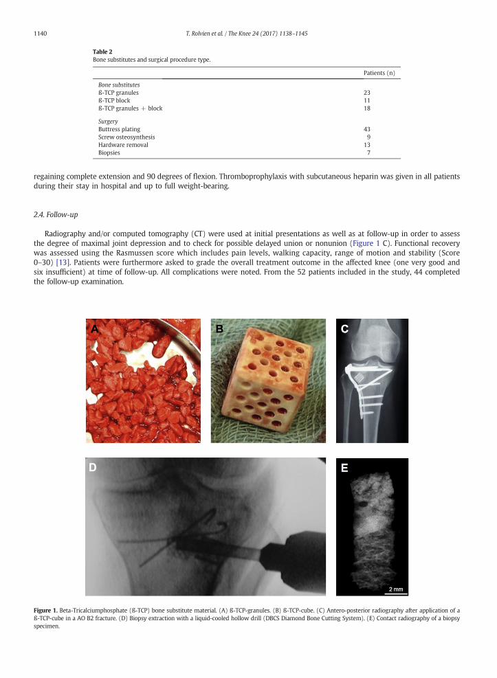
Table 2Bone substitutes and surgical procedure type.
Patients (n)
Bone substitutesß-TCP granules 23ß-TCP block 11ß-TCP granules + block 18
SurgeryButtress plating 43Screw osteosynthesis 9Hardware removal 13Biopsies 7
1140 T. Rolvien et al. / The Knee 24 (2017) 1138–1145
regaining complete extension and 90 degrees of flexion. Thromboprophylaxis with subcutaneous heparin was given in all patientsduring their stay in hospital and up to full weight-bearing.
2.4. Follow-up
Radiography and/or computed tomography (CT) were used at initial presentations as well as at follow-up in order to assessthe degree of maximal joint depression and to check for possible delayed union or nonunion (Figure 1 C). Functional recoverywas assessed using the Rasmussen score which includes pain levels, walking capacity, range of motion and stability (Score0–30) [13]. Patients were furthermore asked to grade the overall treatment outcome in the affected knee (one very good andsix insufficient) at time of follow-up. All complications were noted. From the 52 patients included in the study, 44 completedthe follow-up examination.
Figure 1. Beta-Tricalciumphosphate (ß-TCP) bone substitute material. (A) ß-TCP-granules. (B) ß-TCP-cube. (C) Antero-posterior radiography after application of aß-TCP-cube in a AO B2 fracture. (D) Biopsy extraction with a liquid-cooled hollow drill (DBCS Diamond Bone Cutting System). (E) Contact radiography of a biopsyspecimen.
1141T. Rolvien et al. / The Knee 24 (2017) 1138–1145
2.5. Histology and backscattered electron microscopy
Routine-fashioned cylindrical biopsies were taken with a liquid-cooled hollow drill (DBCS-Diamond Bone Cutting System,Biomet Merck, Darmstadt, Germany) during hardware removal in seven patients (Figure 1 D). Earliest time of hardware removalwas performed 12 months postoperatively. The extraction point was positioned under fluoroscopic guidance towards the centerof the ß-TCP implantation site. The biopsies were approximately 10 mm in length and five millimeters in diameter (Figure 1 E).For undecalcified preparation specimens were fixed in 3.7% formaldehyde for three days, dehydrated, embedded in methyl meth-acrylate, and cut on a Microtec rotation microtome (CVT 4060E, Micro Tec, Walldorf, Germany). Subsequently, the 5-μm sectionswere stained by toluidine blue [14]. Morphological characteristics of the biopsies were analyzed with emphasis on the outcome oftissue-biomaterial interaction using a Zeiss microscope (Carl Zeiss Vision GmbH, Jena, Germany) as well as backscattered electronmicroscopy (LEO 435 VP; LEO Electron Microscopy Ltd., Cambridge, England) as described before by our group [15].
3. Results
Primary trauma mechanisms were falls (34%), car collision (20%), followed by bicycle accident (14%), motorbike accident(11%) and knee distortion 9%. With respect to local swelling, surgery was performed 8 ± 3 days after trauma. Mean durationof stay at the hospital was 18 ± 7 days.
3.1. Complications
There were no delayed or nonunions and no implant breakages. Early complications were seen in seven patients (15.9%).Among these seven cases, superficial wound infections were treated with antibiotics in three cases. Four patients underwent re-vision surgery, one patient with hematoma, one with intra-articular ß-TCP penetration, one with secondary loss of reduction, andone with loosening of the osteosynthesis (Table 3).
3.2. Clinical outcome
Eight patients had to be excluded at follow-up since they didn't show up to the arranged appointment. The remaining 44 pa-tients were clinically and radiologically examined after a mean follow-up time of 36 months (14 to 64 months). The mean Ras-mussen score was 25.6 ± 2.4 points and ranging from nine to 30 points indicating the good clinical outcome (Table 3). Patient'ssatisfaction score was not different with regard to age or fracture subtype (i.e., type B fracture 2.5; type C fracture 2.6) or agegroup. Four patients with a type B fracture presented with a lack of full extension up to five degrees and one patient up to10°. Except one patient with a nearly stiff knee-joint, patients with type C fracture had no problems of knee extension at all. Aknee-flexion of more than 90° was reached in 84% of the patients with a type B fracture (16% 61 to 90°; 37% 90 to 120°,47% N 120°), and 79% of the patients with a type C fracture (11% 61°–90°; 22% 90 to 120°; 57% N 120°).
3.3. Radiographic analysis
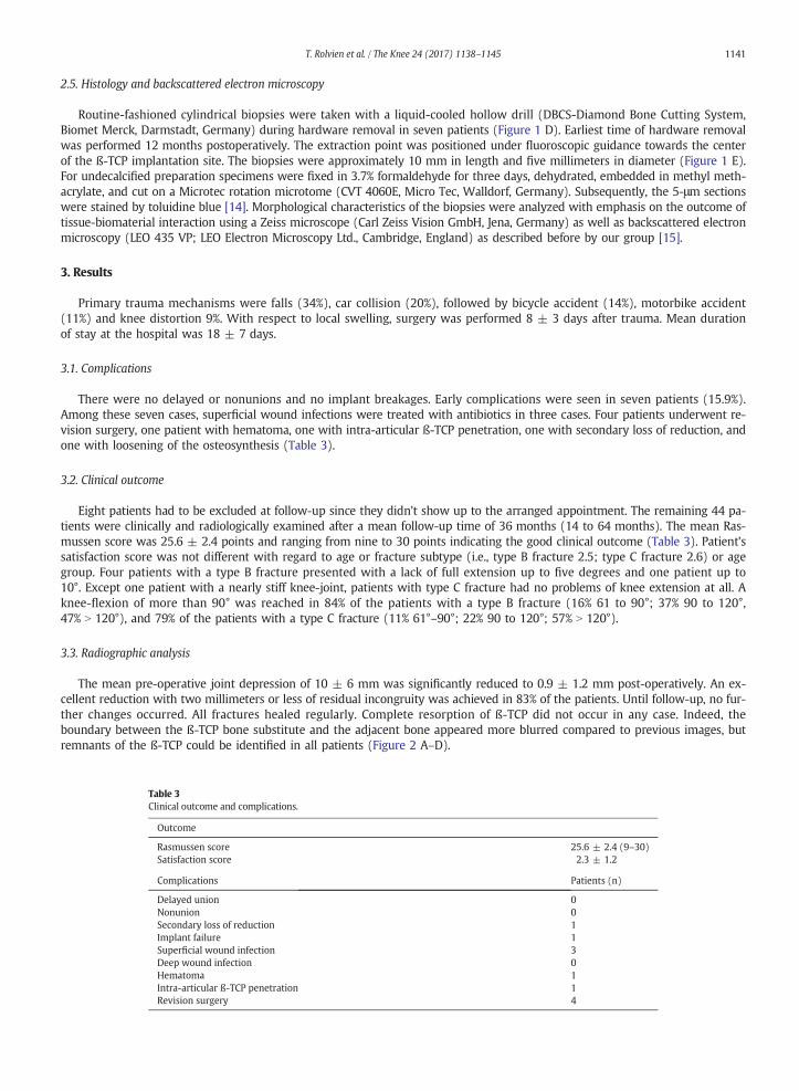
The mean pre-operative joint depression of 10 ± 6 mm was significantly reduced to 0.9 ± 1.2 mm post-operatively. An ex-cellent reduction with two millimeters or less of residual incongruity was achieved in 83% of the patients. Until follow-up, no fur-ther changes occurred. All fractures healed regularly. Complete resorption of ß-TCP did not occur in any case. Indeed, theboundary between the ß-TCP bone substitute and the adjacent bone appeared more blurred compared to previous images, butremnants of the ß-TCP could be identified in all patients (Figure 2 A–D).
Table 3Clinical outcome and complications.
Outcome
Rasmussen score 25.6 ± 2.4 (9–30)Satisfaction score 2.3 ± 1.2
Complications Patients (n)
Delayed union 0Nonunion 0Secondary loss of reduction 1Implant failure 1Superficial wound infection 3Deep wound infection 0Hematoma 1Intra-articular ß-TCP penetration 1Revision surgery 4
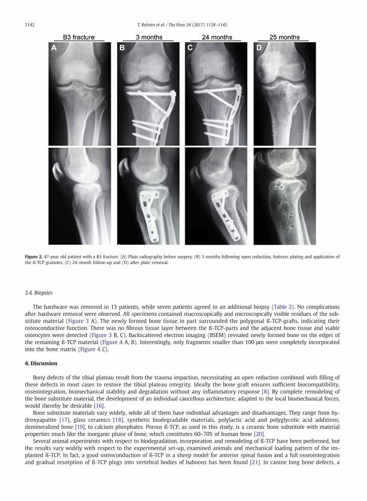
Figure 2. 47-year old patient with a B3 fracture. (A) Plain radiography before surgery. (B) 3 months following open reduction, buttress plating and application ofthe ß-TCP granules, (C) 24 month follow-up and (D) after plate removal.
1142 T. Rolvien et al. / The Knee 24 (2017) 1138–1145
3.4. Biopsies
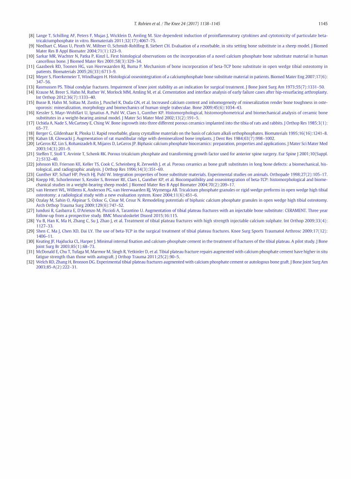
The hardware was removed in 13 patients, while seven patients agreed to an additional biopsy (Table 2). No complicationsafter hardware removal were observed. All specimens contained macroscopically and microscopically visible residues of the sub-stitute material (Figure 3 A). The newly formed bone tissue in part surrounded the polygonal ß-TCP-grafts, indicating theirosteoconductive function. There was no fibrous tissue layer between the ß-TCP-parts and the adjacent bone tissue and viableosteocytes were detected (Figure 3 B, C). Backscattered electron imaging (BSEM) revealed newly formed bone on the edges ofthe remaining ß-TCP material (Figure 4 A, B). Interestingly, only fragments smaller than 100 μm were completely incorporatedinto the bone matrix (Figure 4 C).
4. Discussion
Bony defects of the tibial plateau result from the trauma impaction, necessitating an open reduction combined with filling ofthese defects in most cases to restore the tibial plateau integrity. Ideally the bone graft ensures sufficient biocompatibility,osseointegration, biomechanical stability and degradation without any inflammatory response [8]. By complete remodeling ofthe bone substitute material, the development of an individual cancellous architecture, adapted to the local biomechanical forces,would thereby be desirable [16].
Bone substitute materials vary widely, while all of them have individual advantages and disadvantages. They range from hy-droxyapatite [17], glass ceramics [18], synthetic biodegradable materials, polylactic acid and polyglycolic acid additions,demineralized bone [19], to calcium phosphates. Porous ß-TCP, as used in this study, is a ceramic bone substitute with materialproperties much like the inorganic phase of bone, which constitutes 60–70% of human bone [20].
Several animal experiments with respect to biodegradation, incorporation and remodeling of ß-TCP have been performed, butthe results vary widely with respect to the experimental set-up, examined animals and mechanical loading pattern of the im-planted ß-TCP. In fact, a good osteoconduction of ß-TCP in a sheep model for anterior spinal fusion and a full osseointegrationand gradual resorption of ß-TCP plugs into vertebral bodies of baboons has been found [21]. In canine long bone defects, a
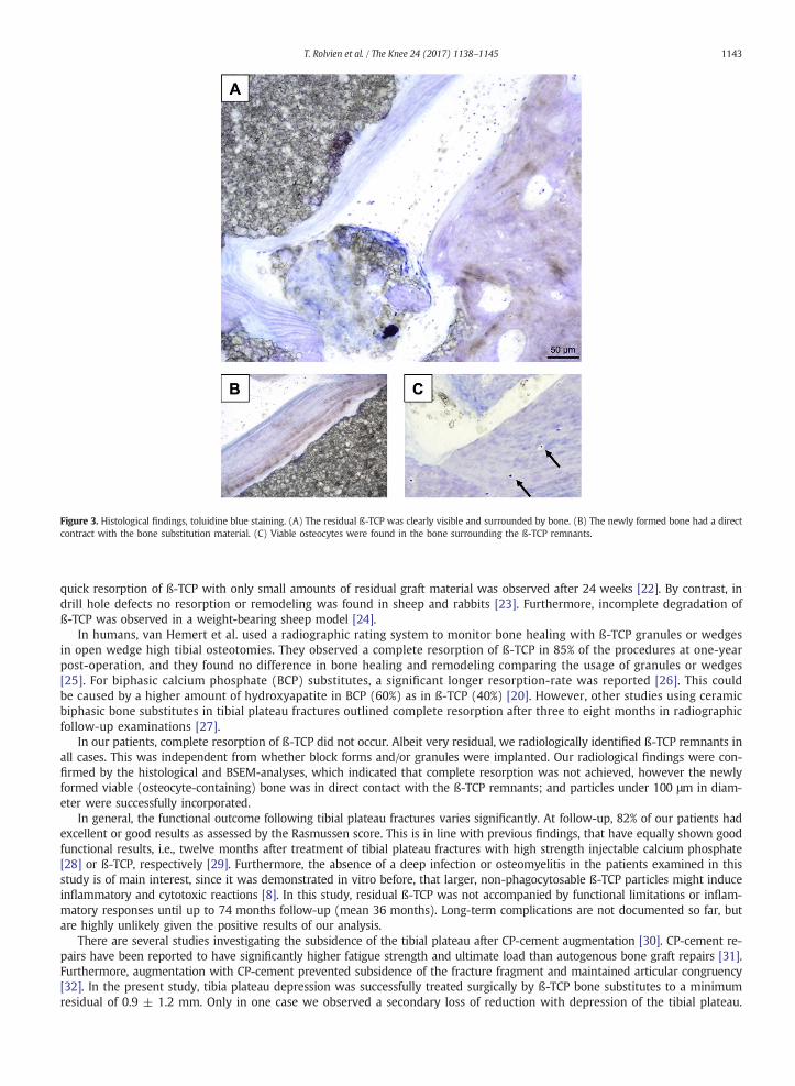
Figure 3. Histological findings, toluidine blue staining. (A) The residual ß-TCP was clearly visible and surrounded by bone. (B) The newly formed bone had a directcontract with the bone substitution material. (C) Viable osteocytes were found in the bone surrounding the ß-TCP remnants.
1143T. Rolvien et al. / The Knee 24 (2017) 1138–1145
quick resorption of ß-TCP with only small amounts of residual graft material was observed after 24 weeks [22]. By contrast, indrill hole defects no resorption or remodeling was found in sheep and rabbits [23]. Furthermore, incomplete degradation ofß-TCP was observed in a weight-bearing sheep model [24].
In humans, van Hemert et al. used a radiographic rating system to monitor bone healing with ß-TCP granules or wedgesin open wedge high tibial osteotomies. They observed a complete resorption of ß-TCP in 85% of the procedures at one-yearpost-operation, and they found no difference in bone healing and remodeling comparing the usage of granules or wedges[25]. For biphasic calcium phosphate (BCP) substitutes, a significant longer resorption-rate was reported [26]. This couldbe caused by a higher amount of hydroxyapatite in BCP (60%) as in ß-TCP (40%) [20]. However, other studies using ceramicbiphasic bone substitutes in tibial plateau fractures outlined complete resorption after three to eight months in radiographicfollow-up examinations [27].
In our patients, complete resorption of ß-TCP did not occur. Albeit very residual, we radiologically identified ß-TCP remnants inall cases. This was independent from whether block forms and/or granules were implanted. Our radiological findings were con-firmed by the histological and BSEM-analyses, which indicated that complete resorption was not achieved, however the newlyformed viable (osteocyte-containing) bone was in direct contact with the ß-TCP remnants; and particles under 100 μm in diam-eter were successfully incorporated.
In general, the functional outcome following tibial plateau fractures varies significantly. At follow-up, 82% of our patients hadexcellent or good results as assessed by the Rasmussen score. This is in line with previous findings, that have equally shown goodfunctional results, i.e., twelve months after treatment of tibial plateau fractures with high strength injectable calcium phosphate[28] or ß-TCP, respectively [29]. Furthermore, the absence of a deep infection or osteomyelitis in the patients examined in thisstudy is of main interest, since it was demonstrated in vitro before, that larger, non-phagocytosable ß-TCP particles might induceinflammatory and cytotoxic reactions [8]. In this study, residual ß-TCP was not accompanied by functional limitations or inflam-matory responses until up to 74 months follow-up (mean 36 months). Long-term complications are not documented so far, butare highly unlikely given the positive results of our analysis.
There are several studies investigating the subsidence of the tibial plateau after CP-cement augmentation [30]. CP-cement re-pairs have been reported to have significantly higher fatigue strength and ultimate load than autogenous bone graft repairs [31].Furthermore, augmentation with CP-cement prevented subsidence of the fracture fragment and maintained articular congruency[32]. In the present study, tibia plateau depression was successfully treated surgically by ß-TCP bone substitutes to a minimumresidual of 0.9 ± 1.2 mm. Only in one case we observed a secondary loss of reduction with depression of the tibial plateau.
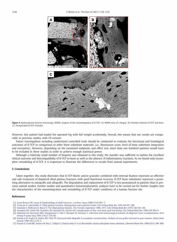
Figure 4. Backscattered electron microscopy (BSEM) analysis of the osseointegration of ß-TCP. (A) BSEM-view of a biopsy. (B) Interface between ß-TCP and bone.(C) Incorporated ß-TCP remnant.
1144 T. Rolvien et al. / The Knee 24 (2017) 1138–1145
However, this patient had loaded the operated leg with full weight accidentally. Overall, this means that our results are compa-rable to previous studies with CP-cement.
Future investigations including randomized controlled trials should be conducted to evaluate the functional and histologicaloutcomes of ß-TCP in comparison to other bone substitute materials (i.e., Rasmussen score, level of bone substitute integrationand resorption). However, depending on the examined endpoints and effect size, more than one hundred patients would haveto be included in these studies in order to achieve enough statistical power.
Although a relatively small number of biopsies was obtained in this study, the number was sufficient to outline the excellentclinical outcome and biocompatibility of ß-TCP to bone as well as the absence of inflammatory reactions. As we found only incom-plete remodeling of ß-TCP, it is important to illustrate the differences to results from animal experiments.
5. Conclusions
Taken together, this study illustrates that ß-TCP blocks and/or granules combined with internal fixation represent an effectiveand safe treatment of displaced tibial plateau fractures with good functional recovery. ß-TCP bone substitutes represent a prom-ising alternative to autografts and allografts. The degradation and replacement of ß-TCP is less pronounced in patients than in pre-vious animal studies. Further studies and quantitative histomorphometric analyses have to be carried out for further insights intothe characteristics of the osseointegration and remodeling of ß-TCP under conditions of a human fracture site.
References
[1] Court-Brown CM, Caesar B. Epidemiology of adult fractures: a review. Injury 2006;37(8):691–7.[2] Tscherne H, Lobenhoffer P. Tibial plateau fractures. Management and expected results. Clin Orthop Relat Res 1993;292:87–100.[3] Schatzker J, McBroom R, Bruce D. The tibial plateau fracture. The Toronto experience 1968–1975. Clin Orthop Relat Res 1979(138):94–104.[4] Arrington ED, Smith WJ, Chambers HG, Bucknell AL, Davino NA. Complications of iliac crest bone graft harvesting. Clin Orthop Relat Res 1996;329:300–9.[5] Hofmann GO, Kirschner MH, Wangemann T, Falk C, Mempel W, Hammer C. Infections and immunological hazards of allogeneic bone transplantation. Arch
Orthop Trauma Surg 1995;114(3):159–66.[6] Hooten Jr JP, Engh CA, Heekin RD, Vinh TN. Structural bulk allografts in acetabular reconstruction. Analysis of two grafts retrieved at post-mortem. J Bone Joint
Surg Br 1996;78(2):270–5.[7] Knaack D, Goad ME, Aiolova M, Rey C, Tofighi A, Chakravarthy P, et al. Resorbable calcium phosphate bone substitute. J Biomed Mater Res 1998;43(4):399–409.
1145T. Rolvien et al. / The Knee 24 (2017) 1138–1145
[8] Lange T, Schilling AF, Peters F, Mujas J, Wicklein D, Amling M. Size dependent induction of proinflammatory cytokines and cytotoxicity of particulate beta-tricalciumphosphate in vitro. Biomaterials 2011;32(17):4067–75.
[9] Niedhart C, Maus U, Piroth W, Miltner O, Schmidt-Rohlfing B, Siebert CH. Evaluation of a resorbable, in situ setting bone substitute in a sheep model. J BiomedMater Res B Appl Biomater 2004;71(1):123–9.
[10] Sarkar MR, Wachter N, Patka P, Kinzl L. First histological observations on the incorporation of a novel calcium phosphate bone substitute material in humancancellous bone. J Biomed Mater Res 2001;58(3):329–34.
[11] Gaasbeek RD, Toonen HG, van Heerwaarden RJ, Buma P. Mechanism of bone incorporation of beta-TCP bone substitute in open wedge tibial osteotomy inpatients. Biomaterials 2005;26(33):6713–9.
[12] Meyer S, Floerkemeier T, Windhagen H. Histological osseointegration of a calciumphosphate bone substitute material in patients. BiomedMater Eng 2007;17(6):347–56.
[13] Rasmussen PS. Tibial condylar fractures. Impairment of knee joint stability as an indication for surgical treatment. J Bone Joint Surg Am 1973;55(7):1331–50.[14] Krause M, Breer S, Hahn M, Ruther W, Morlock MM, Amling M, et al. Cementation and interface analysis of early failure cases after hip-resurfacing arthroplasty.
Int Orthop 2012;36(7):1333–40.[15] Busse B, Hahn M, Soltau M, Zustin J, Puschel K, Duda GN, et al. Increased calcium content and inhomogeneity of mineralization render bone toughness in oste-
oporosis: mineralization, morphology and biomechanics of human single trabeculae. Bone 2009;45(6):1034–43.[16] Kessler S, Mayr-Wohlfart U, Ignatius A, Puhl W, Claes L, Gunther KP. Histomorphological, histomorphometrical and biomechanical analysis of ceramic bone
substitutes in a weight-bearing animal model. J Mater Sci Mater Med 2002;13(2):191–5.[17] Uchida A, Nade S, McCartney E, ChingW. Bone ingrowth into three different porous ceramics implanted into the tibia of rats and rabbits. J Orthop Res 1985;3(1):
65–77.[18] Berger G, Gildenhaar R, Ploska U. Rapid resorbable, glassy crystalline materials on the basis of calcium alkali orthophosphates. Biomaterials 1995;16(16):1241–8.[19] Kaban LB, Glowacki J. Augmentation of rat mandibular ridge with demineralized bone implants. J Dent Res 1984;63(7):998–1002.[20] LeGeros RZ, Lin S, Rohanizadeh R, Mijares D, LeGeros JP. Biphasic calcium phosphate bioceramics: preparation, properties and applications. J Mater Sci Mater Med
2003;14(3):201–9.[21] Steffen T, Stoll T, Arvinte T, Schenk RK. Porous tricalcium phosphate and transforming growth factor used for anterior spine surgery. Eur Spine J 2001;10(Suppl.
2):S132–40.[22] Johnson KD, Frierson KE, Keller TS, Cook C, Scheinberg R, Zerwekh J, et al. Porous ceramics as bone graft substitutes in long bone defects: a biomechanical, his-
tological, and radiographic analysis. J Orthop Res 1996;14(3):351–69.[23] Gunther KP, Scharf HP, Pesch HJ, Puhl W. Integration properties of bone substitute materials. Experimental studies on animals. Orthopade 1998;27(2):105–17.[24] Koepp HE, Schorlemmer S, Kessler S, Brenner RE, Claes L, Gunther KP, et al. Biocompatibility and osseointegration of beta-TCP: histomorphological and biome-
chanical studies in a weight-bearing sheep model. J Biomed Mater Res B Appl Biomater 2004;70(2):209–17.[25] van Hemert WL, Willems K, Anderson PG, van Heerwaarden RJ, Wymenga AB. Tricalcium phosphate granules or rigid wedge preforms in open wedge high tibial
osteotomy: a radiological study with a new evaluation system. Knee 2004;11(6):451–6.[26] Ozalay M, Sahin O, Akpinar S, Ozkoc G, Cinar M, Cesur N. Remodeling potentials of biphasic calcium phosphate granules in open wedge high tibial osteotomy.
Arch Orthop Trauma Surg 2009;129(6):747–52.[27] Iundusi R, Gasbarra E, D'Arienzo M, Piccioli A, Tarantino U. Augmentation of tibial plateau fractures with an injectable bone substitute: CERAMENT. Three year
follow-up from a prospective study. BMC Musculoskelet Disord 2015;16:115.[28] Yu B, Han K, Ma H, Zhang C, Su J, Zhao J, et al. Treatment of tibial plateau fractures with high strength injectable calcium sulphate. Int Orthop 2009;33(4):
1127–33.[29] Shen C, Ma J, Chen XD, Dai LY. The use of beta-TCP in the surgical treatment of tibial plateau fractures. Knee Surg Sports Traumatol Arthrosc 2009;17(12):
1406–11.[30] Keating JF, Hajducka CL, Harper J. Minimal internal fixation and calcium-phosphate cement in the treatment of fractures of the tibial plateau. A pilot study. J Bone
Joint Surg Br 2003;85(1):68–73.[31] McDonald E, Chu T, TufagaM,MarmorM, Singh R, Yetkinler D, et al. Tibial plateau fracture repairs augmented with calcium phosphate cement have higher in situ
fatigue strength than those with autograft. J Orthop Trauma 2011;25(2):90–5.[32] Welch RD, Zhang H, Bronson DG. Experimental tibial plateau fractures augmentedwith calcium phosphate cement or autologous bone graft. J Bone Joint Surg Am
2003;85-A(2):222–31.