© timothy edward andrews 2013
TRANSCRIPT

Enhancing the Potency and Water Solubility of the Sesquiterpene Lactone
Parthenolide
A Thesis SUBMITTED TO THE FACULTY OF
UNIVERSITY OF MINNESOTA BY
Timothy Edward Andrews
IN PARTIAL FULFILLMENT OF THE REQUIREMENTS FOR THE DEGREE OF MASTER OF SCIENCE
Dr. Daniel A. Harki, Advisor
March, 2013

© Timothy Edward Andrews 2013

i
Acknowledgements
First and foremost I’d like to thank my advisor Dr. Daniel A Harki for absolutely
everything he has done for me. I would not be half the scientist that I am today without
him.
Additionally, I’d like to thank the entirety of the Harki lab. You are an amazing
group of people and help foster an environment for not only scientific discussion but also
camaraderie. I will truly miss seeing you guys every day and I wish you all the best.
I’d also like to extend my thanks to the Department of Medicinal Chemistry for
taking me on and believing in me and my potential.
Finally, I’d like to thank my friends and family for their never ending support. I
wouldn’t be where I am without you.

ii
Dedication
To Myron Eugene Andrews,
who inspired me to become a chemist.
May you rest in peace.
iii
Table of Contents
Acknowledgements .............................................................................................................. i
Dedication ........................................................................................................................... ii
Table of Contents ............................................................................................................... iii
List of Figures ................................................................................................................... vii
List of Schemes .................................................................................................................. ix
List of Tables ...................................................................................................................... x
Preface............................................................................................................................... xii
Chapter 1. Cell surface markers of cancer stem cells: diagnostic macromolecules and
targets for drug delivery ..................................................................................................... 1
1.1 Introduction ............................................................................................................ 1
1.2 Cancer Stem Cells Found in Liquid Tumors .......................................................... 6
1.2.1 Leukemia ............................................................................................................. 6
1.2.1.1 Acute Myeloid Leukemia ................................................................................. 6
1.2.1.2 Acute Lymphoblastic Leukemia ...................................................................... 9
1.2.1.3 Chronic Myelogenous leukemia..................................................................... 12
1.3 Cancer Stem Cells Found in Solid Tumors .......................................................... 12
1.3.1 Breast Cancer .................................................................................................... 12
1.3.2 Colorectal Cancer .............................................................................................. 15
1.3.3 Liver Cancer ...................................................................................................... 16
1.3.4 Pancreatic Cancer .............................................................................................. 19
1.3.5 Melanoma .......................................................................................................... 21

iv
1.3.6 Brain Cancer ...................................................................................................... 22
1.3.7 Lung Cancer ...................................................................................................... 25
1.3.8 Bladder Cancer .................................................................................................. 26
1.3.9 Prostate Cancer .................................................................................................. 28
1.3.10 Ovarian Cancer ................................................................................................ 30
1.4 The Chemical Biology and Drug Targeting of Cell Surface Markers on Cancer
Stem Cells ................................................................................................................... 33
1.4.1 CD133 ............................................................................................................... 34
1.4.2 Epithelial Cell Adhesion Molecules .................................................................. 37
1.4.3 CD44 ................................................................................................................. 40
1.4.4 CD33 ................................................................................................................. 42
1.4.5 CD34 ................................................................................................................. 43
1.4.6 ABCB5 .............................................................................................................. 45
1.4.7 CD13 ................................................................................................................. 45
1.4.8 CD123 ............................................................................................................... 46
1.4.9 CD47 ................................................................................................................. 47
1.5 Summary and Conclusion .................................................................................... 48
Chapter 2. Development of Novel Parthenolide Analogues and Evaluation of their
Anticancer Activities ........................................................................................................ 52
2.1 Introduction .......................................................................................................... 52
2.2 Research Objectives ............................................................................................. 56
v
2.3 Results and Discussion ......................................................................................... 57
2.3.1 Synthesis of a Parthenolide Analogue Library .................................................. 57
2.3.2 Biological Evaluation of Parthenolide Analogues ............................................ 62
2.3.2.1 Biological Evaluation of Parthenolide, Costunolide, and Reduced
Parthenolide. ............................................................................................................... 64
2.3.2.2 Biological Evaluation of C1-C10 Olefin Modified Parthenolide Analogues. 66
2.3.2.3 Biological Evaluation of Oxidative Modified Parthenolide Analogues. ........ 69
2.3.2.4 Biological Evaluation of Rearranged Guaianolide Parthenolide Analogues. 72
2.3.2.5 Biological Evaluation of Dimethylamino Fumarate Parthenolide Analogues.
.................................................................................................................................... 74
2.4 Conclusion and Future Direction ......................................................................... 78
2.5 Experimental ........................................................................................................ 79
2.5.1 General .............................................................................................................. 79
2.5.2 Synthesis of Parthenolide Analogues ................................................................ 80
2.5.3 Protocol for Mammalian Cell Culture ............................................................... 89
2.5.4 Protocol for Cell Culture Cytotoxicity Assays .................................................. 90
2.6 NMR ..................................................................................................................... 92
Chapter 3. Raising the Effective Intracellular Concentration of Parthenolide Utilizing a
Gold Nanoparticle Delivery System. .............................................................................. 108
3.1 Introduction ........................................................................................................ 108
3.2 Research Objectives ........................................................................................... 110

vi
3.3 Results and discussion ........................................................................................ 111
3.3.1 Synthesis of 2 nm Gold Nanoparticles ............................................................ 111
3.3.2 Solution Phase Functionalization of Gold Nanoparticles ............................... 112
3.3.3 Solid Phase Synthesis of DMAPT Linker for Gold Nanoparticle
Functionalization ...................................................................................................... 115
3.4 Conclusion and Future Direction ....................................................................... 116
3.5 Experimental ...................................................................................................... 117
3.5.1 Synthesis of 2 nm Gold Nanoparticles ............................................................ 118
3.5.2 Synthesis of Parthenolide Functionalized Gold Nanoparticles in Solution .... 118
3.5.3 Solid Phase Synthesis of DMAPT Linker 17 .................................................. 119
3.6 NMR ................................................................................................................... 121
Bibliography ................................................................................................................... 124
Appendix A: Crystal Structure of Parthenolide Analogue 4 .......................................... 149

vii
List of Figures
Figure 1.1 Depiction of the properties that are characteristic of cancer stem cells
(CSCs). A. CSCs (shown in gray) can self-renew and undergo multi-
lineage differentiation (cells in color). B. Tumorigenicity. CSCs are
more tumorigenic than differentiated bulk tumor cells......................... 3
Figure 2.1 Structure of the three sesquiterpene lactones that have gone to clinical
trials: thapsigargin, artemisinin, and the dimethylamino adduct of
parthenolide......................................................................................... 53
Figure 2.2 Previous SAR studies of the exocyclic methylene of parthenolide. A.
Modification of the exocyclic methylene via a Michael addition of
primary and secondary amines. Neelakantan et al. focused on aliphatic
R groups, while Nasim et al. focused on armotic R groups. B.
Palladium-catawlyzed arylation with aryl iodides to yield R-
alkylidene-γ-butyrolactones by Han et al. .......................................... 55
Figure 2.3 Functional group modifications investigated for their contributions to
the anticancer activity of parthenolide (1, PTL). ............................... 56
Figure 2.4 Synthesized library of parthenolide analogues .................................... 57
Figure 2.5 Synthesized rearranged guaianolide analogues of parthenolide. ......... 61
Figure 3.1 Proposed linkers for conjugation of PTL to AuNP. Conjugation of PTL
via ester linker 15, a nonfunctional control linker 16, and a DMAPT
inspired linker 17. N ranges from 36-55 PEG units CTPEG2000. ...... 111
viii
Figure 3.2 Transition electron microscopy image of synthesized 2 nm gold
nanoparticles. .................................................................................... 112
Figure 3.3 Full spectrum UV-Vis absorption of functionalized (18) and non-
functionalized gold nanoparticles. A reduction of absorbance at the
wavelength max of 15 nm AuNP (λ = 520 nm) was observed upon
functionalizing with thiols. ............................................................... 114
Figure 3.4 Transition electron microscopy image of 15 nm AuNP after
funtionalization with carboxyl thiol PEG (18). Core particle size was
15 nm. ............................................................................................... 114

ix
List of Schemes
Scheme 2.1 Synthesis of exocyclic olefin modified analogues 1a, 2a, and 3 ........ 58
Scheme 2.2 Synthesis of C1-C10 olefin modified PTL analogues. A.
Hydrogenated C1-C10 diastereomers 4 and 5. B. Isomerized Z-isomer
6. ........................................................................................................ 59
Scheme 2.3 Synthesis of melampomagnolide analogues of PTL.
Melampomagnolide B (7) was synthesized as previously described. . 61
Scheme 2.4 Synthesis of fumarate salt analogues of PTL dimethylamino prodrugs
............................................................................................................. 62
Scheme 3.1 Solution phase synthesis of PTL functionalized AuNP. A. Thiol
functionalization of AuNP. B. Attempted Mitsunobu conditions to
synthesize melampomagnolide B (Mel B) functionalized AuNP. C.
Attempted synthesis of non-functional AuNP. ............................... 113
Scheme 3.2 Solid phase synthesis of dimethylamino analogue of PTL (DMAPT)
PEG linker 17. N ranges from 36-55 PEG units CTPEG2000.. ....... 116

x
List of Tables
Table 1.1 Previously reported CSC markers for acute myelogenous leukemia
(AML). ..................................................................................................... 9
Table 1.2 Previously reported CSC markers for acute lymphocytic leukemia
(ALL). .................................................................................................... 11
Table 1.3 Previously reported CSC markers for chronic myelogenous leukemia
(CML)... ................................................................................................. 12
Table 1.4 Previously reported CSC markers for breast cancer. .............................. 15
Table 1.5 Previously reported CSC markers for colorectal cancer. ........................ 16
Table 1.6 Previously reported CSC markers for liver cancer. ................................ 19
Table 1.7 Previously reported CSC markers for pancreatic cancer. ....................... 20
Table 1.8 Previously reported CSC markers for melanoma cancer. ....................... 22
Table 1.9 Previously reported CSC markers for brain cancer. ............................... 25
Table 1.10 Previously reported CSC markers for lung cancer. ................................ 26
Table 1.11 Previously reported CSC markers for bladder cancer.. .......................... 28
Table 1.12 Previously reported CSC markers for prostate cancer.. .......................... 30
Table 1.13 Previously reported CSC markers for ovarian cancer. ........................... 33
Table 1.14 Targeting of CSC populations by antibodies and small molecules. ....... 35
Table 1.15 Alternative names of CSC surface markers that appear in this chapter.. 49
Table 2.1 Half maximal inhibitory concentration (IC50 in µM) of parthenolide (1),
costunolide (2), and reduced parthenolide (3) in nine different cancer
cell lines... .............................................................................................. 65

xi
Table 2.2 Half maximal inhibitory concentration (IC50 in µM) of C1-C10 olefin
modified parthenolide analogues 4 – 6 in nine different cancer cell
lines. ........................................................................................................ 68
Table 2.3 Half maximal inhibitory concentration (IC50 in µM) of oxidative modified
parthenolide analogues 7 and 8 in nine different cancer cell lines.. ........ 71
Table 2.4 Half maximal inhibitory concentration (IC50 in µM) of rearranged
guaianolide analogues 9 and 8 in nine different cancer cell lines.. ......... 73
Table 2.5 Half maximal inhibitory concentration (IC50 in µM) of dimethylamino
fumarate salt compounds 11 – 14 in nine different cancer cell lines... .... 76
xii
Preface
Please Note the following:
Chapter 1. This chapter is a review article co-authored with Dan Wang and Dr. Daniel A.
Harki. Drug Delivery and Translational Research. 2013, 3, 121-141 DOI:
10.1007/s13346-012-0075-1
Chapter 2: Biological testing of parthenolide analogues was assisted by the following
Harki lab members: Joe Hexum, Dan Wang, Fred Meece, Margaret Olson, Matt
Bockman, Erick Carlson, and Ezra Menon. Rearranged parthenolide analogues 9, 10 and
14 were synthesized and characterized by Dan Wang. Crystal structure of PTL analogue
4 was solved by Vic Young (UMN Department of Chemistry) from a crystal grown by
Fred Meece

1
Chapter 1. Cell surface markers of cancer stem cells: diagnostic macromolecules
and targets for drug delivery
1.1 Introduction
Cancer stem cells (CSCs), also known as tumor-propagating cells or tumor-
initiating cells, are subpopulations of undifferentiated, highly tumorigenic cells found
within bulk tumors. CSCs have the ability to self-renew, differentiate, and generate a
phenotype-identical copy of the tumor from which they were isolated upon implantation
in a recipient animal (Fig. 1). Accordingly, CSCs reside at the apex of the tumor cell
hierarchy and can differentiate into all of the cell types that comprise the host tumor.1-8
CSC theory directly challenges the traditional stochastic model of cancer cell growth
that predicts that all cells from a tumor have tumorigenic potential.9
The development of suitable technologies to isolate CSCs from tumors (e.g.,
monoclonal antibodies of unique CSC surface macromolecules coupled with
fluorescence-activated cell sorting (FACS) techniques) and appropriate animal models
for xenotransplantation assays of isolated cell populations (e.g., non-obese diabetic,
severe combined immuno-deficient (NOD/SCID) mice) have facilitated the
identification of CSC populations from a variety of blood and solid tumors. 3 These
technological advancements were prerequisite to the discovery of CSCs in acute
myelogenous leukemia (AML), which is arguably the prototypical human cancer
bearing an established CSC hierarchy. Seminal work by John Dick and colleagues
provided the first critical evidence for CSCs by isolating (with FACS) the CD34+/CD38
−
fraction of human donor AML cells and engrafting those cells into severe combined

2
immunodeficient (SCID) mice.9,10
Engrafted cells were able to proliferate and
differentiate, resulting in identical disease to that of the donor. Isolation of the AML
CSC population (CD34+/CD38
−) from the recipient mouse, followed by serial
transplantation into a secondary recipient mouse yielded the same disease, thereby
demonstrating the self-renewal properties of the initially engrafted CD34+/CD38
− AML
cell fraction. This pioneering study provided insight into the long-standing observation
that AML cells have only limited proliferative capacity by supporting the hypothesis that
a rare leukemic clone must maintain the AML population.11-13
Additionally, this work
provided crucial evidence for the hierarchical model of tumor heterogeneity by
demonstrating that some populations of leukemic cells (CD34+/CD38
−) exhibited CSC
activity, whereas other leukemic cell populations (e.g., CD34+/CD38
+ and CD34
−) did
not.10
The feature that only a sub-population of cells from a tumor can facilitate
tumorigenesis is a tenant of the hierarchical model of tumor cell growth.3,10
AML CSCs
exclusively give rise to clinical AML.9,10

3
Fig. 1.1 Depiction of the properties that are characteristic of cancer stem cells
(CSCs). A. CSCs (shown in gray) can self-renew and undergo multi-lineage
differentiation (cells in color). B. Tumorigenicity. CSCs are more tumorigenic than
differentiated bulk tumor cells.

4
Cancer therapies that are resisted by the CSC population are predicted to fail by
the CSC model.6,7
Although outside the focus of this review, the drug resistance
properties of CSCs may result due to increased cell quiescence,14,15
expression of anti-
apoptotic proteins,16
and upregulation of ABC multidrug resistance transporter
proteins.16,17
The CSC hypothesis is also supported by clinical data. In the case of AML,
approximately 44,000 new cases of AML are diagnosed annually in the United States,
and 5-year survival rates are only 24%.18
Front-line small molecule treatments for AML
include nucleoside analogues (e.g., cytarabine) and anthracyclines (e.g., idarubicin,
daunorubicin), which are designed to induce apoptosis in rapidly dividing cancer
cells.19,20
AML CSCs, which are largely quiescent, are typically resistant to standard
chemotherapeutic agents. Furthermore, recent data have demonstrated that cytarabine
actually facilitates AML CSC entry into G0/G1 phase, thereby promoting cell cycle
quiescence and providing a mechanism that allows AML CSCs to survive
chemotherapy.21
Consequently, viable strategies to eradicate CSC populations are
clearly needed if curative cancer therapies are to be developed. A realized example of
this concern has been documented with patients receiving the targeted bcr-abl tyrosine
kinase antagonist imatinib (Gleevec®) for chronic myelogenous leukemia (CML).
Although imatinib effectively converts CML into a chronically managed disease,22,23
a
patient withdrawing from imatinib treatment will ultimately exhibit disease relapse
because imatinib does not eliminate CML CSCs.24,25
Elucidation of the molecular differences between differentiated cancer cells with
high proliferation rates and nondifferentiated CSCs that are mostly quiescent is an

5
intense area of ongoing research.3,26-28
Equally important is the need to characterize
those chemical features that confer cancerous versus non-cancerous stem cells (e.g., the
differences in biochemistry between a leukemic stem cell and a normal hematopoietic
stem cell).3 A variety of cytosolic and cell surface macromolecules have been
characterized as diagnostic markers for the identification of CSCs from differentiated
cancer cells, normal stem cells, or other cells from tissue. Many of these markers are
utilized in combination for characterization of the CSC population (e.g., CD34+/ CD38
−
for AML CSCs described above).10
However, for isolation and drug targeting of CSCs,
surface markers have proven to be more useful.29-32
Cell surface macromolecules are
readily captured by magnetic micro-bead isolation techniques and are easily detected by
flow cytometry, and the unique ligands presented on the cell surface offer receptors for
targeted drug delivery.33
In this chapter, we have consolidated the known CSC cell
surface markers that have been described for various blood and solid tumors into one
document. Since intracellular markers for CSCs have been described and reviewed
elsewhere,30,34
we have focused this review only on well-characterized cell surface
markers due to their ability to serve as mediators for drug delivery into CSCs. In
addition to compiling CSC surface markers, we review the unique chemical and
structural features of the most well-known CSC surface markers and report recent efforts
to deliver therapeutic agents into CSCs mediated by CSC specific cell surface
macromolecules. Compendium of cell surface markers for identification of cancer stem
cells summarized herein is the current knowledge of CSC markers from the following
human cancers: leukemias (Table 1.1-1.3), breast (Table 1.4), colorectal (Table 1.5),

6
liver (Table 1.6), pancreatic (Table 1.7), prostate (Table 1.8), melanoma (Table 1.9),
brain (Table 1.10), bladder (Table 1.11), lung (Table 1.12), and ovarian carcinomas
(Table 1.13). Recent efforts to deliver therapeutic molecules to CSCs are described in
Table 1.14. Table 1.15 provides a compendium of the other commonly used names of
the CSC markers discussed in the following sections.
1.2 Cancer Stem Cells Found in Liquid Tumors
1.2.1 Leukemia
Leukemia is a family of diseases, comprised of blood, bone marrow, or lymphoid
system cancers that are characterized by abnormal increases in white blood cell count.35
In 2011, more than 44,000 children and adults in the United States were expected to
develop some form of leukemia, and approximately 22,000 deaths were projected.18,36,37
In 1994, Dick and coworkers first characterized the acute myelogenous leukemia stem
cell population utilizing the CD34+/CD38
− cell surface marker combination.
10 This
landmark discovery catalyzed the search for CSC populations from related blood as well
as solid tumors. The repertoire of CSC markers in leukemia is complicated and variable
depending on leukemia subtype and stage of disease.38
1.2.1.1 Acute myelogenous leukemia (AML)
As has been introduced above, CD34+/CD38− (Table 1.1) is the earliest
documented stem cell marker combination in cancer and is still widely used to identify
leukemic CSCs. In a seminal study, Dick and coworkers demonstrated that a small

7
subpopulation of AML cells with the CD34+/CD38
− cell surface phenotype was capable
of producing a large number of colony-forming progenitors when engrafted to SCID
mice. On the other hand, CD34+/CD38
+ and CD34
− cell fractions did not exhibit these
properties.10
Later studies from the same group provided evidence that CD34+/CD38
−
was a common immune phenotype for leukemic CSCs in multiple AML subtypes and
also demonstrated their self-renewal potential.
9
Additional studies have further refined and developed the cell surface
phenotypes of AML. The Suthland group has shown that, in both in vitro and in vivo
models, CD34+/CD90
− (Thy-1), CD34
+/CD71
−/HLA-DR
−, and CD34
+/CD117
− (c-kit)
are unique cell surface marker phenotypes of AML CSCs.39-41
In 2000, the Jordan group
indicated that CD34+/CD38
−/CD123
+ (interleukin-3 receptor α chain) is a specific cell
surface phenotype for human AML stem cells.42
Additionally, CD123 expression has
also been reported on CSCs in CML, myelodysplastic syndrome, and systemic
mastocytosis,43
suggesting that CD123 might be a broadly applicable cell surface marker
for the development of targeted therapies against CSCs across multiple leukemias. CD33
is another important surface marker for characterizing AML CSCs. CD33 has long been
known for its extensive expression on leukemic blasts,44
but only recently has it been
reported as a marker for AML CSCs. Hauswirth et al. have shown that addition of
CD33+ to phenotype CD34
+/CD38
−/CD123
+ yields a robust marker combination for
AML CSCs that is unique to cancerous stem cells.45
Additionally, Florian et al.
demonstrated that CD34+/CD38
−/CD45
+/CD123
+ was a general marker phenotype for
AML CSCs and that additional markers, such as CD13+, CD71
−, CD33
+, CD117
+,
8
CD133+, and HLADR
−, were observed in variable combinations with the
CD34+/CD38
−/CD45
+/CD123
+ phenotype.
43 CD34
+/ CD38
−/CD123
+ CSC populations
have also been noted for their upregulation of drug efflux pumps. Compared with bulk
(differentiated) tumor cells, the upregulation of multidrug-resistance related protein 1,
breast cancer resistance protein, and lung resistance protein have been observed in AML
CSC populations.46
C-type lectin-like molecule-1 (CLL-1), which is a transmembrane
protein with a heavily N-glycosylated extracellular domain, has been found to be
exclusively expressed on AML CSCs in 92% of all studied AML cases. Isolation and
transplantation of CD34+/CD38
−/CLL
-1
+ cells generated AML blasts in NOD/SCID
mice.47
Another important marker of AML CSCs is CD96, which belongs to the
immunoglobulin superfamily. Recent studies have demonstrated that CD96 protein
might be selectively overexpressed as a CD34+/CD38
−/CD90
−/Lin−/CD96
+ phenotype
on AML CSCs as compared with the basal expression level of CD96 on normal HSCs.48
The elevated expression of several isoforms of adhesion molecule CD44 has been
identified in the interaction between hematopoietic progenitor cells and the surrounding
stromal cells (i.e., CSC niche) in primary human AML samples.49
CD44 has been
previously exploited in targeted drug delivery to AML CSCs 20
(see section “The
chemical biology and drug targeting of cell surface markers on cancer stem cells”; Table
1.14). The Ishikawa group has reported the identification of CD25 and CD32 in
conjunction with CD34+/CD38
− as potential CSC markers for AML. In their study, they
found that both CD34+/CD38
−/CD25
+ and CD34
+/CD38
−/CD32
+ leukemia cells are
enriched in quiescent, chemotherapeutic drug-resistant CSCs, are capable of initiating

9
AMLs in vivo, and are expressed at a very limited level in normal HSCs.50
Majeti et al.
also reported CD47 as a new potential target for CSC therapy of AML.
CD34+/CD38
−/CD90
−/Lin
−/CD47
+ expression was observed only in AML CSCs when
compared with their HSC counterparts.51
See Table 1.1 for a listing of AML CSC
surface markers.
Table 1.1. Previously reported CSC markers for acute myelogenous leukemia (AML).
Cancer Type Marker(s) Reference
Acute Myelogenous Leukemia CD34+/CD38
-
10
(AML) CD34+/CD90
-
39
CD34+/CD71
-/HLA
-DR
-
40
CD34+/CD117
-
41
CD34+/CD38
-/CD123
+
42,46
CD34+/CD38
-/CD45
+/CD123
+
43
CD34+/CD38
-/CD123
+/CD33
+
45
CD34+/CD38
-/CLL
-1
+
47
CD34+/CD38
-/CD90
-/Lin
-/CD96
+
48
CD44+
49
CD34+/CD38
-/CD25
+
50
CD34+/CD38
-/CD32
+
50
CD34+/CD38
-/CD90
-/Lin
-/CD47
+
51
1.2.1.2 Acute lymphoblastic leukemia
Acute lymphoblastic leukemia (ALL) is the most common childhood cancer and
is characterized by clonal proliferation of lymphoblasts.18
ALL CSCs present the same
CD34+/CD38
− cell surface marker phenotype as AML CSCs; however, the
CD34+/CD38
−/CD33
−/CD19
− phenotype has been characterized as a more robust marker
to define ALL CSCs.52
A population of ALL CSCs that are committed to lymphoid

10
differentiation has been shown to express the Philadelphia chromosome
(CD34+/CD38
−/Ph
+).
53 The Philadelphia chromosome contains a chromosomal
translocation between the break point cluster region (bcr) and the gene encoding c-Abl
(abl), which encodes for the bcr-abl tyrosine kinase.54
The presence of the Philadelphia
chromosome in ALL indicates an overall poor prognosis.55
Subsequent studies have
shown that, in CD34+/CD38
− bone marrow cells, a predominant and aberrant
CD34+/CD38
−/ CD19
+ cell population that is not present in healthy individuals carries
the Philadelphia chromosome, while the CD19− cell population in this compartment
does not.56
These results indicate that the CD34+/CD38
−/CD19
+ phenotype marrow
compartment in childhood ALL is another potential marker of ALL CSCs,56
which is
contradictory to previous findings.52
However, it has been suggested that CD19+ might
not be a broadly applicable AML CSC marker.56
ALL can be classified as T lineage (T-
ALL) or B lineage (B-ALL).57
Cox et al. have demonstrated that ALL cells capable of
long-term proliferation and differentiation into pre-B-ALL in vitro (suspension culture
assay) and in vivo (NOD/SCID transplant model) were derived from only CD34+/CD10
−
or CD34+/CD19
− sub
-fractions, indicating that the target cells for transformation in pre-
B-ALL have a more immature phenotype than the bulk ALL population.58
The same
group also discovered that CD34+/CD4
− or CD34
+/CD7
− sub-fractions of childhood T-
ALL cells were highly proliferative and capable of NOD/SCID engraftment.59
More
recently, Cox et al. reported that in childhood ALLs, the small subpopulation of
CD133+/CD19
− cells were capable of initiating and maintaining long-term in vitro
cultures of B-ALLs and engrafting serially into NOD/SCID recipient mice.60
In a recent

11
study from the Morimoto group, the stem cell characteristics of CD9+ cell populations in
B-ALL were demonstrated both in vitro and in transplantation experiments, suggesting
that CD9 is a useful positive-selection marker for the identification of CSCs in B-ALL.
Their study also showed that, in some cases, CD9+ in ALL CSCs was a more reliable
CSC marker than CD34+.61
The same group also reported the importance of CD90 (Thy-
1) and CD110 (c-Mpl) as positive-selection markers for T-ALL CSCs. In both in vitro
and in vivo assays, their results suggested that small subpopulations of CD90+/CD110
+
cells isolated from either childhood or adult ALL specimens retained the ability to self-
renew, proliferate, and differentiate 62
. See Table 1.2 for a listing of ALL CSC surface
markers.
Table 1.2. Previously reported CSC markers for acute lymphocytic leukemia (ALL).
Cancer Type Marker(s) Reference
Acute Lymphocytic Leukemia CD34+/CD38
-/CD33
-/CD19
-
52
(ALL) CD34+/CD38
-/Ph
+
53
CD34+/CD38
-/CD19
+
56
CD34+/CD10
-
58
CD34+/CD19
-
58
CD34+/CD4
-
59
CD34+/CD7
-
59
CD133+/CD19
-
60
CD9+
61
CD90+/CD110
+
62

12
1.2.1.3 Chronic myelogenous leukemia (CML)
CML is often characterized by the overproduction of mature myeloid cells. As
with ALL, CML is most closely associated with the bcr-abl chromosomal translocation,
which is present in pluripotent stem cells.38
CML CSCs are also characterized by the
CD34+/Ph
+ cell phenotype (Table 1) and are phenotypically similar to normal HSCs.
63 In
addition to its role as an AML CSC marker, CD123+ has also been characterized as a
surface marker for CML CSCs (phenotype CD34+/CD38
−/CD123
+). Queries for the
presence of other markers in CML cells bearing the CD34+/ CD38
−/CD123
+ phenotype
revealed variable positivity for CD13, CD33, CD44, and CD117 molecules;43
however,
the presence of CD117 on the surface of CML CSCs conflicts with previous reports.41
See Table 1.3 for a listing of CML CSC surface markers
Table 1.3. Previously reported CSC markers for chronic myelogenous leukemia (ALL).
Cancer Type Marker(s) Reference
Chronic Myelogenous CD34+/CD38
-/CD123
+
43
Leukemia (CML) CD34+/Ph
+
63
1.3 Cancer Stem Cells Found in Solid Tumors
1.3.1 Breast cancer
Breast cancer accounts for the deaths of more than 40,000 women each year in
the United States. Although the 5-year survival rate for breast cancer patients has
dramatically increased over the past few decades, tumor relapse and metastasis is still a
significant problem in breast cancer therapy.18,64
The evasion of breast CSCs during
therapy is a significant contributor to disease relapse.65

13
Al-Hajj et al. were the first to identify breast CSCs from whole cell population of
breast cancer specimens using the cell surface marker profile CD44+/CD24
−/low
/Lin−
(Table 2), which was the seminal report of CSCs identified from a solid tumor.66
The
marker profiles CD44+/CD24
−/low
/Lin− has been correlated with high resistance to
traditional cancer therapies,67
poor prognosis,68
and enhanced invasive properties.69
Notably, lineage (Lin) markers are a standard combination of monoclonal antibodies
including CD2, CD3, CD4, CD5, CD8, NK1.1, B220, TER-119, and Gr-1 in mice and
CD3, CD14, CD16, CD19, CD20, and CD56 in humans.70
Addition of epithelial cell
adhesion molecule (EpCAM), yielding marker combination
CD44+/CD24
−/low
/EpCAMhigh
, was later found to be a more robust surface marker
combination for the isolation of human breast CSCs.66
The multiplexing of cytoplasmic
enzyme ALDH1, membrane protein CD44, and cytokeratin has been employed to
identify putative breast CSCs and indicate poor prognosis, which is independent of
tumor size, nodal status, ER-, PR-, and HER2-status, and histological grade.71
Another important cell surface marker of human breast CSCs is Thy1 (CD90). In
an MMTV-Wnt-1 breast cancer mouse model, cancer cells with the phenotype
THY1+/CD24
+ (1–4% of tumor cell population) were identified to be highly tumorigenic
in comparison to non-THY1+/CD24
+ populations and displayed properties of CSCs.
72
The observation of CD24+ phenotype in combination with Thy1 is contradictory to
previous reports that found breast CSCs to have a CD44+/CD24
-/low
/Lin- phenotype.
66
The expression of cell surface marker CD133 is also reported to be of significance and
prognostic value for identifying CSCs in breast tumors, especially when combined with

14
CD44+.73-75
CD173 (H2) and CD174 (Lewis Y) are cell surface carbohydrate antigens
that are expressed to varying extents on different human carcinomas. Co-expression of
CD173 and CD174 with CD44 is another cell surface marker combination proposed to
identify breast CSCs.76
Similar to CD173 and CD174, CD176 is also a cell surface
carbohydrate antigen that has been reported to be co-expressed with CD44 and CD133
in breast carcinoma.77
Another study has shown that in BRCA1 (breast cancer-
associated gene 1)-mutant breast cancer cell lines, a small subpopulation of cancer cells
expressing CD24+/CD29
+ (β integrin) or CD24
+/CD49f
+ (α6 integrin) surface markers
exhibited enhanced proliferation and colony forming ability in vitro and increased tumor
generating ability in vivo. In addition, purified CD24+/CD29
+ cells exhibit self
-renewal
capability, and as low as 500 cells could reconstitute the heterogeneity of the parent
cancer cells in vivo, which is strongly indicative of a CSC population.78
Shipistin et al.
confirmed that CD201, which is also known as protein C receptor (PROCR), was
expressed on 100% of CD44+ breast cancer cells and was used as a cell surface marker
to isolate CSCs from primary invasive breast cancer tumors.79
In addition, high PROCR
expression was also reported to associate with poor prognosis in clinical patients.79
See
Table 1.4 for a listing of breast CSC surface markers.

15
Table 1.4. Previously reported CSC markers for breast cancer.
Solid Tumor Type Marker(s) Reference
Breast CD44+/CD24
-/low/Lin
-
66,69
CD44+/CD24
-/low/EpCAM
high
66
ALDH1+/CD44
+/cytokeratin
+
71
CD90+/CD24
+
72
CD133+/CD44
+
73-75
CD173+/CD174
+/CD44
+
76
CD176+/CD44
+ 77
CD176+/CD133
+
77
CD24+/CD29
+ 78
CD24+/CD49f
+ 78
CD44+/CD201 (PROCR)
+
79
1.3.2 Colorectal cancer
Dalerba et al. have isolated colorectal CSCs from specimens using cell surface
markers CD44 and EpCAM.80
When purified CD44+/EpCAM
high epithelial cells (Table
2) were injected into immunodeficient mice, the engrafted tumors yielded the
differentiated phenotype profile as well as the morphologic heterogeneity of the parent
lesions from which the CD44+/EpCAM
high epithelial cells were isolated. Furthermore,
the authors also reported CD166+ as a co-marker that could be used for validation of
colorectal CSCs in both xenografts and primary tumors, and the
CD44+/EpCAM
high/CD166
+ phenotype was consistent with poor prognosis.
80 However,
the CD44/EpCAM marker phenotypes were not the first reported colorectal CSCs. Prior
studies had first indicated that CD133 was a potential cell surface CSC marker in
primary human colorectal cancer,81,82
but more recent findings by Shmelkov et al. have
shown that both CD133+ and CD133
− populations are tumorigenic and contain tumor
-

16
initiating cells.83
In order to explain this discrepancy, Du et al. have suggested that,
unlike CD44, a surface protein that is of functional importance for the survival of CSCs,
CD133 has not been found to have any essential functions associated with the growth
and survival of colorectal CSCs.84
Although CD44+/EpCAM
high/ CD166
+ is a relatively
more reliable marker for colorectal CSCs than CD133+, some exceptions have also been
documented. Lugli et al. reported that the loss of this combined marker
(CD44+/EpCAM
high/CD166
+) was rather linked to an aggressive tumor phenotype.
85,86
Studies have also shown that CD49f, which has been reported as a CSC marker in breast
cancer,78
is expressed on colon cancer cells, and that even higher levels of CD49f
expression are observed with CD44+ cells, a known marker of colorectal CSCs.
80
Additionally, CD133+/CD24
+ has been identified as a colorectal CSC marker.
87 See
Table 1.5 for a listing of colorectal CSC surface markers.
Table 1.5. Previously reported CSC markers for colorectal cancer.
Solid Tumor Type Marker(s) Reference
Colorectal CD44+/EpCAM
high
80
CD44+/EpCAM
high/CD166
+
80
CD44+/CD49f
+
80
CD133+
81,82
CD44+
88
CD133+/CD24
+
87
1.3.3 Liver cancer
The incidence of hepatocellular carcinoma (HCC) cases has risen by 3% every
year since 1992, and the 5-year survival rate ranges from 14% to 26% depending on the

17
stage of diagnosis.18
In spite of the intensive research efforts devoted to the discovery of
new HCC drugs, tumor relapse is still observed in the majority of cases.89
The recent
identification of liver CSCs has yielded new targets for HCC drug discovery efforts,
which, if successful, may help to address the problem of disease relapse.
The CD133+ phenotype was the first reported putative HCC CSC population.
90
In this report by Ma et al. CD133+ HCC cells exhibited greater colony-forming
capacities and higher proliferation rates than CD133− HCC cells, as well as the ability to
generate new tumors in both in vitro and in vivo models. Additionally, in CD133+ tumor
cells, the expression level of stemness genes such as those involved in Wnt/β-catenin,
Notch, and Hedgehog/SMO signaling were largely upregulated, indicating those
CD133+ tumor cells were putative HCC CSCs. The CD133
+ population was further
characterized for other common CSC markers and CD34 and CD44 were found to be
upregulated when compared with the CD133− population.
90 Another well-established
cell surface marker of HCC stem cell is CD44+. Within the HCC CD133
+ population,
CD44+ cells were found to be more tumorigenic than CD44
− cells in NOD/SCID mouse
model. Recent studies also revealed that the blocking of CD44 function by treatment
with a CD44 specific monoclonal antibody might be a potential strategy to eradicate
liver CSCs91
(see section “The chemical biology and drug targeting of cell surface
markers on cancer stem cells”). Moreover, the cell surface carbohydrate antigen CD176
was found to co-express with CD44 at a high rate on the surface of HCC stem cells in
both cancer cell lines and surgical specimens of malignant tumors.77
Besides CD133 and
CD44, EpCAM was identified as an early biomarker of HCC CSCs.92,93
The sorted

18
EpCAM+ subpopulation of HCC cells yielded more colonies in clonogenicity assays
than the sorted EpCAM− cells from the same cell line. Further in vivo evaluation in
mouse models indicated that as little as 100 EpCAM+ HCC cells were needed to
generate a new tumor, and EpCAM+ cells retained the ability to differentiate into both
EpCAM+ and EpCAM
− cells, while EpCAM
− cells always sustained their phenotype
during cell propagation.92
Additionally, CD133 expression was observed in both
EpCAM+ and EpCAM
− populations, indicating that EpCAM functions as a better
indicator of the CSC population.93
Recently, CD13 (alanine aminopeptidase), which is a
membrane-bound enzyme, has been identified as a marker for semiquiescent CSCs and a
potential therapeutic target for liver cancers. CD13+/CD133
+ and CD13
+/CD90
−
populations were observed to be extremely tumorigenic, requiring only 100 cells for
tumor formation upon transplantation.94
In a xenograft mouse model, CD13 inhibition,
which was achieved with either a CD13 neutralizing antibody or by the CD13 inhibitor
Ubenimex, repressed tumor-initiating and self-renewing capability of the majority of
CSCs94
(see section “The chemical biology and drug targeting of cell surface markers on
cancer stem cells”). CD90 (Thy-1) has also been identified as a CSC marker in HCC cell
lines.95
In comparison to CD90− cells, CD90
+ cells sorted from HCC cells lines were
found to exhibit strong tumorigenic capacity, and the subpopulation of cells with
CD90+/CD45
− phenotype could generate tumor nodules in an immunodeficient mouse
model.95
OV6, which is a marker of hepatic progenitor (oval) cells, has been shown to
be another CSC marker in HCC cells. OV6+ HCC cells are capable of generating new
tumors in vivo and show substantial resistance to standard chemotherapeutic drugs

19
compared with OV6− HCC cells. Furthermore, the inhibition of the Wnt/β-catenin
signaling pathway, which is known to be important for the survival of CSCs,96
decreases
the population of OV6+ cells. The OV6
+ cell population also gained higher proliferation
potential after β-catenin activation.97
See Table 1.6 for a listing of liver CSC surface
markers.
Table 1.6. Previously reported CSC markers for liver cancer.
Solid Tumor Type Marker(s) Reference
Liver CD176+/CD44
+
77
CD133+
90
CD133+/CD44
+
91
EpCAM+
92,93
CD13+/CD133
+
94
CD13+/CD90
-
94
CD90+/CD45
-
95
OV6+
97
1.3.4 Pancreatic cancer
Pancreatic cancer is one of the deadliest forms of human cancers. Currently,
there are no known methods of early detection, and the average 5-year survival rate is
6%.18
The mortality rate of this disease approaches 100% because of its
characteristically high resistance to radiation and chemotherapy, and the tendency of
early systemic dissemination.98
In spite of the significant advances in cancer biology
over the past decade, the efficacy of drugs to treat pancreatic cancer has not substantially
improved.99
Targeting the pancreatic CSC population could allow development of more
efficacious therapies to combat this lethal disease.

20
A pancreatic CSC population was identified only recently.99
A small population
(0.2–0.8%) of highly tumorigenic pancreatic cancer cells with the
CD44+/CD24
+/EpCAM
high phenotype (Table 2) from primary human pancreatic
adenocarcinomas was xenografted into immunocompromised mice. In comparison with
non-tumorigenic cells, the CD44
+/CD24
+/EpCAM
high pancreatic cancer cells exhibited a
100-fold increase in tumorigenic potential, and as few as 100 such cells could
reconstitute tumors that were indistinguishable by histology from the original ones.99
In
related studies, Hermann et al. demonstrated that CD133+ expression on the surface of
pancreatic cancer cells was consistent with increased tumorigenicity and high resistance
to standard chemotherapy. In addition, they further revealed that a unique small
population of CSCs bearing the CD133+/ CXCR4
+ phenotype determined the metastatic
potential of the tumor. Although CD133+/CXCR4
+ cells and CD133
+/ CXCR4
− cells
showed similar tumorigenicity when xenografted into nude mice, only the
CD133+/CXCR4
− cells eliminated tumor metastasis.
100 See Table 1.7 for a listing of
pancreatic CSC surface markers.
Table 1.7. Previously reported CSC markers for pancreatic cancer.
Solid Tumor Type Marker(s) Reference
Pancreatic CD44+/CD24
+/EpCAM
+
99
CD133+
100
CD133+/CXCR4
+
100

21
1.3.5 Melanoma
Approximately 70,000 new cases of human melanoma and 9,000 melanoma-
related deaths were expected in the United States in 2011.18
Although melanoma is not a
leading cause of cancer-related mortality, it is one of few cancer types associated with
both annual increases in incidence and death rate.101
Human malignant melanoma cells
are highly aggressive and drug resistant and contain cell populations with enhanced
tumorigenicity.102
The discovery of putative CSCs in melanoma cell lines was first
reported in 2005,102
preceding their subsequent isolation and characterization from cell
lines and surgical specimens.103
Nestin (Table 2), which is an intermediate filament
protein, was first described as a potential stem cell marker in melanoma cell lines.104
Nestin expression was associated with aggressive behavior of the malignant tumors and
was found to be upregulated during the development of invasive melanoma from banal
nevi (a benign chronic lesion of the skin).104
Co-expression of nestin with CD133 has
been described in context with stem cell populations in circulating melanoma cells,
which are closely related to their metastatic potentials.105
Monzani et al. first utilized
CD133 as a cell surface marker to separate melanoma stem cells. Their data showed that
in contrast to CD133− cells, CD133
+ melanoma cells were highly tumorigenic and able
to generate a Mart-1 (a typical melocytic marker) positive tumor when implanted into
NOD/SCID mice. In the same study, they also revealed that melanoma cells expressing
both CD133 and ABCG2 (ATP-binding cassette sub-family G member 2) were able to
self-renew and differentiate into astrocytes and mesenchymal lineages under specific
conditions, demonstrating the utility of ABCG2 as a CSC marker.103
Schatton et al.

22
identified a small subpopulation of melanoma cells that were enriched for human
malignant melanoma-initiating cells, which were defined by the expression of P-
glycoprotein ABCB5, a well-known chemoresistance mediator. ABCB5+ melanoma
cells were found to be highly aggressive, possess greater tumorigenic capacity than
ABCB5− cells, and could re-establish clinical tumor heterogeneity. ABCB5 has also
been studied as a target of anticancer therapy106,107
(see section “The chemical biology
and drug targeting of cell surface markers on cancer stem cells). See Table 1.8 for a
listing of melanoma CSC surface markers.
Table 1.8. Previously reported CSC markers for skin cancer.
Solid Tumor Type Marker(s) Reference
Skin CD133+
103
CD133+/ABCG
-2
+ 103
Nestin+/CD133
+ 105
ABCB5+
107
1.3.6 Brain cancer
Malignant brain tumors are among the deadliest human cancers. Glioblastoma
(GBM) multiforme is the most common primary brain tumor in adults with 12,000
deaths in the United States annually.108
In children, glioblastoma multiforme occurs less
frequently, accounting for 7–9% of all intracranial tumors, yet the median survival after
diagnosis is 50 weeks.109
Currently, there is a scarcity of effective treatments for brain
cancers, which is attributable to the sensitive environment of the disease and the
inability of many drugs to permeabilize the blood–brain barrier. Unfortunately, most

23
drugs that can access brain malignancies are inefficient at eradicating the tumor cells and
frequently leave behind a side population of radio- and chemoresistant cells. The
existence of this side population provides a mechanism for disease relapse,33
and recent
studies strongly suggest that brain tumors have an established CSC hierarchy.110,111
Drug
targeting of brain CSCs may yield better therapy outcomes.
In 2003, Singh et al. first identified CD133 as a CSC marker (Table 2) in
glioblastomas and medulloblastomas.112
Studies of human medulloblastoma specimens
from children have also revealed a CD133+ subpopulation of cells that is positive for
nestin.113
Sorted CD133+ populations of childhood medulloblastoma specimens have
demonstrated the stem cell characteristics of self-renewal and differentiation, and
subjection of these cells to differentiation conditions resulted in the loss of the CD133+
marker.112
The tumorigenicity of CD133+ human donor medulloblastoma and
glioblastoma cancer cells were evaluated in NOD/SCID mice, and as few as 100 cells
were required for the development of new tumors of the same phenotype.113
Another glioblastoma CSC marker that has been identified is integrin A2B5, a
polysialo ganglioside. Tchoghandjian et al. used A2B5 to characterize CSC populations
in glioblastoma samples using magnetic micro-beads and FACS. In addition, the authors
sorted the cells for a secondary known CSC marker, CD133. A2B5+ populations
expressed characteristic features of CSCs regardless of CD133 expression, and both
A2B5+/CD133
+ and A2B5
+/CD133
− populations were highly tumorigenic, requiring
only 1,000 cells for development of a new tumor of the same phenotype. However, only
the A2B5+/CD133
− population of cells was found to be invasive within the brain.
114

24
Studies by Ogden et al. have suggested that only the A2B5+ phenotype is characteristic
of glioblastoma CSCs, and CD133 positivity is not indicative of a CSC population.115
In
support of the evidence that CD133+ cells are putative CSCs, studies by Wang et al.
found that CD133+ glioblastomas were aggressive tumors and were typified by the onset
of angiogenesis, shorter survival times, and metastatic potential. However, the CD133−
populations were also shown to be capable of differentiating into CD133+ cells, which
suggests that CD133− are stem cell-like.
116 Although characterization of universal
diagnostic markers for CSCs is the ultimate goal for the development of targeted
therapies, these contradictory results with CD133 clearly illustrate that patients can
express different surface markers for the same disease and/or that multiple CSC
populations can be present in glioblastoma specimens.
L1CAM, a transmembrane adhesion protein found in CD133+ glioma cell
populations, was identified as a brain CSC marker in 2008.117
D456MG pediatric
glioblastoma xenografts and primary human samples were utilized to study the role of
L1CAM.118
Neurosphere formation, a cellular morphology consistent with a stem cell-
like state, was quantified as a measure of the self-renewal properties of the CSC
population. Targeting L1CAM with shRNA decreased the ability of the CD133+ cells to
form neurospheres, indicating that the L1CAM+/CD133
+ population was the in vitro
CSC population. In vivo studies conducted in nude mice also showed that knockdown of
L1CAM with shRNA resulted in a reduction in tumor size and increased survival.117
Stage-specific embryonic antigen-1 (SSEA-1), or CD15, has also been identified
as a potential marker of GBM CSCs.119
Utilizing primary human samples, SSEA-1+
25
cells were isolated and characterized for self-renewal, differentiation, and
tumorigenicity. The presence of CD133+ was also investigated in the SSEA-1
+
populations in early studies; however, SSEA-1+/CD133
+ and SSEA-1
+/CD133
- were
both shown to have CSC characteristics. Sorting of primary human GBM specimens for
only SSEA-1 expression and xenograftment of isolated cells into NOD/SCID mice
revealed that SSEA-1+ cells exhibited at least 100-fold greater tumorigenic potential
than SSEA-1− GBM cells. Therefore, SSEA-1
+ can be used to identify CSC populations
in GBM.119
SSEA-1 has also been identified as a medulloblastoma CSC marker.120
See
Table 1.9 for a listing of brain CSC surface markers.
Table 1.9. Previously reported CSC markers for brain cancer.
Solid Tumor Type Marker(s) Reference
Brain CD133+
112
CD133+/Nestin
+
113
CD133+/A2B5
+ and CD133
-/A2B5
+
114
A2B5+
115
CD133+/L1CAM
+
117
SSEA-1
+
119
1.3.7 Lung cancer
Lung cancer is the most common, yet preventable, cancer related death
worldwide, resulting in more mortalities than colon, breast, and prostate cancers
combined. The 5-year survival rate for small cell lung carcinoma is 6% while nonsmall
cell carcinoma is 17%.18
Current lung cancer therapies are usually transient and fail to
completely eradiate the tumor, causing resistance. One potential hypothesis for drug

26
resistance to chemotherapy is the presence of drug refractory lung CSC populations in
the tumor masses.121
Eramo et al. identified CD133+ CSC subpopulations (Table 2) in
small cell and non-small cell lung cancer from primary human donors. CD133
expression was observed on the surface of 0.32–22% of lung cancer cells by flow
cytometry analyses, which varied depending on different disease stages of the sample. In
vivo analysis showed that tumor formation required only 1,000 cells to be implanted into
SCID mice, while differentiated cell populations injected at 50,000 cells per animal
failed to produce tumors.122
CD176 has also been identified as a potential marker for
lung CSC populations and has been observed in combination with CD133 and CD44 cell
surface markers. CD176+/CD133
+ and CD176
+/CD44
+ phenotypes were observed after
sorting cells for CD176 expression, and CD176+ cell populations were found to exhibit
features of CSCs, such as tumorigenicity. These results imply that CD176+ may be used
as a potential lung CSC marker.77
See Table 1.10 for a listing of lung CSC surface
markers.
Table 1.10. Previously reported CSC markers for lung cancer.
Solid Tumor Type Marker(s) Reference
Lung CD176+/CD44
+
77
CD176+/CD133
+
77
CD133+
122
1.3.8 Bladder cancer
Bladder transitional cell carcinoma (BTCC) is the second most prevalent cancer
of the urinary tract.123
Five-year survival rates for BTCC vary by stage of development,

27
ranging from 6% to 97%.18
Traditional therapy consists of surgery, radiation, and
chemotherapy,124
but if disease becomes invasive, then treatment outcomes are typically
poor due to metastasis.125
The development of diagnostic markers and new therapies for
BTCC is currently needed.
Yang and Chang have identified a subpopulation of BTCC samples from primary
human donors that display a CD44+/EMA
− (epithelial membrane antigen) phenotype
(specifically the isoform CD44v6; Table 2). CD44+/EMA
− populations were shown to
overexpress Bmi-1 and EZH2, which are proteins that play a role in self-renewal of
normal bladder stem cells. CD44+/EMA
− cells also were able to form colonies and
proliferate in vitro, which are characteristic features of CSCs.126
Chan et al. have also postulated that BTCC CSCs can be characterized by the
cell surface phenotype Lin−/CD44
+/cytokeratin (CK)5
+/CK20
−. Tumors from primary
human donors were sorted for CD44+ and CD44
− cells and then transplanted into
immunocompromised mice (RAG2−/γc
−). The CD44
+ cells were found to be more
tumorigenic, requiring only 100–1,000 cells to form tumors, while the CD44− population
required 200,000–500,000 cells to elicit tumors in mice. Immunofluorescence analysis
of CD44+ xenografts showed co
-expression of CK5 and lack of expression of CK20
(phenotype Lin−/CD44
+/CK5
+/ CK20
−), and this marker phenotype was deemed to be
the BTCC CSC population. The authors also queried for other common CSC cell surface
markers and discovered that CD47 is highly expressed on CD44+ cells. CD47 inhibits
phagocytosis by macrophages, allowing for pathogenesis of the bladder cancer.125

28
Hoechst 33342 dye is a widely used indicator of side populations with potential
CSC characteristics.127
ATPbinding cassette (ABC) and multidrug resistance efflux
pumps are responsible for the elimination of Hoechst 33342 dye, as well as cytotoxic
drugs, from cells. This process is detectable by FACS and allows for the identification
and isolation of subpopulations of cells displaying these characteristics that are common
of CSCs.127,128
ABC-G2 transporters were detected in the BTCC cell line T24. ABC-G2+
and ABC-G2− cells were cultured in vitro, and their growth characteristics were
evaluated. ABC-G2+ cells demonstrated consistent proliferation, whereas the ABC-G2
−
did not. Side populations of ABC-G2+ also were able to differentiate into ABC-G2
−
cells, which further supports the hypothesis that ABC-G2+ is a marker of BTCC
CSCs.127
See Table 1.11 for a listing of bladder CSC surface markers.
Table 1.11. Previously reported CSC markers for bladder cancer.
Solid Tumor Type Marker(s) Reference
Bladder Lin-/CD44
+/CK5
+/CK20
-
125
CD47+/CD44
+
125
CD44v6+/EMA
-
126
ABC-G2
+
127
1.3.9 Prostate cancer
Prostate cancer is the most commonly diagnosed malignancy and second leading
cause of death in men in the United States.18
More than 80% of men will develop
prostate cancer in their lifetime, and most diseases will not be diagnosed.129
Prostate
cancer is routinely treated by hormone ablation therapy, but this therapy regimen fails

29
when the disease progresses to a hormone-refractory state.
130 Currently, cytotoxic small
molecules are the only therapeutic regimen able to improve outcomes for patients with
hormone-refractory prostate cancers.131
Identification of CSC surface markers could
provide a foundation for the development of next-generation drugs that target prostate
CSCs.130
The first CSC surface marker identified for prostate cancer was CD44 (Table 2).
In 1997, Liu et al. reported their observation that CD44+ prostate cancer cells were
negative for prostate-specific antigen (PSA) and prostate acid phosphatase secretion,
which are proteins that are produced during cancer cell differentiation. On the other
hand, when CD44+ prostate cancer cells were co-cultured with stromal cells, the
secretion of PSA was detected, indicating that the interaction between stromal cells and
CD44+
prostate cancer cells stimulated the differentiation ability of the latter, since PSA
production was not detected when either stromal cells or CD44+
prostate cancer cells
when cultured alone.88
Collins et al. demonstrated that prostate CSCs could be identified
from human specimens with a CD44+/α2β1
high/ CD133
+ phenotype, and this population
comprised 0.1– 0.3% of the total amount of cells in the tumor. In a colony-forming
assay, the CD44+/α2β1
high/CD133
+ population yielded 3.7-fold greater number of
colonies compared with the total cell population and more than 30-fold greater number
of colonies compared with CD44+/α2β1
hi/CD133
− or CD44
+/α2β1
low cells, which
suggests the CD44+/α2β1
high/CD133
+ phenotype has the ability to self-renew.
28
Patrawala et al. performed in vivo studies in NOD/SCID mice utilizing prostate cancer
cell lines that were enriched for the CD44+/α2β1
high combination phenotype.
132,133 They

30
found that the CD44+/α2β1
high population had greater tumorigenicity than other
populations isolated (CD44+/α2β1
low, CD44
−/α2β1
high, CD44
−/α2β1
low), further
supporting the evidence that the CD44+/α2β1
high phenotype is the CSC population.
133
Mulholland et al. later confirmed that the Lin−/Sca-1
+/CD49f
high subpopulation of
prostate cancer cells were CSCs in a murine prostate PTEN− model (PTEN is a tumor
suppressor gene in the protein kinase B pathway).130
The loss of PTEN activity is
strongly associated with the initiation and metastasis of prostate cancer.134
Tissue
samples were taken from murine PTEN mutant prostate tumor grafts on SCID mice after
6–8 weeks, digested into single-cell suspensions, and sorted by FACS. Through a
systematic study of various potential surface markers, Sca-1 and CD49f were identified
as surface markers of prostate CSCs. Sorted cells (Lin−/Sca
-1
+/CD49f
high and Lin
−/Sca-
1+/ CD49f
low) were then injected into SCID mice and only Lin
−/Sca-1
+/CD49f
high cells
induced tumor growth.130
See Table 1.12 for a listing of prostate CSC surface markers.
Table 1.12. Previously reported CSC markers for prostate cancer.
Solid Tumor Type Marker(s) Reference
Prostate CD44+/α2β1
high/CD133
+
28
Lin-/Sca
-1
+/CD49f
high
130
CD44+/α2β1
high
133
1.3.10 Ovarian cancer
The most lethal disease of the female reproductive tract is ovarian cancer.135
High mortality rates occur due to the difficulty in diagnosing early stage ovarian cancer
as well as the high prevalence of drug-refractory relapse after initial treatment, which is

31
likely a result of CSC subpopulations that survive therapy.136
Ovarian cancer 5-year
survival rates vary depending on the stage of development of the tumor. Early stage
diagnosis, when the disease has yet to metastasize, has a 5-year survival rate of 73%,
while late-stage ovarian cancer diagnosis results in a 28% survival rate.18
Zhang et al. first identified CD44 as a potential ovarian CSC surface marker in
2008. Analysis of human ovarian specimens revealed a small population (0.14–0.2%) of
CD44+/CD117
+ cells (Table 2) that were highly tumorigenic and demonstrated self-
renewal by spheroid formation. Tumorigenicity was measured by engraftment of
CD44+/CD117
+ and CD44
−/CD117
− subpopulations into nude mice. The
CD44+/CD117
+ subpopulation was able to repopulate the tumor following injection of
only 100 cells, whereas the isolated CD44−/CD117
− phenotype required 500,000 cells
for tumor development after 3 months.135
Ferrandina et al. identified CD133+ subpopulations as ovarian CSCs in primary
human samples. The CD133+ subpopulation was 4.7-fold more active in a colony-
forming assay, which demonstrated replicative capacity.137
Work by Curley et al.
verified the presence of a CD133+ subpopulation with tumor-initiating properties and
found that 0.2– 12.5% of the tumors expressed this phenotype.138
CD24+ has also been characterized as a potential surface marker of human
ovarian CSCs. Two weeks after the plating of isolated CD24+ ovarian cancer cells,
analysis of the cell population revealed both CD24+ and CD24
− phenotypes, which
suggests that CD24+ cells are CSCs due to their ability to self-renew and differentiate.
Conversely, the same experiment performed with CD24− cells failed to yield CD24
+

32
cells after the same amount of time in culture. Additional in vivo studies demonstrated
that only 5,000 CD24+ cells were needed for tumor formation after injection into nude
mice, whereas CD24− cells injected at the same concentration were unable to generate
tumors.139
Therefore, CD24+ cells are more tumorigenic than CD24
- cells.
To add to the growing number of ovarian CSC markers, Wei et al. isolated a
triple surface marker phenotype, CD44+/CD24
+/EpCAM
+, which best characterized the
most proliferative cell subpopulation when compared with all other marker
combinations. Cells lines OVCAR-5, SKOV-3, and IGROV-1, as well as primary
human donor samples were used for experiments. Colony-formation assays were
performed on CD44+/CD24
+/EpCAM
+ sorted cells to measure proliferation rates and to
characterize self-renewal properties. In vivo studies were performed comparing
CD44+/CD24
+/EpCAM
+ and CD44
−/CD24
−/EpCAM
− phenotypes by injection into
NOD/SCID mice. These studies demonstrated that as few as 100
CD44+/CD24
+/EpCAM
+ cells injected in NOD/SCID mice were capable of tumor
formation, and CD44+/CD24
+/EpCAM
+ cells grew tumors faster and more aggressively
than the CD44−/CD24
−/EpCAM
− population.
140 Consequently, the
CD44+/CD24
+/EpCAM
+ phenotype was deemed to be more tumorigenic and likely a
CSC population. See Table 1.13 for a listing of ovarian CSC surface markers.

33
Table 1.13. Previously reported CSC markers for ovarian cancer.
Solid Tumor Type Marker(s) Reference
Ovarian CD44+/CD117
+
135
CD133+
137,138
CD24+
139
CD44+/CD24
+/EpCAM
+
140
1.4 The chemical biology and drug targeting of cell surface markers on cancer stem
cells
The repertoires of cell surface macromolecules found on CSCs are widely used
as markers for characterizing CSC populations (described previously). In addition to
these diagnostic applications, the unique molecules that are presented on the surfaces of
CSCs are valuable targets for drug delivery. However, many of the surface markers that
are presented on CSCs are known to also be expressed at varying levels on the surfaces
of normal, non-cancerous cells. Some of the reported CSC surface markers, in addition,
have not been rigorously queried for expression across panels of non-cancerous tissue.
Therefore, cell surface markers that are selected for targeted drug delivery applications
must be evaluated for cancer cell selectivity during development studies to probe for off-
target effects.8 In the following section we review, where known, several of the
established chemical and structural features that are characteristic of the most common
CSC surface molecules, as well as previous efforts to deliver therapeutic agents to CSCs
by targeting their unique cell surface markers. We recognize that an equally exciting
area of intense investigation involves the identification of small molecules that possess
CSC inhibitory properties, such as the recent reports of the anti-CSC activities of the

34
natural products parthenolide and salinomycin.19,141
However, we have focused here on
those therapeutic molecules that target CSC surface markers as part of their mechanism
of inhibition.
1.4.1 CD133
CD133 is a five-transmembrane protein consisting of 865 amino acid residues. It
consists of two extracellular glycosylated loops and two cytoplasmic loops, bringing the
final molecular weight of CD133 to 120 kDa.142
Under bioenergetic stresses, such as
hypoxic conditions, CD133 expression is upregulated significantly.143
No ligands or
signaling mechanisms have been defined for CD133. Although the biological functions
of CD133 have not been fully elucidated, the localization of CD133 to protrusions in the
plasma membrane suggests a possible role in membrane organization.143
The rationale
for this hypothesis is the observation that CD133 is commonly located on cell membrane
protrusions, such as microvilli-like structures on epithelial cells, which are important for
increasing the surface area and the reabsorption characteristics of those cells.144,145
In
addition to its presence on CSCs from multiple diseases (vide supra), CD133 is
commonly found as a marker of somatic stem cells, ranging from hematopoietic, neural,
prostate, kidney, liver, and pancreas.146

35
Table 1.14: Targeting of CSC populations by antibodies and small molecules.
Antibody/Agents Description Cancer Indication Reference
Clone7
Monoclonal antibody against
CD133
Brain
147
CD133 targeted
carbon nanotubes
Single walled carbon nanotubes
functionalized with anti-CD133
monoclonal antibodies
Brain
36
CD133 targeted lipid
nanocapsules
Lipid nanocapsules functionalized
with anti-CD133 monoclonal
antibodies
Colorectal
148
Catumaxomab
Monoclonal trifunctional antibody
against EpCAM, CD3 and APCs
Malignant ascites, ovarian and
gastric
149
MT110
Monoclonal single chain
bispecific antibody against
EpCAM and CD3
Colorectal, lung, gastric
150
Edrecolomab
Monoclonal antibody against
EpCAM
Colorectal
151
Adecatumumab
(MT201)
Monoclonal antibody against
EpCAM
Breast, colorectal, prostate
152
H90
Monoclonal antibody against
CD44
Acute myelogenous leukemia
20
P245
Monoclonal antibody against
CD44
Breast
153
Gemtuzumab
ozogamicin (GO)
Small molecule-anti
-CD33
antibody conjugate
Acute myelogenous leukemia
154,155
3C2-1D12
Monoclonal antibody against
ABCB5
Melanoma
156
Ubenimex
Dipeptide small molecule inhibitor
of CD13
Liver
94
7G3
Monoclonal antibody against
CD123
Acute myelogenous leukemia
157
Clone B6H12.2
Monoclonal antibody against
CD47
Acute myelogenous leukemia
51
CD133 is one of the most prominent CSC surface markers. Although a multitude
of monoclonal antibodies against CD133 have been previously described, most
commercially available CD133 antibodies have limited applicability because they only
recognize glycosylated CD133+ epitopes.
158 Consequently, the majority of CD133

36
antibodies fail to identify the entire CD133+ population. Nonetheless, a repertoire of
CD133-recognizing antibodies is available for various uses.
Clone7 is an anti-human CD133+ monoclonal antibody that recognizes
glycosylated, as well as non-glycosylated, epitopes of CD133. Clone7 allows for
multiple experiments with CD133 to be performed regardless of post-translational
glycosylation status. Clone7 can be utilized in Western blotting, immunofluorescence,
flow cytometry, and immunohistochemistry applications.147
Single-walled carbon nanotubes functionalized with anti-CD133 antibodies to
actively target CD133+ have been developed by Wang et al. Selective uptake of the
nanotubes was observed only in the CD133+ population, and phototherapy eradication of
the CD133+ cells bearing the nanotubes was achieved by absorption of 808-nm near IR
light.159
This study has yielded a potential photothermal approach to selectively target
CD133+ glioblastoma cells.
Lipid nanocapsules functionalized with anti-CD133 antibodies have been shown
to target CD133+ CSCs, in recent work by Bourseau-Guilmain et al. Selective uptake by
the CD133+ population was observed by fluorescence of Nile red, which was loaded into
the lipid nanocapsules. Loading of hydrophobic drugs into the lipophilic cores of the
nanocapsules are now being tested for the direct therapeutic targeting of CD133+ tumors
with the goal of diminished side effects.148

37
1.4.2 Epithelial cell adhesion molecules
EpCAM is a single transmembrane domain glycoprotein that plays a major role
in cell-to-cell adhesion, both constructive and disruptive.160
EpCAM is specifically
expressed on epithelial cancers such as breast, colorectal, pancreatic, liver and ovarian
tumors and can interfere with cell-to-cell adhesion in various ways. One interference
method is the disruption of the bonding of α-cadherin and F-actin, which translates into
a loss of E-cadherin-mediated cell-to-cell adhesion.161
Alternatively, EpCAM was
originally observed to prevent cell scattering due to cellular adhesion characteristics.162
Because of this dual role in adhesion of cells, EpCAM expression is both tumor-
promoting as well as tumorsuppressive. The role EpCAM takes within a tumor is
dependent on the tumor type.160
Catumaxomab (Removab®, developed by Fresenium Biotech GmbH and Trion
Phama) is a rat–mouse hybrid monoclonal and tri-functional antibody against EpCAM,
CD3 antigen in T cells, and antigen-presenting cells (APC; such as microphages, natural
killer cells, and dendritic cells).163,164
Its capability of binding three different types of
cells comes from its unique structure: it is a heterozygous antibody with one chain of
mouse anti-EpCAM antibody and one chain of rat anti-CD3 antibody. The heavy-chain
Fc region of both chains can bind to the Fc receptors on APCs. Catumaxomab is more
than 1,000-fold more potent than a monospecific anti-EpCAM antibody in killing cancer
cells.163,164
Catumaxomab does not require the utilization of any additional reagent(s) to
stimulate the immune response. The different specificities of the antigen binding sites of
the two antibody chains enable simultaneous recognition of both tumor cells (especially

38
CSCs) and T cells, and the functionalized heavy chain Fc domain is able to recruit and
stimulate APC, resulting in the initiation of complex immune reactions. In a pivotal
phase II/III clinical study, Catumaxomab treatment lead to activation of peritoneal T
cells and a completion elimination of CD133+/EpCAM
+ cells from the peritoneal fluid
of malignant ascites patients.165,166
Catumaxomab was approved in the European Union
in 2009 and is undergoing phase III clinical trials in the United States for the treatment
of malignant ascites, ovarian cancer, and gastric cancer.149
In addition to Catumaxomab,
there are other bi-functional or trifunctional EpCAM-targeting antibodies undergoing
preclinical studies or clinical trials.
MT110 (developed by Micromet GmbH) belongs to a class of single-chain
antibodies called bispecific T cell engager or BiTE. MT110 was constructed by coupling
the antigen binding domains of α-CD3 and α-EpCAM monoclonal antibodies, yielding a
hybrid antibody with specificity for both EpCAM and CD3.167
A BiTE antibody enables
the generation of a cytolytic synapse between a cytotoxic T cell and a target cell that
binds to the BiTE antibody.168
In the case of MT110, the target cell is a tumor cell that
expresses the EpCAM cell surface marker. The formation of the cytolytic synapse
induces T cells for redirected lysis of the EpCAM+ target cell at pico- to femtomolar
concentrations.167
MT110 has been shown to have high antitumor activity in various
animal models167,169
and is capable of completely eliminating colorectal CSCs in cell
culture and in animal models.170
MT110 is currently in phase I clinical trials in the
United States for lung and gastrointestinal cancers.150

39
Edrecolomab (Mab17-1A, Panorex®, developed by Cancer and Leukemia Group
B) was one of the first monoclonal antibodies administered to humans for cancer
therapy. Edrecolomab is a low-affinity murine IgG2a monoclonal antibody that targets
the cell-surface glycoprotein EpCAM. Because of the extensive basal level expression of
EpCAM molecules on normal epithelial cells,171
the toxicity of EpCAM-targeting
antibodies was viewed as a potential obstacle for the development of this class of
therapies.172
Since the expression level of EpCAM on the surface of CSCs is much
higher than it is on the surface of normal epithelial cells, the designed low-affinity
edrecolomab decreases the possibility of off-target effects while maintaining efficacy for
targeting CSCs, which makes edrecolomab a well-tolerated drug with a relatively wide
therapeutic window.172
Edrecolomab, which is thought to eliminate tumor cells by
antibody-dependent cell-mediated cytotoxicity (ADCC) and/or complement-dependent
cytotoxicity (CDC),152,173
underwent phase II–III clinical trials in the United States as a
potential postoperative adjuvant treatment of stage II colon cancer. However, the clinical
trial was terminated in 2005 because of lack of improvement of overall or disease-free
survival.151
Adecatumumab (MT201, developed by Micromet GmbH) is a fully humanized,
recombinant IgG1 monoclonal antibody that specifically binds to EpCAM with low
affinity. It was designed to retain the safety profile, yet improve the antitumor potency
of edrecolomab. MT201 exhibited EpCAM-specific CDC with potency similar to that of
edrecolomab, but the efficacy of ADCC induction of MT201 was two-fold greater than
that of edrecolomab.152
It also has been reported that the combination of adecatumumab

40
and docetaxel is safe and efficacious in heavily pretreated advanced-stage breast cancer
patients.174
MT201 is now in phase II clinical trials in the United States for the treatment
of prostate, colorectal, and metastatic breast cancers.152
1.4.3 CD44
The lymphocyte adhesion receptor CD44, first described in 1983,175
is a
transmembrane glycoprotein that binds hyaluronate (HA).176
CD44 was first cloned in
1989,177
and the binding domain was crystallized in 2004 (PDB 1POZ).178
CD44 is
involved in many important cellular processes, including cell growth, survival,
differentiation, and apoptosis.179-181
CD44 plays a crucial role in cancer migration and
matrix adhesion in response to the cellular microenvironment, and high CD44 levels are
associated with increasing cellular aggregation and tumor metastatic potential.176
The
importance of CD44 in cancer cells, especially in CSCs, is closely related to its unique
structure and function. The extracellular domain of CD44, which is a globular structure,
contains two domains: a link domain and a HA binding domain.176,182
Binding sites for
microenvironment proteins, such as laminin, collagen, and fibronectin,183,184
receptors
such as E- and L-selectin,185
and other glycosaminoglycans have also been
characterized.186
The transmembrane region of CD44 contains a glycolipid-enriched
microdomain (GEM), which is responsible for the oligomerization of CD44s.187
The
crosslinking and GEM association of CD44s, which is often induced by the binding of
HA or other ligands, leads to a conformational change of the molecule and is important
in CD44-mediated signal transduction.188
The main function of the cytoplasmic domain

41
of CD44 is to recognize its partner molecules ankyrin, ezrin, radixin, and moesin
(ERM). ERM proteins are cytoskeleton proteins that regulate cell migration, cell shape,
and protein resorting in the plasma membrane.189
The unique structure of CD44
molecules indicates that they are functionally important to CSCs. CD44 regulates cancer
cell adhesion and homing,26
cell extravasation and migration,190
the crosstalk between
stem cells and their niche,191
epithelial mesenchymal transition,192
and the repression of
cell apoptosis.193
Each of these functions contributes to the observation that CD44 is one
of the most prevalent surface markers in CSCs.194
CD44 is a cell surface marker
extensively expressed on the surface of a large range of CSCs including AML, bladder,
breast, colorectal, liver, ovarian, pancreatic, and prostate cancers (Tables 1 and 2).
H90 is an anti-CD44 monoclonal antibody that was first reported by John Dick
and colleagues in 2006 as a therapeutic approach to treat AML.20
Currently, the majority
of leukemia chemotherapeutics are antiproliferative drugs that target rapidly cycling
leukemic cells. However, disease relapse is observed in more than 70% of adult patients,
indicating that quiescent AML CSCs have not been effectively removed.195
AML CSCs
are more difficult to target than highly proliferative and differentiated leukemic cells due
to their quiescent nature. However, the survival and self-renewal of AML CSCs requires
attachment to extracellular niche proteins.196
Thus, targeting the interaction between
niche proteins and AML CSCs, which is mediated by the cell surface marker CD44, may
provide a mechanism to eradicate AML CSCs.197
In vivo administration of H90 to
NOD/SCID mice transplanted with human AML showed a significant decrease in
leukemia repopulation.20

42
P245 is another CD44-specific monoclonal antibody with a design and targeting
rationale similar to H90. Administration of P245 to human breast cancer xenografts
markedly decreased the growth of tumor cells and effectively prevented tumor relapse
after chemotherapy-induced remission. P245 is now under preclinical study for the
treatment of breast cancer.153
Both of the above CD44-specific antibodies also have the
effect of inducing differentiation in CSCs.26
1.4.4 CD33
CD33 is a myeloid cell surface antigen that is expressed on leukemic blasts from
85% - 90% of AML patients,44
and the combination phenotype
CD34+/CD38
−/CD123
+/CD33
+ is uniquely expressed on the surface of AML CSCs.
45
CD33 is a single transmembrane protein that is activated by phosphorylation.198
CD33
functions as a putative adhesion molecule of monocytic and myeloid lineage cells and
modulates sialic acid-dependent binding and cell differentiation.44
In immune responses,
CD33 acts as an inhibitory receptor of ligand binding induced tyrosine phosphorylation
by recruiting cellular phosphatases to its SH2 domain and blocking signal transduction
through hydrolysis of the phosphates of signaling molecules.199,200
CD33 also induces
apoptosis in acute myeloid leukemia through mechanisms that are not fully
characterized.201
Gemtuzumab ozogamicin (Mylotarg®
, developed by Wyeth and Pfizer) is a
small-molecule–anti-CD33 monoclonal antibody conjugate. The small-molecule
component of this drug is N-acetyl γ-calicheamicin dimethyl hydrazide, a stable

43
enediyne antitumor antibiotic that was identified from a screen of potent DNA
alkylators.202
The antibody component is a humanized IgG4 anti-CD33 antibody
(hP67.6) that is conjugated to calicheamicin by an acidlabile hydrazone linker. Studies
have shown that the antibody–drug conjugate exhibits good potency, as well as
selectivity, which is attributed to its property of only releasing the DNA alkylator in the
acidic environment of cellular lysosomes (i.e., it is stable in blood).154,155
The antibody
component of gemtuzumab ozogamicin (GO) is largely non-toxic and functions to
facilitate the uptake of calicheamicin into CD33+ AML cells. Once released,
calicheamicin undergoes rearrangement to form a diradical metabolite that is capable of
binding DNA and initiating single- and double-stranded DNA breaks, which results in
cell cycle arrest or apoptosis.203
GO was marketed as an AML therapeutic from 2000 to
2010 but was voluntary withdrawn from the market in June 2010 when postclinical trials
showed an increasing number of deaths and a lack of benefit compared with other
conventional cancer therapies.204
Currently, the combination of GO and other
chemotherapeutics are under various stages of clinical investigations.205
1.4.5 CD34
HSC antigen CD34 is a single transmembrane glycoprotein and belongs to the
CD34 family of proteins.206
CD34 was first identified as a surface marker of immature
lymphohematopoietic progenitor cells in 1984.207
Four years later, Berenson et al.
reported that, following the transplantation of CD34+ human marrow cells,
hematopoietic systems were able to reconstitute in lethally irradiated baboons.208
CD34

44
has been widely used as a surface marker for the identification of non-cancerous
hematopoietic stem cells for over three decades209
and, more recently, has been
identified as a CSC marker for multiple diseases (vide supra). Besides hematopoietic
and leukemic stem and progenitor cells, CD34 is also widely expressed on the surface of
vascular endothelial cells and mast cells.209
In spite of the functional importance of
CD34, the crystal structure of this protein remains unsolved. As a result, the structure
and function of CD34 is not as well studied as CD44. The extracellular domain of CD34
contains a serine-, threonine- and proline-rich domain that is extensively glycosylated
and sialylated.210,211
Moreover, a juxtamembrane stalk region, a cysteine-bonded
globular domain, and a putative N-glycosylation site were also identified in the
extracellular section of CD34.206
The most well-known intercellular binding partner of
CD34 is L-selectin, which is a cellular adhesion molecule expressed on the surface of
leukocytes.212
Other potential ligands of CD34 are also under exploration. In addition,
each CD34 protein comprises a single transmembrane helix and a cytoplasmic domain
that contains highly conserved phosphorylation sites and a C-terminal PDZ
(postsynaptic density protein) binding motif.210,213
The PDZ binding motif of CD34
recognizes Crk-like protein, which is an adaptor protein that further interacts with the N
terminus of NHERF (Na+/H
+ exchange regulatory cofactor) scaffolding proteins. The C
terminus of NHERFs contains an ERM binding domain, which facilitates the indirect
interaction of actin cytoskeleton with other binding partners of NHERF proteins.214
CD34 has been proposed to be involved in enhancing proliferation,213
blocking
differentiation,215
promoting lymphocyte adhesion,211
and the trafficking of

45
hematopoietic cells.206,216
Because of the ubiquitous expression pattern of CD44 on the
surface of both cancerous and normal cells, it has not been utilized for drug delivery to
CSCs.
1.4.6 ABCB5
ATP-binding cassette sub-family B member 5 is a member of ABC transporter
proteins that contains two transmembrane domains and two nucleotide binding
domains.217
ABC transporter proteins are part of the p-glycoprotein family and are
responsible for the transport of a large array of substrates across cellular membranes.
Due to these characteristics, ABC transporter proteins are responsible for the multidrug
resistance profiles of cancer cells due to their ability to export drugs from cells. ABCB5
exists in two isoforms, α and β, and is a cell surface marker that is currently known to be
expressed on skin cancer cells and is closely associated with the chemoresistance of
malignantmelanoma-initiating cells (MMICs).
218
3C2-1D12 is a monoclonal anti-ABCB5 antibody. Systematic administration of
3C2-1D12 into nude mice revealed an increase in ADCC in ABCB5+ MMICs, as well as
tumor-inhibitory effects.107
This anti-ABCB5 monoclonal antibody has not yet reached
preclinical testing status.
1.4.7 CD13
CD13 (alanine aminopeptidase N) is a 967-amino acid single-transmembrane
peptidase, consisting of a small N terminus and a large C terminus that contains the
46
active site.219
CD13 is found ubiquitously in various organs, such as the kidneys, small
intestines, and the liver. CD13 plays an important role in antigen processing for the
immune response and is expressed in T and B cells as well as macrophages.220
CD13 has
been reported as a diagnostic cell surface marker in liver CSCs.94
Ubenimex is a small molecule inhibitor of CD13. Ubenimex (bestatin) is a
dipeptide isolated from Streptomyces olivoreticuli221
and is known to have antitumor
effects through augmentation of the host immune system.222,223
Ubenimex is a
competitive protease inhibitor and deters the growth of lung cancer and leukemic cell
lines.224,225
A mechanism of action has been proposed by the co-crystallization of
ubenimex with leukotriene A4 hydrolase (PDB 1HS6),226
which is structurally
homologous to cell surface antigen CD13.227
Studies have shown that, in mouse
xenograft models, the combination of CD13 inhibitor and cell proliferation inhibitor
fluorouracil dramatically reduces tumor size, as well as tumor progression, as compared
with either single agent therapy.94
Ubenimex is not under clinical investigation at the
present time.
1.4.8 CD123
CD123 is the α subunit of the interleukin-3 cell surface receptor. It has been
elucidated as a CSC marker in AML42
but also has been identified as a marker of normal
hematopoietic stem cells, where it plays an important role in proliferation.228
Structural
information for CD123 is not well established.

47
7G3 is a monoclonal anti-CD123 antibody. Targeting of CD123 by this antibody
has been shown to significantly reduce CD34+/CD38
− AML CSC cell populations and
tumor engraftment in NOD/SCID mice. However, the efficacy of the antibody in
established AML disease treatment models is still limited, and optimization is required
before this strategy is suitable for clinical investigations. The proposed mechanism of
anti-CD123 antibodies (e.g., 7G3) includes the inhibition of AML CSC homing to bone
marrow and prevention of activation of human CD123 by IL-3.157
To our knowledge,
molecules of this class are not yet in clinical trials.
1.4.9 CD47
The leukocyte surface antigen CD47 plays an important role in cell adhesion by
acting as a platelet adhesion receptor.229
CD47 is widely expressed at low levels in many
tissues and highly expressed on AML CSCs, where increased CD47 expression is
indicative of poor prognosis.51
One functional ligand of CD47 is signal regulatory
protein-α (SIRPα), which is expressed on phagocytes. The interaction between CD47
and SIRPα activates a signal transduction cascade that results in the inhibition of
phagocytosis.230
The ectodomain of CD47 was crystallized in 2008 (PDB 2JJS).231
Clone B6H12.2 is a monoclonal antibody that blocks the interaction between
CD47 and SIRPα. It has been demonstrated that B6H12.2 enables phagocytosis of
human AML CSCs in vitro and has therapeutic potential.51
In NOD/SCID mice,
B6H12.2 treatment depleted AML cells in peripheral blood.232
In a preclinical in vivo
mouse treatment model with high levels of engraftment, treatment with anti-CD47

48
antibodies resulted in the rapid clearance of AML and targeting of the CSC
population.51,232
1.5 Summary and conclusions
Numerous studies have reported the existence of tumor cell populations with
abilities to self-renew, differentiate, and potently initiate tumorigenesis. These cell
populations, termed cancer stem cells (CSCs), are strongly implicated in disease
propagation and, if not addressed during drug therapy, can facilitate disease relapse.
Consequently, there is a significant and unmet need to better characterize the unique
biology of CSCs as well as develop therapies to eradicate them. CSC markers, which are
macromolecules (e.g., proteins, carbohydrates, etc.) that are uniquely expressed on the
surface or in the cytoplasm of CSCs, are essential for the isolation and characterization
of CSC populations. Additionally, these CSC markers provide targets for the specific
delivery of therapeutic molecules into CSCs. In this chapter, we reviewed those cell
surface macromolecules that are presented on the surfaces of many tumor CSCs,
described the unique chemical and structural features of the most common markers, and
reported recent efforts to target CSC surface markers as part of drug delivery efforts.
Clearly, the field of CSC biology is still in its infancy and even less advanced are efforts
to target those cells with therapeutic molecules. However, given that the elimination of
CSCs may result in significantly enhanced therapy outcomes in patients, the focus of
cancer drug discovery scientists should shift, at least in part, to designing next
generation drugs that target CSC populations.

49
Table 1.15. Alternative names of CSC surface markers that appear in this chapter.
CSC marker Other common names Cancerous tissue
where expressed
A2B1 α2β1 Integrin; a2b1 integrin; CD49b Prostate
A2B5 α2β5 Integrin; a2b5 integrin Brain
ABCB5 ABCB5 P-gp; ATP-binding cassette sub-family B member 5 Melanoma
ABCG2 ABC transporter G family member 2 Skin, bladder
CD4 Leu-3; T4 ALL
CD7 GP40; Leu-9; TP41 ALL
CD9
BTCC-1; DRAP-27; MIC3; MRP-1; TSPAN-29; 5H9
antigen; p24
ALL
CD10 Atriopeptidase; CALLA; Enkephalinase; Neprilysin; SFE ALL
CD13 Aminopeptidase N; Alanyl aminopeptidase; gp150; Lap1 Liver
CD19 B-lymphocyte surface antigen B4; Leu-12 ALL
CD24
Small cell lung carcinoma cluster 4 antigen
Breast, colorectal,
ovarian, pancreatic
CD25 Interleukin-2 receptor subunit alpha; TAC antigen; p55 AML
CD29
Integrin beta-1; Fibromectin receptor subunit beta; VLA-4
subunit beta
Breast
CD32 Low affinity IgG Fc receptor II-b; CDw32; Fc-gamma RIIb AML
CD33 Siglect-3; gp67 AML, ALL
CD34 Hematopoietic progenitor cell antigen CD34 AML, ALL, CML
CD38 ADP-ribosyl cyclase 1; cADPr hydrolase 1; T10 AML, ALL, CML
CD44
CDw44; Epican; ECMR-III; GP90 receptor; HUTCH-I;
Heparan sulfate proteoglycan; Hermes antigen; PGP-1;
Hyaluronate receptor
AML, bladder, breast,
colorectal, liver,
ovarian, pancreatic,
prostate
CD45
Receptor-type tyrosine
-protein phosphatase C; Leukocyte
common antigen; T200
AML, liver
CD47
Leukocyte surface antigen CD47; antigenic surface
determinant
protein OA3; integrin-associated protein; MER6
AML, bladder

50
Table 1.15. (Continued)
CSC marker Other common names Cancerous tissue
where expressed
CD49f
Integrin alpha-6; VLA-6
Breast, colorectal,
prostate
CD71 Transferrin receptor protein 1; T9; p90 AML, breast, liver
CD90
Thy-1 membrane glycoprotein
ALL, AML, breast, liver
CD96
T cell surface protein tactile; T cell-activated increased late
expression protein
AML
CD110
Thrombopoietin receptor; Myeloproliferative leukemia
protein;
Proto-oncogene c-Mpl
ALL
CD117
Mast/stem cell growth factor receptor kit; Piebald trait
protein; c-kit; p145;
Tyrosine-protein kinase kit
AML, ovarian
CD133
Prominin-1; antigen AC133; Prominin-like protein 1
ALL, brain, breast,
colorectal, liver, lung,
melanoma, ovarian,
pancreatic, prostate
CD166 Activated leukocyte cell adhesion molecule Colorectal
CD173 H2 Breast
CD174 Lewis Y Breast
CD176 Thomsen-Friedenreich antigen (TF) Breast, liver, lung
CD201 Endothelial protein C receptor; PROCR; APC receptor Breast
CK20 Cytokeratin-20; Protein IT Bladder
CK5
Type II keratin Kb5; Cytokeratin-5; 58 kDa cytokeratin;
Keratin-5; Type II cytoskeletal 5
Bladder
CLL-1
C-type lectin domain family 12 member A; DCAL-2;
MICL
AML
CXCR4
FB22; Fusin; HM89; LCR1; LESTR; NPYRL; SDF-1
receptor; CD184; C-X-C chemokine receptor type 4
Pancreatic
Cytokeratin Keratin Breast
51
Table 1.15. (Continued)
CSC marker Other common names Cancerous tissue where
expressed
EMA
Epithelial membrane antigen; MUC-1; CD227;
PEM; PUM; KL-6; H23AG;
Episialin; Carcinoma-associated mucin; Breast
carcinoma associated antigen DF3
Bladder
EpCAM
CD326; GA733-2; KSA; KS1/4 antigen; ESA;
EGP314; EGP-2; Adenocarcinoma-associated
antigen; Cell surface glycoprotein Trop-1
Breast, colorectal, liver,
ovarian, pancreatic
HLA-DR CD74; p33; Ia antigen-associated invariant chain AML
L1CAM CD171 Brain
Lin
Cell lineage protein
AML, bladder, breast,
prostate
OV6 - Liver
Sca-1 Stem cell antigen-1 Prostate
SSEA-1 Stage-specific embryonic antigen-1; CD15 Brain

52
Chapter 2. Development of novel parthenolide analogues and evaluation of their
anticancer activities
2.1 Introduction
Sesquiterpene lactones are a large family of natural products that have broad
therapeutic applications as anti-inflammatory, antimicrobial, antiviral and antitumor
agents. Currently, there are over 5000 known natural sesquiterpene lactones from four
different families;233-236
only three sesquiterpene lactones (and their adducts) have made
it to clinical trials. These sesquiterpene lactones are thapsigargin, artemisinin, and
parthenolide which were evaluated as antimalarial (artemisinin) and anticancer agents
respectively (Thapsigargin and LC-1, Figure 2.1).237
Figure 2.1 Structure of the three sesquiterpene lactones that have gone to clinical trials:
thapsigargin, artemisinin, and the dimethylamino adduct of parthenolide.
Parthenolide (PTL, 1) is a germacranolide sesquiterpene lactone from the
feverfew plant Tanacetum parthenium, a known medical herb utilized for centuries.238

53
Originally isolated by Sorm et al in 1961, PTL can comprise up to 1% of dried feverfew
leaves.239
Out of all the natural products contained in feverfew leaves, PTL is the major
natural product.238
Although known for many therapeutic uses, PTL has been
predominately investigated as an anticancer agent against numerous cancer types, both
in vitro and in vivo. Specifically, PTL has been tested against cervical cancer,240
glioblastoma,241
breast cancer,242,243
leukemia,234,244-246
pancreatic cancer,247
prostate
cancer,248,249
lung cancer,250-252
melanomas,253-255
and colon cancer.256
Although the mechanism of anticancer activity of PTL is not fully elucidated, it
has been demonstrated that PTL has strong NF-κB inhibition.233,257
It has been found
that the NF-κB pathway is constitutively active or dysregulated in numerous disorders,
including cancer.237
Currently, two mechanisms of inhibition have been elucidated for
PTL: inhibition by preventing the phosphorylation and degradation of IκBα,235
and
direct binding to the p65 transcription factor subunit.258
By binding directly to IκB
kinase, the degradation of IκBα is prevented, sequestering the NF-κB transcription
factors in the cytoplasm and activation of the pathway is prevented.259
Additionally,
PTL can bind directly to the p65 subunit of the NF-κB transcription factors preventing
DNA binding.258
Recently, a publication by Jordan et al. has brought PTL into the forefront of
anticancer therapeutic development.246
They discovered that PTL displayed selectivity
against the AML stem cell population (CSC), but when dosed at the same concentration
to normal hematopoietic stem cells, PTL had no cytotoxic effect.246
This selectivity was
due to PTL inhibiting a constitutively active NF-κB pathway previously observed in

54
AML, but is regulated in normal hematopoietic cells.260
In contrast, Cytarabine, a first
line chemotherapeutic for AML, displayed cytotoxicity against both AML cells and
normal hematopoietic stem cells, while sparing the AML CSC population due to their
quiescent nature.246
The ability for a chemotherapeutic to selectively target the CSC
population versus the non-cancerous cell population is a rare characteristic that could
potentially make chemotherapy less toxic to patients. Due to this unique trait, PTL is an
attractive natural product for chemotherapeutic development. (for a comprehensive
background on the CSC model please see Chapter 1).
Unfortunately, PTL is not an ideal drug candidate. PTL suffers from poor
bioavailability and modest in vitro cytotoxicity (low micromolar at best).243,249
Previous
clinical trials investigating the extract of the feverfew plant yielded no detectable serum
concentration of PTL in cancer patients.261
In a similar study in an in vivo mouse model,
a maximum serum concentration of 0.169 µM was detected after an oral gavage of
feverfew dose of 40 mg/kg, far below the required concentration to reach the IC50 which
is in the low micromolar range.261
One way to address the bioavailability is to improve
the aqueous solubility of PTL by conversion to a prodrug. One such compound
(dimethylamino PTL, Figure 2.1) was found to be 1000-times more water soluble than
PTL. However, the modest potency of the compound remains problematic.248
In addition to development of a prodrug, structure activity relationships (SAR)
have been previously investigated on sesquiterpene lactones in an effort to develop
clinical candidates.262-267
Previous SAR studies of PTL have predominately focused on
the exocyclic methylene (Figure 2.2).234,240,268
This is due to the ease of synthetic

55
accessibility of the exocyclic methylene, although it is the primary pharmacophore of
PTL.234,265,269,270
To improve the bioavailability and cytotoxicity of PTL against cancer
cells, we envisioned an SAR study against the various reactive functional groups of the
molecule and to expand on the limited previous investigations in this regard (Figure
2.3). This task will be accomplished through the development of a small library of PTL
analogues.
Figure 2.2 Previous SAR studies of the exocyclic methylene of Parthenolide. A.
Modification of the exocyclic methylene via Michael addition of primary and secondary
amines. Neelakantan et al. focused on aliphatic R groups, while Nasim et al. focused on
aromatic R groups.234,268
B. Palladium-catalyzed arylation with aryl iodides to yield R-
alkylidene-γ-butyrolactones by Han et al.240

56
Figure 2.3 Functional group modifications investigated for their contributions to the
anticancer activities of parthenolide (1, PTL) across a panel of cancer cell types.
2.2 Research Objectives
Using a semi-synthetic approach, we have developed a small library of PTL
analogues (Figure 2.4). With this library of compounds, we elucidated the key structural
features that confer PTL's cytotoxicity, and developed PTL analogues that possess less
off-target cytotoxicity (toxicity to normal cells). Each compound was screened against
nine cell lines to determine their IC50. Described herein is the syntheses and biological
evaluation of each PTL analogue, and its implications to the development of a more
potent and selective antiproliferative agent.

57
Figure 2.4. Synthesized library of parthenolide analogues.
2.3 Results and Discussion
2.3.1 Synthesis of a Library of Parthenolide Analogues
The development of a library of PTL analogues began with the synthesis of
dimethylamino prodrugs DMAPT (LC-1, 1a) and costunolide (CTL, 2) to deduce the
importance of the exocyclic olefin (Scheme 2.1). A dimethylamino modification to the
Michael acceptor allows increased water solubility while maintaining the activity due to
elimination of the dimethylamino group within cells to the parent compound.271,272
Following a published procedure to synthesize DMAPT, PTL was reacted with an
excess of dimethylamine to afford 1a in 97% yield.234
CTL was refluxed with
dimethylamine to yield 2a in 82% yield.273

58
Scheme 2.1 Synthesis of exocyclic olefin modified analogues 1a, 2a, and 3.
In addition to the prodrugs, a reduced exocyclic methylene analogue of PTL,
dihydroparthenolide (3) was synthesized as previously reported.235,274,275
PTL was
hydrogenated in the presence of palladium on carbon under a hydrogen atmosphere.
After 45 min complete reduction of the exocyclic methylene of PTL yielded 3 in 97%
yield (Scheme 2.1), with no reduction of the C1-C10 olefin. We envisioned this PTL
analogue would have no biological activity due to the removal of the Michael acceptor,
the primary pharmacophore of PTL.
The role of the C1-C10 olefin to the cytotoxicity profile of PTL was investigated
by synthesizing compounds 4-6 (Scheme 2.2). In order to synthesize compounds 4 and
5, without reduction of the exocyclic olefin, the PTL prodrug 1a was used. Initially, the
reaction was attempted utilizing palladium on carbon in 1 atm of hydrogen gas, however
no product was observed. The hydrogen pressure was increased to 50 psi with a Parr
shaker which allowed for product formation; however, even after 5 days of shaking
under pressure the reaction did not fully go to completion. Instead, the catalyst was
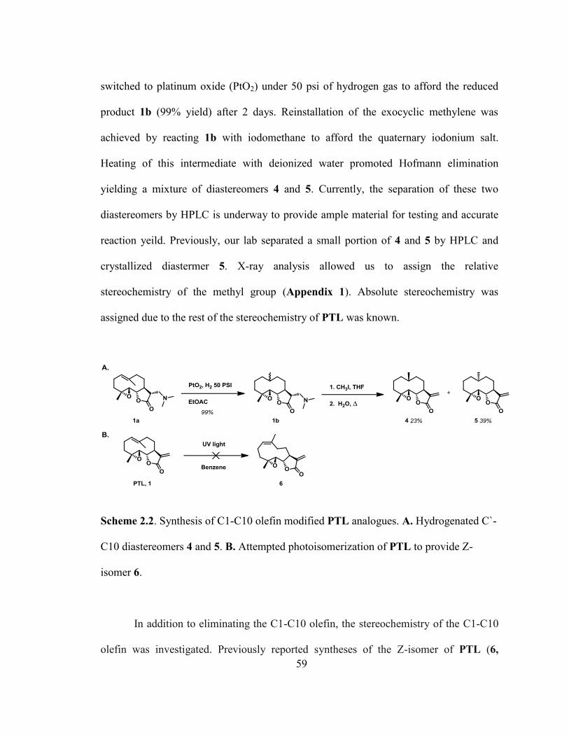
59
switched to platinum oxide (PtO2) under 50 psi of hydrogen gas to afford the reduced
product 1b (99% yield) after 2 days. Reinstallation of the exocyclic methylene was
achieved by reacting 1b with iodomethane to afford the quaternary iodonium salt.
Heating of this intermediate with deionized water promoted Hofmann elimination
yielding a mixture of diastereomers 4 and 5. Currently, the separation of these two
diastereomers by HPLC is underway to provide ample material for testing and accurate
reaction yeild. Previously, our lab separated a small portion of 4 and 5 by HPLC and
crystallized diastermer 5. X-ray analysis allowed us to assign the relative
stereochemistry of the methyl group (Appendix 1). Absolute stereochemistry was
assigned due to the rest of the stereochemistry of PTL was known.
Scheme 2.2. Synthesis of C1-C10 olefin modified PTL analogues. A. Hydrogenated C`-
C10 diastereomers 4 and 5. B. Attempted photoisomerization of PTL to provide Z-
isomer 6.
In addition to eliminating the C1-C10 olefin, the stereochemistry of the C1-C10
olefin was investigated. Previously reported syntheses of the Z-isomer of PTL (6,
60
Scheme 2.2) have been accomplished by photoisomerization using uv light radiation at
254 nm.274,276
After multiple attempts, this method of isomerization could not be reliably
executed in our hands. Instead we acquired the Z-isomer of PTL as a by-product during
the synthesis of melampomagnolide B (7, Scheme 2.3). During the allylic oxidation of
the C1-C10 olefin, a rotation of the vinyl methyl group must occur for the final [2,3]-
sigmatropic rearrangement to occur. Instead of the sigmatropic shift occurring, selenium
is eliminated yielding the Z-isomer of PTL (6) as a byproduct in 4% yield. We used this
material for screening.
Oxidative modifications to the C1-C10 allylic metyl group of PTL were also
investigated. According to an established protocol, PTL was reacted with selenium
dioxide and tert-butyl hydroperoxide in an allylic oxidation of the C1-C10 olefin to
yield melampomagnolide B (7, Scheme 2.3) in 74% yield.277,278
Compound 7 was then
reacted with dimethylamine to produce 7a in 97% yield, protecting the exocyclic olefin.
Compound 7a was then reacted with palladium on carbon under 50 psi of hydrogen gas
to reduce the C1-C10 olefin. Deprotection of the exocyclic olefin was achieved to yield
8 via a Hofmann elimination as previously described for preparation of 4 and 5.

61
Scheme 2.3. Synthesis of melampomagnolide analogues of PTL. Melampomagnolide B
(7) was synthesized as previously described.278,279
The guaianolide analogues (Figure 2.5) were provided by co-worker Dan Wang
(9 and 10). The 5-7-5 ring structure of guaianolide was achieved by reacting PTL with
boron trifluoride diethyl etherate, via reaction of the C1-C10 olefin with the epoxide,
yielding analogues 9 and 10 as previously described.280,281
Figure 2.5 Synthesized rearranged guaianolide analogues of Parthenolide.
In addition to structural modifications of PTL, the fumarate salts of all
dimethylamino analogues of the exocyclic methylene (1a, 2a, 7a, and 9) were

62
synthesized to afford greater water solubility (Scheme 2.4). Generally, the
dimethylamino compounds were reacted with fumaric acid to afford 11-14, in high
yields (range 82% - quant).
Scheme 2.4. Synthesis of fumarate salt analogues of PTL dimethylamino prodrugs.
2.3.2 Biological Evaluation of Parthenolide Analogues
The synthesized analogues of PTL were evaluated for their cytotoxicity in nine
cell lines to probe the effect of structural modification on cellular cytotoxicity profiles
(Table 2.1). Cancer cell lines investigated were DU-145 (prostate cancer), CCRF-CEM
(acute lymphoblastic leukemia), HL-60 (promyelocytic leukemia), HeLa (cervical
63
cancer), MCF-7 (breast cancer), GBM6 (glioblastoma multiforme), U-87MG
(glioblastoma multiforme), and NCI-ADR-RES (multidrug-resistant ovarian
cancer273,282
). Vero cells (African green monkey epithelial kidney cells) were utilized to
gauge cytotoxicity against normal cells. Each compound evaluated for antiproliferative
activity was verified to be ≥ 95% pure by HPLC. Stock solutions were made by
dissolving compounds into molecular biology grade DMSO and were stored at -20 °C.
Cells were plated in 96 well plates and allowed to incubate for 24 hours. The next day,
plates were dosed with molecules, each concentration dosed in triplicate, and then
incubated for 48 hours. The plates were stained with Alamar Blue® metabolic viability
reagent during the final 1-3 hours of dosing. Reaction time with Alamar blue® varied
between cell type, but fluorescent readout was only accepted above 1000 AFU. For
HeLa, DU-145, U-87MG, MCF-7 and Vero cell lines, deoxidative metabolism of the
reagent was observed to be rapid and required an incubation time of 1.5 hours. HL-60
cells metabolized Alamar Blue® at a much slower rate requiring a 3 h incubation time to
observe fluorescence above 1000 AFU. For all remaining cell lines, an incubation time
of 2 h for Alamar Blue® was used. Data collected was normalized by subtracting blank
wells (contained no cells) and dividing by control wells (cells with no drug). Normalized
values were graphed using GraphPad prism using a sigmoidal dose-response equation to
determine the IC50. Sigmoidal dose-response curves were generated in triplicate to
determine the average IC50 and standard deviation. PTL was dosed above and below its
IC50 as a control for each plate.

64
2.3.2.1 Biological Evaluation of Parthenolide, Costunolide, and Reduced
Parthenolide
We found that PTL conveys modest low micromolar cytotoxity against six of the
eight cancer cell lines tested (DU-145, IC50 = 8.9 ± 4.6 µM; CCRF-CEM, IC50 = 4.7 ±
1.6 µM; U-87MG, IC50 = 5.8 ± 2.3 µM; GBM6, IC50 = 3.4 ± 1.1 µM; HL-60, IC50 = 9.3
± 3.8 µM; MCF-7, IC50 = 9.7 ± 2.8 µM Table 2.1). HeLa and NCI/ADR-RES cell lines
required higher concentrations to achieve an IC50 (HeLa, IC50 = 44.1 ± 6.4 µM;
NCI/ADR-RES, IC50 = 57.6 ± 8.9 µM). PTL also conveyed the same cytotoxicity
against the normal Vero cell line (IC50 = 12.4 ± 0.6 µM) when compared to the cancer
cell lines, reinforcing the need for a PTL analogue that can spare normal cells. Previous
studies of PTL against cell lines MCF-7, HL-60, U-87MG, and HeLa were in general
agreement withour data, revealing low micromolar inhibitory activity, with some
variability resulting from different incubation times and methods of viability
measurements.281,283,284

65
Ver
o
12
.4 ±
0.6
25
.7 ±
0.9
> 5
00
NC
I/A
DR
-
RE
S
57
.6 ±
8.9
44
.8 ±
7.4
> 5
00
MC
F-7
9.7
± 2
.8
16
.7, 1
4.3
> 5
00
HeL
a
44
.1 ±
6.4
TB
D
> 5
00
HL
-60
9.3
± 3
.8
35
.4, 3
7.8
> 5
00
GB
M6
3.4
± 1
.1
7.0
± 1
.8
> 5
00
U-8
7M
G
5.8
± 2
.3
9.6
± 0
.8
> 5
00
CC
RF
-CE
M
4.7
± 1
.6
2.3
± 0
.2
> 5
00
DU
-14
5
8.9
± 4
.6
7.5
± 3
.1
> 5
00
Co
mp
ou
nd
1,
PT
L
2
3
Tab
le 2
.1. H
alf
max
imal
inhib
ito
ry c
once
ntr
atio
n (
IC5
0 i
n µ
M)
of
Par
then
oli
de
(1),
Cost
unoli
de
(2),
and r
educe
d
par
then
oli
de
(3)
in n
ine
dif
fere
nt
cance
r ce
ll l
ines
. IC
50 v
alues
are
aver
ages
(n >
3)
± S
tandar
d d
evia
tion. T
BD
= T
o b
e
det
erim
ined
.

66
Biological evaluation of CTL (2) demonstrated that the loss of the epoxide from
PTL was highly tolerated across five cancer cell lines (IC50 for DU-145, CCRF-CEM,
U-87MG, and GBM6 = 2.3 – 9.6 µM vs. PTL = 3.4 – 8.9 µM; IC50 for NCI/ADR-RES
= 44.8 ± 7.4 µM vs. PTL = 57.6 ± 8.9 µM). Loss of the epoxide was not tolerated in the
two remaining cancer cell lines evaluated (HL-60 35.4/37.8 µM vs. PTL = 9.3 ± 3.8
µM; MCF-7, 16.7/14.3 µM vs. PTL = 1.4/6.7 µM) indicating a need for this functional
group for maintaining activity to the level of PTL. Additionally, loss of activity against
Vero cells (IC50 = 25.7 ± 0.9 µM vs. PTL = 12.4 ± 0.6 µM). Costunolide was previously
reported to inhibit growth of DU-145 cells at IC50 = 4.8 µM, verifying our observation
of IC50 = 7.5 ± 3.1 µM.285
HL-60 results were consistent with previous reports of CTL
being dosed up to 8 µM and greater than 97% cell viability observed.286
Reduced PTL analogue 3 was observed to be biologically inactive (IC50 > 500
µM) in all cell lines tested, indicating that the exocyclic olefin must remain intact for
biological activity of PTL. These results are consistant with previous reports of
biological activity of reduced PTL in other cell lines as well as antiviral
activity.234,275,287
Additionally, complete loss of activity of reduced PTL was observed
when dosed to primary leukemia cells.234
2.3.2.2 Biological Evaluation of C1-C10 Olefin Modified Parthenolide Analogues
Analogues with modifications to the C1-C10 olefin (compounds 4-6) were
highly tolerated and demonstrated similar biological activity to the parent compound,

67
PTL (Table 2.2). Diastereomers 4 and 5 were equipotent to PTL in all cancer cell lines
tested (DU-145, CCRF-CEM, U-87MG, and GBM6; IC50 for 4 and 5 = 1.6 – 7.7 ± 0.4 –
2.8 µM vs. PTL = 3.4 – 8.9 ± 1.1 – 4.6 µM). Compounds 4 and 5 also demonstrated the
same cytotoxicity against the normal Vero cell line (IC50 = 13.9 ± 2.2 and 12.1 ± 3.3
µM, respectively) when compared to PTL (IC50 = 12.4 ± 0.6). Biological testing in
HeLa, MCF-7, NCI/ADR-RES, and HL-60 still need to be completed for analogues 4
and 5.
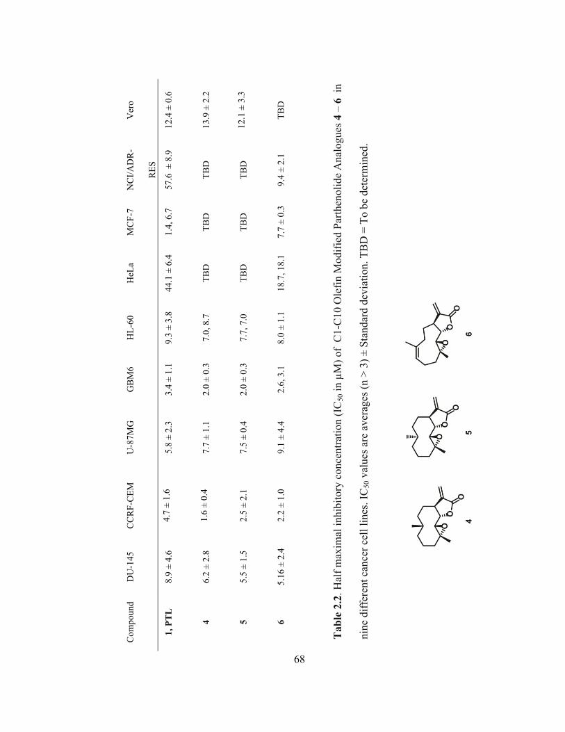
68
Ver
o
12
.4 ±
0.6
13
.9 ±
2.2
12
.1 ±
3.3
TB
D
NC
I/A
DR
-
RE
S
57
.6 ±
8.9
TB
D
TB
D
9.4
± 2
.1
MC
F-7
1.4
, 6.7
TB
D
TB
D
7.7
± 0
.3
HeL
a
44
.1 ±
6.4
TB
D
TB
D
18
.7, 1
8.1
HL
-60
9.3
± 3
.8
7.0
, 8.7
7.7
, 7.0
8.0
± 1
.1
GB
M6
3.4
± 1
.1
2.0
± 0
.3
2.0
± 0
.3
2.6
, 3.1
U-8
7M
G
5.8
± 2
.3
7.7
± 1
.1
7.5
± 0
.4
9.1
± 4
.4
CC
RF
-CE
M
4.7
± 1
.6
1.6
± 0
.4
2.5
± 2
.1
2.2
± 1
.0
DU
-14
5
8.9
± 4
.6
6.2
± 2
.8
5.5
± 1
.5
5.1
6 ±
2.4
Co
mp
ou
nd
1,
PT
L
4
5
6
Tab
le 2
.2. H
alf
max
imal
inhib
ito
ry c
once
ntr
atio
n (
IC5
0 i
n µ
M)
of
C1
-C10 O
lefi
n M
odif
ied P
arth
enoli
de
Anal
ogues
4 –
6 in
nin
e dif
fere
nt
can
cer
cell
lin
es.
IC5
0 v
alues
are
aver
ages
(n >
3)
± S
tandar
d d
evia
tion.
TB
D =
To b
e det
erm
ined
.
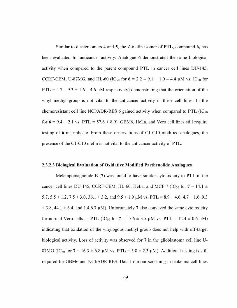
69
Similar to diastereomers 4 and 5, the Z-olefin isomer of PTL, compound 6, has
been evaluated for anticancer activity. Analogue 6 demonstrated the same biological
activity when compared to the parent compound PTL in cancer cell lines DU-145,
CCRF-CEM, U-87MG, and HL-60 (IC50 for 6 = 2.2 – 9.1 ± 1.0 – 4.4 µM vs. IC50 for
PTL = 4.7 – 9.3 ± 1.6 – 4.6 µM respectively) demonstrating that the orientation of the
vinyl methyl group is not vital to the anticancer activity in these cell lines. In the
chemoresistant cell line NCI/ADR-RES 6 gained activity when compared to PTL (IC50
for 6 = 9.4 ± 2.1 vs. PTL = 57.6 ± 8.9). GBM6, HeLa, and Vero cell lines still require
testing of 6 in triplicate. From these observations of C1-C10 modified analogues, the
presence of the C1-C10 olefin is not vital to the anticancer activity of PTL.
2.3.2.3 Biological Evaluation of Oxidative Modified Parthenolide Analogues
Melampomagnolide B (7) was found to have similar cytotoxicity to PTL in the
cancer cell lines DU-145, CCRF-CEM, HL-60, HeLa, and MCF-7 (IC50 for 7 = 14.1 ±
5.7, 5.5 ± 1.2, 7.5 ± 3.0, 36.1 ± 3.2, and 9.5 ± 1.9 µM vs. PTL = 8.9 ± 4.6, 4.7 ± 1.6, 9.3
± 3.8, 44.1 ± 6.4, and 1.4,6.7 µM). Unfortunately 7 also conveyed the same cytotoxicity
for normal Vero cells as PTL (IC50 for 7 = 15.6 ± 3.5 µM vs. PTL = 12.4 ± 0.6 µM)
indicating that oxidation of the vinylogous methyl group does not help with off-target
biological activity. Loss of activity was observed for 7 in the glioblastoma cell line U-
87MG (IC50 for 7 = 16.3 ± 6.8 µM vs. PTL = 5.8 ± 2.3 µM). Additional testing is still
required for GBM6 and NCI/ADR-RES. Data from our screening in leukemia cell lines

70
matches previous reported IC50 results observed from dosing 7 to primary leukemia
cells.278
71
Ver
o
12
.4 ±
0.6
15
.6 ±
3.5
TB
D
NC
I/A
DR
-RE
S
57
.6 ±
8.9
12
.8, 1
9.9
, 20
.6
TB
D
MC
F-7
1.4
, 6.7
9.5
± 1
.9
TB
D
HeL
a
44
.1 ±
6.4
36
.1 ±
3.2
TB
D
HL
-60
9.3
± 3
.8
7.5
± 3
.0
41
.6, 4
2.0
GB
M6
3.4
± 1
.1
3.5
, 6.6
17
.6,
16
.6
U-8
7M
G
5.8
± 2
.3
16
.3 ±
6.8
26
.8 ±
2.3
CC
RF
-CE
M
4.7
± 1
.6
5.5
± 1
.2
5.9
, 1.5
DU
-14
5
8.9
± 4
.6
14
.1 ±
5.7
26
.7 ±
6.5
Co
mp
ou
nd
1,
PT
L
7
8
Tab
le 2
.3. H
alf
max
imal
inhib
itory
conce
ntr
atio
n (
IC5
0 i
n µ
M)
of
oxid
ativ
e m
odif
ied p
arth
enoli
de
anal
ogues
7
and 8
in n
ine
dif
fere
nt
cance
r ce
ll l
ines
. IC
50 v
alues
are
av
erag
es (
n >
3)
± S
tandar
d d
evia
tion. T
BD
= T
o b
e
det
erm
ined
.

72
Reduction of the C1-C10 olefin of Melampomagnolide B (7) was also performed
to investigate if the orientation of the alcohol had an impact on the anticancer activity of
PTL. After hydrogenation, the two diastereomers were tested as a mixture (8). A loss of
activity in DU-145 and U-87MG cells was observed for 8 (IC50 = 26.7 ± 6.5, and 26.8 ±
2.3 µM, respectively) when compared to PTL (IC50 = 8.9 ± 4.6 and 5.8 ± 2.3 µM,
respectively). Data has been collected for CCRF-CEM, GBM6, and HL-60 but not yet in
triplicate. HeLa, MCF-7, NCI/ADR-RES and Vero all still require triplicate evaluation
of 8.
2.3.2.4 Biological Evaluation of Rearranged Guaianolide Parthenolide Analogues
Rearranged guaianolide analogue 9 demonstrated equipotent cytotoxicity in three
cancer cell lines (DU-145, CCRF-CEM, and HL-60: IC50 = 14.8 ± 4.0, 5.2 ± 1.8, and 7.1
± 1.3 µM respectively) when compared to PTL (DU-145, CCRF-CEM, and HL-60:
PTL = 8.9 ± 4.6, 4.7 ± 1.6, and 9.3 ± 3.8 µM respectively). It was observed with the
glioblastoma cell lines (U-87MG and GBM6), as well as the metastatic breast cancer
cell line MCF-7, that a higher concentration were required to achieve IC50 when
compared to PTL (IC50 for U-87MG, GBM6, and MCF-7 for 9: 16.2 ± 3.6, 8.7 ± 0.6,
9.6 ± 1.0 µM vs. PTL = 5.8 ± 2.3, 3.4 ± 1.1, and 1.4/6.7 µM, respectively).
Interestingly, 9 also demonstrated less cytotoxicity against the normal Vero cell line
(IC50 = 36.1 ± 9.4 µM vs. PTL = 12.4 ± 0.6 µM)

73
Ver
o
12
.4 ±
0.6
36
.1 ±
9.4
54
.5 ±
3.0
NC
I/A
DR
-
RE
S
57
.6 ±
8.9
12
.5, 1
03,
102
18
.1, 9
7.1
, 13
1
MC
F-7
1.4
, 6.7
9.6
± 1
.0
25
.0 ±
4.3
HeL
a
44
.1 ±
6.4
35
.6, 3
4.0
44
.2
HL
-60
9.3
± 3
.8
7.1
± 1
.3
11
.6 ±
0.2
GB
M6
3.4
± 1
.1
8.7
± 0
.6
8.4
± 1
.8
U-8
7M
G
5.8
± 2
.3
16
.2 ±
3.6
29
.3 ±
2.9
7
CC
RF
-CE
M
4.7
± 1
.6
5.2
± 1
.8
4.8
± 1
.5
DU
-14
5
8.9
± 4
.6
14
.8 ±
4.0
13
.9 ±
3.7
Co
mp
ou
nd
1,
PT
L
9
10
Tab
le 2
.4. H
alf
max
imal
inhib
itory
conce
ntr
atio
n (
IC5
0 i
n µ
M)
of
rear
ranged
guai
anoli
de
anal
ogu
es 9
and 8
in n
ine
dif
fere
nt
cance
r ce
ll l
ines
. IC
50 v
alues
are
aver
ages
(n >
3)
± S
tandar
d d
evia
tion. T
BD
= T
o b
e det
erm
ined
.

74
Rearranged guaianolide analogue 10 was also found to have similar cytotoxicity
in three cancer cell lines (DU-145, CCRF-CEM, and HL-60: IC50 = 13.9 ± 3.7, 4.8 ±
1.5, 11.6 ± 0.2 µM respectively) when compared to PTL (DU-145, CCRF-CEM, and
HL-60: PTL = 8.9 ± 4.6, 4.7 ± 1.6, and 9.3 ± 3.8 µM respectively). Similar to 9,
analogue 10 lost activity against the glioblastoma cell lines U-87MG and GBM6 (IC50 =
29.3 ± 3.0, and 8.4 ± 1.8 µM respectively) when compared to PTL (IC50 = 5.8 ± 2.3 and
3.4 ± 1.1 µM respectively). A decrease in activity was also observed in the breast
cancer cell line MCF-7 (IC50 = 25.0 ± 4.3 µM) as compared to PTL (IC50 = 1.4, 6.7
µM). Analogue 10 also showed the lowest cytotoxicity against the normal cell line Vero
(IC50 = 54.5 ± 3.0 µM) out of all the other analogues when compared to PTL (IC50 =
12.4 ± 0.6 µM). Triplicate data has not been completed yet for HeLa and NCI/ADR-
RES.
2.3.2.5 Biological Evaluation of Dimethylamino Fumarate Parthenolide Analogues.
Dimethylamino furmarate salts were highly tolerated as modifications of PTL.
LC-1 fumarate salt (11) displayed the same cytotoxicity against all the cancer cell lines
when compared to PTL (IC50 for 11 = 1.9 – 63.5 ± 0.4 – 11.8 µM vs. IC50 for PTL = 3.4
– 44.1 ± 1.6 – 6.4 µM). Unfortunately, the same cytotoxicity was observed for 11
against the normal Vero cell line (IC50 = 15.6 ± 7.8 µM vs. PTL = 12.4 ± 0.6 µM).
Although not primary leukemia cell lines themselves, HL-60 and CCRF-CEM leukemia
cell lines match previous data performed in primary leukemia cell lines.234
Although not

75
tested in vivo for our studies, LC-1 has been previously investigated in pancreatic and
prostate murine models resulting in tumor suppression.288,289

76
Ver
o
12
.4 ±
0.6
15
.6 ±
7.8
21
.1 ±
1.2
20
.6, 2
8.0
48
.7 ±
5.9
NC
I/A
DR
-RE
S
57
.6 ±
8.9
38
.5 ±
2.1
11
8 ±
18
19
.6, 3
9, 4
0.6
74
.5, 2
0.8
, 22
.9
MC
F-7
1.4
, 6.7
10
.4 ±
1.2
23
.7 ±
1.2
16
.7 ±
0.9
3.3
HeL
a
44
.1 ±
6.4
63
.8 ±
11.8
82
.3 ±
1.9
11
8, 1
04
, 4
42
> 2
50
HL
-60
9.3
± 3
.8
7.1
± 0
.4
35
.1 ±
5.5
36
.2 ±
6.1
32
.3 ±
10.4
GB
M6
3.4
± 1
.1
3.5
± 1
.1
14
.6 ±
0.9
5.8
± 1
.2
27
.8 ±
4.8
U-8
7M
G
5.8
± 2
.3
8.8
± 1
.9
25
.2 ±
2.4
28
.3 ±
2.3
77
.5 ±
7.3
CC
RF
-CE
M
4.7
± 1
.6
1.9
± 0
.4
6.6
± 2
.5
2.9
, 3.3
45
.0 ±
7.4
DU
-14
5
8.9
± 4
.6
8.4
± 4
.5
20
5 ±
4.7
12
.0
60
.7 ±
7.5
Co
mp
ou
nd
1,
PT
L
11
12
13
14
Tab
le 2
.5 H
alf
max
imal
inhib
itory
conce
ntr
atio
n (
IC5
0 i
n µ
M)
of
dim
ethyla
min
o f
um
arat
e sa
lt c
om
pounds
11 –
14 i
n
nin
e dif
fere
nt
can
cer
cell
lin
es. IC
50 v
alues
are
aver
ages
(n >
3)
± S
tandar
d d
evia
tion. T
BD
= T
o b
e det
erm
ined
.

77
CTL fumarate salt 12 only retained the same activity as 11 in the leukemia cell
line CCRF-CEM and normal Vero cells (CCRF-CEM and Vero IC50s for 12 = 6.6 ± 2.5
and 21.1 ±1.2 µM vs. 11 =1.9 ± 0.4 and 15.6 ± 7.8 µM respectively). 12 lost activity
against seven other cancer cell lines (IC50 for DU-145, U-87MG, HL-60, HeLa, MCF-7,
NCI/ADR-Res, and GBM6 = 6.6 -118 ± 0.9 - 18 µM vs. 11 (IC50 = 3.5 – 63.8 ± 0.4 –
11.8 µM). Previous DU-145 cytotoxicity data for only the dimethylamino compound 2a,
and not the fumarate salt was available. Our findings agreed with the previous DU-145
anticancer data.273
The Melampomagnolide B fumarate salt 13 is currently under additional
biological evaluation to determine its IC50 values in all nine cell lines. It has been
elucidated that it has lower cytotoxicity in U-87MG, HL-60, and MCF-7 (IC50 = 28.3 ±
2.3, 36.2 ± 6.1, and 16.7 ± 0.9 µM vs. PTL = 5.8 ± 2.3, 9.3 ± 3.8, and 1.4, 6.7
respectively). Compound 13 was found to be equipotent as PTL in the glioblastoma cell
line GBM6 (IC50 for 13 = 5.8 ± 1.2 vs. PTL = 3.4 ± 1.1).
The dimethylamino fumarate salt of guaianolide 9 (14) was not as well tolerated
as other dimethylamino fumarates. Guaianolide fumarate salt 14 lost activity against all
cell lines tested to date (IC50 for DU-145, CCRF-CEM, U-87MG, HL-60, and GBM6 =
27.8 – 77.5 ± 7.3 – 10.4 µM vs. PTL = 3.4 – 9.3 ± 1.1 – 4.6 µM; NCI/ADR-RES = 74.5
µM vs. PTL = 57.6 ± 8.9 µM). In HeLa cells 100% cell viability was not achievable
with 500 µM being the largest concentration dosed. The extrapolated IC50 value was >
78
250 µM. Additionally, activity was also lost against the normal Vero cell line (IC50 for
14 = 48.7 ± 5.9 µM vs. PTL = 12.4 ± 0.6 µM).
2.4 Conclusion and Future Direction
In summary, we have synthesized a library of PTL analogues to elucidate the
importance of various functional groups to its cytotoxicity profile. The modified
functional groups of PTL included alterations to the exocyclic olefin, C1-C10 olefin, the
vinyl methyl group, as well as loss of the epoxide. In addition, rearranged 5-7-5
guainanolide analogues were synthesized to observe the importance of the ring structure
of PTL to its cytotoxicity profile. Dimethylamino prodrugs were synthesized to address
the water solubility issues of PTL and evaluated as fumarate salts for their biological
activity. The only modification to completely ablate activity of PTL across all cell lines
was removal of the exocyclic olefin, the primary pharmacophore of PTL.
Modifications to the C1-C10 olefin were tolerated across all cell lines tested for
analogues 4-6 indicating that alteration or complete removal of the internal olefin of
PTL has no effect on the biological activity. Oxidative modifications to the vinyl methyl
group were also highly tolerated with slight loss of activity observed in the glioblastoma
cell line U-87MG. Tolerance of the installation of the allylic alcohol can allow for
further modifications to this site to prepare PTL probe compounds and additional
analogues. Dimethylamino fumarate salt analogues 11-14 all displayed low cytotoxicity
within 5-fold of PTL against the cancer cell lines tested, confirming that the
79
dimethylamino prodrug of PTL is a viable modification to improve water solubility
while retaining biological activity.
The rearranged guaianolide analogues 9 and 10 demonstrated the most
interesting biological activity of all the PTL analogues. Although analogues 9 and 10
displayed similar activity when compared to PTL in most cancer cell lines (except U-
87MG, 10 was 6-fold less potent than PTL), the greatest finding was a loss in activity
against the normal cell line Vero. Both rearranged PTL analogues 9 and 10 had up to a
5-fold loss in activity against Vero cells, indicating that the more ridged 5-7-5
guaianolide ring structure was not as cytotoxic to normal cells. From these results we
can conclude that the guaianolide ring structure should serve as a new basis for further
SAR studies. Other future direction for this SAR could include modifications to the
butyrolactone, as well alterations in ring sizes for new potential clinical leads. Re-
synthesis and purification of analogues 4, 5 and 8 to finish biological testing is currently
underway.
2.5 Experimental
2.5.1 General
All reactions were performed under an anhydrous nitrogen atmosphere unless
otherwise noted. Parthenolide was purchased from Enzo Life Sciences and repurified by
SiO2 chromatography prior to biological studies. Costunolide was purchased from
Tocris Biosciene (Boston, MA) and was used without further purification. Commercial
grade reagents (Aldrich, Acros, Alfa Aesar) were used without further purification

80
unless otherwise noted. Tetrahydrofuran and dichloromethane were rendered anhydrous
by passing through the resin column of a solvent purification system. Benzene was
purified by distillation over calcium hydride. Methanol and N,N-dimethylformamide
were purchased as anhydrous solvent. High pressure hydrogenation reactions were
performed in a Parr hydrogenation apparatus (Moline, IL; model # 3916). All reactions
were monitored by thin-layer chromatography (TLC) using silica gel 60-F254 TLC plates
(Merck), and visualized using cerium ammonium molybdate stain. Silica gel
chromatography was performed using a Teledyne-Isco Combiflash Rf 200 instrument
using Redisep Rf Gold High-Performance silica gel columns (Teledyne-Isco). HPLC
purification was performed using an Agilent 1200 series instrument equipped with a
Zorbax (Agilent) preparatory column (21.2 x 250 mm, 7 µm). Analysis of compound
purity was conducted on the aforementioned HPLC, using a Zorbax analytical column
(4.6 x 150 mm, 5 µm). Nuclear magnetic resonance (NMR) spectroscopy employed a
Bruker Avance instrument operating at 400 MHz (for 1H) and 100 MHz (for
13C) at
ambient temperature. Internal solvent peaks were referenced in each case. Mass spectral
data was obtained from the University of Minnesota Department of Chemistry Mass
Spectrometry lab, employing a Bruker BioTOF II instrument.
2.5.2 Synthesis of Parthenolide Analogues

81
Dimethylamino Parthenolide 1a.
Known compound 1a was prepared by the method of Neelakantan et al.234
Parthenolide
(16.90 mg, 0.07 mmol) was dissolved in methanol (4 mL) and dimethylamine was added
(0.10 mL, 0.20 mmol). The reaction was allowed to stir for 5 h at rt, and then
concentrated in vacuo to yield compound 4 (19.90 mg, 97%) as an off white crystalline
solid. 1H NMR (400 MHz, CDCl3) δ = 5.19 (dd, J = 12.3, 3.8 Hz, 1H), 3.81 (t, J = 9.0
Hz, 1H), 2.78 – 2.68 (m, 2H), 2.62 (dd, J = 13.2, 4.8 Hz, 1H), 2.45 – 2.31 (m, 2H), 2.25
(s, 6H), 2.22 (s, 1H) , 2.21 – 2.01 (m, 5H), 1.68 (t, J = 1.2 Hz, 3H), 1.66 – 1.57 (m, 1H),
1.28 (s, 3H), 1.21 (td, J = 13.0, 5.8 Hz, 1H). 13
C NMR (100 MHz, CDCl3) δ = 176.6,
134.8, 125.2, 82.3, 66.7, 61.6, 57.8, 48.0, 46.6, 46.3, 46.3, 41.2, 36.8, 30.1, 24.2, 17.3,
17.1. MS-ESI+ m/z [M+H] calcd for C17H28NO3: 294.2, found: 294.2. MS-ESI
+ m/z
[M+Na] calcd for C17H27NO3Na: 316.1, found: 316.2.
Dimethylamino Costunolide 2a
The known compound 2a was prepared by as described by Srivastava et al.273
Costunolide (10.0 mg, 0.04 mmol) was dissolved into ethanol (15 mL). An excess of
dimethylamine (0.1 mL, 0.2 mmol) was added and the reaction was refluxed at 90 °C

82
overnight. The reaction was concentrated in vacuo after reacting for 12 h. The crude
product was purified by flash chromatography over SiO2 (gradient 0-10% methanol in
dichloromethane) to yield compound 2a (9.8 mg, 82%) as an off-white crystalline solid.
1H NMR (400 MHz, CDCl3) δ 4.82 (dd, J = 11.5, 4.5 Hz, 1H), 4.66 (d, J = 9.7 Hz, 1H),
4.55 (t, J = 9.3 Hz, 1H), 2.74 (dd, J = 13.2, 5.1 Hz, 1H), 2.60 (dd, J = 13.2, 4.8 Hz, 1H),
2.39 – 2.29 (m, 2H), 2.27 (s, 6H), 2.25 – 2.12 (m, 2H), 2.12 – 1.91 (m, 4H), 1.68 (d, J =
1.4 Hz, 3H), 1.64 – 1.54 (m, 1H), 1.40 (s, 3H), 1.25 (s, 1H). 13
C NMR (100 MHz,
CDCl3) δ 177.7, 140.6, 137.2, 127.5, 127.0, 81.5, 58.1, 51.3, 46.3, 46.2, 41.2, 39.7, 28.5,
26.3, 17.4, 16.3. MS-ESI+ m/z [M+H] calcd for C17H28NO2: 278.2, found: 278.2.
Reduced Parthenolide 3
Known compound 3 was prepared by the method of Govindachari et al.282
Parthenolide
(13.50 mg, 0.054 mmol) and palladium on carbon (14.20 mg, 0.13 mmol) were added to
a flask and ethanol (10 mL). The reaction was placed under 1 atm of H2, and allowed to
stir for 45 mins. Upon completion, the reaction was filtered through celite and the
solvent was removed in vacuo. The product was purified by flash chromatography over
SiO2 (10-50% ethyl acetate in hexanes gradient) to yield compound 3 (13.30 mg, 97%
yield). 1H NMR (400 MHz, CDCl3) δ = 5.19 (dd, J = 11.8, 1.8 Hz, 1H), 3.81 (t, J = 9.1
Hz, 1H), 2.70 (d, J = 9.0 Hz, 1H), 2.46 – 2.22 (m, 3H), 2.16 (m, 2H), 2.05 (t, J = 12.3
Hz, 1H), 1.95 – 1.79 (m, 3H), 1.70 (s, 3H), 1.68 – 1.35 (m, 3H), 1.29 (s, 3H). MS-ESI+

83
m/z [M+H] calcd for C15H23O3Na: 273.1, Found: 273.1. MS-ESI+ m/z [M+H] calcd for
C15H22O3K: 289.1, found: 289.1.
Reduced C1-C10 Olefin of Dimethylamino Parthenolide 1b
Compound 1a (29.10 mg, 0.10 mmol) was dissolved into ethyl acetate, and PtO2 (29.50
mg, 0.28 mmol) was added. The reaction was placed under an atmosphere of H2 (50 psi)
and the reaction was shook for 2 days with a Parr shaker. The reaction was then filtered
through a celite plug and washed with ethyl acetate (5 × 5 ml) and dichloromethane (5 x
4 ml). The solvent was removed in vacuo to yield compound 1b (29.30 mg, 99%) as a
white crystalline solid. 1H NMR (400 MHz, CDCl3) δ = 3.82 (t, J = 9.4 Hz, 1H), 3.00 (d, J =
9.6 Hz, 1H), 2.72 (dd, J = 12.9, 4.0 Hz, 1H), 2.58 (dd, J = 12.9, 5.0 Hz, 1H), 2.37 (m,
2H), 2.25 (s, 6H), 2.15 (m, 2H), 1.73 (m, 2H), 1.66 – 1.52 (m, 2H), 1.49 (s, 3H), 1.47 –
1.36 (m, 3H), 1.10 (m, 2H), 0.88 (d, J = 6.4 Hz, 3H). 13
C NMR (100 MHz, CDCl3) δ=
177.1, 80.8, 76.7, 66.6, 61.1, 58.2, 46.2, 45.6, 44.3, 36.6, 35.7, 30.8, 27.6, 25.7, 21.2,
20.5, 19.0. HRMS-ESI+ m/z [M+H] calcd for C17H30NO3: 296.2220, found: 296.2229.

84
and
C1-C10 Reduced PTL Analogues 4 and 5
Compound 1b (28.70 mg, 0.01 mmol) was dissolved into THF (2 ml) and iodomethane
was added in excess (0.10 ml, 1.60 mmol). The reaction was allowed to stir at rt for 20
h. The solvent and excess iodomethane was removed in vacuo resulting in 30.6 mg of
yellowish solid. Distilled water (10 mL) was added and the reaction was heated to 45
°C. Complete solvation of the yellowish material resulted within minutes of heating. The
reaction was allowed to stir with heating for 3 h, and then solvent was removed in
vacuo. Saturated NaHCO3 aqueous solution (5 mL) was added to the reaction mixture,
and the product was extracted with DCM (3 × 20 mL). The combined organic layers
were washed with brine (20 mL), and dried with Na2SO4. The reaction was purified by
flash chromatography over SiO2 (10%-50% ethyl acetate in hexanes gradient) to yield
12.3 mg of crude product. HPLC separation and characterization is currently underway.
Cis Parthenolide 6
Previously reported synthesis of cis parthenolide were attempted but could not be
replicated in my hands.274,279
Cis parthenolide was isolated as a side product from

85
synthesis of melampomagnolide B (7). Crude material was purified by SiO2 (gradient 10
– 50% ethyl acetate in Hexanes) to yield compound 6 (7.90 mg, 4% yield) as a white
crystalline solid. 1H NMR (400 MHz,CDCl3) δ = 6.24 (d, J = 3.5 Hz, 1H), 5.53 (d, J =
3.2 Hz, 1H), 5.33 (t, J = 7.3 Hz, 1H), 3.83 (t, J = 9.3 Hz, 1H), 2.90 (d, J = 9.4 Hz, 1H),
2.77 (M, 1H), 2.50 – 2.18 (m, 3H), 2.10 (m, 3H), 1.71 (s, 3H), 1.69 – 1.58 (m, 1H), 1.53
(s, 3H), 1.07 (t, J = 12.1 Hz, 1H). MS-ESI+ m/z [M+Na] calcd for C15H20NaO: 271.1,
Found: 271.1. MS-ESI+ m/z [M+K] calcd for C15H20KO: 287.1, found: 287.1.
Melampomagnolide B (7)
Known compound 7 was prepared by the method of Nasim et al.277,278
Parthenolide (49
mg, 0.20 mmol) and selenium dioxide (22 mg, 0.20 mmol) were suspended in anhydrous
dichloromethane (15 mL) and t-BuOOH (0.32 mL, 3.33 mmol, 70% in H2O) and
refluxed at 40 °C for 4.5 h. The reaction mixture was cooled to rt, and water (15 mL)
was added to the reaction. The crude material was extracted with DCM (3 × 15 mL).
The combined organics were dried over Na2SO4. The solvent was then removed in
vacuo, and the product was purified by chromatography over SiO2 (gradient 10-50%
ethyl acetate in hexanes) to yield compound 3 (38.60 mg, 74%) as an off-white
crystalline solid. 1H NMR (400 MHz, CDCl3) δ = 6.25 (d, J = 3.5 Hz, 1H), 5.66 (t, J =
8.3 Hz, 1H), 5.55 (d, J = 3.2 Hz, 1H), 4.23 – 4.04 (q, J = 12.6 Hz, 2H), 3.86 (t, J = 9.3

86
Hz, 1H), 2.92 – 2.76 (m, 2H), 2.42 (s, 2H), 2.37 – 2.11 (m, 3H), 1.74 – 1.61 (m, 1H),
1.56 (s, 3H), 1.18 – 1.04 (m, 1H). 13
C NMR (100 MHz, CDCl3) δ= 169.6, 139.7, 139.0,
127.7, 120.4, 81.3, 66.0, 63.5, 60.2, 43.0, 37.0, 25.7, 24.1, 23.8, 18.2 ppm; MS-ESI+ m/z
[M+Na] calcd for C15H20NaO4: 287.1, found: 287.1.
Dimethylamino adduct of Melampomagnolide B (7a)
Melampomagnolide B, 7, (32.50 mg, 0.10 mmol) was dissolved in methanol (4 ml) and
dimethylamine was added (0.10 mL, 0.20 mmol). The reaction was allowed to stir
overnight, and was then concentrated in vacuo to yield compound 7a (32.70 mg 86%) as
a white solid. 1H NMR (400 MHz, CDCl3) δ = 5.59 (t, J = 7.9 Hz, 1H), 4.10 (q, J = 13.0
Hz, 2H), 3.85 (t, J = 9.2 Hz, 1H), 2.80 (d, J = 9.4 Hz, 1H), 2.73 (dd, J = 12.9, 5.1 Hz,
1H), 2.61 (dd, J = 12.9, 5.4 Hz, 1H), 2.51 – 2.35 (m, 4H), 2.25 (m, 2H), 2.21 (s, 6H),
2.12 (m, 2H), 1.59 (m, 1H), 1.52 (s, 3H), 1.07 (t, J = 11.4 Hz, 1H). 13
C NMR (100 MHz,
CDCl3) δ = 177.0, 141.1, 127.2, 81.6, 77.5, 66.0, 64.1, 60.0, 57.7, 45.7, 44.3, 42.2, 37.1,
27.5, 25.8, 23.8, 18.0. HRMS-ESI+ m/z [M+H] calcd for C17H28NO4: 310.2013, found:
310.1999.

87
Reduced C1-C10 olefin of Melampomagnolide B 8
Compound 7a (46.90 mg, 0.15 mmol) was dissolved into ethyl acetate (10 mL), and
10% palladium on carbon (46.30 mg, 0.44 mmol) was added to the reaction mixture.
The reaction was put under an atmosphere of 50 psi hydrogen gas, and shook for 5 days.
The reaction mixture was filtered through a celite plug and washed with ethyl acetate (5
× 5 mL) and DCM (5 × 5 mL). The solvent was removed in vacuo and the crude
material was purified by flash chromatography over SiO2 (0 to 10% Methanol in DCM)
to yield (14.00 mg) as a white solid. The compound was then dissolved into THF (2 ml)
and iodomethane was added in excess (0.10 mL, 1.60 mmol). The reaction was allowed
to stir at rt for 20 h. The solvent and excess methyl iodine was removed in vacuo
resulting in a yellowish solid. Distilled water (10 mL) was added and the reaction and
was heated to 45°C. Complete solvation of the yellowish material resulted within
minutes of heating. The reaction was allowed to stir with heating for 3 h, then solvent
was removed in vacuo. Saturated NaHCO3 aqueous solution (10 mL) was added to the
reaction mixture, and the product was extracted with DCM (3 × 20 mL). The combined
organic layers were washed with brine, and dried over Na2SO4. Purification and
characterization is currently underway.

88
LC-1 (11)
Known compound 11 was prepared by the method of Neelakantan et al.234
Compound
1a (14.20 mg, 0.05 mmol) was dissolved into ethyl ether (5 mL) and fumaric acid (5.62
mg, 0.05 mmol) was added. The reaction was allowed to stir for two days and solvent
was removed in vacuo to yield compound 11 (16.30 mg, 82 %) as a white solid. 1H
NMR (400 MHz, DMSO-d6) δ = 13.00 (s, 2H), 6.62 (s, 2H), 5.22 (dd, J = 11.9, 2.4 Hz,
1H), 3.97 (t, J = 9.0 Hz, 1H), 2.80 (d, J = 9.0 Hz, 1H), 2.63 (m, 3H), 2.42 – 2.26 (m, 2H)
2.21 (s, 6H), 2.14 (m, 1H), 2.10 – 1.91 (m, 4H), 1.64 (s, 3H), 1.20 (s, 3H), 1.17 – 1.05
(m, 2H). Spectral data matches previously reported.234
Costunolide Fumarate Salt 12
Compound 1b (15.10 mg, 0.055 mmol) was dissolved into ethyl ether (5 mL) and
fumaric acid (6.34 mg, 0.06 mmol) was added. The reaction was allowed to stir for two
days, at which time solvent was removed in vacuo to yield compound 12 (22.20 mg,
quantitative yield) as an off white solid. 1H NMR (400 MHz, DMSO-d6) δ = 6.60 (s,
2H), 4.78 (dd, J = 10.6, 2.2 Hz, 1H), 4.70 (t, J = 9.7 Hz, 1H), 4.64 (d, J = 10.2 Hz, 1H),
2.70 – 2.52 (m, 4H), 2.35 – 2.21 (m, 3H), 2.18 (s, 6H), 2.12 – 1.88 (m, 5H), 1.63 (d, J =

89
1.3 Hz, 3H), 1.36 (s, 3H). 13
C NMR (100 MHz, DMSO-d6) δ = 177.1, 166.2, 139.5,
136.9, 134.1, 127.5, 126.2, 80.5, 57.0, 50.3, 45.3, 45.2, 44.4, 40.5, 27.1, 25.8, 16.9, 15.9.
HRMS-ESI+ m/z [M+H] Calcd for C17H28NO2 (without salt): 278.2115, found:
278.2126.
Melampomagnolide B fumarate salt 13
7a (40.50 mg, 0.15 mmol) was dissolved into DCM (5 mL) and fumaric acid (17.70 mg,
0.15 mmol) was added. The reaction was allowed to stir overnight, at which time solvent
was removed in vacuo to yield compound 13 (55.30 mg, 85%) as a white solid. 1H NMR
(400 MHz, DMSO-d6) δ 6.61 (s, 2H), 5.49 (t, J = 7.7 Hz, 1H), 4.01 (t, J = 9.6 Hz, 1H),
3.92 (q, J = 13.3 Hz, 2H), 2.73 – 2.55 (m, 4H), 2.31-2.14 (m, 10H), 2.13 – 1.99 (m, 3H),
1.57 (t, J = 10.6 Hz, 1H), 1.47 (s, 3H), 0.96 – 0.86 (m, 1H). 13
C NMR (100 MHz,
DMSO-d6) δ = 177.1, 166.2, 141.0, 134.1, 123.5, 123.4, 80.6, 63.3, 59.9, 57.6, 45.2,
45.2, 43.4, 42.7, 40.2, 36.9, 25.7, 24.0, 23.0, 17.5. HRMS-ESI+ m/z [M+H] calcd for
C17H28NO4 (without salt): 310.2013, found: 310.2015.
2.5.3 Protocol for Mammalian Cell Culture
All cell lines were maintained in a humidified 5% CO2 environment at 37 °C in
culture flasks (Corning). Adherent cells were dissociated using 0.25% Trypsin-EDTA
solution (Gibco). CCRF-CEM cells (ATCC, CCL-119) and NCI/ADR-RES were

90
cultured in RPMI-1640 media (Cellgro) supplemented with 10% fetal bovine serum
(FBS, Gibco), penicillin (100 I.U./mL), and streptomycin (100 µg/mL, ATCC) at a
density of 2 × 10⁵ - 2 × 10
6 cells/mL. DU-145 cells (ATCC, HTB-81), Vero cells
(ATCC, CCL-81), U-87MG cells (ATCC, HTB-14), and HeLa cells (ATCC, CCL-2)
were cultured in MEM media (Cellgro) supplemented with 10% FBS (Gibco), penicillin
(100 I.U./mL), and streptomycin (100 µg/mL, ATCC). HL-60 cells (ATCC, CCL-240)
were cultured in IMDM media (Cellgro) supplemented with 20% FBS (Gibco),
penicillin (100 I.U./mL), and streptomycin (100 µg/mL, ATCC). MCF-7 cells (ATCC,
HTB-22) were cultured in MEM media (Cellgro) supplemented with 10% FBS (Gibco),
bovine insulin (0.01 mg/mL, Sigma) penicillin (100 I.U./mL), and streptomycin (100
µg/mL, ATCC). GBM6 cells (gift from the lab of professor John Ohlfest) were cultured
with DMEM/F12 (1:1) with L-Glutamine, without HEPES (Thermo Scientific)
supplemented with Normocin (100 µg/mL, Invivogen), 50X B-27 supplement (10 mL,
Gibco), 100X N-2 supplement (5 mL, Gibco), human EGF (20 ng/µL, Peprotech),
human FGF-basic (20 ng/µL, PeproTech), penicillin (50 I.U./mL), and streptomycin (50
µg/mL, ATCC).
2.5.4 Protocol for Cell Culture Cytotoxicity Assays
The protocol for cellular cytotoxicity assays was previously published by our
laboratory (Hexum et al).264
CCRF-CEM and HL-60 cells were cultured at a density of
10,000 cells/well in cell culture media (50 µL) in standard 96-well plates (Costar) 24 h
prior to treatment. DU-145, Vero, U-87MG, GBM6, HeLa, MCF-7, and NCI/ADR-RES

91
cells were seeded at a density of 5,000 cells/well in cell culture media (50 µL) in
standard 96-well plates (Costar). Blank (no cells) wells and control (vehicle control
treated) wells were prepared for every experiment to normalize the data. All PTL
analogues were serially diluted in pre-warmed media and dosed to 96 well plates (final
volume/well = 100 µL; final DMSO concentration = 0.5%). Approximately 1-3 h before
the end of the treatment period (48 h), Alamar Blue (Invitrogen) cell viability reagent
was added to the 96 well plates (10 µL). A quantitative measure of cell viability can be
achieved from evaluating the ability of metabolically active cells (which are proportional
to the number of living cells) to convert resazurin (non-fluorescent dye) to red-
fluorescent resorufin. Fluorescence data were obtained on either a Molecular Devices
SpectraMax M2 plate reader or an LJL BioSystems HT Analyst plate reader.
Background fluorescence (no cell controls) was subtracted from each well and cellular
viability values following compound treatment were normalized to vehicle-only treated
wells (control wells only treated with aqueous DMSO, which were arbitrarily assigned
100% viability). Individual IC50 curves were generated by fitting data to the sigmoidal
(dose response) function of varied slope in GraphPad Prism (v. 5.0) software. Only
curve fits with R2 > 0.95 were deemed sufficient. Each experiment was performed in
biological triplicate and mean IC50 values were calculated from the individual IC50
values obtained from each replicate. Standard deviation was calculated from the
individual IC50 values obtained for each biological replicate.

92
2.6 NMR
Dim
ethyla
min
o P
arth
en
olid
e 1a
1H N
MR
(CD
Cl3 ):
CD
Cl3
TM
S

93
Dim
ethyla
min
o P
arth
en
olid
e 1a
13C
NM
R (C
DC
l3 ):
CD
Cl3

94
Dim
ethyla
min
o C
ostu
nolid
e 2a
1H
NM
R (C
DC
l3 ):
CD
Cl3
TM
S

95
Dim
ethyla
min
o C
ostu
nolid
e 2a
1
3C N
MR
(CD
Cl3 ):
CD
Cl3

96
Red
uced
Parth
enolid
e 3
1H N
MR
(CD
Cl3 ):
CD
Cl3
TM
S

97
Red
uced
C1
-C10 O
lefine o
f Dim
thyla
min
o P
arth
enolid
e 1b
1H
NM
R (C
DC
l3 ):
CD
Cl3
TM
S

98
Red
uced
C1
-C10 O
lefine o
f Dim
thyla
min
o P
arth
enolid
e 1b
1
3C N
MR
(CD
Cl3 ):
CD
Cl3

99
Cis P
arth
enolid
e 6
1H N
MR
(CD
Cl3 ):
CD
Cl3
TM
S

100
Mela
mp
om
agn
olid
e B (7
) 1H
NM
R (C
DC
l3 ):
CD
Cl3
TM
S
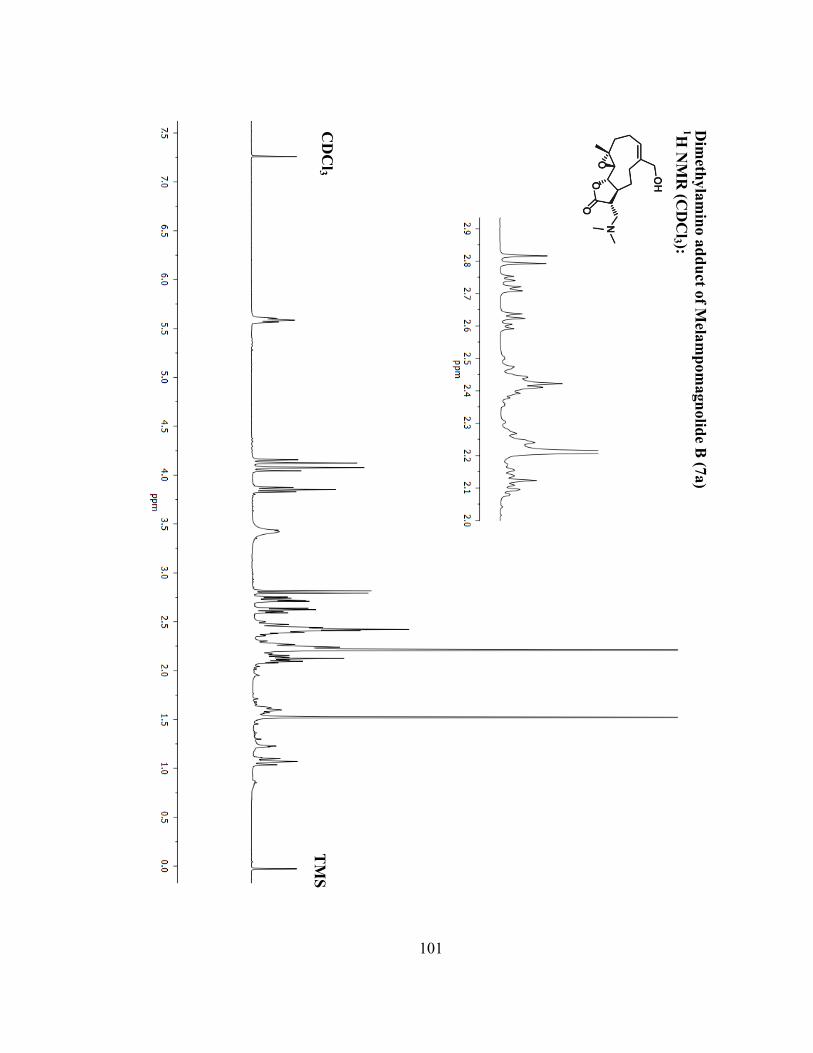
101
Dim
ethyla
min
o a
dd
uct o
f Mela
mp
om
agn
olid
e B (7
a)
1H N
MR
(CD
Cl3 ):
CD
Cl3
TM
S

102
Dim
ethyla
min
o a
dd
uct o
f Mela
mp
om
agn
olid
e B (7
a)
13C
NM
R (C
DC
l3 ):
CD
Cl3

103
LC
-1 (1
1)
1H N
MR
(DM
SO
-d6 ):
DM
SO
DM
SO

104
Costu
nolid
e Fu
mara
te Salt 1
2
1H N
MR
(DM
SO
-d6 ):
DM
SO
DM
SO

105
DM
SO
Costu
nolid
e Fu
mara
te Salt 1
2
13C
NM
R (D
MS
O-d
6 ):

106
Mela
mp
om
agn
olid
e B F
um
ara
te Salt 1
3
1H N
MR
(DM
SO
-d6 ):
DM
SO
DM
SO

107
Mela
mp
om
agn
olid
e B F
um
ara
te Salt 1
3
13C
NM
R (D
MS
O-d
6 ): D
MS
O

108
Chapter 3. Raising the Effective Intracellular Concentration of Parthenolide
Utilizing a Gold Nanoparticle Delivery System.
3.1 Introduction
Nanoparticles are particles on the order of nanometers made out of various
metals (copper, platinum, silver, or gold) or semiconductors (titanium dioxide, cadmium
selenide, cadmium sulfide, silver sulfide, and lead sulfide).290
Ligands are used during
the synthesis of nanoparticles to control growth and prevent aggregation.291
Compared to
the size of a cell, nanoparticles are up to ten thousand times smaller, around the size of
receptors, enzymes, and antibodies.292
Due to their small size, nanoparticles are capable
of interactions both extracellular and intracellular.292
Nanoparticles have been intensely investigated as potential drug carriers for
targeted delivery.291,293-295
Nanoparticle delivery systems take advantage of
pathophysiological alterations observed in tumors, such as increased diffusion and
permeability due to leaky vasculature of tumors.296
Nanoparticles ranging between 20-
100 nm can take advantage of the enhanced permeability and retention effect (EPR),
allowing passive diffusion into tumors, thereby permitting greater accumulation when
compared to free drugs.297
Moreover, nanoparticle delivery systems can increase drug
solubility and allow for timed release of drugs conjugated to them.298,299
However, nanoparticles do have their flaws. The reticuloendothelial system
(RES) will opsonize large nanoparticles (> 100 nm) and eliminate them through the
liver.292,300,301
To avoid excretion through the RES, smaller particles functionalized with

109
a secondary layer of hydrophilic polymer are used.301
Polyethylene glycol (PEG) has
been widely used to mask nanoparticles from the RES, while also improving water
solubility.302
Out of all the nanomaterials utilized today, gold nanoparticles (AuNP) have
become a primary interest in drug delivery research.295,303,304
Gold has excellent
properties that lead to ease of synthesis and functionalization of nanoparticles.303,305
Compared to other materials used, gold has greater chemical stability.305
Gold also has
intrinsic characterization properties that include visible light scattering/absorption, low
biological concentrations, and large electron density that allows for easy quantification,
location, and imaging in biological organisms.305,306
Additionally, routes have been
developed for controlled synthesis of AuNP allowing the shape and size of the AuNP to
be regulated.307-310
AuNPs have been previously loaded with anticancer drugs, such as
paclitaxel,301,311
methotrexate,312
daunorubicin,313
gemcitabine,314
5-fluoruracil,315
cisplatin,316
tamoxifen,317
trastuzumab,318
and doxorubicin.319
Agasti et al demonstrated
that by conjugation to a AuNP delivery system, a 100-fold decrease in the IC50 value of
5-fluorouracil (5-FU) was observed, from 11.9 µM to 0.7 µM in MCF-7 cells. AuNP in
this study were used to cage 5-FU, and upon photolysis, 5-FU was released.315
Gold is also readily functionalized by thiols. Utilization of an alkanethiol linker
to anchor drug payloads to the AuNP is often observed in the literature.303,320,321
Thiol
ligand exchange is a fairly rapid conversion that depends on chain length and
sterics.320,322
Because of these characteristics we have chosen to synthesize parthenolide

110
(PTL) functionalized AuNPs utilizing a PEG-thiol, similar to that of a previously
reported functionalization of paclitaxel to a AuNP.311
By conjugating PTL to a AuNP
we hope to improve the water solubility of the drug and increase the effective
concentration delivered to the target cancer cells.
3.2 Research Objectives
Current progress on the synthesis and anticancer evaluation of a PTL
functionalized AuNP is herein reported. We have envisioned three different linkers
(Figure 3.1) to investigate the effects of conjugating PTL to AuNP: melampomagnolide
B linker 15, a non-functional linker 16 and a dimethylamino analogue of PTL
(DMAPT) linker 17. Once synthesized, we will investigate the AuNP’s ability to address
two major problems with PTL: its poor water solubility and low micromolar anticancer
activity. A AuNP delivery system should increase the effective concentration of PTL
delivered to cancer cells allowing detectable levels of PTL, and ultimately reaching the
IC50 in vivo. Additionally, water solubility of the drug would improve by utilization of a
PEG linker as well as the innate characteristic of AuNP to improve drug solubility.
Described here are the efforts made towards the synthesis of a PTL functionalized
AuNP.

111
Figure 3.1 Proposed linkers for conjugation of PTL to AuNP. Conjugation of PTL via
ester linker 15, a nonfunctional control linker 16, and a DMAPT inspired linker 17. N
ranges from 36-55 PEG units CTPEG2000.
3.3 Results and Discussion
3.3.1 Synthesis of 2 nm Gold Nanoparticles
2 nm gold nanoparticles were synthesized from a known procedure.307
Tetrachloroaurate trihydrate and p-mercaptophenol were dissolved into methanol. Acetic
acid was added to the stirring reaction to prevent deprotonation of p-mercaptophenol.
Next, NaBH4 was added to the reaction gradually, causing a brown precipitate to form,
which was a clear indication of formation of 2 nm gold nanoparticles. After complete
addition of NaBH4 the reaction was allowed to stir for 30 minutes then the solvent was
removed in vacuo. The brown solid was washed with ethyl ether and distilled and
deionized H2O to remove any excess reagent. Transition electron microscopy was

112
utilized to image the nanoparticles verifying the size of the particles and to observe if
aggregation had occurred. Particles were found to have an average of 2 nm in diameter
(Figure 3.2).
Figure 3.2 Transition electron microscopy image of synthesized 2 nm gold
nanoparticles.
3.3.2 Solution Phase Functionalization of Gold Nanoparticles
Originally, use of 2 nm AuNP for conjugation of PTL was planned; however,
concerns were raised. Two nm AuNP fall short of the requirements for the EPR effect.
Additionally, TEM images of larger AuNP are easier to resolve. We opted to purchase
15 nm tannic acid capped AuNP to continue our studies. Tannic acid capped 15 nm
AuNPs were reacted with the 2,000 mw (Da) carboxyl thiol polyethylene glycol
(CTPEG2000) at rt for 12 h to yield 18 (Scheme 3.1). AuNPs were then filtered through
a 10,000 kDa molecular weight filter to remove excess thiol and tannic acid.

113
Scheme 3.1 Solution phase synthesis of PTL functionalized AuNP. A. Thiol
functionalization of AuNP. B. Attempted Mitsunobu conditions to synthesize
Melampomagnolide B (Mel B) functionalized AuNP. C. Attempted synthesis of non-
functional AuNP.
After resuspension of the AuNP 18 in Millipore water, UV-Vis full spectrum
analysis was performed. We observed that functionalization of the AuNP via ligand
exchange of the thiol for tannic acid had occurred due to an observed reduction in the
wavelength max (520 nm) of 15 nm AuNPs (Figure 3.3).306
Additionally, we could
confirm a 15 nm core particle size (Figure 3.4).

114
Figure 3.3 Full spectrum UV-Vis absorption of functionalized 18 and non-
functionalized gold nanoparticles. A reduction of absorbance at the wavelength max of
15 nm AuNP (λ = 520 nm) was observed upon functionalizing with thiols.
Figure 3.4 Transition electron microscopy image of 15 nm AuNP after funtionalization
with carboxyl thiol PEG 18. Core particle size was 15 nm.
0
0.2
0.4
0.6
0.8
1
1.2
300 400 500 600 700 800
Absorbance (Relative Units)
Wavelength, nm
Commercial tannic acid-capped AuNP
Functionalized AuNP 18

115
Following the protocol to conjugate paclitaxel to a AuNP, Mitsunobu conditions
were performed to conjugate melampomagnolide B (7) to 18 (Scheme 3.1).311
DMF was
added to the solution of 18, and the water was removed in vacuo. 4-(N,N-
Dimethylamino)pyridinium-4-toluenesulfonate and N,N’-diisopropylcarbodiimide was
added to the reaction and left to stir overnight. Irreversbile Aggregation of the AuNP
was observed the next day.
In an effort to synthesize the non-functional AuNP, peptide coupling conditions
were performed to conjugate 3-(dimethylamino)-1-propylamine to the free carboxylic
acid of 18. PyBOP and dimethylamine were reacted with 18 overnight. Aggregation of
the AuNP was observed similar to that observed with coupling to melampomagnolide B.
Based on these unexpected difficulties, it was decided to first synthesize the PTL-linker-
thiol reagent, and then couple the pre-assembled payload to the nanoparticle.
3.3.3 Solid Phase Synthesis of DMAPT Linker for Gold Nanoparticle
Functionalization
In an effort to avoid the aggregation issues of solution phase synthesis of the
PTL functionalized linker on a AuNP, solid phase synthesis was performed. FMOC
protected Rink amide MHBA resin was deprotected with piperidine. After washing the
resin with DMF, it was reacted with mercaptoacetic acid in the presence of EDCI and
TEA to yield the free thiol on resin (Scheme 3.1).

116
Scheme 3.2 Solid phase synthesis of dimethylamino analogue of PTL (DMAPT) PEG
linker 17. N ranges from 36-55 PEG units CTPEG2000.
Any disulfide bonds formed from the free thiol of mercaptoacetic acid were then
cleaved with DTT. Rigourous washing to remove the DTT was performed and the
resulting free thiol was reacted with the 2000 MW thiol carboxyl PEG (CTPEG2000)
yielding compound 21. In an effort to prevent linkers on resin to react with each other
during peptide synthesis with N,N-dimethyl-1,3-propanediamine, the diamine was
monofunctionalized beforehand with PTL in a Michael addition to yield compound 22
in 98% yield. Current effort are focused on exploring peptide coupling reagents to
conjugate 22 to 21, and then cleaving 23 from resin to yield the DMAPT linker 17.
3.4 Conclusion and Future Direction
In summary, we have attempted to synthesize a PTL functionalized AuNP.
Initial solution phase synthetic routes lead to irreversible aggregation of the AuNP via

117
Mitsunobu and peptide coupling reactions. Functionalization of the AuNP with
CTPEG2000 was achieved by a thiol exchange reaction to replace the tannic acid capping
the AuNP. To circumvent the observed aggregation, we have begun solid phase
synthesis of the PTL functionalized linkers. Once synthesized, we will react the linkers
with tannic acid capped AuNP for a facile thiol exchange reaction. Anticancer activity of
the PTL functionalized AuNP will be evaluated in vitro upon completion of the
synthesis.
3.5 Experimental
Parthenolide was purchased from Enzo Life Sciences. Commercial grade
reagents (Aldrich, Acros, Alfa Aesar) were used without further purification unless
otherwise noted. 15 nm gold nanoparticles were purchased from Ted Pella Inc
(Redding, CA). Carboxyl thiol polyethylene glycol (mw = 2000) was purchased from
JenKem Technology USA (Allen, TX). Centrifuge filters were purchased from Millipore
(Billerica, MA). 2 nm Transition electron microscopy images were peformed by the
UMN characterization facilty on a on a Tecnai G2 F30 microscope. 15 nm AuNP
Transition electron microscopy images were also performed by the UMN
characterization facility on a Tecnai T12 microscope. UV-Vis full specrum analysis was
performed on a Molecular Devices SpectraMax M2 plate reader using millipore water as
a solvent Nuclear magnetic resonance (NMR) spectroscopy employed a Bruker Avance
instrument operating at 400 MHz (for 1H) and 100 MHz (for
13C) at ambient
temperature. Internal solvent peaks were referenced in each case.

118
3.5.1 Synthesis of 2nm Gold Nanoparticles
Two nm gold nanoparticles were synthesized from the Brust method.307
Tetrachloroauric(III) acid (298.5 mg, 0.88 mmol) and p-mercaptophenol (230 mg, 1.82
mmol) were dissolved into 150 mL of MeOH. AcOH (3 mL) was then added. NaBH4
(30 mL, 0.4 M) was added slowly (1 mL every 30 seconds) affording a brown ppt.
Reaction was then allowed to stir for 30 minutes after complete addition of NaBH4.
Solvent was then removed in vacuo and the remaining brown solid was washed with
ethyl ether (20 mL) and then deionized water (20 mL) to yield a brown solid (1.26 g).
Transition electron microscopy images showed an average size of 2 nm of the particles
(Figure 3.2).
3.5.2 Synthesis of Parthenolide Functionalized Gold Nanoparticles in Solution.
Synthesis of CTPEG2000 Functionalized AuNP (18)
A 15 nm tannic acid-capped gold nanoparticle solution in distilled and deionized
water (2mL, 1.1x10¹²
particles/mL) was reacted with a non-discrete CTPEG2000 (50.0
mg, 0.03 mmol) overnight. The next day, the reaction was filtered through a 10,000 mw
centrifuge filter, washed with distilled and deionized water (3 x 0.5 mL) through the
centrifuge filter. Remaining nanoparticle solution that did not filter was removed from
the centrifuge filter and resuspended into distilled and deionized water (total volume of
2 mL). A total volue of 2 mL was required to be compare the UV-Vis spectrum of the
functionalized AuNP to the starting tannic acid-capped AuNP. Full spectrum UV-Vis

119
was performed to gauge functionalization. 1H NMR (400 MHz, D2O) δ = 3.71 (s, 189H)
2.74 (t, J = 6.3 Hz, 2 H, -CH2-SH). 189 protons correlates to 47 repeat PEG units, which
falls into the range of PEG repeat units observed by maldi of CTPEG2000 starting
material.
3.5.3 Solid Phase Synthesis of DMAPT Linker 17
Synthesis of 21.
Rink amide MHBA resin (21.4 mg) was swelled in DMF (1 mL) for 10 minutes
in a solid phase reaction vessel. Piperidine (0.2 mL, 0.002 mmol) was added and
allowed to shake for 1 h. The reaction was filtered and washed with DMF (5 x 1 mL)
with manual shaking between washes. Kaiser test verified deprotection of the amine was
achieved. The resin was swelled with DMF (1 mL) for 10 minutes. Mercaptoacetic acid
(0.01 mL, 0.14 mmol), EDCI (24.3 mg, 0.16 mmol), and Et3N (0.02 mL, 0.14 mmol)
were added to the reaction vessel and left to shake over night. Reaction was filtered and
washed with DMF (5 x 1 mL) with manual shaking between washes to form the amide
bond on resin.
The resin was then swelled with DMF (1 mL) for 10 minutes. Dithiothreitol
(28.5 mg, 0.19 mmol) was added and reaction was allowed to shake for 15 minutes.
Reaction was filtered and washed with DMF (5 x 1 mL) with manual shaking between
washes. The resin was reswelled with DMF (1 mL) for 10 minutes, and CTPEG2000

120
(23.4 mg, 0.0117 mmol) was added to the reaction vessel and left to shake over night.
Next day reaction was filtered and washed with DMF (5 x 1 mL) with manual shaking
between washes to yield compound 21. Future direction would involve peptide coupling
with 22 to anchor PTL to resin. Cleavage of the disulfide bond would then yield the
DMAPT Linker which can be functionalized to a gold nanoparticle.
Dimethyl-Propanediamine Parthenolide 22
Parthenolide (32.6 mg, 0.13 mmol) as dissolved into MeOH (5 mL) and an
excess of N,N’-Dimethyl-1,3-propanediamine (0.1 mL, 0.80 mmol) was added and the
reaction was allowed to stir overnight at rt. The next day solvent and excess N,N’-
Dimethyl-1,3-propanediamine was removed in vacuo to yield compound 23 (45.2 mg,
98%) as a clear oil. 1H NMR (400 MHz, CDCl3) δ = 5.17 (d, J = 11.5 Hz, H), 3.79 (t, J
= 9.0 Hz, 1H), 2.77 (dd, J = 13.4, 4.8 Hz, 1H), 2.69 (m, 2H), 2.60 (t, J = 6.9 Hz, 2H),
2.45 – 2.32 (m, 7H), 2.26 (m, 2H), 2.20 (s, 3H), 2.16 – 2.01 (m, 5H), 1.67 (s, 3H), 1.63
(m, 2H), 1.27 (s, 3H), 1.20 (td, J = 13.0, 5.6 Hz, 1H). 13
C NMR (100 MHz, CDCl3) δ =
176.6, 134.7, 125.2, 125.1, 82.2, 66.7, 61.4, 57.0, 56.5, 50.5, 48.0, 46.6, 42.7, 41.3, 36.8,
30.1, 27.7, 24.2, 17.1. MS-ESI+ m/z calcd for C20H35N2O3: 351.2648, found: 351.2611.

121
3.6 NMR
CT
PE
G20
00 F
un
ction
alized
Au
NP
(18)
1H N
MR
(D2 O
):
D2 O

122
Dim
ethyl-P
rop
an
edia
min
e Parth
enolid
e 22
1H N
MR
(CD
Cl3 ):
CD
Cl3
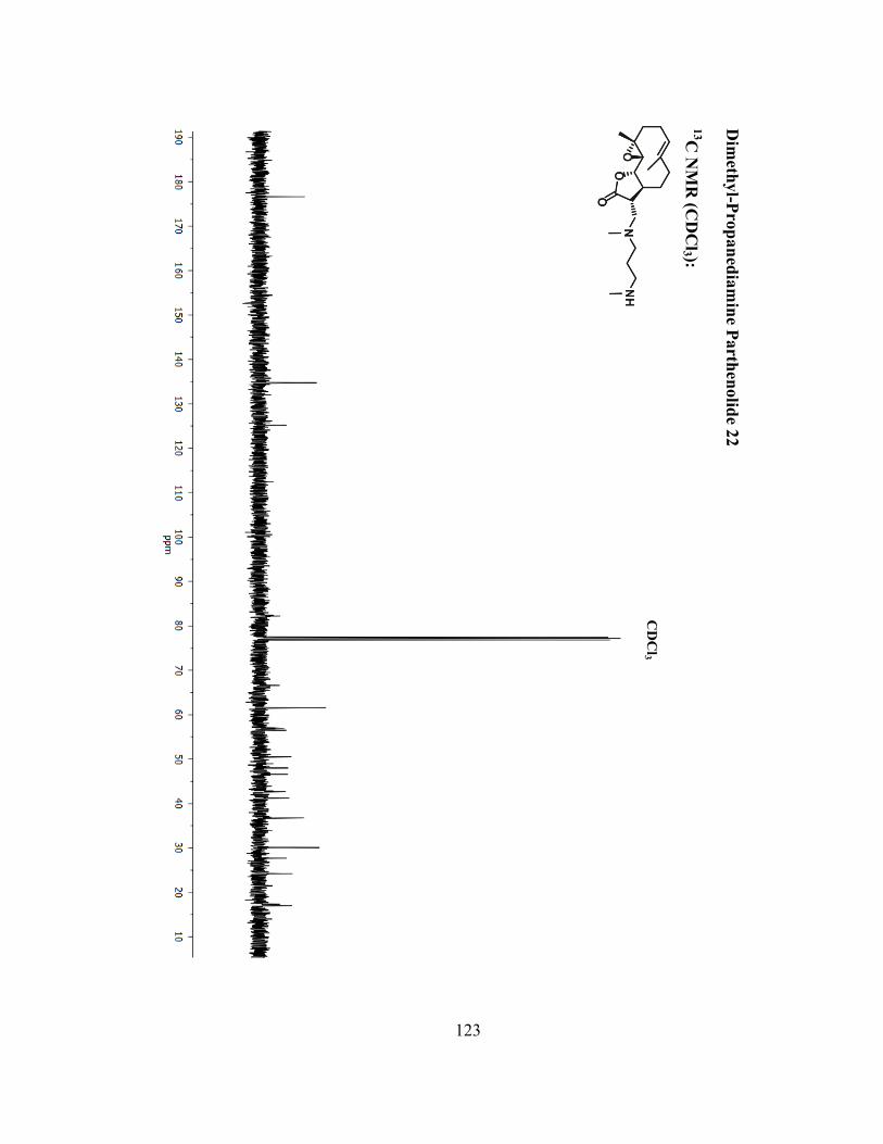
123
Dim
ethyl-P
rop
an
edia
min
e Parth
enolid
e 22
13C
NM
R (C
DC
l3 ):
C
DC
l3

124
Bibliography
(1) Al-Hajj, M. Cancer stem cells and oncology therapeutics Curr. Opin.
Oncol. 2007, 19, 61.
(2) Behbod, F.; Rosen, J. M. Will cancer stem cells provide new therapeutic
targets? Carcinogenesis 2005, 26, 703.
(3) Dick, J. E. Stem cell concepts renew cancer research Blood 2008, 112,
4793.
(4) Reya, T.; Morrison, S. J.; Clarke, M. F.; Weissman, I. L. Stem cells,
cancer, and cancer stem cells Nature 2001, 414, 105.
(5) Saini, V.; Shoemaker, R. H. Potential for therapeutic targeting of tumor
stem cells Cancer Sci. 2010, 101, 16.
(6) Wang, J. C. Y. Evaluating therapeutic efficacy against cancer stem cells:
New challenges posed by a new paradigm Cell Stem Cell 2007, 1, 497.
(7) Ward, R. J.; Dirks, P. B. Cancer Stem Cells: At the Headwaters of Tumor
Development Annu. Rev. Pathol-Mech. 2007, 2, 175.
(8) Zhou, B. B. S.; Zhang, H. Y.; Damelin, M.; Geles, K. G.; Grindley, J. C.;
Dirks, P. B. Tumour-initiating cells: challenges and opportunities for anticancer
drug discovery Nat. Rev. Drug Discov. 2009, 8, 806.
(9) Bonnet, D.; Dick, J. E. Human acute myeloid leukemia is organized as a
hierarchy that originates from a primitive hematopoietic cell Nat. Med. 1997, 3,
730.
(10) Lapidot, T.; Sirard, C.; Vormoor, J.; Murdoch, B.; Hoang, T.;
Cacerescortes, J.; Minden, M.; Paterson, B.; Caligiuri, M. A.; Dick, J. E. A Cell
initiating human acute myeloid leukemia after transplantation into SCID mice
Nature 1994, 367, 645.
(11) Fialkow, P. J.; Singer, J. W.; Raskind, W. H.; Adamson, J. W.; Jacobson,
R. J.; Bernstein, I. D.; Dow, L. W.; Najfeld, V.; Veith, R. Clonal development,
stem-cell differentiation, and clinical remission in acute nonlymphocytic
leukemia New Engl. J. Med. 1987, 317, 468.
(12) Griffin, J. D.; Lowenberg, B. Clonogenic cells in acute myeloblastic
leukemia Blood 1986, 68, 1185.
(13) McCulloch, E. A. Stem cells in normal and leukemic hematopoiesis
(Henry Stratton Lecture) Blood 1983, 62, 1.
(14) Li, L.; Bhatia, R. Stem Cell Quiescence Clin. Cancer Res. 2011, 17,
4936.
(15) Sehl, M.; Zhou, H.; Sinsheimer, J. S.; Lange, K. L. Extinction models for
cancer stem cell therapy Math. Biosci. 2011, 234, 132.
(16) Dey, M.; Ulasov, I. V.; Tyler, M. A.; Sonabend, A. M.; Lesniak, M. S.
Cancer Stem Cells: The Final Frontier for Glioma Virotherapy Stem Cell Rev.
Rep. 2011, 7, 119.
(17) Natarajan, K.; Xie, Y.; Baer, M. R.; Ross, D. D. Role of breast cancer
resistance protein (BCRP/ABCG2) in cancer drug resistance Biochem.
Pharmacol. 2012, 83, 1084.

125
(18) American Cance Society. Cancer Facts & Figures 2011 ACS 2011.
(19) Guzman, M. L.; Rossi, R. M.; Karnischky, L.; Li, X. J.; Peterson, D. R.;
Howard, D. S.; Jordan, C. T. The sesquiterpene lactone parthenolide induces
apoptosis of human acute myelogenous leukemia stem and progenitor cells
Blood 2005, 105, 4163.
(20) Jin, L.; Hope, K. J.; Zhai, Q.; Smadja-Joffe, F.; Dick, J. E. Targeting of
CD44 eradicates human acute myeloid leukemic stem cells Nat. Med. 2006, 12,
1167.
(21) Chen, Q.; Du, L.; Wang, H. Y.; He, L. Y.; Zhang, J. Y.; Ni, B. Y.; Wang,
X. H.; Jin, H. J.; Cahuzac, N.; Mehrpour, M.; Lu, Y. Y. CD44 is of Functional
Importance for Colorectal Cancer Stem Cells Clin. Cancer Res. 2008, 14, 6751.
(22) Druker, B. J.; Talpaz, M.; Resta, D. J.; Peng, B.; Buchdunger, E.; Ford, J.
M.; Lydon, N. B.; Kantarjian, H.; Capdeville, R.; Ohno-Jones, S.; Sawyers, C. L.
Efficacy and safety of a specific inhibitor of the BCR-ABL tyrosine kinase in
chronic myeloid leukemia. New Engl. J. Med. 2001, 344, 1031.
(23) Kantarjian, H.; Talpaz, M.; O'Brien, S. S.; Garcia-Manero, G.;
Verstovsek, S.; Giles, F.; Rios, M. B.; Shan, J. Q.; Letvak, L.; Thomas, D.;
Faderl, S.; Ferrajoli, A.; Cortes, J. High-dose imatinib mesylate therapy in newly
diagnosed Philadelphia chromosome-positive chronic phase chronic myeloid
leukemia Blood 2004, 103, 2873.
(24) Nicholson, E.; Holyoake, T. The Chronic Myeloid Leukemia Stem Cell
Clin. Lymphoma Myeloma 2009, 9, S376.
(25) Graham, S. M.; Jorgensen, H. G.; Allan, E.; Pearson, C.; Alcorn, M. J.;
Richmond, L.; Holyoake, T. L. Primitive, quiescent, Philadelphia-positive stem
cells from patients with chronic myeloid leukemia are insensitive to STI571 in
vitro Blood 2002, 99, 319.
(26) Zoeller, M. CD44: can a cancer-initiating cell profit from an abundantly
expressed molecule? Nat. Rev. Cancer 2011, 11, 254.
(27) Garvalov, B. K.; Acker, T. Cancer stem cells: a new framework for the
design of tumor therapies J. Mol. Med-Jmm 2011, 89, 95.
(28) Collins, A. T.; Berry, P. A.; Hyde, C.; Stower, M. J.; Maitland, N. J.
Prospective Identification of Tumorigenic Prostate Cancer Stem Cells Cancer
Res. 2005, 65, 10946.
(29) Clayton, S.; Mousa, S. A. Therapeutics formulated to target cancer stem
cells: Is it in our future? Cancer Cell Int. 2011, 11.
(30) Klonisch, T.; Wiechec, E.; Hombach-Klonisch, S.; Ande, S. R.;
Wesselborg, S.; Schulze-Osthoff, K.; Los, M. Cancer stem cell markers in
common cancers – therapeutic implications Trends Mol. Med. 2008, 14, 450.
(31) Liu, H.-G.; Chen, C.; Yang, H.; Pan, Y.-F.; Zhang, X.-H. Cancer stem
cell subsets and their relationships J. Transl. Med. 2011, 9.
(32) Visvader, J. E.; Lindeman, G. J. Cancer stem cells in solid tumours:
accumulating evidence and unresolved questions Nat. Rev. Cancer 2008, 8, 755.
(33) Gilbert, C. A.; Ross, A. H. Cancer Stem Cells: Cell Culture, Markers, and
Targets for New Therapies J. Cell Biochem. 2009, 108, 1031.

126
(34) Alison, M. R.; Lim, S. M. L.; Nicholson, L. J. Cancer stem cells:
problems for therapy? J. Pathol. 2011, 223, 147.
(35) Jordan, C. T. Cancer sten cell biology: from leukemia to solid tumors
Curr. Opin. Cell Biol. 2004, 16, 708.
(36) Jemal, A.; Bray, F.; Center, M. M.; Ferlay, J.; Ward, E.; Forman, D.
Global Cancer Statistics CA-A Cancer Journal for Clinicians 2011, 61, 69.
(37) Siegel, R.; Ward, E.; Brawley, O.; Jemal, A. Cancer statistics, 2011: the
impact of eliminating socioeconomic and racial disparities on premature cancer
deaths CA Cancer J Clin 2011, 61, 212.
(38) Testa, U. Leukemia stem cells Ann. Hematol. 2011, 90, 245.
(39) Blair, A.; Hogge, D. E.; Ailles, L. E.; Lansdorp, P. M.; Sutherland, H. J.
Lack of expression of Thy-1 (CD90) on acute myeloid leukemia cells with long-
term proliferative ability in vitro and in vivo Blood 1997, 89, 3104.
(40) Blair, A.; Hogge, D. E.; Sutherland, H. J. Most acute myeloid leukemia
progenitor cells with long-term proliferative ability in vitro and in vivo have the
phenotype CD34(+)/CD71(-)/HLA-DR Blood 1998, 92, 4325.
(41) Blair, A.; Sutherland, H. J. Primitive acute myeloid leukemia cells with
long-term proliferative ability in vitro and in vivo lack surface expression of c-kit
(CD 117) Exp. Hematol. 2000, 28, 660.
(42) Jordan, C. T.; Upchurch, D.; Szilvassy, S. J.; Guzman, M. L.; Howard, D.
S.; Pettigrew, A. L.; Meyerrose, T.; Rossi, R.; Grimes, B.; Rizzieri, D. A.; Luger,
S. M.; Phillips, G. L. The interleukin-3 receptor alpha chain is a unique marker
for human acute myelogenous leukemia stem cells Leukemia 2000, 14, 1777.
(43) Florian, S.; Sonneck, K.; Hauswirth, A. W.; Krauth, M. T.; Schernthaner,
G. H.; Sperr, W. R.; Valent, P. Detection of molecular targets on the surface of
CD34+/CD38-stem cells in various myeloid malignancies Leukemia Lymphoma
2006, 47, 207.
(44) Dinndorf, P. A.; Andrews, R. G.; Benjamin, D.; Ridgway, D.; Wolff, L.;
Bernstein, I. D. Expression of normal myeloid-associated antigens by acute
leukemia cells Blood 1986, 67, 1048.
(45) Hauswirth, A. W.; Florian, S.; Printz, D.; Sotlar, K.; Krauth, M. T.;
Fritsch, G.; Schernthaner, G. H.; Wacheck, V.; Selzer, E.; Sperr, W. R.; Valent,
P. Expression of the target receptor CD33 in CD34(+)/CD38(-)/CD123(+) AML
stem cells European Journal of Clinical Investigation 2007, 37, 73.
(46) de Figueiredo-Pontes, L. L.; Pintao, M.-C. T.; Oliveira, L. C. O.;
Dalmazzo, L. F. F.; Jacomo, R. H.; Garcia, A. B.; Falcao, R. P.; Rego, E. M.
Determination of P-glycoprotein, MDR-related protein 1, breast cancer
resistance protein, and lung-resistance protein expression in leukemic stem cells
of acute myeloid leukemia Cytom. Part B-Clin. Cy. 2008, 74B, 163.
(47) van Rhenen, A.; van Dongen, G. A. M. S.; Kelder, A.; Rombouts, E. J.;
Feller, N.; Moshaver, B.; Stigter-van Walsum, M.; Zweegman, S.; Ossenkoppele,
G. J.; Schuurhuis, G. J. The novel AML stem cell-associated antigen CLL-1 aids
in discrimination between normal and leukemic stem cells Blood 2007, 110,
2659.

127
(48) Hosen, N.; Park, C. Y.; Tatsumi, N.; Oji, Y.; Sugiyama, H.; Gramatzki,
M.; Krensky, A. M.; Weissman, I. L. CD96 is a leukemic stem cell-specific
marker in human acute myeloid leukemia Proc. Natl. Acad. Sci. USA. 2007, 104,
11008.
(49) Bendall, L. J.; Bradstock, K. F.; Gottlieb, D. J. Expression of CD44
variant exons in acute myeloid leukemia is more common and more complex
than that observed in normal blood, bone marrow or CD34(+) cells Leukemia
2000, 14, 1239.
(50) Saito, Y.; Kitamura, H.; Hijikata, A.; Tomizawa-Murasawa, M.; Tanaka,
S.; Takagi, S.; Uchida, N.; Suzuki, N.; Sone, A.; Najima, Y.; Ozawa, H.; Wake,
A.; Taniguchi, S.; Shultz, L. D.; Ohara, O.; Ishikawa, F. Identification of
Therapeutic Targets for Quiescent, Chemotherapy-Resistant Human Leukemia
Stem Cells Sci. Transl. Med. 2010, 2.
(51) Majeti, R.; Chao, M. P.; Alizadeh, A. A.; Pang, W. W.; Jaiswal, S.;
Gibbs, K. D., Jr.; van Rooijen, N.; Weissman, I. L. CD47 Is an Adverse
Prognostic Factor and Therapeutic Antibody Target on Human Acute Myeloid
Leukemia Stem Cells Cell 2009, 138, 286.
(52) Quijano, C. A.; Moore, D.; Arthur, D.; Feusner, J.; Winter, S. S.;
Pallavicini, M. G. Cytogenetically aberrant cells are present in the
CD34(+)CD33(-)38(-)19(-) marrow compartment in children with acute
lymphoblastic leukemia Leukemia 1997, 11, 1508.
(53) Cobaleda, C.; Gutierrez-Cianca, N.; Perez-Losada, J.; Flores, T.; Garcia-
Sanz, R.; Gonzalez, M.; Sanchez-Garcia, I. A primitive hematopoietic cell is the
target for the leukemic transformation in human Philadelphia-positive acute
lymphoblastic leukemia Blood 2000, 95, 1007.
(54) Advani, A. S.; Pendergast, A. M. Bcr-Abl variants: biological and clinical
aspects Leukemia Res. 2002, 26, 713.
(55) Faderl, S.; Garcia-Manero, G.; Thomas, D. A.; Kantarjian, H. M.
Philadelphia chromosome-positive acute lymphoblastic leukemia– current
concepts and future perspectives Rev. Clin. Exp. Hematol. 2002, 6, 142.
(56) Castor, A.; Nilsson, L.; Astrand-Grundstrom, I.; Buitenhuis, M.;
Ramirez, C.; Anderson, K.; Strombeck, B.; Garwicz, S.; Bekassy, A. N.;
Schmiegelow, K.; Lausen, B.; Hokland, P.; Lehmann, S.; Juliusson, G.;
Johansson, B.; Jacobsen, S. E. W. Distinct patterns of hematopoietic stem cell
involvement in acute lymphoblastic leukemia Nat. Med. 2005, 11, 630.
(57) Yeoh, E. J.; Ross, M. E.; Shurtleff, S. A.; Williams, W. K.; Patel, D.;
Mahfouz, R.; Behm, F. G.; Raimondi, S. C.; Relling, M. V.; Patel, A.; Cheng, C.;
Campana, D.; Wilkins, D.; Zhou, X. D.; Li, J. Y.; Liu, H. Q.; Pui, C. H.; Evans,
W. E.; Naeve, C.; Wong, L. S.; Downing, J. R. Classification, subtype discovery,
and prediction of outcome in pediatric acute lymphoblastic leukemia by gene
expression profiling Cancer Cell 2002, 1, 133.
(58) Cox, C. V.; Evely, R. S.; Oakhill, A.; Pamphilon, D. H.; Goulden, N. J.;
Blair, A. Characterization of acute lymphoblastic leukemia progenitor cells
Blood 2004, 104, 2919.

128
(59) Cox, C. V.; Martin, H. M.; Kearns, P. R.; Virgo, P.; Evely, R. S.; Blair,
A. Characterization of a progenitor cell population in childhood T-cell acute
lymphoblastic leukemia Blood 2007, 109, 674.
(60) Cox, C. V.; Diamanti, P.; Evely, R. S.; Kearns, P. R.; Blair, A.
Expression of CD133 on leukemia-initiating cells in childhood ALL Blood 2009,
113, 3287.
(61) Nishida, H.; Yamazaki, H.; Yamada, T.; Iwata, S.; Dang, N. H.; Inukai,
T.; Sugita, K.; Ikeda, Y.; Morimoto, C. CD9 correlates with cancer stem cell
potentials in human B-acute lymphoblastic leukemia cells Biochem. Bioph. Res.
Co. 2009, 382, 57.
(62) Yamazaki, H.; Nishida, H.; Iwata, S.; Dang, N. H.; Morimoto, C. CD90
and CD110 correlate with cancer stem cell potentials in human T-acute
lymphoblastic leukemia cells Biochem. Bioph. Res. Co. 2009, 383, 172.
(63) Holyoake, T.; Jiang, X. Y.; Eaves, C.; Eaves, A. Isolation of a highly
quiescent subpopulation of primitive leukemic cells in chronic myeloid leukemia
Blood 1999, 94, 2056.
(64) DeSantis, C.; Siegel, R.; Bandi, P.; Jemal, A. Breast cancer statistics,
2011 CA Cancer J Clin 2011, 61, 409.
(65) Takahashi, R. T., Fumitaka; Fujiwara,Tomohiro; Ono, Makiko and
Ochiya, Takahiro Cancer stem cells in breast cancer Cancers 2011, 3, 1311.
(66) Al-Hajj, M.; Wicha, M. S.; Benito-Hernandez, A.; Morrison, S. J.;
Clarke, M. F. Prospective identification of tumorigenic breast cancer cells Proc.
Natl. Acad. Sci. USA. 2003, 100, 3983.
(67) Phillips, T. M.; McBride, W. H.; Pajonk, F. The response of CD24(-
/low)/CD44(+) breast cancer-initiating cells to radiation J. Natl. Cancer I. 2006,
98, 1777.
(68) Buess, M.; Rajski, M.; Vogel-Durrer, B. M. L.; Herrmann, R.; Rochlitz,
C. Tumor-Endothelial Interaction Links the CD44(+)/CD24(-) Phenotype with
Poor Prognosis in Early-Stage Breast Cancer Neoplasia 2009, 11, 987.
(69) Sheridan, C.; Kishimoto, H.; Fuchs, R. K.; Mehrotra, S.; Bhat-Nakshatri,
P.; Turner, C. H.; Goulet, R.; Badve, S.; Nakshatri, H. CD44(+)/CD24(-) breast
cancer cells exhibit enhanced invasive properties: an early step necessary for
metastasis Breast Cancer Res. 2006, 8.
(70) Donnelly, D. S.; Zelterman, D.; Sharkis, S.; Krause, D. S. Functional
activity of murine CD34(+) and CD34(-) hematopoietic stem cell populations
Exp. Hematol. 1999, 27, 788.
(71) Neumeister, V.; Agarwal, S.; Bordeaux, J.; Camp, R. L.; Rimm, D. L. In
Situ Identification of Putative Cancer Stem Cells by Multiplexing ALDH1,
CD44, and Cytokeratin Identifies Breast Cancer Patients with Poor Prognosis
Am. J. Pathol. 2010, 176, 2131.
(72) Cho, R. W.; Wang, X.; Diehn, M.; Shedden, K.; Chen, G. Y.; Sherlock,
G.; Gurney, A.; Lewicki, J.; Clarke, M. F. Isolation and molecular
characterization of cancer stem cells in MMTV-Wnt-1 murine breast tumors
Stem Cells 2008, 26, 364.

129
(73) Wright, M. H.; Calcagno, A. M.; Salcido, C. D.; Carlson, M. D.;
Ambudkar, S. V.; Varticovski, L. Brca1 breast tumors contain distinct
CD44(+)/CD24(-) and CD133(+) cells with cancer stem cell characteristics
Breast Cancer Res. 2008, 10.
(74) Zhao, P.; Lu, Y.; Jiang, X.; Li, X. Clinicopathological significance and
prognostic value of CD133 expression in triple-negative breast carcinoma
Cancer Sci. 2011, 102, 1107.
(75) Liu, Q.; Li, J.-g.; Zheng, X.-y.; Jin, F.; Dong, H.-t. Expression of CD133,
PAX2, ESA, and GPR30 in invasive ductal breast carcinomas Chinese Med. J-
Peking 2009, 122, 2763.
(76) Lin, W.-M.; Karsten, U.; Goletz, S.; Cheng, R.-C.; Cao, Y. Co-expression
of CD173 (H2) and CD174 (Lewis Y) with CD44 suggests that fucosylated
histo-blood group antigens are markers of breast cancer-initiating cells Vichows
Arch. 2010, 456, 403.
(77) Lin, W.-M.; Karsten, U.; Goletz, S.; Cheng, R.-C.; Cao, Y. Expression of
CD176 (Thomsen-Friedenreich antigen) on lung, breast and liver cancer-
initiating cells Int. J. Exp. Pathol. 2011, 92, 97.
(78) Vassilopoulos, A.; Wang, R.-H.; Petrovas, C.; Ambrozak, D.; Koup, R.;
Deng, C.-X. Identification and characterization of cancer initiating cells from
BRCA1 related mammary tumors using markers for normal mammary stem cells
Int. J. Biol. Sci. 2008, 4, 133.
(79) Shipitsin, M.; Campbell, L. L.; Argani, P.; Werernowicz, S.; Bloushtain-
Qimron, N.; Yao, J.; Nikolskaya, T.; Serebryiskaya, T.; Beroukhim, R.; Hu, M.;
Halushka, M. K.; Sukumar, S.; Parker, L. M.; Anderson, K. S.; Harris, L. N.;
Garber, J. E.; Richardson, A. L.; Schnitt, S. J.; Nikolsky, Y.; Gelman, R. S.;
Polyak, K. Molecular definition of breast tumor heterogeneity Cancer Cell 2007,
11, 259.
(80) Dalerba, P.; Dylla, S. J.; Park, I.-K.; Liu, R.; Wang, X.; Cho, R. W.;
Hoey, T.; Gurney, A.; Huang, E. H.; Simeone, D. M.; Shelton, A. A.; Parmiani,
G.; Castelli, C.; Clarke, M. F. Phenotypic characterization of human colorectal
cancer stem cells Proc. Natl. Acad. Sci. USA. 2007, 104, 10158.
(81) O'Brien, C. A.; Pollett, A.; Gallinger, S.; Dick, J. E. A human colon
cancer cell capable of initiating tumour growth in immunodeficient mice Nature
2007, 445, 106.
(82) Ricci-Vitiani, L.; Lombardi, D. G.; Pilozzi, E.; Biffoni, M.; Todaro, M.;
Peschle, C.; De Maria, R. Identification and expansion of human colon-cancer-
initiating cells Nature 2007, 445, 111.
(83) Shmelkov, S. V.; Butler, J. M.; Hooper, A. T.; Hormigo, A.; Kushner, J.;
Milde, T.; St Clair, R.; Baljevic, M.; White, I.; Jin, D. K.; Chadburn, A.;
Murphy, A. J.; Valenzuela, D. M.; Gale, N. W.; Thurston, G.; Yancopoulos, G.
D.; D'Angelica, M.; Kemeny, N.; Lyden, D.; Rafii, S. CD133 expression is not
restricted to stem cells, and both CD133+ and CD133- metastatic colon cancer
cells initiate tumors J Clin Invest 2008, 118, 2111.

130
(84) Du, L.; Wang, H.; He, L. CD44 is of functional importance for colorectal
cancer stem cells. (vol 14, pg 6751, 2008) Clin. Cancer Res. 2008, 14, 7964.
(85) Horst, D.; Kriegl, L.; Engel, J.; Kirchner, T.; Jung, A. Prognostic
Significance of the Cancer Stem Cell Markers CD133, CD44, and CD166 in
Colorectal Cancer Cancer Invest. 2009, 27, 844.
(86) Lugli, A.; Iezzi, G.; Hostettler, I.; Muraro, M. G.; Mele, V.; Tornillo, L.;
Carafa, V.; Spagnoli, G.; Terracciano, L.; Zlobec, I. Prognostic impact of the
expression of putative cancer stem cell markers CD133, CD166, CD44s,
EpCAM, and ALDH1 in colorectal cancer Brit. J. Cancer 2010, 103, 382.
(87) Vermeulen, L.; Todaro, M.; Mello, F. d. S.; Sprick, M. R.; Kemper, K.;
Alea, M. P.; Richel, D. J.; Stassi, G.; Medema, J. P. Single-cell cloning of colon
cancer stem cells reveals a multi-lineage differentiation capacity Proc. Natl.
Acad. Sci. USA. 2008, 105, 13427.
(88) Liu, A. Y.; True, L. D.; LaTray, L.; Nelson, P. S.; Ellis, W. J.; Vessella,
R. L.; Lange, P. H.; Hood, L.; vandenEngh, G. Cell-cell interaction in prostate
gene regulation and cytodifferentiation Proc. Natl. Acad. Sci. USA. 1997, 94,
10705.
(89) Eguchi, S.; Kanematsu, T.; Arii, S.; Omata, M.; Kudo, M.; Sakamoto,
M.; Takayasu, K.; Makuuchi, M.; Matsuyama, Y.; Monden, M.; Liver Canc
Study Grp, J. Recurrence-free survival more than 10 years after liver resection
for hepatocellular carcinoma Brit. J. Surg. 2011, 98, 552.
(90) Ma, S.; Chan, K.-W.; Hu, L.; Lee, T. K.-W.; Wo, J. Y.-H.; Ng, I.-L.;
Zheng, B.-J.; Guan, X.-Y. Identification and characterization of tumorigenic
liver cancer stem/progenitor cells Gasterenterol. 2007, 132, 2542.
(91) Zhu, Z.; Hao, X.; Yan, M.; Yao, M.; Ge, C.; Gu, J.; Li, J. Cancer
stem/progenitor cells are highly enriched in CD133(+)CD44(+) population in
hepatocellular carcinoma Int. J. Cancer 2010, 126, 2067.
(92) Kimura, O.; Takahashi, T.; Ishii, N.; Inoue, Y.; Ueno, Y.; Kogure, T.;
Fukushima, K.; Shiina, M.; Yamagiwa, Y.; Kondo, Y.; Inoue, J.; Kakazu, E.;
Iwasaki, T.; Kawagishi, N.; Shimosegawa, T.; Sugamura, K. Characterization of
the epithelial cell adhesion molecule (EpCAM) plus cell population in
hepatocellular carcinoma cell lines Cancer Sci. 2010, 101, 2145.
(93) Yamashita, T.; Ji, J. F.; Budhu, A.; Forgues, M.; Yang, W.; Wang, H. Y.;
Jia, H. L.; Ye, Q. H.; Qin, L. X.; Wauthier, E.; Reid, L. M.; Minato, H.; Honda,
M.; Kaneko, S.; Tang, Z. Y.; Wang, X. W. EpCAM-Positive Hepatocellular
Carcinoma Cells Are Tumor-Initiating Cells With Stem/Progenitor Cell Features
Gasterenterol. 2009, 136, 1012.
(94) Haraguchi, N.; Ishii, H.; Mimori, K.; Tanaka, F.; Ohkuma, M.; Kim, H.
M.; Akita, H.; Takiuchi, D.; Hatano, H.; Nagano, H.; Barnard, G. F.; Doki, Y.;
Mori, M. CD13 is a therapeutic target in human liver cancer stem cells J. Clin.
Invest. 2010, 120, 3326.
(95) Yang, Z. F.; Ho, D. W.; Ng, M. N.; Lau, C. K.; Yu, W. C.; Ngai, P.; Chu,
P. W. K.; Lam, C. T.; Poon, R. T. P.; Fan, S. T. Significance of CD90(+) cancer
stem cells in human liver cancer Cancer Cell 2008, 13, 153.

131
(96) Polakis, P. Wnt signaling and cancer Gene. Dev. 2000, 14, 1837.
(97) Yang, W.; Yan, H.-X.; Chen, L.; Liu, Q.; He, Y.-Q.; Yu, L.-X.; Zhang,
S.-H.; Huang, D.-D.; Tang, L.; Kong, X.-N.; Chen, C.; Liu, S.-Q.; Wu, M.-C.;
Wang, H.-Y. Wnt/beta-catenin signaling contributes to activation of normal and
tumorigenic liver progenitor cells Cancer Res. 2008, 68, 4287.
(98) Jemal, A.; Siegel, R.; Ward, E.; Hao, Y.; Xu, J.; Murray, T.; Thun, M. J.
Cancer statistics, 2008 Ca-Cancer J. Clin. 2008, 58, 71.
(99) Li, C.; Heidt, D. G.; Dalerba, P.; Burant, C. F.; Zhang, L.; Adsay, V.;
Wicha, M.; Clarke, M. F.; Simeone, D. M. Identification of pancreatic cancer
stem cells Cancer Res. 2007, 67, 1030.
(100) Hermann, P. C.; Huber, S. L.; Herrler, T.; Aicher, A.; Ellwart, J. W.;
Guba, M.; Bruns, C. J.; Heeschen, C. Distinct populations of cancer stem cells
determine tumor growth and metastatic activity in human pancreatic cancer Cell
Stem Cell 2007, 1, 313.
(101) Jemal, A.; Siegel, R.; Xu, J.; Ward, E. Cancer Statistics, 2010 Ca-Cancer
J. Clin. 2010, 60, 277.
(102) Fang, D.; Nguyen, T. K.; Leishear, K.; Finko, R.; Kulp, A. N.; Hotz, S.;
Van Belle, P. A.; Xu, X. W.; Elder, D. E.; Herlyn, M. A tumorigenic
subpopulation with stem cell properties in melanomas Cancer Res. 2005, 65,
9328.
(103) Monzani, E.; Facchetti, F.; Galmozzi, E.; Corsini, E.; Benetti, A.;
Cavazzin, C.; Gritti, A.; Piccinini, A.; Porro, D.; Santinami, M.; Invernici, G.;
Parati, E.; Alessandri, G.; La Porta, C. A. M. Melanoma contains CD133 and
ABCG2 positive cells with enhanced tumourigenic potential Eur. J. Cancer
2007, 43, 935.
(104) Grichnik, J. M.; Burch, J. A.; Schulteis, R. D.; Shan, S.; Liu, J.; Darrow,
T. L.; Vervaert, C. E.; Seigler, H. F. Melanoma, a tumor based on a mutant stem
cell? J. Invest. Dermatol. 2006, 126, 142.
(105) Fusi, A.; Reichelt, U.; Busse, A.; Ochsenreither, S.; Rietz, A.; Maisel,
M.; Keilholz, U. Expression of the Stem Cell Markers Nestin and CD133 on
Circulating Melanoma Cells J. Invest. Dermatol. 2011, 131, 487.
(106) Schatton, T.; Frank, N. Y.; Frank, M. H. Identification and targeting of
cancer stem cells Bioessays 2009, 31, 1038.
(107) Schatton, T.; Murphy, G. F.; Frank, N. Y.; Yamaura, K.; Waaga-Gasser,
A. M.; Gasser, M.; Zhan, Q.; Jordan, S.; Duncan, L. M.; Weishaupt, C.;
Fuhlbrigge, R. C.; Kupper, T. S.; Sayegh, M. H.; Frank, M. H. Identification of
cells initiating human melanomas Nature 2008, 451, 345.
(108) Davis, F. G.; Kupelian, V.; Freels, S.; McCarthy, B.; Surawicz, T.
Prevalence estimates for primary brain tumors in the United States by behavior
and major histology groups Neuro-Oncology 2001, 3, 152.
(109) Khalatbari, M. R.; Hamidi, M.; Moharamzad, Y. Glioblastoma
multiforme with very rapid growth and long-term survival in children: report of
two cases and review of the literature Child. Nerv. Syst. 2012, 28, 9.

132
(110) Eyler, C. E.; Rich, J. N. Survival of the fittest: Cancer stem cells in
therapeutic resistance and angiogenesis J. Clin. Oncol. 2008, 26, 2839.
(111) Binello, E.; Germano, I. M. Targeting glioma stem cells: A novel
framework for brain tumors Cancer Sci. 2011, 102, 1958.
(112) Singh, S. K.; Clarke, I. D.; Terasaki, M.; Bonn, V. E.; Hawkins, C.;
Squire, J.; Dirks, P. B. Identification of a cancer stem cell in human brain tumors
Cancer Res. 2003, 63, 5821.
(113) Singh, S. K.; Hawkins, C.; Clarke, I. D.; Squire, J. A.; Bayani, J.; Hide,
T.; Henkelman, R. M.; Cusimano, M. D.; Dirks, P. B. Identification of human
brain tumour initiating cells Nature 2004, 432, 396.
(114) Tchoghandjian, A.; Baeza, N.; Colin, C.; Cayre, M.; Metellus, P.; Beclin,
C.; Ouafik, L. H.; Figarella-Branger, D. A2B5 Cells from Human Glioblastoma
have Cancer Stem Cell Properties Brain Pathol. 2010, 20, 211.
(115) Ogden, A. T.; Waziri, A. E.; Lochhead, R. A.; Fusco, D.; Lopez, K.;
Ellis, J. A.; Kang, J.; Assanah, M.; McKhann, G. M.; Sisti, M. B.; McCormick,
P. C.; Canoll, P.; Bruce, J. N. Identification of A2B5(+)CD133-tumor-initiating
cells in adult human gliomas Neurosurgery 2008, 62, 505.
(116) Wang, J.; Sakariassen, P. O.; Tsinkalovsky, O.; Immervoll, H.; Boe, S.
O.; Svendsen, A.; Prestegarden, L.; Rosland, G.; Thorsen, F.; Stuhr, L.; Molven,
A.; Bjerkvig, R.; Enger, P. O. CD133 negative glioma cells form tumors in nude
rats and give rise to CD133 positive cells Int. J. Cancer 2008, 122, 761.
(117) Bao, S.; Wu, Q.; Li, Z.; Sathornsumetee, S.; Wang, H.; McLendon, R. E.;
Hjehneland, A. B.; Rich, J. N. Targeting cancer stem cells through L1CAM
suppresses glioma growth Cancer Res. 2008, 68, 6043.
(118) Bao, S.; Wu, Q.; Sathornsumetee, S.; Hao, Y.; Li, Z.; Hjelmeland, A. B.;
Shi, O.; McLendon, R. E.; Bigner, D. D.; Rich, J. N. Stem cell-like glioma cells
promote tumor angiogenesis through vascular endothelial growth factor Cancer
Res. 2006, 66, 7843.
(119) Son, M. J.; Woolard, K.; Nam, D.-H.; Lee, J.; Fine, H. A. SSEA-1 Is an
Enrichment Marker for Tumor-Initiating Cells in Human Glioblastoma Cell Stem
Cell 2009, 4, 440.
(120) Read, T.-A.; Fogarty, M. P.; Markant, S. L.; McLendon, R. E.; Wei, Z.;
Ellison, D. W.; Febbo, P. G.; Wechsler-Reya, R. J. Identification of CD15 as a
Marker for Tumor-Propagating Cells in a Mouse Model of Medulloblastoma
Cancer Cell 2009, 15, 135.
(121) Dean, M.; Fojo, T.; Bates, S. Tumour stem cells and drug resistance Nat.
Rev. Cancer 2005, 5, 275.
(122) Eramo, A.; Lotti, F.; Sette, G.; Pilozzi, E.; Biffoni, M.; Di Virgilio, A.;
Conticello, C.; Ruco, L.; Peschle, C.; De Maria, R. Identification and expansion
of the tumorigenic lung cancer stem cell population Cell Death Differ 2007, 15,
504.
(123) Dinney, C. P. N.; McConkey, D. J.; Millikan, R. E.; Wu, X. F.; Bar-Eli,
M.; Adam, L.; Kamat, A. M.; Siefker-Radtke, A. O.; Tuziak, T.; Sabichi, A. L.;

133
Grossman, H. B.; Benedict, W. F.; Czerniak, B. Focus on bladder cancer Cancer
Cell 2004, 6, 111.
(124) Su, Y.; Qiu, Q.; Zhang, X.; Jiang, Z.; Leng, Q.; Liu, Z.; Stass, S. A.;
Jiang, F. Aldehyde Dehydrogenase 1 A1-Positive Cell Population Is Enriched in
Tumor-Initiating Cells and Associated with Progression of Bladder Cancer
Cancer Epidem. Biomar. 2010, 19, 327.
(125) Chan, K. S.; Espinosa, I.; Chao, M.; Wong, D.; Ailles, L.; Diehn, M.;
Gill, H.; Presti, J.; Chang, H. Y.; van de Rijn, M.; Shortliffe, L.; Weissman, I. L.
Identification, molecular characterization, clinical prognosis, and therapeutic
targeting of human bladder tumor-initiating cells Proc. Nat. Acad. Sci. 2009,
106, 14016.
(126) Yang, Y. M.; Chang, J. W. Bladder cancer initiating cells (BCICs) are
among EMA(-)CD44v6(+) subset: Novel methods for isolating undetermined
cancer stem (initiating) cells Cancer Invest. 2008, 26, 725.
(127) Ning, Z. F.; Huang, Y. J.; Lin, T. X.; Zhou, Y. X.; Jiang, C.; Xu, K. W.;
Huang, H.; Yin, X. B.; Huang, J. Subpopulations of Stem-like Cells in Side
Population Cells from the Human Bladder Transitional Cell Cancer Cell Line
T24 J. Int. Med. Res. 2009, 37, 621.
(128) Oates, J. E.; Grey, B. R.; Addla, S. K.; Samuel, J. D.; Hart, C. A.;
Ramani, V. A. C.; Brown, M. D.; Clarke, N. W. Hoechst 33342 Side Population
Identification Is a Conserved and Unified Mechanism in Urological Cancers
Stem Cells Dev. 2009, 18, 1515.
(129) Bostwick, D. G. E., J. N. Urological Surgical Pathology; 2nd ed.; Mosby,
2007.
(130) Mulholland, D. J.; Xin, L.; Morim, A.; Lawson, D.; Witte, O.; Wu, H.
Lin(-)Sca-1(+)CD49f(high) Stem/Progenitors Are Tumor-Initiating Cells in the
Pten-Null Prostate Cancer Model Cancer Res. 2009, 69, 8555.
(131) Beltran, H.; Beer, T. M.; Carducci, M. A.; de Bono, J.; Gleave, M.;
Hussain, M.; Kelly, W. K.; Saad, F.; Sternberg, C.; Tagawa, S. T.; Tannock, I. F.
New Therapies for Castration-Resistant Prostate Cancer: Efficacy and Safety
Eur. Urol. 2011, 60, 279.
(132) Patrawala, L.; Calhoun, T.; Schneider-Broussard, R.; Li, H.; Bhatia, B.;
Tang, S.; Reilly, J. G.; Chandra, D.; Zhou, J.; Claypool, K.; Coghlan, L.; Tang,
D. G. Highly purified CD44(+) prostate cancer cells from xenograft human
tumors are enriched in tumorigenic and metastatic progenitor cells Oncogene
2006, 25, 1696.
(133) Patrawala, L.; Calhoun-Davis, T.; Schneider-Broussard, R.; Tang, D. G.
Hierarchical organization of prostate cancer cells in xenograft tumors: The
CD44(+)alpha 2 beta 1(+) cell population is enriched in tumor-initiating cells
Cancer Res. 2007, 67, 6796.
(134) Suzuki, A.; Nakano, T.; Mak, T. W.; Sasaki, T. Portrait of PTEN:
Messages from mutant mice Cancer Sci. 2008, 99, 209.
(135) Zhang, S.; Balch, C.; Chan, M. W.; Lai, H.-C.; Matei, D.; Schilder, J. M.;
Yan, P. S.; Huang, T. H.-M.; Nephew, K. P. Identification and Characterization

134
of Ovarian Cancer-Initiating Cells from Primary Human Tumors Cancer Res.
2008, 68, 4311.
(136) Choi, Y. P.; Shim, H. S.; Gao, M.-Q.; Kang, S.; Cho, N. H. Molecular
portraits of intratumoral heterogeneity in human ovarian cancer Cancer Lett.
2011, 307, 62.
(137) Ferrandina, G.; Bonanno, G.; Pierelli, L.; Perillo, A.; Procoli, A.;
Mariotti, A.; Corallo, M.; Martinelli, E.; Rutella, S.; Paglia, A.; Zannoni, G.;
Mancuso, S.; Scambia, G. Expression of CD133-1 and CD133-2 in ovarian
cancer Int. J. Gynecol. Cancer 2008, 18, 506.
(138) Curley, M. D.; Therrien, V. A.; Cummings, C. L.; Sergent, P. A.;
Koulouris, C. R.; Friel, A. M.; Roberts, D. J.; Seiden, M. V.; Scadden, D. T.;
Rueda, B. R.; Foster, R. CD133 Expression Defines a Tumor Initiating Cell
Population in Primary Human Ovarian Cancer Stem Cells 2009, 27, 2875.
(139) Gao, M. Q.; Choi, Y. P.; Kang, S.; Youn, J. H.; Cho, N. H. CD24+ cells
from hierarchically organized ovarian cancer are enriched in cancer stem cells
Oncogene 2010, 29, 2672.
(140) Wei, X.; Dombkowski, D.; Meirelles, K.; Pieretti-Vanmarcke, R.; Szotek,
P. P.; Chang, H. L.; Preffer, F. I.; Mueller, P. R.; Teixeira, J.; MacLaughlin, D.
T.; Donahoe, P. K. Mullerian inhibiting substance preferentially inhibits
stem/progenitors in human ovarian cancer cell lines compared with
chemotherapeutics Proc. Natl. Acad. Sci. USA. 2010, 107, 18874.
(141) Gupta, P. B.; Onder, T. T.; Jiang, G. Z.; Tao, K.; Kuperwasser, C.;
Weinberg, R. A.; Lander, E. S. Identification of Selective Inhibitors of Cancer
Stem Cells by High-Throughput Screening Cell 2009, 138, 645.
(142) Shmelkov, S. V.; St Clair, R.; Lyden, D.; Rafii, S.
AC133/CD133/Prominin-1 Int J Biochem Cell B 2005, 37, 715.
(143) Griguer, C. E.; Oliva, C. R.; Gobin, E.; Marcorelles, P.; Benos, D. J.;
Lancaster, J. R., Jr.; Gillespie, G. Y. CD133 Is a Marker of Bioenergetic Stress in
Human Glioma Plos One 2008, 3.
(144) Weigmann, A.; Corbeil, D.; Hellwig, A.; Huttner, W. B. Prominin, a
novel microvilli-specific polytopic membrane protein of the apical surface of
epithelial cells, is targeted to plasmalemmal protrusions of non-epithelial cells
Proc. Natl. Acad. Sci. USA. 1997, 94, 12425.
(145) Rappa, G.; Fodstad, O.; Lorico, A. The Stem Cell-Associated Antigen
CD133 (Prominin-1) Is a Molecular Therapeutic Target for Metastatic
Melanoma Stem Cells 2008, 26, 3008.
(146) Wu, Y. J.; Wu, P. Y. CD133 as a Marker for Cancer Stem Cells:
Progresses and Concerns Stem Cells Dev. 2009, 18, 1127.
(147) Swaminathan, S. K.; Olin, M. R.; Forster, C. L.; Cruz, K. S. S.; Panyam,
J.; Ohlfest, J. R. Identification of a novel monoclonal antibody recognizing
CD133 J. Immunol. Methods. 2010, 361, 110.
(148) Bourseau-Guilmain, E.; Bejaud, J.; Griveau, A.; Lautram, N.; Hindre, F.;
Weyland, M.; Benoit, J. P.; Garcion, E. Development and characterization of
135
immuno-nanocarriers targeting the cancer stem cell marker AC133 Int. J. Pharm.
2012, 423, 93.
(149) Seimetz, D.; Lindhofer, H.; Bokemeyer, C. Development and approval of
the trifunctional antibody catumaxomab (anti-EpCAM x anti-CD3) as a targeted
cancer immunotherapy Cancer Treat. Rev. 2010, 36, 458.
(150) Haas, C.; Krinner, E.; Brischwein, K.; Hoffmann, P.; Lutterbuese, R.;
Schlereth, B.; Kufer, P.; Baeuerle, P. A. Mode of cytotoxic action of T cell-
engaging BiTE antibody MT110 Immunobiology 2009, 214, 441.
(151) Hartung, G.; Hofheinz, R. D.; Dencausse, Y.; Sturm, J.; Kopp-Schneider,
A.; Dietrich, G.; Fackler-Schwalbe, I.; Bornbusch, D.; Gonnermann, M.;
Wojatschek, C.; Lindemann, W.; Eschenburg, H.; Jost, K.; Edler, L.; Hochhaus,
A.; Queisser, W. Adjuvant therapy with edrecolomab versus observation in stage
II colon cancer: A multicenter randomized phase III study Onkologie 2005, 28,
347.
(152) Naundorf, S.; Preithner, S.; Mayer, P.; Lippold, S.; Wolf, A.; Hanakam,
F.; Fichtner, I.; Kufer, P.; Raum, T.; Riethmuller, G.; Baeuerle, P. A.; Dreier, T.
In vitro and in vivo activity of MT201, a fully human monoclonal antibody for
pancarcinoma treatment Int. J. Cancer 2002, 100, 101.
(153) Marangoni, E.; Lecomte, N.; Durand, L.; de Pinieux, G.; Decaudin, D.;
Chomienne, C.; Smadja-Joffe, F.; Poupon, M. F. CD44 targeting reduces tumour
growth and prevents post-chemotherapy relapse of human breast cancers
xenografts Brit. J. Cancer 2009, 100, 918.
(154) Hamann, P. R.; Hinman, L. M.; Hollander, I.; Beyer, C. F.; Lindh, D.;
Holcomb, R.; Hallett, W.; Tsou, H. R.; Upeslacis, J.; Shochat, D.; Mountain, A.;
Flowers, D. A.; Bernstein, I. Gemtuzumab ozogamicin, a potent and selective
anti-CD33 antibody-calicheamicin conjugate for treatment of acute myeloid
leukemia Bioconjugate chemistry 2002, 13, 47.
(155) Hamann, P. R.; Hinman, L. M.; Beyer, C. F.; Lindh, D.; Upeslacis, J.;
Flowers, D. A.; Bernstein, I. An anti-CD33 antibody-calicheamicin conjugate for
treatment of acute myeloid leukemia. Choice of linker Bioconjugate chemistry
2002, 13, 40.
(156) Frank, M. H.; Schatton, T.; Murphy, G. F.; Frank, N. Y.; Yamaura, K.;
Waaga-Gasser, A. M.; Gasser, M.; Zhan, Q.; Jordan, S.; Duncan, L. M.;
Weishaupt, C.; Fuhlbrigge, R. C.; Kupper, T. S.; Sayegh, M. H. Identification of
cells initiating human melanomas Nature 2008, 451, 345.
(157) Jin, L. Q.; Lee, E. M.; Ramshaw, H. S.; Busfield, S. J.; Peoppl, A. G.;
Wilkinson, L.; Guthridge, M. A.; Thomas, D.; Barry, E. F.; Boyd, A.; Gearing,
D. P.; Vairo, G.; Lopez, A. F.; Dick, J. E.; Lock, R. B. Monoclonal Antibody-
Mediated Targeting of CD123, IL-3 Receptor alpha Chain, Eliminates Human
Acute Myeloid Leukemic Stem Cells Cell Stem Cell 2009, 5, 31.
(158) Bidlingmaier, S.; Zhu, X.; Liu, B. The utility and limitations of
glycosylated human CD133 epitopes in defining cancer stem cells J. Mol. Med-
Jmm 2008, 86, 1025.

136
(159) Wang, C.-H.; Chiou, S.-H.; Chou, C.-P.; Chen, Y.-C.; Huang, Y.-J.;
Peng, C.-A. Photothermolysis of glioblastoma stem-like cells targeted by carbon
nanotubes conjugated with CD133 monoclonal antibody Nanomedicine-
Nanotechnology Biology and Medicine 2011, 7, 69.
(160) van der Gun, B. T. F.; Melchers, L. J.; Ruiters, M. H. J.; de Leij, L. F. M.
H.; McLaughlin, P. M. J.; Rots, M. G. EpCAM in carcinogenesis: the good, the
bad or the ugly Carcinogenesis 2010, 31, 1913.
(161) Winter, M. J.; Nagelkerken, B.; Mertens, A. E. E.; Rees-Bakker, H. A.
M.; Briaire-de Bruijn, I. H.; Litvinov, S. V. Expression of Ep-CAM shifts the
state of cadherin-mediated adhesions from strong to weak Exp. Cell Res. 2003,
285, 50.
(162) Litvinov, S. V.; Velders, M. P.; Bakker, H. A. M.; Fleuren, G. J.;
Warnaar, S. O. Ep-CAM - A human epithelial antigen is a homophilic cell-cell
adhesion molecule J. Cell Biol. 1994, 125, 437.
(163) Ruf, P.; Gires, O.; Jager, M.; Fellinger, K.; Atz, J.; Lindhofer, H.
Characterisation of the new EpCAM-specific antibody HO-3: implications for
trifunctional antibody immunotherapy of cancer Brit. J. Cancer 2007, 97, 315.
(164) Chelius, D.; Ruf, P.; Gruber, P.; Ploscher, M.; Liedtke, R.; Gansberger,
E.; Hess, J.; Wasiliu, M.; Lindhofer, H. Structural and functional characterization
of the trifunctional antibody catumaxomab Mabs-Austin 2010, 2, 309.
(165) Lindhofer, H.; Schoberth, A.; Pelster, D.; Hess, J.; Herold, J.; Jager, M.
Elimination of cancer stem cells (CD133+/EpCAM+) from malignant ascites by
the trifunctional antibody catumaxomab: Results from a pivotal phase II/III study
J. Clin. Oncol. 2009, 27.
(166) Jager, M.; Schoberth, A.; Ruf, P.; Hess, J.; Hennig, M.; Schmalfeldt, B.;
Wimberger, P.; Strohlein, M.; Theissen, B.; Heiss, M. M.; Lindhofer, H.
Immunomonitoring Results of a Phase II/III Study of Malignant Ascites Patients
Treated with the Trifunctional Antibody Catumaxomab (Anti-EpCAM x Anti-
CD3) Cancer Res. 2012, 72, 24.
(167) Brischwein, K.; Schlereth, B.; Guller, B.; Steiger, C.; Wolf, A.;
Lutterbuese, R.; Offner, S.; Locher, M.; Urbig, T.; Raum, T.; Kleindienst, P.;
Wimberger, P.; Kimmig, R.; Fichtner, I.; Kufer, P.; Hofmeister, R.; da Silva, A.
J.; Baeuerle, P. A. MT110: A novel bispecific single-chain antibody construct
with high efficacy in eradicating established tumors Mol. Immunol. 2006, 43,
1129.
(168) Baeuerle, P. A.; Kufer, P.; Bargou, R. BiTE: Teaching antibodies to
engage T-cells for cancer therapy Curr. Opin. Mol. Ther. 2009, 11, 22.
(169) Amann, M.; Brischwein, K.; Lutterbuese, P.; Parr, L.; Petersen, L.;
Lorenczewski, G.; Krinner, E.; Bruckmeier, S.; Lippold, S.; Kischel, R.;
Lutterbuese, R.; Kufer, P.; Baeuerle, P. A.; Schlereth, B. Therapeutic window of
MuS110, a single-chain antibody construct bispecific for murine EpCAM and
murine CD3 Cancer Res. 2008, 68, 143.
(170) Herrmann, I.; Baeuerle, P. A.; Friedrich, M.; Murr, A.; Filusch, S.;
Ruttinger, D.; Majdoub, M. W.; Sharma, S.; Kufer, P.; Raum, T.; Munz, M.

137
Highly Efficient Elimination of Colorectal Tumor-Initiating Cells by an
EpCAM/CD3-Bispecific Antibody Engaging Human T Cells Plos One 2010, 5.
(171) Balzar, M.; Winter, M. J.; de Boer, C. J.; Litvinov, S. V. The biology of
the 17-1A antigen (Ep-CAM) J. Mol. Med-Jmm 1999, 77, 699.
(172) Riethmuller, G.; Schneidergadicke, E.; Schlimok, G.; Schmiegel, W.;
Raab, R.; Hoffken, K.; Gruber, R.; Pichlmaier, H.; Hirche, H.; Pichlmayr, R.;
Buggisch, P.; Witte, J.; Eigler, F. W.; Facklerschwalbe, I.; Funke, I.; Schmidt, C.
G.; Schreiber, H.; Schweiberer, L.; Eibleibesfeldt, B. Randomized Trial of
Monoclonal-Antibody for Adjuvant Therapy of Resected Dukes-C Colorectal-
Carcinoma Lancet 1994, 343, 1177.
(173) Braun, S.; Hepp, F.; Kentenich, C. R. M.; Janni, W.; Pantel, K.;
Riethmuller, G.; Willgeroth, F.; Sommer, H. L. Monoclonal antibody therapy
with Edrecolomab in breast cancer patients: Monitoring of elimination of
disseminated cytokeratin-positive tumor cells in bone marrow Clin. Cancer Res.
1999, 5, 3999.
(174) Schmidt, M.; Ruttinger, D.; Sebastian, M.; Hanusch, C. A.; Marschner,
N.; Baeuerle, P. A.; Wolf, A.; Goppel, G.; Oruzio, D.; Schlimok, G.; Steger, G.
G.; Wolf, C.; Eiermann, W.; Lang, A.; Schuler, M. Phase IB study of the
EpCAM antibody adecatumumab combined with docetaxel in patients with
EpCAM-positive relapsed or refractory advanced-stage breast cancer Ann.
Oncol. 2012.
(175) Gallatin, W. M.; Weissman, I. L.; Butcher, E. C. A Cell-Surface
Molecule Involved in Organ-Specific Homing of Lymphocytes Nature 1983,
304, 30.
(176) Aruffo, A.; Stamenkovic, I.; Melnick, M.; Underhill, C. B.; Seed, B.
CD44 is the Principle Cell-Surface Receptor For Hyaluronate Cell 1990, 61,
1303.
(177) Stamenkovic, I.; Amiot, M.; Pesando, J. M.; Seed, B. A Lymphocyte
Molecule Implicated in Lymph-Node Homing Is a Member of the Cartilage Link
Protein Family Cell 1989, 56, 1057.
(178) Teriete, P.; Banerji, S.; Noble, M.; Blundell, C. D.; Wright, A. J.;
Pickford, A. R.; Lowe, E.; Mahoney, D. J.; Tammi, M. I.; Kahmann, J. D.;
Campbell, I. D.; Day, A. J.; Jackson, D. G. Structure of the regulatory
hyaluronan binding domain in the inflammatory leukocyte homing receptor
CD44 Mol. Cell 2004, 13, 483.
(179) Nagano, O.; Saya, H. Mechanism and biological significance of CD44
cleavage Cancer Sci. 2004, 95, 930.
(180) Vigetti, D.; Viola, M.; Karousou, E.; Rizzi, M.; Moretto, P.; Genasetti,
A.; Clerici, M.; Hascall, V. C.; De Luca, G.; Passi, A. Hyaluronan-CD44-
ERK1/2 regulate human aortic smooth muscle cell motility during aging J. Biol.
Chem. 2008, 283, 4448.
(181) Cheng, C. H.; Sharp, P. A. Regulation of CD44 alternative splicing by
SRm160 and its potential role in tumor cell invasion Mol. Cell. Biol. 2006, 26,
362.

138
(182) Peach, R. J.; Hollenbaugh, D.; Stamenkovic, I.; Aruffo, A. Identification
of Hyaluronic-Acid Binding-Sites in the Extracellular Domain of Cd44 J. Cell
Biol. 1993, 122, 257.
(183) Ishii, S.; Ford, R.; Thomas, P.; Nachman, A.; Steele, G.; Jessup, J. M.
Cd44 Participates in the Adhesion of Human Colorectal-Carcinoma Cells to
Laminin and Type-Iv Collagen Surg. Oncol. 1993, 2, 255.
(184) Jalkanen, S.; Jalkanen, M. Lymphocyte Cd44 Binds the Cooh-Terminal
Heparin-Binding Domain of Fibronectin J. Cell Biol. 1992, 116, 817.
(185) Konstantopoulos, K.; Thomas, S. N. Cancer Cells in Transit: The
Vascular Interactions of Tumor Cells Ann. Rev. Biomed. Eng. 2009, 11, 177.
(186) Toyamasorimachi, N.; Miyasaka, M. A Novel Ligand for Cd44 Is
Sulfated Proteoglycan Int. Immunol. 1994, 6, 655.
(187) Liu, D. C.; Sy, M. S. Phorbol myristate acetate stimulates the
dimerization of CD44 involving a cysteine in the transmembrane domain J.
Immunol. 1997, 159, 2702.
(188) Oliferenko, S.; Paiha, K.; Harder, T.; Gerke, V.; Schwarzler, C.;
Schwarz, H.; Beug, H.; Gunthert, U.; Huber, L. A. Analysis of CD44-containing
lipid rafts: Recruitment of annexin II and stabilization by the actin cytoskeleton
J. Cell Biol. 1999, 146, 843.
(189) Fehon, R. G.; McClatchey, A. I.; Bretscher, A. Organizing the cell
cortex: the role of ERM proteins Nat. Rev. Mol. Cell Bio. 2010, 11, 276.
(190) Siegelman, M. H.; Stanescu, D.; Estess, P. The CD44-initiated pathway
of T-cell extravasation uses VLA-4 but not LFA-1 for firm adhesion J. Clin.
Invest. 2000, 105, 683.
(191) Williams, D. A.; Cancelas, J. A. Leukaemia - Niche retreats for stem cells
Nature 2006, 444, 827.
(192) Yang, J.; Weinberg, R. A. Epithelial-mesenchymal transition: At the
crossroads of development and tumor metastasis Dev. Cell 2008, 14, 818.
(193) Toole, B. P. Hyaluronan: From extracellular glue to pericellular cue Nat.
Rev. Cancer 2004, 4, 528.
(194) Lobo, N. A.; Shimono, Y.; Qian, D.; Clarke, M. F. The biology of cancer
stem cells Annu. Rev. Cell Dev. Bi. 2007, 23, 675.
(195) Tallman, M. S. New strategies for the treatment of acute myeloid
leukemia including antibodies and other novel agents Hematology Am Soc
Hematol Educ Program 2005, 143.
(196) Wilson, A.; Trumpp, A. Bone-marrow haematopoietic-stem-cell niches
Nat. Rev. Immunol. 2006, 6, 93.
(197) Lapidot, T.; Dar, A.; Kollet, O. How do stem cells find their way home?
Blood 2005, 106, 1901.
(198) Ulyanova, T.; Blasioli, J.; Woodford-Thomas, T. A.; Thomas, M. L. The
sialoadhesin CD33 is a myeloid-specific inhibitory receptor Eur. J. Immunol.
1999, 29, 3440.
(199) Taylor, V. C.; Buckley, C. D.; Douglas, M.; Cody, A. J.; Simmons, D. L.;
Freeman, S. D. The myeloid-specific sialic acid-binding receptor, CD33,

139
associates with the protein-tyrosine phosphatases, SHP-1 and SHP-2 J. Biol.
Chem. 1999, 274, 11505.
(200) Vitale, C.; Romagnani, C.; Puccetti, A.; Olive, D.; Costello, R.;
Chiossone, L.; Pitto, A.; Bacigalupo, A.; Moretta, L.; Mingari, M. C. Surface
expression and function of p75/AIRM-1 or CD33 in acute myeloid leukemias:
Engagement of CD33 induces apoptosis of leukemic cells Proc. Natl. Acad. Sci.
USA. 2001, 98, 5764.
(201) Schwemmlein, M.; Peipp, M.; Barbin, K.; Saul, D.; Stockmeyer, B.;
Repp, R.; Birkmann, J.; Oduncu, F.; Emmerich, B.; Fey, G. H. A CD33-specific
single-chain immunotoxin mediates potent apoptosis of cultured human myeloid
leukaemia cells Br J Haematol 2006, 133, 141.
(202) Damle, N. K.; Frost, P. Antibody-targeted chemotherapy with
immunoconjugates of calicheamicin Curr Opin Pharmacol 2003, 3, 386.
(203) Linenberger, M. L. CD33-directed therapy with gemtuzumab ozogamicin
in acute myeloid leukemia: progress in understanding cytotoxicity and potential
mechanisms of drug resistance Leukemia 2005, 19, 176.
(204)
http://www.fda.gov/Safety/MedWatch/SafetyInformation/SafetyAlertsfor
HumanMedicalProducts/ucm216458.htm.
(205) Walter, R. B.; Appelbaum, F. R.; Estey, E. H.; Bernstein, I. D. Acute
myeloid leukemia stem cells and CD33-targeted immunotherapy Blood 2012.
(206) Furness, S. G. B.; McNagny, K. Beyond mere markers - Functions for
CD34 family of sialomucins in hematopoiesis Immunol. Res. 2006, 34, 13.
(207) Civin, C. I.; Strauss, L. C.; Brovall, C.; Fackler, M. J.; Schwartz, J. F.;
Shaper, J. H. Antigenic Analysis of Hematopoiesis .3. A Hematopoietic
Progenitor-Cell Surface-Antigen Defined by a Monoclonal-Antibody Raised
against Kg-1a Cells J. Immunol. 1984, 133, 157.
(208) Berenson, R. J.; Andrews, R. G.; Bensinger, W. I.; Kalamasz, D.; Knitter,
G.; Buckner, C. D.; Bernstein, I. D. Antigen Cd34+ Marrow-Cells Engraft
Lethally Irradiated Baboons J. Clin. Invest. 1988, 81, 951.
(209) Nielsen, J. S.; McNagny, K. M. Novel functions of the CD34 family J.
Cell Sci. 2008, 121, 3683.
(210) Krause, D. S.; Ito, T.; Fackler, M. J.; Smith, O. M.; Collector, M. I.;
Sharkis, S. J.; May, W. S. Characterization of Murine Cd34, a Marker for
Hematopoietic Progenitor and Stem-Cells Blood 1994, 84, 691.
(211) Healy, L.; May, G.; Gale, K.; Grosveld, F.; Greaves, M.; Enver, T. The
stem cell antigen CD34 functions as a regulator of hemopoietic cell adhesion
Proc. Natl. Acad. Sci. USA. 1995, 92, 12240.
(212) Baumhueter, S.; Singer, M. S.; Henzel, W.; Hemmerich, S.; Renz, M.;
Rosen, S. D.; Lasky, L. A. Binding of L-Selectin to the Vascular Sialomucin
Cd34 Science 1993, 262, 436.
(213) Krause, D. S.; Fackler, M. J.; Civin, C. I.; May, W. S. CD34: Structure,
biology, and clinical utility Blood 1996, 87, 1.

140
(214) Tan, P. C.; Furness, S. G. B.; Merkens, H.; Lin, S. J.; McCoy, M. L.;
Roskelley, C. D.; Kast, J.; McNagny, K. M. Na+/H+ exchanger regulatory
factor-1 is a hematopoietic ligand for a subset of the CD34 family of stem cell
surface proteins Stem Cells 2006, 24, 1150.
(215) Fackler, M. J.; Krause, D. S.; Smith, O. M.; Civin, C. I.; May, W. S. Full-
Length but Not Truncated Cd34 Inhibits Hematopoietic-Cell Differentiation of
M1 Cells Blood 1995, 85, 3040.
(216) Gangenahalli, G. U.; Singh, V. K.; Verma, Y. K.; Gupta, P.; Sharma, R.
K.; Chandra, R.; Luthra, P. M. Hematopoietic stem cell antigen CD34: Role in
adhesion or homing Stem Cells Dev. 2006, 15, 305.
(217) Kawanobe, T.; Kogure, S.; Nakamura, S.; Sato, M.; Katayama, K.;
Mitsuhashi, J.; Noguchi, K.; Sugimoto, Y. Expression of human ABCB5 confers
resistance to taxanes and anthracyclines Biochem. Bioph. Res. Co. 2012, 418,
736.
(218) Frank, N. Y.; Margaryan, A.; Huang, Y.; Schatton, T.; Waaga-Gasser, A.
M.; Gasser, M.; Sayegh, M. H.; Sadee, W.; Frank, M. H. ABCB5-mediated
doxorubicin transport and chemoresistance in human malignant melanoma
Cancer Res. 2005, 65, 4320.
(219) Wickstrom, M.; Larsson, R.; Nygren, P.; Gullbo, J. Aminopeptidase N
(CD13) as a target for cancer chemotherapy Cancer Sci. 2011, 102, 501.
(220) Luan, Y. P.; Xu, W. F. The structure and main functions of
aminopeptidase N Curr. Med. Chem. 2007, 14, 639.
(221) Umezawa, H.; Aoyagi, T.; Suda, H.; Hamada, M.; Takeuchi, T. Bestatin,
an Inhibitor of Aminopeptidase-B, Produced by Actinomycetes J. Antibiot. 1976,
29, 97.
(222) Talmadge, J. E.; Lenz, B. F.; Pennington, R.; Long, C.; Phillips, H.;
Schneider, M.; Tribble, H. Immunomodulatory and Therapeutic Properties of
Bestatin in Mice Cancer Res. 1986, 46, 4505.
(223) Mathe, G. Bestatin, an Aminopeptidase Inhibitor with a Multi-
Pharmacological Function Biomed. Pharmacother. 1991, 45, 49.
(224) Sekine, K.; Fujii, H.; Abe, F. Induction of apoptosis by bestatin
(ubenimex) in human leukemic cell lines Leukemia 1999, 13, 729.
(225) Ezawa, K.; Minato, K.; Dobashi, K. Induction of apoptosis by ubenimex
(Bestatin(R)) in human non-small-cell lung cancer cell lines Biomed.
Pharmacother. 1996, 50, 283.
(226) Thunnissen, M. M. G. M.; Nordlund, P.; Haeggstrom, J. Z. Crystal
structure of human leukotriene A(4) hydrolase, a bifunctional enzyme in
inflammation Nat. Struct. Biol. 2001, 8, 131.
(227) Tholander, F.; Rudberg, P.; Thunnissen, M.; Haeggstrom, J. Z.
Leukotriene A4 hydrolase, the gatekeeper of chemotactic leukotriene B4
biosynthesis Prostag. Oth. Lipid M. 2006, 79, 157.
(228) Fromm, J. R. Flow Cytometric Analysis of CD123 is Useful for
Immunophenotyping Classical Hodgkin Lymphoma Cytom. Part B-Clin. Cy.
2011, 80B, 91.

141
(229) Lindberg, F. P.; Gresham, H. D.; Schwarz, E.; Brown, E. J. Molecular-
Cloning of Integrin-Associated Protein - an Immunoglobulin Family Member
with Multiple Membrane-Spanning Domains Implicated in Alpha-Nu-Beta-3-
Dependent Ligand-Binding J. Cell Biol. 1993, 123, 485.
(230) Brown, E. J.; Frazier, W. A. Integrin-associated protein (CD47) and its
ligands Trends Cell Biol. 2001, 11, 130.
(231) Hatherley, D.; Graham, S. C.; Turner, J.; Harlos, K.; Stuart, D. I.;
Barclay, A. N. Paired receptor specificity explained by structures of signal
regulatory proteins alone and complexed with CD47 Mol. Cell 2008, 31, 266.
(232) Majeti, R. Monoclonal antibody therapy directed against human acute
myeloid leukemia stem cells Oncogene 2011, 30, 1009.
(233) Merfort, I. Perspectives on Sesquiterpene Lactones in Inflammation and
Cancer Curr. Drug Targets 2011, 12, 1560.
(234) Neelakantan, S.; Nasim, S.; Guzman, M. L.; Jordan, C. T.; Crooks, P. A.
Aminoparthenolides as novel anti-leukemic agents: Discovery of the NF-kappa
B inhibitor, DMAPT (LC-1) Bioorg. Med. Chem. Lett. 2009, 19, 4346.
(235) Kwok, B. H. B.; Koh, B.; Ndubuisi, M. I.; Elofsson, M.; Crews, C. M.
The anti-inflammatory natural product parthenolide from the medicinal herb
Feverfew directly binds to and inhibits I kappa B kinase Chem. Biol. 2001, 8,
759.
(236) Picman, A. K. Biological activities of sesquiterpene lactones Biochem.
Syst. Ecol. 1986, 14, 255.
(237) Ghantous, A.; Gali-Muhtasib, H.; Vuorela, H.; Saliba, N. A.; Darwiche,
N. What made sesquiterpene lactones reach cancer clinical trials? Drug Discov.
Today 2010, 15, 668.
(238) Knight, D. W. Feverfew: Chemistry and biological activity Nat. Prod.
Rep. 1995, 12, 271.
(239) Soucek, M.; Herout, V.; Sorm, F. Terpenes. CXVIII. Constitution of
parthenolide Collect. Czech. Chem. Commun. 1961, 26, 803.
(240) Han, C.; Barrios, F. J.; Riofski, M. V.; Colby, D. A. Semisynthetic
Derivatives of Sesquiterpene Lactones by Palladium-Catalyzed Arylation of the
α-Methylene-γ-lactone Substructure J. Org. Chem. 2009, 74, 7176.
(241) Anderson, K. N.; Bejcek, B. E. Parthenolide induces apoptosis in
glioblastomas without affecting NF-kappa B J. Pharmacol. Sci. 2008, 106, 318.
(242) Nakshatri, H.; Rice, S. E.; Bhat-Nakshatri, P. Antitumor agent
parthenolide reverses resistance of breast cancer cells to tumor necrosis factor-
related apoptosis-inducing ligand through sustained activation of c-Jun N-
terminal kinase Oncogene 2004, 23, 7330.
(243) Sweeney, C. J.; Mehrotra, S.; Sadaria, M. R.; Kumar, S.; Shortle, N. H.;
Roman, Y.; Sheridan, C.; Campbell, R. A.; Murry, D. J.; Badve, S.; Nakshatri,
H. The sesquiterpene lactone parthenolide in combination with docetaxel reduces
metastasis and improves survival in a xenograft model of breast cancer Mol.
Cancer Ther. 2005, 4, 1004.

142
(244) Steele, A. J.; Jones, D. T.; Ganeshaguru, K.; Duke, V. M.; Yogashangary,
B. C.; North, J. M.; Lowdell, M. W.; Kottaridis, P. D.; Mehta, A. B.; Prentice, A.
G.; Hoffbrand, A. V.; Wickremasinghe, R. G. The sesquiterpene lactone
parthenolide induces selective apoptosis of B-chronic lymphocytic leukemia
cells in vitro Leukemia 2006, 20, 1073.
(245) Hewamana, S.; Alghazal, S.; Lin, T. T.; Clement, M.; Jenkins, C.;
Guzman, M. L.; Jordan, C. T.; Neelakantan, S.; Crooks, P. A.; Burnett, A. K.;
Pratt, G.; Fegan, C.; Rowntree, C.; Brennan, P.; Pepper, C. The NF-κB subunit
Rel A is associated with in vitro survival and clinical disease progression in
chronic lymphocytic leukemia and represents a promising therapeutic target
Blood 2008, 111, 4681.
(246) Guzman, M. L.; Rossi, R. M.; Karnischky, L.; Li, X.; Peterson, D. R.;
Howard, D. S.; Jordan, C. T. The sesquiterpene lactone parthenolide induces
apoptosis of human acute myelogenous leukemia stem and progenitor cells
Blood 2005, 105, 4163.
(247) Ramachandran, P. V.; Pratihar, D.; Nair, H. N. G.; Walters, M.; Smith,
S.; Yip-Schneider, M. T.; Wu, H. B.; Schmidt, C. M. Tailored alpha-methylene-
gamma-butyrolactones and their effects on growth suppression in pancreatic
carcinoma cells Bioorg. Med. Chem. Lett. 2010, 20, 6620.
(248) Sweeney, C.; Li, L.; Shanmugam, R.; Bhat-Nakshatri, P.; Jayaprakasan,
V.; Baldridge, L. A.; Gardner, T.; Smith, M.; Nakshatri, H.; Cheng, L. Nuclear
factor-kappa B is constitutively activated in prostate cancer in vitro and is
overexpressed in prostatic intraepithelial neoplasia and adenocarcinoma of the
prostate Clin. Cancer Res. 2004, 10, 5501.
(249) Shanmugam, R.; Jayaprakasan, V.; Gokmen-Polar, Y.; Kelich, S.; Miller,
K. D.; Yip-Schneider, M.; Cheng, L.; Bhat-Nakshatri, P.; Sledge, G. W.;
Nakshatri, H.; Zheng, Q. H.; Miller, M. A.; DeGrado, T.; Hutchins, G. D.;
Sweeney, C. J. Restoring chemotherapy and hormone therapy sensitivity by
parthenolide in a xenograft hormone refractory prostate cancer model Prostate
2006, 66, 1498.
(250) Fang, L.-J.; Shao, X.-T.; Wang, S.; Lu, G.-H.; Xu, T.; Zhou, J.-Y.
Sesquiterpene lactone parthenolide markedly enhances sensitivity of human
A549 cells to low-dose oxaliplatin via inhibition of NF-κB activation and
induction of apoptosis Planta Med. 2010, 76, 258.
(251) Zhang, D.; Qiu, L.; Jin, X.; Guo, Z.; Guo, C. Nuclear Factor-κB
Inhibition by Parthenolide Potentiates the Efficacy of Taxol in Non-Small Cell
Lung Cancer In vitro and In vivo Mol. Cancer Res. 2009, 7, 1139.
(252) Jin, X.; Qiu, L.; Zhang, D.; Zhang, M.; Wang, Z.; Guo, Z.; Deng, C.;
Guo, C. Chemosensitization in non-small cell lung cancer cells by IKK inhibitor
occurs via NF-κB and mitochondrial cytochrome c cascade J. Cell. Mol. Med.
2009, 13, 4596.
(253) Suvannasankha, A.; Crean, C. D.; Shanmugam, R.; Farag, S. S.;
Abonour, R.; Boswell, H. S.; Nakshatri, H. Antimyeloma effects of a
sesquiterpene lactone parthenolide Clin. Cancer Res. 2008, 14, 1814.

143
(254) Lesiak, K.; Koprowska, K.; Zalesna, I.; Nejc, D.; Duchler, M.; Czyz, M.
Parthenolide, a sesquiterpene lactone from the medical herb feverfew, shows
anticancer activity against human melanoma cells in vitro Melanoma Res. 2010,
20, 21.
(255) Heon, S. K.; Ko, H.-M.; Kim, H.-A.; Choi, J.-H.; Jun, P. S.; Kim, K.-J.;
Lee, H.-K.; Im, S.-Y. Platelet-activating factor induces up-regulation of
antiapoptotic factors in a melanoma cell line through nuclear factor-kappaB
activation Cancer Res. 2006, 66, 4681.
(256) Zhang, S.; Ong, C.-N.; Shen, H.-M. Critical roles of intracellular thiols
and calcium in parthenolide-induced apoptosis in human colorectal cancer cells
Cancer Lett. (Amsterdam, Neth.) 2004, 208, 143.
(257) Hehner, S. P.; Hofmann, T. G.; Droge, W.; Schmitz, M. L. The
antiinflammatory sesquiterpene lactone parthenolide inhibits NF-kappa B by
targeting the I kappa B kinase complex J. Immunol. 1999, 163, 5617.
(258) Garcia-Pineres, A. J.; Castro, V.; Mora, G.; Schmidt, T. J.; Strunck, E.;
Pahl, H. L.; Merfort, I. Cysteine 38 in p65/NF-kappa B plays a crucial role in
DNA binding inhibition by sesquiterpene lactones J. Biol. Chem. 2001, 276,
39713.
(259) Garcia-Pineres, A. J.; Lindenmeyer, M. T.; Merfort, I. Role of cysteine
residues of p65/NF-kappa B on the inhibition by the sesquiterpene lactone
parthenolide and-N-ethyl maleimide, and on its transactivating potential Life Sci.
2004, 75, 841.
(260) Guzman, M. L.; Neering, S. J.; Upchurch, D.; Grimes, B.; Howard, D. S.;
Rizzieri, D. A.; Luger, S. M.; Jordan, C. T. Nuclear factor-kappa B is
constitutively activated in primitive human acute myelogenous leukemia cells
Blood 2001, 98, 2301.
(261) Curry, E. A.; Murry, D. J.; Yoder, C.; Fife, K.; Armstrong, V.; Nakshatri,
H.; O'Connell, M.; Sweeney, C. J. Phase I dose escalation trial of feverfew with
standardized doses of parthenolide in patients with cancer Invest. New Drug
2004, 22, 299.
(262) Lee, K. H.; Meck, R.; Piantado.C; Huang, E. S. Antitumor agents. 4.
Cytotoxicity and in-vivo activity of helenalin esters and related derivatives J.
Med. Chem. 1973, 16, 299.
(263) Schmidt, T.; Nour, A.; Khalid, S.; Kaiser, M.; Brun, R. Quantitative
Structure ‒ Antiprotozoal Activity Relationships of Sesquiterpene Lactones
Molecules 2009, 14, 2062.
(264) Reddy, D. M.; Qazi, N. A.; Sawant, S. D.; Bandey, A. H.; Srinivas, J.;
Shankar, M.; Singh, S. K.; Verma, M.; Chashoo, G.; Saxena, A.; Mondhe, D.;
Saxena, A. K.; Sethi, V. K.; Taneja, S. C.; Qazi, G. N.; Sampath Kumar, H. M.
Design and synthesis of spiro derivatives of parthenin as novel anti-cancer agents
Eur. J. Med. Chem. 2011, 46, 3210.
(265) Kupchan, S. M.; Eakin, M. A.; Thomas, A. M. Tumor inhibitors. 69.
Structure-cytotoxicity relations among the sesquiterpene lactones J. Med. Chem.
1971, 14, 1147.

144
(266) de Alvarenga, E. S.; Silva, S. A.; Barosa, L. C. A.; Demuner, A. J.;
Parreira, A. G.; Ribeiro, R. I. M. A.; Marcussi, S.; Ferreira, J. M. S.; Resende, R.
R.; Granjeiro, P. A.; Silva, J. A.; Soares, A. M.; Marangoni, S.; Da Silva, S. L.
Synthesis and evaluation of sesquiterpene lactone inhibitors of phospholipase A2
from Bothrops jararacussu Toxicon 2011, 57, 100.
(267) Fernandes, M. B.; Scotti, M. T.; Ferreira, M. J. P.; Emerenciano, V. P.
Use of self-organizing maps and molecular descriptors to predict the cytotoxic
activity of sesquiterpene lactones Eur. J. Med. Chem. 2008, 43, 2197.
(268) Nasim, S.; Crooks, P. A. Antileukemic activity of aminoparthenolide
analogs Bioorg. Med. Chem. Lett. 2008, 18, 3870.
(269) Guzman, M. L.; Rossi, R. M.; Neelakantan, S.; Li, X.; Corbett, C. A.;
Hassane, D. C.; Becker, M. W.; Bennett, J. M.; Sullivan, E.; Lachowicz, J. L.;
Vaughan, A.; Sweeney, C. J.; Matthews, W.; Carroll, M.; Liesveld, J. L.; Crooks,
P. A.; Jordan, C. T. An orally bioavailable parthenolide analog selectively
eradicates acute myelogenous leukemia stem and progenitor cells Blood 2007,
110, 4427.
(270) Lee, K.-H.; Huang, E.-S.; Piantadosi, C.; Pagano, J. S.; Geissman, T. A.
Cytotoxicity of Sesquiterpene Lactones Cancer Res. 1971, 31, 1649.
(271) Hejchman, E.; Haugwitz, R. D.; Cushman, M. Synthesis and Cytotoxicity
of Water-Soluble Ambrosin Prodrug Candidates J. Med. Chem. 1995, 38, 3407.
(272) Woods, J. R.; Mo, H.; Bieberich, A. A.; Alavanja, T.; Colby, D. A.
Amino-derivatives of the sesquiterpene lactone class of natural products as
prodrugs MedChemComm 2013, 4, 27.
(273) Srivastava, S. K.; Abraham, A.; Bhat, B.; Jaggi, M.; Singh, A. T.; Sanna,
V. K.; Singh, G.; Agarwal, S. K.; Mukherjee, R.; Burman, A. C. Synthesis of 13-
amino costunolide derivatives as anticancer agents Bioorg. Med. Chem. Lett.
2006, 16, 4195.
(274) Neukirch, H.; Kaneider, N. C.; Wiedermann, C. J.; Guerriero, A.;
D'Ambrosio, M. Parthenolide and its photochemically synthesized 1(10)Z
isomer: Chemical reactivity and structure-activity relationship studies in human
leucocyte chemotaxis Bioorg. Med. Chem. 2003, 11, 1503.
(275) Hwang, D. R.; Chang, C. W.; Lien, T. W.; Chen, W. C.; Tan, U. K.; Hsu,
J. T. A.; Hsieh, H. P. Synthesis and anti-viral activity of a series of sesquiterpene
lactones and analogues in the subgenomic HCV replicon system Bioorg. Med.
Chem. 2006, 14, 83.
(276) Dell’Agli, M.; Galli, G. V.; Bosisio, E.; D’Ambrosio, M. Inhibition of
NF-kB and metalloproteinase-9 expression and secretion by parthenolide
derivatives Bioorg. Med. Chem. Lett. 2009, 19, 1858.
(277) Macías, F. A.; Galindo, J. C. G.; Massanet, G. M. Potential allelopathic
activity of several sesquiterpene lactone models Phytochemistry 1992, 31, 1969.
(278) Nasim, S.; Pei, S. S.; Hagen, F. K.; Jordan, C. T.; Crooks, P. A.
Melampomagnolide B: A new antileukemic sesquiterpene Bioorg. Med. Chem.
2011, 19, 1515.
145
(279) Neukirch, H.; Guerriero, A.; D’Ambrosio, M. Transannular Cyclization
in Cyclodecenes: The Case Study of Melampolides Eur. J. Org. Chem. 2003,
2003, 3969.
(280) Castanedaacosta, J.; Fischer, N. H.; Vargas, D. Biomimetic
transformation of parthenolide J. Nat. Prod. 1993, 56, 90.
(281) Zhang, Q.; Lu, Y.; Ding, Y.; Zhai, J.; Ji, Q.; Ma, W.; Yang, M.; Fan, H.;
Long, J.; Tong, Z.; Shi, Y.; Jia, Y.; Han, B.; Zhang, W.; Qiu, C.; Ma, X.; Li, Q.;
Shi, Q.; Zhang, H.; Li, D.; Zhang, J.; Lin, J.; Li, L.-Y.; Gao, Y.; Chen, Y.
Guaianolide Sesquiterpene Lactones, a Source To Discover Agents That
Selectively Inhibit Acute Myelogenous Leukemia Stem and Progenitor Cells J.
Med. Chem. 2012, 55, 8757.
(282) Govindachari, T. R.; Joshi, B. S.; Kamat, V. N. Structure of parthenolide
Tetrahedron 1965, 21, 1509.
(283) Collu, F.; Bonsignore, L.; Casu, M.; Floris, C.; Gertsch, J.; Cottiglia, F.
New cytotoxic saturated and unsaturated cyclohexanones from Anthemis
maritima Bioorg. Med. Chem. Lett. 2008, 18, 1559.
(284) Zanotto-Filho, A.; Braganhol, E.; Schröder, R.; de Souza, L. H. T.;
Dalmolin, R. J. S.; Pasquali, M. A. B.; Gelain, D. P.; Battastini, A. M. O.;
Moreira, J. C. F. NFκB inhibitors induce cell death in glioblastomas Biochem.
Pharmacol. 2011, 81, 412.
(285) Hsu, J.-L.; Pan, S.-L.; Ho, Y.-F.; Hwang, T.-L.; Kung, F.-L.; Guh, J.-H.
Costunolide induces apoptosis through nuclear calcium2+ overload and DNA
damage response in human prostate cancer J Urol 2011, 185, 1967.
(286) Kim, S. H.; Kang, S. N.; Kim, H. J.; Kim, T. S. Potentiation of 1,25-
dihydroxyvitamin D3-induced differentiation of human promyelocytic leukemia
cells into monocytes by costunolide, a germacranolide sesquiterpene lactone
Biochem. Pharmacol. 2002, 64, 1233.
(287) Ogura, M.; Cordell, G. A.; Farnsworth, N. R. Anticancer sesquiterpene
lactones of Michelia compressa (magnoliaceae) Phytochemistry 1978, 17, 957.
(288) Yip-Schneider, M. T.; Wu, H.; Njoku, V.; Ralstin, M.; Holcomb, B.;
Crooks, P. A.; Neelakantan, S.; Sweeney, C. J.; Schmidt, C. M. Effect of
Celecoxib and the Novel Anti-Cancer Agent, Dimethylamino-Parthenolide, in a
Developmental Model of Pancreatic Cancer Pancreas (Hagerstown, MD, U. S.)
2008, 37, e45.
(289) Shanmugam, R.; Kusumanchi, P.; Appaiah, H.; Cheng, L.; Crooks, P.;
Neelakantan, S.; Peat, T.; Klaunig, J.; Matthews, W.; Nakshatri, H.; Sweeney, C.
J. A water soluble parthenolide analog suppresses in vivo tumor growth of two
tobacco-associated cancers, lung and bladder cancer, by targeting NF-κB and
generating reactive oxygen species Int. J. Cancer 2011, 128, 2481.
(290) Katz, E.; Willner, I. Integrated Nanoparticle–Biomolecule Hybrid
Systems: Synthesis, Properties, and Applications Angew. Chem. Int. Ed. 2004,
43, 6042.
(291) Sperling, R. A.; Parak, W. J. Surface modification, functionalization and
bioconjugation of colloidal inorganic nanoparticles Philosophical Transactions
146
of the Royal Society A: Mathematical, Physical and Engineering Sciences 2010,
368, 1333.
(292) Cai, W.; Gao, T.; Hong, H.; Sun, J. Applications of gold nanoparticles in
cancer nanotechnology Nanotechnol., Sci. Appl. 2008, 1, 17.
(293) Gabizon, A. A. Stealth Liposomes and Tumor Targeting: One Step
Further in the Quest for the Magic Bullet Clin. Cancer Res. 2001, 7, 223.
(294) Pang, Z.; Gao, H.; Yu, Y.; Chen, J.; Guo, L.; Ren, J.; Wen, Z.; Su, J.;
Jiang, X. Brain delivery and cellular internalization mechanisms for transferrin
conjugated biodegradable polymersomes Int. J. Pharm. 2011, 415, 284.
(295) Pissuwan, D.; Niidome, T.; Cortie, M. B. The forthcoming applications
of gold nanoparticles in drug and gene delivery systems J. Controlled Release
2011, 149, 65.
(296) Sahoo, S. K.; Parveen, S.; Panda, J. J. The present and future of
nanotechnology in human health care Nanomedicine (N. Y., NY, U. S.) 2007, 3,
20.
(297) Maeda, H.; Wu, J.; Sawa, T.; Matsumura, Y.; Hori, K. Tumor vascular
permeability and the EPR effect in macromolecular therapeutics: a review J.
Control. Release 2000, 65, 271.
(298) Chen, P. C.; Mwakwari, S. C.; Oyelere, A. K. Gold nanoparticles: from
nanomedicine to nanosensing Nanotechnol., Sci. Appl. 2008, 1, 45.
(299) Verma, A.; Simard, J. M.; Worrall, J. W. E.; Rotello, V. M. Tunable
Reactivation of Nanoparticle-Inhibited β-Galactosidase by Glutathione at
Intracellular Concentrations J. Am. Chem. Soc. 2004, 126, 13987.
(300) Woodle, M. C.; Engbers, C. M.; Zalipsky, S. New Amphipatic Polymer-
Lipid Conjugates Forming Long-Circulating Reticuloendothelial System-
Evading Liposomes Bioconjugate Chem. 1994, 5, 493.
(301) Paciotti, G. F.; Kingston, D. G. I.; Tamarkin, L. Colloidal gold
nanoparticles: a novel nanoparticle platform for developing multifunctional
tumor-targeted drug delivery vectors Drug Dev. Res. 2006, 67, 47.
(302) Mishra, S.; Webster, P.; Davis, M. E. PEGylation significantly affects
cellular uptake and intracellular trafficking of non-viral gene delivery particles
Eur. J. Cell. Biol. 2004, 83, 97.
(303) Daniel, M.-C.; Astruc, D. Gold Nanoparticles: Assembly,
Supramolecular Chemistry, Quantum-Size-Related Properties, and Applications
toward Biology, Catalysis, and Nanotechnology Chem. Rev. 2004, 104, 293.
(304) Dykman, L.; Khlebtsov, N. Gold nanoparticles in biomedical
applications: recent advances and perspectives Chem. Soc. Rev. 2012, 41, 2256.
(305) Alkilany, A. M.; Lohse, S. E.; Murphy, C. J. The Gold Standard: Gold
Nanoparticle Libraries To Understand the Nano–Bio Interface Acc. Chem. Res.
2012.
(306) Haiss, W.; Thanh, N. T. K.; Aveyard, J.; Fernig, D. G. Determination of
Size and Concentration of Gold Nanoparticles from UV−Vis Spectra Anal.
Chem. 2007, 79, 4215.

147
(307) Brust, M.; Fink, J.; Bethell, D.; Schiffrin, D. J.; Kiely, C. Synthesis and
reactions of functionalized gold nanoparticles J. Chem. Soc. Chem. Comm. 1995,
1655.
(308) Kennedy, L. C.; Bickford, L. R.; Lewinski, N. A.; Coughlin, A. J.; Hu,
Y.; Day, E. S.; West, J. L.; Drezek, R. A. A New Era for Cancer Treatment:
Gold-Nanoparticle-Mediated Thermal Therapies Small 2011, 7, 169.
(309) Turkevich, J.; Stevenson, P. C.; Hillier, J. A study of the nucleation and
growth processes in the synthesis of colloidal gold Farad. Discuss. 1951, 11, 55.
(310) Weare, W. W.; Reed, S. M.; Warner, M. G.; Hutchison, J. E. Improved
Synthesis of Small (dCORE ≈ 1.5 nm) Phosphine-Stabilized Gold Nanoparticles
J. Am. Chem. Soc. 2000, 122, 12890.
(311) Gibson, J. D.; Khanal, B. P.; Zubarev, E. R. Paclitaxel-Functionalized
Gold Nanoparticles J. Am. Chem. Soc. 2007, 129, 11653.
(312) Chen, Y.-H.; Tsai, C.-Y.; Huang, P.-Y.; Chang, M.-Y.; Cheng, P.-C.;
Chou, C.-H.; Chen, D.-H.; Wang, C.-R.; Shiau, A.-L.; Wu, C.-L. Methotrexate
Conjugated to Gold Nanoparticles Inhibits Tumor Growth in a Syngeneic Lung
Tumor Model Mol. Pharmaceutics 2007, 4, 713.
(313) Li, J.; Wang, X.; Wang, C.; Chen, B.; Dai, Y.; Zhang, R.; Song, M.; Lv,
G.; Fu, D. The enhancement effect of gold nanoparticles in drug delivery and as
biomarkers of drug-resistant cancer cells ChemMedChem 2007, 2, 374.
(314) Patra, C. R.; Bhattacharya, R.; Wang, E.; Katarya, A.; Lau, J. S.; Dutta,
S.; Muders, M.; Wang, S.; Buhrow, S. A.; Safgren, S. L.; Yaszemski, M. J.;
Reid, J. M.; Ames, M. M.; Mukherjee, P.; Mukhopadhyay, D. Targeted Delivery
of Gemcitabine to Pancreatic Adenocarcinoma Using Cetuximab as a Targeting
Agent Cancer Res. 2008, 68, 1970.
(315) Agasti, S. S.; Chompoosor, A.; You, C.-C.; Ghosh, P.; Kim, C. K.;
Rotello, V. M. Photoregulated Release of Caged Anticancer Drugs from Gold
Nanoparticles J. Am. Chem. Soc. 2009, 131, 5728.
(316) Patra, C. R.; Bhattacharya, R.; Mukherjee, P. Fabrication and functional
characterization of gold nanoconjugates for potential application in ovarian
cancer J. Mater. Chem. 2010, 20, 547.
(317) Dreaden, E. C.; Mwakwari, S. C.; Sodji, Q. H.; Oyelere, A. K.; El-Sayed,
M. A. Tamoxifen-Poly(ethylene glycol)-Thiol Gold Nanoparticle Conjugates:
Enhanced Potency and Selective Delivery for Breast Cancer Treatment
Bioconjugate Chem. 2009, 20, 2247.
(318) Eghtedari, M.; Liopo, A. V.; Copland, J. A.; Oraevsky, A. A.; Motamedi,
M. Engineering of Hetero-Functional Gold Nanorods for the in vivo Molecular
Targeting of Breast Cancer Cells Nano Lett. 2009, 9, 287.
(319) Asadishad, B.; Vossoughi, M.; Alemzadeh, I. Folate-Receptor-Targeted
Delivery of Doxorubicin Using Polyethylene Glycol-Functionalized Gold
Nanoparticles Ind. Eng. Chem. Res. 2010, 49, 1958.
(320) Hostetler, M. J.; Templeton, A. C.; Murray, R. W. Dynamics of Place-
Exchange Reactions on Monolayer-Protected Gold Cluster Molecules Langmuir
1999, 15, 3782.

148
(321) Woehrle, G. H.; Brown, L. O.; Hutchison, J. E. Thiol-Functionalized,
1.5-nm Gold Nanoparticles through Ligand Exchange Reactions: Scope and
Mechanism of Ligand Exchange J. Am. Chem. Soc. 2005, 127, 2172.
(322) Templeton, A. C.; Wuelfing, W. P.; Murray, R. W. Monolayer-Protected
Cluster Molecules Acc. Chem. Res. 1999, 33, 27.

149
Appendix A: Crystal Structure of Parthenolide Analogue 4

150

151