sharedocs.casharedocs.ca/documents/docshare2004-25.docx · web viewretinoblastoma. helen dimaras,...
TRANSCRIPT

RetinoblastomaHelen Dimaras, Tim Corson, David Cobrinik, Abby White, Junyang Zhao, Francis L Munier, David Abramson, Carol Shields, Guillermo Chantada, Festus Njuguna, Brenda Gallie
Brenda Gallie Overall Canada
Helen Dimaras Global Canada
Tim Corson Basic Molecular USA
Francis L. Munier Ophthalmology Switzerland
David Abramson Ophthalmology USA
Carol Shields Ophthalmology USA
Junyang Zhao Ophthalmology China
Guillermo Chantada Peds Oncology Argentina
Festus Njuguna Peds Oncology Kenya
Abby White Patient Advocate/Survivor UK
David Cobrinik Basic Science Cell of Origin USA
[No titles or qualifications are permitted, but these can be included in the author biographies at the end of the document]
[[Total: maximum 7,500–8,000 words]] might be 11,000 now….
Abstract 228/200
[The unstructured abstract should describe the disease/disorder, and summarize the key points from the Primer. 200 words maximum. ]
Retinoblastoma is the rare cancer of infant retina that revealed the genetic basis of human cancer. Tumors form when both RB1 alleles mutate in a susceptible retinal cell, likely a cone photoreceptor precursor cell. The tumor suppressor functions of RB1 are related to cell division and genomic stability, but the molecular basis for tissue specificity is unknown.
Although retinoblastoma is the same disease in all 8000 children diagnosed each year, >95% survive in high-income countries, but <30% may survive globally. Collaborations of stakeholders are improving outcomes, by increasing awareness for earlier diagnosis, sharing expertise, and developing guidelines.

Two new therapies are have emerged to salvage eyes with retinoblastoma: intra-arterial and intra-vitreal chemotherapy. Retinoblastoma responds dramatically to these therapies, used in the “right” context. We look forward to international collaboration replacing the multiple different classifications of eye involvement with clear definitions, to facilitate rigorous assessment of eligibility, efficacy and safety.
Retinoblastoma survivors want solutions to second cancers. We look forward to the molecular basis of RB1 retinal specificity that may explain specificity of second cancers and lead to cancer prevention.
The Internet empowers a world map (1rbw.org) and retinoblastoma-specific medical record. We look forward to new kinds of evidence: a learning health system can mine previous outcomes to provide evidence for the next child’s management. We look forward to optimize care for every child with retinoblastoma, wherever they live.
Keywords 6/10
[Please include a list of up to 10 keywords. These will be placed in a box on the online (HTML) version of the article and link to the relevant subject page on nature.com. Please note that not all your key words might be dedicated subject terms on the website, but our editors will make alternative suggestions should this be the case.]
retinoblastoma
pediatric oncology
pediatric ophthalmology
tumour suppressor gene
MYCN oncogene
Intra-arterial chemotherapy
Intra-vitreal chemotherapy
Enucleation
Introduction 333/300
Gallie
Retinoblastoma is a rare cancer initiated by mutation of the retinoblastoma gene (RB1) in a specific developing retinal cell, resulting in cell division rather than differentiation. Biological processes first revealed in retinoblastoma led to recognition that all cancer is initiated and progresses, by altered genes. Cost-effective translation of this knowledge has improved outcomes for affected families.

The number of children affected depends on the birth rate and infant death rate, available for each country. The disease starts in an RB1-/- susceptible retinal cell that fails differentiation (retinoma) (Figure 1) . Genomic changes release proliferation to intraretinal tumours, which form a white tumor that is visible through the pupil of the eye (most common first sign) or block vision causing the eye to lose central visual fixation (second most common sign). When these signs are first noticed, prompt treatment likely cures. If not, the cancer invades beyond the eye, (into the optic nerve, brain; usually incurable) or metastasizes through blood (particularly bone marrow; may be curable with modern medicine).
Clinical trials in retinoblastoma are difficult for multiple reasons: too few patients in high income countries; complex disease presentation (two eyes of different severity); too rare to interest the pharmaceutical industry; multidisciplinary collaboration is necessary; and eyes and vision have high value in society and blindness is poorly understood. New technologies showing a dramatic primary response in the intraocular tumour have been quickly embraced for eye salvage. Overall world survival may be as low as 30%; but with resources retinoblastoma survival is >95%.
The Internet has opened many avenues for retinoblastoma: parents make the diagnosis themselves; colleagues discuss and share patients around the globe; centers of retinoblastoma excellence are mapped (Figure 2); and a common database for all children no matter where they live is within sight, that could empower a learning health system to achieve an evidence base for retinoblastoma care. We review retinoblastoma now, at a time when new science, new ideas, new therapies and global collaboration are unprecedented. The concept of One Retinoblastoma World (1RBW) is a reality.
Epidemiology 789/500
Dimaras, Festus, Zhao, Chantada
We first explore through the lens of epidemiology, “the branch of medicine that deals with the incidence, distribution and control of disease”.
Distribution of patients & resources
The expected retinoblastoma patients annually per country can be calculated by multiplying the retinoblastoma incidence (1 in 16,000-18,000 live births) by forecast births (forecast births = population x birth rate x [1 - infant mortality rate]). (Table 1, Extended Data Table 1).1-3
Global retinoblastoma patients reside in high-income (11%), middle-income (69%) and low-income (20%) countries. Most centres are in middle- and high-income countries, but most children are in middle- to low-income countries, creating a gap in healthcare access (Figure 1).
The 1RBW Map of Treatment Centers (www.1rbw.org) (Figures 12, 2Table 2) aims to connect affected families to expert care; promote evidence-based retinoblastoma treatment; and facilitate enhanced collaboration. Understanding where and how retinoblastoma children are worldwide can empower multicenter referral and co-management, keeping children close to home while optimizing their access to advanced therapies.

Solutions for global retinoblastoma
In epidemiology and global health, income is a surrogate (reliable figures exist, e.g. World Bank) for non-economic measures of quality of life (e.g. life expectancy, child mortality, education). Retinoblastoma in low-income countries is associated with low survival (30%4, 5) compared with high-income countries (>95%6), but solid data is lacking.
Poor outcome correlates with late presentation, difficulty accessing retinoblastoma-specific health care, and socio-economic issues leading to poor compliance (family decline of enucleation and abandonment of therapy).7-9 Primary enucleation is the most expeditious treatment that minimizes on going interventions, enabling high-risk pathological features to guide further treatments. Without timely diagnosis and appropriate treatment, difficult-to-cure metastatic disease may ensue. The good news is that with early diagnosis, many eyes can be safely treated to support a lifetime of good vision, pointing to key elements to focus on globally: awareness, collaboration, and affordable expert care.
Guidelines
In 2009, Canadian National Rb Strategy (NRbS) published the first-ever retinoblastoma clinical practice guidelines,6 adapted by the Kenyan National Retinoblastoma Strategy (KNRbS) and published in 2014 in partnership with the Kenyan Department of Health10. In 2013, the Pediatric Oncology in Developing Countries Committee of the SIOP (International Society of Pediatric Oncology) published a consensus guideline for management of retinoblastoma in countries with limited resources11 with clear ideas that can shape resource development.
Twinning programs
Peer-to-peer collaborations and twinning programs build a framework for knowledge and expertise exchange, filling gaps in specialized training, and source donations of equipment and resources with the ultimate aim of sustainable local capacity.12 Retinoblastoma-specific twinning programs include partnerships between St. Jude’s Children’s Research Hospital (USA) and the Middle-East,13 Central America14 and Mexico,15 and between the Institut Curie and a centre in Bamako, Mali.16 The Central American Association of Pediatric Hematology Oncology (AHOPCA) created a cooperative group and implemented multicentre protocols for retinoblastoma treatment,14, 17-19 a major achievement, not yet paralleled in developed countries. The AHOPCA funding is now 90% local. Twinning programs benefit from strong participation of both non-governmental and governmental organizations. However, where government may prove volatile and unpredictable, sustainable local capacity is a challenge. Less formal cooperation between developed and less developed countries can also result in highly efficient programs.20, 21
National retinoblastoma strategies
The Canadian NRbS and the KNRbS include all concerned about retinoblastoma (ophthalmologists, oncologists, pediatricians, pathologists, nurses, Child Life22 specialists, parents, survivors and others) and facilitate collaboration among the institutions that treat retinoblastoma. A major was publication of the KNRbS Guidelines10 by the Kenyan Ministry of Health. The Mexican National Strategy15 and the Brazilian SOBOPE (Brazilian Society of Pediatric Oncology)23 guidelines are also applicable at a national level with governmental support for treatment.
The KNRbS achieved standardization of pathology processing and reporting which supports treatment decisions and discussion of prognosis with families. Adoption of upfront enucleation with implants and immediate prosthetics eyes (sourced from India), parent to parent interactions to allay uninformed fears, and standardization of information provided to parents has reduced the rate of non compliance with treatment.10, 24
A unique model has developed in China. About 1100 newly diagnosed cases are forecasted annually, scattered over 32 provinces imposing high costs related to travel. Before 2005, enucleation was the only treatment available for most children. For the better treatment and follow-up, multiple centres classified by expertise and resources were established in 28 hospitals covering 25 provinces (over 90% of the population) (http://www.1rbw). With efficiency and collaboration, 2097 new diagnosed retinoblastoma patients were classified and treated on common protocols from 2006 to 2014 with improvements of survival (unpublished data).
In different socio-economic situation, a single centre with high expertise coordinated affiliated retinoblastoma clinics in Argentina. This collaboration of governmental hospital, and local and international NGO’s, significantly improved survival in two decades with prospective protocols.25 In addition, translational research programs and population-based incidence and survival studies are reported.26, 27
Display items
Table/Figure:
Estimated Global Distribution of Retinoblastoma // Figure Combo with heat map from www.1RBW.org
Supplementary Table: Global Retinoblastoma Burden by Country (excel sheet)
Table shows estimated retinoblastoma casesfor each country.
Forecast births were calculated using most recent data (2012) for population, birth rate and mortality rate (World Bank (http://data.worldbank.org] accessed on 5 February 2015).
Low (1:18000 live births) and high estimates (1:16000 live births)calculated, following the example ofKivela.1
Disease mechanisms/pathophysiology 1684/1500
Corson, Cobrinik, Gallie
Genetic changes underlying retinoblastoma initiation and progression
Retinoblastoma has provided numerous insights into tumorigenesis mechanisms. In 1972, Knudson plotted the age at diagnosis of bilateral vs. unilateral patients and discovered that bilateral patients were diagnosed earlier. His mathematical analysis showed that retinoblastoma formation was consistent with one rate-limiting event in bilaterally affected, predisposed individuals (i.e., in those with heritable disease), and with two events in unilaterally affected children with no family history.28 Based on these results, Comings

proposed that hereditary cancers result from a heritable germline mutation (first hit) and an acquired somatic mutation (second hit) in a single transformation suppressor gene in the cell of origin.29
Subsequently, this two-hit model guided investigators in their search for the retinoblastoma gene, RB1. Chromosomal deletions in some patients led investigators to chromosome 13q14. Loss of heterozygosity (LOH) of 13q14 polymorphic markers in 70% of retinoblastoma tumours30, 31 implied that the second, somatic mutation involved the same genetic region, consistent with two hits in the same gene. Eventually, Southern blotting showed complete loss from a tumour of a 13q probe31 that was expressed in mRNA and turned out to be a conserved, exonic sequence of RB1.31-34 While RB1 was the first tumour suppressor gene to be cloned, similar searches for genomic LOH led to discovery of many suppressors of heritable cancers.35
RB1 is a large (190 kbp) gene with 27 exons. A huge number of different mutations knock out function, including point mutations, promoter methylation, and small and large deletions.36 RB1 encodes a 4.7 kb mRNA, translated into the 928 aa protein, pRB. The A/B “pocket” region37 is best characterized, mediating protein-protein interactions and harboring missense mutations. The A/B pocket is conserved in two other pRB family members or “pocket proteins”, p107 (RBL1) and p130 (RBL2). These proteins are rarely mutated in cancer, although a large region of chromosome 16q containing p130 is lost in some retinoblastoma and other cancers.
pRB is best known as a cell cycle regulator, with function mediated by binding to E2F transcription factors, repressing cell proliferation-related genes. Hyperphosphorylation of pRB by cyclin-dependent kinases in response to mitogenic signals relieves repression and promotes the G1 to S phase transition. Notably, p107 and p130 regulate distinct E2Fs and cell cycle genes, suggesting that the few genes that are uniquely regulated by pRB have special importance. However, additional pRB functions in the cell cycle, maintenance of genomic stability, and apoptosis could also contribute to tumour suppression.37
Although biallelic loss of RB1 is necessary to initiate most retinoblastomas, this is not sufficient; RB1-/- retinal cells likely either undergo apoptosis, or proliferate in a limited fashion to form the benign retinal lesion, retinoma (Figure 3).38 Further genetic or epigenetic lesions are associated with malignant transformation.39 Comparative genomic hybridization studies identified common regions of DNA gain or loss in retinoblastoma, which led to identification of candidate oncogenes: the mitotic kinesin KIF14 and p53 regulator MDM4 (1q32), transcription factors E2F3 and DEK (6p22), and the oncomiR clusters miR-106b~25 (7q22.1) and miR-17~92 (13q31). Loss at 16q22 led to identification of cadherin-11 (CDH11) as a tumour suppressor gene, while whole-genome sequencing identified inactivating mutations in the transcriptional corepressor BCOR in some samples.40 Epigenetic alterations may also drive retinoblastoma formation: the oncogenic kinase SYK has an activated histone signature in retinoblastoma.40 Numerous other genes show altered expression in retinoblastoma compared to normal retina,39 including those altered at the DNA level (above), several microRNAs, and the multidrug resistance gene (MDR1).
Gene expression profiles may segregate RB1-/- retinoblastomas into two subtypes, perhaps consistent with histology and cytogenetic aberrations. However, it remains uncertain whether the tumours instead display a spectrum rather than a dichotomy of phenotypes.41-43 Similar to other pediatric cancers, retinoblastomas are a “small, round, blue cell tumour”. Well-differentiated regions form rosette structures: Flexner-Wintersteiner rosettes are

pathognomonic of retinoblastoma while Homer Wright rosettes are common in diverse neural cancers (Fig x).44 Retinomas feature more differentiated photoreceptor-like clusters of cells termed fleurettes.38 Poor prognosis histopathologic features associated with increased metastasis risk include invasion of tumour into the optic nerve, choroid, or sclera.
While nearly all retinoblastomas have mutation of both copies of RB1, 1.4% of unilateral patients have tumours with no detectable RB1 mutation (Figure 3), and instead primary high-level amplification of the oncogene MYCN (MYCNA) drives retinoblastoma initiation.43 While RB1+/+MYCNA retinoblastomas share certain secondary genomic changes with RB1-/- tumours, they show very early onset and a distinct histology, reflecting a unique subtype.
Cone-precursor circuitry sensitizes to RB1 loss
Since pRB is expressed in most if not all cell types, the retina’s unique sensitivity to pRB loss has been perplexing. Identifying the retinoblastoma cell-of-origin could solve this conundrum by identifying cell type-specific circuitry collaborating with RB1. Different retinal cell type markers in retinoblastoma suggested a multipotent cell-of-origin, yet could also represent normal RB1-positive retinal cells within the tumour mass.45 Moreover, single retinoblastoma cells co-express RNAs characteristic of diverse retinal cell types,41 suggesting “hybrid” gene expression of a multipotent origin. However, these features could also simply reflect oncogenic transformation, rather than cell-of-origin properties. Mouse retinoblastoma have not clarified the cell-of-origin, since they require loss of RB1 plus p107, p130, or p2746 and express different retinal markers, and may not extrapolate to humans.47
The first suggestion that retinoblastoma originate in cone precursor cells came from study of infants with familial retinoblastoma, showin that new emerging small tumors have a topography that mimics the horizontal visual streak characteristic of red/green cone cell distribution.48 RB1-/- retinoblastomas show consistent expression of cone photoreceptor- but not other retinal cell type-specific proteins (Figure 4). Maturing cone precursors prominently express oncoproteins (MDM2 and N-Myc) that could collaborate with RB1 loss to enable retinoblastoma growth,45 supporting a cone precursor origin. Direct experimental depletion of RB1 induced cone precursor proliferation, and orthotopic xenografts elicited tumours with histology, protein expression, and lack of cytogenetic changes typical of differentiated retinoblastomas.49 Proliferation depended cone precursorl N-Myc and MDM2 high expression, cone-specific transcription factors RXR and TR2, and down-regulation of p2749 during cone precursor maturation.50 These features suggest that normal RB1 is intrinsic to cone precursor maturation.
Despite progress, it remains uncertain whether cone precursors originate all RB1-/- retinoblastomas, or only highly differentiated tumors that also have few cytogenetic changes.42 Moreover, the cone precursor origin of retinoblastoma is challenged by detection by optical coherence tomography (OCT) of very early tumours in infants carrying an inherited RB1 mutation. The smallest tumours are centered in the inner nuclear layer of the retina, not the outer nuclear layer where mature cones reside (Fig. x).51 Perhaps RB1-deficient cone precursors migrate inappropriately when they are born or begin to divide. The location of these early tumours could reflect a retinoblastoma cell requirement to interact with blood vessels and retinal astrocytes, which are present only in the inner retina and ubiquitous in tumours, and promote retinoblastoma cell growth in vitro.52 However, such inward migration still requires experimental support.

The pRB functions that normally suppress retinoblastoma with such strong specificity for one embryonic cell type remain unknown. In addition to E2F suppression, pRB can up-regulate p27 via stabilization of SKP2, and is implicated in cell differentiation, apoptosis, and genomic integrity.37 pRB seems unlikely to be required to sustain p27, since increasing pRB expression is associated with decreasing p27 during cone precursor maturation.50 It is tempting to speculate that pRB is primarily needed to suppress E2F. However, several “low penetrance” RB1 mutations encode proteins that have minimal ability to bind E2F, yet predispose carriers to only one or no retinoblastomas53 in contrast to the mean of 6 tumours in carriers of RB1 null alleles.48 Such E2F-binding defective alleles may suppress retinoblastomas that would form with RB1 null alleles through an E2F-independent mechanism. Clearly, there is opportunity to further define tumour suppressor functions in the clinical retinoblastoma scenario.
RB1+/+MYCNA retinoblastomas are diagnosed much younger age than RB1-/- tumours, even those that arise in predisposed persons carrying RB1 mutations. The trend of increasing MYCN copy number with age at diagnosis, and the very young age of presentation of very large tumours, suggests that the cell of origin of RB1+/+MYCNA tumours is developmentally earlier than that of RB1-/-tumours.43
Translating advances in retinoblastoma pathogenesis
Knowledge of retinoblastoma signalling pathways could lead to treatment and prevention opportunities. For example, pathways specific to maturing cone precursors could be targeted after retinal maturation is complete. Oncoproteins such as MYCN could be targeted54 both in RB1+/+MYCNA tumours43 and in RB1-/- tumours that are also MYCN-dependent.45 There are murine studies suggesting that the retinoblastoma development can be prevented by blocking some effects of loss of pRB.46 It would be very exciting to find a way to reduce the second cancer risk for person carrying RB1 germline mutations.
These new opportunities require cell line and mouse models that accurately reflect retinoblastoma cell responses. Most cell culture studies of retinoblastoma have used just two cell lines, Y79 and WERI-Rb1, developed in the 1970s.55, 56 Many other lines exist57 and with further characterization could provide insights into the therapeutic consequences of different genomic changes. Primary retinoblastoma cells form xenografts in immune deficient mice58 and can bypass the need to establish cell lines, as recently used to evaluate SYK inhibitors as preclinical therapies.40
Genetically engineered mouse models are also powerful for treatment testing.59 While Rb1+/- mice do not develop retinoblastoma, retinal deletion of Rb1 (using Pax6a, Nestin, or Chx10 promoters) in mutant p107, p130 or p27 backgrounds achieves retinal tumour formation. These models have been used to examine genetic interactions in vivo, such as the role of miR-17~92 overexpression in Pax6a-Cre;Rb1lox/lox; p107-/- mice.60 However, since the mouse tumours require different collaborating mutations and seem not to originate from the same retinal cell type, their ability to predict human retinoblastoma treatment responses is not certain. Viral oncoproteins can promote murine retinoblastoma, such as by delivery of adenovirus E1A to the retinas of p53-/- mice, and by expression of Simian Virus 40 T-antigen in developing Müller cells.61 Subconjunctival topotecan was tested in this latter model and is now in the clinic, and Cdh11 was confirmed as a tumour suppressor by crossing a knockout into this model.18 Thus, much may be learned and translated to the bedside with available systems while awaiting models that more precisely simulate retinoblastoma pathogenesis.

References: Nature Reviews Style. Must annotate 2-3 references total as of key importance; please indicate 1-2 nominees for each subsection.
Figures: ideas (at least two desired by editor): Genomic events leading to retinoblastoma Retinal precursor cells of potential origin of retinoblastoma Human retinoblastoma comparing retinal OCT and human in vitro retinal stem
cells culture??
Diagnosis, screening and prevention 2036/1500
Dimaras, Gallie, White, Zhao
This section describes diagnosis (primary detection by a healthcare professional), screening (prospective procedures to diagnose as early as possible) and prevention (pre-empt the development) of retinoblastoma and second cancers.
Clinical Diagnosis
The most common first sign worldwide of retinoblastoma is leukocoria (white pupil), due to light reflecting directly off white tumor into the observer’s eye. The second most common sign is strabismus, or misaligned eyes, due to tumour blocking central vision. Other signs of advanced disease include change in iris color, an enlarged eye due to glaucoma, and orbital cellulitis. Proptosis (protrusion of the eye from the socket) is very late presentation, but common where awareness and resources are poor. Often parents notice leukocoria, but cannot convince their health advisors that there is a problem. Importantly, as awareness efforts are being implemented globally, advanced retinoblastoma is declining.
Unlike most cancers where pathology provides the definitive diagnosis, retinoblastoma diagnosis is clinical; biopsy incurs high risk of metastasis. Observation of leukocoria necessitates rapid referral to an expert who can usually diagnose retinoblastoma by simple clinical examination after pharmacologic dilation of the pupil. A dilated eye exam with indirect ophthalmoscope or retinal camera, and ultrasound determines estimate extent of tumor. A major retinoblastoma centre would also have Ultrasound biomicroscopy (UBM) to image the anterior extension of tumor,62 and Optical coherent tomography (OCT) discovers invisible tumors in infants with familial diease.63 MRI screens for optic nerve invasion and trilateral tumor (pinealoblastoma, primitive neuroectodermal tumor). CT Scan is avoided if possible due to oncogenic toxicity of radiation in these children. Such imaging supports classification of each eye and cancer stage, documents treatment responses, supports consultation with colleagues, and helps parents to understand treatment options.
Fundus Photography
Wide-angle retinal imaging with the hand-held fundus camera (RetCam®) is used to view and record the whole retina during EUA or awake infants. Systematically, pan-retinal views of posterior pole, superior, inferior, nasal and temporal fields are taken with the wide field lens (130° or 120°) and scleral depression provides visualization of including the ora serrata. An 80° lens reveals high contrast detail. Pressure on the eye can be monitored by noting ophthalmic artery pulsations.

Compliant older patients can be imaged with standard table-top fundus cameras for specific tumours or scar locations, or a general sweep of the posterior pole and mid-periphery. Most have a narrow to medium field of view. The OPTOS, a non-mydriatic, wide field camera of 200° may be very useful for a rapid view of the retina, with minimal intrusion on the child.
Intravenous Fluorescein Angiogram (IVFA) may be performed with the RetCam® to evaluate vascular abnormalities, suspicious residual or recurrent tumour, new vessel activity in scars, and areas of non-perfusion. However, IVFA does not show very tiny intra-retinal tumors because they are not yet vascularized; they are better discovered with OCT. It may also help differentiate retinoblastoma from other pediatric retinal diseases.
B-scan Ultrasound
Demonstration of calcium is important in the diagnosis of retinoblastoma and well demonstrated using the 10 MHz b-scan. Echographically, retinoblastoma tumours may have a smooth, dome shape or an irregular configuration. B-scan is used to measure tumour height, evaluate the eye when there is a poor view, monitor progression or regression of tumour masses, assess retinal changes including retinal detachment, and evaluate the optic nerve. However, infiltration of the optic nerve, choroid or sclera is not well resolved by b-scan ultrasound. Non-calcified tumours have low to medium reflectivity, whereas calcium produces extremely high internal reflectivity and shadows underlying sclera and orbit.
Optical Coherence Tomography (OCT)
OCT provides a cross-sectional “microscopic” view of full thickness retina and choroid. A hand-held OCT is used at EUA in patients less than 1 year of age, to sweep the posterior pole to detect primary small tumours which are invisible by indirect ophthalmoscope or RetCam®.63 The OCT is also used to monitor treatment response and tumour activity, evaluate foveal architecture/disruption at the center of vision, scars, dragging or detachment, and investigate visual loss for causes such as retinal atrophy, retinal edema, persistent retinal detachment, optic disk edema or atrophy.
Ultrasound Biomicroscopy (UBM)
UBM uses high frequency ultrasound (50 MHz) to evaluate the anterior segment of the eye, the otherwise hidden ciliary region behind the iris, for tumour not easily seen with other imaging methods.62 It is especially important prior to IViC treatment to confirm that the injection site is tumor-free.
External Photography
Digital cameras are useful to document external features including the face, both eyes, the socket, implant extrusions, granulomas and features related to the ocular prosthesis. The photo slit lamp will also provide high magnification the socket and adnexa. External photography by parents is often the first clue of the diagnosis of retinoblastoma.
Clinical Eye Classification and Cancer Staging
The first classification for intraocular retinoblastoma was the Reese-Ellsworth (RE) classification.64 It predicted outcomes of external beam radiation largely based on size, retinal location of tumour and vitreous seeding. When intravenous/systemic chemotherapy (IVC)/focal therapy became the common primary treatment to salvage eyes, Murphree led

international collaboration toward the International Classification of Intraocular Retinoblastoma (IIRC) which better predicted responses to IVC than the RE scheme.65 The subsequent Shields version (also called IIRC)66, 67 results in discrepant classification of 25% of the most severely involved eyes (Figure x: Inconsistent Classification of Intraocular Retinoblastoma).68, 69 Subsequently, the Children’s Oncology Group (COG) used a variation (also called IIRC)70 with minor measurement differences (Figure x: Inconsistent Classification of Intraocular Retinoblastoma). For this Nature Reviews Disease Primer, we choose the Murphree IIRC.65
Similarly, for extraocular retinoblastoma, a number of staging systems co-exist and none has gained uniform acceptanceREF The St Jude Staging System, reported decades ago is still used by some groups, especially in Latin America. In a coordinated action with the Murphree international IIRC collaboration, the International Retinoblastoma Staging System focused on the overall staging of cancer for retinoblastoma.71 The American Joint Committee on Cancer (AJCC)/International Union Against Cancer (UICC) staging system (TNM, defined by primary tumour (T), lymph node extension (N), and distant metastasis (M) for retinoblastoma72 is not widely used. There is a strong need for a single, collaborative, consensus classification of eyes and cancer staging.
Pathology
Retinoblastomas are a “small, round, blue cell tumour”. Well-differentiated regions form rosette structures: Flexner-Wintersteiner rosettes, virtually pathognomonic of retinoblastoma; Homer Wright rosettes, common in diverse neural cancers (Fig x).44 Retinomas feature more differentiated photoreceptor-like clusters of cells termed fleurettes.38 Poor prognosis histopathological features increasing the risk of metastasis, include invasion of tumour into the optic nerve, choroid, or sclera.
For clinical IIRC65 Group E eyes, enucleation and histopathology is important to assess risk for metastases and need for adjuvant therapy.73, 74 Pathology specimens are staged using the pathology TNM (pTNM) (Supplementary pdf) and the International Retinoblastoma Staging System.72, 75 The preparation and examination of the enucleated eye have been optimized to completely evaluate risk features.76 However, too frequently retrospective examination finds a previously un-noticed risk, after metastatic disease is diagnosed. Highly retinoblastoma-specific stains such as CRX77 and NeuGc-GM378 may play facilitate observation of a few tumours cells lurking in a high-risk location.
Genetic Diagnosis
The majority (94%) of patients with retinoblastoma are the first individuals to be diagnosed in a family (141/2141 probands (6%) indicated one or more family members previously diagnosed with retinoblastoma when tested for RB1 mutations, data from Impact Genetics, January 2015). Approximately 50% of people with retinoblastoma carry one RB1 mutation in their constitutional cells (all bilateral and 15% of unilateral patients).79 The remaining unilateral retinoblastomas arise by somatic mutational events (biallelic RB1 loss or somatic amplification of the MYCN oncogene) with no additional cancer risks.
Knowledge of the proband’s RB1 mutation enables precise screening of relatives and subsequent generations. The mutation can be detected prenatally and labour induced near term to detect and treat retinoblastoma tumours early. Without genetic testing, it is recommended that children who may (e.g. unilateral, positive family history) continue to

undergo multiple exams under anaesthetic. After genetic testing, 85% of unilateral retinoblastoma patients will test negative with <1% residual risk for undetectable low level mosaicism, reducing the intensity of surveillance and accurate determination of cancer risks of family members.6 Pre-implantation genetic diagnosis offers the family the option for unaffected children when the mutation of a parent is identified. 80, 81
The unique RB1 mutations of the eye tumor (if tumor DNA is available) can also be a highly sensitive marker to screen CSF, bone marrow and harvested stem cells for minimal residual disease. Markers that have been used are the RB1 tumour mutation (when different from germline mutation, or the child is not germline carrier),82 the M3-Mn signature83 and CRX.77
Best outcomes in familial retinoblastoma depend on the effectiveness of the healthcare team to identify and counsel families. Parents need appropriate genetic counselling and understanding of the risks and actions for each at-risk pregnancy. Alarmingly, a study of retinoblastoma in several developing countries observed that familial cases were diagnosed later than non-familial probands.84 The authors inferred that the probands did not understood the risks to their children, but they also might be frightened that there would be no treatment available. Alternatively, socioeconomic or geographic barriers may have reduced desired healthcare access. Clearly, study of social determinants of health, such as health seeking behaviour, perceptions of medical care, and sociocultural issues related to cancer inheritance would inform counselling approaches that meet the needs of families.85
Vision Screening / Eye Examination
To detect retinoblastoma as early as possible, vision screening and eye examination have been developed. General recommendations for childhood vision screening with effective training to detect signs of retinoblastoma do occasionally identify a child with retinoblastoma. However, one clear childhood screening test doesn’t mean this child is clear since a tumour m appear later, or may have been in the periphery and not visible.
Leukocoria is most often first noted by parents, rarely first by physicians. Too often health workers fail to take the parent’s complaint seriously, due to lack of awareness. Photoleukocoria refers to the appearance of leukocoria on flash photographs. Awareness campaigns bring children to attention, but are lose effectiveness with time. An innovative study called PhotoRed in India trained healthcare professionals to use flash photography to identify childhood eye diseases, including retinoblastoma.86 Another pioneering project is developing software to enable cameras to detect photoleukocoria.87
A camera’s Red Eye Reduction technology constricts pupils with a pre-photograph flash, limiting photoleukocoria detection. Red-eye and pet-eye correction tools also enable unsuspecting parents to remove photoleukocoria (Figure: Lancet photos or example photo). The global imaging industry could play a role in early diagnosis of retinoblastoma.
Second Cancer Surveillance
Individuals with germline RB1 mutation and/or treated with radiotherapy have an elevated risk of developing specific second cancers, including leiomyosarcoma, osteosarcoma, melanoma, lung and bladder cancer.88 Surveillance screening for second cancers is a pressing need in the opinion of retinoblastoma survivors. The first study to evaluate annual whole body MRI surveillance for individuals with predisposing RB1 mutation showed it was feasible to detect second cancers but with modest sensitivity.89 This is an important area for

further research so that early intervention can reduce mortality from second cancers, as has been demonstrated in Li-Fraumeni syndrome.90
Prevention
Lifestyle counseling educates survivors on ways to mitigate their second cancer risk. In addition to being vigilant about reporting unexplained lesions, they are encouraged to avoid an unnecessary radiation and carcinogens (e.g. cigarettes and excessive alcohol). The extent to which these ideas will prevent second cancers is unknown.
The future of retinoblastoma care is to move from prediction to prevention. Retinoblastoma survivors are desperate for a solution that would eliminate cancer risk for themselves and their family members. Transgenic murine retinoblastoma can be prevented.46 As molecular research further elucidates the markers of the cell-of-origin, and the molecular paths in susceptible cells are elucidated, targeted therapies to block development will emerge. Familial newborn infants (known RB1 mutation carriers) present the opportunity for a clinical trial of this new therapy, potentially targeted to eyes alone. Clinical trial of this ocular therapy could be randomized to one eye, the other a control, and effect determined by counting emerging tumours in each eye. Such a trial would only take a few years, since retinoblastoma develops quickly; this therapy we dream of could then be considered to reduce risk of second cancers as well.
References
36 references max
Display item
(only 1, we have 3 potentials, but perhaps we could refer to another section for some of these, if overlap)
Table X: Staging Differences (from Brenda’s TNM database table) Table Y: Impact Genetics numbers on familial cases Figure: Leukocoria image?
Management 4624/3000
Munier, Abramson, Shields, Chantada, Njuguna, Gallie, Zhao
Management of retinoblastoma depends on extent of disease at diagnosis (classification of intraocular disease, stage of systemic disease), status of the opposite eye, overall health of the child, socioeconomic opportunities for the family, and accessibility to expert care where the child lives.91, 92 Diagnosis is usually clear from presenting signs92 (See DimarasDx section) and clinical examination. Biopsies are not performed on presumed intraocular retinoblastoma due to risk of seeding tumour outside of the eye. The leading simulators of retinoblastoma include Coats disease, persistent fetal vasculature, and vitreous hemorrhage.93
INTRAOCULAR RETINOBLASTOMA PRIMARY TREATMENT
Choice of primary treatment reflects first, consideration of patient survival, and second, eye salvage and ultimate visual potential, both weighed against short term and long term

complications of treatment.91 In order of approximate frequency of use globally, primary treatment options for presumed intraocular disease include enucleation, intravenous chemotherapy (IVC) with focal therapy (laser, cryotherapy), intra-arterial chemotherapy (IAC) with focal therapy, and focal therapy alone for patients/eyes with small tumours at diagnosis. External beam radiotherapy (EBR) is now reserved for refractory cancer in the last eye with a chance for useful vision, since radiation incurs a very high risk of second cancers on persons carrying an RB1mutation, especially in the first year of life.94, 95
Treatments considered for intraocular retinoblastoma are dependent on the classification of severity of disease (section diagnosis…). We use the Murphree IIRC65 for this Review (Figure x: Inconsistent Classification of Intraocular Retinoblastoma;Table x: Treatments for Retinoblastoma Based on Classification and Stage). The availability of treatments globally varies directly with expertise and resources. The safest treatment for cure, available everywhere, is enucleation. Eye salvage can be achieved using conservative methods. Choices of primary treatment for each child are best arrived at by fully informed discussion of all factors with the parents and guardians.
Primary treatment options depend on the IIRC of each eye (Table x: Primary Treatment for Intraocular Retinoblastoma). Group A eyes can be treated with laser/cryo focal therapy only. If the other eye is Group B, C or D, laser to the Group A eye may be delayed to allow the systemic chemotherapy to reach the tumour before the blood supply is cut off by laser. Group B eyes require several chemotherapy cycles followed by focal laser/cryo to optimize vision by minimizing focal therapy near the macula and optic nerve. Group C eyes require several chemotherapy cycles because minimal extension beyond the retina is evident as vitreous or subretinal seeding; intravitreal melphalan (IViC) for vitreous seeds may be included in the primary plan, after systemic chemotherapy. Isolated Group B, C eyes with one tumour may occasionally be appropriate for primary radioactive plaque therapy.
Group D eyes can be managed by enucleation, IVC or IAC. Enucleation is the fastest and cheapest treatment for such eyes96, 97 but the eye is lost. Eye salvage with either IVC or IAC, or both, followed by repeated EUAs for focal therapy offers hope to save the eye safely. The success to save a Group D eye with ICV is 47% with high dose vincristine, etoposide and carboplatin (VEC) (Table x: Chemotherapy drugs and doses.) and optimal focal therapy.98 The success rate for IAC for Group D eyes (Shields IIRC)66, 99 was 94%.99 The major reason for failure of IVC and IAC was recurrence of subretinal or vitreous seeding within 3 years.100,
101 Since vitreous seeds at diagnosis predicts recurrence, IViC may be included in the initial treatment plan, to be delivered after control of the source of seeds by IVC or IAC102.
IIRC65 Group E eyes have clinical features that suggest risk for imminent or actual extension of retinoblastoma beyond the confines of the eye, with 10% risk of metastatic death.103-105 Attempts to salvage Group E eyes have a negative impact on survival,105 since pre-enucleation treatment may mask the high risk for metastases.105 When high risk features are documented, adjuvant chemotherapy may abort incipient metastases.103, 106-109 Metastatic disease is reported after IAC in a Group E eye.110 Evaluation of the present literature is difficult, since the Shields IIRC67 Group E includes many Murphree IIRC65 Group D eyes.
Bilateral retinoblastoma in much of the less developed world is best treated with bilateral enucleation, where there is lack of expertise, equipment and resources, and especially when there are difficulties with close monitoring.11, 111 Many bilaterally enucleated retinoblastoma survivors lead active, productive and satisfying lives because they were cured by timely surgery as infants.
Intraocular Retinoblastoma: Second Line (Salvage) Therapy
Second line salvage means initiating a new plan of therapies, to make a second attempt to save an eye that has failed the first plan. All retinoblastoma treatments involve multiple modalities, and a range of modalities is appropriate for second line therapy. However, each subsequent plan has a lower success rate101 and long drawn out attempts to salvage an eye incur high costs of many kinds for the child and family.112, 113
Second line treatments have included focal therapies including peri-ocular chemotherapy,114 repeated systemic chemotherapy,114, 115 repeated IAC,101 iodine or ruthenium brachytherapy,116,
117 EBRT98, 118, 119 and tantalum ring localization120 or whole-eye121 proton beam radiotherapy. Criteria for secondary enucleation after to salvage an eye are not well defined but are dominated by refractory subretinal and vitreous seeding,115, 122 complications such as vitreous hemorrhage and secondary neovascular glaucoma suggesting risk of extraocular extension,121 and socio-economic and psychological fatigue to save an eye with poor vision.113 Unlike pathologic risk factors following primary enucleation, scleral invasion in secondarily enucleated eyes was most associated with extra-ocular relapse.123
Prior to the advent of IAC, over 60% of eyes with advanced retinoblastoma treated by chemoreduction and focal therapy failed (required salvage external beam radiotherapy and/or enucleation).124 First-line IAC reduced the recurrence rate in group D eyes.125 Combination of IAC and intravitreal chemotherapy (IViC) can play an important role to save eyes that have failed IVC.99, 125, 126 However, extensive treatments to save an eye may increase risk for metastases.101, 110, 127
Despite advances in the conservative management of advanced retinoblastoma, the major cause of failure remains the persistence or recurrence of vitreous seeding. Pharmacokinetic studies have shown poor vitreous levels of drugs administered by either systemic chemotherapy or IAC.128 The highest drug bioavailability in the vitreous, is achieved by IViC using a safety-enhanced injection technique in carefully selected eligible eyes.129 Following control of the source of seeds, IViC achieved two-year Kaplan-Meier estimates of 98.5% and 90.4% event-free survival for target seeds and ocular survival respectively.102, 129, 130 The number of IViC treatments to attain control of vitreous seeds is dependent on the type and extent of seeding.102 The efficacy of IViC has eliminated the need for EBR, and decreased patient exposure to salvage systemic or intra-arterial chemotherapy. The toxicity of IViC is limited to localized peripheral salt-and-pepper retinopathy, the extent of which is technique-dependent.131
Combinations of new routes for therapy can target salvage therapy to the site and extent of relapse. For relapse confined to the retina and/or vitreous, salvage therapy can consist of focal therapy and/or IViC, as long as whole-eye therapy is not required. Conversely, eyes with relapse touching the optic nerve head and/or vision-critical regions such as the maculo-papillary bundle, and eyes with diffuse retinal/subretinal recurrence, represent good indications for IAC, which might achieve better visual outcome than focal treatments. However, it is clear that therapies that have already failed to control the intraocular tumour are unlikely to succeed as salvage therapy for the same eye.

Ocular Therapies
Enucleation
Enucleation is a first-line therapy for the majority of eyes with retinoblastoma globally. The procedure is readily available wherever there are ophthalmologists. Since the majority of children with intraocular retinoblastoma have Group D or E eyes at diagnosis, and more than 50% have unilateral disease with another normal eye, cure can be achieved with enucleation. All IIRC65 Group E eyes require enucleation since by definition, they carry risk of extraocular extension, determined only by pathology. Best cosmetic outcome is achieved by replacement of the volume of the eye with an implant buried in the orbit, and provision of a prosthetic eye, worn in the conjunctival sac. Many different reconstruction techniques are used worldwide;132 Comparative studies have shown enhanced prosthetic eye motility with the myoconjunctival approach (Fig. Triplets), which is also affordable world-wide.133, 134 Complex integrated implants are commonly used but have a higher rate of infection and extrusion and are more costly. Provision of a temporary prosthetic eye at the time of enucleation has a positive psychological impact on families,135 observed in Kenya to help the next family accept enucleation for their child.
Histological study of the enucleated eye the only way to evaluate high-risk features and establish pathological staging136 (tumour invasion into the optic nerve, post lamina cribosa, cut end of nerve; invasion of uvea ≥3 mm dimension; or both optic nerve and uveal invasion).74, 104, 108, 136 High-risk features are observed in 17% of IIRC65 Group D eyes and 24% of Group E eyes.74, 104 The role of adjuvant systemic chemotherapy to reduce risk of metastatic relapse in patients with high-risk pathological features is reviewed.108, 109
In this paper, the 8 deaths from metastases of 343 patients cannot be clearly assigned to IAC alone since a minority of the patients received exclusively IAC. On the other hand, similar figures of deaths from metastasis have been reported with other conservative therapeutic modalities.137from DA, echo here in context of taking out eye primarily
Intravenous chemotherapy (IVC) and focal therapy Since 1996 first-line therapy to control IIRC Groups B, C and D eyes has been IVC with different combinations, doses, schedules, and durations of carboplatin, etoposide and vincristine (CEV) (Table x. Chemotherapy for retinoblastoma) followed by focal therapy to consolidate the chemotherapy responses.122, 138, 139 One group used high dose acute cyclosporine (CSA) to modulate multidrug resistance.140, 141 The Groups B and C eyes do well with CEV and focal therapy. With follow-up of 54 months, 47% of IIRC65 Group D eyes98 and 47% of RE Group V122 avoided enucleation or external beam radiation.67 Fundamental principles for systemic cancer therapy apply to retinoblastoma: optimized outcomes are achieved by high dose intensity and combination of several agents with complementary mechanisms of action. This is illustrated by the reduced effectiveness of single agent low dose carboplatin for retinoblastoma.142 Acute toxicities of IVC for retinoblastoma are as for other pediatric cancers, including short-term transient pancytopenia, hair loss, vincristine-induced neurotoxicity, and infections. Long term toxicities include carboplatin-induced ototoxicity,143 second non-ocular cancer risk with alkylating agents144, 145 and secondary acute myeloid leukemia following intense chemotherapy including topoisomerase inhibitors, doxorubricin and alkylating agents.146, 147
IVC alone rarely eradicates the last retinoblastoma cell in the eye, and focal therapy consolidation is very important with repeated EUAs.100, 137, 148 Tumours in the macular region

are at risk of recurrence without focal therapy149 and better treatments are required for these visually threatening tumours.
Following control of retinoblastoma with IVC, 50% of patients have visual acuity at 5-years of 20/20-20/40; 67% have 20/200 or better.150, 151 Foveal involvement with tumour or subretinal fluid at presentation contribute to poor vision. There is no documented local toxicity of IVC to the eye.
Intra-arterial chemotherapy (IAC)
Intra-arterial chemotherapy and intra-vitreal chemotherapy for intraocular retinoblastoma have had the greatest transformative effect on retinoblastoma management since the introduction of radiation more than 100 years ago as they have allowed saving eyes that were previously unsalvageable.
Method of delivery: There have been 3 major breakthroughs in intra-arterial treatment for retinoblastoma. It was first done in the USA sixty years ago where 54 patients were treated in via intra-carotid injections of TEM (called “intra-arterial chemotherapy”).152 This was followed 30 years later in Japan where more than 300 patients were treated via a catheter with an inflatable balloon near the tip which was fed into the internal carotid artery. During the procedure the tip was inflated, temporarily occluding the internal carotid artery and Melphalan injected “over a few seconds” below the balloon with the intention of having the drug go into the ophthalmic artery-(this was called “selective ophthalmic treatment”).127 In 2006 a technique was introduced in the USA in which a micro catheter (450 microns in diameter) was inserted into the femoral artery after heparinization and passed up to the orifice of the ophthalmic artery (but not into the artery) where drug or combination of drugs (Melphalan, Carboplatin, Topotecan) were infused in a pulsatile fashion over many minutes (called “Superselective ophthalmic artery infusion or ophthalmic artery chemosurgery (OAC)).153 In both the Japanese and USA techniques, cannulation has been successful in almost 99% of cases. Currently, OAC is performed in more than 30 countries and nearly half of these are in developing nations.154
OAC is usually done via the internal carotid artery but at times treatment can only be delivered through the external carotid artery through the middle meningeal artery (15% of cases) or a modification of the balloon technique (10% of cases).155
Although the initial group of patients who received OAC were unilateral, bilateral treatment is often done in some centers at the same session-within one hour (utilizing combinations of drugs in the two eyes) with similar success-this has been called “tandem therapy”.156
Patient survival: Of the more than 2,500 infusions and >800 patients in the literature using OAC only two patients have died of metastatic retinoblastoma. In the USA, patient survival was 100% at 5 years.99 However, Retinoblastoma death from metastases can occur more than five years after any treatment. The longest follow-up study is described for the Japanese series using IA delivery (not OAC) (including death from metastatic retinoblastoma and second cancers) with an overall survival of 95% at 15 years.127 In this paper, the 8 deaths from metastases of 343 patients cannot be clearly assigned to IAC alone since a minority of the patients received exclusively IAC. On the other hand, similar figures of deaths from metastasis have been reported with other conservative therapeutic modalities.137

Ocular Survival: Many patients treated with intrarterial chemotherapy had advanced disease and would have been candidates for enucleation but for cultural reasons families (and often physicians) refused this option. Despite this ocular survival exceeds all other approaches for advanced eyes (which represent the majority of eyes at diagnosis worldwide). It should be stressed that a comprehensive analysis of the published literature on eye survival following IAC is confounded by the concomitant use of 3 different IIRC65, 66, 70 (Table x), which prevents a clear-cut comparison of the results between centers, especially regarding Group D and E eyes. A universal classification should be proposed and adopted in the near future in order to avoid confusion regarding the effective salvage rates, and to clarify the indications of IAC.
Ocular survival with the Japanese technique was: Group C (65%) and Group D (45%). 127 Using OAC in the USA, ocular survival was 96% in both Groups B and C, and 94% in Group D.99, 157 Eyes with neovascular glaucoma, pthisis bulbi, and anterior chamber involvement were universally enucleated primarily.
Ocular survival is highest in naive eyes with extensive retinal detachments (Figure 1). With OAC eyes with >50% retinal detachments demonstrated ocular survival (Kaplan-Meier) of 87.9% at two years and 76% had complete retinal reattachment as a result of IAC alone.158
The most common reason for secondary enucleation of eyes with retinoblastoma in developed countries has always been the presence of extensive vitreous seeding.159 Only 20% of such eyes can be salvaged with external beam irradiation.160 With OAC 74% of eyes with seeding have been salvaged (Figure 2).99, 101, 126
Ocular complications: Complications following OAC are currently few and include both short-term and long-term effects. In an analysis of 198 catheterizations of the ophthalmic artery in 70 consecutive eyes with retinoblastoma, minor transient complications included transient eyelid edema (5%), blepharoptosis (5%), and forehead hyperemia (2%).99 More lasting complications included vitreous hemorrhage (2%), branch retinal artery obstruction (1%), ophthalmic artery spasm with reperfusion (2%), ophthalmic artery obstruction (2%), partial choroidal ischemia (2%), and optic neuropathy (< 1%).99 These complications are minimized at experienced centers.
Doses and drugs: Melphalan by OAC has been given at doses of 2.5-7.5 mg based on eye size, extent of disease and whether the disease was naive or recurrent tumor.161 Because the ophthalmic artery has laminar flow, the drug is delivered in pulses manually given over 10-30 minutes in equally divided doses a minute apart. Carboplatin has also been used as a single agent in doses of 25-50 mg.162-164 The two-year KM ocular survival was 89.9%. A two-drug regimen has been used in selected cases. For very advanced eyes a three-drug regimen has been used (Melphalan, Carboplatin and Topotecan).165 The drugs are delivered in the same session within an hour. These are more advanced eyes (92% Reese-Ellsworth V) and for naive eyes 86% of them were salvaged (Figure 3).165
Number of cycles: In the initial report on OAC patients were treated as few as 2 times (and as many as 9) with complete response. Subsequently it was realized that some patients (even recurrent cases) achieved complete responses with as little as one treatment session.157, 161, 166-
168
Consolidation: Historically consolidation was necessary in the majority of radiated eyes and in nearly 100% of the eyes initially treated with systemic chemotherapy so it was initially

used in almost all of the original patients treated with OAC. Subsequently experience has shown that consolidation was not needed in 23-33% of cases.166, 167
Bridge Therapy: Although this procedure has been performed in children as young as three weeks of age, most centers withhold canulation until the patient is at least 3 months of age and 6-7 kg in weight because of concerns about repeated puncture of the femoral artery. As a result these very young children are given single agent (Carboplatin) intravenous chemotherapy in modest doses (18.7 mg/kg) as an outpatient until they attain the 6-7 kg/3 month goal when they are suitable for OAC. This approach is called “bridge therapy” and 94.7% of such eyes (Kaplan-Meier) have been salvaged without the need of radiation.169
ERG/Vision: In the Japanese experience, 58% of children with foveal tumors retained a visual acuity of >0.01 and for those without foveal tumors 51% retained visual acuity of >0.5 with 36% >1.0 (2). ERG monitoring of OAC patients153, 163, 170-172 demonstrated remarkable stability of the ERG.
Prevention of new tumors: OAC decreases the appearance of subsequent, new (usually peripheral) intra-ocular tumors which commonly develop after systemic chemotherapy or radiation in genetic cases resulting in fewer overall treatments for the children.173
Focal Therapy
Focal therapy is local application of anti-cancer therapy to the eye, under direct visualization through the pharmacologically dilated pupil. This approach is useful for primary treatment of IIRC Group A eyes and “consolidation” therapy for residual or recurrent small volume active tumour after systemic or intra-arterial chemotherapy. Focal Therapy generally is repeated monthly until the tumour is completely atrophic or calcified.
Transpupillary thermotherapy is 810 nm diode laser delivered through the dilated pupil at sub-photocoagulation level for a period of 3-5 minutes per spot. Photocoagulation treatment with 532 nm, 810 nm or continuous wave 1064 nm laser is directly applied by multiple short (0.7 s) burns to small volume active or suspicious tumour, starting at a sub-coagulation power intensity and increasing to attain white, opaque coagulation. Both laser treatments are repeated monthly until the tumour is flat, atrophic or calcified.
Cryotherapy is freezing of tumour through the sclera with a nitrous oxide probe; the tumour is directly visualized and duration of freeze judged to completely encompass the tumour. However, since tumour cells die when thawing, one minute is allowed for each thaw. Cryotherapy is effective to destroy small primary tumour(s) or recurrences in the periphery of the retina.
Plaque radiotherapy is trans-scleral radiotherapy to deliver an apex dose of 35 Gy to an isolated single intraocular tumour or recurrence, over 4-7 days. Plaque focal radiation has not been associated with second primary tumours. Plaque radiotherapy is effective for treatment of a single primary or recurrent tumour in a location that will not compromise vision.
Paraocular Chemotherapy ……174-176
Intravitreal Chemotherapy
Vitreous seeds are the major cause of failure (enucleation or external beam radiation) of primary treatments. IViC is adjunctive to many other treatments, initiated after source of the

seeds is controlled, with promising results. IViC using a safety-enhanced injection technique in carefully selected eligible eyes has shown excellent responses with the most difficult to control form of retinoblastoma.102, 129, 131, 177, 178
After induction of anesthesia, the intraocular pressure was lowered with an anterior chamber paracentesis or by digital massage. Intravitreal melphalan (20-40 µg in 0.05 to 0.15 ml) is
injected through the conjunctival, sclera, and pars plana with a 32- or 33-gauge needle. On needle withdrawal, the injection site is sealed and sterilized with cryotherapy and the eye is shaken gently to distribute the drug though the vitreous. Three classes of vitreous seeds have been identified with significantly different median times to regression, mean number of injections and cumulative and mean melphalan dose.102
Extraocular retinoblastoma
Extraocular at presentation
Retinoblastoma may present with evident extraocular disease, especially in low income countries. Children with orbital retinoblastoma, which may be massive and disfiguring, benefit from up-front adjuvant chemotherapy. The preferred chemotherapeutic agents are carboplatin, etoposide and vincristine, as for intra-ocular retinoblastoma; other agents that are useful include cisplatin, cyclophosphamide and anthracyclines adriamycin.11
Those who present with overt extraocular disease have a low chance of survival, especially in low income settings. Chemotherapy followed by enucleation, orbital radiation, adjuvant chemotherapy, intrathecal chemotherapy and high dose chemotherapy with stem cell rescue have potential for cure.
Adjuvant therapy for high-risk pathology
Extraocular retinoblastoma can develop despite initial diagnosis of intraocular disease. Recognition of high-risk pathological features of primarily enucleated eyes followed by adjuvant chemotherapy with tight surveillance for metastatic disease (repeated BM, LP and MRI, , etc) has good outcomes.REF Enthusiasm to salvage eyes increases metastatic risk by both masking primary extraocular disease105 and continued hope eye salvage in the face of failure110. Bone marrow metastasis without central nervous system disease has potential for cure with extensive therapy including stem cell transplant. However, extension of retinoblastoma into the brain has a very low likelihood of cure.
Palliation
Palliation includes pain management, symptom relief, nutritional support, and psychosocial support for the child and families.11
Untreated retinoblastoma is highly sensitive to most chemotherapy agents. Children presenting with orbital retinoblastoma are usually in severe pain and discomfort that may be alleviated with judicious use of anticancer therapy even when no curative intent is pursued. These children usually present with severe emaciation needing prompt medical treatment. Easily available, moderate intensity chemotherapy should be offered to these children since life prolongation will be likely achieved and their quality of life will significantly improve. Options include the combination of cyclophosphamide (which may also be administered orally) and vincristine, or carboplatin and etoposide, which will seldom cause severe toxicity.

Radiotherapy may also be helpful, especially for a CNS relapse or for the treatment of massive orbital extension. External beam radiotherapy and chemotherapy are useful for pain control in palliation but are often unavailable in low income countries. However, in these cases, it is more convenient to administer it after the tumour has shrunk with chemotherapy. Radiotherapy may not be easily available in many developing countries. Intrathecal chemotherapy may be considered when leptomeningeal dissemination is present (when it is not contraindicated by a CNS mass), but active agents like topotecan are not always available in these settings. Widely used combinations for intrathecal therapy for acute leukemias such as methotrexate or cytarabine are less active. There have been anecdotal responses to oral etoposide (Dunkel, 2004). Tumour response will likely occur within a few weeks after these agents and children may be managed on an outpatient basis.
Role of being at home; pain control; oral morphine; short cycle chemo? Benefit or not? Etc.
Long-term surveillance planning
Surveillance: 89, 179
Biomarkers for drug response
No retinoblastoma biomarkers for drugs
Treatments in the pipeline
The distress experienced by patients and their families facing cancer and the need for enucleation has prompted a search for eye-salvaging treatments. Each innovative idea will most rapidly achieve its long-term relevance by
Clinical trial standards include approval from research ethics boards, detailed informed consent processes, and the registration of patients to avoid selective reporting of outcomes with substandard follow-up timelines. Under the American College of Surgeons (ACS) published guidelines on “Issues to be Considered Before New Surgical Technology is Applied to the Care of Patients”,180 IAC treatment for unilateral retinoblastoma requires careful clinical trialsX, Y COG planned study??? to assess safety and efficacy compared with the current standard of care, enucleation.181
Informed consent can be complicated by the emotional challenges faced by families of retinoblastoma patients, with fear of death and blindness.112 Qualitative literature reports stories of parents negotiating with physicians to avoid removing their child’s eye. Further, given excellent long-term survival in developed countries, ophthalmologists with less resources may believe it is no longer a fight for life, but rather a fight for an eye.112
Preclinical studies are crucial to identify new molecular targets to block the drivers of tumourigenesis in retinoblastoma and for pharmacokinetic evaluation of new agents alone or in combination182 before their incorporation to the clinics and for assessing their toxicity profile. However, very few translational studies have actually resulted in significant changes in clinical practice for retinoblastoma. In most instances, preclinical studies have helped in optimizing treatments already available in the clinic, mostly by providing information on the ocular pharmacokinetics of drugs by comparing different routes183. In addition, preclinical studies have been carried out for the development of innovative delivery systems to the vitreous for the treatment of vitreous seeding such as devices for sustained released

preparations or metronomic administration for periocular or intravitreal routes. Fibrin-sealant carboplatin and topotecan176, episcleral implants184 or nanoparticles185 and exoplants have been evaluated, but only the first one is currently used in clinical practice albeit for restricted indications. Other agents such as those targeting the tumour vasculature186 or hypoxia187 have been evaluated with some degree of detail in preclinical models but they have not yet progressed to clinical use. As new biomarkers for molecular dissemination of retinoblastoma outside the eye are identified, treatments may be targeted based on that information.188
One of the earliest targeted therapies developed for retinoblastoma that was based on preclinical information generated by transgenic mice was reported for Nutlins.189 Nutlin-3 showed promising activity in combination with topotecan for retinoblastoma control in preclinical models. Nutlin-3targets the MDM2/MDMXpathway as a negative regulator of p53 resulting in apoptotic cell death mediated by p53.This drug combination is currently undergoing evaluation in a prospective study in combination with local chemotherapy. Based upon information obtained from sequencing the whole genome and the epigenome of retinoblastoma tumours, thespleen tyrosine kinase (SYK), an upregulated proto-oncogene required for retinoblastoma cell survival has been identified as a potential new target for the treatment of retinoblastoma.40 The SYK antagonist R406,wasconsidered a promising candidate from preclinical studies, but later it was found that its ocular pharmacokinetics was not favorable for clinical useby periocular administration.190 Drugs targeting MYCN, which has been recently identified as a candidate driver for retinoblastoma tumourigenesis in cases with no RB1 gene mutation,43 may also be considered for targeted therapy but this is still under development.
Transgenic mice models of retinoblastoma do not entirely recapitulate the tumourigenic steps of human retinoblastoma, so drugs targeting specific molecular pathways may behave differently in these models. On the other hand, xenografts from patient-derived specimens show a different eye anatomy since injected human retinoblastoma cells in the vitreous or the subretinal space modify the natural anatomic barriers of the eye potentially affecting drug distribution and limiting translation to the patient.
In summary, novel targeted agents undergoing intensive investigation for retinoblastoma, may significantly change future treatment.
Quality of life 1081/500
White, A
“Quality of life” (QoL) describes the level of physical, emotional and psychological wellbeing experienced by an individual. Cancer significantly decreases QoL, with implications for treatment decisions, supportive and long-term care.191-194
Measured life-long impact
Life-long impacts of retinoblastoma and its various treatments show surprising results.195, 196 Overall, survivors diagnosed under 1 year of age performed significantly better compared with those diagnosed at over 1 year of age. Whole brain radiation exposure was significantly associated with poorer verbal memory. There is altered development of visual, auditory and multisensory brain morphology in adults who lost one eye to retinoblastoma in early life, suggesting that the remaining eye acquired increased contralateral visual cortical
connections.197, 198 In children followed from diagnosis to age 5 years, trajectories of developmental functioning decline over time. 196
There is clinical and animal evidence that repeated anesthesia of young children may impair neurocognitive development. Repeat anaesthesia is necessary in eye salvage and in very young children to discover and treat small tumors. Going forward, one outcome to record in retinoblastoma clinical studies is number of EUAs and age.96, 199, 200
Direct insight from survivors
Most important are insights from the retinoblastoma survivors themselves. Social media brings retinoblastoma parents and survivors into peer-support communities. Research processes that deepen understanding of QoL following retinoblastoma are needed to learn from these valuable evidence sources. Quotes are used with author permission.
Coping During Treatment
A mother describes her son’s response to radiotherapy aged 16 months: “In the beginning he was extremely combative. At the end of the treatment course, he was a broken child, withdrawn and passively accepting what was happening to him. The long term damage caused took years of therapy to start to heal". Children’s perception of pain and medical interventions changes over time.201-207 Repeated procedures cause anticipation anxiety and intolerance of even minimally invasive experiences and mild pain. The child’s initial strong emotions may be suppressed as the child gives up, and re-emerge as depression, post-traumatic stress or developmental trauma disorder.112, 208-214 Child Life promotes effective coping through play, preparation, education, positive-touch and self-expression activities based on natural child development.22 Child Life interventions at any age and with any treatment help children thrive during treatment, reduce treatment costs, ease family stress and improve long-term mental health.88, 215-223
Treatment Choices
When only one eye is involved, choice between eye salvage (with potential long term intensive therapies) and enucleation (loss of the eye) is complex. A comparative retrospective study of socioeconomic and psychosocial impacts of attempted ocular salvage in a middle-income country concluded that primary enucleation is a good treatment for unilateral retinoblastoma.113 Challenges following enucleation range from discomfort with appearance or handling their prosthesis, to fear, shame and non-compliance. Facilitated self-expression activities that build self-esteem and confidence help children overcome these negative feelings.
Shopping for therapy at multiple centres is a complex journey that can ravage family life and finances. Delays while seeking alternatives can result in curable children dying.224-228 Prospective protocols can build collaboration, communication and efficiency, to achieve optimized care as close to home as possible, with coordinated trips for special needs.
Radiotherapy Late Effects
While radiotherapy is now rarely used for retinoblastoma, thousands of adult survivors live with its long-term effects. Many feel neglected and demoralized by lack of follow up and prospective management. Facial deformity causes low self-confidence and social anxiety. Reconstructive surgery is a painful process that may impact remaining vision, but its
cosmetic effects can dramatically improve QoL. Dry eye is very painful, and corneal vascularization reduces already limited vision. Use of ocular lubricants may prevent complications, best started early before pain and vision loss occur. Chronic primary headaches, hormone dysfunction and seizures also impact QoL after radiotherapy.
Second Cancer Risk
Individuals at risk of second primary cancers require life-long oncology follow up for second cancers.144, 229, 230 Lack of agreed protocols causes confusion, frustration and fear as adults struggle to access informed follow up care, compounded by inaction of primary doctors unfamiliar with late effects and lifelong implications of an RB1 mutation. Full information about their cancer history, genetic status and life-long risks will empower survivors to be advocates for their own and their children’s health.231-234 They seek honesty, compassion and support in learning about how their cancer may impact them throughout life.
Psychosocial Outcomes
Retinoblastoma treatment is often the child’s only life experience, forming the centerpiece of their earliest memories. While adult survivors may not remember being anaesthetized, many describe acute fear of their mouth and nose being covered. One adult describes how the scent and taste of strawberries makes her nauseous – her mask was always coated with strawberry scent.
Extended isolation during therapy may impact social functioning. While most adult survivors perform well socially, many report low confidence and intense anxiety, especially in large groups and crowded environments. Most survivors are high cognitive performers.195 However, reduced vision causes some children to become frustrated by their inability to keep up with peers, damaging self-esteem and confidence.235-237
Family Planning
Many adult survivors have little knowledge of retinoblastoma genetics, genetic counselling or testing, their status, or of options for their baby. Cost and availability of genetic testing and Pre-implantation Genetic Diagnosis is often prohibitive. Lack of an agreed screening protocol for at-risk babies causes anxiety among survivor-parents. Profound anger and guilt about somehow being responsible for the child’s cancer is amplified when diagnosis is delayed by inadequate screening. Agreed screening protocols for at-risk children will reduce survivor-parent anxiety and enhance early diagnosis to achieve minimally invasive therapy.
Outlooks 821/1000
Gallie, Dimaras, Corson, Munier, Zhao, and all.
[Maximum 1,000 words. What are the key outstanding research questions, and why? Where will the field focus its efforts on in the next 5–10 years? What are the advances to look out for, for bench researchers and clinicians? Will emerging technologies and advances in other fields influence the research trajectory in this disease?]
We envision a day where we can celebrate zero deaths from retinoblastoma. Our anti-retinoblastoma arsenal will consist of not only treatments that cure, but also measures to
prevent development of disease in predisposed individuals. To achieve this vision, critical efforts are needed to build upon the current state-of-the-art for retinoblastoma as presented in this Primer. We have suggested that progress in treatment of retinoblastoma is critically dependent on achieving consensus on the standard classification of intraocular disease. A current international survey of outcomes will compare initial clinical staging by Murphree IIRC,65 Shields IIRC66 and the 7th edition TNM72. This evidence will be used to formulate the 8th edition TNM for retinoblastoma, to achieve one cancer staging and eye classification with consensus of retinoblastoma experts, with an official cancer organization that can control versions, endorsed by eye journals.
We have described the vast body of scientific work that is steadily increasing our knowledge of the retinoblastoma cell of origin and its unique sensitivities, which may reveal modes to prevent tumour. Advances in therapeutic development and prevention depend on these molecular studies. Refinement of preclinical models, such as that established at St. Jude’s Children’s Research Hospital, and committed multicentre collaboration for eventual clinical trials will be essential in this regard.
Without thinking about the lifelong outlook for patients, however, all of the above endeavours become moot. One survivor observes “We're the center of the world when we go through treatment, but the moment we survive, we're last on the list, and no one wants to be anywhere near us, as if surviving cancer is the black plague, especially if you survive with vision loss – I can't imagine doing this for the rest of my life”. Patient-oriented research that aims to develop and validate life-long survivor follow-up care protocols, including centralized specialist eye care, genetic testing, second cancer screening, ongoing psychological support and fertility care, will educate primary physicians and survivor clinics, reduce survivor morbidity and aid research that can inform patient management to improve quality of life during retinoblastoma treatment and throughout life.
We have outlined the global burden of retinoblastoma, which shows the inherent inequity in how patients are diagnosed and cared for around the world. Documentation of human and material resources required for optimal retinoblastoma care, drawn from established clinical care guidelines, represent the first step in the path towards achieving health equity. We will develop of a “retinoblastoma index” relating levels of awareness, access to centres with the necessary resources and expertise, and application of evidence-based care, as predictors of outcome. National strategies in areas currently under-represented in the retinoblastoma literature are poised to fill the knowledge gap with new approaches and evidence.
We suggest that a coordinated interdisciplinary, international effort is required to address the global retinoblastoma burden, easier said that done. The documented difficulty in achieving consensus on so many important retinoblastoma issues, including clinical and pathologic staging, and treatment approach, suggests a failure in one or more of the major components of consensus-making: inclusion, participation, cooperation, collaboration, egalitarianism. Far too often, stakeholders from low-and-middle income countries have been absent from efforts to innovate. Much of the evidence-base for care has been established in high-income countries, often not tested for feasibility or acceptability in other settings. Even when attempts are made to form consensus, such as the group that developed the TNM guidelines, no low-and-middle income country participants were consulted. Add to this the potential isolating effects of twinning programs, where capacity is built on the basis of expertise of a

single partner, this can further reduce the capacity to truly work together and build a joint consensus (e.g. Central America has adopted St. Jude’s classification, largely because that is who trained them).
Also absent from much of the research and development is the patient voice. The comment from the survivor above indicates that the end-beneficiaries of this work are not being heard. It naturally points to a truly inclusive, participatory global network to advance evidence, one that links clinicians and scientists to the patient and families, and efforts are pursed with the input of all.
One Retinoblastoma World (1RBW) is an attempt toward complete and absolute inclusion and participation from all those who share in the bold idea that all children with retinoblastoma can have equal opportunity to optimal care. A global collaborative, constellation-modelled approach aims to harness the power of all the individual activities, institutions and national strategies and networks, and deliver coordinated, evidence-based retinoblastoma care. The Internet networking links retinoblastoma experts everywhere, proportional to numbers of patients and their geographical distribution. Without prior awareness of retinoblastoma, parents now commonly self-diagnose on the Internet, arrive at the 1RBW map and discover means to directly connect them to experts, without delay and obstacles. We look to a day when equality for retinoblastoma comes by education (Internet access), retinoblastoma expertise developed locally (proportional to local burden of retinoblastoma), with shared care coordinated online, can improve outcomes for patients and families.
The difficulty in achieving consensus on so many important retinoblastoma issues (e.g. clinical and pathologic staging; treatment approach) suggests a failure in one or more of the major components of consensus-making: inclusion, participation, cooperation, collaboration, egalitarianism. It is clear that far too often, stakeholders from low-and-middle income countries have been left out of efforts to innovate (e.g. TNM had no developing country pediatric oncologist on the team) or included in vertical, potentially isolating twinning initiatives (e.g. Central America has adopted St. Jude’s classification, largely because that is who trained them), which have possibly reduced ownership and adoption of new ideas.
National strategies in areas currently under-represented in the retinoblastoma literature are poised to fill the knowledge gap with new approaches and evidence. For example, much of the evidence-base for care has been built in high-income countries, yet socio-cultural issues (e.g. treatment compliance) are a bigger factor in low-income countries, yet another form of resource-gap. New research approaches can be focused to address such gaps.
Perhaps we can develop a “retinoblastoma index”, relating levels of awareness, access to centres with the necessary resources and expertise, and application of evidence-based care, as predictors of outcome.
Progress in treatment of retinoblastoma is critically dependent on standard classification of intraocular disease. A current international survey of outcomes will compare initial clinical staging by Murphree IIRC,65 Shields IIRC66 and the 7th edition TNM72. This evidence will be used to formulate the 8th edition TNM for retinoblastoma, to achieve one cancer staging and

eye classification recognized by all cancer-related organizations and endorsed by eye journals.
Reiterate something about therapeutic development here? Increased knowledge of cell of origin and sensitivities of RB cells (KIF14 as e.g.?) Refinement of preclinical models (e.g. St. Jude protocol), multicentre collaboration for eventual clinical trials.
One survivor observes “We're the center of the world when we go through treatment, but the moment we survive, we're last on the list, and no one wants to be anywhere near us, as if surviving cancer is the black plague, especially if you survive with vision loss – I can't imagine doing this for the rest of my life”.
Agreed life-long survivor follow-up care protocols, including centralized specialist eye care, genetic testing, second cancer screening, ongoing psychological support and fertility care, will educate primary physicians and survivor clinics, reduce survivor morbidity and aid research that can inform patient management to improve quality of life during retinoblastoma treatment and throughout life.
CHantada: please draw that up. it does not have to great art at this stage, just great concepts and clarity.
John Zhao also had a very interesting idea: let examine every death from rb we can find, and conclude what went wrong: i.e. delayed till extra ocular, missed pathology (no specimen or, like two children with mets in OUR clinic, failed interpretation of pathology, etc……
that would fit with your diagram,….NOT in the paper cause it is not done; but a project to DO, all together.
lets make RB a zero death cancer! it is achievable if we look at death right up front and fix the “errors”.
238 metronomic chemotherapy and valproic acid in a low-income country: 239 IAC going forward
One Retinoblastoma World
1RBW (www.1rbw.org) is a global network with the bold idea that all children with retinoblastoma can have equal opportunity to optimal care. A global collaborative, constellation-modelled approach aims to harness the power of all the individual activities, institutions and national strategies and networks, and deliver coordinated, evidence-based retinoblastoma care. The Internet networking links retinoblastoma experts everywhere, proportional to numbers of patients and their geographical distribution. Without prior awareness of retinoblastoma, parents now commonly self-diagnose on the Internet, arrive at the 1RBW map and discover means to directly connect them to experts, without delay and

obstacles. We look to a day when equality for retinoblastoma comes by education (Internet access), retinoblastoma expertise developed locally (proportional to local burden of retinoblastoma), with shared care coordinated online, can improve outcomes for patients and families.
Display Item Legends
Figure 1. Progression of retinoblastoma. a) Biallelic loss of RB1 may block differentiation (retinoma) of a cone precursor cell that fails to localize normally. b) Genomic instability leads to intraretinal retinoblastoma, which c) grows beyond the retina seeding under the retina and into vitreous. d) Invasion past lamina cribrosa of optic nerve, uvea, sclera constitute High Risk pathologic features. e) Tumor extends extraocularly into orbit, brain and metastasizes to bone marrow.Global Retinoblastoma Prevalence. Heat Map of estimated distribution of the global retinoblastoma patient population. A) Retinoblastoma Prevalence by country. B) Retinoblastoma Prevalence by world bank country income level. The majority of cases reside in low and middle income countries. Images from www.1rbw.org.
Figure 2. Global Retinoblastoma Prevalence. Heat Map of estimated distribution of the global retinoblastoma patient population. A) Retinoblastoma Prevalence by country. B) Retinoblastoma Prevalence by world bank country income level. The majority of cases reside in low and middle income countries. Images from www.1rbw.org.
Figure 3.
Figure 4. Development of retinoblastoma in the developing human retina.
a, The retina has a complex structure and contains multiple cell types. Hematoxylin and eosin staining of post-fertilization week 19 retina showing three post-mitotic nuclear layers in the central retina in the vicinity of the fovea (left) and two nuclear layers in the less mature periphery of the same histologic section (right). Cell types in each layer are indicated.
b, Cone precursors normally have high expression of pRB. Immunofluorescence staining of post-fertilization week 19 retina showing especially strong pRB signal (red) in maturing cone precursors in the central retina (left, stained for cone arrestin, green) and in retinal progenitor cells in the peripheral retina of the same histologic section (right). pRB staining is less intense in DAPI-stained nuclei (blue) in other retinal cell types.
c, Early retinoblastoma appear to arise in the inner nuclear layer. Optical coherence tomography (OCT) image of nascent retinoblastoma centered in the inner nuclear layer.
Abbreviations: ONL, outer nuclear layer; INL, inner nuclear layer; GCL, ganglion cell layer; NBL, neuroblastic layer; RPE, retinal pigment epithelium. [any OCT label abbreviations?]
Retinoblastomas are thought to originate in developing retinal cells that have biallelic RB1 inactivation. The prominent expression of cone-specific proteins in retinoblastoma cells combined with high-level pRB, MYCN, and MDM2 in cone precursors and the proliferative response of RB1-depleted cone precursors have provided compelling evidence of a cone precursor origin 34,38,39 . Although cone precursors are among the earliest post-mitotic retinal cell types produced, they lie dormant for several weeks to months prior to expressing pRB, cone phototransduction proteins, and

the oncoproteins that may sensitize to pRB loss. 34,39 Hence, retinoblastomas have been proposed to arise following cell cycle re-entry of this normally post-mitotic retinal cell type. 38
The figure displays the locations of maturing cone precursors relative to other cell types in the developing retina. Because the retina develops in a central-to-peripheral direction, the central retina is developmentally far more advanced than the periphery, and different retinal cell maturation states can be viewed in a single histologic section. Panels (a) and (b) display the different histologic appearance and pRB expression patterns in the relatively mature and post-mitotic central retina ( left) and in the immature far periphery (right). In the central retina, postmitotic cone precursors in the outer nuclear layer highly express pRB (red) and cone arrestin (green). However, in the immature peripheral retina, pRB is most highly expressed in the retinal progenitor cells in the neuroblastic layer, but neither pRB nor cone arrestin is detected in immature post-mitotic cone precursors. 39
Despite that maturing cone precursors but not other retinal cell types proliferate in response to pRB depletion38 , and that cone precursors are located in the outer nuclear layer, the smallest and presumably earliest retinoblastomas that have been detected in infants using optical coherence tomography have been centered in the inner nuclear layer (c). This poses a challenge to the cone precursor origin model, yet might be consistent with a migration of RB1-deficient cone precursors from the outer nuclear layer to the inner nuclear layer, where they could interact with astrocytes and blood vessels that enable tumorigenesis.41
Reference
Maximum 200 references. Please annotate 2-3 references from each section, describing why these papers are of particular importance.
Supplementary information
Acknowledgement
can contain grant and contribution numbers. should be brief, and should not include thanks to anonymous referees and editors, inessential words, or effusive comments
Author contributions
Where appropriate, we would like to attribute sections to individual authors, either with their initials at the end of each section or with a statement at the end of the article (referring authors by initials here, too). Please make clear author contributions in the main document.
Competing interests
In the interests of transparency, the Nature Clinical Reviews journals have a competing financial interests policy. A detailed explanation of the policy can be found in the Nature website (http://www.nature.com/nature/submit/policies/competing/index.html). Each author is required to disclose any relationship, financial or otherwise, that could be perceived as a conflict of interest.
General instructions
Each major section must have at least one display item—be it Figure, Table or Box. Our art editors will offer support in developing and drawing these elements. The mechanisms section must have at least two figures: one showing the ‘healthy’ state, and the other depicting the pathogenesis/molecular biology aspects of the disease. The figures may be inserted within the text at the appropriate positions

or grouped at the end, and each figure legend should be presented together with its figure. Also, please include line numbers within the text.
Use Oxford UK English spelling
Supplementary materials: should be provided in a format suitable for converting to PDF for publication. Items should be cited in first
Author biographies: for each listed author, require a 100 word. Current/past roles, training and research,society memberships, only main affiliation will appear in print
Formatting: Nature will edit; http://www.nature.com/nature/authors/gta/index.html#a4; 12-point Times New Roman
Corresponding author: At submission, the corresponding author must include written permission from the authors of the work concerned for mention of any unpublished material included in the manuscript, for example others' data, in press manuscripts, personal communications or work in preparation, previously published materials, for example, figures
References
1. Kivela, T. The epidemiological challenge of the most frequent eye cancer: retinoblastoma, an issue of birth and death. Br J Ophthalmol 93, 1129-31 (2009).
2. Broaddus, E., Topham, A. & Singh, A.D. Incidence of retinoblastoma in the USA: 1975-2004. Br J Ophthalmol 93, 21-3 (2009).
3. Seregard, S., Lundell, G., Svedberg, H. & Kivela, T. Incidence of retinoblastoma from 1958 to 1998 in Northern Europe: advantages of birth cohort analysis. Ophthalmology 111, 1228-32 (2004).
4. Nyawira, G., Kahaki, K. & Kariuki-Wanyoike, M. Survival among retinoblastoma patients at the Kenyatta National Hospital, Kenya. Journal of Ophthalmology of Eastern Central and Southern Africa, 15-19 (2013).
5. Dean, M. et al. Increased incidence and disparity of diagnosis of retinoblastoma patients in Guatemala. Cancer Lett 351, 59-63 (2014).
6. National Retinoblastoma Strategy Canadian Guidelines for Care / Stratégie thérapeutique du rétinoblastome guide clinique canadien. Can J Ophthalmol 44, S1-88 (2009).
7. Kumar, A., Moulik, N.R., Mishra, R.K. & Kumar, D. Causes, outcome and prevention of abandonment in retinoblastoma in India. Pediatr Blood Cancer 60, 771-5 (2013).

8. Gichigo, E.N., Kariuki-Wanyoike, M.M., Kimani, K. & Nentwich, M.M. [Retinoblastoma in Kenya : Survival and prognostic factors.]. Ophthalmologe (2014).
9. Moreno, F. et al. A population-based study of retinoblastoma incidence and survival in Argentine children. Pediatr Blood Cancer 61, 1610-5 (2014).
10. (2014).
11. Chantada, G. et al. SIOP-PODC recommendations for graduated-intensity treatment of retinoblastoma in developing countries. Pediatr Blood Cancer 60, 719-27 (2013).
12. Naseripour, M. "Retinoblastoma survival disparity": The expanding horizon in developing countries. Saudi J Ophthalmol 26, 157-61 (2012).
13. Qaddoumi, I. et al. Team management, twinning, and telemedicine in retinoblastoma: a 3-tier approach implemented in the first eye salvage program in Jordan. Pediatr Blood Cancer 51, 241-4 (2008).
14. Wilimas, J.A. et al. Development of retinoblastoma programs in Central America. Pediatr Blood Cancer 53, 42-6 (2009).
15. Perez-Cuevas, R. et al. Scaling up cancer care for children without medical insurance in developing countries: The case of Mexico. Pediatr Blood Cancer 60, 196-203 (2013).
16. Traore, F. et al. [Retinoblastoma: inventory in Mali and program to develop early diagnosis, treatments and rehabilitation]. Bull Cancer 100, 161-5 (2013).
17. Luna-Fineman, S. et al. Retinoblastoma in Central America: report from the Central American Association of Pediatric Hematology Oncology (AHOPCA). Pediatr Blood Cancer 58, 545-50 (2012).
18. Marchong, M.N. et al. Cdh11 acts as a tumor suppressor in a murine retinoblastoma model by facilitating tumor cell death. PLos Genetics 6, e1000923 (2010).
19. Barr, R.D. et al. Asociacion de Hemato-Oncologia Pediatrica de Centro America (AHOPCA): a model for sustainable development in pediatric oncology. Pediatr Blood Cancer 61, 345-54 (2014).
20. Waddell, K.M. et al. Improving survival of retinoblastoma in Uganda. Br J Ophthalmol (2015).
21. Waddell, K.M. et al. Clinical features and survival among children with retinoblastoma in Uganda. Br J Ophthalmol 99, 387-90 (2015).
22. Committee on Hospital, C. & Child Life, C. Child life services. Pediatrics 133, e1471-8 (2014).
23. de Castro Junior, C.G. & Macedo, C.R. Brazilian Society of Pediatric Oncology - SOBOPE: 30 years of history, a lot in the present, full of the future. Rev Bras Hematol Hemoter 33, 326-7 (2011).

24. Dimaras, H., White, A. & Gallie, B.L. in Ophthalmology Rounds (Toronto, 2008).
25. Schvartzman, E. et al. Results of a stage-based protocol for the treatment of retinoblastoma. J Clin Oncol 14, 1532-6 (1996).
26. Chantada, G.L. et al. Impact of chemoreduction for conservative therapy for retinoblastoma in Argentina. Pediatr Blood Cancer 61, 821-6 (2014).
27. Chantada, G.L. et al. Results of a prospective study for the treatment of unilateral retinoblastoma. Pediatr Blood Cancer 55, 60-6 (2010).
28. Knudson, A.G. Mutation and cancer: statistical study of retinoblastoma. Proceedings of the National Academy of Science, USA 68, 820-823 (1971).
29. Comings, D.E. A general theory of carcinogenesis. Proc Natl Acad Sci U S A 70, 3324-8 (1973).
30. Cavenee, W.K. et al. Expression of recessive alleles by chromosomal mechanisms in retinoblastoma. Nature 305, 779-784 (1983).
31. Dryja, T.P., Friend, S. & Weinberg, R.A. Genetic sequences that predispose to retinoblastoma and osteosarcoma. Symp Fundam Cancer Res 39, 115-9 (1986).
32. Lee, W.H. et al. Human retinoblastoma susceptibility gene: cloning, identification, and sequence. Science 235, 1394-9 (1987).
33. Friend, S.H. et al. A human DNA segment with properties of the gene that predisposes to retinoblastoma and osteosarcoma. Nature 323, 643-6 (1986).
34. Lee, W.H., Bookstein, R., Wheatley, W., Benedict, W.F. & Lee, E.Y. A null allele of esterase D is a marker for genetic events in retinoblastoma formation. Hum Genet 76, 33-6 (1987).
35. Presneau, N., Manderson, E.N. & Tonin, P.N. The quest for a tumor suppressor gene phenotype. Curr Mol Med 3, 605-29 (2003).
36. Lohmann, D.R. RB1 gene mutations in retinoblastoma. Hum Mutat 14, 283-288 (1999).
37. Dick, F.A. & Rubin, S.M. Molecular mechanisms underlying RB protein function. Nat Rev Mol Cell Biol 14, 297-306 (2013).
38. Dimaras, H. et al. Loss of RB1 induces non-proliferative retinoma: increasing genomic instability correlates with progression to retinoblastoma. Hum Mol Genet 17, 1363-72 (2008).
39. Theriault, B.L., Dimaras, H., Gallie, B.L. & Corson, T.W. in Clin Experiment Ophthalmol 33-52 (2014).
40. Zhang, J. et al. A novel retinoblastoma therapy from genomic and epigenetic analyses. Nature 481, 329-34 (2012).
41. McEvoy, J. et al. Coexpression of normally incompatible developmental pathways in retinoblastoma genesis. Cancer Cell 20, 260-75 (2011).
42. Kapatai, G. et al. Gene expression profiling identifies different sub-types of retinoblastoma. Br J Cancer 109, 512-25 (2013).
43. Rushlow, D.E. et al. Characterisation of retinoblastomas without RB1 mutations: genomic, gene expression, and clinical studies. Lancet Oncol 14, 327-34 (2013).
44. Eagle, R.C., Jr. The pathology of ocular cancer. Eye (Lond) 27, 128-36 (2013).
45. Xu, X.L. et al. Retinoblastoma has properties of a cone precursor tumor and depends upon cone-specific MDM2 signaling. Cell 137, 1018-31 (2009).
46. Sangwan, M. et al. Established and new mouse models reveal E2f1 and Cdk2 dependency of retinoblastoma, and expose effective strategies to block tumor initiation. Oncogene 31, 5019-28 (2012).
47. Cobrinik, D. in Animal Models of Brain Tumors (eds. Martinez-Murillo, R. & Martinez, A.) 141-152 (Springer, New York, 2013).
48. Munier, F.L., Balmer, A., van Melle, G. & Gailloud, C. Radial asymmetry in the topography of retinoblastoma. Clues to the cell of origin. Ophthalmic Genet 15, 101-6 (1994).
49. Xu, X.L. et al. Rb suppresses human cone-precursor-derived retinoblastoma tumours. Nature 514, 385-8 (2014).
50. Lee, T.C., Almeida, D., Claros, N., Abramson, D.H. & Cobrinik, D. Cell cycle-specific and cell type-specific expression of Rb in the developing human retina. Invest Ophthalmol Vis Sci 47, 5590-8 (2006).
51. Rootman, D.B. et al. Hand-held high-resolution spectral domain optical coherence tomography in retinoblastoma: clinical and morphologic considerations. Br J Ophthalmol 97, 59-65 (2013).
52. Xu, X.L. et al. Tumor-associated retinal astrocytes promote retinoblastoma cell proliferation through production of IGFBP-5. Am J Pathol 177, 424-35 (2010).
53. Lohmann, D.R., Brandt, B., Hopping, W., Passarge, E. & Horsthemke, B. Distinct RB1 gene mutations with low penetrance in hereditary retinoblastoma. Human Genetics 94, 349-354 (1994).
54. Hook, K.E. et al. An integrated genomic approach to identify predictive biomarkers of response to the aurora kinase inhibitor PF-03814735. Mol Cancer Ther 11, 710-9 (2012).
55. Reid, T.W. et al. Characteristics of an established cell line of retinoblastoma. J National Cancer Inst 53:1347-360 (1974).
56. McFall, R.C., Sery, T.W. & Makadon, M. Characterization of a new continuous cell line derived from a human retinoblastoma. Cancer Res 37, 1003-10 (1977).

57. Gallie, B.L., Trogadis, J. & Han, L.-P. in Human Cell Culture (ed. Masters, J.) 361-374 (1999).
58. Gallie, B.L., Albert, D.M., Wong, J.J., Buyukmihci, N. & Puliafito, C.A. Heterotransplantation of retinoblastoma into the athymic "nude" mouse. Invest Ophthalmol Vis Sci 16, 256-9 (1977).
59. Pacal, M. & Bremner, R. Insights from animal models on the origins and progression of retinoblastoma. Curr Mol Med 6, 759-81 (2006).
60. Conkrite, K. et al. miR-17~92 cooperates with RB pathway mutations to promote retinoblastoma. Genes Dev 25, 1734-45 (2011).
61. Windle, J.J. et al. Retinoblastoma in transgenic mice. Nature 343, 665-9. (1990).
62. Moulin, A.P., Gaillard, M.C., Balmer, A. & Munier, F.L. Ultrasound biomicroscopy evaluation of anterior extension in retinoblastoma: a clinicopathological study. Br J Ophthalmol 96, 337-40 (2012).
63. Rootman, D.B. et al. Hand-held high-resolution spectral domain optical coherence tomography in retinoblastoma: clinical and morphologic considerations. Br J Ophthalmol (2012).
64. Reese, A.B. & Ellsworth, R.M. The evaluation and current concept of retinoblastoma therapy. Trans Am Acad Ophthalmol-Otolaryngol, 164-172 (1963).
65. Murphree, A.L. Intraocular retinoblastoma: the case for a new group classification. Ophthalmology Clinics of North America 18, 41-53 (2005).
66. Shields, C.L. & Shields, J.A. Basic understanding of current classification and management of retinoblastoma. Curr Opin Ophthalmol 17, 228-34 (2006).
67. Shields, C.L. et al. The International Classification of Retinoblastoma predicts chemoreduction success. Ophthalmology 113, 2276-80 (2006).
68. Novetsky, D.E., Abramson, D.H., Kim, J.W. & Dunkel, I.J. Published international classification of retinoblastoma (ICRB) definitions contain inconsistencies--an analysis of impact. Ophthalmic Genet 30, 40-4 (2009).
69. Mallipatna, A., Dimaras, H., Héon, E. & Gallie, B. Published International Classification of Retinoblastoma (Icrb) Definitions Contain Inconsistencies: An Analysis of Impact. Evidence-Based Ophthalmology 10, 183-185 (2009).
70. Group, C.s.O. (2011).
71. Chantada, G. et al. A proposal for an international retinoblastoma staging system. Pediatr Blood Cancer 47, 801-5 (2006).
72. Finger, P.T. et al. in AJCC Cancer Staging Manual. (eds. Edge, S.B., Byrd, D.R., Carducci, M.A. & Compton, C.C.) 561-568 (Springer, New York, NY, 2010).

73. Sullivan, E.M. et al. Pathologic risk-based adjuvant chemotherapy for unilateral retinoblastoma following enucleation. J Pediatr Hematol Oncol 36, e335-40 (2014).
74. Kaliki, S. et al. High-risk retinoblastoma based on international classification of retinoblastoma: analysis of 519 enucleated eyes. Ophthalmology 120, 997-1003 (2013).
75. Sastre, X. et al. Proceedings of the consensus meetings from the International Retinoblastoma Staging Working Group on the pathology guidelines for the examination of enucleated eyes and evaluation of prognostic risk factors in retinoblastoma. Arch Pathol Lab Med 133, 1199-202 (2009).
76. Grossniklaus, H.E., Kivëla, T., Harbour, J.W. & Finger, P.T. (College of American Pathologists, 2011).
77. Torbidoni, A.V. et al. The cone-rod homeobox transcription factor (CRX) mRNA as a molecular marker in metastatic retinoblastoma. JAMA Ophthalmology (In press).
78. Torbidoni, A.V. et al. Immunoreactivity of the 14F7 Mab raised against N-Glycolyl GM3 Ganglioside in retinoblastoma tumours. Acta Ophthalmol (2014).
79. Gallie, B., Erraguntla, V., Heon, E. & Chan, H. in Pediatric Ophthalmology and Strabismus (eds. Taylor, D. & Hoyt, C.) 486-504 (ELSEVIER, 2004).
80. Xu, K. et al. Preimplantation genetic diagnosis for retinoblastoma: the first reported liveborn. Am J Ophthalmol 137, 18-23 (2004).
81. Dommering, C.J., Moll, A.C., Imhof, S.M., de Die-Smulders, C.E. & Coonen, E. Another liveborn after preimplantation genetic diagnosis for retinoblastoma. Am J Ophthalmol 138, 1088-9 (2004).
82. Dimaras, H. et al. Retinoblastoma CSF metastasis cured by multimodality chemotherapy without radiation. Ophthalmic Genet 30, 121-6 (2009).
83. Bowles, E., Corson, T.W. & Gallie, B.L. in International Congress of Ocular Oncology (Whistler, British Columbia, 2005).
84. Chantada, G.L. et al. Familial retinoblastoma in developing countries. Pediatr Blood Cancer 53, 338-42 (2009).
85. He, L.Q. et al. Developing clinical cancer genetics services in resource-limited countries: the case of retinoblastoma in Kenya. Public Health Genomics 17, 221-7 (2014).
86. Patel, T. et al. Consumer Digital Cameras: A feasible strategy for the early detection of childhood blindness. Association for Research in Vision and Ophthalmology (ARVO) (2012).
87. Abdolvahabi, A. et al. Colorimetric and longitudinal analysis of leukocoria in recreational photographs of children with retinoblastoma. PLoS One 8, e76677 (2013).

88. McCarthy, M. et al. Comfort First: an evaluation of a procedural pain management programme for children with cancer. Psychooncology 22, 775-82 (2013).
89. Friedman, D.N. et al. Whole-body magnetic resonance imaging (WB-MRI) as surveillance for subsequent malignancies in survivors of hereditary retinoblastoma: a pilot study. Pediatr Blood Cancer 61, 1440-4 (2014).
90. Villani, A. et al. Biochemical and imaging surveillance in germline TP53 mutation carriers with Li-Fraumeni syndrome: a prospective observational study. Lancet Oncol 12, 559-67 (2011).
91. Shields, C.L. et al. Retinoblastoma frontiers with intravenous, intra-arterial, periocular, and intravitreal chemotherapy. Eye (Lond) 27, 253-64 (2013).
92. Abramson, D.H. et al. Presenting signs of retinoblastoma. J Pediatr 132, 505-8 (1998).
93. Shields, C.L. et al. Lesions simulating retinoblastoma (pseudoretinoblastoma) in 604 cases: results based on age at presentation. Ophthalmology 120, 311-6 (2013).
94. Eng, C. et al. Mortality from second tumors among long-term survivors of retinoblastoma. J Natl Cancer Inst 85, 1121-8 (1993).
95. MacCarthy, A. et al. Non-ocular tumours following retinoblastoma in Great Britain 1951 to 2004. Br J Ophthalmol 93, 1159-62 (2009).
96. Rappaport, B.A., Suresh, S., Hertz, S., Evers, A.S. & Orser, B.A. Anesthetic neurotoxicity--clinical implications of animal models. N Engl J Med 372, 796-7 (2015).
97. Mallipatna, A.C., Sutherland, J.E., Gallie, B.L., Chan, H. & Heon, E. Management and outcome of unilateral retinoblastoma. J AAPOS 13, 546-50 (2009).
98. Berry, J.L. et al. Long-term outcomes of Group D eyes in bilateral retinoblastoma patients treated with chemoreduction and low-dose IMRT salvage. Pediatr Blood Cancer 60, 688-93 (2013).
99. Shields, C.L. et al. Intra-arterial chemotherapy for retinoblastoma in 70 eyes: outcomes based on the international classification of retinoblastoma. Ophthalmology 121, 1453-60 (2014).
100. Shields, C.L. et al. Factors predictive of recurrence of retinal tumors, vitreous seeds, and subretinal seeds following chemoreduction for retinoblastoma. Arch Ophthalmol 120, 460-4 (2002).
101. Francis, J.H. et al. Efficacy and Toxicity of Second-Course Ophthalmic Artery Chemosurgery for Retinoblastoma. Ophthalmology (2015).
102. Francis, J.H. et al. The classification of vitreous seeds in retinoblastoma and response to intravitreal melphalan. Ophthalmology (2015).

103. Chantada, G.L. et al. Outcome of patients with retinoblastoma and postlaminar optic nerve invasion. Ophthalmology 114, 2083-9 (2007).
104. Wilson, M.W., Qaddoumi, I., Billups, C., Haik, B.G. & Rodriguez-Galindo, C. A clinicopathological correlation of 67 eyes primarily enucleated for advanced intraocular retinoblastoma. The British journal of ophthalmology 95, 553-8 (2011).
105. Zhao, J. et al. Pre-enucleation chemotherapy for eyes severely affected by retinoblastoma masks risk of tumor extension and increases death from metastasis. Journal of clinical oncology : official journal of the American Society of Clinical Oncology 29, 845-51 (2011).
106. Uusitalo, M.S. et al. Evaluation of chemoprophylaxis in patients with unilateral retinoblastoma with high-risk features on histopathologic examination. Arch Ophthalmol 119, 41-8 (2001).
107. Honavar, S.G. et al. Postenucleation adjuvant therapy in high-risk retinoblastoma. Arch Ophthalmol 120, 923-31 (2002).
108. Kaliki, S. et al. Postenucleation adjuvant chemotherapy with vincristine, etoposide, and carboplatin for the treatment of high-risk retinoblastoma. Arch Ophthalmol 129, 1422-7 (2011).
109. Kim, J.W. Retinoblastoma: evidence for postenucleation adjuvant chemotherapy. Int Ophthalmol Clin 55, 77-96 (2015).
110. Mathew, A.A., Sachdev, N., Staffieri, S.E., McKenzie, J.D. & Elder, J.E. Superselective intra-arterial chemotherapy for advanced retinoblastoma complicated by metastatic disease. J AAPOS 19, 72-4 (2015).
111. Dimaras, H. et al. Retinoblastoma. Lancet 379, 1436-46 (2012).
112. Hamama-Raz, Y., Rot, I. & Buchbinder, E. The coping experience of parents of a child with retinoblastoma-malignant eye cancer. J Psychosoc Oncol 30, 21-40 (2012).
113. Soliman, S.E., Dimaras, H., Souka, A.A., Ashry, M.H. & Gallie, B.L. Socioeconomic and Psychological Impact of Treatment for Unilateral Intraocular Retinoblastoma. French Journal of Ophthalmology (In Press).
114. Manjandavida, F.P., Honavar, S.G., Reddy, V.A. & Khanna, R. Management and outcome of retinoblastoma with vitreous seeds. Ophthalmology 121, 517-24 (2014).
115. Gunduz, K. et al. Causes of chemoreduction failure in retinoblastoma and analysis of associated factors leading to eventual treatment with external beam radiotherapy and enucleation. Ophthalmology 111, 1917-24 (2004).
116. Shields, C.L. et al. Iodine 125 plaque radiotherapy as salvage treatment for retinoblastoma recurrence after chemoreduction in 84 tumors. Ophthalmology 113, 2087-92 (2006).
117. Abouzeid, H. et al. (106)Ruthenium brachytherapy for retinoblastoma. Int J Radiat Oncol Biol Phys 71, 821-8 (2008).

118. Pica, A. et al. Preliminary experience in treatment of papillary and macular retinoblastoma: evaluation of local control and local complications after treatment with linear accelerator-based stereotactic radiotherapy with micromultileaf collimator as second-line or salvage treatment after chemotherapy. Int J Radiat Oncol Biol Phys 81, 1380-6 (2011).
119. Chan, M.P., Hungerford, J.L., Kingston, J.E. & Plowman, P.N. Salvage external beam radiotherapy after failed primary chemotherapy for bilateral retinoblastoma: rate of eye and vision preservation. Br J Ophthalmol 93, 891-4 (2009).
120. Munier, F.L. et al. New developments in external beam radiotherapy for retinoblastoma: from lens to normal tissue-sparing techniques. Clin Experiment Ophthalmol 36, 78-89 (2008).
121. Mouw, K.W. et al. Proton radiation therapy for the treatment of retinoblastoma. Int J Radiat Oncol Biol Phys 90, 863-9 (2014).
122. Shields, C.L. et al. Chemoreduction plus focal therapy for retinoblastoma: factors predictive of need for treatment with external beam radiotherapy or enucleation. Am J Ophthalmol 133, 657-64 (2002).
123. Chantada, G.L. et al. Risk factors for extraocular relapse following enucleation after failure of chemoreduction in retinoblastoma. Pediatr Blood Cancer 49, 256-60 (2007).
124. Abramson, D.H. & Schefler, A.C. Update on retinoblastoma. Retina 24, 828-48 (2004).
125. Schaiquevich, P. et al. Intra-arterial chemotherapy is more effective than sequential periocular and intravenous chemotherapy as salvage treatment for relapsed retinoblastoma. Pediatr Blood Cancer 60, 766-70 (2013).
126. Abramson, D.H. et al. Intra-arterial chemotherapy for retinoblastoma in eyes with vitreous and/or subretinal seeding: 2-year results. Br J Ophthalmol 96, 499-502 (2012).
127. Suzuki, S., Yamane, T., Mohri, M. & Kaneko, A. Selective ophthalmic arterial injection therapy for intraocular retinoblastoma: the long-term prognosis. Ophthalmology 118, 2081-7 (2011).
128. Schaiquevich, P. et al. Pharmacokinetic analysis of topotecan after superselective ophthalmic artery infusion and periocular administration in a porcine model. Retina 32, 387-95 (2012).
129. Munier, F.L. et al. Intravitreal chemotherapy for vitreous disease in retinoblastoma revisited: from prohibition to conditional indications. Br J Ophthalmol 96, 1078-83 (2012).
130. Shields, C.L. et al. Intravitreal melphalan for persistent or recurrent retinoblastoma vitreous seeds: preliminary results. JAMA Ophthalmol 132, 319-25 (2014).

131. Munier, F.L. Classification and management of seeds in retinoblastoma. Ellsworth Lecture Ghent August 24th 2013. Ophthalmic Genet 35, 193-207 (2014).
132. Mourits, D.L., Hartong, D.T., Bosscha, M.I., Kloos, R.J. & Moll, A.C. Worldwide enucleation techniques and materials for treatment of retinoblastoma: an international survey. PLoS One 10, e0121292 (2015).
133. Shome, D., Honavar, S.G., Raizada, K. & Raizada, D. Implant and prosthesis movement after enucleation: a randomized controlled trial. Ophthalmology 117, 1638-44 (2010).
134. Yadava, U., Sachdeva, P. & Arora, V. Myoconjunctival enucleation for enhanced implant motility. result of a randomised prospective study. Indian J Ophthalmol 52, 221-6 (2004).
135. Vincent, A.L., Webb, M.C., Gallie, B.L. & Heon, E. Prosthetic conformers: a step towards improved rehabilitation of enucleated children. Clin Experiment Ophthalmol 30, 58-9 (2002).
136. The 7th edition AJCC staging system for eye cancer: an international language for ophthalmic oncology. Arch Pathol Lab Med 133, 1197-8 (2009).
137. Lee, V. et al. Globe conserving treatment of the only eye in bilateral retinoblastoma. Br J Ophthalmol 87, 1374-80 (2003).
138. Gallie, B.L. et al. Chemotherapy with focal therapy can cure intraocular retinoblastoma without radiotherapy. Arch Ophthalmol 114, 1321-8 (1996).
139. Kingston, J.E., Hungerford, J.L., Madreperla, S.A. & Plowman, P.N. Results of combined chemotherapy and radiotherapy for advanced intraocular retinoblastoma. Archives of Ophthalmology 114, 1339-1343 (1996).
140. Chan, H.S. et al. Combining cyclosporin with chemotherapy controls intraocular retinoblastoma without requiring radiation. Clin Cancer Res 2, 1499-508 (1996).
141. Chan, H.S.L. (ClinicalTrials.gov, 2005).
142. Dunkel, I.J. et al. A phase II trial of carboplatin for intraocular retinoblastoma. Pediatr Blood Cancer (2007).
143. Jehanne, M. et al. Analysis of ototoxicity in young children receiving carboplatin in the context of conservative management of unilateral or bilateral retinoblastoma. Pediatr Blood Cancer 52, 637-43 (2009).
144. Wong, J.R. et al. Risk of subsequent malignant neoplasms in long-term hereditary retinoblastoma survivors after chemotherapy and radiotherapy. J Clin Oncol 32, 3284-90 (2014).
145. Draper, G.J., Sanders, B.M. & Kingston, J.E. Second primary neoplasms in patients with retinoblastoma. Br J Cancer 53, 661-71 (1986).
146. Gombos, D.S. et al. Secondary acute myelogenous leukemia in patients with retinoblastoma: is chemotherapy a factor? Ophthalmology 114, 1378-83 (2007).
147. Smith, M.A. et al. Secondary leukemia or myelodysplastic syndrome after treatment with epipodophyllotoxins. J Clin Oncol 17, 569-77 (1999).
148. Gombos, D.S., Kelly, A., Coen, P.G., Kingston, J.E. & Hungerford, J.L. Retinoblastoma treated with primary chemotherapy alone: the significance of tumour size, location, and age. Br J Ophthalmol 86, 80-3 (2002).
149. Shields, C.L. et al. Macular retinoblastoma managed with chemoreduction: analysis of tumor control with or without adjuvant thermotherapy in 68 tumors. Arch Ophthalmol 123, 765-73 (2005).
150. Demirci, H., Shields, C.L., Meadows, A.T. & Shields, J.A. Long-term visual outcome following chemoreduction for retinoblastoma. Arch Ophthalmol 123, 1525-30 (2005).
151. Narang, S., Mashayekhi, A., Rudich, D. & Shields, C.L. Predictors of long-term visual outcome after chemoreduction for management of intraocular retinoblastoma. Clin Experiment Ophthalmol 40, 736-42 (2012).
152. Abramson, D.H. Retinoblastoma: saving life with vision. Annu Rev Med 65, 171-84 (2014).
153. Abramson, D.H., Dunkel, I.J., Brodie, S.E., Kim, J.W. & Gobin, Y.P. A phase I/II study of direct intraarterial (ophthalmic artery) chemotherapy with melphalan for intraocular retinoblastoma initial results. Ophthalmology 115, 1398-404, 1404 e1 (2008).
154. Grigorovski, N. et al. Use of intra-arterial chemotherapy for retinoblastoma: results of a survey. Int J Ophthalmol 7, 726-30 (2014).
155. Klufas, M.A. et al. Intra-arterial chemotherapy as a treatment for intraocular retinoblastoma: alternatives to direct ophthalmic artery catheterization. AJNR Am J Neuroradiol 33, 1608-14 (2012).
156. Abramson, D.H., Dunkel, I.J., Brodie, S.E., Marr, B. & Gobin, Y.P. Bilateral superselective ophthalmic artery chemotherapy for bilateral retinoblastoma: tandem therapy. Arch Ophthalmol 128, 370-2 (2010).
157. Abramson, D.H. et al. Ophthalmic artery chemosurgery for less advanced intraocular retinoblastoma: five year review. PLoS ONE 7, e34120 (2012).
158. Palioura, S. et al. Ophthalmic artery chemosurgery for the management of retinoblastoma in eyes with extensive (>50%) retinal detachment. Pediatr Blood Cancer 59, 859-64 (2012).
159. Abramson, D.H. Retinoblastoma in the 20th century: past success and future challenges the Weisenfeld lecture. Invest Ophthalmol Vis Sci 46, 2683-91 (2005).

160. Abramson, D.H., Beaverson, K.L., Chang, S.T., Dunkel, I.J. & McCormick, B. Outcome following initial external beam radiotherapy in patients with Reese-Ellsworth group Vb retinoblastoma. Arch Ophthalmol 122, 1316-23 (2004).
161. Abramson, D.H., Gobin, Y.P., Marr, B.P., Dunkel, I.J. & Brodie, S.E. Intra-arterial chemotherapy for retinoblastoma. Ophthalmology 119, 1720-1; author reply 1721 (2012).
162. Francis, J.H. et al. Carboplatin +/- topotecan ophthalmic artery chemosurgery for intraocular retinoblastoma. PLoS One 8, e72441 (2013).
163. Francis, J.H. et al. Electroretinogram monitoring of dose-dependent toxicity after ophthalmic artery chemosurgery in retinoblastoma eyes: six year review. PLoS One 9, e84247 (2014).
164. Francis, J.H. et al. Experience of intra-arterial chemosurgery with single agent carboplatin for retinoblastoma. Br J Ophthalmol 96, 1270-1 (2012).
165. Marr, B.P., Brodie, S.E., Dunkel, I.J., Gobin, Y.P. & Abramson, D.H. Three-drug intra-arterial chemotherapy using simultaneous carboplatin, topotecan and melphalan for intraocular retinoblastoma: preliminary results. Br J Ophthalmol 96, 1300-3 (2012).
166. Gobin, Y.P., Dunkel, I.J., Marr, B.P., Brodie, S.E. & Abramson, D.H. Intra-arterial Chemotherapy for the Management of Retinoblastoma: Four-Year Experience. Arch Ophthalmol (2011).
167. Abramson, D.H. Chemosurgery for retinoblastoma: what we know after 5 years. Arch Ophthalmol 129, 1492-4 (2011).
168. Shields, C.L. et al. Minimal exposure (one or two cycles) of intra-arterial chemotherapy in the management of retinoblastoma. Ophthalmology 119, 188-92 (2012).
169. Gobin, Y.P. et al. Combined, sequential intravenous and intra-arterial chemotherapy (bridge chemotherapy) for young infants with retinoblastoma. PLoS ONE 7, e44322 (2012).
170. Brodie, S.E. et al. ERG monitoring of retinal function during systemic chemotherapy for retinoblastoma. Br J Ophthalmol 96, 877-80 (2012).
171. Brodie, S.E., Pierre Gobin, Y., Dunkel, I.J., Kim, J.W. & Abramson, D.H. Persistence of retinal function after selective ophthalmic artery chemotherapy infusion for retinoblastoma. Doc Ophthalmol 119, 13-22 (2009).
172. Francis, J.H., Abramson, D.H., Marr, B.P. & Brodie, S.E. Ocular manipulation reduces both ipsilateral and contralateral electroretinograms. Doc Ophthalmol 127, 113-22 (2013).
173. Abramson, D.H. et al. Ophthalmic artery chemosurgery for retinoblastoma prevents new intraocular tumors. Ophthalmology 120, 560-5 (2013).

174. Abramson, D.H., Frank, C.M. & Dunkel, I.J. A phase I/II study of subconjunctival carboplatin for intraocular retinoblastoma. Ophthalmology 106, 1947-50 (1999).
175. Mulvihill, A. et al. Ocular motility changes after subtenon carboplatin chemotherapy for retinoblastoma. Arch Ophthalmol 121, 1120-4 (2003).
176. Mallipatna, A.C., Dimaras, H., Chan, H.S., Heon, E. & Gallie, B.L. Periocular topotecan for intraocular retinoblastoma. Arch Ophthalmol 129, 738-45 (2011).
177. Ghassemi, F. & Amoli, F.A. Pathological findings in enucleated eyes after intravitreal melphalan injection. Int Ophthalmol 34, 533-40 (2014).
178. Ghassemi, F., Shields, C.L., Ghadimi, H., Khodabandeh, A. & Roohipoor, R. Combined intravitreal melphalan and topotecan for refractory or recurrent vitreous seeding from retinoblastoma. JAMA Ophthalmol 132, 936-41 (2014).
179. Malkin, D. Surveillance for children at genetic risk for cancer: are we ready? Pediatr Blood Cancer 61, 1337-8 (2014).
180. Statement on issues to be considered before new surgical technology is applied to the care of patients. Bulletin of the American College of Surgeons 80, 46-47 (1995).
181. Ferris, F.L. & Chew, E.Y. A new era for the treatment of retinoblastoma. Archives of Ophthalmology 114, 1412 (1996).
182. Mahida, J.P. et al. A synergetic screening approach with companion effector for combination therapy: application to retinoblastoma. PLoS ONE 8, e59156 (2013).
183. Taich, P. et al. Clinical pharmacokinetics of intra-arterial melphalan and topotecan combination in patients with retinoblastoma. Ophthalmology 121, 889-97 (2014).
184. Carcaboso, A.M. et al. Episcleral implants for topotecan delivery to the posterior segment of the eye. Invest Ophthalmol Vis Sci 51, 2126-34 (2010).
185. Kang, S.J., Durairaj, C., Kompella, U.B., O'Brien, J.M. & Grossniklaus, H.E. Subconjunctival nanoparticle carboplatin in the treatment of murine retinoblastoma. Arch Ophthalmol 127, 1043-7 (2009).
186. Jockovich, M.E., Murray, T.G., Escalona-Benz, E., Hernandez, E. & Feuer, W. Anecortave acetate as single and adjuvant therapy in the treatment of retinal tumors of LH(BETA)T(AG) mice. Invest Ophthalmol Vis Sci 47, 1264-8 (2006).
187. Pina, Y. et al. Focal, periocular delivery of 2-deoxy-D-glucose as adjuvant to chemotherapy for treatment of advanced retinoblastoma. Invest Ophthalmol Vis Sci 51, 6149-56 (2010).
188. Laurent, V.E. et al. Detection of minimally disseminated disease in the cerebrospinal fluid of children with high-risk retinoblastoma by reverse transcriptase-polymerase chain reaction for GD2 synthase mRNA. Eur J Cancer 49, 2892-9 (2013).
189. Laurie, N.A. et al. Inactivation of the p53 pathway in retinoblastoma. Nature 444, 61-6 (2006).

190. Pritchard, E.M. et al. Pharmacokinetics and efficacy of the spleen tyrosine kinase inhibitor r406 after ocular delivery for retinoblastoma. Pharm Res 31, 3060-72 (2014).
191. Weintraub, N., Rot, I., Shoshani, N., Pe'er, J. & Weintraub, M. Participation in daily activities and quality of life in survivors of retinoblastoma. Pediatric blood & cancer 56, 590-4 (2011).
192. Rueegg, C.S. et al. Health-related quality of life in survivors of childhood cancer: the role of chronic health problems. J Cancer Surviv 7, 511-22 (2013).
193. Darcy, L., Bjork, M., Enskar, K. & Knutsson, S. The process of striving for an ordinary, everyday life, in young children living with cancer, at six months and one year post diagnosis. Eur J Oncol Nurs 18, 605-12 (2014).
194. Li, H.C., Chung, O.K., Ho, K.Y., Chiu, S.Y. & Lopez, V. A descriptive study of the psychosocial well-being and quality of life of childhood cancer survivors in Hong Kong. Cancer Nurs 35, 447-55 (2012).
195. Brinkman, T.M. et al. Cognitive function and social attainment in adult survivors of retinoblastoma: a report from the St. Jude Lifetime Cohort Study. Cancer 121, 123-31 (2015).
196. Willard, V.W. et al. Developmental and adaptive functioning in children with retinoblastoma: a longitudinal investigation. J Clin Oncol 32, 2788-93 (2014).
197. Kelly, K.R., McKetton, L., Schneider, K.A., Gallie, B.L. & Steeves, J.K. Altered anterior visual system development following early monocular enucleation. Neuroimage Clin 4, 72-81 (2014).
198. Kelly, K.R., DeSimone, K.D., Gallie, B.L. & Steeves, J.K. Increased cortical surface area and gyrification following long-term survival from early monocular enucleation. Neuroimage Clin 7, 297-305 (2015).
199. Ing, C. et al. Long-term differences in language and cognitive function after childhood exposure to anesthesia. Pediatrics 130, e476-85 (2012).
200. Rappaport, B., Mellon, R.D., Simone, A. & Woodcock, J. Defining safe use of anesthesia in children. N Engl J Med 364, 1387-90 (2011).
201. Khin Hla, T. et al. Perception of pediatric pain: a comparison of postoperative pain assessments between child, parent, nurse, and independent observer. Paediatr Anaesth 24, 1127-31 (2014).
202. Romsing, J., Dremstrup Skovgaard, C., Friis, S.M. & Henneberg, S.W. Procedure-related pain in children in a Danish University Hospital. A qualitative study. Paediatr Anaesth 24, 602-7 (2014).
203. Fox, J.K., Halpern, L.F., Dangman, B.C., Giramonti, K.M. & Kogan, B.A. Children's anxious reactions to an invasive medical procedure: The role of medical and non-medical fears. J Health Psychol (2014).

204. Chen, E., Zeltzer, L.K., Craske, M.G. & Katz, E.R. Children's memories for painful cancer treatment procedures: implications for distress. Child Dev 71, 933-47 (2000).
205. Silver, G.H., Kearney, J.A., Kutko, M.C. & Bartell, A.S. Infant delirium in pediatric critical care settings. Am J Psychiatry 167, 1172-7 (2010).
206. Caes, L. et al. The relationship between parental catastrophizing about child pain and distress in response to medical procedures in the context of childhood cancer treatment: a longitudinal analysis. J Pediatr Psychol 39, 677-86 (2014).
207. Dahlquist, L.M. & Pendley, J.S. When distraction fails: parental anxiety and children's responses to distraction during cancer procedures. J Pediatr Psychol 30, 623-8 (2005).
208. van Dijk, J. et al. Behavioural functioning of retinoblastoma survivors. Psychooncology 18, 87-95 (2009).
209. van Dijk, J. et al. Coping strategies of retinoblastoma survivors in relation to behavioural problems. Psychooncology 18, 1281-9 (2009).
210. Cloitre, M. et al. A developmental approach to complex PTSD: childhood and adult cumulative trauma as predictors of symptom complexity. J Trauma Stress 22, 399-408 (2009).
211. van der Kolk, B. Developmental Trauma Disorder. Psychiatric Annuals, 401-408 (2005).
212. Kaplan, L.M., Kaal, K.J., Bradley, L. & Alderfer, M.A. Cancer-related traumatic stress reactions in siblings of children with cancer. Fam Syst Health 31, 205-17 (2013).
213. Buchbinder, D. et al. Psychological outcomes of siblings of cancer survivors: a report from the Childhood Cancer Survivor Study. Psychooncology 20, 1259-68 (2011).
214. Gold, J.I. in RESEARCHLA Blog (Children's Hospital Los Angeles The Saban Research Institute, Los Angeles).
215. Care, C.o.H. & Council, C.L. Child life services. Pediatrics 133, e1471-8 (2014).
216. Landier, W. & Tse, A.M. Use of complementary and alternative medical interventions for the management of procedure-related pain, anxiety, and distress in pediatric oncology: an integrative review. J Pediatr Nurs 25, 566-79 (2010).
217. Shockey, D.P. et al. Preprocedural distress in children with cancer: an intervention using biofeedback and relaxation. J Pediatr Oncol Nurs 30, 129-38 (2013).
218. Wohlheiter, K.A. & Dahlquist, L.M. Interactive versus passive distraction for acute pain management in young children: the role of selective attention and development. J Pediatr Psychol 38, 202-12 (2013).
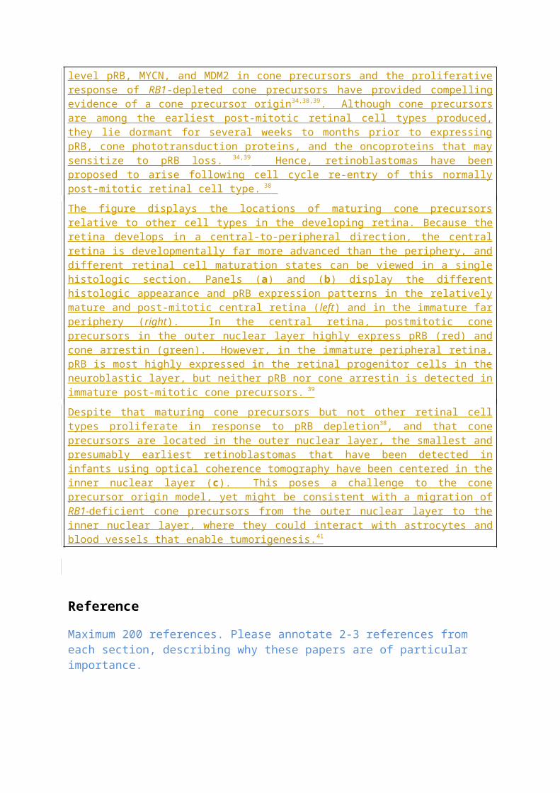
219. Willis, D. & Barry, P. Audiovisual interventions to reduce the use of general anaesthesia with paediatric patients during radiation therapy. J Med Imaging Radiat Oncol 54, 249-55 (2010).
220. O'Callaghan, C., Sexton, M. & Wheeler, G. Music therapy as a non-pharmacological anxiolytic for paediatric radiotherapy patients. Australas Radiol 51, 159-62 (2007).
221. Post-White, J. et al. Massage therapy for children with cancer. J Pediatr Oncol Nurs 26, 16-28 (2009).
222. Sposito, A.M. et al. Coping Strategies Used by Hospitalized Children With Cancer Undergoing Chemotherapy. J Nurs Scholarsh (2015).
223. Hildenbrand, A.K., Clawson, K.J., Alderfer, M.A. & Marsac, M.L. Coping with pediatric cancer: strategies employed by children and their parents to manage cancer-related stressors during treatment. J Pediatr Oncol Nurs 28, 344-54 (2011).
224. Olteanu, C. & Dimaras, H. Enucleation Refusal for Retinoblastoma: A Global Study. Ophthalmic Genet, 1-7 (2014).
225. Brink, A., Correa, Z.M., Geller, J., Abruzzo, T. & Augsburger, J.J. Managing the consequences of aggressive conservative treatment for refractory retinoblastoma with vitreous seeding. Arquivos Brasileiros de Oftalmologia 77 (2014).
226. White, A. & Gallie, B.L. Life Before Eye: Implications for the Whole Child and Family of Attempted Eye Salvage for Unilateral and Severe Bilateral Retinoblastoma. 46th World Congress of the International Society of Paediatric Oncology (2014).
227. Miller, V.A., Cousino, M., Leek, A.C. & Kodish, E.D. Hope and persuasion by physicians during informed consent. J Clin Oncol 32, 3229-35 (2014).
228. White, A. Managing International Patients. 1st meeting of One Retinoblastoma World (2012).
229. MacCarthy, A. et al. Second and subsequent tumours among 1927 retinoblastoma patients diagnosed in Britain 1951-2004. Br J Cancer 108, 2455-63 (2013).
230. Shinohara, E.T., DeWees, T. & Perkins, S.M. Subsequent malignancies and their effect on survival in patients with retinoblastoma. Pediatr Blood Cancer 61, 116-9 (2014).
231. Ford, J.S., Chou, J.F. & Sklar, C.A. Attendance at a survivorship clinic: impact on knowledge and psychosocial adjustment. J Cancer Surviv 7, 535-43 (2013).
232. Schulz, C.J., Riddle, M.P., Valdimirsdottir, H.B., Abramson, D.H. & Sklar, C.A. Impact on survivors of retinoblastoma when informed of study results on risk of second cancers. Med Pediatr Oncol 41, 36-43 (2003).
233. Rowland, E. & Metcalfe, A. Communicating inherited genetic risk between parent and child: a meta-thematic synthesis. Int J Nurs Stud 50, 870-80 (2013).

234. Clarke, S.A., Sheppard, L. & Eiser, C. Mothers' Explanations of Communicating Past Health and Future Risks to Survivors of Childhood Cancer. Clinical Child Psychology and Psychiatry 13, 157-170 (2008).
235. van Dijk, J. et al. Restrictions in daily life after retinoblastoma from the perspective of the survivors. Pediatr Blood Cancer 54, 110-5 (2010).
236. Friedman, D.N. et al. Long-term medical outcomes in survivors of extra-ocular retinoblastoma: the Memorial Sloan-Kettering Cancer Center (MSKCC) experience. Pediatr Blood Cancer 60, 694-9 (2013).
237. van Dijk, J. et al. Quality of life of adult retinoblastoma survivors in the Netherlands. Health Qual Life Outcomes 5, 30 (2007).
238. Traore, F., Togo, B., Pasquier, E., Dembele, A. & Andre, N. Preliminary evaluation of children treated with metronomic chemotherapy and valproic acid in a low-income country: Metro-Mali-02. Indian J Cancer 50, 250-3 (2013).
239. Zanaty, M. et al. Update on intra-arterial chemotherapy for retinoblastoma. ScientificWorldJournal 2014, 869604 (2014).