mrslampsbiocorner.weebly.commrslampsbiocorner.weebly.com/uploads/1/6/0/4/16048104/... · web...
TRANSCRIPT

•A
•,•
'--'
t
•bout 36% of the mass in female bodies and about 42% of the mass in male bodies i s composed of skeletal muscle.
In this exercise, some of the larger skel etal muscles of the human body are presented for study.
A. Identifying musclesUsing models and cha rts of the human muscu la ture, locate the skeletal muscles ou tlined in the succeed in g pages. As yo u do, note the size and shape of each mu scle.
Before you begin 0 10 Read the appropriate chapte r in your tex tbook.
• CJ Set your learning goa ls. \!Vhen you finish thi s exercise,
Identify these muscles of the h('11d and nt>ck (Figure 18-1):::J Occipitofrontalis (epicranius)
Orbicularis oculi
you should be able to:0 identify the followin g on a model and in figures:
muscles of the head and n ec k muscles of the trunkmuscles of the upper ex tremit y muscles of the lower ex tremitv muscles of the pelvic floor
::J name the origin and insertion of each major muscle of the body
0 demonstrate the action of each muscle studi ed
Prepare your materials:o models and charts of human musculatureo demonstration pointers::J computer setup with D ISSECTIBLE HUt-- I AN or simi la r
human dissection program (optiona l)
0 Read the directions and safety tips for this exercise ca refully before starting any procedure.
v==================(In this exercise, the muscles are presented by their location(e.g., head and trunk). Another common approach is to group muscles by the part they move (e.g., muscles that mo\'e the head, or muscles that move the arm)_ Yet another manner of grouping muscles involves their action (e.g., flex ors, extensors, and adductors). You may want to keep these groupings in mind as you explore the muscular system .
Using the D I SSECTIBLE HUMAN or similar comp uterized hu man dissection program, explore the human body and tr y to find the structures listed in this activi t y Check them off in your Lab Report as you find them.
Indi\·idu al muscles can be l ocated on a model of hum a n mu sculature by locatin g them in the diagrams on the foll owing pages, then checking their origins, insertions, andactions a s li sted on the tables for this exercise. Another method - more cha llenging, but more informative- is to try to identify them by their attachme nt points and actions befcw identifying them in a diagram
Co p>-ri c;ht D 200:i bv Mosby, Lnc. A ll ri g hts reser,·ed .

::J0 Orbicularis oris::J Buccinator::J Zygomaticus (two muscles)::J Levator labii superioris, major and minorD Depressor anguli oriso Temporaliso Massetero Pterygoids (two muscles, internal and extern a l )o Sternocleidomastoid o Trapezius
0 2 Id enti fy th ese muscles of the trunk (F i gure 18-2):o Erector spinae (divides into three muscles)0 Deep back muscles o
External intercostals CJInternal intercostals o Rectus abdominis
o External abdominal oblique0 Internal abdominal oblique o Transversus abdominiso TrapeziusQ Levator scapulaeQ Rhomboideus (two muscles), major and minorCJ Serratus anter.jor o Pectoralis minor o Pectoralis major0 Teres majoro Latissimus dorsi o
Infraspinatuso Supraspinatuso Subscapularis o Teres minoro Deltoid
!t£)'[j'==:::::::::::::::::::::::::::::::::::::::::::::::::::::::::::::::::::::::::::::::;Notice that many of the muscles li sted for the trunk in clude should er (upper trunk) muscles thilt insert on the arm.
163

5 ••
•
IICOLORING EXERCISEUsing colored pens or pencils, shade in the figures and accompanying labels incontrasting colors of your choice as indicated by the red numerals.
Muscles of the Head and Neck
5
Figure 18-1 Muscles of facial expression and mastication.
4
'4c f f4
•· -4\
4
•5
c
-•·•

• Copyright© 2003 by Mosby, Inc. All rights reserved.

©cscso ou(Q) [Yl©u&l1D 1 Occipital Skin of eyebrow, nose Elevates brows; moves scalp
@[YlrnDCSM [YlO ©CSMO:,O 2 Maxilla, frontal Encircles eye, near Closes eyeorigin
@[{rnOCSM lf.lO ©m.O J Maxilla, mandible Lips Closes lips
rnMcscso&u©[Yl 4 Maxilla, mandible Angle of mouth Compresses cheeks
®©Gal&'U'OCSMs (two) Zygomatic bone Angle of mouth, upper Elevates angle oflip mouth, upper lip
l1@\:J&v©m. l1&rnoo Maxilla Upper lip, nose Elevates upper lip,11D[p@[Il0@[Il0£B 6 nose
[Q)[][p[Yl@ £B@[Il & ®MD:,O@m,O?:B 1 Mandible Lower lip near angle Depresses angle ofmouth
urn lP@[f O:,Of0 s Temporal aspect of Mandible Closes jawskull
u £B£B[]u[][Yl 9 Zygomatic arch Mandible Closes jaw
PTERYGOIDS (two) Inferior aspect of skull Mandible Medial closes jaw;lateral opens jaw
vrnlil ©CSO:,[]O©(§>L f0u©ow 1o Sternum, clavicle Mastoid process Rotates, extends head(skull)
u lP[]L£0M?:B 11 Skull, upper vertebral column
Scapula Extends h ead, n eck
Anatomy & Physiology Laboratory Manual 165
•••
Use te rm s in @@Ulli][/:!][;l type as colorin g labels. Terms in SOLID type do not appea r in th e colorin g pla t e.
•••
Copy right © 2003 by Mosby, lnc All rights reserved.

••••
•
• COLORING EXERCISEUsing colored pens or pencils, shade in the figure and accompanying labels in contrast i ng colors of you r choice as indicated by t he red n u merals.
Muscles of the Trunk 4
-
Figure 18-2 Muscles of the trunk and abdominal wai L
c
c
•tf
c•4
•-
20
I
24 •••••


•
"
t
••
•
•
••
•
(I
,C;;;
Anatomy & Physiology Laboratory Manual 16:
;;; 'U'@[Rl 0 & 12 Vertebrae, pelvis Superior vertebrae, ribs Holds body upright
j (divides into three)
4 DEEP BACK MUSCLES Vertebrae Vertebrae Flexes or extends trunk
,;j b'U''1 [1 Ribs Edge of next rib Expands thorax
..;; O 'U' © 'TI'U: l1:0 13 (inferiorly)
o :mL lb Ribs Edge of next rib Compresses thorax
OrKJu( ©:0uE lbill 14 (superiorly)
ij CSuM&wlD©WDOO 15 Pubis Inferior thoracic cage Flexes waist
u [KJL [b &fl)[Q)@OrKJ&[b Inferior thoracic cage Midline of abdomen Compresses abdomen
©wlbO@l1D 16
OrKJ'U' [b [l)@@OrKJ&[b©wl1D@M 1 7
Pelvis Midline of abdomen Compresses abdomen
• U '-'v, r::o;-;:v·-,n "R Vertebrae, pelvis, ribs Midline of abdomen Compresses abdomenG "'U. \ C'-:-:'J•'.'-'';::...J..:..'Jl.J'I..2..:>."·1, .:J:._I.:;:.V...
&w©@G0\l0lKlD 18
• rotates scapula"-----"' 'U' LZO\\.D 11 Skull, upper vertebral Scapula Extends head, neck;
column
-•- [1 \::'&l 'U'::Q! _,[§)QD[l,& 19 Vertebra e Scapula Elevates scapula
D=D@G j@O[Q) QJJ 20 (two) Vertebrae Scapula Retract scapula
'U'G:DJ rKJ'DT O@ 21 Ribs Scapula
Protracts scapula rnCS'U'@ [10ltv OrKJ@ 22 Ribs Scapula
Depresses sca pu la [p CSu© f1oc© 23 Ribs,
clavicle Humerus Adducts, flexes arm
'ITUf® @{fl 24 Scapula Humerus Extends, adducts,rotates arm
l1&'U'O Ou M©@0 25 Vertebrae Humerus Extends arm
OrKJl? lPOrKJ3iuM 26 Scapula Hume rus Extends, rotates arm
ru:J[plfu LPDG &'U'M 27 Scapula Humerus Abducts arm
SUBSCAPULARIS Scapula Humerus Extends, rotates arm
TERES MINOR Scapula Humerus Adducts, rotates arm

•© [1'U'@O[Q) 28 Scapula, clavicle Humerus Abducts arm
Use t e rms in @Mil'!!J] !] ty pe as col oring labels.Terms in SOLID type do not appear in the colorin g pl a t e.Copyright © 2003 by Mosby, Inc. All rights reserv ed.

I
•IICOLORING EXERCISEJsing colored pens or pencils, shade in the figures and accompanying labels in:ontrasting colors of your choice as indicated by the red numerals.
-
\1uscles of the Lower Extremity IecI
•I
••...
1
••••
•.•.Figure 18-4 Muscles of the leg.
Copyright© 2003 by Mosby, Inc. All rights reserved.

•
•
••
,.r,>Jr Ilium, vertebrae Femur Flexes thigh
--- ---- ---------------------r------------ -----;---- --------------r-----------------
;1 0· \:2...0f!. ·;\15u _"\...Is <::::J...L:_..-.._,b
GLUTEAL GROUP (a, b, c )
•
Hip
Hip
Hip
Tibia Abducts thigh
Femur Extends thigh
c GLUTEUS MINIMUS llium
QUADRICEPS FEMORIS(a, b, c, d)
Femur Extends thigh
Femur Abducts thigh
',t
• d VASTUS INTERMEDIUS
t
• HAMSTRING GROUP (a, b, c)
Ilium Tibia Extends leg, flexes thigh
Femur Tibia Ex tend s l eg
Fe mur Tibia Extends leg
Femur Tibia Extends l eg
Ilium Tibia Flexes thigh; flexes, rotates leg
Ischium, femur Fibula Flexes l eg, exte nds thigh
I sc hium Tibia Flexes l eg, extends thigh
ADDUCTOR GROUP (a, b)Ischium Tibia Flexes leg, extends thigh
Pubis Metatarsal bones Extends (dorsi fl exes)foot
Pubis
Tibia
Tibia
Femur
Tibia, fibula
Tibia, fibula
Tibia Adducts thi g h
M e tatarsal bones Extends (dorsifl exes)
foot
Phalanges Ex tends toes
Calcaneus (Plantar) fl exes foot
Calcaneus (Plantar) fl exes foot
Tarsal, metatarsal bones Flexes, eve rts foot

Use te rm s in @Gd!l!f',utype as col orin g labels. Term s in SOLID type do n ot appear in th e col orin g plat e. Copyri ght © 2003 by Mosby, lnc. A ll rights resened.

•
•
172 Lab Exercise 18 • Skeletal Muscle Identification
m- ======================By now you have noticed that several of the muscles illus /
trated in the coloring exercises do not have labels. Some of them are muscles that you were asked to identify but are not labeled for coloring because they are too far away from the outline labels. Go back to the figures and try to identify the unlabeled muscles. Use your textbook and anatomy reference books in your school library to help you.
(
61A
p
Female
61
B
p
ale
c
4
44
-
----••••
-••
M
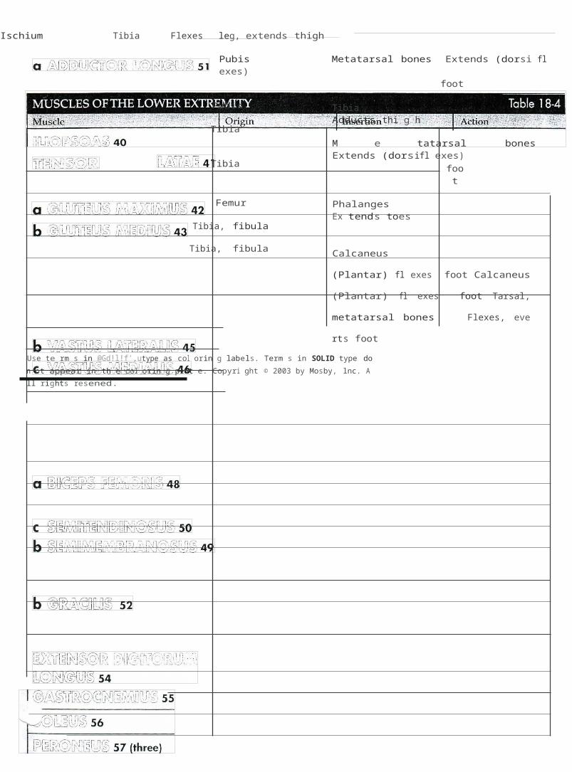
Figure 18-5 Inferior view of the pelvic muscles in the female (A) and male (B). ••
Copyright© 2003 by Mosby, Inc. All rights reserved- .

•
''
Anatomy & Physiology Laboratory Manual 17
I [1J W&u©&lf80 58 Pubis, ischium Sacrum, coccyx Elevates anus
'•.[lfl[b @ [p@ @O@ [lfl£0 60 MALE: bulb of penis
O CSC O@CS&W [f\:J@ llJJ 59 Ischium Clitoris or penis Compresses baseof clitoris or penis
t CPG=QOlf8CSu O'l(t L1u lf8M&lf8D 61
FEMALE: central tendon of perineum
Coccyx
MALE: central tendon ofperineumFEMALE: base of clitoris
Central tendon(median raphe)
MALE: constricts urethra; erects penisFEMALE: erects clitoris
Closes anal canal
'•'•..•
Femur
Use terms in @l)[l'jj'[l,Oittltype as coloring labels.Terms in SOLID type do not appear in the coloring plate.
Extends thigh
'••

•
'
',
A• Copyright © 2003 by Mosby, lnc. AU rights reserved.

ii
174 lab Exercise 1 8 • Skeletal Muscle Identification
:J 3 Identify these muscles of the upper extremity(Figure 18-3):CJ Triceps brachii:::::1 Biceps brachii':J BrachialisCJ Pronators (two muscles, teres and quadratus)o flexor carpi ulnaris':J Flexor carpi radialisCJ Flexor digitorum (two muscles)::J Brachioradialis:J SupinatorCJ Extensor carpi ulnaris::J Extensor carpi radialis (two muscles, profundus
and superficialis):J Extensor digitorum
:J 4 Identify these muscles of the lower extrernity(Figure 18-4): · -0 Iliopsoas0 Tensor fasciae lataeo Gluteal group: gluteus maxim us,
gluteus medius, gluteus minimuso Quadriceps femoris group: rectus femoris, vas
tus lateralis, vastus medialis, vastus inter medius
'.J SartoriusD Hamstring group: biceps femoris, semimembra-
nosus, semitendinosusCJ Adductor group: adductor longus, gracilis:J Tibialis anterior::J Extensor digitorum longus:::1 GastrocnemiusD Soleus::J Peroneus (three muscles, longus, brevis,
and tertius)
0 5 Identify these muscles of the pelvic floor (Figure 18-.5):CJ Levator ani:J Ischiocavernosus·:J Bulbospongiosus·::l Sphincter extern us ani::J Gluteus maximus
B. Demonstrating muscle actionFor each of the muscles found in Activity A of this PXPrcisP. demonstrate its action with your ovvn muscle (if pc·si bl2). As you contract the muscle, palpate it and note its size and loca tion.
For a more detailed description of the origin, l' l'>< ·t i·ll tion, and innervation of some of these mnscL s, _Jlc J:o e L
fer to your textbook."\\)
Copyright © 2003 by Moshy. Inc. All rights reserved.
•;\
.J·

anne: _ Date: _ Section:
lAB REPORT 1 8
Skeletal Muscle Identification
Table Completion (list each muscle name only once in the table below)
Adductor group Biceps brachii DeltoidErector spinae Gastrocnemius Gluteus maximus Hamstring group Iliopsoas
Latissimus dorsiPectoralis majorPectoralis major /latissimus dorsi combinationQuadriceps femoris groupRectus abdominis Tensor fasciae latae Tibialis anterior Triceps brachii
Part Moved Flexor Extensor Abductor Adductor
Upper arm
Lower arm
Thigh
Lower leg
Foot
Trunk
Copy right © 2003 by Mosby, Inc All right s reserved.

176
Fill-in
1.
2. ---------------------
3. -----------------------
Lab Exercise 1 8 • Skeletal Muscle Identification
Fill-in (complete each item with the correct term)
1. The occipitofrontalis, or epicranius, originates on the ? bone.2. The masseter, temporalis, and pterygoids all insert on the ? .3. The sternocleidomastoid inserts on the ?4. The temporalis closes the ? .5. The ? closes the eye.6. The pectoralis ? inserts on the humerus.
4. ----------------------- 7. The deltoid inserts on the ?8. The ? intercostals help accomplish forced expiration.
5. --------------------- 9. The erector ? . help maintain posture.6. -------------------------
7. -----------------------
8. ---------------------
9. -----------------------
10.
11. ------------------------
12. ------------------------
13. -----------------------
14. ------------------------
15. ---------------------
16. ------------------------
17. ------------------------
18.
19. -----------------------
20.
Z1.
------------------------
U. -----------------------
U.
4- --------------------------
5. --------------------------
10. The pectoralis major is ? to the pectoralis minor.11. The latissimus dorsi is on the ? side of the trunk.12. The ? intercostals contract when you take a deep breath.13. The rippling effect seen on the lower midline of the abdomens of some athletes
is caused by hypertrophy of the ? muscle.14. The origin of the brachialis is ? to its insertion.15. In grasping a baseball tightly in the hand, one would likely use the ?
muscle.16. Both the biceps brachii and the ? flex the forearm.17. The extensor digitorum inserts on the ? _18. The adductor longus and the ? are both part of the adductor group.19. The hamstring muscles all ? the leg.20. The muscle that pulls the leg so that you can cross it over the other le wh i
le sitting is called the ? muscle.21. Both the gastrocnemius and the ? plantar flex the foot.22. Muscles of the quadriceps group all ? the leg.23. In the male, the ? pulls the penis erect but also can constrict the ureth ra. \24. The large muscle that extends the thigh is the"_] _
· 25. The levator ani raises the ?
\
Copyright© 2003 by Mosby, lnc. All rights reserved. t

1cor;ing cc
'-'"'ll'/5 colored pens or pencils, shade in the figures and accompanying labels in contrasting colors of your choice as indicated by the red numerals.
ntrast Muscles of the Upper Extremity
IRightlimb
IRightlimb
Figure 18-3 Muscles of the arm.

Anatomy & Physiology Laboratory Manual 169
MUSCLES OF THE UPPER EXTREMITY . " Table 1 8-3.scle Ongm InsertiOn Action
uffiO rnDD ffi& [}:i]OO 29 Humerus, scapula Ulna Extends forearm
o rnDD ffi& [}:i]OO 30 Humerus, scapula Radius Flexes, supinates forearm
ffi& [}:i]O&[LO 31 Humerus Ulna Flexes forearm
ffi© &u©ffi 32 (two) Ulna, humerus Radius Pronates forearm
csa,rn&@ffi &ffiDDD Humerus Carpal bone Flexes, abducts wrist
ruJ[LCK!J&ffiO 33 (medial epicondyle)
t?a,rn&@ffi &ffiODO Humerus Metacarpal bones Flexes, abducts wrist
ffi&lli>O&[LO 34 (medial epicondyle)
csa,rn&@ffi Humerus Phalanges Flexes fingers
@O®Ou@ffiM 35 (two) (medial epicondyle, ulna, radius)
ffi& [}:i]O@ffi&@O&[LO 36 Humerus Radius (distal) Flexes, pronates forearm
SUPINATOR Ulna Radius Supinates forearm
;;?.urn @ffi &ffiDDD Humerus Metacarpal bones Extends, abducts wrist
'----" &ffiD 37 (lateral epicondyle)
rn&urn @ffi &ffiDDD Humerus Metacarpal bones Extends, abducts wrist
ffi&[Q)0&[b0 38 (two) (lateral epicondyle)
rn&urn ©ffi Humerus Phalanges Extends fingers
C0D®Du@ffiM 39 (lateral epicondyle)
Use terms in @(!D'[j'[],[]lt:!)type as coloring labels.Terms in SOLID type do not appear in the coloring plate.