01 prosp dmire 2 e.qxp (page 1) - unimore · the growing complexity of the challenges faced by...
TRANSCRIPT
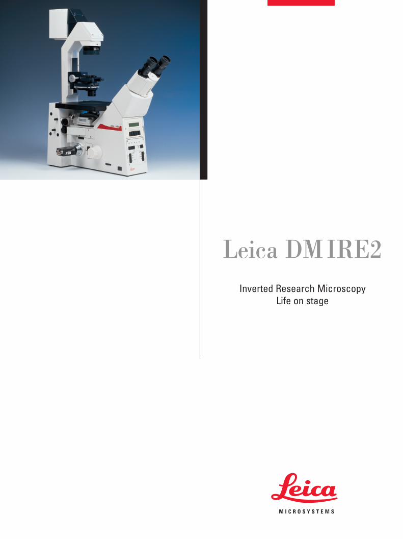
Leica DM IRE2Inverted Research Microscopy
Life on stage

Leica DMIRE2 –The latest generation of inverted
research microscopesDemands on research microscopes are growing at breathtaking
speed. Users of inverted microscopes in particular are expecting
more and more of their equipment, motivated by rapid advances
in medicine and biology where research scientists are conti-
nuously breaking through into new dimensions. The increasing
complexity of research tasks relies on high measuring accuracy
and the processing of large amounts of data.
With our new generation of inverted research microscopes, we at
Leica have managed once more to exactly meet our users’ needs.
The Leica DMIRE2 is the ideal combination of motorization, auto-
mation, high stability, optimized system compatibility, ergonomy
and user convenience. And yet again, we have proved that Leica
is not only keeping pace with progress, but is actually setting the
pace.
2
Chicken embryo - brightfield
3
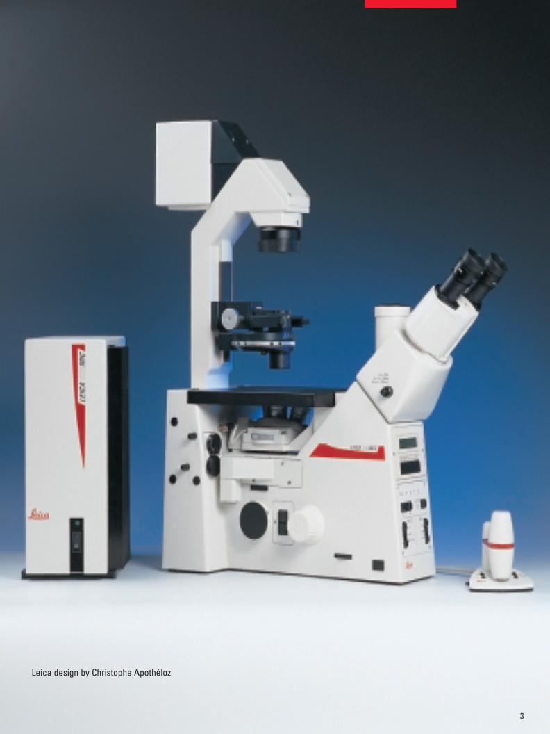
Leica design by Christophe Apothéloz
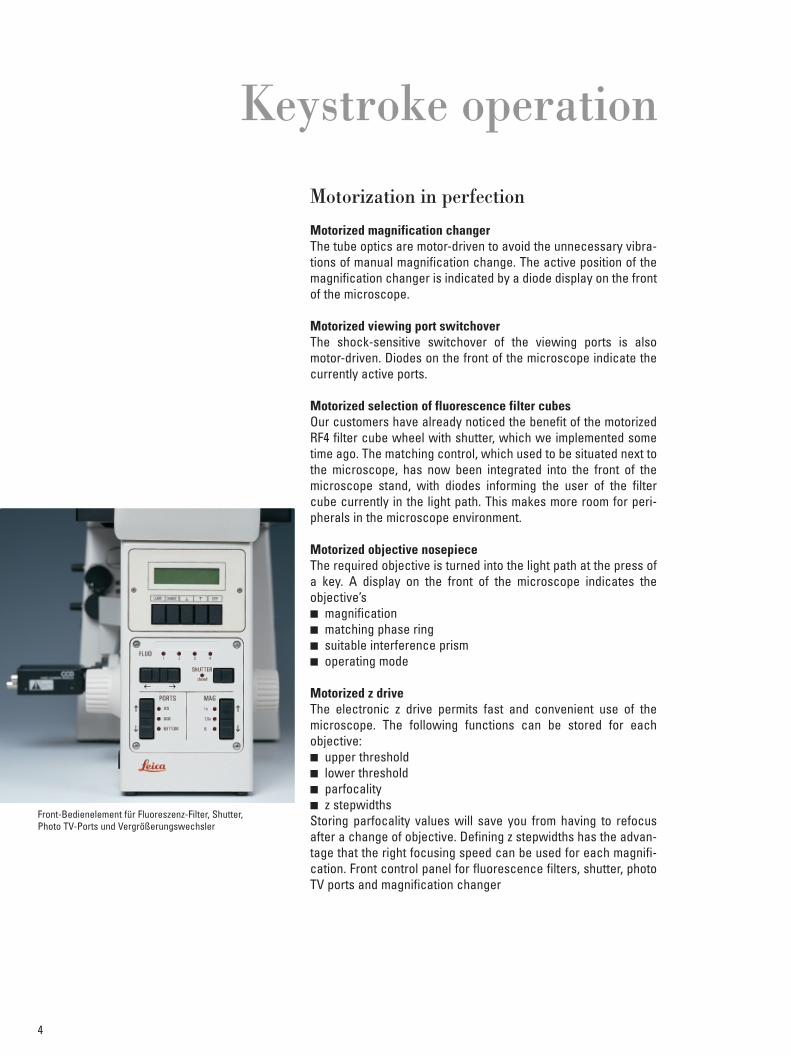
Motorization in perfection
Motorized magnification changerThe tube optics are motor-driven to avoid the unnecessary vibra-tions of manual magnification change. The active position of themagnification changer is indicated by a diode display on the frontof the microscope.
Motorized viewing port switchoverThe shock-sensitive switchover of the viewing ports is alsomotor-driven. Diodes on the front of the microscope indicate thecurrently active ports.
Motorized selection of fluorescence filter cubesOur customers have already noticed the benefit of the motorizedRF4 filter cube wheel with shutter, which we implemented sometime ago. The matching control, which used to be situated next tothe microscope, has now been integrated into the front of themicroscope stand, with diodes informing the user of the filtercube currently in the light path. This makes more room for peri-pherals in the microscope environment.
Motorized objective nosepieceThe required objective is turned into the light path at the press ofa key. A display on the front of the microscope indicates theobjective’s� magnification� matching phase ring� suitable interference prism � operating mode
Motorized z driveThe electronic z drive permits fast and convenient use of themicroscope. The following functions can be stored for eachobjective:� upper threshold� lower threshold� parfocality� z stepwidthsStoring parfocality values will save you from having to refocusafter a change of objective. Defining z stepwidths has the advan-tage that the right focusing speed can be used for each magnifi-cation. Front control panel for fluorescence filters, shutter, photoTV ports and magnification changer
Keystroke operation
4
Front-Bedienelement für Fluoreszenz-Filter, Shutter, Photo TV-Ports und Vergrößerungswechsler
Encoded prism wheel for interference contrastThe interference prism that is currently in the light path is indicatedin the front display of the microscope. If the objective in the lightpath does not suit this prism, the display will contain a warning toavoid operation errors.
Separate accommodation of electronicsAlmost all the electronics have been moved out of the microscopeinto the CTR MIC electronics box, effectively suppressing electronicnoise, which can easily lead to interpretation errors in electro-physiological examinations.
5
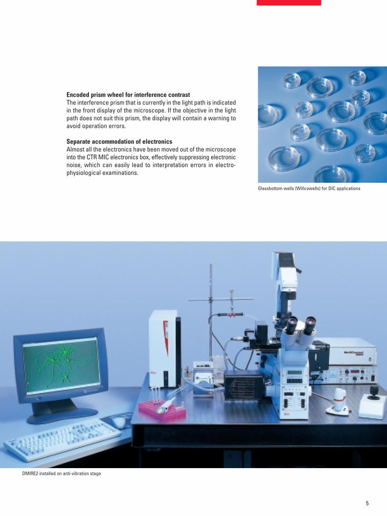
Glassbottom wells (Willcowells) for DIC applications
DMIRE2 installed on anti-vibration stage

The growing complexity of the challenges faced by today’s rese-arch scientists has to be matched by complex examination tech-niques. The microscope must satisfy this need as part of a systemof various accessories and peripherals. Today, intricate examina-tions or manipulation processes are expected to be computer-controlled to attain a high degree of reproducibility and thereforeensure comparability of results.Leica has made provision for the exchange of data betweenmicroscope and computer by fitting the DMIRE2 with a standardi-zed RS 232 interface. Using the SDK system software, which canbe downloaded from the internet free of charge, the data of allmotorized or coded elements of the microscope can be called upon the screen. At the same time, it is possible to address all moto-rized components via the program. The user-friendly softwarealso allows the creation and activation of automated microscoperoutines.
Living Cell Microscopy makes particularly heavy demands onsystem integration.In the special applications IVF (in-vitro fertilization), ET (embryotransfer) or ICSI (intra cytoplasmatic sperm injection) of transge-nics, successful micromanipulation and -injection depends on aperfect match of components.Multidimensional microscopy requires a high degree of micros-cope automation. Climate chambers and evaluation software arenecessary for time-lapse experiments. The microscope must alsofeature excellent stability.To be able to measure the extremely small electric signals gene-rated in electrophysiological examinations, microscopes needoptimum electric noise suppression..Finally, confocal laser scanning microscopes satisfy top require-ments of resolution in z direction and are used in optimally mat-ched conjunction with a light microscope.
Leica provides the ideal platform for these and other systemapplications. Not only is the Leica DMIRE2 ideally prepared for itsjob at the heart of an integrated system - accessories such asheating stages, climate boxes, micromanipulators, high-resoluti-on cameras or evaluation programs work in perfect harmony withthe microscope.
6
Integration into the systemsof today and tomorrow
Accessories for the DMIRE2 (climate chambers, temperaturecontrol and various specimen photos)

Remote control operationThe remote control function (z control) permits non-contact ope-ration of the main electronic elements. The following elementscan be addressed:� focus (z drive)� objective nosepiece and � illumination intensity.
PC operationPC control is realized with the SDK software that can be downlo-aded free of charge from the internet. All electronically availabledata can then be called up on the screen. At the same time it ispossible to drive all motorized microscope elements from thecomputer.
Elimination of vibrationsThe microscope’s remote control facility enables the user to eli-minate virtually all vibrations from his experiments.On the DMIRE2, particularly shock-sensitive manual controlssuch as magnification change or viewing port switchover havebeen motorized as a further improvement to the stability of themicroscope.
More free spaceThe microscope’s remote control facility also creates spaceround the microscope that would otherwise have been occupiedby manual controls. This is an advantage for users who need tosurround the microscope with bulky equipment.
7
Non-contact operation
Remote control of the DMIRE2
Easy operation due to electronic support
Storage of user settingsPersonal preferences of users such as left- or right-handed ope-ration are effectively supported by the DMIRE2. The z drive move-ment or objective change functions can be optionally assigned tothe keys on either the left or right side of the microscope. In addi-tion, the z drive hand wheels can be operated either clockwise orcounterclockwise.
No collisions of the objective with the specimenOnce the upper threshold has been stored, this z position cannotbe inadvertently overridden with the electronics.Empty nosepiece positions are not turned into the light path.For objective change, only nosepiece positions containing objec-tives are turned into the light path.
Operating modesIf IMMERSION mode is selected, only immersion objectives willbe turned into the light path. This prevents dry objectives fromcoming into contact with immersion oil or, in DRY mode, an immer-sion oil being used without oil by mistake.Combined objectives are available for both modes.
Display and diode indicatorsThe display and diode indicators are situated on the front of themicroscope for easy and immediate viewing.The display of the currently active components� objective� interference prism� fluorescence filter cube� tube optics� viewing portmakes work easier and helps to avoid operation errors.
User convenience
8
Front control panel for fluorescence filters, shutter, photo TVports and magnification changer
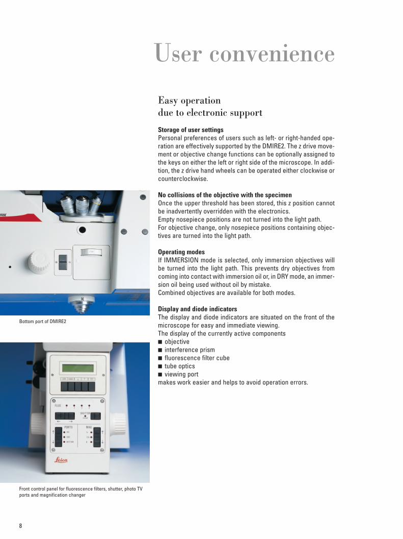
Bottom port of DMIRE2
Ergonomically optimized controls
ErgotubeThe ergonomic observation tube features stepless height adjust-ment, allowing the user to adopt a comfortable sitting position andvary it as necessary.
Fatigue-free viewing with fov 22Eyepieces with this field of view index show an area of the imagethat can be surveyed without pupil movement, reducing eye fati-gue during spells of concentrated observation.
Ergonomic layout of controlsAll controls on the side of the microscope are designed to enableyou to rest the ball of your thumb against them for relaxed opera-tion.
Easily accessible objectivesThe objective nosepiece is mounted at a slant, providing easieraccess to and convenient handling of objectives.
Angled lamphousing for easier centrationTo facilitate centration of the mercury lamp, the lamphousing isangled on the right-hand side and has the centration devices onthis side face.
3-plate mechanical stage with extra-long coaxial driveExtending as far as the desktop, the coaxial drive enables you toadjust the stage, i.e. position the required specimen detail in thefield of view, with the ball of your thumb resting on the control.
9
Ergonomy
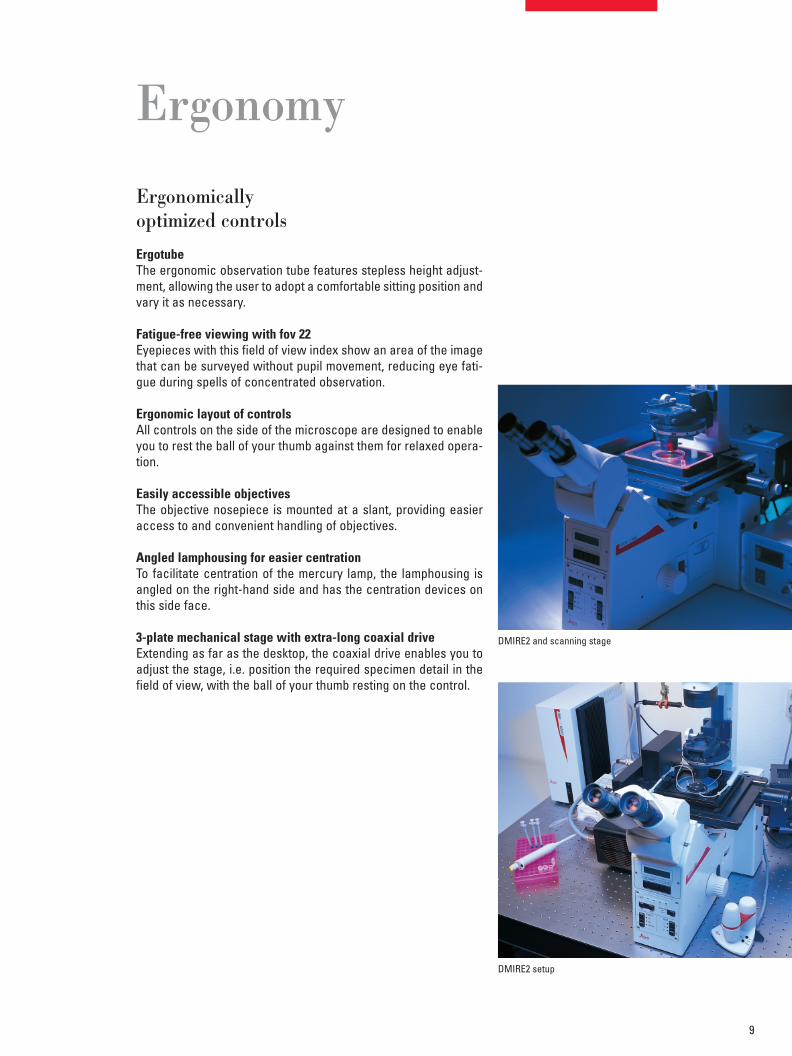
DMIRE2 setup
DMIRE2 and scanning stage

10
Contrasting techniquesThe Leica DMIRE2 will produce brilliant and high-contrast imagesof all your specimens, providing the right contrasting techniquefor every application.
Phase contrastPhase contrast is used for high-contrast imaging of faint, unstai-ned specimens that are typically encountered in fresh samplesand cell and tissue cultures.We have designed special phase contrast objectives for invertedmicroscopy with long free working distances and magnificationsfrom 5x to 100x.
Differential interference contrast (DIC)Like phase contrast, interference contrast is used for visualizingunstained transparent specimens in transmitted light. With thismethod, however, a 3D impression is produced, making it highlysuitable for manipulation work.Leica DIC will prove particularly useful in IVF or ICSI, i.e. micro-manipulation or microinjection. The IC prisms on the side of theillumination are inserted into the 6-position condenser disk, whilethe objective-side prisms are accommodated in a turret on thestandard objective nosepiece. The Leica DMIRE2 produces DICup to a working distance of 70 mm with objectives up to 100xmagnification. Fast switching between DIC, brightfield and phasecontrast is possible at any time.
Leica Modulation Contrast (LMC)Based on Hoffman Modulation Contrast, this technique producesrelief images similar to those obtained with interference contrast.Unlike DIC however, LMC can be used for viewing specimensthrough birefringent plastics, and is therefore an excellent alter-native to interference contrast.For this technique, Leica offers objectives that are specially mat-ched for inverted microscopy with long free working distancesand magnifications between 10x and 63x.
FluorescenceFluorescence microscopy produces images of fluorochromedsubstances or sections of cell or tissue. When irradiated at a spe-cific wavelength, the fluorochrome emits visible light (fluorescen-ce), visualizing the substance under examination.
3-plate mechanical stage with glass insert
Microinjection nucleus Acetabularia acetabulum
Fibroblasts Fluorescence cytoskeletonand nucleus stained
11
Leica offers fluorescence filter cubes for the excitation wave-lengths of all important fluorochromes. The filter turret (RF4) takesup to 4 filter cubes to allow synchronized viewing of more thanone fluorochrome in one specimen. The excitation light is conve-niently switched off with a motorized switch.
All transmitted light techniques can be used at the same time asincident light fluorescence, permitting the simultaneous observa-tion of non-fluorescing specimen details.
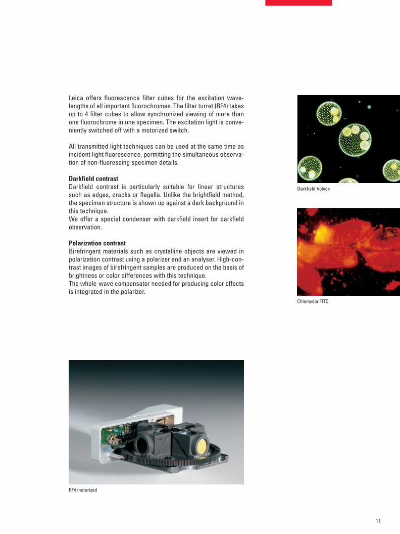
Darkfield contrastDarkfield contrast is particularly suitable for linear structuressuch as edges, cracks or flagella. Unlike the brightfield method,the specimen structure is shown up against a dark background inthis technique.We offer a special condenser with darkfield insert for darkfieldobservation.
Polarization contrastBirefringent materials such as crystalline objects are viewed inpolarization contrast using a polarizer and an analyser. High-con-trast images of birefringent samples are produced on the basis ofbrightness or color differences with this technique.The whole-wave compensator needed for producing color effectsis integrated in the polarizer.
RF4 motorized
Darkfield Volvox
Chlamydia FITC

Leica Microsystems – the brandfor outstanding products
Copy
right
©Le
ica
Mic
rosy
stem
s W
etzla
r Gm
bH •
Erns
t-Lei
tz-S
traße
•35
578
Wet
zlar •
Germ
any
2000
•Te
l. (0
6441
) 29-
0 •
Fax
(064
41) 2
9-25
99
LEI
CA a
nd th
e Le
ica
Logo
are
regi
ster
ed tr
adem
arks
of L
eica
Tec
hnol
ogy
BV.
Orde
r nos
. of t
he e
ditio
ns in
: Eng
lish
9141
97•
Germ
an 9
1419
6 •
Fren
ch 9
1419
8 •
Italia
n 91
4199
•Sp
anis
h 91
4200
•Pa
rt-N
o. 5
01-1
90
Prin
ted
on c
hlor
ine-
free
blea
ched
pap
er.
•/••
/???
?
Leica Microsystems’ mission is to be the world’s first-choice provider of innovativesolutions to our customers’ needs for vision, measurement, lithography and analysisof microstructures.
Leica, the leading brand for microscopes and scientific instruments, developed fromfive brand names, all with a long tradition: Wild, Leitz, Reichert, Jung and CambridgeInstruments. Yet Leica symbolizes innovation as well as tradition.
Leica Microsystems – an international companywith a strong network of customer servicesAustralia: Gladesville Tel. +61 2 9879 9700 Fax +61 2 9817 8358Austria: Vienna Tel. +43 1 486 80 50 0 Fax +43 1 486 80 50 30Canada: Willowdale/Ontario Tel. +1 416 497 2860 Fax +1 416 497 8516Denmark: Herlev Tel. +45 4454 0101 Fax +45 4454 0111France: Rueil-Malmaison Tel. +33 1 473 285 85 Fax +33 1 473 285 86
CedexGermany: Bensheim Tel. +49 6251 136 0 Fax +49 6251 136 155Italy: Milan Tel. +39 0257 486.1 Fax +39 0257 40 3273Japan: Tokyo Tel. +81 3 5435 9600 Fax +81 3 5435 9618Korea: Seoul Tel. +82 2 514 65 43 Fax +82 2 514 65 48Netherlands: Rijswijk Tel. +31 70 4132 100 Fax +31 70 4132 109Portugal: Lisbon Tel. +351 21 388 9112 Fax +351 21 385 4668Republic of China: Hong Kong Tel. +852 2564 6699 Fax +852 2564 4163Singapore: Tel. +65 779 7823 Fax +65 773 0628Spain: Barcelona Tel. +34 93 494 95 30 Fax +34 93 494 95 32Sweden: Sollentuna Tel. +46 8 625 45 45 Fax +46 8 625 45 10Switzerland: Glattbrugg Tel. +41 1 809 34 34 Fax +41 1 809 34 44United Kingdom: Milton Keynes Tel. +44 1908 246 246 Fax +44 1908 609 992USA: Bannockburn/llinois Tel. +1 847 405 0123 Fax +1 847 405 0030
and representatives of Leica Microsystemsin more than 100 countries.
Leica Microsystems Ing.2345 Waukegan RoadBannockburn IL 60015
Tel.: (800) 2 48-01 23Fax: (847) 4 05-01 64www.leica-microsystems.com
The companies of the Leica MicrosystemsGroup operate internationally in five businesssegments, where we rank with the marketleaders.
MicroscopyOur expertise in microscopy is the basis for allour solutions for visualization, measurementand analysis of microstructures in life sciencesand industry.
Specimen PreparationWe specialize in supplying complete solutionsfor histology and cytopathology.
Imaging SystemsWith confocal laser technology and imageanalysis systems, we provide three-dimensionalviewing facilities and offer new solutions forcytogenetics, pathology and material sciences.
Medical EquipmentInnovative technologies in our surgical micro-scopes offer new therapeutic approaches inmicrosurgery. With automated instruments forophthalmology, we enable new diagnosticmethods to be applied.
Semiconductor EquipmentOur automated, leading-edge measurementand inspection systems and our E-beam lithog-raphy systems make us the first choice suppli-er for semiconductor manufacturers all overthe world.