019 ' # '7& *#0 & 81 1 1 1 ð1 1 1 1 1 1 1 ýz._ þð1 1 1 1 1 1 1 1 1 1 1 1ý...
TRANSCRIPT

3,350+OPEN ACCESS BOOKS
108,000+INTERNATIONAL
AUTHORS AND EDITORS114+ MILLION
DOWNLOADS
BOOKSDELIVERED TO
151 COUNTRIES
AUTHORS AMONG
TOP 1%MOST CITED SCIENTIST
12.2%AUTHORS AND EDITORS
FROM TOP 500 UNIVERSITIES
Selection of our books indexed in theBook Citation Index in Web of Science™
Core Collection (BKCI)
Chapter from the book Plasma Science and Technology - Progress in Physical Statesand Chemical ReactionsDownloaded from: http://www.intechopen.com/books/plasma-science-and-technology-progress-in-physical-states-and-chemical-reactions
PUBLISHED BY
World's largest Science,Technology & Medicine
Open Access book publisher
Interested in publishing with IntechOpen?Contact us at [email protected]

Chapter 6
Physicochemical Analysis of Argon Plasma-Treated CellCulture Medium
Claudia Bergemann, Cornelia Hoppe, Maryna Karmazyna, Maxi Höntsch,Martin Eggert, Torsten Gerling and Barbara Nebe
Additional information is available at the end of the chapter
http://dx.doi.org/10.5772/61980
Abstract
The effects of cold plasma under atmospheric pressure are being explored for medical ap‐plications. It was found that plasma effects on cells correspond to a plasma–medium in‐teraction; thus, plasma-treated cell culture medium alone is able to influence the cellbehavior. Here, we discovered that the liquid-mediated effect of atmospheric-pressure ar‐gon plasma on mouse liver epithelial cells persists up to 21 days of storage; i.e., the liquidpreserves the characteristics once induced by the argon plasma. Earlier investigations ofour group revealed that temperature and pH, hydrogen peroxide production and oxygencontent can be excluded as initiators of the detrimental biological changes. As we foundhere, the increased osmolality in the media caused by plasma treatment can also be ex‐cluded as a reason for the observed cell effects. Conversely, we found changes in thecomponents of cell culture medium by fast protein liquid chromatography (FPLC) anddecreased cell viability in plasma-treated media independent of the presence of fetal calfserum (FCS) during plasma treatment. The persistent biological effect on plasma-treatedliquids observed here could open up new medical applications. Stable plasma-treated liq‐uids could find application for dermatological, dental, or orthopedic therapy.
Keywords: Non-thermal atmospheric-pressure plasma jet, Plasma–liquid interaction, Os‐molality, Size-exclusion chromatography, Cell viability
1. Introduction
Plasma medicine is an emerging field of interdisciplinary research combining physics, biology,and clinical medicine [1,2]. In the first applications, gas plasma was used in the form of hotplasma for cauterization [1,3]. Currently, the effects of cold plasma under atmospheric pressureare being explored for medical applications. Several experiments showed the achievement of
© 2016 The Author(s). Licensee InTech. This chapter is distributed under the terms of the Creative CommonsAttribution License (http://creativecommons.org/licenses/by/3.0), which permits unrestricted use, distribution,and reproduction in any medium, provided the original work is properly cited.
blood coagulation and bacterial decontamination, with little effect on the surrounding tissue[4–9], as well as the promotion of wound healing and tissue regeneration [10–13]. Thisproliferative reaction was correlated with the secretion of growth factors induced by plasmatreatment [14]. Conversely, adverse effects such as anti-proliferative and anti-tumorigeniceffects were reported [15–18]. Also, the detachment of cells from the substrate, an effect thataccompanies the change to a rounded cell morphology, and apoptosis were reported for animalcell lines like CHO-K1 (Chinese hamster ovary epithelial cells), 3T3 (mouse fibroblasts), BAEC(bovine aorta endothelial cells), H5V (mouse endothelial cells), and RASMC (rat aorta smoothmuscle cells) [1,19,20]. This is in line with the observation that the expression of cell adhesionmolecules is changed upon plasma treatment [21]. Detrimental cell effects like DNA damagewere shown by Kalghatgi et al. [22]. Plasma treatment of epithelial cells led to cell detachmentand additional DNA damage, which was proven by the phosphorylation of the histone proteinH2AX [22].
It was discovered that these effects can be mediated by the use of plasma-treated liquids alone[23]. Plasma treatment of cell culture medium resulted in DNA damage to the subsequentlyincubated cells [22,24]. Similar to the direct treatment, liver epithelial cells lost their substrateattachment when incubated with argon plasma-treated medium alone [25]. In addition, cell–cell contact proteins, e.g., the tight junction protein zonula occludens (ZO-1), as well as the cellmembrane morphology were shown to be impaired [25,26]. Based on these observations, itcould be concluded that plasma effects are not only solely based on the direct plasma–cellinteraction but also on the interaction between plasma and medium, and these plasma-inducedchanges in the complex Dulbecco’s modified Eagle’s medium (DMEM) were long lasting: upto 7 days and more [26].
Further analysis revealed that neither lipid oxidation [24] nor the generation of ozone [27] wasresponsible for these effects. Also, changes in temperature, pH-value, or the concentration ofhydrogen peroxide could be excluded as a reason [25,26]. In addition, it was shown thatdecreasing the oxygen concentration of the medium as a result of plasma treatment was notthe reason for the respective cell defects [26]. Several publications speculated that the gener‐ation of reactive oxygen species (ROS) through plasma treatment would be an explanation forDNA damage [20,28,29]. However, the cell-damaging effect of plasma-treated medium couldbe detected after 1 h as well as up to 7 days after treatment [22,25,26], indicating that thecomponent(s) remained stable for a long time. This result excludes the short-lived ROS as aninducing agent.
The present work aimed to examine the contribution of different medium components, e.g.,fetal calf serum and gentamicin, to the detrimental cell effects resulting from plasma treatment.Furthermore, physicochemical parameters other than temperature, pH, oxygen concentration,or the presence of hydrogen peroxide, which had already been excluded as a reason [25, 26],were investigated. Taking into consideration that the plasma effect is mediated only by liquids[22,23,25,26,29], the cell culture medium was treated with argon plasma and cells wereincubated afterward with the medium. The plasma treatment was performed using anatmospheric-pressure argon plasma jet.
Plasma Science and Technology - Progress in Physical States and Chemical Reactions156

2. Material and methods
2.1. Argon plasma source
The atmospheric-pressure plasma jet kINPen®09 (neoplas tools GmbH, Greifswald) was usedfor all experiments. This plasma source consists of a quartz capillary (inner diameter of 1.6mm) with a needle electrode (diameter of 1 mm). A high-frequency voltage of 1.1 MHz/2–6 kVis applied at this electrode. Argon gas (purity 99.996%) is used as a feed gas with a gas flow of1.9 slm. A gas discharge is ignited at the tip of the high-voltage needle exciting the argon gas.In this way, low-temperature plasma is generated and blown out of the capillary. The so-calledplasma jet outside the nozzle has a length of 12–14 mm and is about 1 mm wide. The temper‐ature at the visible tip of the plasma jet does not exceed 50°C. Details for the characterizationof the plasma source are given by Weltmann et al. [12,26].
2.2. Argon plasma treatment of cell culture media
One-hundred microliters of the defined cell culture medium (see below) was supplied per wellin a 96-well plate (Greiner Bio-One). The kINPen®09 was mounted vertically and the quartzcapillary was positioned at the top edge of each well in the 96-well plate. The distance betweenthe tip of the quartz capillary and the 96-well plate does not exceed 1 mm. In this way, theargon plasma had an immediate contact with the cell culture medium (Figure 1).
Figure 1. Experimental setup for argon plasma treatment of cell culture media. The kINPen®09 was mounted vertical‐ly. The distance between the tip of the quartz capillary and the 96-well plate does not exceed 1 mm.
Physicochemical Analysis of Argon Plasma-Treated Cell Culture Mediumhttp://dx.doi.org/10.5772/61980
157

Dulbecco’s modified Eagle’s medium (DMEM; Invitrogen) was used for all experiments. Forcell culture experiments, DMEM is usually supplemented with 10% fetal calf serum (PAAGold, PAA Laboratories) and 1% gentamicin (Ratiopharm). To explore the effect of thesupplements on plasma treatment results, pure DMEM without supplements or with onesupplement was treated as well. We refer to the different media as follows: pure DMEM =DMEM; DMEM with 10% FCS and 1% gentamicin = DMEM+FCS+Genta; DMEM with 10%FCS = DMEM+FCS; DMEM with 1% gentamicin = DMEM+Genta. The different mediaunderwent argon plasma treatment for 60 or 120 s as indicated for the specific experiment.Argon gas treatment using the kINPen®09 without igniting the plasma was used as a control.Untreated medium was included as a further control. After plasma treatment, the differentsamples and controls were pooled in separate tubes (1.5 ml, Eppendorf) and analyzed for theirphysicochemical parameters. For subsequent cell experiments, the medium was stored at 37°Cand 5% CO2 for at least 24 h to ensure that the cell effects observed were neither caused by thechanged oxygen concentrations nor by the persistence of hydrogen peroxide; these wereshown to be balanced over 24 h after plasma treatment [26]. For investigations regarding thepersistent effect of plasma-treated medium on cells, the medium was stored at 37°C and 5%CO2 for 7 or 21 days. For all experiments, with the exception of the size-exclusion chromatog‐raphy, the supplements omitted during plasma treatment were added afterward to ensureanalogous conditions.
2.3. Physicochemical analysis of argon plasma-treated cell culture media
2.3.1. Osmolality
To acquire the loss of solvent during argon plasma treatment, the osmotic strength was utilizedby the measurement of the freezing point depression. The respective medium (50 µl) wasapplied to an Osmomat 030 (Gonotec) and analyzed in triplicate. The determination ofosmolality was performed in three independent experiments for DMEM+FCS+Genta, whichhad been argon plasma-treated for 60 or 120 s, and for the same medium after argon flow for60, 120, or 180 s versus the untreated control.
2.3.2. Size-exclusion chromatography
Size-exclusion chromatography was performed by gel filtration on a fast performance liquidchromatography (FPLC) system equipped with a UV detector (ÄKTA purifier, GE Health‐care). After argon plasma treatment, samples were pooled and the protein concentration wasmeasured. The samples were diluted with distilled water to a concentration of 4 mg/ml protein,sterile-filtrated (0.2 µm), applied to a size-exclusion column (Superdex 200, 10/300 GL, GEHealthcare) and separated with a constant flow of 0.5 ml/min for 60 min, using a phosphatebuffer (34.3 mM Na2HPO4, 14.5 mM NaH2PO4, 200 mM NaCl, pH 7.14). The samples weremonitored by the UV detector (280 nm) and collected in fractions of 500 µl. Chromatogramswere conducted for DMEM, DMEM+FCS, and DMEM+Genta, which were argon plasma-treated for 120 s, for the untreated control and for the same medium after argon flow for 120 s.
Plasma Science and Technology - Progress in Physical States and Chemical Reactions158

2.4. Epithelial cell experiments
2.4.1. Cell culture of mHepR1
The epithelial cell line mHepR1 (murine hepatocytes) [30] was used throughout the experi‐ments. These immortalized mHepR1 cells represent a clone of the HepSV40 line derived fromtransgenic mice [31] and exhibit characteristic markers for epithelial cells like the tight-junction-associated protein ZO-1. The epithelial cells were cultured in DMEM supplementedwith 10% FCS and 1% gentamicin at 37°C and 5% CO2. Near confluence, cells were detachedwith 0.25% trypsin/0.38% EDTA (Invitrogen) for 10 min. After stopping trypsinization by theaddition of cell culture medium, an aliquot of 100 µL was put into 10 mL of CASY® ton buffersolution (Roche Innovatis) and the cell number was measured in the counter CASY® ModelDT (Schärfe System).
2.4.2. Immunofluorescence staining of ZO-1
Into a 24-well plate (Greiner), which was provided with a round coverslip (diameter of 12 mm,MENZEL) and 1 ml DMEM+FCS+Genta, 5×10⁴ cells/well were seeded. The mHepR1 cells wereincubated for 2 days at 37°C and 5% CO2 to achieve a confluent cell layer for ZO-1 observation.Then the culture medium was replaced by plasma-treated or control DMEM+FCS+Genta,prepared as described in paragraph 2.2. and stored for 21 days at 37°C and 5% CO2. The platewas incubated for another 24 h at 37°C and 5% CO2 followed by immunofluorescence stainingof the cells for ZO-1 protein. The cells were permeabilized with ice-cold acetone (−20°C, 200ml, Lab-Scan) for 5 min and, after washing three times with phosphate buffer solution (PBS;PAA Laboratories), the cells were incubated with rabbit anti-ZO-1 antibody (diluted 1:100,Invitrogen) for 30 min at room temperature. After washing again with PBS, the cells wereincubated with fluorescein-conjugated Alexa Fluor 488 secondary antibody goat anti-rabbit(1:100, Invitrogen) for 30 min at room temperature in the dark to avoid fading of the fluorescentdye. Finally, the cells were embedded in mounting medium (FluroShield, Sigma Aldrich) witha coverslip. The ZO-1 protein was analyzed by the inverted confocal laser scanning microscopeLSM 780 (Carl Zeiss, Oberkochen, Germany), equipped with a 63× oil-immersion differentialinterference contrast (DIC) objective.
2.4.3. Cell viability
To monitor the viability of the cells in plasma-treated medium, the CellTiter 96®AQueous Non-Radioactive Cell Proliferation Assay (Promega) was used. For each medium sample and thecontrol (prepared as described in paragraph 2.2.), three wells containing 100 µl of mediumwere prepared in a 96-well plate and 2×10⁴ mHepR1 cells were added to each well. After anincubation time of 24 h (37°C and 5% CO2), 20 µl of the MTS solution (Promega) was added toeach well and incubated for 2 h (37°C and 5% CO2) with manual shaking for every 30 min.Afterward, 60 µl of each well was transferred into a fresh 96-well plate and the absorption wasdetermined at 492 nm (reference 620 nm) using a microplate reader (Anthos). The appropriatemedium without cells served as a blank. The viability of cells was calculated correspondingto cells in the untreated control medium (100%). The determination of cell viability was
Physicochemical Analysis of Argon Plasma-Treated Cell Culture Mediumhttp://dx.doi.org/10.5772/61980
159

performed in three independent experiments for DMEM+FCS+Genta, which had been argonplasma-treated for 60 or 120 s, for the untreated control and for the same medium after argonflow for 60, 120, or 180 s. Three additional experiments were performed for DMEM and DMEM+FCS, which were argon plasma-treated for 120 s, for the untreated control and for the samemedia after argon flow for 120 s. The supplements omitted during plasma treatment wereadded before cell culture to ensure analogous conditions.
2.4.4. Scanning Electron Microscopy (SEM)
Into a 24-well plate (Greiner), which was provided with a round coverslip and 1 ml DMEM+FCS+Genta, 5×10⁴ cells/well were seeded. The mHepR1 cells were incubated for 2 days at 37°Cand 5% CO2 to achieve a confluent cell layer. Then the culture medium was replaced by plasma-treated DMEM+FCS+Genta (60 s) or control medium, prepared as described in paragraph 2.2.and stored for 7 days at 37°C and 5% CO2. After 24 h of incubation, the mHepR1 monolayerwas washed three times with PBS and subsequently fixed in glutardialdehyde (GDA, SigmaAldrich, 0.5% in PBS) and stored for at least 24 h at 4°C. Afterward, the mHepR1 monolayerwas critical point-dried. For this purpose, the GDA PBS solution was first gradually removedby drainage using acetone with increasing concentrations of 30, 50, 70, 90, and 100% for 10 s,5, 10, and 15 min and twice for 10 min, respectively. Subsequently, the acetone was substitutedby critical point drying (K 850 EMITECH). Then samples were sputter-coated with goldparticles for 100°s (layer thickness approximately 20 nm), achieving a conductive dissipationfor SEM. The cell surface structure was analyzed with a DSM 960 A (Carl Zeiss), operated at10 kV at a magnification of 5,000× and a tilt angle of 60°.
3. Results and discussion
The atmospheric-pressure plasma jet kINPen09 [12] was applied throughout the experiments.The same plasma jet was used in our recent work [25], where we found that plasma effects oncells correspond to a plasma–medium interaction, and thus plasma-treated DMEM+FCS+Genta alone is able to open cell–cell contacts in a confluent cell monolayer of mHepR1 cells.Immunostaining of the zonula occludens tight junction protein (ZO-1) showed large openingsbetween cells, which led to the complete degradation of the tight junction proteins. Normally,untreated cells represent clear, continuous ZO-1 bands between adjoining cells. In contrast,large intercellular openings were observed using plasma-treated DMEM+FCS+Genta. Thiseffect was verified also for the medium that was stored up to 7 days after plasma treatmentshowing a persistent effect of plasma-treated medium on cells [26].
As we discovered here, this long-lasting effect is persistent for up to 21 days after argon plasmatreatment. The tight junction protein ZO-1 of murine hepatocytes mHepR1 is shown in Figure2. After incubation of cells with DMEM+FCS+Genta, which was plasma-treated for 120 s andstored for 21 days, the expression of ZO-1 in the cell contact zone was either reduced in differentareas or disappeared due to the retraction in a centripetal direction. In consequence, the cell
Plasma Science and Technology - Progress in Physical States and Chemical Reactions160

size increased due to the loss of cell–cell contacts in the monolayer. In normal controls, tightcell–cell contacts could be found between neighboring cells.
Figure 2. ZO-1 of mHepR1 cells after 21 days. (A) In control cells, the tight cell–cell contacts between neighboring cellsare clearly visible. (B) After incubation of cells with plasma-treated DMEM+FCS (120 s) – which was stored for 21 days– either the expression of ZO-1 in the cell contact zone was reduced (arrow) or ZO-1 disappeared due to the retractionin a centripetal direction (arrowhead). Confocal microscopy (LSM 780, Carl Zeiss, bars 10 µm).
Accompanied with the long-lasting effect of plasma-treated medium on the distribution ofZO-1 in mHepR1 cells, we found changes in the cell surface morphology by SEM. The mHepR1cells exhibited shortened microvilli with a lower density resulting from the plasma-treated cellculture medium [26]. These findings on microvilli shortening were revealed after a 1-day aswell as a 7-day storage time of plasma-treated medium (60 s; complete DMEM). SEM imagesof mHepR1 cells (Figure 3), incubated for 24 h with the plasma-treated (60 s), 7-day storedDMEM+FCS+Genta illustrate this effect. Untreated and even argon-treated mHepR1 cellspresent elongated microvilli covering the cell surface at a high density. In contrast, the plasma-treated mHepR1 cells showed greatly shortened microvilli and the density over the entire cellsurface seems to be reduced.
The formation of cell surface structures, e.g., microvilli, is essential for characteristic functionsof specialized cells in tissues. Microvilli increase the cell surface and play an important role inmetabolic processes: they regulate cellular functions by external signals as well as Ca2+
signaling [32]. Effects similar to those we observed in our work were detected by Pfister andBurstein after the treatment with ophthalmic drugs [33]. They observed the loss of surfaceepithelial microvilli as well as rupture of the tight junctions on corneal epithelial cells.
As a consequence, the question arises of which parameter that can persist as long as 21 daysis changed in cell culture medium due to plasma treatment. As recently shown, plasma effectsdue to thermal damage of differential proteins in cell culture medium could be excluded for
Physicochemical Analysis of Argon Plasma-Treated Cell Culture Mediumhttp://dx.doi.org/10.5772/61980
161

treatment times below 180 s for the atmospheric-pressure plasma jet kINPen09 [26]. Thetemperature in the cell culture medium did not exceed 25°C. Plasma–liquid interactions werestudied by Oehmigen et al. [34] and von Woedtke et al. [35] relating to the effective disinfectionof liquids. It was shown that the inactivation of bacteria is strongly dependent on the acidifi‐cation of aqueous liquids (pH decrease). In our work, we could show that the pH of thecomplete cell culture medium DMEM remained constant. The buffering capacity of sodiumhydrogen carbonate in the medium was sufficient to maintain the pH even after a plasmatreatment. Furthermore, the redox amphoteric hydrogen peroxide was found in liquids afterplasma treatment in various studies [10,34]. As hydrogen peroxide also occurs in the cellmetabolism, it can be degraded by various repair and protection mechanisms in the cell.Antioxidants inside the cell are able to detoxify “natural” amounts of ROS. In the presence ofan overabundance of ROS, these mechanisms fail. Thus, the concentration of hydrogenperoxide and oxygen in the medium after plasma treatment was investigated earlier. Ourinvestigations revealed that hydrogen peroxide and oxygen concentrations were balancedover the time period of up to 1 day, but the cell behavior was affected by argon plasma-treatedmedium stored for 1 or 7 days [26] or as described here for 21 days.
These detrimental effects on mHepR1 cells due to argon plasma-treated cell culture mediumlead us to investigate other physicochemical parameters, which could change during theplasma treatment of a medium. One parameter to which cells react sensitively is the osmolality[36]. Due to evaporation processes induced by the argon gas flow, the medium volume in thewell could be reduced during plasma treatment. Accordingly, the osmotic strength in themedium would increase. To examine this, we utilized the osmolality by measuring the freezingpoint depression for argon plasma-treated DMEM+FCS+Genta. The results in Figure 4 show
Figure 3. Microvilli of mHepR1 epithelial cells (A) in DMEM+FCS+Genta as control and (B) in the same medium,which was argon plasma-treated for 60 s and stored for 7 days (culture time 24 h). Scanning electron microscopy (SEM,DSM 910, magnification 5,000×, tilt angle 60°, and bar 4 µm).
Plasma Science and Technology - Progress in Physical States and Chemical Reactions162

an increasing osmolality for the medium after argon plasma treatment for 60 or 120 s. Inter‐estingly, argon gas flow alone did not influence osmolality as high as argon plasma did. Thus,osmolality in the medium after 60 s of plasma treatment is higher (444.11 mOsmol/kg) than inthe argon gas-treated control (384.67 mOsmol/kg). To reach the same osmolality level as after60 s of plasma treatment, the argon gas flow needs to impact on the medium for 180 s.
Figure 4. The osmolality of treated DMEM+FCS+Genta increased significantly with longer exposition times. Note thatargon plasma treatment (plasma) over 60 s and argon gas treatment (argon) over 180 s result in similar values. (mean ±SD, *** p < 0.001, t-test, n = 3).
To determine the effect of increasing osmolality on the cell behavior, we studied the viabilityof mHepR1 cells cultured for 24 h in DMEM+FCS+Genta, which was treated similarly to thesamples used for osmolality measurements. As can be seen in Figure 5, the viability of the cellswas massively reduced due to the 60 s plasma-treated medium. Interestingly, cells in 180 sargon gas-treated medium kept a viability of around 80%, although the osmolality values of60 s argon plasma versus 180 s argon gas were in the same range.
Thus, increasing osmolality during argon plasma treatment is not the reason for the detri‐mental cell effects we observed, neither the changing of the pH nor the oxygen concentrationnor the persistence of hydrogen peroxide after plasma treatments, which were precludedearlier.
Besides physical parameters, which could be changed due to plasma treatment, alterations inthe chemical composition of the cell culture medium were investigated. In a recent study,Kalghatgi et al. [24] demonstrated an induction of DNA damage in mammalian breast epithelialcells by plasma-treated cell culture medium. This effect was not reduced if the medium hadbeen stored up to 1 h prior to addition to the cells. The authors hypothesized that this remainingbiological effect of plasma-treated cell culture medium is caused by stable organic peroxides
Physicochemical Analysis of Argon Plasma-Treated Cell Culture Mediumhttp://dx.doi.org/10.5772/61980
163

made up of medium compounds like amino acids [24]. The formation of stable peroxides fromamino acids and proteins by reactive oxygen species like an OH radical is a well-known process[37]. In recent years, an active discussion has begun about the very complex ROS chemistry inplasma-treated liquids and its biological effects, including reactive species like OH radical,hyperoxide anion, and also the relatively stable hydrogen peroxide [1,38,39].
As we showed recently by size-exclusion chromatography on an FPLC analysis system,medium components are modified by argon plasma treatment [26]. We detected an additionalpeak upon plasma treatment of DMEM+FCS+Genta compared with the argon gas-treatedcontrol. In these experiments, we separated the components after plasma treatment by gelfiltration. Interestingly, the peak height increased depending on the treatment time of themedium. In the present experiments, we examined the contribution of different mediumcomponents, FCS and gentamicin, to this additional peak and to the effects on cells. For thispurpose, we investigated plasma treatment for the medium with (DMEM+FCS) and withoutsupplements (DMEM). The different medium samples were analyzed after argon gas flow for120 s and after argon plasma for 120 s by gel filtration with an FPLC system and comparedwith untreated controls (Figure 6).
All chromatograms of the analyzed media show peaks at a retention volume of about 22 ml(21.67–22.10 ml) and 26 ml (25.41–25.77 ml). The characteristic peak for albumin at about 15ml (14.38–14.70 ml) was found in DMEM supplemented with FCS. An additional peak wasfound for the samples, which were treated for 120 s with argon plasma (see arrow) in basicDMEM, in DMEM+FCS (see Figure 6) and in DMEM+Genta (data not shown). No signalaround the retention volume of 20 ml could be detected in untreated media and media that
Figure 5. Difference in the viability of cells cultured in argon plasma- and argon gas-treated DMEM+FCS+Genta. Notethat argon gas treatment over 180 s did not decrease cell viability as much as the argon plasma treatment did. (mean ±SD, *** p < 0.001, t-test, n = 3).
Plasma Science and Technology - Progress in Physical States and Chemical Reactions164
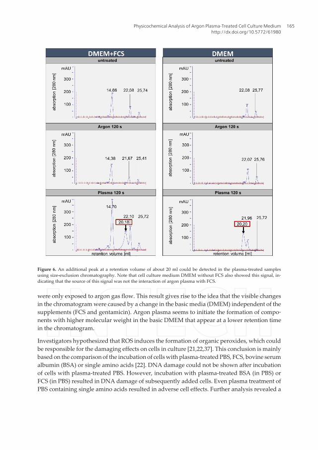
were only exposed to argon gas flow. This result gives rise to the idea that the visible changesin the chromatogram were caused by a change in the basic media (DMEM) independent of thesupplements (FCS and gentamicin). Argon plasma seems to initiate the formation of compo‐nents with higher molecular weight in the basic DMEM that appear at a lower retention timein the chromatogram.
Investigators hypothesized that ROS induces the formation of organic peroxides, which couldbe responsible for the damaging effects on cells in culture [21,22,37]. This conclusion is mainlybased on the comparison of the incubation of cells with plasma-treated PBS, FCS, bovine serumalbumin (BSA) or single amino acids [22]. DNA damage could not be shown after incubationof cells with plasma-treated PBS. However, incubation with plasma-treated BSA (in PBS) orFCS (in PBS) resulted in DNA damage of subsequently added cells. Even plasma treatment ofPBS containing single amino acids resulted in adverse cell effects. Further analysis revealed a
Figure 6. An additional peak at a retention volume of about 20 ml could be detected in the plasma-treated samplesusing size-exclusion chromatography. Note that cell culture medium DMEM without FCS also showed this signal, in‐dicating that the source of this signal was not the interaction of argon plasma with FCS.
Physicochemical Analysis of Argon Plasma-Treated Cell Culture Mediumhttp://dx.doi.org/10.5772/61980
165

correlation between the peroxidation efficiency of single amino acids and their potential toinduce DNA damage in cells [22]. These findings supported the thesis of amino acid peroxideformation and its potential contribution to DNA damage generation. The damage could bemediated either by single amino acids or by FCS, both being usual components of cell culturemedia.
As shown in Figure 5 and also observed in an earlier work of our group, DMEM+FCS+Gentaimpaired cell vitality after argon plasma treatment [25]. Based on the results deduced fromsize-exclusion chromatography (see Figure 6), it was deemed important to analyze the viabilityof mHepR1 cells dependent on the medium supplements during plasma treatment. For thispurpose, DMEM and DMEM+FCS were argon plasma-treated for 60 and 120 s. Controlswithout treatment or only argon gas flow were also investigated. It was found that the mHepR1cell viability decreased significantly in plasma-treated media after 24-h incubation for bothapproaches (Figure 7). In particular, in the presence of FCS during plasma treatment, theinhibitory effect is more obvious.
Figure 7. Cell viability after incubation of mHepR1 cells in plasma-treated DMEM and DMEM+FCS. (mean ± SD, ** p <0.005, *** p < 0.001, t-test, n = 3).
Therefore, it can be assumed that components in the basic DMEM without supplements werechanged due to the impact of argon plasma and influenced cell viability.
Based on the hypothesis of Kalghatgi et al., for the generation of stable organic peroxides fromamino acids during plasma treatment [22] and our cell viability results in mHepR1 cells inplasma-treated DMEM versus DMEM+FCS, it can be concluded that peroxides formed fromsingle amino acids by plasma treatment could be the reason for the detrimental cell effects wefound. However, peroxidated amino acids are semi-reactive compounds and it seems possiblethat they generate compounds with higher molecular weight during storage, e.g., by poly‐
Plasma Science and Technology - Progress in Physical States and Chemical Reactions166

merization. The identification of the newly generated substances in the cell culture mediumawaits further analysis.
There is increasing evidence that plasma treatment promotes the healing process of tissue andaccelerates wound healing [13,40–42]. The skin is a complex architectural multilayer cellsystem and processes concerning wound healing can be examined on cell monolayers as anin vitro model system. However, it is important to keep in sight that a cell monolayer in vitrois much more sensitive to agents because there are no cells in the “second row” to protect orreplace cells in the apical row [43]. Here, we described effects of argon plasma-treated liquids(e.g., cell culture medium) as an indirect approach for plasma application in medicine.Transferred to in vivo systems, the opening of cell–cell contacts we observed by plasma-treatedliquids could have a positive effect on the penetration of conventional therapeutics (antibioticsand disinfectants) on skin. Often, the application of conventional liquid antiseptics is notsufficient and sustainable as the borders and the surrounding of chronic wounds frequentlyconsist of sclerotic skin, impeding an effectual penetration of these products [44]. With regardto disinfection, direct plasma treatment of living intact and wounded skin was found to be safefor doses even higher than required for inactivation of bacteria [45,46]. Impaired cell adhesionand reduced cell viability due to plasma-treated liquids are important starting points forfurther investigations concerning cancer therapy. The work presented here, focused on thebasic mechanisms in the interaction of plasma-treated cell culture medium with cells and onthe components in the treated medium, which are responsible for the persistent biologicaleffect.
The vision could be the establishment of local plasma centers to prepare relatively stableplasma liquids for dermatological (chronic wounds and tumors), dental (peri-implantitis), ororthopedic (joint rinsing) applications to support or replace the conventional therapy.
4. Summary and conclusions
This study focused on the physicochemical analysis of argon plasma-treated cell culturemedium DMEM with the additives FCS and gentamicin. In addition, the efficacy of plasma-treated complete cell culture medium DMEM upon storage and its impact on the cell physi‐ology of epithelial mHepR1 cells were ascertained. We discovered that the liquid-mediatedeffect of atmospheric-pressure argon plasma on mouse liver epithelial cell–cell contacts andcell membrane microvilli persists up to even 21 days or 7 days of storage, respectively. Earlierinvestigations of our group revealed that temperature and pH (both were constant) as well ashydrogen peroxide production and oxygen content (both decreased within 1 day) can beexcluded as initiators of the detrimental biological changes. As we found here, increasedosmolality in the media caused by plasma treatment can also be excluded as a reason for theobserved cell effects. On the other hand, we found an additional peak in size-exclusionchromatography analysis in basic DMEM after plasma treatment and significantly decreasedcell viability in plasma-treated media independent of the presence of FCS during plasmatreatment. High molecular compounds generated during plasma treatment of DMEM without
Physicochemical Analysis of Argon Plasma-Treated Cell Culture Mediumhttp://dx.doi.org/10.5772/61980
167

FCS give an impulse for further investigations on the formation, stability, and reaction of aminoacid peroxides in this medium. The persistent biological effect on plasma-treated liquidsobserved here could open up new medical applications. Stable plasma-treated liquids couldfind applications for dermatological, dental, or orthopedic therapy.
Acknowledgements
The authors acknowledge the financial support of the University Medical Center Rostock,Germany (FORUN 889061, PlasmaBiomedicine) for C.H., M.K., and C.B.. M.H. is grateful forthe doctoral stipendium of the state Mecklenburg-Vorpommern (Germany) and of theUniversity of Rostock, Interdisciplinary Faculty, Department of Life, Light and Matter. Theauthors appreciate the BMBF Germany Pilot Program Campus PlasmaMed (13N11183). Theywould also like to express their thanks to the Electron Microscopic Center (EMZ) of theUniversity Medical Center Rostock (M. Frank, G. Fulda) for qualified technical assistance.
Author details
Claudia Bergemann1, Cornelia Hoppe1,2, Maryna Karmazyna1,3, Maxi Höntsch1,4,Martin Eggert5, Torsten Gerling6 and Barbara Nebe1*
*Address all correspondence to: [email protected]
1 University Medical Center Rostock, Department of Cell Biology, Schillingallee, Rostock,Germany
2 Technical University Dresden, DFG-Center for Regenerative Therapies, Dresden, Germany
3 Seracell Stem Cell Technology GmbH, Schillingallee, Rostock, Germany
4 Sarstedt AG and Co, Quality Management/EWZ, Nümbrecht, Germany
5 University Medical Center Rostock, Department of Internal Medicine, Center forExtracorporeal Organ Support, Schillingallee, Rostock, Germany
6 Leibniz-Institute for Plasma Science and Technology e.V., Greifswald, Germany
References
[1] von Woedtke T, Reuter S, Masur K, Weltmann KD. Plasmas for medicine. Phys Rep.2013;530:291–320.
Plasma Science and Technology - Progress in Physical States and Chemical Reactions168
[2] von Woedtke T, Metelmann HR, Weltmann KD. Clinical plasma medicine: State andperspectives of in vivo application of cold atmospheric plasma. Contrib Plasma Phys.2014;54:104–17.
[3] Fridman G, Friedman G, Gutsol A, Shekhter AB, Vasilets VN, Fridman A. Appliedplasma medicine. Plasma Process Polym. 2008;5:503–33.
[4] Fridman G, Peddinghaus M, Ayan H, Fridman A, Balasubramanian M, Gutsol A, etal. Blood coagulation and living tissue sterilization by floating-electrode dielectricbarrier discharge in air. Plasma Chem Plasma P. 2006;26:425–42.
[5] Kalghatgi SU, Fridman G, Cooper M, Nagaraj G, Peddinghaus M, BalasubramanianM, et al. Mechanism of blood coagulation by nonthermal atmospheric pressure die‐lectric barrier discharge plasma. IEEE T Plasma Sci. 2007;35:1559–66.
[6] Daeschlein G, von Woedtke T, Kindel E, Brandenburg R, Weltmann KD, Junger M.Antibacterial activity of an atmospheric pressure plasma jet against relevant woundpathogens in vitro on a simulated wound environment. Plasma Process Polym.2010;7:224–30.
[7] Daeschlein G, Scholz S, Ahmed R, von Woedtke T, Haase H, Niggemeier M, et al.Skin decontamination by low-temperature atmospheric pressure plasma jet and die‐lectric barrier discharge plasma. J Hosp Infect. 2012;81:177–83.
[8] Daeschlein G, Scholz S, Ahmed R, Majumdar A, von Woedtke T, Haase H, et al. Coldplasma is well-tolerated and does not disturb skin barrier or reduce skin moisture. JDtsch Dermatol Ges. 2012;10:509–15.
[9] Daeschlein G, Scholz S, Arnold A, von Podewils S, Haase H, Emmert S, et al. In vitrosusceptibility of important skin and wound pathogens against low temperature at‐mospheric pressure plasma jet (APPJ) and dielectric barrier discharge plasma (DBD).Plasma Process Polym. 2012;9:380–89.
[10] Nosenko T, Shimizu T, Morfill GE. Designing plasmas for chronic wound disinfec‐tion. New J Phys. 2009;11:115013.
[11] Nastuta AV, Topala I, Grigoras C, Pohoata V, Popa G. Stimulation of wound healingby helium atmospheric pressure plasma treatment. J Phys D Appl Phys.2011;44:105204-0.
[12] Weltmann KD, Kindel E, Brandenburg R, Meyer C, Bussiahn R, Wilke C, et al. At‐mospheric pressure plasma jet for medical therapy: Plasma parameters and risk esti‐mation. Contrib Plasma Phys. 2009;49:631–40.
[13] Heinlin J, Isbary G, Stolz W, Morfill G, Landthaler M, Shimizu T, et al. Plasma appli‐cations in medicine with a special focus on dermatology. J Eur Acad Dermatol.2011;25:1–11.
Physicochemical Analysis of Argon Plasma-Treated Cell Culture Mediumhttp://dx.doi.org/10.5772/61980
169

[14] Kalghatgi S, Friedman G, Fridman A, Clyne AM. Endothelial cell proliferation is en‐hanced by low dose non-thermal plasma through fibroblast growth factor-2 release.Ann Biomed Eng. 2010;38:748–57.
[15] Vandamme M, Robert E, Lerondel S, Sarron V, Ries D, Dozias S, et al. ROS implica‐tion in a new antitumor strategy based on non-thermal plasma. Int J Cancer.2012;130:2185–94.
[16] Plewa JM, Yousfi M, Frongia C, Eichwald O, Ducommun B, Merbahi N, et al. Low-temperature plasma-induced antiproliferative effects on multi-cellular tumor sphe‐roids. New J Phys. 2014;16:043027.
[17] Kalghatgi S, Kelly C, Cerchar E, Azizkhan-Clifford J. Selectivity of non-thermal at‐mospheric-pressure microsecond-pulsed dielectric barrier discharge plasma inducedapoptosis in tumor cells over healthy cells. Plasma Med. 2011;1:249–63.
[18] Kaushik NK, Kaushik N, Attri P, Kumar N, Kim CH, Verma AK, et al. Biomedicalimportance of indoles. Molecules. 2013;18:6620–62.
[19] Stoffels E, Sakiyama Y, Graves DB. Cold atmospheric plasma: Charged species andtheir interactions with cells and tissues. IEEE T Plasma Sci. 2008;36:1441–57.
[20] Kieft IE, Broers JLV, Caubet-Hilloutou V, Slaaf DW, Ramaekers FCS, Stoffels E. Elec‐tric discharge plasmas influence attachment of cultured CHO k1 cells. Bioelectro‐magnetics. 2004;25:362–68.
[21] Haertel B, Wende K, von Woedtke T, Weltmann KD, Lindequist U. Non-thermal at‐mospheric-pressure plasma can influence cell adhesion molecules on HaCaT-kerati‐nocytes. Exp Dermatol. 2011;20:282–84.
[22] Kalghatgi S, Kelly CM, Cerchar E, Torabi B, Alekseev O, Fridman A, et al. Effects ofnon-thermal plasma on mammalian cells. Plos One. 2011;6:16270.
[23] Haertel B, Hahnel M, Blackert S, Wende K, von Woedtke T, Lindequist U. Surfacemolecules on HaCaT keratinocytes after interaction with non-thermal atmosphericpressure plasma. Cell Biol Int. 2012;36:1217–22.
[24] Kalghatgi S, Azizkhan-Clifford J. DNA damage in mammalian cells by atmosphericpressure microsecond-pulsed dielectric barrier discharge plasma is not mediated vialipid peroxidation. Plasma Med. 2011;1:167–77.
[25] Hoentsch M, von Woedtke T, Weltmann KD, Nebe JB. Time-dependent effects oflow-temperature atmospheric-pressure argon plasma on epithelial cell attachment,viability and tight junction formation in vitro. J Phys D Appl Phys. 2012;45:025206.
[26] Hoentsch M, Bussiahn R, Rebl H, Bergemann C, Eggert M, Frank M, et al. Persistenteffectivity of gas plasma-treated, long time-stored liquid on epithelial cell adhesioncapacity and membrane morphology. Plos One. 2014;9:e104559.
Plasma Science and Technology - Progress in Physical States and Chemical Reactions170
[27] Kalghatgi S, Fridman A, Azizkhan-Clifford J, Friedman G. DNA damage in mamma‐lian cells by non-thermal atmospheric pressure microsecond pulsed dielectric barrierdischarge plasma is not mediated by ozone. Plasma Process Polym. 2012;9:726–32.
[28] Stoffels E, Kieft IE, Sladek REJ, van den Bedem LJM, van der Laan EP, Steinbuch M.Plasma needle for in vivo medical treatment: Recent developments and perspectives.Plasma Sources Sci Technol. 2006;15:S169–S80.
[29] Dobrynin D, Fridman G, Friedman G, Fridman A. Physical and biological mecha‐nisms of direct plasma interaction with living tissue. New J Phys. 2009;11:115020.
[30] Nebe B, Rychly J, Knopp A, Bohn W. Mechanical induction of beta-1-integrin-medi‐ated calcium signaling in a hepatocyte cell-line. Exp Cell Res. 1995;218:479–84.
[31] Paul D, Hohne M, Pinkert C, Piasecki A, Ummelmann E, Brinster RL. Immortalizeddifferentiated hepatocyte lines derived from transgenic mice harboring Sv40 T-anti‐gen genes. Exp Cell Res. 1988;175:354–62.
[32] Lange K. Role of microvillar cell surfaces in the regulation of glucose uptake and or‐ganization of energy metabolism. Am J Physiol-Cell Physiol. 2002;282:C1–C26.
[33] Pfister RR, Burstein N. Effects of ophthalmic drugs, vehicles, and preservatives oncorneal epithelium – scanning electron-microscope study. Invest Ophth Visual.1976;15:246–59.
[34] Oehmigen K, Winter J, Hahnel M, Wilke C, Brandenburg R, Weltmann KD, et al. Es‐timation of possible mechanisms of Escherichia coli inactivation by plasma treated so‐dium chloride solution. Plasma Process Polym. 2011;8:904–13.
[35] von Woedtke T, Oehmigen K, Brandenburg R, Hoder T, Wilke C, Hähnel M, et al.Plasma-liquid interactions: Chemistry and antimicrobial effects. In: Machala Z, Hen‐sel K, Akishev Y, editors. Plasma for bio-decontamination, medicine and food securi‐ty: Springer, Netherlands; 2012. pp. 67–78.
[36] Waymouth C. Osmolality of mammalian blood and of media for culture of mamma‐lian cells. In Vitro Cell Dev Biol Plant. 1970;6:109–27.
[37] Gebicki S, Gebicki JM. Formation of peroxides in amino-acids and proteins exposedto oxygen free-radicals. Biochem J. 1993;289:743–49.
[38] van Gils CAJ, Hofmann S, Boekema BKHL, Brandenburg R, Bruggeman PJ. Mecha‐nisms of bacterial inactivation in the liquid phase induced by a remote RF cold at‐mospheric pressure plasma jet. J Phys D Appl Phys. 2013;46:175203.
[39] Machala Z, Tarabova B, Hensel K, Spetlikova E, Sikurova L, Lukes P. Formation ofROS and RNS in water electro-sprayed through transient spark discharge in air andtheir bactericidal effects. Plasma Process Polym. 2013;10:649–59.
[40] Barekzi N, Laroussi M. Dose-dependent killing of leukemia cells by low-temperatureplasma. J Phys D Appl Phys. 2012;45:422002.
Physicochemical Analysis of Argon Plasma-Treated Cell Culture Mediumhttp://dx.doi.org/10.5772/61980
171

[41] Ermolaeva SA, Varfolomeev AF, Chernukha MY, Yurov DS, Vasiliev MM, Kamin‐skaya AA, et al. Bactericidal effects of non-thermal argon plasma in vitro, in biofilmsand in the animal model of infected wounds. J Med Microbiol. 2011;60:75–83.
[42] Emmert S, Brehmer F, Hänßle H, Helmke A, Mertens N, Ahmed R, et al. Atmospher‐ic pressure plasma in dermatology: Ulcus treatment and much more. Clin PlasmaMed. 2013;1:24–29.
[43] Hoene A, Prinz C, Walschus U, Lucke S, Patrzyk M, Wilhelm L, et al. In vivo evalua‐tion of copper release and acute local tissue reactions after implantation of copper-coated titanium implants in rats. Biomed Mater. 2013;8:035009.
[44] Lademann O, Kramer A, Richter H, Patzelt A, Meinke MC, Czaika V, et al. Skin dis‐infection by plasma-tissue interaction: Comparison of the effectivity of tissue-tolera‐ble plasma and a standard antiseptic. Skin Pharmacol Physiol. 2011;24:284–88.
[45] Dobrynin D, Wu A, Kalghatgi S, Park S, Shainsky N, Wasko K, et al. Live pig skintissue and wound toxicity of cold plasma treatment. Plasma Med. 2011;1:93–108.
[46] Fluhr JW, Sassning S, Lademann O, Darvin ME, Schanzer S, Kramer A, et al. In vivoskin treatment with tissue-tolerable plasma influences skin physiology and antioxi‐dant profile in human stratum corneum. Exp Dermatol. 2012;21:130–34.
Plasma Science and Technology - Progress in Physical States and Chemical Reactions172