0830 saturday pfts correct performance (koulouris)
TRANSCRIPT

ATHENS 2019GREECE | 27-29 JUNE

ATHENS 2019GREECE | 27-29 JUNE
Conflict of interest disclosureqI have no, real or perceived, direct or indirect
conflicts of interest that relate to this presentation.

ATHENS 2019GREECE | 27-29 JUNE
PFTs: The Correct Performance and
Interpretation
Nickolaos G Koulouris MD, PhD, FCCP

Learning Objectives:ØExplain how to perform spirometry testing.ØLearn how to detect airway obstruction.ØBe able to give a preoperative assessment.ØDescribe the current algorithm for preoperative assessment.
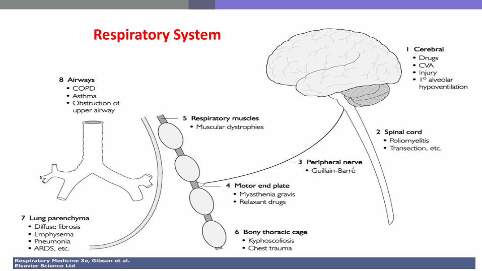
Respiratory System
Pulmonary lung function tests are designed toidentify and quantify defects and abnormalities in thefunction of the respiratory system (ventilatory defects orfunctional syndromes) and answer several crucialquestions, such as:ØDoes a patient has a ventilatory limitation?Ø If yes, on what basis?ØHow severe it is?ØAnd many more questions………
Pulmonary lung function tests

How lung function testing helps us?
Measurements determine ¯
The Ventilatory Defect of a patient. Combining these data¯
with History and Clinical Examination we make the correct ¯
Diagnosis

The ventilatory defects are: a) obstructive characterized by obstruction or narrowing of the airways, b) restrictivechararacterized by a reduction of Total Lung Capacity (TLC)due to i) pulmonary diseases per se (i.e., pulmonary fibrosis), ii) extrapulmonary diseases and disorders (i.e., chest wall deformities, neuromuscular diseases, etc),and c) mixed characterized by the presence ofboth, obstructive and restrictive
ventilatory defects.
VENTILATORY DEFECTS

ATHENS 2019GREECE | 27-29 JUNE
Routine Lung Function Tests
§ Simple spirometry and maximum flow-volume curve before and after bronchodilation (PEF, FEVC, FEV1, MMEF), seated and supine
It is the best test to detect airways obstruction§ Static Lung Volumes and Capacities (TLC, FRC, RV)TLC is the best measurement to detect restriction § Diffusing Capacity or Transfer Factor and Carbon Monoxide Rate Constant
(DLCO ή TLCO, KCO)Assesses the transfer of gas between the alveoli and pulmonary capillary blood flow
Maximum Static Mouth Pressures (Pimax, Pemax)Assessment of Respiratory Muscle Strength§ Blood Gases (PaO2, PaCO2, pH), pulse oximetry

ATHENS 2019GREECE | 27-29 JUNE
Miller et al, Eur Respir J 2005; 26: 153–161

ATHENS 2019GREECE | 27-29 JUNE
DEFINITION OF SPIROMETRY
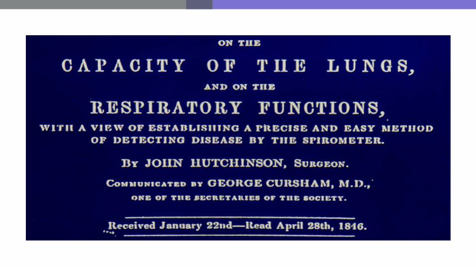
Spirometry, Spirometer : Introduced in Oxford Dictionary in 1846
From the Latin Spiro, Respiro (breathing) and the Greek Μετρώ (measure)
Spirometry is a physiological test that measures how an individual inhales or exhales volumes of air as a function of time or flow.
In practice, Spirometry is the measurement of Forced Vital Capacity (FVC). FVC is the maximum volume of air expelled after a maximum inhalation.

John Hutchinson invented andpresented the first spirometric device in1846. (Spirometer is a device thatmeasures air volumes exhaled from thelung). He also described in detail themeasurement of the first spirometric testthe so called Slow Vital Capacity (SVC).


ATHENS 2019GREECE | 27-29 JUNE
BTPS CORRECTIONBody Conditions: Body Temperature, Ambient Pressure, and Saturation with water vapor
üCharles Law states that the volume occupied by any given quantity of gas is directly related to temperature. As the exhaled air travels from a patient (BTPS conditions) to a spirometer it cools (ATPS conditions). Therefore, the volume of air exhaled is reduced and its water saturation changes.

Miller et al, EurRespir J 2005; 26: 319–338

ATHENS 2019GREECE | 27-29 JUNE
Miller et al, Eur Respir J 2005; 26: 153–161

Slow Vital Capacity (SVC), used as thesole lung function test for 101 years, it wasnot capable of detecting the commonestlung defect, i.e., the obstructive lung defect.Most lung diseases cause obstructive lungdefect (~70%).


In 1947, the detection of the “obstructive lung defect” was madepossible only when two French investigators Tiffeneau & Pinelliproposed the measurement of two new parameters derived from theForced Spirogram. a) Forced Expired Volume in the 1st second(FEV1), and b) FEV1/FVC, % ratio (also known as Tiffeneau’s ratio).
Volume-Time curveExpiration
Inspiration

In 1958, Hyatt presented the MaximumExpiratory Flow Volume (MEFV) curve as analternative expression of the classical Volume-Timecurve (FVC-t).
Flow-Volume Volume-Time


Miller et al, EurRespir J 2005; 26: 319–338

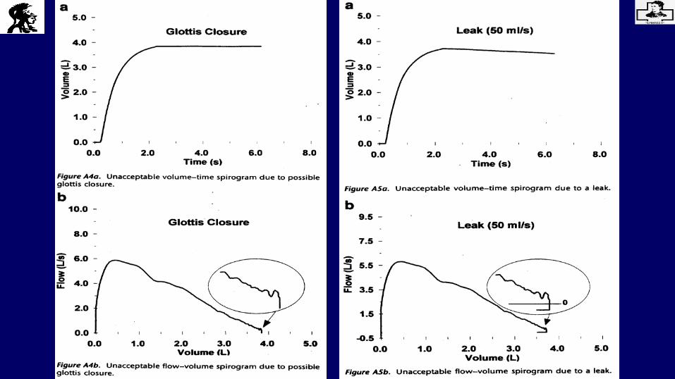
Time to reach PEF is 100ms.The volume–time curve shows no change in volume (0.025 L) for ≥1 s, and the subject has tried to exhale for≥ 3 s in children aged ≤10 yrs and for ≥ 6 s in subjects aged ≥ than10 yrs.For patients with airways obstruction or older subjects, exhalation times of ≥ 6 s are frequently needed. However,exhalation times of ≥15 s will rarely change clinical decisions.


The EV must be less than 0,5% of the FVC or 0.150 L, whichever is greater.


Time dependence of Forced Vital Capacity (FVC)
Extensive guidelines have been provided for the measurement procedure of FVC. In the early guidelines , however,the inspiratory manoeuvre preceding the expiratory effort was not standardized. In practice, the FVC was preceded by
1) maximal inspirations made at different speeds, and2) variable pauses at full inspiration
The time course of inspiration preceding the FVC has a marked effect on PEF, FEV1, and MEFV curves both in normal subjects and patients with obstructive and restrictive lung disease.

Tracings showing the time course of changes in lung volume (ΔV) obtained in aCOPD patient during an FVC manoeuvre preceded by a) a rapid inspiration withoutbreathhold at end inspiration, and b) a slow inspiration with a 5s breathhold. With theslow manoeuvre the PEF and FEV1 were 23% lower than the fast one, whilst FVC didnot change. D’Angelo et al, Am J Respir Crit care Med 1994; 150: 1581-1586

Miller et al, Eur Respir J 2005; 26: 319–338


Lung Volumes
1. Tidal volume (Vt) is the volume of air that is inspired and expiredwith each breath during normal breathing.
2. Residual Volume (RV) is the volume of air remaining in the lungs atthe end of a maximum expiration.
3. Inspiratory Reserve Volume (IRV) is the maximum amount of air thatcan be inhaled beyond the tidal volume end-inspiratory level.
4. Expiratory Reserve Volume (ERV) is the maximum amount of airthat can be exhaled below the tidal volume end-expiratory level.

Lung Capacities
1. Functional Residual Capacity (FRC) is the volume remaining in thelungs at the tidal volume end-expiratory level.
2. Total Lung Capacity (TLC) is the volume of air in the lungs after amaximum inspiration.
3. Vital Capacity (VC) is the volume of air that can be exhaled from thelungs after a maximum inhalation.
4. Inspiratory Capacity (IC) is the maximum amount of air that can beinhaled from the tidal volume end-expiratory level.

Techniques for measuring Static Lung Volumes(essentially FRC)
1) Body Plethysmography2) Nitrogen Washout3) Helium Dilution4) Imaging (CXR, CT, MRI)

Definition of Diffusion Capacity (USA)or Transfer Factor (Europe)
DLCO* 0.33 = TLCO
DLCO is actually the diffusive conductance,meaning the “ease of transfer” for CO moleculespassing from alveolar gas to pulmonary capillaryhemoglobin. It reflects the surface area of the lungavailable for gas exchange.

1. Membrane Conductance2. Reactive Conductance
Hughes & Pride (eds). Lung Function Tests. London WB Saunders, 1999.
1/DLCO=1/Dm+1/ θ.QcRoughton FJW, Forster RE. JAP
1957;11: 290-302

Factors that reduce DLCO
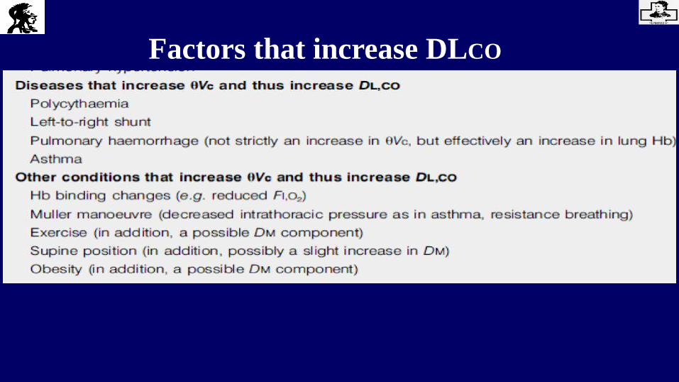
Factors that increase DLCO

Surgery remains the best treatment option for non-smallcell lung cancer, but only 20–25% of lung cancer patients areoperable. Therefore, offering surgery to patients deemed to beinoperable remains highly relevant.
In order to construct a reasonable algorithm it seemsnecessary to use FEV1, Diffusion Capacity, and exercise testing.

ATHENS 2019GREECE | 27-29 JUNE
FEV1Spirometry is widely available, well standardized, and cheap.
Among the multiple parameters measured, FEV1 has stood the test of time and has been included in all the published functional algorithms. However, its predictive value for postoperative complications is not very high, even if the extent of resection is taken into account through the calculation of a ppoFEV1. For these reasons, the decision to operate or not should not be based on ppoFEV1 alone.
Licker M, Schnyder JM, Frey JG, et al. Impact of aerobic exercice capacity and procedure-related factors in lungcancer surgery. Eur Respir J 2010 DOI: 10.1183/09031936.00069910
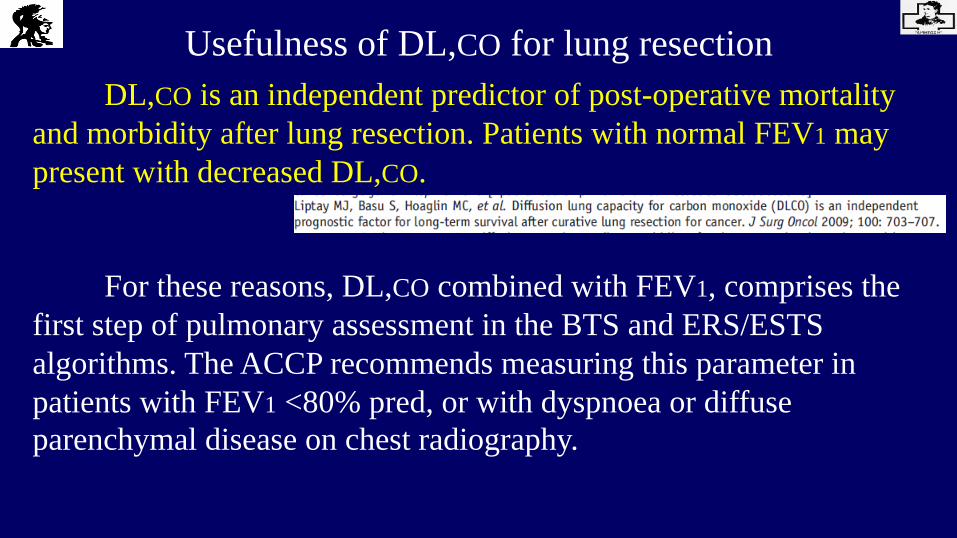
DL,CO is an independent predictor of post-operative mortality and morbidity after lung resection. Patients with normal FEV1 may present with decreased DL,CO.
For these reasons, DL,CO combined with FEV1, comprises the first step of pulmonary assessment in the BTS and ERS/ESTS algorithms. The ACCP recommends measuring this parameter in patients with FEV1 <80% pred, or with dyspnoea or diffuse parenchymal disease on chest radiography.
Usefulness of DL,CO for lung resection

In the literature, V´O2,max appears to be 1) an independent risk factor of bothCardiovascular and Pulmonary Complications, and 2) a very strong predictor ofPostoperative Complications, as well as a good predictor of long-term post-operativeexercise capacity.
Therefore, the most used and best validated exercise parameter is V´O2, max (mlper Kg per min).
Exercise tests
Koulouris et al, JAP 1997; 82: 723-31


Low-technology exercise testsFormal CPET with V´O2,max (ml per Kg per min) measurements may not be
readily available in all centres. Therefore, low-technology tests have been used toevaluate fitness before lung resectionØThe 6MWT is not recommended to select patients for lung resection because does notcorrelate with V´O2,max.ØIn contrast, there is a good correlation between the distance walked during a shuttletest and V´O2,max. Chronic obstructive pulmonary disease patients walking 420 m havea mean V´O2,max of 21 mL per kg per min and those walking 120 m of 11 mL per kgper min.ØThe stair climbing test has also been used as a screening test. The height of ascentcorrelates with V´O2,max, 98% of patients climbing>22 m demonstrating V´O2,max>15 mL per min per kg. The speed of ascent also correlates with V´O2,max, a speed >15m per min corresponding to V´O2,max >20 mL per kg per min.

Cardiological assessmentA cardiological assessment has been
intergrated in all guidelines
A cardiological evaluation is justified, as 10% of major complications and 50% of minor complications after lung resection have a cardiovascular cause.
The guidelines published by the BTS,ACCP and ERS/ESTS recommend using the American College of Cardiology and American Heart Association guidelines.
Lee TH, Marcantonio ER, Mangione CM, et al. Derivation and prospective validation of a simple index for prediction of cardiac risk of major noncardiacsurgery. Circulation 1999; 100: 1043–1049.9. Poldermans D, Bax JJ, Boersma E, et al. Guidelines for pre-operative cardiac risk assessment and perioperative cardiac management in non-cardiac surgery: Eur Heart J 2009; 30: 2769–2812.

It is recommended to express FEV1 as % predicted rather than an absolute value.
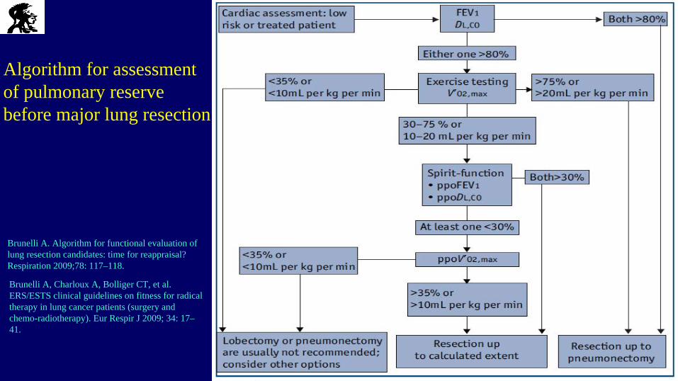
Algorithm for assessmentof pulmonary reserve before major lung resection
Brunelli A. Algorithm for functional evaluation of lung resection candidates: time for reappraisal? Respiration 2009;78: 117–118.
Brunelli A, Charloux A, Bolliger CT, et al. ERS/ESTS clinical guidelines on fitness for radical therapy in lung cancer patients (surgery and chemo-radiotherapy). Eur Respir J 2009; 34: 17–41.

Calculation of predictive post operative (ppo) forced expiratory volumein 1 s (FEV1), diffusing capacity of the lung for carbon dioxide (DL,CO) or
maximal oxygen uptake (V´O2,max), ppoFEV1 is taken as a model.Similar equations are used for the calculation of ppoDL,CO or ppo V´O2,max, and include preoperative DL,CO or V´O2,max, respectively. For ppoFEV1 before lobectomy, the calculation is based on the segment counting method, as follows. Number of functional segments: 19Right lung: Left lung:Upper lobe: 3 Upper lobe: 3Middle lobe: 2 Lingula: 2Lower lobe: 5 Lower lobe: 4
ppoFEV1=pre-operative FEV1X(1 - a/b) where a is the number of unobstructed segments to be resected and b is the total number of unobstructed segments. An unobstructive segment is defined as one where the patency of the bronchus and the segment structure are preserved, according to bronchoscopy and computed tomography (CT) scan.For ppoFEV1 before pneumonectomy, the calculation is based on scintigraphy or quantitative CT scan, as follows.ppoFEV1=pre-operative FEV1X(1 - FP)where FP is the fraction of total perfusion for the lung to be resected.

Respiratory Function Tests, represent the most powerful tools we have available for the diagnosis of many respiratory diseases.Just as one cannot make a diagnosis of hypertension without
measuring blood pressure, so one cannot diagnose COPD, asthma, restrictive disorders, respiratory muscle weakness, and many other chest diseases without respiratory function tests. To understand the
natural history of many lung diseases and to determine how they respond to therapy also requires function tests. Sadly, this seem to
have been forgotten in recent years.
Peter T Macklem

Ναύπλιο Ελλάς
Thanks for your attention

ATHENS 2019GREECE | 27-29 JUNE
Questions1) What is the best respiratory function parameter to
define a “Restrictive Ventilatory Defect” ?
a) Functional Residual Capacity (FRC)b) Airway Resistance (Raw)c) Residual Volume (RV)d) Total Lung Capacity (TLC)e) Forced Vital Capacity (FVC)

ATHENS 2019GREECE | 27-29 JUNE
2) What test is best for detecting “Obstructive Ventilatory Defect”?
a) Measurement of Lung Volumesb) DLCOc) Spirometryd) Respiratory Muscle Testinge) Specific airway Conductance (SGaw)

ATHENS 2019GREECE | 27-29 JUNE
1) Formal CPET including V´O2,max (ml per Kg per min) measurement
2) Spirometry3) Diffusion Capacity 4) Static Lung Volumes5) A combination of 1, 2, 3 tests
3) If you like construct an algorithm for preoperative assessment for lung resection, which test(s) you choose?