09 urinary system
TRANSCRIPT
URINARY SYSTEM
• ONE OF THE SYSTEM THAT CONTRIBUTES HEMOSTASIS
• THIS SYSTEM REGULATES BLOOD PLASMA COMPOSTION THROUGH CONTROLLED EXCRETION OF ORGANIC WASTES, SALTS & WATER
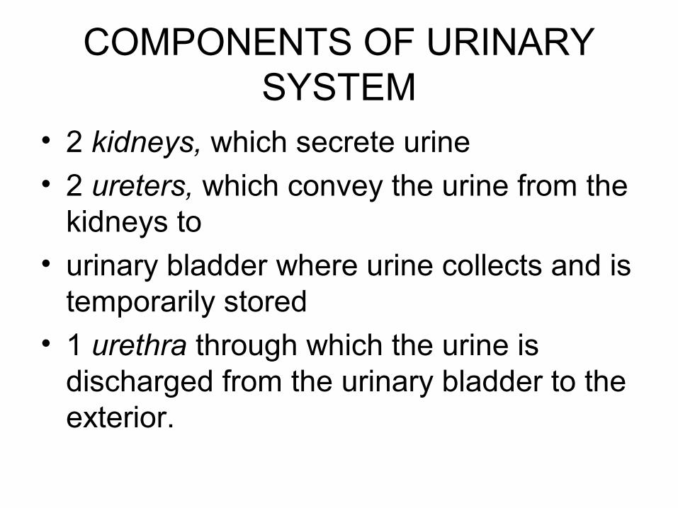
COMPONENTS OF URINARY SYSTEM
• 2 kidneys, which secrete urine
• 2 ureters, which convey the urine from the kidneys to
• urinary bladder where urine collects and is temporarily stored
• 1 urethra through which the urine is discharged from the urinary bladder to the exterior.
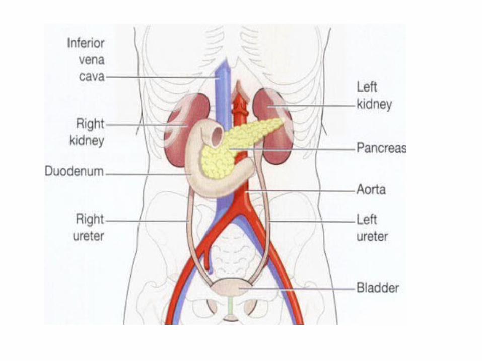
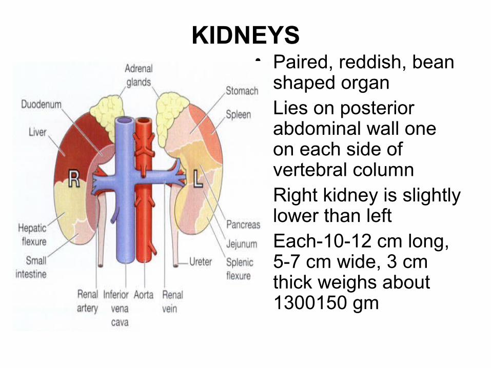
KIDNEYS• Paired, reddish, bean
shaped organ• Lies on posterior
abdominal wall one on each side of vertebral column
• Right kidney is slightly lower than left
• Each-10-12 cm long, 5-7 cm wide, 3 cm thick weighs about 1300150 gm
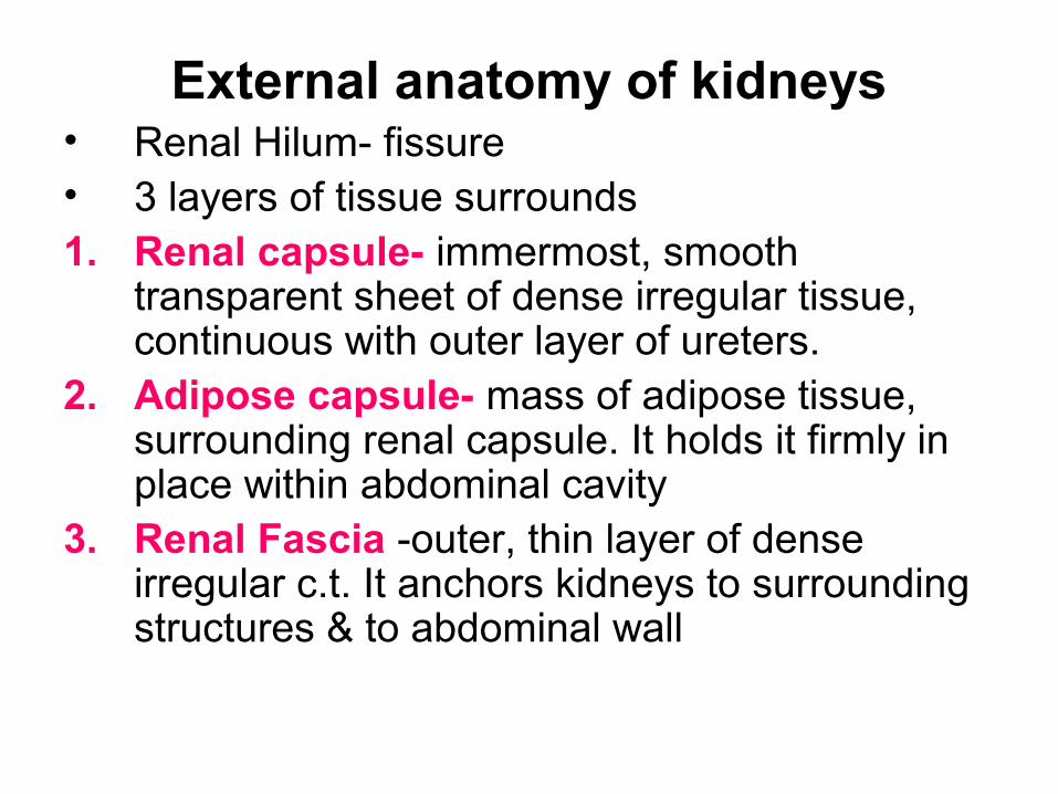
External anatomy of kidneys• Renal Hilum- fissure• 3 layers of tissue surrounds 1. Renal capsule- immermost, smooth
transparent sheet of dense irregular tissue, continuous with outer layer of ureters.
2. Adipose capsule- mass of adipose tissue, surrounding renal capsule. It holds it firmly in place within abdominal cavity
3. Renal Fascia -outer, thin layer of dense irregular c.t. It anchors kidneys to surrounding structures & to abdominal wall
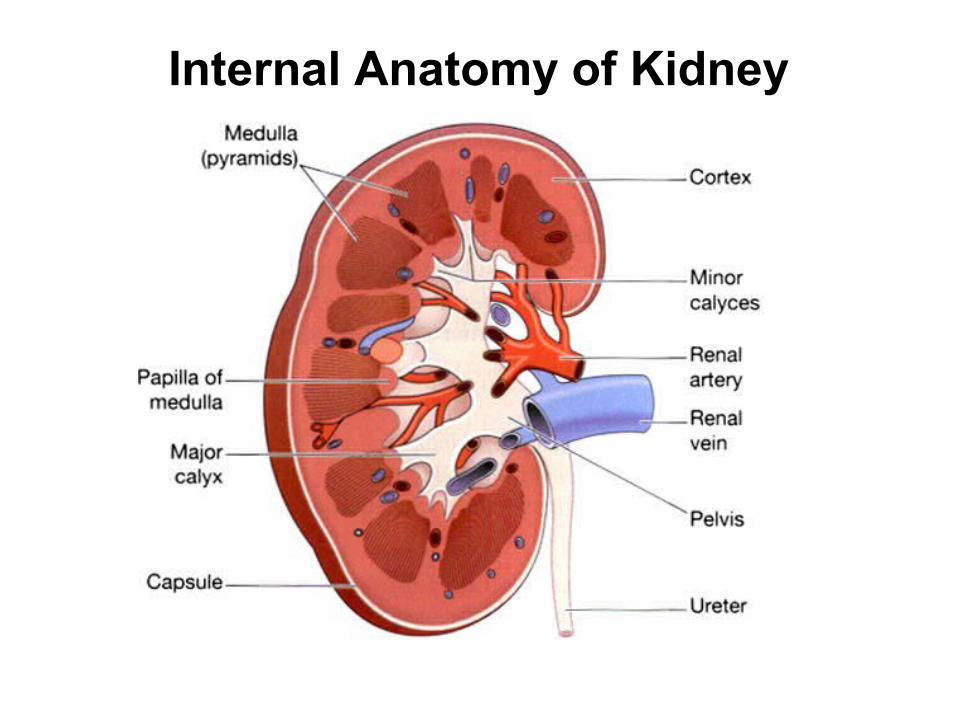
Internal Anatomy of Kidney
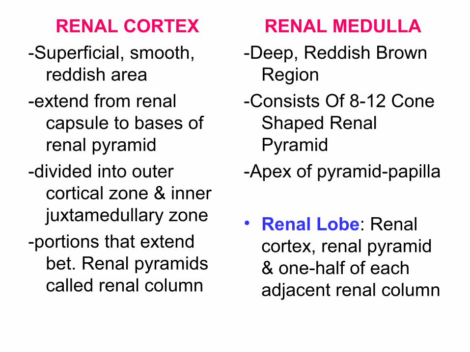
RENAL CORTEX
-Superficial, smooth, reddish area
-extend from renal capsule to bases of renal pyramid
-divided into outer cortical zone & inner juxtamedullary zone
-portions that extend bet. Renal pyramids called renal column
RENAL MEDULLA
-Deep, Reddish Brown Region
-Consists Of 8-12 Cone Shaped Renal Pyramid
-Apex of pyramid-papilla
• Renal Lobe: Renal cortex, renal pyramid & one-half of each adjacent renal column
• The renal pelvis is the funnel-shaped structure which acts as a receptacle for the urine formed by the kidney. It has a number of distal branches called calyces, each of which surrounds the apex of a renal pyramid
• Urine formed in the kidney passes through a papilla at the apex of a pyramid into a minor calyx, then into a major calyx before passing through the pelvis into the ureter.
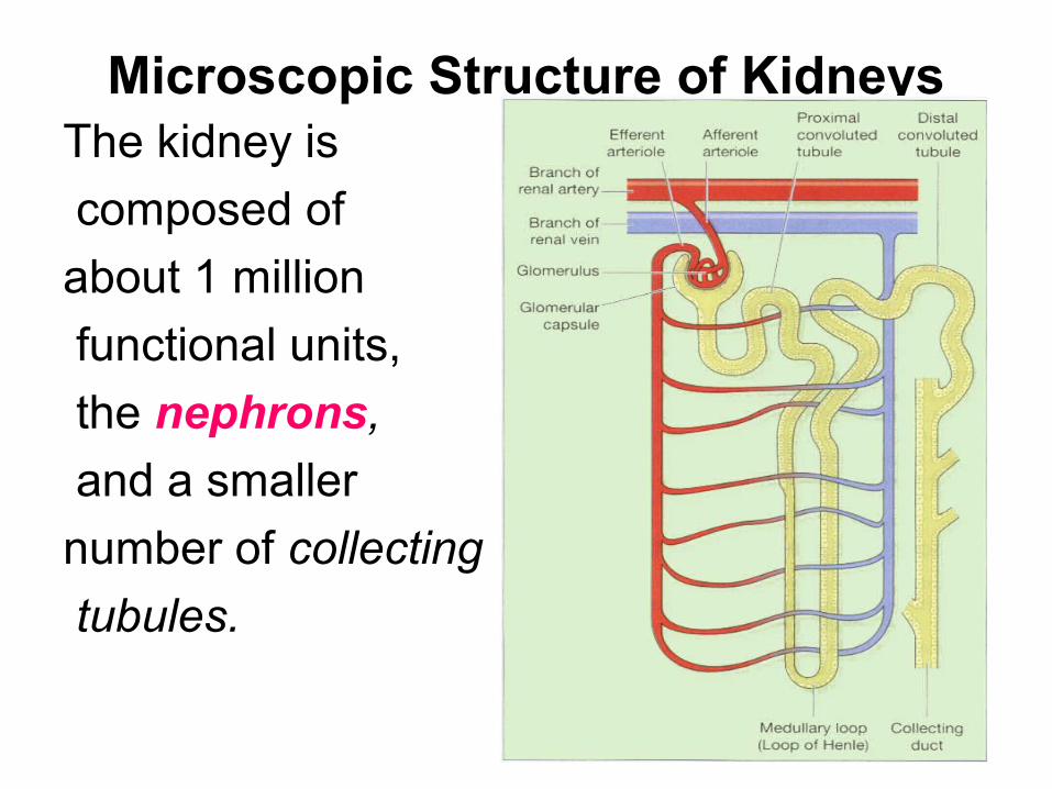
Microscopic Structure of KidneysThe kidney is
composed of
about 1 million
functional units,
the nephrons,
and a smaller
number of collecting
tubules.
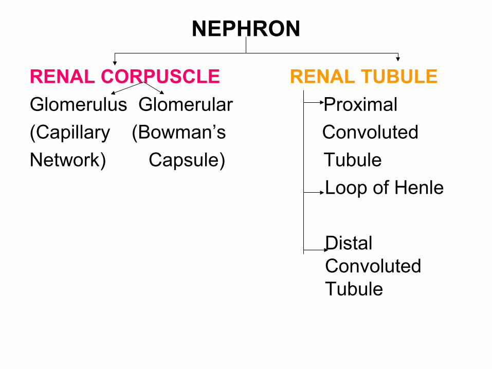
NEPHRON
RENAL CORPUSCLE RENAL TUBULE
Glomerulus Glomerular Proximal
(Capillary (Bowman’s Convoluted
Network) Capsule) Tubule
Loop of Henle
Distal Convoluted Tubule
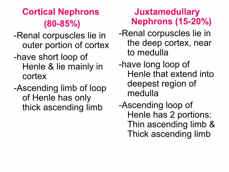
Cortical Nephrons (80-85%)
-Renal corpuscles lie in outer portion of cortex
-have short loop of Henle & lie mainly in cortex
-Ascending limb of loop of Henle has only thick ascending limb
Juxtamedullary Nephrons (15-20%)
-Renal corpuscles lie in the deep cortex, near to medulla
-have long loop of Henle that extend into deepest region of medulla
-Ascending loop of Henle has 2 portions: Thin ascending limb & Thick ascending limb
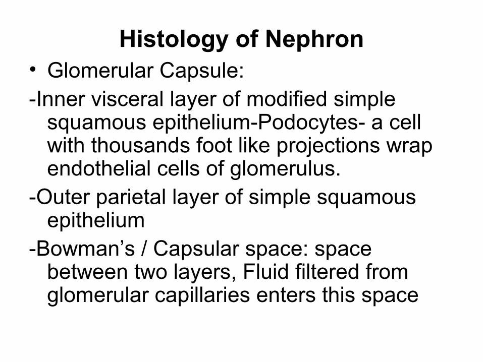
Histology of Nephron• Glomerular Capsule:-Inner visceral layer of modified simple
squamous epithelium-Podocytes- a cell with thousands foot like projections wrap endothelial cells of glomerulus.
-Outer parietal layer of simple squamous epithelium
-Bowman’s / Capsular space: space between two layers, Fluid filtered from glomerular capillaries enters this space
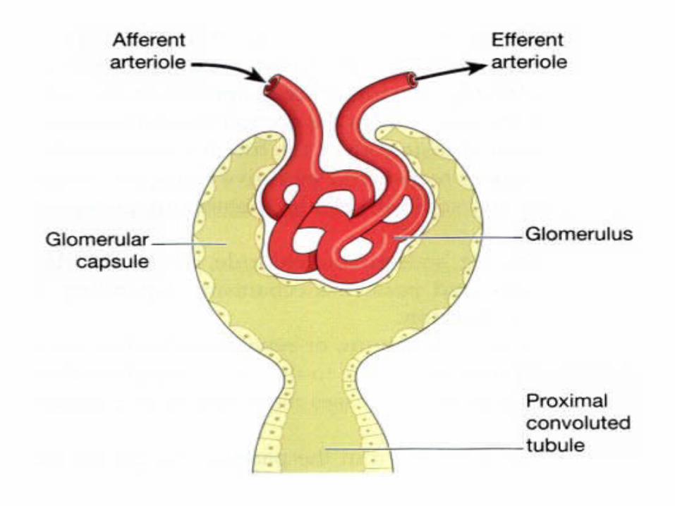
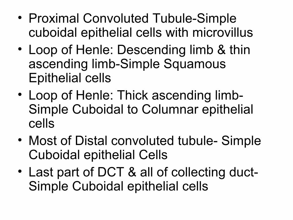
• Proximal Convoluted Tubule-Simple cuboidal epithelial cells with microvillus
• Loop of Henle: Descending limb & thin ascending limb-Simple Squamous Epithelial cells
• Loop of Henle: Thick ascending limb-Simple Cuboidal to Columnar epithelial cells
• Most of Distal convoluted tubule- Simple Cuboidal epithelial Cells
• Last part of DCT & all of collecting duct-Simple Cuboidal epithelial cells
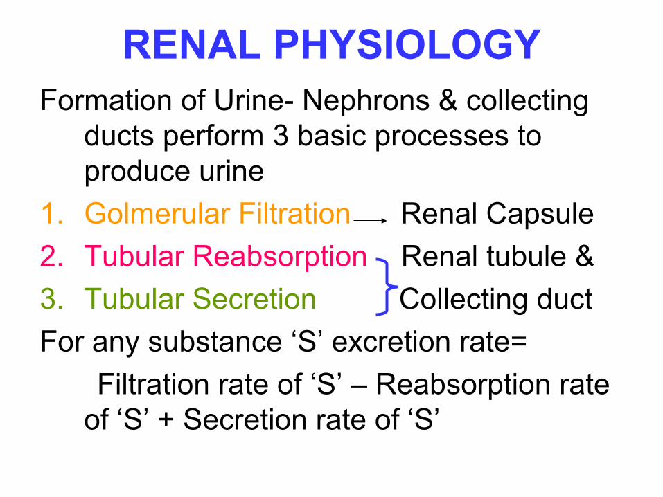
RENAL PHYSIOLOGYFormation of Urine- Nephrons & collecting
ducts perform 3 basic processes to produce urine
1. Golmerular Filtration Renal Capsule
2. Tubular Reabsorption Renal tubule &
3. Tubular Secretion Collecting duct
For any substance ‘S’ excretion rate=
Filtration rate of ‘S’ – Reabsorption rate of ‘S’ + Secretion rate of ‘S’
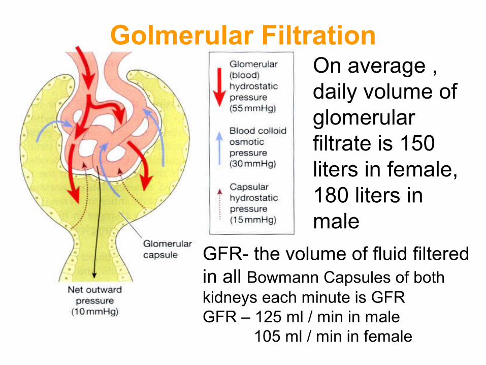
Golmerular FiltrationOn average , daily volume ofglomerular filtrate is 150 liters in female, 180 liters in male
GFR- the volume of fluid filteredin all Bowmann Capsules of bothkidneys each minute is GFRGFR – 125 ml / min in male 105 ml / min in female
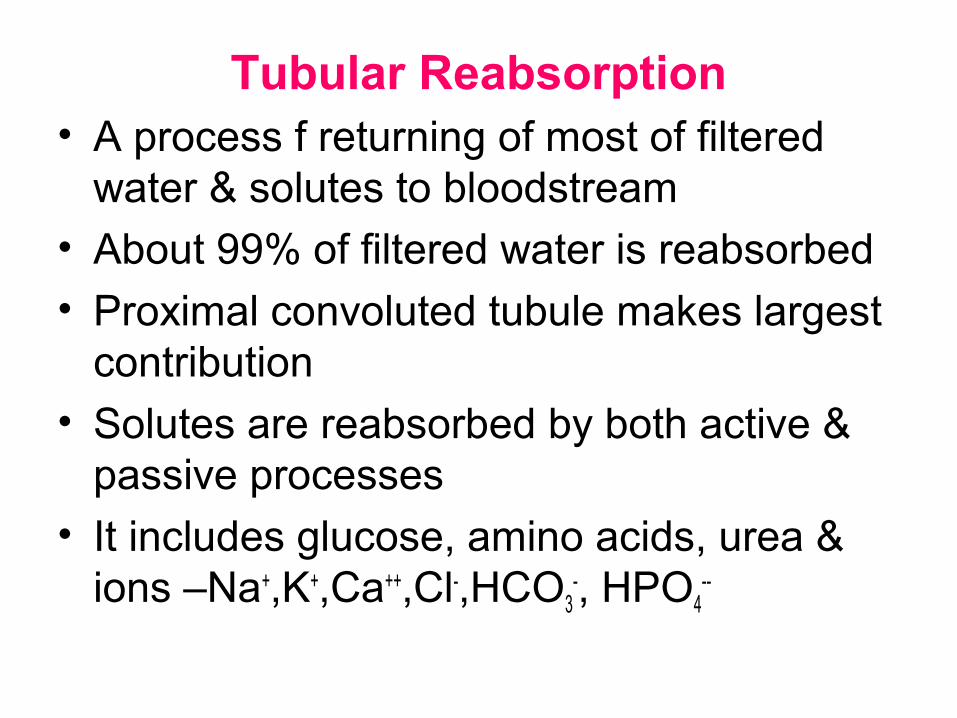
Tubular Reabsorption• A process f returning of most of filtered
water & solutes to bloodstream
• About 99% of filtered water is reabsorbed
• Proximal convoluted tubule makes largest contribution
• Solutes are reabsorbed by both active & passive processes
• It includes glucose, amino acids, urea & ions –Na+,K+,Ca++,Cl-,HCO3
-, HPO4--
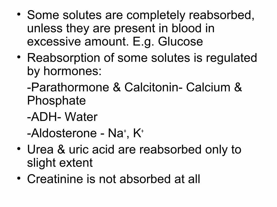
• Some solutes are completely reabsorbed, unless they are present in blood in excessive amount. E.g. Glucose
• Reabsorption of some solutes is regulated by hormones:-Parathormone & Calcitonin- Calcium & Phosphate-ADH- Water-Aldosterone - Na+, K+
• Urea & uric acid are reabsorbed only to slight extent
• Creatinine is not absorbed at all
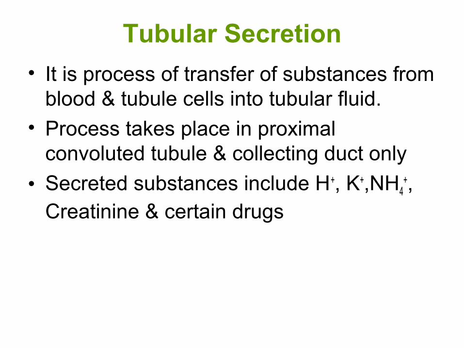
Tubular Secretion
• It is process of transfer of substances from blood & tubule cells into tubular fluid.
• Process takes place in proximal convoluted tubule & collecting duct only
• Secreted substances include H+, K+,NH4+,
Creatinine & certain drugs

Kidney controls urine output & maintain water balance by 3 ways
ANTI-DIURETIC HORMONE
Increased blood osmotic pressure
Osmoreceptors in hypothalamus
Stimulation of post.Pituitary Release of ADH
promotes Reabsorption of water
Reduces loss of water in urine
(reduced blood osmotic pressure)
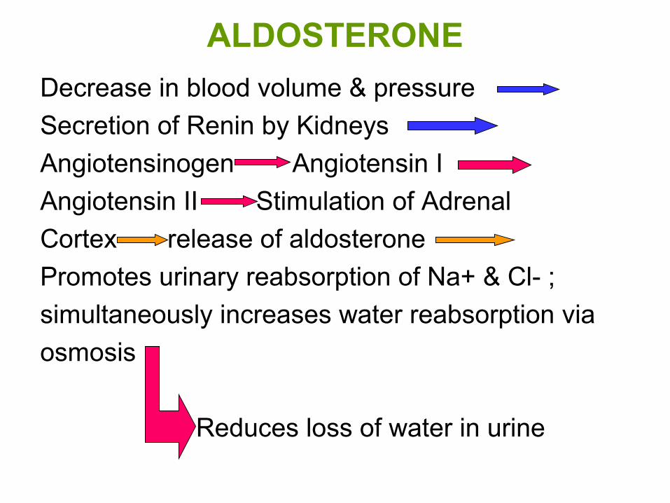
ALDOSTERONE
Decrease in blood volume & pressure
Secretion of Renin by Kidneys
Angiotensinogen Angiotensin I
Angiotensin II Stimulation of Adrenal
Cortex release of aldosterone
Promotes urinary reabsorption of Na+ & Cl- ;
simultaneously increases water reabsorption via
osmosis
Reduces loss of water in urine
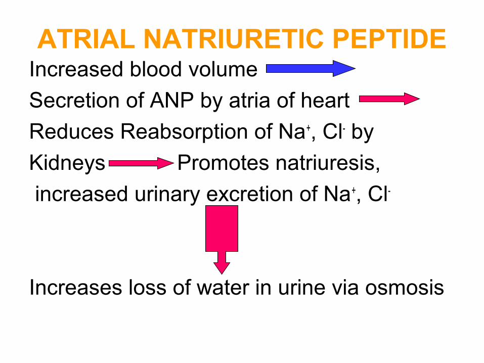
ATRIAL NATRIURETIC PEPTIDEIncreased blood volume
Secretion of ANP by atria of heart
Reduces Reabsorption of Na+, Cl- by
Kidneys Promotes natriuresis,
increased urinary excretion of Na+, Cl-
Increases loss of water in urine via osmosis
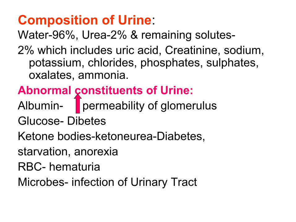
Composition of Urine:Water-96%, Urea-2% & remaining solutes-2% which includes uric acid, Creatinine, sodium,
potassium, chlorides, phosphates, sulphates, oxalates, ammonia.
Abnormal constituents of Urine:Albumin- permeability of glomerulusGlucose- DibetesKetone bodies-ketoneurea-Diabetes,starvation, anorexia RBC- hematuriaMicrobes- infection of Urinary Tract
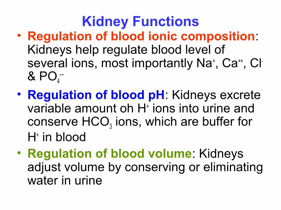
Kidney Functions• Regulation of blood ionic composition:
Kidneys help regulate blood level of several ions, most importantly Na+, Ca++, Cl- & PO4
—
• Regulation of blood pH: Kidneys excrete variable amount oh H+ ions into urine and conserve HCO3 ions, which are buffer for H+ in blood
• Regulation of blood volume: Kidneys adjust volume by conserving or eliminating water in urine
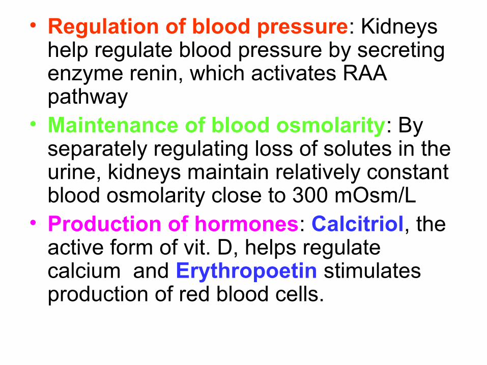
• Regulation of blood pressure: Kidneys help regulate blood pressure by secreting enzyme renin, which activates RAA pathway
• Maintenance of blood osmolarity: By separately regulating loss of solutes in the urine, kidneys maintain relatively constant blood osmolarity close to 300 mOsm/L
• Production of hormones: Calcitriol, the active form of vit. D, helps regulate calcium and Erythropoetin stimulates production of red blood cells.
• Regulation of blood glucose level: Like liver, kidneys can use amino acid glutamine in gluconeogenesis. It release glucose into blood to help maintain normal blood glucose level
• Excretion of wastes & foreign substances: By forming urine, kidneys excrete wastes- substances that have no useful function in body. These include ammonia, urea, bilirubin, creatinine & uric acid.Foreign substances such as metabolites of drugs and environmental toxins are excreted
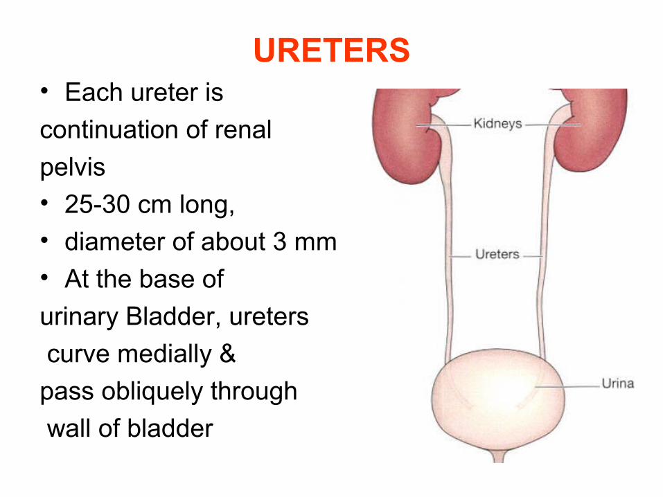
URETERS• Each ureter is
continuation of renal
pelvis• 25-30 cm long, • diameter of about 3 mm• At the base of
urinary Bladder, ureters
curve medially &
pass obliquely through
wall of bladder
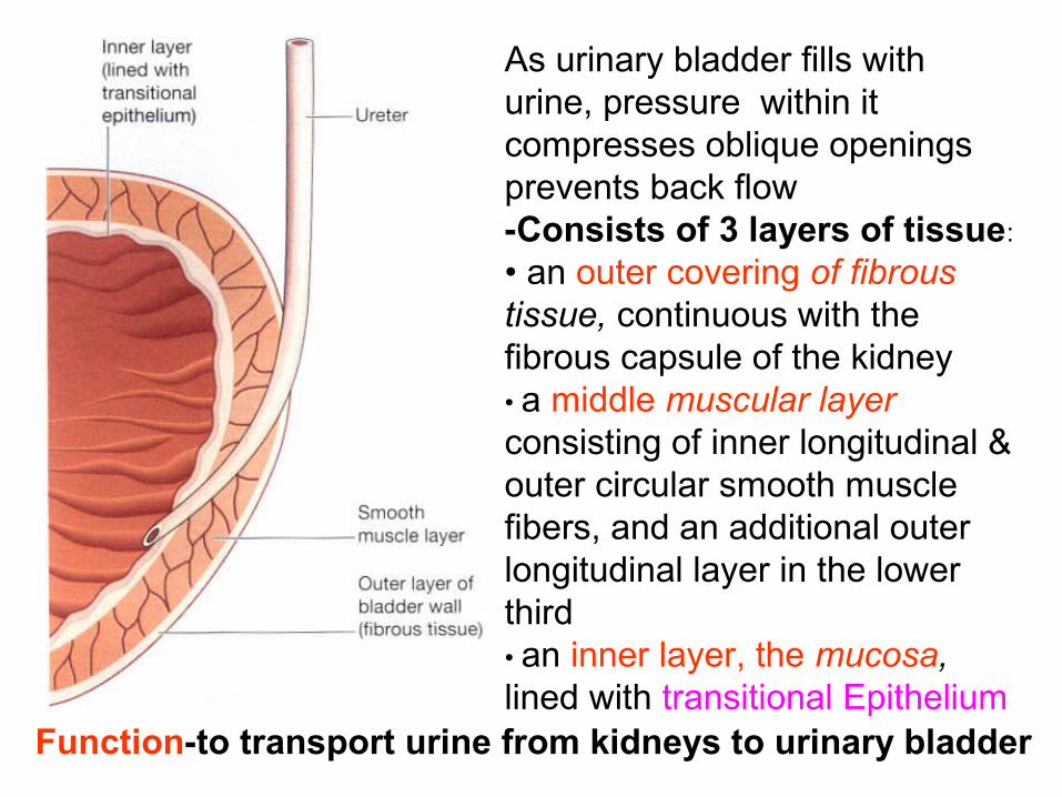
As urinary bladder fills with urine, pressure within it compresses oblique openings prevents back flow-Consists of 3 layers of tissue:
• an outer covering of fibrous tissue, continuous with the fibrous capsule of the kidney• a middle muscular layer consisting of inner longitudinal & outer circular smooth muscle fibers, and an additional outer longitudinal layer in the lower third• an inner layer, the mucosa, lined with transitional Epithelium
Function-to transport urine from kidneys to urinary bladder
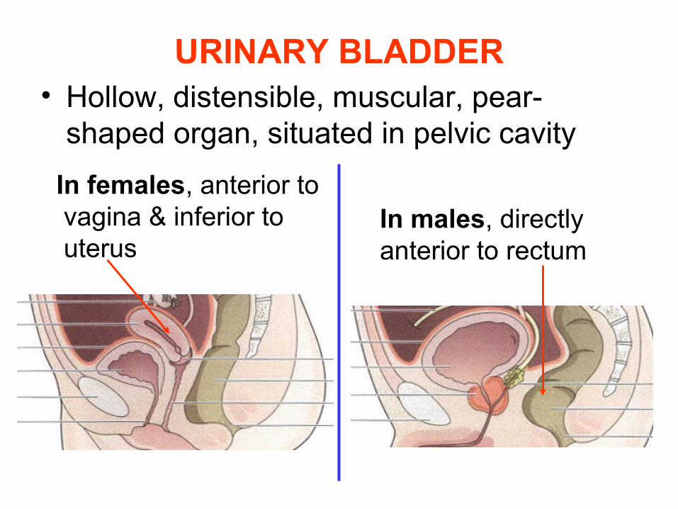
URINARY BLADDER• Hollow, distensible, muscular, pear-
shaped organ, situated in pelvic cavity
In males, directly anterior to rectum
In females, anterior to vagina & inferior to uterus
• The bladder wall is composed of three layers:
• the outer layer of loose connective tissue, containing blood and lymphatic vessels and nerves, covered on the upper surface by the peritoneum
• the middle layer, Muscularis (detrusor muscle) -consisting of 3 layers of smooth muscle fibers : inner Longitudinal, middle Circular & outer Longitudinal
• the mucosa, lined with transitional epithelium
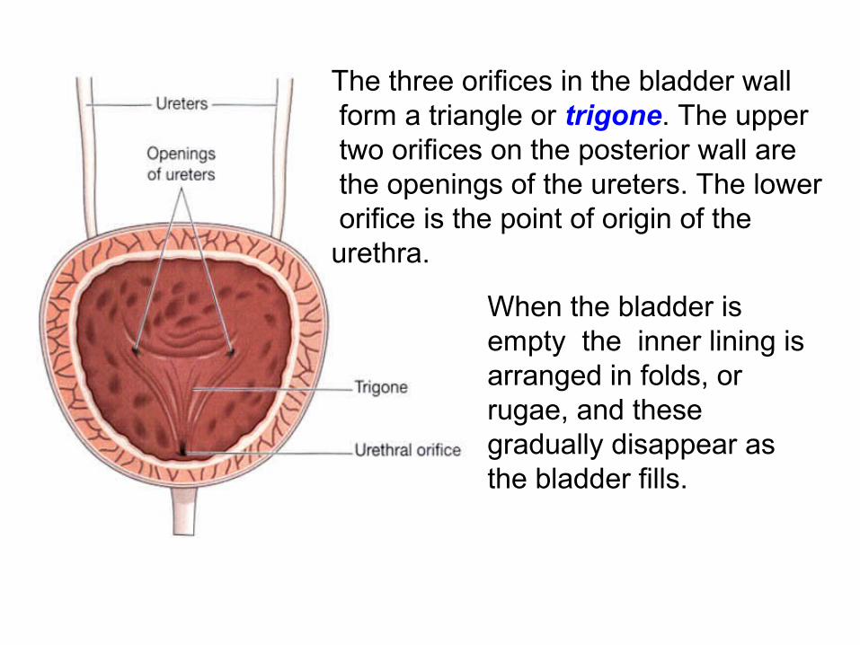
The three orifices in the bladder wall form a triangle or trigone. The upper two orifices on the posterior wall are the openings of the ureters. The lower orifice is the point of origin of the urethra.
When the bladder is empty the inner lining is arranged in folds, or rugae, and these gradually disappear as the bladder fills.
• The bladder is distensible but when it contains 300 to 400 ml the awareness of the desire to urinate is initiated. The total capacity is rarely more than about600 ml.
• Where the urethra commences is a thickening of the smooth muscle layer forming the internal urethral sphincter. This sphincter is not under voluntary control.
Functions: 1.Reservoir of Urine
2.Micturition
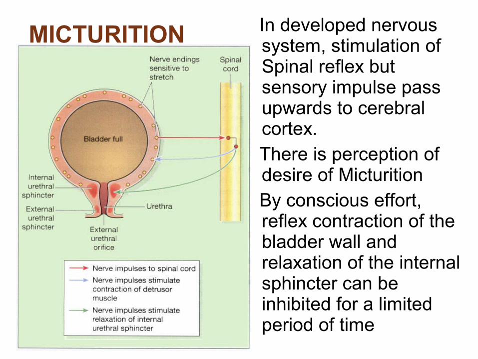
MICTURITION In developed nervous system, stimulation of Spinal reflex but sensory impulse pass upwards to cerebral cortex.
There is perception of desire of Micturition
By conscious effort, reflex contraction of the bladder wall and relaxation of the internal sphincter can be inhibited for a limited period of time
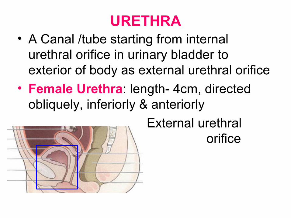
URETHRA• A Canal /tube starting from internal
urethral orifice in urinary bladder to exterior of body as external urethral orifice
• Female Urethra: length- 4cm, directed obliquely, inferiorly & anteriorly
External urethral orifice located between clitoris & vaginal opening
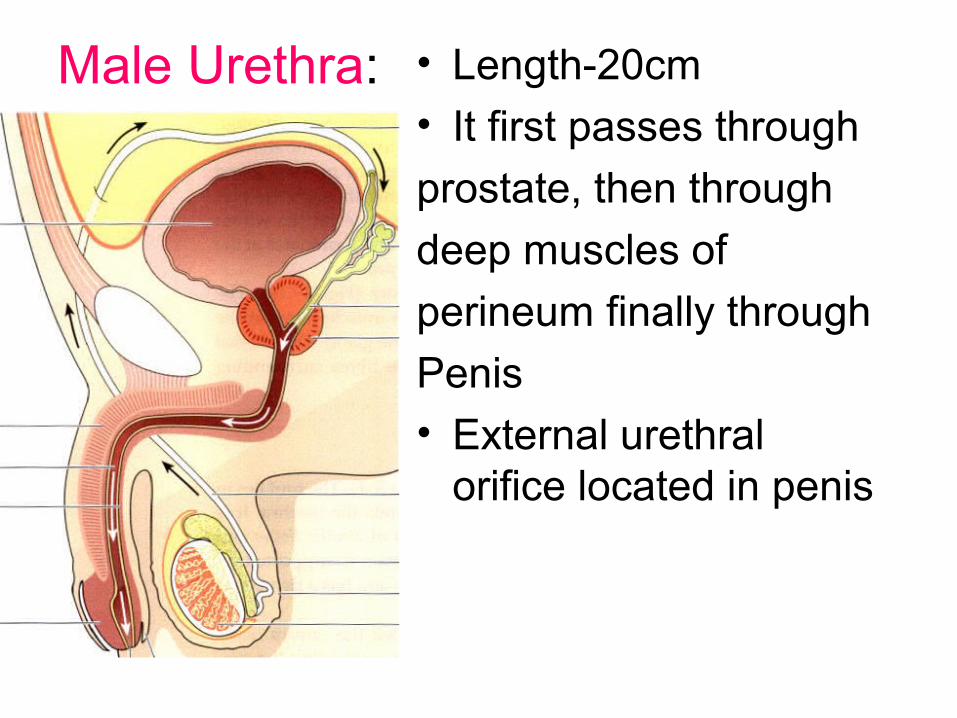
Male Urethra: • Length-20cm
• It first passes through
prostate, then through
deep muscles of
perineum finally through
Penis
• External urethral orifice located in penis
Walls of urethra: In both sexes the structure is the sameIts walls consist of three layers of tissue:
• the muscle layer, continuous with that of the bladder. At its origin there is the internal urethral sphincter, consisting mainly of elastic tissue and smooth muscle fibers, under autonomic nerve control. Slow and continuous contraction of this sphincter keeps the urethra closed. In the middle third there is skeletal muscle surrounding the urethra, under voluntary nerve control, that forms the external urethral sphincter
• the submucosa, a spongy layer containing blood vessels and nerves
• the mucosa, which is continuous with that of the bladder in the upper part. In the lower part the lining consists of stratified squamous epithelium, continuous externally with the skin of the vulva.
Function: Discharge of urine. In males, it also discharges semen