0hgldwruv ,q . %[1 0xulqh 0rgho )ru 5khxpdwrlg …
TRANSCRIPT
�5ROH�2I�3DWKRJHQLF�$XWR�$QWLERGLHV�$QG�,QQDWH�,PPXQLW\�
0HGLDWRUV�,Q�.�%[1�0XULQH�0RGHO�)RU�5KHXPDWRLG�$UWKULWLV
'LVVHUWDWLRQ�Zur Erlangung des akademischen Grades eines
Doktors der Naturwissenschaften
an der Universität Konstanz
Fakultät für Biologie
vorgelegt von
6DPXHO�5DM�6RORPRQ
Konstanz, im Dezember 2002
���
7DJ�GHU�P�QGOLFKHQ�3U�IXQJ������0lU]�������
5HIHUHQW��3URI��'U��5ROI�.QLSSHUV�5HIHUHQW��3URI��'U��0DUFXV�*URHWWUXS

��
,�ZLOO�LQVWUXFW�WKHH�DQG�WHDFK�WKHH�LQ�WKH�ZD\�ZKLFK�WKRX�VKDOW�JR��,�ZLOO�JXLGH�WKHH�ZLWK�PLQH�H\H��
3VDOPV��������������������
'HGLFDWHG�WR�$SSD��$PPD��%DEX�

Acknowledgements
My special thanks to Dr. Harald Illges� for giving an opportunity to carry out this work, for
financial support from his research grants, for his guidance and helpful discussions.
My special thanks to Prof. Dr. Rolf Knippers for being my doctoral father and for his valuable
guidance during my PhD studies.
My sincere thanks to Prof. Dr. Marcus Groettrup for his support and for accepting to be a
referee for my PhD thesis and oral examinations.
My sincere thanks to Prof. Dr. W. Welte for accepting to be my examiner for oral
examinations.
I am greatly indebted to Dr. Cornelia Kolb for her help, especially in establishing the
monoclonal antibodies.
My sincere thanks to Prof. Michael Glocker for letting me, to make use of the mass
spectrometry facility at the Proteome center, University Rostock.
Thanks to Elvira Jeisy for the technical assistance in histology.
My sincere thanks to Uli beck, Elisabeth Naidoo, Elisabeth von Seydlitz, Alex Titz, and
Brigitte Shanze for their valuable assistance in my work.
Finally I thank all my lab colleagues at University Konstanz, Jorg Schwab, Attef Allam,
Subhasis Mohanty, Madhan Masilamani, Lawrence Rajendran, Daniella Kasshahn,
Narendiran Rajasekaran and at BITG, Melanie Hoefer, Vera Klett, Eva Maria, and Annette
Aichem for their kind help and co-operation.
Publications
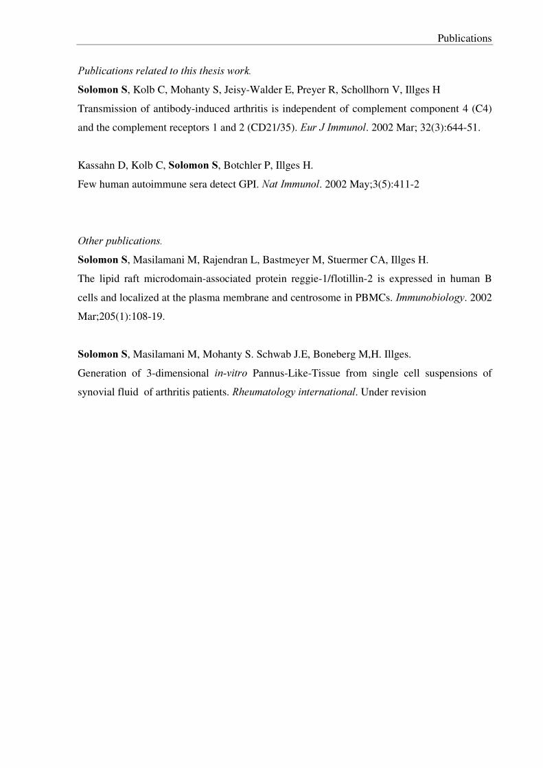
3XEOLFDWLRQV�UHODWHG�WR�WKLV�WKHVLV�ZRUN��6RORPRQ�6, Kolb C, Mohanty S, Jeisy-Walder E, Preyer R, Schollhorn V, Illges H
Transmission of antibody-induced arthritis is independent of complement component 4 (C4)
and the complement receptors 1 and 2 (CD21/35). (XU�-�,PPXQRO. 2002 Mar; 32(3):644-51.
Kassahn D, Kolb C, 6RORPRQ�6, Botchler P, Illges H.
Few human autoimmune sera detect GPI. 1DW�,PPXQRO. 2002 May;3(5):411-2
2WKHU�SXEOLFDWLRQV��6RORPRQ�6, Masilamani M, Rajendran L, Bastmeyer M, Stuermer CA, Illges H.
The lipid raft microdomain-associated protein reggie-1/flotillin-2 is expressed in human B
cells and localized at the plasma membrane and centrosome in PBMCs. ,PPXQRELRORJ\. 2002
Mar;205(1):108-19.
6RORPRQ�6, Masilamani M, Mohanty S. Schwab J.E, Boneberg M,H. Illges.
Generation of 3-dimensional LQ�YLWUR Pannus-Like-Tissue from single cell suspensions of
synovial fluid of arthritis patients. 5KHXPDWRORJ\�LQWHUQDWLRQDO. Under revision�

Contents
v
�����,QWURGXFWLRQ………………………………………………………………………….. 1 1.1. Scope for Rheumatoid Arthritis research……………………………………………..
1
1.2. KBN spontaneous mice model for Rheumatoid Arthritis…………………………….. 2 1.3. Note on auto-antigen in K/BxN mice model…………………………………………. 4 1.4. Innate immunity mediators of inflammation in Rheumatoid Arthritis……………….. 5 1.4.1. Role of autoantibodies in RA……………………………………………………... 5 1.4.2. Role of complements and complement activation products………………………. 5 1.4.3. Role of complement receptors in RA……………………………………………... 7 1.4.4. Role of Fcγ Receptors in RA……………………………………………………… 7 1.4.5. Role of cellular mediators of inflammation in RA………………………………... 8 1.4.5.1. Role of mast cell and mediators……………………………………………….. 8 1.4.5.2. Role of neutrophils…………………………………………………………….. 9 1.4.5.3. Role of macrophages…………………………………………………………... 9 1.5. Role of inflammatory cytokines and receptors in Rheumatoid Arthritis……………... 10 1.5.1. TNFα super-family and TNFα receptors…………………………………………. 10 1.5.2. Role of Interleukin–� «««««««««««««««««««««««� 11 1.6. Role of chemokines and chemokine receptors in RA………………………………… 11 1.7. Role of cell surface adhesion molecules in RA………………………………………. 13 1.8. Role of leukocyte proteases and protease activated receptors in RA………………… 14 1.9. Role of animal models in RA research………………………………………………. 15 ������3ULPDU\�DLPV�RI�WKH�VWXG\««««««««««««««««««««««����
17
�����0DWHULDOV�DQG�PHWKRGV««««««««««««««««««««««««�� 18
3.1. Materials………………………...……………………………………………………. 18 3.1.1. General chemicals and reagents……………………..……………………………. 18 3.1.2. Molecular biology reagents………………………...……………………………... 18 3.1.3. Antibodies…………………………………………………………………………. 18 3.1.4. Cell culture reagents…………………………………..………………………….. 19 3.1.5. Protein chemistry reagents………………………………………………………… 19 3.1.6. Experimental animals……………...……………………………………………… 19 3.1.7.Therapy reagents…………………………………………………………………… 19 3.2. Methods………………………………………………………………………………. 20 3.2.1. Recombinant DNA techniques…………………………………..………………... 20 3.2.1.1.Preparation of bacterial media…………………..…………………………….... 20 3.2.1.2. Isolation of RNA…...…………………………………………..………………. 20 3.2.1.3. cDNA synthesis and polymerase chain reaction (RT-PCR)…..……………..… 20

Contents
vi
3.2.1.4. Addition of 3´adenosine to PCR product… … … … ..… … … … ..… … … … … … . 21 3.2.1.5. TOPO cloning of PCR product… … … … … … … … … … … … … ..… … … … … .. 21 3.2.1.6. Extraction of plasmid-DNA… … … … … … … … … … … .… … … ..… … … … … . 21 3.2.1.7. Restriction digestions of plasmid DNA… … … … … … … … … ..… … … … … ..... 21 3.2.1.8. Isolation of DNA and plasmids from preparative agarose gel… … ..… … ..… … 22 3.2.1.9. DNA ligation… … … … … … ..… … … … … … … … … … … … … … … … … ..… ... 22 3.2.1.10. Preparation of competent bacteria… … .… … … .… … … … … … … … … ..… … . 22 3.2.1.11. Transformation of competent bacteria… … … … … … .… ..… … … … … ..… .… . 22 3.2.2. Protein techniques… … … … … … … … … … … … … … … … … … ..… … … .… … … 22 3.2.2.1. Gel electrophoresis of protein… … … … … ..… … … … … … … … ..… … … … … . 22 3.2.2.2. Expression and purification of GST fusion recombinant proteins… ..… ..… … .. 23 3.2.3. Immunological techniques… … … … … … … … .… … … … … … … … … … ..… .… … 23 3.2.3.1.Western blots and dot blots… … … … … ..… … … … … … .… … … ..… … … … … . 23 3.2.3.2. GPI Enzyme-Linked Immunosorbent Assay (ELISA)… ..… … … ....… … .… .... 24 3.2.3.3. Preparation of antigen for immunization… … … … … ..… … … … ..… … … .… … 24 3.2.3.4. Production of GPI specific monoclonal antibodies… … … … … … ..… … … ...… 25 3.2.3.5. Limited dilution cloning of hybridomas and adaptation to serum free media… . 25 3.2.3.6. Purification of monoclonal antibodies… … … … … … … .… … … … … … … .… ... 26 3.2.4. Epitope analysis by affinity proteomics… … … … ....… … … … … … ..… … … … … . 26 3.2.4.1. Cross linking of GPI antibody to epoxy dynabeads… … … … … ..… … ..… … … 27 3.2.4.2. Proteolytic digestion of purified recombinant mouse GPI… … ..… … ..… … … .. 27 3.2.4.3. Epitope extraction… .… … … … … … … … … … … … … .… … … … … … ..… … … 27 3.2.4.4. Mass spectrometry analysis… … ..… ...… … … … … … … … … … … … ..… … … .. 28 3.2.5. Animal experimentation… … ..… … … ..… … … … … … … … … … … … … … … … ... 28 3.2.5.1. Establishment of KBN mice model and production of KBN sera… … … .....… .. 28 3.2.5.2. Establishment of antibody induced arthritis in mice… … … ...… ..… … … ...… ... 28 3.2.5.3. Assessment of antibody induced arthritis by ankle thickness and clinical index score… … … … … … … … … … … … … … … … … … … … … … … … … … … .… … … … … …
28
3.2.5.4. Treatments protocols for inhibition studies… … … … ..… … ..… … … … … … … . 29 3.2.5.4.1. Complement depletion by cobra venom factor treatment… ..… … ..… ..… … 29 3.2.5.4.2. Depletion of macrophages… … … … ...… … … … .… … … … ..… … … … … … 29 3.2.5.4.3. In vivo inhibition of mast cell degranulation… … … … ..… … ..… … .… .… … 29 3.2.4.3.4. Administration of histamine receptor antagonists… ..… .… .… ..… … … … … 30 3.2.5.4.5. Administration of IL8R antagonist… … … … … … ...… … … … ..… ..… … … . 30 3.2.5.4.6. Administration of blocking antibodies… … … … … ..… … … … ..… … … ...… 30 3.2.6. Histochemical techniques… … … … … … … … … … … .… … … … … … ...… … … … . 30 � � �
Contents
vii
���5HVXOWV«««««««««««««««««««««««««««««««���
32
4.1. Generation and characterization of glucose-6-phosphate isomerase specific monoclonal antibodies from K/BxN mice… … … … … … … … … … … … … … … … … … …
32
4.1.1. Cloning, expression and purification of recombinant mouse glucose-6-hosphate isomerase… … … … … … … … … … … … … … … … … … … … … … … … … … … … … … … ..
32
4.1.2. Cloning and expression of truncated mouse glucose-6-phosphate isomerase… … .. 34 4.1.3. Cloning, expression and purification of recombinant human glucose-6-phosphate isomerase… … … … … … … … … … … … … … … … … … … … … … … … … … … … … … … ..
36
4.1.4. Glucose-6-phosphate isomerase reactive hybridomas cloned from un-manipulated K/BxN RA mice produced only IgM antibodies and were cross reactive… ..
38
4.1.5.Glucose-6-phosphate isomerase reactive hybridomas cloned from GPI immunized K/BxN RA mice produced GPI reactive antibodies of both IgM and IgG1 isotype… … …
39
4.1.6.Purification of glucose-6-phosphate isomerase specific monoclonal antibodies… .. 41 4.1.7. Differentiation of GPI reactive antibodies reacting to different epitopes by a novel peptide fingerprint western blot technique… … … … … … … … … … … … … … … … .
43
4.1.8. Epitope mapping of GPI antibodies by mass spectrometry… … … … … … … … … .. 44 4.1.9. Protein truncation epitope mapping confirms with the mass spectrometry identified epitope regions… … … … … … … … … … … … … … … … … … … … … … … … … .
46
4.1.10. Modeling of epitope residues on crystal structure … … … … … … … … … … … … . 47 4.2. Role of Innate immunity mediators in KBN Rheumatoid Arthritis mice model..… … 48 4.2.1. KBN Sera transfer induced RA in laboratory mice strains… … .… … … … … … … . 49 4.2.2 .Role of complement activation and complement receptors in K/BxN sera induced RA… … … … … … … … … … … … … … … … … … … … … … … … … … … … … … … … … …
50
4.2.2.1 Complement C4-/- mice defective in classical complement and lectin complement pathway are susceptible to KBN sera induced arthritis… … … … … … … … ...
50
4.2.2.2. Mice lacking complement C3 or C5 are resistant to KBN sera induced RA… . 52 4.2.2.3. No modulatory role for complement receptors CR1(CD35) and CR2(CD21) on the outcome of RA KBN sera induced mice model… … … … … … … … … … … … … …
54
4.2.2.3.1. &U� � � � mice lacking complement receptors CR1 and CR2 are susceptible to
RA… … … … … … … … … … … … … … … … … … … … … … … … … … … … … … … … … … 54
4.2.2.3.2. Clearance of GPI-antibodies from peripheral blood and disease severity are correlated… … … … … … … … … … … … … … … … … … … … … … … … … … … … … … … ..
55
4.3. )Fγ5,,, �� � mice are completely resistant whereas )Fγ5,,E � � �
have exaggerated disease in KBN sera induced RA model… … … … … … … … … … … … … … … … … … … … … … ...
57
4.4. Role of innate immunity cellular players in RA… … … … … … … … … … … … … … … 59 4.4.1. Mast cell de-granulation inhibitors and Histamine H1 receptor antagonist ameliorates RA… … … … … … … … … … … … … … … … … … … … … … … … … … … … … .
59

Contents
viii
4.4.2. Role of neutrophil recruitment mediators in KBN sera induced RA pathogenesis.. 61 4.4.3. Role of macrophage and macrophage chemoattractant protein-1 in KBN sera induced RA pathogenesis… … … … … … … … … … … … … … … … … … … … … … … … … .
63
4.5. Role of TNFα and TNF receptors in KBN sera induced RA pathogenesis 65 4.6. New glucose-6-phosphate isomerase specific antibody induced mice models for Rheumatoid Arthritis… … … … … … … … … … … … … … … … … … … … … … … … … … …
68
4.6.1. Establishment of glucose-6-phosphate isomerase specific monoclonal antibody antibody transfer induced mice model for Rheumatoid Arthritis… … … … … … … … … …
68
4.6.2. Establishment of GPI anti-sera induced mice model for Rheumatoid Arthritis… .. 70 �����'LVFXVVLRQV«««««««««««««««««««««««««««««�
72
5.1. Characterization of GPI reactive auto-antibodies from K/BxN mice… … … … … … … 72 5.2. Role of Innate immunity mediators… … … … … … … … … … … … … … … … … … … .. 74 5.2.1. K/BxN sera induced arthritis is dependent on the alternative complement pathway activation and no role for classical complement pathway… … … … … … … … … .
74
5.2.2. No role for Complement receptors CR1 and CR2 in K/BxN sera induced RA… ... 75 5.2.3. Activation of FcγRIII receptors mediates inflammation in K/BxN sera induced RA… … … … … … … … … … … … … … … … … … … … … … … … … … … … … … … … … …
75
5.2.4. Inhibition of mast cells degranulation and administration of histamine H1 receptors antagonists ameliorates K/BxN sera induced RA… … … … … … … … … … … … .
76
5.2.5. Administration of chemokine receptor CXCR2 antagonists ameliorates K/BxN sera induced RA… … … … … … … … … … … … … … … … … … … … … … … … … … … … ...
77
5.2.6. Macrophages depleted mice are resistant to K/BxN sera induced RA… … … … … . 78 5.2.7. TNFα has a dual role in K/BxN sera induced RA… … … … … … … … … … … … … 78 5.3. New animal models in RA… … … … … … … … … … … … … … … … … … … … … … … 79 �����6XPPDU\«««««««««««««««««««««««««««««��� 81 �����=XVDPPHQIDVVXQJ«««««««««««««««««««««««««���� 82 �����5HIHUHQFHV«««««««««««««««««««««««««««««� 85 �

Figures and tables
ix
)LJXUHV«««««««««««««««««««««««««««««««««��
Figure 1. Affected limb of a patient with Rheumatoid Arthritis............................................ 1 Figure 2: Illustration of joint tissue in normal health and RA with parallels of ankle joint histology of normal and Rheumatoid Arthritis affected joint in mice....................................
2
Figure 3: Illustration showing generation of the K/BxN spontaneous RA mice model......... 3 Figure 4: An illustration showing the complement activation pathways............................... 6 Figure 5: Illustration showing principle of Mass spectrometry based epitope analysis ......... 26 Figure 6. PCR amplification of mouse GPI… … … ................................................................ 33 Figure 7. Screening of pGEX-4T-3 plasmids for recombinant mouse GPI inserts by restriction enzyme digestion analysis................................................................................… ..
33
Figure 8. SDS-PAGE showing expression and purification of recombinant mouse GPI from BL21 cells.......................................................................................................................
34
Figure 9. PCR amplification of truncated mouse GPI… … ................................................ 35 Figure 10. Screening of PGEX-4T3 plasmids for truncated mouse GPI inserts by restriction enzyme digestion analysis......................................................................................
35
Figure 11. SDS-PAGE showing expression of truncated recombinant mouse GPI in BL21 cells .....................................................................................................................… … … … …
36
Figure 12. PCR amplification of human GPI… … .… ............................................................. 36 Figure 13. Screening of PGEX-4T3 plasmids for human GPI DNA inserts by restriction digestion analysis..............................................................................................................… ..
37
Figure 14. SDS-PAGE showing expression and purification of recombinant human GPI from BL21 cells..............................................................................................................… …
37
Figure 15. Illustration showing scheme for production GPI monoclonal antibodies from naïve K/BxN mice............................................................................................................… ..
38
Figure 16. Illustration showing scheme of production GPI monoclonal antibodies from GPI immunized K/BxN mice...........................................................................................… ..
40
Figure 17. GPI monoclonal antibody Protein G affinity purification profile......................... 42 Figure 18. SDS-PAGE gel showing Protein G purified mGPI monoclonal antibodies.......... 42 Figure 19. Dot blot of purified mGPI monoclonal antibodies showing reactivity with kappa light chain antibodies................................… … … … … … … … … … … … … … … … …
42
Figure 20. Western blot showing the reactivity of purified mGPI mAB to recombinant mouse GPI and human GPI..........................................................................................… … ..
43
Figure 21. Peptide fingerprint western blot epitope mapping of Trypsin partial digest of mouse GPI by mGPI mAB antibodies....................................................................................
44
Figure 22. Peptide fingerprint western blot epitope mapping of Glu-C partial digest of mouse GPI by mGPI mAB antibodies...........................................................................… … .
44
Figure 23. Mass spectrometric epitope mapping profile of epitope extracted Glu-C digested GPI..............................................................................................................… … … ..
45

Figures and tables
x
Figure 24. Western blot showing the reactivities of mGPI mAb to expressed truncated proteins of mouse GPI.............................................................................................................
46
Figure 25. Modeling of epitopes residues identified by mass spectrometric on rabbit GPI crystal structure........................................................................................................................
47
Figure 26. K/BxN sera transfer in laboratory mice strains...… … … … … … … … … … … … ... 48 Figure. 27. Histology of ankle joints of K/BxN serum induced RA in KRN, C57BL/6 and BALB/c mice.........................................................................… … … … … … .… … … … … …
49
Figure 28. C4-/- mice are susceptible to K/BxN induced RA.................................................. 50 Figure 29. Histology of C4-/- ankle joints.......................................................................… … . 51 Figure 30. Complement C5-/- NOD mice are resistant to K/BxN sera induced RA...........… . 52 Figure 31. C3 depleted mice are resistant to K/BxN sera induced RA................................... 53 Figure 32. No role of CR1 and CR2 in K/BxN sera induced RA..................................… … . 54 Figure 33. Histology of &U� � � � ankle joints..................................................… … … … … … … 55 Figure 34. GPI antibody clearance in the peripheral blood of the sera transferred mice..........… … … … … … … … … … … … … … … … … … … … … … … … … … … … … … … ..
56
Figure 35. )Fγ5,,, � � � mice are resistant and )Fγ5,,E � � � are highly susceptible to K/BxN sera
induced RA...........................… … … … … … … … … … … … … … … … … … … … … … … … .. 57
Figure 36. Joint histology of )Fγ5,,, � � � and )Fγ5,,E � � � mice.............................................… .. 58
Figure 37. Inhibition of mast cell degranulation ameliorates RA.......................… … … … … 59 Figure 38. H1 histamine receptor antagonists ameliorates RA while H2 histamine receptor does not.............................................… … … … … … … … … … … … … … … … … … … … … ..
61
Figure 39. Role of neutrophil attracting CXC chemokine receptor. CXCR2 and CXC chemokine MIP-2 in RA........................................................................… … … … … … … … .
62
Figure 40. Macrophage depleted mice are resistant to RA.............................................… … . 63 Figure 41. Role of chemokine Macrophage Chemoattractant Protein -1 in RA................… . 64 Figure 42. Neutralizing antibodies to TNFα reduces inflammation in RA............................. 65 Figure 43. 71)5� � � � and 71)5� � � � mice are not protected from RA.............................… ....... 66 Figure 44. Histology of ankle of 71)5� � � � and 71)5� � � � mice before and after RA induction… … … … … … … … … … … … … … … … … … … … … … … … … … … … … … … … .
67
Figure 45. Illustration of scheme of GPI monoclonal induced arthritis in mice................… . 68 Figure 46. Joint inflammation in GPI monoclonal induced RA mice model..............… … ... 69 Figure 47. Illustration of scheme for generation of rabbit GPI sera induced RA mode......… … … … … … … … … … … … … … … … … … … … … … … … … … … … … … … … ..
70
Figure 48. Reactivity of rabbit sera to recombinant GPI shown by western blotting............. 71 Figure 49. Ankle thickness and clinical index in rabbit anti mGPI sera induced RA in mice… … … … … … … … … … … … … … … … … … … … … … … … … … … … … … … … … … ..
72

Figures and tables
xi
7DEOHV«««««««««««««««««««««««««««««««««����
Table 1. Chemokines and chemokine receptors… … … … … … … … … … … … … … … … … .. 12 Table 2.Cell adhesion molecules and function… … … … … … … … … … … … … … … … … .... 13 Table 3. Classification of leukocyte proteases according to catalytic mechanism… … … … .. 14 Table 4: Serine proteases of complement activation pathway… … … … … … … … … … … … 15 Table 5: Current therapies under development for Rheumatoid Arthritis… … … … … … … .. 16 Table 6: Panel summarizing GPI reactive hybridomas of IgM isotype cloned from non-manipulated K/BxN mice… … … … … … … … … … … … … … … … … … … … … … … … … …
39
Table 7. Panel summarizing GPI reactive hybridomas clones of IgM isotype cloned from GPI boosted K/BxN mice… … … … … … … … … … … … … … … … … … … … … … … … … ....
40
Table 8. Panel summarizing GPI reactive hybridomas clones of IgG1 isotype cloned from GPI boosted K/BxN mice… … … … … … … … … … … … … … … … … … … … … … … … … …
41
Table A. Gene knockout mice studies in K/BxN sera induced model… … … … … … … … … 82 Table B. ,Q�YLYR therapy/ antibody blocking studies in K/BxN sera induced model… … … ... 82 Tabelle A. Die genetische Knockoutmaus-Studie im K-/BxN- Serum induzierten Model..... 84 Tabelle B. In vivo Therapie /Antikörper-Studien im K-/BxN-Serum induzierten Model.....
84

Abbreviations
viii
Ab antibody
Ag antigen
AT ankle thickness
BSA bovine serum albumin
cDNA complementary DNA
CI clinical index
CIA collagen induced arthritis
CO2 carbondioxide
CR1 complement receptor 1
CR2 complement receptor 2
CVF cobra venom factor
DEPC diethylene pyrocarbonate
DMSO dimethylsulfoxide
DNA deoxy ribonucleic acid
DTT Dithiothreitol
E. coli escherichia coli
e.g. for example
ECM extra cellular matrix
EDTA Ethylene diaminetetra acetic acid
ELISA enzyme linked immunosorbant assay
EtBr ethidium bromide
FACS flourescent activated cell scan
FcγR fc gamma receptor
FCS fetal calf serum
FITC flourescien isothicyanate
FPLC fast protein liquid chromatography
GAPDH glyceraldehyde phosphate dehydrogenase
GPI glucose–6-phosphate isomerase
GRO growth related oncogene
GST glutathione –s-transferase
h hour
HAT hypoxanthine aminopterin thymidine
HLA human leucocyte antigen
HPLC high performance liquid chromatography
HRP horse radish peroxidase
i.p intra peritoneal
i.v intra venous
IC immune complex
ICAM-1 intra cellular adhesion molecule-1
Ig immunoglobulin
IL interleukin
IMDEM iscoves dulbecco´s modified medium
IPTG isopropyl-beta-D-thiogalactopyranoside
KDa kilo daltons
M, mM molar, millimolar
mA milli amphere
MALDI- matrix assisted laser desorption ionisation
MCP monocyte chemoattractant protein
MeOH methanol
MHC major histocompatibility complex
MIP-2 macrophage inflammatory protein
MMP matrix metallo protease
MP milk powder
NO nitric oxide
NTP nucleotide tri phosphate oC degree celsius
OD optical density
PAGE polyacrylamide gel electrophoresis
PAR protease activated receptor
PBS phosphate buffered saline
PCR polymerase chain reaction
PE phycoerythrin
PEG polyethyleneglycol
PMN polymorphonuclear monocytes
RA rheumatoid arthritis
RNA ribonucleic acid
RPM rotations per minute
RT room temperature
SDS sodium dodecyl sulfate
SEM standard error mean
SFM serum free medium
SLE systemic lupus erythematosus
TAE tris acetate edta
TBE tris borate edta
TBS tris buffered saline
TBST tris buffered saline tween
TCR t cell recptor
TE tris edta
TFA triflouroacetic acid
TNFα tumour necrosis factor α
TNFR tumour necrosis factor receptor
XO� micro liter

Abbreviations
ix
UV ultra violet
V volt
v/v , w/v volume/volume, weight/volume
VCAM vascular cell adhesion molecule

Introduction �
1
�����,QWURGXFWLRQ������6FRSH�IRU�5KHXPDWRLG�$UWKULWLV�UHVHDUFK��Rheumatoid Arthritis (RA) is chronic progressive, systemic inflammatory disorder affecting
the synovial joints. It affects 1 to 2% of population, with prevalence increasing with age, and
can reach upto 5% in women over age 55. If left untreated it leads to joint destruction
responsible for the deformity and disability seen in the disease. The resulting morbidity and
mortality has a substantial socio-economic impact on the society.
��)LJXUH����$IIHFWHG�OLPE�RI�D�SDWLHQW�ZLWK�5KHXPDWRLG�$UWKULWLV���The etiology of RA is still not very clear. Even a basic question whether it is primarily an
autoimmune disease, or an inflammatory response/ to infection, is yet to be resolved. Genetic
predisposition has been implicated, while failure to demonstrate Mendelian inheritance means
multiple genetic factors are probably involved. In humans the MHC class II HLA-DR4 allele
is shown to be associated with development and severity of RA. Controversy exists also about
the role of the effector leukocyte populations in RA pathogenesis. The dominating paradigm
in the field until recently was that antigen-specific T cells in the joint incites the inflammatory
cascade by triggering macrophages and synoviocytes. But key observations such as paucity of
T cell and its derived cytokines in inflamed joints disapproved such views. Recent studies of
new animal models have shifted this importance to B-cell secreted autoantibodies and innate
immunity mediators (Benoist and Mathis 2000). The current concept being that while T and B
cells are important in initiation of RA the pathogenicity is mostly mediated by autoantibodies
and innate immune mediators. The role of inflammatory cytokines such as IL-1 and TNFα are
well known and are current targets in therapy. The emerging key role of autoantibodies and
innate immunity mediators has renewed hopes for discovery of new targets for RA treatment.
��

Introduction �
2
�����.%1�VSRQWDQHRXV�PLFH�PRGHO�IRU�5KHXPDWRLG�$UWKULWLV-D�SDUDGLJP�VKLIW�IURP�7�FHOO�FHQWULF�WR�DQ�DXWRDQWLERGLHV�FHQWULF�YLHZ�RI�5$�SDWKRJHQHVLV��Murine RA models like collagen or pristane induced arthritis models have been of tremendous
use in RA research. These animals models have been parallels to understand human RA, yet
are more simple to elucidate RA mediators, for hypothesis and drug testing. Generally these
models were studied with a notion that T cell, through interaction with antigen, is the primary
cell responsible for initiating the disease as well as for driving the chronic inflammatory
process by activating macrophages and fibroblasts. However a very recent model of RA the
spontaneous RA K/BxN murine model (Kouskoff, Korganow et al. 1996) has changed this
thought. RA in these mice develops spontaneously in the offspring of the α -TCR-transgenic
(specific for RNAse peptide residues 41–61) KRN mice crossed with the NOD diabetes mice
strain harboring the MHC II Ag7 allele (figure 3). RA development in K/BxN offsprings is
visible at about 3 weeks of age, characterized by rapid onset of joint inflammation and
cartilage destruction. The pathology is similar in several aspects to human RA, with
symmetrical involvement of peripheral joints, pannus formation, synovial hyperplasia, bone
and cartilage destruction, with subsequent anarchic remodeling of the joints.
)LJXUH����,OOXVWUDWLRQ�RI�MRLQW�WLVVXH�LQ�QRUPDO�KHDOWK�DQG�5$�ZLWK�SDUDOOHOV�RI�DQNOH�MRLQW�KLVWRORJ\�RI�QRUPDO�DQG�5KHXPDWRLG�$UWKULWLV�DIIHFWHG�MRLQW�LQ�PLFH��

Introduction �
3
In the K/BxN mice RA model, initiation of RA is dependent on KRN T cells. But
unexpectedly the B-cell immunoglobulins were found to be the arthritogenic effectors as the
disease could be transferred to naïve mice and also in mice lacking T and B cells by injection
K/BxN mice immunoglobulins (Korganow, Ji et al. 1999).
)LJXUH����,OOXVWUDWLRQ�VKRZLQJ�JHQHUDWLRQ�RI�WKH�.�%[1�VSRQWDQHRXV�5$�PLFH�PRGHO���Subsequent studies showed that the disease depends on the KRN T cell recognition of a
ubiquitous glycolytic enzyme, glucose-6-phosphate isomerase (GPI), on I-Ag7 MHC-II
molecule. The arthritogenic sera of sick K/BxN mice also recognized a 60KDa band in cell
lysates, corresponding to ubiquitous glyolytic enzyme glucose-6-phosphate isomerase (GPI)
(Matsumoto, Staub et al. 1999). Transfer of, as little as 100 O�of K/BxN serum or GPI affinity
purified antibodies could induce arthritis in naive recipient mice and in mice lacking
lymphocytes. The T-cell epitopes (GPI residues 282-294) on GPI have been reported (Basu,
Horvath et al. 2000) but the B-cell epitopes of arthritogenic GPI specific antibody have not
yet been elucidated. The joint tissue specificity of this very interesting RA model is also yet to
be clearly explained. Recently it was shown that GPI deposited on ankle joint surfaces of sick
as well normal mice, could probably target GPI antibodies to the joints (Matsumoto, Maccioni
et al. 2002). Also recently (Schaller, Burton et al. 2001) a study, showed that 64% of humans
with RA had increased concentrations of anti-GPI antibodies in serum and synovial fluid.
NOD
MHC-Ag7 +ve
KRN
KRN TCR +ve
KBN
KBN KBN
MHC-Ag7 and KRN TCR +ve KBN RA SPONTANEOUS MI CE MODEL
BALB/ C
KBN SERA I NDUCED RA MODEL
KBN SERUM
X
Introduction �
4
�����1RWH�RQ�DXWR�DQWLJHQ�LQ�.%1�PLFH�PRGHO�DQ�XELTXLWRXV�DQWLJHQ�DQG�PXFK�PRUH���Several proteins like collagen, gp39, proteoglycans, RA33, p205, and citrullinated proteins
are implicated as autoantigens in RA. glucose-6-phosphate isomerase has been the most
recent in this list. An interesting point to be noted is the multiple functions of GPI, GPI as a
homodimer, in cytosol functions as glycoltic enzyme catalyzing the reversible isomerization
of glucose 6-phosphate to fructose 6-phosphate, whereas as a monomeric phosphorlyated
form it is secreted from the cytoplasm and can be detected at low concentration (0.24-0.54
Xg/ml) in serum. Several interesting extracellular functions have been attributed to the
monomeric GPI, like in mineralization of osteoblasts (Zhi, Sommerfeldt et al. 2001), in
stimulation of B-cells to secrete immunoglobulins (Gurney, Apatoff et al. 1986), as an
angiogenic factor that stimulates endothelial cell motility (Funasaka, Haga et al. 2001) and in
survival of spinal and sensory neurons (Gurney, Heinrich et al. 1986). Interestingly also the
GPI receptor (gp78) is a seven membrane G-protein coupled receptor, a receptor most often
implicated in inflammation, cell activation and motility (Shimizu, Tani et al. 1999). Till date
no investigations have been carried out with a view that GPI protein as such could have role
in pathogenesis of RA. It can be expected that the tissue specificity in RA depends on a
putative joint specific antigen. In fact, immunization of mice with collagen type II a protein
found in joints induces pathogenic autoantibodies targeted to murine type II collagen in joints
and resulting in arthritis affecting joints(collagen-induced arthritis, CIA) (Holmdahl, Mo et al.
1989). The autoimmune target in KBN RA model is a ubiquitous glycolytic enzyme, GPI
(Matsumoto, Staub et al. 1999). The question how a ubiquitous protein leads to joint specific
autoimmunity and systemic disease has been one of the most puzzling questions in this
model. Also how T cell tolerance is broken for this enzyme is a unresolved question in this
RA model. No evidence has been provided for the possibility of modified forms of GPI or
mimicry antigens being recognized by KRN T cells or the by GPI autoantibodies till now.
�������
Introduction �
5
����,QQDWH�LPPXQLW\�PHGLDWRUV�RI�LQIODPPDWLRQ�LQ�5KHXPDWRLG�$UWKULWLV����������5ROH�RI�DXWRDQWLERGLHV�LQ�5$±�D�WLVVXH�VSHFLILFLW\�WDUJHW�IRU�LQQDWH�LPPXQH�V\VWHP��A important question in RA pathogenesis had been, whether immune complexes represent
primary disease pathology mediators or are “innocent bystanders” resulting from tissue
damage. But now increasing evidence exists from mice models (Stuart and Dixon 1983;
Korganow, Ji et al. 1999) that autoantibodies are principle effectors for the initiation and
propagation of joint inflammation in RA. Immune complexes targeted to joints can lead to
complement activation and/or directly effect cell activation through Fcγ receptors(Baumann,
Kohl et al. 2000; Heyman 2000). The complement cascade so initiated can recruit cell
mediators of inflammation by the activating and chemo-attractant property of complement
activation products. Post-translational modification on antibodies like lack of galactose on
asparagine-linked oligosaccharides on the Ig CH2 domain has been associated with severity
of RA(Rademacher, Williams et al. 1994). Also these antibodies have impaired immune
complex clearance and are defective in down regulating activated B cells. Most often immune
complexes are targeted to the site of inflammation due to the presence of auto-antigen. Hence
immunizing mice with joint/cartilage protein like collagen or proteogylcan can induce RA
affecting joints. Recent studies from mice models have shown strong parallels between auto-
antigens recognized by autoantibodies in human RA and mice. Collagen-II antibodies in RA
patients was shown to recognize the very same pathogenic epitopes on collagen-II as in CIA
mice model (Burkhardt, Koller et al. 2002). Likewise some RA patients have high titers of
GPI autoantibodies reflecting the similarities of the KBN mice model to human RA(Schaller,
Burton et al. 2001; Kassahn, Kolb et al. 2002; Schubert, Schmidt et al. 2002). The elucidation
of the reason for pathogenicity and epitopes recognized by the GPI antibodies in KBN mice
would be of utmost importance in the above context.
�������5ROH�RI� FRPSOHPHQWV�DQG�FRPSOHPHQW�DFWLYDWLRQ�SURGXFWV�DQ�RSVRQLF�KDQGOH� IRU�FRPSOHPHQW�UHFHSWRUV�DQG�PRUH��The formation and deposition of soluble immune complexes(IC) results in a variety of
autoimmune disease like arthritis, vasculitis and glomerulo-nephritis. Generally initiation of
the immune complex mediated tissue injury is mainly attributed to the activation of the
classical complement pathway, while bacterial products are known to activate the alternative
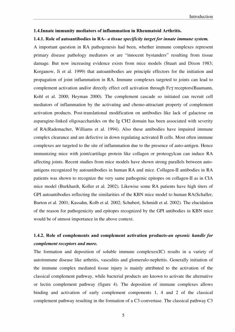
or lectin complement pathway (figure 4). The deposition of immune complexes allows
binding and activation of early complement components 1, 4 and 2 of the classical
complement pathway resulting in the formation of a C3-convertase. The classical pathway C3
Introduction �
6
convertase cleaves C3 leading to C3b-deposition on ICs through covalent bonding. A further
product of this proteolytic process is the formation of the C5-convertase, leading finally to the
assembly of the membrane attack complex (MAC).
�
�
�
�
�
�
)LJXUH����$Q�LOOXVWUDWLRQ�VKRZLQJ�WKH�FRPSOHPHQW�DFWLYDWLRQ�SDWKZD\V���WDNHQ�IURP�.XE\��,PPXQRORJ\���WK�(G����A common outcome of complement activation is the covalent attachment of the C3 split
product, C3b, iC3b and C3d, to the target surface. These products modulate the inflammatory
process by providing the opsonic handle recognized by the complement receptors
(CR1/CD35, CR2/CD21, and CR3/CD11b-CD18) expressed on leukocytes. The small
complement activation byproducts C3a and C5a are potent chemotactic peptides and induce
mast cell/neutrophil influx and degranulation leading to tissue injury (Baumann 2001).
Rightly so complement C3-deficient mice have reduced or no inflammation due to reduced
mast cell degranulation, TNFα production and decreased neutrophil infiltration (Prodeus,
Zhou et al. 1997). Also the complement C5 deficient mice have defective inflammatory
responses (Wang, Kristan et al. 2000), which strongly implicate the role of complement
activation and C5a in activating cell mediators. Also complement proteins are implicated in
IC-clearance e.g. a deficiency in the C4 complement component results in a systemic lupus
erythematosus (SLE)-like disease in humans (Gatenby 1991).
�

Introduction �
7
�������5ROH�RI�&RPSOHPHQW�UHFHSWRUV�LQ�5$�5HJXODWRUV�RI�LQIODPPDWLRQ��Complement receptors especially CR1(CD35), CR2(CD21) and C5aR(CD88) can have a
modulatory role on the outcome and severity of autoimmune inflammatory disease.
Complement activation products C3b and C4b can attach covalently to proteins/antibodies,
also these and other complement split products (C4b, C4d, iC3b, C3dg and C3d) are ligands
for complement receptors CR1 and CR2. The complement receptor CR1 is known to have an
important role in the clearance of IC from the circulation, in inhibition of C3 and C5
convertase (decay accelerating activity) and as a receptor for C3b/C4b products (Krych-
Goldberg and Atkinson 2001). In SLE, the disposal of IC fails as a consequence of a disease-
induced reduction in the number of CR1 on erythrocytes resulting in deposition of IC in
glomeruli and small vessels leading to inflammatory reactions (Gatenby 1991). Another clear
evidence of protective role for CR1 comes from the reduced disease severity of CIA in mice
over expressing soluble CR1 (Dreja, Annenkov et al. 2000). The complement receptor CR2
has a role in inflammation by being a receptor for C3dg-bound immune complexes, in
reducing the threshold for B cell activation, involved in germinal center reactions and in the
activation of the alternative complement pathway on binding iC3 (Schwendinger, Spruth et al.
1997). Also C5a receptor has been strongly implicated in inflammation (Höpken 1997;
Baumann, Kohl et al. 2000). Genetic deletion of C5aR leads to impaired inflammatory
responses in mice (Höpken 1997). C5aR on mast cells and chondrocytes are strongly up
regulated in RA (Kiener, Baghestanian et al. 1998; Onuma, Masuko-Hongo et al. 2002). The
triggering of mast cell de-granulation by the complement activation product C5a links the
immune complex mediated complement activation to the innate immunity cellular component
in RA inflammation.
�������5ROH�RI�)FJ�5HFHSWRUV�LQ�5$�OLQNLQJ�DQWLERGLHV�WR�HIIHFWRU�FHOOV��The Fc component of immune complexes can directly interact with the cellular components of
innate immune system via Fcγ receptors (FcγRs) and thereby link antibody-antigen complexes
to innate immune effector cell activation. FcγRs, belong to the immunoglobulin superfamily
that bind IgG. Three classes of leucocyte FcγR have been described (FcγRI, RII, and RIII)
(Dearon 1997). Binding of these receptors initiates signaling cascades that can lead to either
activation(RI and RIII) or deactivation(RII) of effector cells. The coordinated expression of
activating and inhibitory Fcγ receptors has been suggested to drive immune-complex-
mediated diseases. In recent studies, the importance of these receptors in inflammation and
tissue damage in various autoimmune and inflammatory diseases have been highlighted by

Introduction �
8
several knockout animal studies. )Fγ5,,, � � � mice show a marked reduction of inflammatory
cytokines TNFα and IL-1β in inflammatory responses. Cross-linking of FcγRs on monocytes
leads to the production of inflammatory cytokines (Kindt, Moore et al. 1993) and chemokines
(Marsh, Wewers et al. 1997). )Fγ5,,, � � � mice are highly protected from IgG-induced
hemolytic anemia, CIA (Stahl, Andren et al. 2002) and show an impaired Arthus reaction
(Baumann, Kohl et al. 2000), suggesting a dominant role of FcγRIII (Marsh, Gadek et al.
1995). Also strong correlation exist on FcγRI/III expression on macrophages and the severity
of cartilage destruction in human RA (Heller, Gessner et al. 1999; Blom, van Lent et al.
2000).
�������5ROH�RI�FHOOXODU�PHGLDWRUV�RI�LQIODPPDWLRQ�LQ�5$�7KH�NH\�LQQDWH�LPPXQLW\�FHOOXODU�WULDG�0DVW�FHOO��1HXWURSKLO�DQG�0DFURSKDJH�����������5ROH�RI�0DVW�FHOO�DQG�PHGLDWRUV��WULJJHUV�LQ�LQIODPPDWLRQ��D�OLQN�EHWZHHQ�FRPSOHPHQW�DFWLYDWLRQ�WR�FHOOXODU�LQIODPPDWLRQ��Mast cells have a key role as `triggers´ of inflammatory responses. Mast cells are strategically
located at high density around the blood vessels that are the principal sites of plasma efflux
and leukocyte transmigration. Activation of mast cells leads to immediate degranulation and
release of stored inflammation mediators such as TNFα, vasoactive mediators like histamine,
proteases like tryptase and chemokines like IL-8 (Krishnaswamy, Kelley et al. 2001). Mast
cells along with complement activation are implicated in immune-complex induced injury
(Baumann 2001); zymosan induced peritoneal inflammation (Kolaczkowska 2001) and in
Arthus reaction (Sylvestre and Ravetch 1996). Mast cells are implicated in production of the
first wave of TNFα secretion, recruiting neutrophils to sites of inflammation (von Stebut,
Metz et al. 2002). Moreover synovial mast cells have been known to up regulate C5aR during
inflammation (Kiener, Baghestanian et al. 1998) and activation of mast cells by C5a results in
inflammatory cytokine secretion (Woolley and Tetlow 2000). An important vaso-active amine
mediator stored in mast cell granules is histamine��Histamine has diverse functions including
inducing local dilation of small vessels and increase vascular permeability by contraction of
endothelial cells. Histamine functions by activating three G coupled histamine receptors H1,
H2 and H3 receptors (Del Valle and Gantz 1997; Baroody and Naclerio 2000). Histamine is
also a potent inducer of IL-8 from monocytes (Kohka, Nishibori et al. 2000). Also the serine
proteases tryptase, plasmin and thrombin released by mast cells are implicated in degradation
of matrix macromolecules like fibronectin, fibrinogen, type VI collagen and in activation of
cells by cleaving protease activated receptors (PAR) (Akers, Parsons et al. 2000). Mast cells
Introduction �
9
also serve as functional interface between the neuroendocrine and the immune system as a
number of neuropeptides (e.g. substance P, calcitonin gene–related peptide, VIP and the
related pituitary adenylate cyclase-activating polypeptide) have been reported to induce mast
cell degranulation (Stassen, Hultner et al. 2002).
����������5ROH�RI�QHXWURSKLOV��PHGLDWRU�RI�DFXWH�LQIODPPDWLRQ��Polymorphonuclear leukocytes (PMN) are the most abundant leukocytes, comprising about
two-thirds of peripheral blood leukocytes. Upon tissue injury and inflammation circulating
PMNs infiltrate rapidly into inflamed tissue. Recent evidence suggests that Neutrophils are
recruited by mast cells to sites of inflammation by producing TNFα and MIP-2 (functional
murine homologue of human IL-8) (Biedermann, Kneilling et al. 2000; von Stebut, Metz et al.
2002). Neutrophil are also known to be recruited to sites of complement activation by C5a
(Höpken 1997). Activated neutrophils produce and release pro-inflammatory mediators, such
as IL-1, IL-8, macrophage inflammatory protein (MIP) and proteases (Scapini, Lapinet-Vera
et al. 2000). Neutrophils have been clearly implicated in the recruitment of macrophages into
sites of inflammation by producing macrophage chemoattractants MIP-1α� �� 0&3-1 (von
Stebut, Metz et al. 2002) and inactive myeloperoxidase (iMPO) (Lefkowitz and Lefkowitz
2001). Neutrophils are involved in local production of complement C3 at sites of
inflammation (Yu, Tsai et al. 1995) leading to a positive feedback loop of neutrophil-
complement activation axis. Also the respiratory burst activity of Neutrophils can lead to
tissue destruction by reactive nitrogen and oxygen radicals and inducible NO synthase2
(L126�) or gp91 (SKR[) gene knockout mice are defective in inflammation. Induction of
neutropenia leads to complete reduction of inflammation and macrophage infiltration in
animal models (Chen 2001; Wipke and Allen 2001). IL8-R (CXCR2) knockout mice are
defective for neutrophil migration proving the important role of MIP-2 and other related CXC
chemokine (White 1998; Godaly, Hang et al. 2000).
��������� 5ROH� RI� 0DFURSKDJHV-D� IDFWRU\� RI� SUR�LQIODPPDWRU\� F\WRNLQHV� DQG� PHGLDWRU� RI�FKURQLF�LQIODPPDWLRQ��Macrophages have been described as `factories´ of pro-inflammatory cytokines and hence
have key role in inflammation and RA (Kinne, Bräuer et al. 2000). Macrophages have a broad
pro-inflammatory, tissue destructive and remodeling capacity both during acute and chronic
inflammatory conditions. At sites of tissue destruction macrophages produce high amounts of
inflammatory cytokines TNFα, IL1- �� ,/-8, prostaglandins and tissue degrading proteases-

Introduction �
10
stromelysin, collagenase, gelatinase B and leukocyte elastase. FcγR crosslinking on
monocytes induces TNFα and IL-� �VHFUHWLRQ�(Kindt, Moore et al. 1993). Activation through
CD40 further enhances their release (Wagner, Stout et al. 1994). These cells are present in
high numbers in inflamed tissues especially at the cartilage-pannus interface in RA and
correlate with severity of disease. Depletion/inactivation of monocytes/macrophages by drugs
or inhibiting macrophage migration to sites of inflammation markedly reduces inflammation
and tissue destruction (Ricote, Li et al. 1998; Richards, Williams et al. 1999). Macrophages
are also involved in the tissue degrading cascade by activation of fibroblasts/osteoclasts in
RA. Synovial monocytes are also known to differentiate into osteoclasts in RA leading to
bone damage (Danks, Sabokbar et al. 2002). Activation of macrophages FcγR by immune
complexes at sites of inflammation seems to be crucial in the pathogenesis of RA as the
abundance of FcγR expression is shown strongly correlated to the extent of cartilage
destruction (Heller, Gessner et al. 1999; Blom, van Lent et al. 2000). Activated monocytes
express chemokine receptors CCR1, CCR2 and CCR5 and are predominantly known to be
recruited to sites of inflammation by local production of chemokines MCP-1, RANTES, MIP-
1α/ and IL-8 (Gillitzer and Goebeler 2001). Recent studies have shown that monocytes are
recruited to sites of inflammation through CCR2 receptors by MCP-1 and after differentiation
into macrophages, up regulation of CCR1 and CCR5 leads to predominant recruitment by
MIP-1α (Kaufmann, Salentin et al. 2001). Macrophages are also activated by/ attracted to
protease degraded extra-cellular matrix components like fibronectin (White, Livant et al.
2001) and hyaluronan (Horton, Shapiro et al. 1999) at sites of tissue damage during
inflammation.
�����5ROH�RI�LQIODPPDWRU\�F\WRNLQHV�DQG�UHFHSWRUV�LQ�5KHXPDWRLG�$UWKULWLV��7KH�71)D�,/�� �FRQQHFWLRQ��������71)D�VXSHU�IDPLO\�DQG�71)D�UHFHSWRUV�NH\�UROH�LQ�LQLWLDWLRQ�RI�LQIODPPDWLRQ��Tumor Necrosis Factor α( TNFα) is one of the first inflammatory cytokine produced during
inflammation and has a pivotal role in the cytokine cascade that results in joint inflammation
and destruction in rheumatoid arthritis (RA). The role of TNFα in RA has to do with the up
regulation of the key pro-inflammatory cytokine IL-1 and other mediators like
cyclooxygenase-1 (COX-1), prostaglandin E2 (PGE2), nitric oxide (NO), adhesion molecules,
chemokines, collagenases and apoptosis. In initial stages of inflammation degranulating mast
cells release stored TNFα to recruit neutrophils (Chen 2001; Stebut 2002). Inhibition of
TNFα production has been shown to reduce inflammation and tissue damage (Shealy,

Introduction �
11
Wooley et al. 2002). The blockade of TNFα and its receptor TNFR1 and TNFR2 with TNFα
antibodies and soluble TNFR has been one of much talked about contemporary therapies for
RA (Shanahan and St Clair 2002). Though such a therapy has been successful in reducing
joint inflammation in RA about 30% of the patients do not respond to the therapy. Recently
studies with 71)α � � � mice have shown that RA could proceed in the absence of TNFα
(Campbell, O’Donnell et al. 2001) or TNFR (Mori, Iselin et al. 1996). In contrast collagen-II
mAb induced RA mice model administration of neutralizing TNFα antibodies protected from
RA (Kagari, Doi et al. 2002). These studies question the obligate role/requirement of TNFα in
disease pathogenesis. The TNF family member receptor activator of NFκB ligand (RANKL)
mediates joint damage as the RANK/RANKL interaction is required for the generation and
differentiation of osteoclasts (Theill, Boyle et al. 2002). Deregulated RANK/RANKL
expression and pathogenic bone resorption by activated osteoclasts have been implicated in
RA. In CIA model blockade of RANKL by osteoprotegerin, a RANKL homologue protects
from cartilage and bone damage (Romas, Sims et al. 2002).
�������5ROH�RI�,QWHUOHXNLQ±� �D�FHQWUDO�F\WRNLQH�LQ�LQIODPPDWLRQ�DQG�MRLQW�GDPDJH��Interleukin-� � �,/-� � is a key inflammatory cytokine in RA. Transgenic mice over-
expressing IL-� � GHYHORS� IXOO-blown destructive inflammatory arthritis, while complete
protection from RA was seen in absence of IL-� � LQ�PLFH� LPSOLFDWLQJ� WKH�GRPLQDQW� UROH�RI�this cytokine (Joosten, Helsen et al. 1999; Ji, Pettit et al. 2002; Kagari, Doi et al. 2002). IL-� �stimulates production of inflammation mediators PGE2, NO, matrix metallo-proteases
(MMPs), and COX-2. IL-1 is also implicated in the migration of neutrophils, lymphocytes,
and macrophage to the synovium. IL-1 plays a dominating role in cartilage thinning and
destruction by activating osteoclast and enhancing secretion of tissue degrading enzymes
(Tatakis 1993). ,/�� � � � mice were shown to be protected from collagen monoclonal antibody
induced RA (Kagari, Doi et al. 2002). Blockade of IL-� �LQ�UKHXPDWRLG�DUWKULWLV�SDWLHQWV��E\�an IL-1 receptor antagonist, suppressed inflammatory symptoms and reduced the rate of
progression of joint destruction (Cohen, Hurd et al. 2002). �������5ROH�RI�FKHPRNLQHV�DQG�FKHPRNLQH�UHFHSWRUV�LQ�5$�JDWHZD\V�WR�LQIODPPDWLRQ��Chemokines are key molecules that activate and direct the migration of different types of
leucocytes from the blood stream into sites of infection and inflammation. Chemokines
mediate their pro-inflammatory effects by binding to a variety of specific receptors, belonging
to the G protein-coupled receptors. The CXC chemokines with ELR motif also have

Introduction �
12
important roles in angiogenesis (Belperio 2000). Table 1 reviews the major chemokines and
their receptors and the cells involved.
�7DEOH����&KHPRNLQHV�DQG�FKHPRNLQH�UHFHSWRUV��
5HFHSWRU� /LJDQGV� 0DLQ�FHOO�W\SH�CXCR1 IL-8 neutrophils
CXCR2 IL-8, NAP-2, Groα, ENA-78 neutrophils
CXCR3 IP-10, MIG, ITAC activated T cells (TH1)
CXCR4 SDF-1 naïve T cells, B cells
CXCR5 BCA-1 B cells
CCR1 MCP-3, RANTES, MIP-1α activated T cells, monocytes, eosinophils,DCs
CCR2 MCP-1,-2,-3,-4,-5 monocytes, macrophages, activated T cells
CCR3 eotaxin, MCP-3,-4; RANTES eosinophils; basophils, activated T cells (TH2)
CCR4 TARC, MIP-1α, RANTES, MDC activated T cells (TH2); basophils; platelets
CCR5 MIP-� ��5$17(6��0,3-1α activated T cells, monocyte/macrophages; DCs
CCR6 MIP-3α DCs, T cells
CCR7 MIP-� B cells, T cells, DCs
CCR8 I-309 monocytes; macrophages
CCR9 CC chemokines non-haematopoietic cells
Duffy antigen IL-8, Groα, RANTES, MCP-1 erythrocytes
CX3CR1 fractalkine (neurotactin) NK cells
�$GDSWHG�IURP�7�1��&��:HOOV�DQG�$�(�,��3URXGIRRW��,QIODPP��UHV���������������±������,QWHUOHXNLQ����D�NH\�FKHPRDWWUDFWDQW��DQG�DQJLRJHQHVLV�IDFWRU��IL-8 (MIP-2 in mice), is a potent chemoattractant, proliferative and angiogenesis inducing
cytokine during inflammation (Ajuebor, Hogaboam et al. 2001). It is produced in high
amounts in RA joints (Koch, Volin et al. 2001). IL-8R (CXCR2) deficient mice show
defective neutrophil migration into sites of inflammation (White, Lee et al. 1998; Godaly,
Hang et al. 2000). Synovial cells, mast cells, neutrophils, endothelial cells and macrophages at
sites of inflammation produce IL-8. Blockade of IL-8 or IL8 receptor has been shown to
reduce inflammation (Miura, Fu et al. 2001).
�0DFURSKDJH�FKHPRNLQHV�DQG�FKHPRNLQH�UHFHSWRUV��MCP-1, MIP-1α� �DQG�5$17(6�DUH�FKHPRDWWUDFWDQW�WR�PRQRF\WHV�PDFURSKDJHV�DQG�LQ�VRPH�cases neutrophils (Yuan, Masuko-Hongo et al. 2001). The production of these mediators in
the inflammatory milieu leads to recruitment and activation of monocytes/macrophages

Introduction �
13
through the C-C chemokine receptors CCR1, 2 and 5. The chemokines MCP-1 and RANTES
act on chondrocytes leading to production of MMPs (Robinson, Scott et al. 2002). The
inhibition of these chemokines leads to marked reduction in recruitment of monocyte/
macrophage and hence tissue damage. Antagonist to MCP-1 inhibited arthritis in MRL-lpr
mice model (Gong, Ratkay et al. 1997). RANTES has been shown to be highly elevated in the
chronic phase of a rat colitis model and antagonist Met-RANTES could inhibit inflammation
(Ajuebor, Hogaboam et al. 2001).
�����5ROH�RI�FHOO�VXUIDFH�DGKHVLRQ�PROHFXOHV�LQ�5$��Leukocyte/endothelial cell adhesion molecules are essential mediators of both immune and
inflammatory responses, involved in cell rolling, activation, adhesion and diapedesis
(reviewed in Table 2).
7DEOH���&HOO�DGKHVLRQ�PROHFXOHV�DQG�IXQFWLRQ��
6WHS� )DFWRUV�RQ�HQGRWKHOLXP� )DFWRUV�RQ�OHXNRF\WHV�P-selectin PSGL-1
E-selectin ESL-1
Rolling
L-selectin ligand ?
Sialyl Lewis-X, CLA, L-selectin
Chemokines (IL-8, MCP-1), PAF Cytokine and chemokine receptors
PECAM-1 PECAM-1 Activation
E-selectin
PSGL-1, ESL-1
Firm adhesion ICAM-1, VCAM-1
1�� 2 DQG� 7 integrins
Diapedesis ICAM-1, VCAM-1, PECAM-1 1�� 2 DQG� 7 integrins, PECAM-1
Taken from 6]HNDQHF]�DQG�.RFK��$UWKULWLV�5HVHDUFK�9RO���1R������Firm adhesion and emigration of rolling leukocytes such as neutrophils are mostly dependent
on twR� PHPEHUV� RI� WKH� &'��� � ��-integrin family; CD11a/CD18 [lymphocyte function-
associated antigen-1 (LFA-1)] and CD11b/CD18 (Mac-1) on neutrophil surface and
intercellular adhesion molecules (ICAM)-1 and ICAM-2 on the endothelium. ICAM-1 and
VCAM-1 bind to inWHJULQV�RQ�WKH�VXUIDFH�RI�OHXNRF\WHV��7KH� 2 integrin, lymphocyte LFA-1
is a monocyte/macrophage ligand for ICAM-1. Gene knockout studies suggest that absence of
ICAM-1 significantly inhibits the development of arthritis and glomerulonephritis, while
selectin deficiency results in accelerated development of joint and kidney inflammation
(Bullard 2002)��6WXGLHV�DOVR�VKRZHG�WKDW� �� LQWHJULQV�PD\�SOD\�D�NH\�UROH� LQ�UHJXODWLQJ� WKH�initiation of psoriasis like skin diseases (Bullard 2002). Antibody and peptide inhibition

Introduction �
14
studies have suggested an important role for αY �� LQWHJULQV� LQ� DQJLRJHQHVLV� GXULQJ�inflammation (Maeshima, Yerramalla et al. 2001).
������ 5ROH� RI� OHXNRF\WH� SURWHDVHV� DQG� SURWHDVH� DFWLYDWHG� UHFHSWRUV� LQ� 5$±D� ILQDO�FRXQWGRZQ�WR�WLVVXH�GDPDJH��Leukocyte-derived proteases (Owen 1999) have the capacity to degrade every component of
the extracellular matrix (ECM), and thereby play fundamental roles in physiological
processes. In RA however the activity of these proteases is uncontrolled or deregulated
contributing to tissue injury. The table. 3 reviews the major classes of leukocyte proteases
involved in inflammation and tissue remodeling.
7DEOH����&ODVVLILFDWLRQ�RI�OHXNRF\WH�SURWHDVHV�DFFRUGLQJ�WR�FDWDO\WLF�PHFKDQLVP��
0HFKDQLVP� $FWLYH�VLWH�
S+�RSWLPXP� ([DPSOHV� /RFDWLRQ�
Serine proteases
Catalytic triad Asp, His, Ser
Neutral (pH 7–9)
Human leukocyte elastase Cathepsin G Proteinase 3 Urokinase-type plasminogen activator Tryptase Chymase Granzyme A and B
PMN, monocytes, mast cells, Eosinophils, basophils PMN, P monocytes PMN, P monocytes, mast cells PMN, monocytes, macrophages Mast cells and basophils Mast cells Cytolytic T lymphocytes and natural killer cells
Metallo-proteases
Zn+2 coordinated to amino acids
Neutral (pH 7–9)
Interstitial collagenase (MMP-1) Neutrophil collagenase(MMP-8) 72-kDa gelatinase (MMP-2) 92-kDa gelatinase(MMP-9) Stromelysin-1, -2, -3 (MMP-3,-10,-11) Matrilysin (MMP-7) Metalloelastase (MMP-12)
Mononuclear phagocytes, eosinophils PMN, eosinophils Mononuclear phagocytes PMN, mononuclear phagocytes Mononuclear phagocytes Monocytes Macrophages
Cysteine proteases
Cys, His Acidic
(pH 3–6) At pH 7, cathepsin S retains
25% of its catalytic activity.
Cathepsin S Cathepsin L Cathepsin B Cathepsin H
Lysosomes of most cells
Aspartic proteases
Asp (2 residues)
Acidic (pH 2–5)
Cathepsin D
Lysosomes of most cells
�$GDSWHG�IURP�2ZHQ�DQG�&DPSEHOO���-�/HXN�%LRO�9ROXPH�����������The serine proteases and metallo-proteinases play a major role in RA pathogenesis. Serine
proteases such as elastase and cathepsin G hydrolyze proteins of ECM- Collagen, fibronectin
and hyaluronan. The ECM breakdown products have chemotactic activity for neutrophils,
monocytes, and fibroblasts (Horton, Shapiro et al. 1999; White, Livant et al. 2001). The serine

Introduction �
15
proteases tryptase, plasmin and thrombin produced by mast cells and neutrophils can activate
cells by cleaving Protease activated receptors(PAR) (Asokananthan, Graham et al. 2002;
Burysek, Syrovets et al. 2002; Frungieri, Weidinger et al. 2002). Inhibition of serine protease
thrombin ameliorated CIA in mice (Marty, Peclat et al. 2001). The serine proteases (reviewed
in table 4) of the complement system have an important role in complement activation and
hence in inflammation mediated by autoantibodies.
7DEOH����6HULQH�SURWHDVHV�RI�FRPSOHPHQW�DFWLYDWLRQ�SDWKZD\���
3URWHDVH� $FWLYDWHG�E\��
$FWLYH�IRUP� 6XEVWUDWH�
C1r C1r C1qr2s2
C1r and C1s
C1s C1r C1qr2s2
C2 and C4
MASP1 ? MBL–MASPs complex
?
MASP2 ? MBL–MASPs complex
C2 and C4
MASP3 ? MBL–MASPs complex
?
C2 C1s or MASP2 C4b2a
C3 and C5
Factor B Factor D C3bBb
C3 and C5
Factor D Contact with substrate– cofactor complex
C3bBD complex Factor B
Factor I Contact with substrate– cofactor complex
Factor I complex with cofactor and C3b or C4b
C3b or C4b
�$GDSWHG�IURP�%LRFKHPLFDO�6RFLHW\�7UDQVDFWLRQV��������9ROXPH�����SDUW����
Matrix metalloproteinases (MMPs) are a group of zinc dependent endopeptidases that can
degrade every component of the extracellular matrix. Deregulation of MMPs expression has
been implicated in the pathogenesis of various diseases, such as arthritis, atherosclerosis, and
tumor invasion and metastasis. A recent study showed that in mice MMP-2- deficiency results
in severe arthritis, while MMP-9 deficient mice showed milder arthritis (Itoh, Matsuda et al.
2002).
1.9.Role of animal models in RA research– outlook for human RA treatment.
Animal models provide excellent opportunities to dissect mechanisms involved in RA
pathogenesis. Though no single animal model for human disease can completely mimic a
muti-factorial disease like RA, the simplicity of animal models means understanding the role
of individual factors in the light of its genetic, environmental and pathogenic effects becomes
Introduction �
16
easy using gene knockout and in-vivo blocking/inhibition studies. Different animals models
of same disease are valuable as each of them highlight different aspect of the disease e.g.
some may highlight genetic factors, others may highlight effector mechanisms/or both. Hence
there has always been a great demand for generation of new animal models for RA. Animals
models have been used not only in disease hypothesis testing but as well in screening and
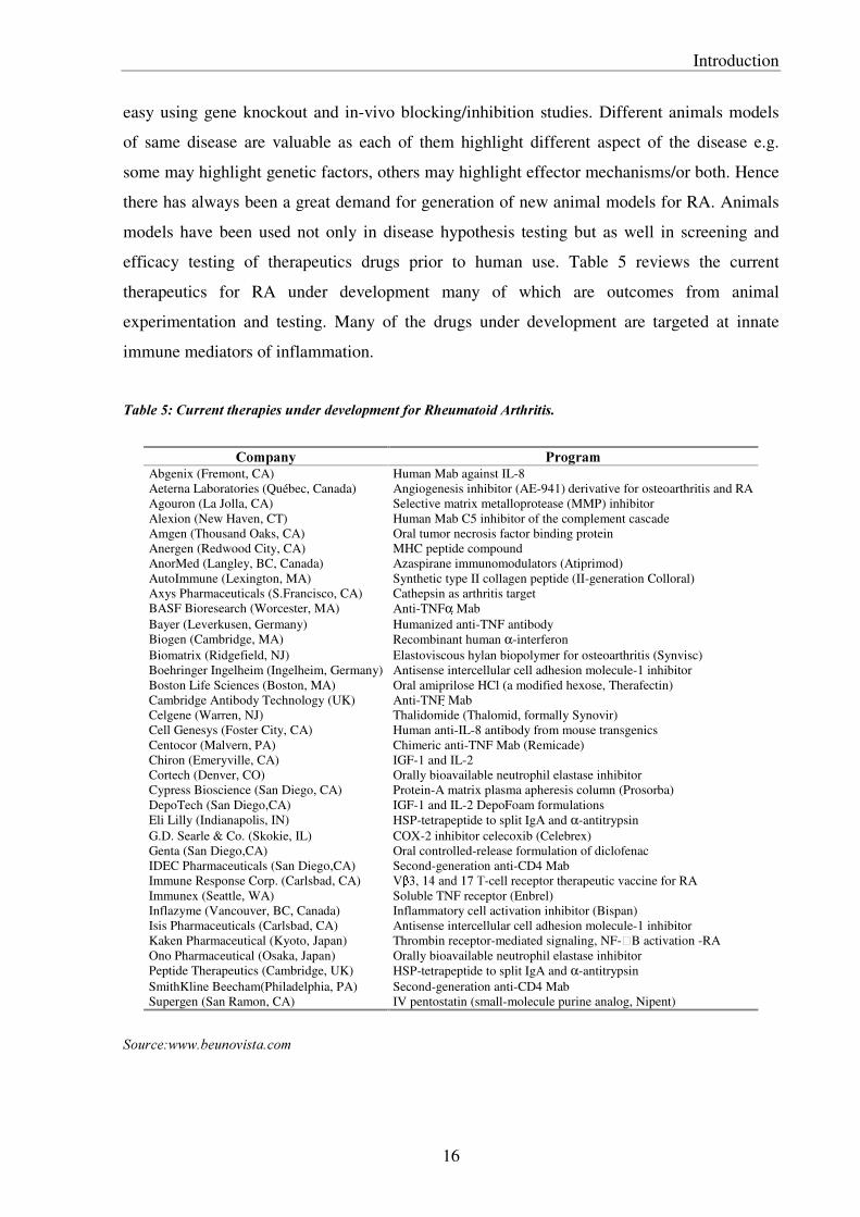
efficacy testing of therapeutics drugs prior to human use. Table 5 reviews the current
therapeutics for RA under development many of which are outcomes from animal
experimentation and testing. Many of the drugs under development are targeted at innate
immune mediators of inflammation.
�7DEOH����&XUUHQW�WKHUDSLHV�XQGHU�GHYHORSPHQW�IRU�5KHXPDWRLG�$UWKULWLV���
&RPSDQ\� 3URJUDP�Abgenix (Fremont, CA) Human Mab against IL-8 Aeterna Laboratories (Québec, Canada) Angiogenesis inhibitor (AE-941) derivative for osteoarthritis and RA Agouron (La Jolla, CA) Selective matrix metalloprotease (MMP) inhibitor Alexion (New Haven, CT) Human Mab C5 inhibitor of the complement cascade Amgen (Thousand Oaks, CA) Oral tumor necrosis factor binding protein Anergen (Redwood City, CA) MHC peptide compound AnorMed (Langley, BC, Canada) Azaspirane immunomodulators (Atiprimod) AutoImmune (Lexington, MA) Synthetic type II collagen peptide (II-generation Colloral) Axys Pharmaceuticals (S.Francisco, CA) Cathepsin as arthritis target BASF Bioresearch (Worcester, MA) Anti-TNFα Mab Bayer (Leverkusen, Germany) Humanized anti-TNF antibody Biogen (Cambridge, MA) Recombinant human α-interferon Biomatrix (Ridgefield, NJ) Elastoviscous hylan biopolymer for osteoarthritis (Synvisc) Boehringer Ingelheim (Ingelheim, Germany) Antisense intercellular cell adhesion molecule-1 inhibitor Boston Life Sciences (Boston, MA) Oral amiprilose HCl (a modified hexose, Therafectin) Cambridge Antibody Technology (UK) Anti-TNF Mab Celgene (Warren, NJ) Thalidomide (Thalomid, formally Synovir) Cell Genesys (Foster City, CA) Human anti-IL-8 antibody from mouse transgenics Centocor (Malvern, PA) Chimeric anti-TNF Mab (Remicade) Chiron (Emeryville, CA) IGF-1 and IL-2 Cortech (Denver, CO) Orally bioavailable neutrophil elastase inhibitor Cypress Bioscience (San Diego, CA) Protein-A matrix plasma apheresis column (Prosorba) DepoTech (San Diego,CA) IGF-1 and IL-2 DepoFoam formulations Eli Lilly (Indianapolis, IN) HSP-tetrapeptide to split IgA and α-antitrypsin G.D. Searle & Co. (Skokie, IL) COX-2 inhibitor celecoxib (Celebrex) Genta (San Diego,CA) Oral controlled-release formulation of diclofenac IDEC Pharmaceuticals (San Diego,CA) Second-generation anti-CD4 Mab Immune Response Corp. (Carlsbad, CA) 9 ������DQG����7-cell receptor therapeutic vaccine for RA Immunex (Seattle, WA) Soluble TNF receptor (Enbrel) Inflazyme (Vancouver, BC, Canada) Inflammatory cell activation inhibitor (Bispan) Isis Pharmaceuticals (Carlsbad, CA) Antisense intercellular cell adhesion molecule-1 inhibitor Kaken Pharmaceutical (Kyoto, Japan) Thrombin receptor-mediated signaling, NF-�B activation -RA Ono Pharmaceutical (Osaka, Japan) Orally bioavailable neutrophil elastase inhibitor Peptide Therapeutics (Cambridge, UK) HSP-tetrapeptide to split IgA and α-antitrypsin SmithKline Beecham(Philadelphia, PA) Second-generation anti-CD4 Mab Supergen (San Ramon, CA) IV pentostatin (small-molecule purine analog, Nipent)
�6RXUFH�ZZZ�EHXQRYLVWD�FRP�

Primary aims of the study
17
�����3ULPDU\�DLPV�RI�WKH�VWXG\��
i. Characterize the pathogenic GPI-specific auto-antibodies from the K/BxN murine
model for Rheumatoid Arthritis.
ii. Dissect the roles of key innate immunity players in Rheumatoid Arthritis
pathogenesis.
iii. Establish new murine models for Rheumatoid Arthritis.
.
Materials and methods �
�
18
�����0DWHULDOV�DQG�PHWKRGV�������0DWHULDOV���������*HQHUDO�FKHPLFDOV�DQG�5HDJHQWV� All chemicals, reagents and solutions were of analytical grade and/or of highest commercially
available purity. When not stated the chemicals were purchased from the Roth, Sigma, ICN or
Merck. Microfuge tubes, centrifuge tubes, pipette tips were from Eppendorf (Hamburg),
Biozym (Hess. Oldendorf) and TPP(Trassadingen, Swiss). ELISA plates were from TPP.
BioMax Light X-Ray films were from Kodak.
�������0ROHFXODU�ELRORJ\�UHDJHQWV��Bacterial strains XL2 Blue and TOP10F´were from Stratagene (Heidelberg) and BL21 was
from Novagen (Heidelberg).
;/�� %OXH� 05)¶� ∆(mrcA)183 ∆(mcrCB-hsdSMR-mrr)173 endA1 supE44 thi-1 recA1
gyrA96 relA1 lac[F’ proAB lacIqZ∆M15 Tn10 (Tetr) Amy Camr]a.
723��)¶�� F’{lacIq Tn10(TetR)}mcrA (mrr-hsdRMS-mcrBC) Φ80lacZ M15 lacX74 recA1
deoR araD139 (ara-leu)7697galU galK rpsL (StrR) endA1 nupG.
%/���'(��� F- ompT hsdSB(rB-mB-) gal dcm (srl-recA)306::Tn10(DE3).
Restriction enzymes and buffers were from Fermentas and Promega. 7DT and 3IX DNA
polymerase, T4 DNA ligase and Alkaline phosphatase were from Gibco BRL(Eggenstein).
TA TOPO CloningR kit was from Invitrogen (Groningen, Netherlands). cDNA synthesis kit
was from Gibco BRL. DNA MW markers were from Gibco BRL. Plasmid mini extraction
kits were from Amersham. Midi plasmid extraction kit and DNA gel extraction kit were from
Qiagen (Munich). RNA extraction reagent RNAzolR was from WAK-Chemie(Bad Soden).
GTG low melting agarose was from Biozym. Oligonucleotides for PCR were from MWG-
Biotech (München). DNA sequencing was done by GATC (Konstanz).
��������$QWLERGLHV��Anti mouse CD4-PE and anti-Vβ6 antibodies were from BD Pharmingen (Heidelberg).
Rabbit anti-mouse kappa light chain and rabbit anti-mouse lambda light chain were from BD
pharmingen. Secondary antibodies goat anti-mouse antibody-HRP and Goat anti-rabbit
antibody-HRP were from Pierce (Rockford, USA).

Materials and methods �
�
19
�������&HOO�FXOWXUH�UHDJHQWV��Iscove´s modified Dulbeccos Eagle medium for cell culture and Hybridoma-SFM media, fetal
calf serum, penicillin, streptomycin, glutamine, HAT and PEG were bought from Gibco BRL.
Cell culture plates, serological pipettes and confocal dishes were obtained from Costar and
Greiner (Frickenhausen).
��������3URWHLQ�FKHPLVWU\�UHDJHQWV��Acrylamide solutions for protein gels were from Roth. Protein gel MW markers were from
Amersham( Frieburg) and prestained markers from Invitrogen. Protease inhibitor cocktail was
from Boehringer (Mannheim). Nitrocellulose membranes were from Schleicer and Schuell.
Enhanced chemiluminescent reagent was from Pierce. GST bulk purification module and Hi
Trap Protein G column were from Amersham. Epoxy magnetic beads for antibody
crosslinking were from Dynal Biotech. Trypsin and Glu-C were from Promega. Microcon
concentrators were from Millipore. �
�������([SHULPHQWDO�DQLPDOV��KRN TCR mice on C57Bl/6 background (Kouskoff, Korganow et al. 1996) was a kind gift
from D. Mathis and C. Benoist, IGMBC France, and maintained and bred at animal facility of
University Konstanz.
BALB/C, C567BL6 and NOD/lt mice were from animal facility University Konstanz.
&U�-/- (Chen, Koralov et al. 2000) and &� � �(Fischer, Ma et al. 1996) mice are a kind gift from
M.C. Carroll, Boston, USA and were maintained and bred at animal facility, University
Konstanz.
)Fγ5,,,-/- (Hazenbos, Gessner et al. 1996) and )F 5,,E-/- (Takai, Ono et al. 1996)�mice on
mixed (B6 x 129) background were from Jackson laboratory,USA; bred and maintained at
animal facility, University konstanz.�71)5�-/- (Pfeffer, Matsuyama et al. 1993) and 71)5�-/- (Erickson, de Sauvage et al. 1994)�mice were a kind gift from Dr.Wendel, University Konstanz
Chinchilla rabbits for anti-sera production were from animal facility, University konstanz.
������7KHUDS\�UHDJHQWV��Cromolyn, Cimetidine, and Mepyramine, were from Sigma (Deisenhofen), SB225002 from
Calbiochem, Tranilast was from Biotrend, sheep anti- mouse TNFα polyclonal antibodies was
a gift from Dr.Wendel, University konstanz, polyclonal anti-mouse MCP-1 and anti-mouse

Materials and methods �
�
20
MIP-2 antibodies were from DPC Biermann (Germany), Cobra venom factor was from
Quidel biotech. Clodronate liposomes was a gift from N. Van Roojen Vrije University,
Holland.
�����0HWKRGV���������5HFRPELQDQW�'1$�WHFKQLTXHV�����������3UHSDUDWLRQ�RI�EDFWHULDO�PHGLD��Bacterial culture media-LB (Luria-Bertani)-Medium, LB agar, NZYM-Medium und SOC-
medium were prepared according to Maniatis HW�DO.1991.�����������,VRODWLRQ�RI�51$���RNA was extracted by RNAzol B reagent. Cells/tissue was homogenized with 2 ml RNAzol
B per 100 mg tissue or 107 cells. Phase separation was done by adding 0.2 ml of chloroform
per 2.0 ml homogenate and spun at 12,000g for 15 min at 4oC. The upper colorless aqueous
phase was transferred into clean tube and RNA precipitated by adding equal volume of
isopropanol and spinning at 12,000g for 15 min. The pellet washed 75% ethanol, air dried and
suspended in DEPC water. The quality of the RNA was verified by gel electrophoresis.
���������F'1$�V\QWKHVLV�DQG�SRO\PHUDVH�FKDLQ�UHDFWLRQ��57�3&5����cDNA synthesis was done according to manufacturer protocol. Briefly 5 J of total RNA with
1 O (50 J) primer oligo(12-18) dT in volume of 12 O DEPC water was incubated at 70oC for
10 min. 7 O of reaction mix solution (10X PCR buffer, 25mM MgCl2, 10 mM dNTP mix and
0.1M DTT) was mixed with RNA primer mix and incubated at 25oC for 5 min and for 10 min
after adding 1 O (200U) Superscript II reverse transcriptase enzyme. RT reaction was carried
out at 42oC for 50 min and terminated at 70oC for 15 min. RNA was removed by treatment
with 1 O RNAse H at 37oC for 15 min. The cDNA quality was checked by GAPDH PCR. For
general PCR, 7DT DNA polymerase and for PCR cloning, 3IX�DNA polymerase was used. A
standard 50 O�PCR mix consisted of 5 O of 10X PCR buffer, 1 O of 0.25-1 J of template, 5
O�RI���� 0�SULPHUV�HDFK, 2 O�RI���� 0�G173�PL[���� O of 1.5 mM MgCl2. The standard
PCR cycling conditions were- an initial denaturation at 95oC for 2 min, followed by 30 cycles
of denaturation at 95oC for 45 sec, annealing at 50oC to 60oC for 1 min and extension of 72oC
for 1 min/Kb pair for 7DT polymerase and 2 min/Kb pair for 3IX� polymerase extension
reaction. A final extension reaction was done at 72oC for 5 min.

Materials and methods �
�
21
���������$GGLWLRQ�RI���DGHQRVLQH�WR�3&5�SURGXFW��Prior to TOPO cloning of 3IX DNA polymerase amplified PCR products addition of poly A
ends was done by adding 1 O�of Taq pol (3U/� O) and 1 O of 10mM ATP to 50 O PCR mix
and incubation at 72oC for 10 min.
���������7232�FORQLQJ�RI�3&5�SURGXFW��The TOPO TA cloning of PCR amplified products into pCR2.1-TOPO vector were done
according to manufacturers recommendations. The ligated products were transformed into
T0P10F´ cells and screened by blue white selection.
���������([WUDFWLRQ�RI�SODVPLG�'1$��Plasmids preps from 1-3 ml cultures were prepared using GFXTM Micro Plasmid Prep Kit.
Bacterial cells from 1.5 ml cultures was suspended in 150 O solution I (100 mM Tris-HCl
(pH7.5), 10 mM EDTA, 400 J /ml RnaseI) and lysed in 150 O alkali solution II (1M NaOH,
5.3% SDS) and then neutralized by 300 O solution III (buffered solution containing acetate
and chaotrope). The lysate was pelleted and supernatant applied to glass fiber GFXTM column
and washed with wash buffer (Tris-EDTA buffer with 80% ethanol). Plasmid was eluted by
Tris-EDTA buffer pH 8.0. The Qiagen plasmid midi kit was used for plasmid extraction from
25-50 ml cultures. Overnight culture was diluted 1/500 in 25 ml medium and grown at 37oC
for 12-16 h. The bacterial cells were suspended in 4 ml buffer P1 (50 mM Tris-HCl ,pH 8.0,
10 mM EDTA, 100 J /ml Rnase A), alkali lysed by 4 ml of buffer P2 (200 mM NaOH, 1%
SDS) and then neutralized by buffer P3 (3.0 M Potassium acetate). The lysate were pelleted
and supernatant loaded on equilibrated Qiagen-tip 100 column to bind DNA. The columns
were washed by wash buffer (1M NaCl, 50 mM MOPS, pH7.0, 15% Isoproponal Y�Y� 0.15%
Triton X-100) and plasmids were eluted with 5 ml elution buffer (1M NaCl, 50 mM MOPS
pH 7.0, 15% Isopropanol Y�Y). The plasmid was precipitated by 0.7 volumes of isoproponal
and by centrifugation at 15000g for 30 min, The DNA pellet was washed in 70% ethanol,
dried and dissolved in 10 mM Tris Cl pH 8.0.
����������5HVWULFWLRQ�GLJHVWLRQV�RI�SODVPLG�'1$��Upto 5 Xg plasmid DNA was suspended in 20 O of restriction enzyme buffer. 1-5 units of
restriction enzyme was added and reaction mix incubated at 37oC for 2h-4h. The reaction was
stopped by 0.2 O 500 mM EDTA pH8.0. The restriction digests was analyzed by gel
electrophoresis. �

Materials and methods �
�
22
���������,VRODWLRQ�RI�'1$�DQG�SODVPLGV�IURP�SUHSDUDWLYH�DJDURVH�JHO��Purification of plasmid and DNA inserts was carried out on 0.8-2% low melting point agarose
gels. EtBr stained gels were visualized on a UV trans-illuminator (λ 265 nm). A clean scalpel
was used to excise the insert bands of interest and DNA was extracted using the QIA quick
gel extraction kit. Briefly 3 volumes of buffer QG was added to 1 volume of gel and
incubated at 50oC for 10 min followed by 1 gel volume of Isopropanol. The mix was applied
to QIA quick column to bind DNA and washed with buffer PE. The DNA was eluted with 10
mM Tris-Cl, pH 8.5.
���������'1$�OLJDWLRQ��For ligation reaction 1U of T4 ligase was added in 1:3 molar ratio of plamid to insert DNA
(0.5-1 J total DNA) in 20 O 1X ligase buffer mix. The reaction was incubated at RT for 3-5
hours. 2-4 O of reaction mix was used to transform 100 O competent cells.
����������3UHSDUDWLRQ�RI�FRPSHWHQW�EDFWHULD��Single colony from LB plate without antibiotic was picked and inoculated into 100 ml SOCS
medium. The culture was grown at 37oC until an OD (λ600nm) of 0.5. The bacteria were
pelleted at 4000 rpm and resuspended in ice-cold 50 mM CaCl2 and incubated on ice for 30
min. The CaCl2 treated bacteria were pelleted again at 2600g at 4oC for 5 min and carefully
resuspended in 4 ml of 50 mM CaCl2. The bacteria were aliquoted and stored at -70oC until
use.
�����������7UDQVIRUPDWLRQ�RI�FRPSHWHQW�EDFWHULD��The competent cells were thawed on ice and mixed with 0.1-2 J of purified plasmid DNA or
with ligation mix and incubated on ice for 30 min. Subsequently the cells were heat shocked
at 42oC for exactly 45 sec and incubated on ice for 2 min. The cells were suspended in 200 O SOCS media and incubated for 45 min at 37oC with shaking. 10-200 O of the transformation
mix was plated onto petri plates with selection medium and incubated overnight at 37oC.
�������3URWHLQ�WHFKQLTXHV������������*HO�HOHFWURSKRUHVLV�RI�SURWHLQ���SDS-polyacrylamide-gel electrophoresis was performed according to Laemmli. Gels of
different percentages were casted with acrylamide/bisacrylamide (30:0.8) depending on the
size of the investigated protein. All samples were supplemented with 0.25x vol. sample buffer

Materials and methods �
�
23
����� P0� 7ULV�� S+� ����� ���� 6'6�� ���� -mercaptoethanol, 0.5% bromophenolblue), and
denatured at 95oC for 5 min and loaded. Gels were run with a constant current of 80 mA/gel
in Laemmli buffer (25 mM Tris, 192 mM Glycine, 0.1% SDS). Staining of gels were done
with Coomassie stain (40% methanol, 10% acetic acid, 0.2% Brilliant Blue R250). The gels
were destained with destaining solution (40% methanol, 10% acetic acid).
���������([SUHVVLRQ�DQG�SXULILFDWLRQ�RI�*67�IXVLRQ�UHFRPELQDQW�SURWHLQV��10 ml of LB ampicillin media was inoculated with recombinant protein expressing
BL21(DE3) (�FROL and grown overnight (o/n) at 37oC. 5 ml from o/n culture was used to
inoculate 200 ml LB ampicillin media in 1litre flask and shaken at 37oC for 2-3 hours until
OD λ 600 nm was 0.6. The cultures were induced with 1 mM IPTG (end concentration) and
shaken at 37oC for 3 hours to express the protein. The cells were pelleted at 3500 rpm in
sorvall GSA rotor for 30 min at 4oC. For every gram of cell pellet, 3 ml of ice cold lysis
buffer (50mM Tris pH 8.3, 100mM NaCl, 1mM EDTA pH 8.0), 8 O of 50mM PMSF, 125 O of 25X Protease inhibitor cocktail and 80 O of lysozyme (10mg/ml) were added and
incubated on ice for 20 min with occasional mixing. 4 mg of deoxycholic acid was then added
to lysate with stirring at 37oC until the lysate became viscous. To this lysate 20 O of DNAseI
(1mg/ml) was added and placed at RT until viscosity was lost (30 min). The lysate was then
sonicated on ice with at micro-tip limit for 10 times (30 sec intervals each) or until OD λ600
nm was 1/5 the initial. The sonicate was pelleted at 10,000 rpm in sorvall F6 rotor for 20 min
at 4oC and supernatant collected. 1 ml Glutathoine beads for every for 5 mg GST-fusion
protein (yield from approximately 2 liter of culture) was re-suspended in 1ml lysis buffer and
the sonicate supernatant added to beads and mixed at RT for 30 min. The beads were pelleted
at 500g for 5 min, supernatant removed and were washed three times with 10 bed volumes of
1X PBS, 0.5% Triton X. The recombinant GST-fusion protein was cleaved off from beads
incubated overnight at RT after adding 10 U of Thrombin protease/mg of recombinant
protein. The cleaved GPI was eluted from beads and checked on SDS-PAGE for purity.
��������,PPXQRORJLFDO�WHFKQLTXHV����������:HVWHUQ�EORWV�DQG�GRW�EORWV��After protein gel electrophoresis. Two layers of Whatman 3MM paper, nitrocellulose
membrane, the gel, and two more layers of 3MM paper all soaked in blotting-buffer (20%
methanol, 48 mM Tris, 39 mM glycine), were stacked onto the anode of a semi-dry-blotter.
Air bubbles between the layers were removed by rolling a glass pipette over the sandwich.

Materials and methods �
�
24
Proteins were transferred at 2.75 mA/cm2 for 75 min. After the blotting, complete transfer and
equal loading of the proteins were controlled by staining the gel with Coomassie and the
membrane with Ponceau red (0.2% Ponceau S, 5% acetic acid). The membranes were then
incubated at RT for 1 h or at 4oC overnight, in TBST (150 mM NaCl, 50 mM Tris/HCl pH 8,
0.05% Tween 20), 5% non-fat dry milk to block non-specific binding of the antibody. Excess
milk protein was removed by several washing steps with TBST. The membrane was
incubated at RT for 1h with primary antibody diluted in TBST. After further washing in
TBST (3x 5 min), the membrane was incubated at RT for 1h with secondary antibody. This
was a goat anti-mouse HRP (1:10,000 in TBST) or a goat anti-rabbit HRP (1:5000 in TBST).
After another three washing steps, HRP activity was detected by Enhanced Chemi
Luminescence (ECL) on X-ray film. For dot blots the proteins were spotted on nitrocellulose
membranes and processed in same manner as mentioned for immunoblots for antibody
staining.
���������*3,�(Q]\PH�/LQNHG�,PPXQRVRUEHQW�$VVD\��(/,6$���Purified, GST-cleaved GPI was coated (0.125 �J/well in PBS, pH 7) onto 96 well ELISA
microtiter plates. The nonspecific binding sites were blocked with 1% fat-free milk
powder/PBS. Sera from K/BxN serum-injected mice as well as the control serum-injected
mice were diluted 1:300 in PBS with 1% milk powder and added to the wells. After
incubation for 2 h at 37oC the plates were washed with PBS/Tween 20 and incubated for 2 h
with rabbit anti-mouse-IgG-HRP antibodies (1:20,000). The plates were developed with
ortho-phenylene-diamine as substrate. The reaction was stopped by adding 1M HCl and read
at 495nm OD.
���������3UHSDUDWLRQ�RI�DQWLJHQ�IRU�LPPXQL]DWLRQ��For mice immunization 250 J of recombinant GPI was dissolved in 1 ml of 5% Alum
(Aluminum potassium sulphate) as adjuvant. Then the pH was then shifted to pH 6.5 using
1.25 N NaOH to induce precipitation of GPI-alum complex. Each mice was immunized by
injecting 75 J (300 O) of the antigen-adjuvant mixture intra-peritoneally. For rabbits
immunization with 200 J�of recombinant GPI plus 500 O of complete Freunds adjuvant for
primary dose or in 500- O�LQFRPSOHWH�)UHXQGV�DGMXYDQW�IRU�ERRVWHU�GRVH�LQ�D�WRWDO�YROXPH�RI���ml saline was injected sub-cutaneously.
Materials and methods �
�
25
���������3URGXFWLRQ�RI�*3,�VSHFLILF�PRQRFORQDO�DQWLERGLHV��A week prior fusion SP2/0 myeloma fusion partner cells were expanded in Iscoves medium
10% FCS,�96 well plates were plated with peritoneal cells obtained from C57Bl6 mice and the
KBN mice were immunized with purified recombinant GST cleaved GPI. On day of fusion
the mice were sacrificed; spleens harvested and teased to obtain a single cell suspension.
Debris were removed by passage through fine steel mesh and washed cells were suspended in
serum free Iscoves media. The spleen cells were mixed in 1:1 ratio of SP2/0 in 50 ml conical
centrifuge tube, pelletted at 500g and the supernatant discarded. The cell fusion was
performed in laminar hood with cell pellet in 37oC water bath. Using a 1 ml pipette 1 ml
prewarmed 50% PEG solution was added drop-by-drop over 1 min with stirring with pipette
after each drop. This was repeated with additional 1 ml of prewarmed serum free Iscoves
medium. A further 7 ml of prewarmed serum free Iscoves medium was added drop-by-drop
over 2-3 min. The fusion mix was pelletted at 500g for 5 min at RT and supernatant
discarded. Placing the cell pellet at 37oC, 10 ml of prewarmed Iscoves medium with 20% FCS
was forcibly added to disrupt the cell pellet and the volume made to final cell density of
2.5x10⁶ cells/ml. The cell suspension is then gently pipetted out at 100-125� O /well into a 96
well flat bottom plate with feeder cells and incubated in 37oC, 5% CO2. On days 2,3,4,5,7,9,
and 11 half the volume of each well was aspirated using sterile Pasteur pipette attached to
vacuum pump and 2 drops of Iscoves with 20% FCS, and 1X HAT was added. On day 14 the
feeding was done with Iscoves with 20% FCS and 1X HT. From day 15 onwards the cells
were fed with Iscoves medium 20% FCS. The hybridomas were screened when the cells
showed 10%-25% confluence. About 100 O supernatant from each well were removed and
tested for GPI reactivity by GPI ELISA.
���������/LPLWHG�GLOXWLRQ�FORQLQJ�RI�K\EULGRPDV�DQG�DGDSWDWLRQ�WR�VHUXP�IUHH�PHGLD��10 days prior to cloning, 96 well plates were plated with peritoneal cells obtained from
C57BL/6 mice and grown at 37oC. On the day of cloning hybridoma cells were counted and
checked for viability by trypan blue dye exclusion stain. The viability must be atleast 80% for
successful cloning. For each hybridoma, the cells were serially diluted in Iscoves medium
with 20% FCS to give 4, 2 and 1 cell/ml. The diluted cells were then plated at 250 O /well for
each dilution in prepared 96 well plates and incubated at 37oC, 7.5% CO2 for 5-7 days. The
plates were microscopically examined for plating and cloning efficiency. Wells with single
clones were followed up for GPI reactivity by ELISA and positive clones were expanded for
freezing. The hybridomas were adapted to grow in serum free media (SFM) to reduce protein

Materials and methods �
�
26
loads in downstream processing. Briefly the hybridoma was grown in Iscoves media 10%
FCS. The growing cells were then serially expanded in Iscoves media 10% FCS with 25%
SFM followed by 50% SFM medium and finally in complete 100% SFM media.
���������3XULILFDWLRQ�RI�PRQRFORQDO�DQWLERGLHV��The hybridomas were grown in hybridoma medium to confluency and the medium turned
yellow. The supernatants were collected and concentrated 10 times using an Amicon
concentrator (Millipore) having a membrane cutoff 100 KDa. The purification of antibodies
from the concentrated hybridoma supernatants was done on AKTATM Prime instrumentation
(Amersham) using Hi Trap Protein G columns. Briefly concentrated supernatants were loaded
onto Protein G columns equilibrated with binding buffer (20 mM sodium phosphate pH 7.0).
The columns were washed with binding buffer to remove non-specific proteins. The antibody
was then eluted with elution buffer (0.1 M Glycine-HCl, pH 2.7). The eluted antibody was
immediately neutralized with 1M Tris pH 9.0 and buffer exchanged into PBS buffer using a
microcon-50 concentrator (Millipore).
�������(SLWRSH�DQDO\VLV�E\�DIILQLW\�SURWHRPLFV��
�)LJXUH����,OOXVWUDWLRQ�VKRZLQJ�SULQFLSOH�RI�0DVV�VSHFWURPHWU\�EDVHG�HSLWRSH�DQDO\VLV���Rapid developments in mass spectrometry in combination with the proteolytic digestion of
antigen have opened new methods of epitope mapping (Macht, Fiedler et al. 1996; Parker,
Papac et al. 1996). The method is based on the fact that antibody in general are very resistant

Materials and methods �
�
27
to proteolytic digestion. The epitope analysis can be done by epitope excision or extraction In
epitope excision immobilized monoclonal antibody is incubated with whole antigen and the
bound antigen is digested with a proteolytic enzyme The antibody bound surface of the
antigen is protected from proteolysis. In epitope extraction protease digested antigen peptide
mixture is affinity bound to immobilized antibody. In both the above cases the peptides
containing the epitopes are eluted from immobilized antibody by low pH buffer and analyzed
by mass spectrometric peptide fingerprinting. The high accuracy and sensitivity of mass
spectrometer helps in identifying the peptides by molecular weights.
����������&URVV�OLQNLQJ�RI�*3,�DQWLERG\�WR�HSR[\�G\QDEHDGV��Dynabeads® M-270 Epoxy are uniform, super paramagnetic beads with surface reactive
epoxy groups for binding of antibody or other ligands by covalent bond formation at neutral
pH. Binding of antibodies occurs through amine or thiol groups occur with no further
activation of the surface. For antibody cross-linking, the beads were washed and resuspended
in 0.1 M sodium phosphate buffer pH 7.4 at 109 beads/ml. 3 J of antibodies/107 beads in
phosphate buffer, 1 M ammonium sulfate (to enhance binding of antibody) were mixed,
incubated for 24 hours at RT with gentle mixing. After incubation the tube with the beads
were placed in magnet and supernatant removed. The beads were washed with PBS buffer and
stored at 2-8oC.
����������3URWHRO\WLF�GLJHVWLRQ�RI�SXULILHG�UHFRPELQDQW�PRXVH�*3,��Digestion of GPI was carried with Trypsin or Glu-C protease for peptide fingerprinting
western blot of monoclonal antibodies and for epitope analysis by mass spectrometry��Trypsin
is a serine protease, which cleaves peptide bonds C-terminally of arginine or lysine. Trypsin
digestion of GPI was done at a enzyme/substrate ratio of 1/50 in 0.1 M Tris HCl buffer pH 8.0
containing 0.01% Z�Y SDS at 37oC for 12 hours. The reaction was stopped by adding 0.1%
TFA. Glu-C is a serine protease, which cleaves peptide bonds C-terminally to glutamic acid.
The digestion of GPI was done at an enzyme substrate ratio of 1:50 in 25 mM ammonium
carbonate buffer pH 7.8 containing 0.01% w/v SDS at 25oC for 12 hours.
����������(SLWRSH�H[WUDFWLRQ��For epitope extraction 108 Dynabeads immobilized with GPI monoclonal antibodies were
incubated with 50 J peptides of Trypsin or Glu-C digested mouse GPI in 1 ml PBS buffer,
pH 7.0 for 4 hours with shaking. The beads were washed 10X with 1 ml PBS, 0.1% Tween on
Materials and methods �
�
28
a magnet. The peptides were eluted with 50 O of 0.1% TFA solution. The eluates were
concentrated in a speed vac and resuspended in 0.1% TFA for mass spectrometric analysis.
���������0DVV�VSHFWURPHWU\�DQDO\VLV��MALDI time-of-flight mass spectrometry was performed with a Bruker Biflex-DE mass
spectrometer equipped with a Scout MALDI source and video system, a nitrogen UV laser (
max 337 nm), dual-channel plate detector and XMASS software. Sample preparation was
performed with 1 O of a freshly prepared saturated solution of 4-α-hydroxy cyanocinnamic
acid in 0.1% TFA in H2O and 0.1% TFA in Acetonitrile (Y�Y 1:2), which was directly mixed
with 0.5 O of the peptide solution and crystallized on targets. Spectra were recorded at an
acceleration voltage of 25 kV and were averaged over 5 single laser shots. Calibration was
carried out using the singly and doubly protonated ions of bovine insulin as internal standard.
�������$QLPDO�H[SHULPHQWDWLRQ�����������(VWDEOLVKPHQW�RI�.%1�PLFH�PRGHO�DQG�SURGXFWLRQ�RI�.%1�VHUD��Arthritic mice were obtained by crossing KRN-TCR transgenic maintained on C57BL/6( K/B)
with NOD/Lt (N) animals (K/BxN). Progeny bearing the V ��WUDQVJHQLF�7&5�ZHUH�LGHQWLILHG�by cytofluorometry of PBL using anti-CD4 PE and anti-9 ��),7&�ODEHOHG�$EV�
���������(VWDEOLVKPHQW�RI�DQWLERG\�LQGXFHG�DUWKULWLV�LQ�PLFH��Sera were obtained by tail bleeding of K/BxN mice aged 1-6 months. Sera were pooled and
stored at –20oC until use. 100 O equivalent of sera was injected L�S per mice to induce arthritis
on day 0 and in some cases again on day 10 to prolong inflammation. For GPI monoclonal
induced arthritis 4.0 mg of total GPI mAb cocktail in a volume of 200 O saline was injected
L�Y per mice on day 0 followed injection of 50 �J LPS (�(�FROL�strain 0111B4) L�S. on day 1.
For Rabbit anti- mouse GPI sera induced arthritis 300 O of rabbit anti-sera was injected L�S�per mice to induce arthritis.
���������$VVHVVPHQW�RI�DQWLERG\� LQGXFHG�DUWKULWLV�E\�DQNOH� WKLFNQHVV�DQG�FOLQLFDO� LQGH[�VFRUH��Development of RA in the mice was assessed by calliper (Mitutoyo, Japan) measurement of
ankle thickness and by clinical index score-a visual score based on number of ankles affected.
Ankle thickness for each mice was expressed in millimeters as summed average of thickness
of the four limbs of each mice. The error was expressed as standard error from mean (SEM).
Materials and methods �
�
29
Clinical Index(I) score was obtained by summing of ankles scores: Ankle score is 0 if ankle
not affected; 1 if ankle affected and 2 if 2 ankles affected and so on. Clinical index(II) score
was obtained by sum of multiplied values of each ankle score with its inflammation score.
Ankle score is 0, if ankle not affected and 1, if ankle affected. Inflammation score is 0 ankle
not affected; 0.25 if reddening and slightly swollen ankle; 0.75 if reddening and moderately
swollen ankle; 1 if reddening and highly swollen ankle. The error was expressed as standard
error from mean (SEM).
The percentage reduction in maximum ankle thickness was obtained by formula [1-
(maximum average ankle thickness of treated mice/ maximum average ankle thickness of
non- treated mice] x 100. The percentage reduction in clinical index was calculated similarly.
���������7UHDWPHQWV�SURWRFROV�IRU�DQG�LQKLELWLRQ�VWXGLHV�������������&RPSOHPHQW�GHSOHWLRQ�E\�FREUD�YHQRP�IDFWRU�WUHDWPHQW��Cobra Venom Factor was injected into mice to deplete complement C3 and C5. A single L�S
injection of 500U CVF/Kg body weight was done for this purpose. Stock solution of 3U/� O in saline were prepared and stored at -70oC and were diluted 10 times with saline prior to
injection.
�����������'HSOHWLRQ�RI�PDFURSKDJHV��Macrophages were depleted by Clodronate liposomes treatment (Van Rooijen and Sanders
1994; van Rooijen, Sanders et al. 1996). Two L�S. injections of 200 O of liposomes were done
on day day-3 and -1 prior to sera transfer to deplete macrophages.
�����������,Q�YLYR�LQKLELWLRQ�RI�PDVW�FHOO�GHJUDQXODWLRQ.
For mast cell degranulation inhibition with cromolyn (Kolaczkowska 2001), mice were
treated with Cromolyn sodium 10mg/mice/injection to inhibit Mast cell degranulation. A 50
mg/ml solution of Cromolyn was prepared in saline. Three L�S injection, of 10 mg each (200
O) were made on day -1, 0 and 2 with regard to sera transfer. 100 O of KBN sera was injected
L�S 2 hours after the day 0 cromolyn treatment. For Mast cell degranulation inhibition with
tranilast, 2-[[3-(3,4-Dimethoxyphenyl)-1-oxo-2-propenyl]amino] benzoic acid.(Hara, Ono et
al. 2002), mice were treated with tranilast 250 XJ/mice/injection. A solution of 2mg/ml
solution of tranilast was prepared in saline. Three L�S injections of 250 J�each (125 O) were
made on day -1, 0 and 1 with regard to sera transfer. 100 O of KBN sera was injected L�S 2
hours after the day 0 treatment. The mice were assessed for arthritis development.
Materials and methods �
�
30
�����������$GPLQLVWUDWLRQ�RI�KLVWDPLQH�UHFHSWRU�DQWDJRQLVWV��Mepyramine, a H1 histamine receptor antagonist or cimetidine, a H2 histamine receptor
antagonist(Kolaczkowska 2001) were administrated at 450 XJ/mice/injection to study the role
of histamine and its receptors. A solution of 5mg/ml mepyramine was prepared in saline.
Cimetidine base was dissolved in a small quantity of 0.1 N HCl, neutralized with NaOH, and
made to 5mg/ml in saline. Three L�S injections of 450 XJ each (90 O) were made with
mepyramine or cimetidine into each mice on day -1,0 and 2 with regard to sera transfer. 100
O of KBN sera was injected L�S 2 hours after day 0 treatment. The mice were assessed for
arthritis development.
�����������$GPLQLVWUDWLRQ�RI�,/�5�DQWDJRQLVW�� The compound SB225002-a small molecule IL8 receptor-b (CXCR2) antagonist (White, Lee
et al. 1998) was administered at 80 J/mice/injection. A solution of 400 J/ml SB225002 was
prepared in 25% DMSO, saline. Four L�S injections of 80 J each (200 O) were administered
on day -1, 0, 2 and 4 with regard to sera transfer. 100 O of KBN sera was injected L�S after 30
min of day 0 treatment. The mice were assessed for arthritis development.
������������$GPLQLVWUDWLRQ�RI�EORFNLQJ�DQWLERGLHV��i. Rabbit anti-mouse/rat Macrophage inflammatory protein-2 (MIP-2) antibodies were
administered at 150 XJ/mice/injection. Two L�Y injections of 150 J�each (150 O) were made
with the antibodies resuspended in saline on day 0 and 3 with regard to sera transfer.
ii. Rabbit anti-mouse/rat Macrophage Chemoattractant Protein-1 (MCP-1) antibodies were
administered at 150 XJ/mice/injection. Two L�Y injections of 150 J each (150 O) were made
with the antibodies re-suspended in saline on day 0 and 3 with regard to sera transfer.
iii. Sheep anti- mouse TNFα were administered at 500 J /mice/injection. Two L�Y injections
of 500 J each (100 O) were made with the antibodies re-suspended in saline on day 0 and 3
with regard to sera transfer.
�������+LVWRFKHPLFDO�WHFKQLTXHV��Mice were sacrificed by cervical dislocation .Tissue samples were prepared by fixing tissues
24 h in 4% phosphate-buffered formaldehyde. Fixed joints were decalcified by treatment with
14% EDTA solution for 2 weeks with gentle rocking and daily replacement of the solution.
Samples were then washed with PBS, dehydrated with a series of ethanol washes (50%

Materials and methods �
�
31
ethanol, followed by 70% ethanol), and embedded in paraffin. Sections of tissue 4 µm thick
were cut by microtomy and stained with haematoxylin and eosin.

Results
32
���5HVXOWV�������� *HQHUDWLRQ� DQG� FKDUDFWHUL]DWLRQ� RI� JOXFRVH���SKRVSKDWH� LVRPHUDVH� VSHFLILF�PRQRFORQDO�DQWLERGLHV�IURP�.�%[1�PLFH���GPI specific antibodies from K/BxN mice are arthritogenic and can transfer arthritis into
naive mice. The reason for the arthritogenicity nor the joint specificity and cross reactivity of
GPI antibodies is still not known. And moreover a comparative study between the auto-
antibodies in the K/BxN murine RA model and to that in human RA patients would be
interesting. For these reasons it was decided to elucidate the epitopes recognized by the GPI
specific pathogenic antibodies from the K/BxN mice. GPI-specific ELISA and immuno-
blotting screening assays were needed to generate the GPI reactive hybridomas from K/BxN
mice. Therefore full-length recombinant mouse GPI was cloned expressed and purified in
bacteria and mouse GPI specific ELISA and immunoblots screening assays established. Also
the truncated versions of mouse GPI proteins were cloned and expressed in bacteria for use in
epitope mapping investigations of the GPI antibodies. To study the possibility that the RA
patients had similar autoantibodies to GPI as in K/BxN mice, human GPI specific ELISA and
GPI immunoblot screening assays were setup from the recombinant human GPI cloned
expressed and purified from bacteria.
�������� &ORQLQJ�� H[SUHVVLRQ� DQG� SXULILFDWLRQ� RI� UHFRPELQDQW� PRXVH� JOXFRVH���KRVSKDWH�LVRPHUDVH��The mouse full length GPI was cloned by RT-PCR from total C57BL6 thymus RNA using
forward primer, KN143 -5´GGA GAA TTC TAT GGC TGC GCT CAC CCG GAA 3´ and
reverse primer KN144 -5´TTC CTC GAG TCC CAA CAG CCT TGT AGT CC 3´. The
primers had EcoRI and XhoI recognition sites for in-frame cloning into pGEX-4T-3 GST
fusion vector. 3IX DNA polymerase was used for error free amplification of the product. The
PCR cycling conditions were 94oC for 2 min, followed by 30 cycles of denaturation at 94oC
for 45 sec, annealing at 60oC for 1 min and extension at 72oC for 4 min, followed by a final
extension reaction at 72oC. Figure 6 shows the PCR amplified full-length mouse GPI
amplicon.

Results
33
���)LJXUH����3&5�DPSOLILFDWLRQ�RI�PRXVH�*3,��(A) Marker. (B) Amplified PCR mGPI gene product.
The PCR product was cloned into TOPO vector; transformed into TOP10F (�FROL and
recombinant clones selected by blue/white screening. The insert DNA were excised from
recombinant vectors by EcoRI and XhoI digestion, separated on low melting point agarose
gel, extracted and sub-cloned into pGEX-4T-3 GST fusion vector. Positive clones with the
right size DNA insert (figure 7) were further amplified and sequenced.
�)LJXUH� ��� 6FUHHQLQJ� RI� S*(;��7��� SODVPLGV� IRU� UHFRPELQDQW� PRXVH� *3,� LQVHUWV� E\� UHVWULFWLRQ� HQ]\PH�GLJHVWLRQ�DQDO\VLV��(A) Marker. (B) Plasmid without insert. (C-F) Plasmids with insert.
�
Recombinant plasmids with mouse GPI inserts were confirmed by DNA sequencing and
transformed into BL21 (�FROL for protein expression. The bacterial clones were screened by
SDS-PAGE for recombinant protein expression on IPTG addition. For purification of GPI
protein the recombinant BL21 (�FROL were grown to log phase and the protein was induced
with IPTG. The bacterial cells were lysed, sonicated and the supernatant containing the
recombinant GST-GPI fusion protein was affinity purified on Glutathione sepharose beads.
The recombinant mouse GPI was cleaved from the beads using thrombin protease. The purity
of the cleaved mouse GPI protein was checked by SDS-PAGE. Figure 8 shows a SDS-PAGE

Results
34
gel with various stages of mouse GPI expression and purification. The expression of GST-
GPI was found to be very good as more than estimated 25% of total bacterial protein being
the recombinant protein The purity of the purified GPI protein was more than 95% as assessed
by coomassie stained gels. The purified mouse GPI was used for epitope mapping studies and
to establish mouse GPI ELISA and immunoblot screening assays.
��
���)LJXUH����6'6�3$*(�JHO�VKRZLQJ�H[SUHVVLRQ�DQG�SXULILFDWLRQ�RI�UHFRPELQDQW�PRXVH�*3,�IURP�%/���FHOOV��($) Marker. (B) Un-induced bacterial lysate. (&) IPTG induced bacterial lysate. (') Glutathione affinity purified
GST-mGPI fusion protein. (() GST alone. ()��*) Thrombin cleaved and purified mGPI.
�������&ORQLQJ�DQG�H[SUHVVLRQ�RI�WUXQFDWHG�PRXVH�JOXFRVH���SKRVSKDWH�LVRPHUDVH��In order to determine the epitopes recognized by the GPI-specific monoclonal antibodies by
protein truncation assay, we cloned the following four truncated forms of mouse GPI protein
into GST fusion vector pGEX-4T-3. i. GPI-I (GPI amino acid residues 1-190); ii. GPI-II (GPI
amino acid residues 1-368); iii. GPI-III (GPI amino acid residues 324-559); iv. GPI-IV (GPI
amino acid residues 485-559). The partial GPI clones were RT-PCR amplified from total
C57BL6 thymus RNA using the following pairs of PCR primers respectively i. Forward-
KN143 5 GGA GAA TTC TAT GGC TGC GCT CAC CCG GAA 3 and Reverse-KN222
5 TTC CTC GAG TCG AGA GCT TCA GTC ACC ATG AG 3 ; ii. Forward-KN143 5 GGA
GAA TTC TAT GGC TGC GCT CAC CCG GAA 3 and Reverse-KN224 5 TTC CTC GAG
TCA GCA AAG CGG TGC ATG TAC TG 3 ; iii. Forward-KN223 5 GGA GAA TTC TGG
CAT CTG GTA CAT CAA CTG GC 3 and Reverse-KN 144 5 TTC CTC GAG TCC CAA
CAG CCT TGT AGT CC 3 ; iv. Forward-KN 225 5 GGA GAA TTC TTT CAT TCT GGG
GGC CTT GAT TG 3 and Reverse-KN144 5 TTC CTC GAG TCC CAA CAG CCT TGT
AGT CC 3 . The primers had EcoRI and XhoI recognition sites for in-frame cloning into
pGEX-4T-3 vector. PCR was done with 3IX DNA polymerase for error free amplification.
The PCR cycling parameters for GPI-I and GPI-IV were an initial denaturation at 95oC for 2