1 acquired tnfrsf14 mutations in follicular - cancer research
TRANSCRIPT
Author manuscripts have been peer reviewed and accepted for publication but have not yet been edited.
Copyright © 2010 American Association for Cancer Research
1
Acquired TNFRSF14 mutations in follicular lymphoma are associated with worse prognosis K-John J. Cheung,1 Nathalie A. Johnson,1 Joslynn G. Affleck,2 Tesa Severson,3 Christian Steidl,1 Susana Ben-Neriah,1 Jacqueline Schein,3 Ryan D. Morin,3 Richard Moore,3 Sohrab P. Shah,4 Hong Qian,3 Jessica E. Paul,3 Adele Telenius,1 Thomas Relander,1 Wan Lam,5 Kerry Savage,1 Joseph M. Connors,1 Carolyn Brown,2 Marco A. Marra,3 Randy D. Gascoyne,1 and Douglas E. Horsman1
1Center for Lymphoid Cancer, British Columbia Cancer Agency; 2Department of Medical Genetics, University of British Columbia; 3Genome Sciences Center, British Columbia Cancer Research Center; 4Department of Computer Science, University of British Columbia; 5Cancer Genetics and Developmental Biology, University of British Columbia, Vancouver, BC Canada. Support: Terry Fox Foundation New Frontiers Program Project Grant (#019001) and Genome Canada/BC Grant Competition III (J.M.C., R.D.G. and D.E.H.); National Cancer Institute of Canada (NCIC) (#019005), Michael Smith Foundation for Health Research (#ST-PDF-01793) and Canadian Institute of Health Research (#STP-53912) (N.A.J.); The Deutsche Forschungsgemeinschaft (STE-1706/1-1) (C.S.); Terry Fox Young Investigator and Michael Smith Senior Research Scholar (M.A.M.). Requests for reprints: K-John J. Cheung, Ph.D., Center for Lymphoid Cancer, British Columbia Cancer Agency, 600 West 10th Avenue, Vancouver, British Columbia V5Z 4E6, Canada (E-mail: [email protected]) Conflict of interest: None Running title: TNFRSF14 mutations in follicular lymphoma Key words: Array comparative genomic hybridization, single nucleotide polymorphism, conditional Markov model, follicular lymphoma, overall survival, transformation risk, Kaplan-Meier analysis, Cox proportional-hazards model Word counts: Abstract-213; Text-4079 Abbreviations: array CGH, Array comparative genomic hybridization; SNP, single nucleotide polymorphism; FISH, fluorescence in situ hybridization; FL, follicular lymphoma; DLBCL, diffuse large B-cell lymphoma; CMM, conditional Markov model; OS, overall survival; BAC, bacterial artificial chromosome; MRD, minimum region of deletion; cnLOH, copy neutral loss of heterozygosity; HR, hazard ratio.
Published OnlineFirst on September 30, 2010 as 10.1158/0008-5472.CAN-10-2460
on March 26, 2019. © 2010 American Association for Cancer Research.cancerres.aacrjournals.org Downloaded from
Author manuscripts have been peer reviewed and accepted for publication but have not yet been edited. Author Manuscript Published OnlineFirst on September 30, 2010; DOI: 10.1158/0008-5472.CAN-10-2460

Author manuscripts have been peer reviewed and accepted for publication but have not yet been edited.
Copyright © 2010 American Association for Cancer Research
2
Abstract
Clinical correlative studies have linked 1p36 deletions with worse prognosis in follicular
lymphoma (FL). In this study, we sought to identify the critical gene(s) in this region that
is responsible for conferring inferior prognosis. BAC array technology applied to 141 FL
specimens detected a minimum region of deletion (MRD) of ~97 kb within 1p36.32 in
20% of these cases. Frequent SNP-detected copy-neutral loss of heterozygosity was
also found in this region. Analysis of promoter CpGs in the MRD did not reveal
differential patterns of DNA methylation in samples that differed in 1p36 status. Exon
sequencing of MRD genes identified somatic alterations in the TNFRSF14 gene in 3/11
selected cases with matching normal DNA. An expanded cohort consisting of 251
specimens identified 46 cases (18.3%) with nonsynonymous mutations affecting
TNFRSF14. Overall survival (OS) and disease-specific survival (DSS) were associated
with the presence of TNFRSF14 mutation in patients whose overall treatment
included rituximab. We further demonstrated that inferior OS and DSS were most
pronounced in patients whose lymphomas contained both TNFRSF14 mutations and
1p36 deletions after adjustment for the International Prognostic Index (HR=3.65,
95%CI=1.35-9.878, p=0.011 and HR=3.19, 95%CI=1.06-9.57, p=0.039, respectively).
Our findings identify TNFRSF14 as a candidate gene associated with a subset of FL,
based on frequent occurrence of acquired mutations and their correlation with inferior
clinical outcomes.
on March 26, 2019. © 2010 American Association for Cancer Research.cancerres.aacrjournals.org Downloaded from
Author manuscripts have been peer reviewed and accepted for publication but have not yet been edited. Author Manuscript Published OnlineFirst on September 30, 2010; DOI: 10.1158/0008-5472.CAN-10-2460

Author manuscripts have been peer reviewed and accepted for publication but have not yet been edited.
Copyright © 2010 American Association for Cancer Research
3
Introduction
Follicular lymphoma (FL) is characterized at the karyotype level by the
t(14;18)(q32;q21) that leads to overexpression of the anti-apoptotic gene BCL2 due to
relocation in proximity to the IGH enhancer (1-6). In the great majority of cases
additional chromosomal changes are evident, some of which represent disease-
associated events of clonal evolution. Alterations resulting in deletion of chromosome
band 1p36 constitute the most frequent of these secondary karyotypic alterations. The
1p36 region has been the focus of attempts to refine the deletion breakpoints and
identify driver or tumor suppressor gene(s), but historically, these efforts have been
hampered by relatively small sample cohorts, the inclusion of heterogeneous subtypes
of lymphoma and the low resolution of analytic tools available including standard
cytogenetic analysis and chromosome-based comparative genomic hybridization
(CGH). Most of these studies have reported 1p36 aberrations in the form of
intrachromosomal deletions and unbalanced translocations (7-10). Recently developed
high resolution technologies have shed further light on the issue. Of interest, a study by
Ross et al of 58 FL cases analyzed using a 50k single nucleotide polymorphism (SNP)
array demonstrated that 50% of these cases had either copy number loss or copy-
neutral loss of heterozygosity (cnLOH) at 1p36 (5). O’Shea et al, using a lower
resolution 10k SNP assay, found 8% of cases to show cnLOH but found no evidence of
deletions, possibly due to the low resolution capability of the array, however, correlation
between 1p36 cnLOH and overall survival (OS) was significant (11). These studies
have highlighted the importance of alternate mechanisms of gene inactivation that may
on March 26, 2019. © 2010 American Association for Cancer Research.cancerres.aacrjournals.org Downloaded from
Author manuscripts have been peer reviewed and accepted for publication but have not yet been edited. Author Manuscript Published OnlineFirst on September 30, 2010; DOI: 10.1158/0008-5472.CAN-10-2460

Author manuscripts have been peer reviewed and accepted for publication but have not yet been edited.
Copyright © 2010 American Association for Cancer Research
4
be operative in FL. Although the affected region of alteration identified in these studies
was still relatively large at 8.5Mb (5), a specific candidate gene was not identified.
In a previous study we have demonstrated by karyotype and array CGH that a region of
~11 Mb from 1p36.22 to p36.33 was deleted at a frequency of ~25% in 106 FL
specimens and the deletion was shown to correlate with risk for development of
transformed diffuse large B cell lymphoma (DLBCL) and for poor survival outcome
independent of clinical risk predictors (6). In this study we have applied tiling path BAC
array CGH, with a resolution of 80-150 kb (12), to a larger cohort of 141 FL specimens
and applied robust computational algorithm to further fine-map the 1p36 deleted region.
We have also used a custom-designed oligonucleotide microarray targeted to band
1p36, promoter methylation analysis and exon sequencing to identify the potential gene
of interest. Further, in an expanded cohort of 251 FL patients, we have assessed the
frequency and clinical impact of somatic mutations of a candidate gene in the 1p36
region.
Materials and Methods
Patient materials
For breakpoint mapping of 1p36 deletions, 141 FL cases were selected from the
Lymphoid Cancer Database of the British Columbia Cancer Agency diagnosed between
1987 and 1996 based on the availability of sufficient frozen tumor material for DNA
extraction. 103 of these 141 cases were included in a previous study (6). For the
on March 26, 2019. © 2010 American Association for Cancer Research.cancerres.aacrjournals.org Downloaded from
Author manuscripts have been peer reviewed and accepted for publication but have not yet been edited. Author Manuscript Published OnlineFirst on September 30, 2010; DOI: 10.1158/0008-5472.CAN-10-2460

Author manuscripts have been peer reviewed and accepted for publication but have not yet been edited.
Copyright © 2010 American Association for Cancer Research
5
assessment of mutation frequency and clinical correlative analysis, we included 115 of
the 141 cohort plus 136 additional specimens from our archive, totaling 251 cases with
sufficient DNA for sequencing analysis. Each case had a lymph node biopsy containing
FL obtained at the time of diagnosis or prior to systemic therapy and available clinical
information that included initial clinical characteristics, age, stage, lactate
dehydrogenase (LDH) level, performance status, number of extranodal sites, treatment
regimen and clinical outcome (Table 1). It should be noted that the two cases with
homozygous deletion of TNFRSF14 were not included in the 251 cohort as there was
insufficient follow-up data for survival analysis. Of these 251 patients, 35 had paired
biopsies taken at the time of diagnosis and at transformation to aggressive diffuse large
B cell lymphoma (DLBCL). The samples were accrued over a 20 year period (16 March
1987 to 11 September 2007) and were reviewed by an expert hematopathologist
(R.D.G), and classified according to 2008 World Health Organization (WHO) criteria
(13). Treatment regimens evolved over the 20 years and included observation,
localized radiation and systemic chemotherapy with or without rituximab, including
rituximab maintenance. Chemotherapeutic regimens ranged from single agent
fludarabine and chlorambucil to multi-agent regimens such as CVP (cyclophosphamide,
vincristine and prednisone) or CHOP (cyclophosphamide, doxorubicin, vincristine and
prednisone). Information on the percentages of patients receiving rituximab as part of
their first-line therapy is listed in Table 1. This study was approved by the University of
British Columbia-British Columbia Cancer Agency Research Ethics Board.
Cytogenetic analysis
on March 26, 2019. © 2010 American Association for Cancer Research.cancerres.aacrjournals.org Downloaded from
Author manuscripts have been peer reviewed and accepted for publication but have not yet been edited. Author Manuscript Published OnlineFirst on September 30, 2010; DOI: 10.1158/0008-5472.CAN-10-2460
Author manuscripts have been peer reviewed and accepted for publication but have not yet been edited.
Copyright © 2010 American Association for Cancer Research
6
Cytogenetic analysis of lymph node specimens was performed as previously described
(8). FISH was performed on residual cell suspensions using the LSI IGH/BCL2 probe
according to the manufacturer’s protocol (Vysis, Downers Grove, IL, USA) to detect the
presence of IGH/BCL2 genomic fusion. For validation of deletion of the 1p36.32 locus,
the BAC clones RP13-493G06, RP13-586E24 and RP11-756P03 were selected from
the array CGH profile and prepared for use as FISH probes as previously described
(14). BAC RP11-229M05 at 1q32.3 was used as a copy number control. All BAC
clones had previously been identity-verified by BAC-end sequencing and by
hybridization to normal metaphases to confirm the expected site of chromosomal
localization. The frequency of false deletion for each BAC FISH probe was established
by hybridization to normal lymphocyte cell suspensions and ranged from 0.5 to 3.0%.
For the purpose of this study the cut-off value for true deletion was set at >5% of 200
interphase nuclei. All of the FISH studies were performed on residual methanol-acetic
acid preserved cell pellets.
Sample preparation
Genomic tumor DNA was extracted from frozen tissue or frozen cell suspensions using
the ALLPREP kit (Qiagen, Germany). Germline DNA was extracted from peripheral
blood in 11 patients using PureGene DNA purification kit (GENTRA, MN).
BAC array CGH and oligonucleotide array analysis
The sub-megabase resolution tiling (SMRT) BAC array contains 26,819 BAC clones
spotted in duplicate on glass slides and covers >95% of the human genome (15, 16).
on March 26, 2019. © 2010 American Association for Cancer Research.cancerres.aacrjournals.org Downloaded from
Author manuscripts have been peer reviewed and accepted for publication but have not yet been edited. Author Manuscript Published OnlineFirst on September 30, 2010; DOI: 10.1158/0008-5472.CAN-10-2460
Author manuscripts have been peer reviewed and accepted for publication but have not yet been edited.
Copyright © 2010 American Association for Cancer Research
7
Array CGH using this platform was performed as previously described (17). A custom
built high density Agilent oligonucleotide array was designed using the eArray software
(Agilent Technologies, Santa Clara, CA) and contained ~12,000 60-mer probes with an
average of one probe per 500 basepairs spanning the first 5,983,478 bp of the 1p
terminus. Hybridization to the custom Agilent array was performed using 500 ng of
tumor DNA according to the manufacturer’s protocol (Agilent Technologies). Analysis of
BAC array CGH data was performed using the joint inference Hidden Markov Model
called Conditional Markov Model (CMM) proposed by Colella et al, adapted for array
CGH to detect subtle clone shifts in recurrent cases (18). The model consists of a non-
stationary transition matrix indexed by a probe that is shared across samples, thus
allowing for borrowing statistical strength that may be present in recurrent alterations.
Data from the Agilent oligo array were visualized using the CGHAnalytics 3.0 software
(Agilent Technologies) and analyzed using the supplied statistical algorithm ADM-1 with
sensitivity threshold set to 5.0, the moving average window to 1 Mb, and the
requirement of 10 consecutive probes to annotate a loss or a gain.
DNA methylation analysis
CpG's were selected for pyrosequencing based on their location within the promoter of
the associated gene. Specifically, if the gene had a CpG island promoter, CpG's from
within the island were chosen. If the gene did not have an identified CpG island
promoter, CpG's closest to the transcription start site were chosen. PCR primer and
sequencing primer design were performed using the PyroMark software (Qiagen).
Genomic DNA (500 ng) was bisulfite-converted using the EZ DNA Methylation Gold Kit
on March 26, 2019. © 2010 American Association for Cancer Research.cancerres.aacrjournals.org Downloaded from
Author manuscripts have been peer reviewed and accepted for publication but have not yet been edited. Author Manuscript Published OnlineFirst on September 30, 2010; DOI: 10.1158/0008-5472.CAN-10-2460
Author manuscripts have been peer reviewed and accepted for publication but have not yet been edited.
Copyright © 2010 American Association for Cancer Research
8
(Zymo Research Corporation) according to the manufacturers’ protocol. Cycling
conditions were as follows: 95°C for 15 minutes, 50 cycles of [94°C for 30 seconds,
57°C for 30 seconds, and 72°C for 1 minute], 72°C for 10 minutes. PCR products were
then prepared for pyrosequencing as described by Tost and Gut (19) using the
PyroMark reagent kit (Qiagen) and sequencing primers as listed in Supplemental Table
1. Pyrosequencing was performed using a PyroMark Q96 ID instrument (Qiagen) and
bisulfite DNA conversion was confirmed using internal bisulfite conversion controls.
DNA Sequencing
Exon sequencing was performed using an ABI 3730xl DNA analyzer and the Jumpstart
Taq DNA polymerase (Sigma-Aldrich, St Louis, MO) according to manufacturer’s
instructions. Each amplification primer set was tagged with M13F(-21) forward (5’-
TGTAAAACGACGGCCAGT-3'), and M13 reverse (5'-
CAGGAAACAGCTATGAC-3') for direct sequencing. Cycling conditions were as
follows: 94°C for 1 minute, 30 cycles of [98°C for 10 seconds, 68°C for 6 minutes, and
72°C for 10 minutes]. Mutation Surveyor software (SoftGenetics LLC, State College,
PA) was used to analyze the sequence data. Information on primer sequences is
provided in Supplemental Table 1.
Statistical analysis
Statistical analyses were performed using SPSS Software version 14.0. For methylation
analysis, differences among groups were evaluated by one-way ANOVA. Pearson Chi-
Square test was used to determine the significance of any differences between the
on March 26, 2019. © 2010 American Association for Cancer Research.cancerres.aacrjournals.org Downloaded from
Author manuscripts have been peer reviewed and accepted for publication but have not yet been edited. Author Manuscript Published OnlineFirst on September 30, 2010; DOI: 10.1158/0008-5472.CAN-10-2460
Author manuscripts have been peer reviewed and accepted for publication but have not yet been edited.
Copyright © 2010 American Association for Cancer Research
9
clinical variables in mutated versus wild type samples. Survival curves were plotted
using the Kaplan-Meier method and compared using the log-rank test. OS was defined
as the time from diagnosis to date of most recent follow-up (censored) or death from
any cause (event). The Cox Proportional hazards model that included the International
Prognostic Index (IPI) Score was used to determine the significance of variables in the
univariate survival analyses. All statistical tests were two-sided and considered
significant at p ≤ 0.05.
Results
Identification of the Minimum Region of Deletion on 1p36
In a previous study we demonstrated a deletion frequency of ~25% at chromosome
band 1p36 based on the BAC array CGH analysis of 106 diagnostic FL biopsies (6).
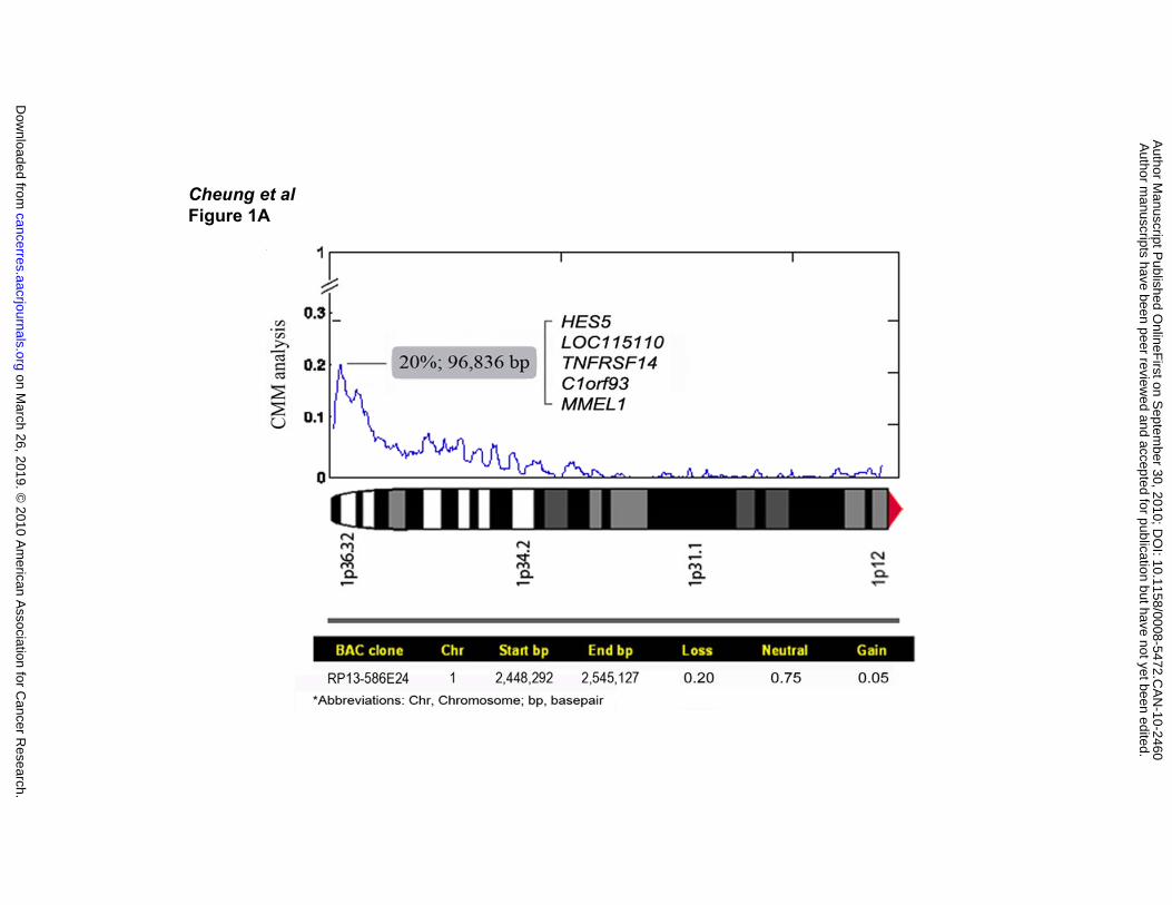
Herein we report a frequency of 20% in 141 FL cases using an algorithm called CMM
joint analysis that identified a minimum region of deletion (MRD) corresponding to a
single BAC clone with the highest frequency shift (Figure 1A). This area is
approximately 97 kb in size and contains five genes including HES5, LOC115110,
TNFRSF14, C1orf93 and MMEL1. Information on the BAC clone within the MRD and
the frequency of loss, gain and copy-neutral state is shown in the lower panel of Figure
1A.
On the right of Figure 1B, four representative array CGH cases are shown with
heterozygous (-/+) and homozygous deletions (-/-), and the corresponding FISH profiles
on March 26, 2019. © 2010 American Association for Cancer Research.cancerres.aacrjournals.org Downloaded from
Author manuscripts have been peer reviewed and accepted for publication but have not yet been edited. Author Manuscript Published OnlineFirst on September 30, 2010; DOI: 10.1158/0008-5472.CAN-10-2460

Author manuscripts have been peer reviewed and accepted for publication but have not yet been edited.
Copyright © 2010 American Association for Cancer Research
10
(1p36: red arrowhead; 1q32 control: green arrowhead). All deletions detected by array
CGH were validated by FISH analysis using one or more of the BAC probes RP13-
493G06, RP13-586E24 and RP11-756P03 with probe RP11-229M05 mapping at
1q32.3 as a copy number control. The majority of the deletions were shown to be
heterozygous, while two were homozygous. High resolution oligonucleotide array
analysis was performed on case #1, which had the smallest array CGH-detected 1p36
deletion, to fine-map the breakpoints (left panel of Figure 1B). Due to numerous
sequence repeats in this area a portion of the deleted region was not covered by the
probes, however, the deletion spanned 224,674 bp containing the five genes identified
in the MRD by array CGH analysis (red/black/blue dots are data points representing
gain/neutral/loss status).
The MRD region was also examined with Affymetrix mapping 500k SNP arrays to detect
possible cnLOH using 27 selected cases with available matched germline DNA (from
the 141 cases used to determine the MRD). Six of the 27 cases (22.2%) showed cnLOH
affecting chromosome arm 1p with an overlapping region of cnLOH of approximately
15Mb that included the 97 kb-MRD identified by array CGH (data not shown).
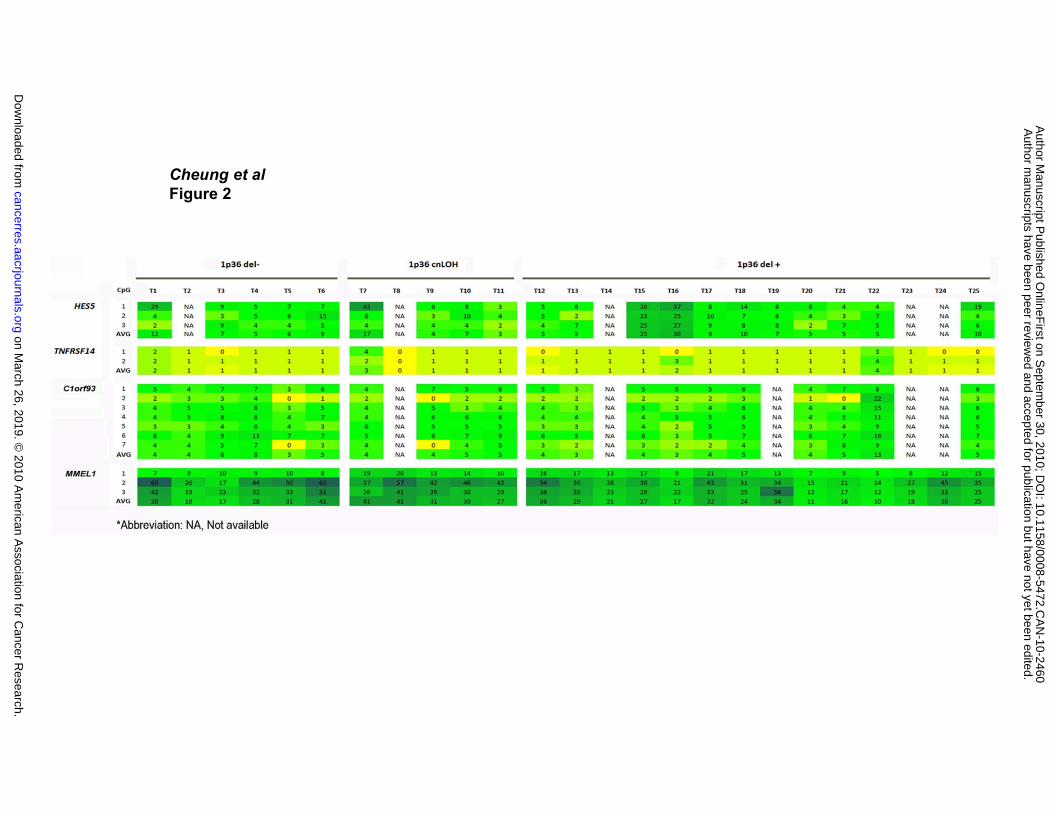
Methylation Status of Genes in the MRD
The methylation pattern of four of the five genes within the MRD was investigated using
the pyrosequencing technique (Figure 2). Technical failures prevented the analysis of
LOC115110, due likely to abundant sequence repeats in the promotor region (see
Figure 2). A number of samples were not investigated due to the lack of available tumor
on March 26, 2019. © 2010 American Association for Cancer Research.cancerres.aacrjournals.org Downloaded from
Author manuscripts have been peer reviewed and accepted for publication but have not yet been edited. Author Manuscript Published OnlineFirst on September 30, 2010; DOI: 10.1158/0008-5472.CAN-10-2460
Author manuscripts have been peer reviewed and accepted for publication but have not yet been edited.
Copyright © 2010 American Association for Cancer Research
11
DNA. The number of CpG dinucleotides analyzed at each gene promoter is indicated
beside each gene in Figure 2. The percent methylation of each CpG is presented both
numerically and by color coding (with yellow being the least methylated, green being
intermediate and blue being highly methylated). None of the genes examined showed a
differential methylation pattern among 1) six cases without array CGH-detected 1p36
deletion (1p36 del -), 2) five cases with cnLOH (1p36 cnLOH), and 3) 14 cases with
1p36 deletion (1p36 del +) (p> 0.05, one-way ANOVA).
Identification of Somatic Mutations in TNFRSF14
Exon sequencing of the five genes identified within the MRD was performed on eleven
cases: five cases with and six cases without deletion of 1p36 as determined by array
CGH analysis. We searched for somatic mutations in the sequence data from the
lymphoma cells by comparison with constitutional DNA obtained from peripheral blood
lymphocytes from the same patient. The exons and exon/intron boundaries for HES5
(exons 1 to 3), LOC115110 (exons 1 and 2), TNFRSF14 (exons 1 to 8), C1orf93 (exons
1 to 7) and MMEL1 (exons 18 to 23) were studied. Exons 1 to 17 and 24 of MMEL1
could not be covered by PCR amplicons due to high content of GC repeats, therefore,
the existence of mutations in these exons cannot be excluded. Of the five genes, only
TNFRSF14 displayed somatic mutations. Four somatic mutations were found in the
tumor DNA from three of the five cases with 1p36 deletion. One case had an A > G
transition in exon 1 resulting in an amino acid change from methionine to valine, plus a
synonymous change from G > A also in exon 1. Another case had a G > T transversion
in exon 2 resulting in truncation. The last case had a G > A transition in the first base of
on March 26, 2019. © 2010 American Association for Cancer Research.cancerres.aacrjournals.org Downloaded from
Author manuscripts have been peer reviewed and accepted for publication but have not yet been edited. Author Manuscript Published OnlineFirst on September 30, 2010; DOI: 10.1158/0008-5472.CAN-10-2460

Author manuscripts have been peer reviewed and accepted for publication but have not yet been edited.
Copyright © 2010 American Association for Cancer Research
12
intron 4. There were no somatic mutations detected in the six cases without a 1p36
deletion.
Validation of TNFRSF14 Mutation Frequency and Correlation with Inferior
Prognosis
To determine the frequency of TNFRSF14 mutation and to undertake clinical correlative
analysis, we selected 115 cases with sufficient DNA from our 141 discovery cohort
(used in determining the MRD and for which 1p36 deletion status was known) plus 136
additional FL specimens from our archive in which 1p36 deletion status was not known,
totaling 251 primary FL cases. The clinical features and mutation findings for these
cases are summarized in Tables 1 and 2, respectively. Briefly, in 46 cases (18.3%) we
identified a total of 47 previously undescribed nonsynonymous mutations in TNFRSF14
after screening to exclude known polymorphic variants using several databases
including NCBI dbSNP build 130 (http://www.ncbi.nlm.nih.gov/projects/SNP/), J. Craig
Venter HuRef Genome Browser (http://huref.jcvi.org/), James Watson Genotype viewer
(http://jimwatsonsequence.cshl.edu/cgi-perl/gbrowse/jwsequence/) and the 1000
genomes project (http://www.1000genomes.org/page.php). The 47 nonsynonymous
mutations included 22 transitions, 13 transversions, 8 deletions and 4 insertions
resulting in 16 truncations and 10 frameshifts (Table 2). All exons except 7 and 8 were
affected by mutations. One case showed two mutations in
TNFRSF14.
On examination of the relationship between 1p36 deletion and TNFRSF14 mutation, we
on March 26, 2019. © 2010 American Association for Cancer Research.cancerres.aacrjournals.org Downloaded from
Author manuscripts have been peer reviewed and accepted for publication but have not yet been edited. Author Manuscript Published OnlineFirst on September 30, 2010; DOI: 10.1158/0008-5472.CAN-10-2460

Author manuscripts have been peer reviewed and accepted for publication but have not yet been edited.
Copyright © 2010 American Association for Cancer Research
13
found in the 114 cases where both deletion status and mutation status was known, eight
cases (7%) were positive for both 1p36 deletion and TNFRSF14 mutation, 10 cases
(8.8%) were TNFRSF14 mutation positive and 1p36 deletion negative, and 23 cases
(20.2%) were 1p36 deletion positive and TNFRSF14 mutation negative. The clinical
impact of these findings is presented below. Furthermore, we found that among the 35
cases with paired specimens taken at diagnosis and later at the time of development of
transformed DLBCL, a TNFRSF14 mutation was detected in both samples from five
paired specimens and in two of the transformed samples where the corresponding prior
FL samples had no mutation.
Analysis of clinical correlation with TNFRSF14 mutations in an expanded cohort of 251
cases revealed that TNFRSF14 mutations were associated with high-risk clinical
features (see Table 1). Specifically, patients with mutated TNFRSF14 were more likely
to have a poor performance status and their FL involved a greater number of extranodal
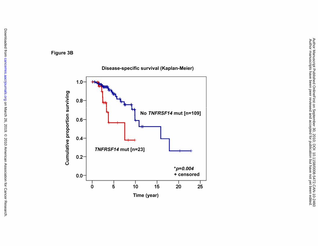
sites. When patients, whose overall treatment included rituximab, were analyzed,
inferior OS and disease-specific survival (DSS) were observed in those with a mutation
in TNFRSF14 (median 3.73 years and 7.52 years, respectively) compared to those
without (median 15.85 years and 15.85 years) (log-rank test, p<0.001 and p=0.004,
respectively) (Figure 3A and B). On multivariate analysis using Cox regression,
TNFRSF14 mutation emerged as a stronger predictor of inferior OS and DSS after
adjustment for the IPI in rituximab-treated patients (hazard ratio [HR]=4.54, 95%
confidence interval [CI] 1.80-11.44, p=0.001 and HR=3.97, 95% CI 1.50-10.51, p=0.006,
respectively). Where information was available, all patients were classified into groups
on March 26, 2019. © 2010 American Association for Cancer Research.cancerres.aacrjournals.org Downloaded from
Author manuscripts have been peer reviewed and accepted for publication but have not yet been edited. Author Manuscript Published OnlineFirst on September 30, 2010; DOI: 10.1158/0008-5472.CAN-10-2460
Author manuscripts have been peer reviewed and accepted for publication but have not yet been edited.
Copyright © 2010 American Association for Cancer Research
14
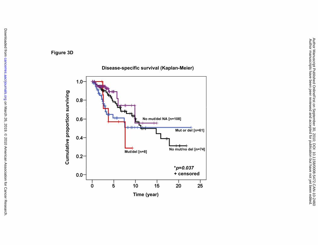
based on the presence or absence of 1p36 deletion and/or TNFRSF14 mutation.
Patients with one alteration (“mut or del”; median 7.52 years) or both alterations
(“mut/del”; median 6.09 years) displayed a significantly inferior OS compared to those
without either alteration (“no mut/no del”; median 11.23 years) or cases without any
mutation but in whom the 1p36 deletion status was unknown (“no mut/del NA”; median
11.69 years) (log-rank test, p=0.005) (see Figure 3C). On multivariate analysis, cases
with both aberrations (1p36 deletion and TNFRSF14 mutation) remained a significant
predictor of OS independent of the IPI compared to the group of no aberrations
(HR=3.65, 95% CI 1.35-9.878, p=0.011). In the analysis of DSS, patients with both
alterations (“mut/del”; median 7.61 years) displayed a significantly inferior DSS
compared to those without either alteration (“no mut/no del”; median 11.23 years), those
with one alteration (“mut or del”; median not reached), or cases without any mutation
but in whom the 1p36 deletion status was unknown (“no mut/del NA”; median not
reached) (log-rank test, p=0.037) (see Figure 3D). Cases with both aberrations (1p36
deletion and TNFRSF14 mutation) remained a significant predictor independent of the
IPI compared to the group of no aberrations (HR=3.19, 95% CI 1.06-9.57, p=0.039).
Discussion
Based on microarray-based copy number and cnLOH data analyses, we have identified
a MRD at chromosome band 1p36 in FL that measures approximately 97kb. This
region contains five genes; HES5, LOC115110, TNFRSF14, C1orf93 and MMEL1.
Direct DNA sequencing of the exons of these genes in the 11 cases with matching
on March 26, 2019. © 2010 American Association for Cancer Research.cancerres.aacrjournals.org Downloaded from
Author manuscripts have been peer reviewed and accepted for publication but have not yet been edited. Author Manuscript Published OnlineFirst on September 30, 2010; DOI: 10.1158/0008-5472.CAN-10-2460

Author manuscripts have been peer reviewed and accepted for publication but have not yet been edited.
Copyright © 2010 American Association for Cancer Research
15
tumor and constitutional DNA revealed that only TNFRSF14 was affected by acquired
somatic mutations in the clonal cells. In the cohort of 251 cases where matching
constitutional DNA was not examined, 47 nonsynonymous mutations were detected in
46 cases (18.3%) (after elimination of known polymorphic variants). Although the
mutations detected in the 251 case study were not definitively confirmed to be somatic
by parallel constitutional DNA sequencing, the lack of similar mutations in large public
genomic databases and the significant correlation of these observed mutations with
inferior clinical outcomes provide strong evidence that these mutations are somatic in
nature and that TNFRSF14 is the target gene in the 1p36 MRD.
In the clinical correlative analysis of the 251 patient cohort, we report three interesting
observations. First, patients with TNFRSF14 mutations were more likely to have high-
risk clinical features such as poor performance status and involvement of multiple
extranodal sites. Second, TNFRSF14 mutation alone or combined with 1p36 deletion
correlated with inferior prognosis. Third, the adverse impact of TNFRSF14 mutation on
FL patients was independent of rituximab treatment. The implications of these findings
are of clinical significance. Essentially, there appears to be a dose effect where
homozygosity for a somatic TNFRSF14 gene defect (deletion plus mutation) was
correlated with the poorest clinical outcomes. In addition, treatment with rituximab
conferred less than expected benefit in patients with a mutation in TNFRSF14.
Furthermore, our observation that TNFRSF14 mutations were detected in both the
paired diagnostic and transformed specimens or only in transformed specimens
suggests that TNFRSF14 mutation may play a role in transformation of FL to DLBCL.
on March 26, 2019. © 2010 American Association for Cancer Research.cancerres.aacrjournals.org Downloaded from
Author manuscripts have been peer reviewed and accepted for publication but have not yet been edited. Author Manuscript Published OnlineFirst on September 30, 2010; DOI: 10.1158/0008-5472.CAN-10-2460

Author manuscripts have been peer reviewed and accepted for publication but have not yet been edited.
Copyright © 2010 American Association for Cancer Research
16
The TNFRSF14 gene is located on the chromosome 1 minus DNA strand from base
pair 2,479,150 to 2,486,757 and is composed of eight exons, which together produce an
RNA transcript 1,731 bp in size and a protein product consisting of 283 amino acids.
The significance of such acquired mutations on the structure-function relationship of
TNFRSF14 is unclear, however, a previous study has found that alterations of amino
acid residues at positions 14, 23 and 26 (corresponding to exons 1, 1 and 2,
respectively) significantly reduce its binding affinity to BTLA, one of the protein ligands
for TNFRSF14 in negatively modulating T-cell immune responses (20). Of interest in
light of this report, we found one case with a frameshift mutation corresponding to
amino acid position 14, another frameshift mutation corresponding to position 23, and a
nonsynonymous mutation corresponding to position 26. Among the mutations detected
in TNFRSF14, we found an exon-intron junction alteration. As TNFRSF14 can produce
15 different splice variants, alterations at splice sites will likely disrupt their recognition
by the transcription machinery, leading to introns not being removed from the pre-
mRNA and/or frameshifts (21, 22). In vitro investigation will be needed to further
examine the impact of mutations on cellular function in different parts of the gene.
Furthermore, CpG methylation analysis of the five genes in the MRD showed that
acquired methylation was unlikely to be a mechanism involved in transcriptional
silencing of TNFRSF14.
In conclusion, we have identified a minimum region of deletion at 1p36 that
encompasses five genes, of which only TNFRSF14 (also called HVEA, HVEM, ATAR,
on March 26, 2019. © 2010 American Association for Cancer Research.cancerres.aacrjournals.org Downloaded from
Author manuscripts have been peer reviewed and accepted for publication but have not yet been edited. Author Manuscript Published OnlineFirst on September 30, 2010; DOI: 10.1158/0008-5472.CAN-10-2460
Author manuscripts have been peer reviewed and accepted for publication but have not yet been edited.
Copyright © 2010 American Association for Cancer Research
17
TR2 and LIGHTR) is associated with FL based on the presence of somatic mutations
and the observation that FL patients with TNFRSF14 mutation alone or combined with a
1p36 deletion have an inferior prognosis. To the best of our knowledge, no other
studies have been able to provide strong evidence for a candidate gene in the 1p36
deleted region in FL. Although the physiologic role of TNFRSF14 in humans is still
being deciphered, recent evidence indicates that in addition to being a member of the
TNF receptor superfamily, normal TNFRSF14 is capable of inhibiting the proliferation of
adenocarcinoma cells and enhancing Fas-induced apoptosis in non-Hodgkin’s
lymphoma cells, suggesting a tumor suppressor role (23, 24). Functional analysis of
this gene, specifically the impact of mutations on the function of TNFRSF14, is the
subject of ongoing investigations. The identification of TNFRSF14 as a candidate gene
in the second most frequently altered region in the FL genome will not only open the
possibility of it being developed as a prognostic marker for identifying high-risk patients
as candidates for risk-adapted therapies, TNFRSF14 may provide pharmacological
clues as to why rituximab treatment in some cases of FL may have a less than expected
beneficial effect on survival.
on March 26, 2019. © 2010 American Association for Cancer Research.cancerres.aacrjournals.org Downloaded from
Author manuscripts have been peer reviewed and accepted for publication but have not yet been edited. Author Manuscript Published OnlineFirst on September 30, 2010; DOI: 10.1158/0008-5472.CAN-10-2460

Author manuscripts have been peer reviewed and accepted for publication but have not yet been edited.
Copyright © 2010 American Association for Cancer Research
18
Acknowledgements
We thank Dr. Stephane Le Bihan, Anne Haegert and Robert Bell from the Vancouver
Prostate Center Microarray Facility for their Agilent oligo array services; A.M. Devlin for
the use of pyrosequencing instruments; Jane Donaldson and Suman Singh for
maintenance of the BCCA Lymphoid Cancer Database.
on March 26, 2019. © 2010 American Association for Cancer Research.cancerres.aacrjournals.org Downloaded from
Author manuscripts have been peer reviewed and accepted for publication but have not yet been edited. Author Manuscript Published OnlineFirst on September 30, 2010; DOI: 10.1158/0008-5472.CAN-10-2460

Author manuscripts have been peer reviewed and accepted for publication but have not yet been edited.
Copyright © 2010 American Association for Cancer Research
19
References
1. Yunis JJ, Frizzera G, Oken MM, McKenna J, Theologides A, Arnesen M. Multiple
recurrent genomic defects in follicular lymphoma. A possible model for cancer. N Engl J
Med 1987; 316(2): 79-84.
2. Tilly H, Rossi A, Stamatoullas A, Lenormand B, Bigorgne C, Kunlin A, et al.
Prognostic value of chromosomal abnormalities in follicular lymphoma. Blood 1994;
84(4): 1043-9.
3. Tsujimoto Y, Finger LR, Yunis J, Nowell PC, Croce CM. Cloning of the
chromosome breakpoint of neoplastic B cells with the t(14;18) chromosome
translocation. Science 1984; 226(4678): 1097-9.
4. Graninger WB, Seto M, Boutain B, Goldman P, Korsmeyer SJ. Expression of Bcl-2
and Bcl-2-Ig fusion transcripts in normal and neoplastic cells. J Clin Invest 1987; 80(5):
1512-5.
5. Ross CW, Ouillette PD, Saddler CM, Shedden KA, Malek SN. Comprehensive
analysis of copy number and allele status identifies multiple chromosome defects
underlying follicular lymphoma pathogenesis. Clin Cancer Res 2007; 13(16): 4777-85.
6. Cheung KJ, Shah SP, Steidl C, Johnson N, Relander T, Telenius A, et al.
Genome-wide profiling of follicular lymphoma by array comparative genomic
hybridization reveals prognostically significant DNA copy number imbalances. Blood
2009; 113(1): 137-48.
on March 26, 2019. © 2010 American Association for Cancer Research.cancerres.aacrjournals.org Downloaded from
Author manuscripts have been peer reviewed and accepted for publication but have not yet been edited. Author Manuscript Published OnlineFirst on September 30, 2010; DOI: 10.1158/0008-5472.CAN-10-2460

Author manuscripts have been peer reviewed and accepted for publication but have not yet been edited.
Copyright © 2010 American Association for Cancer Research
20
7. Lestou VS, Gascoyne RD, Sehn L, Ludkovski O, Chhanabhai M, Klasa RJ, et al.
Multicolour fluorescence in situ hybridization analysis of t(14;18)-positive follicular
lymphoma and correlation with gene expression data and clinical outcome. Br J
Haematol 2003; 122(5): 745-59.
8. Horsman DE, Connors JM, Pantzar T, Gascoyne RD. Analysis of secondary
chromosomal alterations in 165 cases of follicular lymphoma with t(14;18). Genes
Chromosomes Cancer 2001; 30(4): 375-82.
9. Lestou VS, Ludkovski O, Connors JM, Gascoyne RD, Lam WL, Horsman DE.
Characterization of the recurrent translocation t(1;1)(p36.3;q21.1-2) in non-Hodgkin
lymphoma by multicolor banding and fluorescence in situ hybridization analysis. Genes
Chromosomes Cancer 2003; 36(4): 375-81.
10. Rajgopal A, Carr IM, Leek JP, Hodge D, Bell SM, Roberts P, et al. Detection by
fluorescence in situ hybridization of microdeletions at 1p36 in lymphomas, unidentified
on cytogenetic analysis. Cancer Genet Cytogenet 2003; 142(1): 46-50.
11. O'Shea D, O'Riain C, Gupta M, Waters R, Yang Y, Wrench D, et al. Regions of
acquired uniparental disomy at diagnosis of follicular lymphoma are associated with
both overall survival and risk of transformation. Blood 2009; 113(10): 2298-301.
12. Garnis C, Coe BP, Lam SL, MacAulay C, Lam WL. High-resolution array CGH
increases heterogeneity tolerance in the analysis of clinical samples. Genomics 2005;
85(6): 790-3.
13. Harris NL, Jaffe ES, Diebold J, Flandrin G, Muller-Hermelink HK, Vardiman J, et
on March 26, 2019. © 2010 American Association for Cancer Research.cancerres.aacrjournals.org Downloaded from
Author manuscripts have been peer reviewed and accepted for publication but have not yet been edited. Author Manuscript Published OnlineFirst on September 30, 2010; DOI: 10.1158/0008-5472.CAN-10-2460

Author manuscripts have been peer reviewed and accepted for publication but have not yet been edited.
Copyright © 2010 American Association for Cancer Research
21
al. The World Health Organization classification of neoplasms of the hematopoietic and
lymphoid tissues: report of the Clinical Advisory Committee meeting--Airlie House,
Virginia, November, 1997. Hematol J 2000; 1(1): 53-66.
14. Henderson LJ, Okamoto I, Lestou VS, Ludkovski O, Robichaud M, Chhanabhai
M, et al. Delineation of a minimal region of deletion at 6q16.3 in follicular lymphoma and
construction of a bacterial artificial chromosome contig spanning a 6-megabase region
of 6q16-q21. Genes Chromosomes Cancer 2004; 40(1): 60-5.
15. Ishkanian AS, Malloff CA, Watson SK, DeLeeuw RJ, Chi B, Coe BP, et al. A tiling
resolution DNA microarray with complete coverage of the human genome. Nat Genet
2004; 36(3): 299-303.
16. Davies JJ, Wilson IM, Lam WL. Array CGH technologies and their applications to
cancer genomes. Chromosome Res 2005; 13(3): 237-48.
17. de Leeuw RJ, Davies JJ, Rosenwald A, Bebb G, Gascoyne RD, Dyer MJ, et al.
Comprehensive whole genome array CGH profiling of mantle cell lymphoma model
genomes. Hum Mol Genet 2004; 13(17): 1827-37.
18. Colella S, Yau C, Taylor JM, Mirza G, Butler H, Clouston P, et al. QuantiSNP: an
Objective Bayes Hidden-Markov Model to detect and accurately map copy number
variation using SNP genotyping data. Nucleic Acids Res 2007; 35(6): 2013-25.
19. Tost J, Gut IG. DNA methylation analysis by pyrosequencing. Nat Protoc 2007;
2(9): 2265-75.
on March 26, 2019. © 2010 American Association for Cancer Research.cancerres.aacrjournals.org Downloaded from
Author manuscripts have been peer reviewed and accepted for publication but have not yet been edited. Author Manuscript Published OnlineFirst on September 30, 2010; DOI: 10.1158/0008-5472.CAN-10-2460

Author manuscripts have been peer reviewed and accepted for publication but have not yet been edited.
Copyright © 2010 American Association for Cancer Research
22
20. Compaan DM, Gonzalez LC, Tom I, Loyet KM, Eaton D, Hymowitz SG.
Attenuating lymphocyte activity: the crystal structure of the BTLA-HVEM complex. J Biol
Chem 2005; 280(47): 39553-61.
21. Mount SM. A catalogue of splice junction sequences. Nucleic Acids Res 1982;
10(2): 459-72.
22. Kahl U. 50 years after the discovery of the DNA structure its implications still
have not reached clinical practice. Our genetic material is "a big tangle of nucleic acids
and proteins". Lakartidningen 2003; 100(25): 2218-21.
23. Harrop JA, McDonnell PC, Brigham-Burke M, Lyn SD, Minton J, Tan KB, et al.
Herpesvirus entry mediator ligand (HVEM-L), a novel ligand for HVEM/TR2, stimulates
proliferation of T cells and inhibits HT29 cell growth. J Biol Chem 1998; 273(42): 27548-
56.
24. Costello RT, Mallet F, Barbarat B, Schiano De Colella JM, Sainty D, Sweet RW,
et al. Stimulation of non-Hodgkin's lymphoma via HVEM: an alternate and safe way to
increase Fas-induced apoptosis and improve tumor immunogenicity. Leukemia 2003;
17(12): 2500-7.
on March 26, 2019. © 2010 American Association for Cancer Research.cancerres.aacrjournals.org Downloaded from
Author manuscripts have been peer reviewed and accepted for publication but have not yet been edited. Author Manuscript Published OnlineFirst on September 30, 2010; DOI: 10.1158/0008-5472.CAN-10-2460
Author manuscripts have been peer reviewed and accepted for publication but have not yet been edited.
Copyright © 2010 American Association for Cancer Research
23
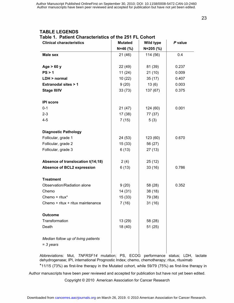
TABLE LEGENDS Table 1. Patient Characteristics of the 251 FL Cohort
Clinical characteristics Mutated
N=46 (%)
Wild type
N=205 (%)
P value
Male sex
Age > 60 y
PS > 1
LDH > normal
Extranodal sites > 1
Stage III/IV
IPI score
0-1
2-3
4-5
Diagnostic Pathology
Follicular, grade 1
Follicular, grade 2
Follicular, grade 3
Absence of translocation t(14;18)
Absence of BCL2 expression
Treatment
Observation/Radiation alone
Chemo
Chemo + ritux*
Chemo + ritux + ritux maintenance
Outcome
Transformation
Death
Median follow up of living patients
= 3 years
21 (46)
22 (49)
11 (24)
10 (22)
9 (20)
33 (73)
21 (47)
17 (38)
7 (15)
24 (53)
15 (33)
6 (13)
2 (4)
6 (13)
9 (20)
14 (31)
15 (33)
7 (16)
13 (29)
18 (40)
114 (56)
81 (39)
21 (10)
35 (17)
13 (6)
137 (67)
124 (60)
77 (37)
5 (3)
123 (60)
56 (27)
27 (13)
25 (12)
33 (16)
58 (28)
38 (18)
79 (38)
31 (16)
58 (28)
51 (25)
0.4
0.237
0.009
0.407
0.003
0.375
0.001
0.670
0.786
0.352
Abbreviations: Mut, TNFRSF14 mutation; PS, ECOG performance status; LDH, lactate dehydrogenase; IPI, international Prognostic Index; chemo, chemotherapy; ritux, rituximab
*11/15 (73%) as first-line therapy in the Mutated cohort, while 59/79 (75%) as first-line therapy in
on March 26, 2019. © 2010 American Association for Cancer Research.cancerres.aacrjournals.org Downloaded from
Author manuscripts have been peer reviewed and accepted for publication but have not yet been edited. Author Manuscript Published OnlineFirst on September 30, 2010; DOI: 10.1158/0008-5472.CAN-10-2460

Author manuscripts have been peer reviewed and accepted for publication but have not yet been edited.
Copyright © 2010 American Association for Cancer Research
24
Chr1 position Exon Transcript positon: alteration Amino acid position: alteration
2486246 1 368:het_delG 23:Deletion/Frameshift
2486260 1 354:G>C 19:D>H:Transversion
2486272 1 342 to 346:het_delACCCC 15:Deletion/Frameshift
2486274 1 340 to 344:het_delCCACC 14:Deletion/Frameshift
2486279 1 335:G>A 12:W>X:Transition/Truncation
2486280 1 334:G>A 12:W>X:Transition/Truncation
2486295 1 319:G>A 7:W>X:Transition/Truncation
2486312 1 302:G>A 1:M>I:Transition
2486312 1 302:G>A 1:M>I:Transition
2486314 1 300:A>T 1:M>L:Transversion
2485148 2 1466:C>T 59:P>S:Transition
2485152 2 1462:C>A 57:C>X:Transversion/Truncation
2485182 2 1432:C>A 47:Y>X:Transversion/Truncation
2485197 2 1417:C>G 42:C>W:Transversion
2485197 2 1417:C>G 42:C>W:Transversion
2485224 2 1390 to 1391:het_insC 34:Insertion/Frameshift
2485230 2 1384 to 1385:het_insC 32:Insertion/Frameshift
2485245 2 1369:T>A 26:Y>X:Transversion/Truncation
2485247 2 1367:T>A 26:Y>N:Transversion
2485249 2 1365:T>G 25:L>R:Transversion
2484526 3 2088:C>T 97:Q>X:Transition/Truncation
2484571 3 2043:A>C 82:T>P:Transversion
2484583 3 2031:T>C 78:C>R:Transition
2484590 3 2024 to 2025:delTG 75:Deletion/Frameshift
2484600 3 2014:G>A 72:G>D:Transition
2484622 3 1992:G>T 65:E>X:Transversion/Truncation
2484633 3 1981:A>G 61:Y>C:Transition
2484636 3 1978:G>A 60:G>D:Transition
2483010 4 3604:C>T 151:Q>X:Transition/Truncation
2483012 4 3602 to 3611:het_delTGCAGAAGGG 150:Deletion/Frameshif t
2483019 4 3595:C>T 148:Q>X:Transition/Truncation
2483050 4 3564 to 3567:het_delGTGC 137:Deletion/Frameshif t
2483056 4 3558 to 3567:het_delCGCCGCGTGC 136:Deletion/Frameshif t
2483086 4 3528 to 3529:het_insGCCGTGTGTGGCTGCAGCCCAGGCCAC 126:Insertion
2483099 4 3515:G>A 121:C>Y:Transition
2483116 4 3498 to 3499:het_insACACCGTGTGTGGCTGCAGCCCAGGCC 116:Insertion
2483136 4 3478:C>T 109:R>W:Transition
2482330 5 4284:T>A 162:C>X:Transversion/Truncation
2482344 5 4270:C>T 158:Q>X:Transition/Truncation
2481164 6 5450:G>A 232:G>S:Transition
2481214 6 5400:T>A 215:V>D:Transversion
2481246 6 5368:G>A 204:W>X:Transition/Truncation
2481249 6 5365:G>A 203:W>X:Transition/Truncation
2481256 6 5358:G>A 201:W>X:Transition/Truncation
2481282 6 5332 to 5341:het_delCGGAGCTGGG 193:Deletion/Frameshif t
2481297 6 5317:G>A 187:W>X:Transition/Truncation
2481297 6 5317:G>A 187:W>X:Transition/Truncation
Abbreviations: chr1, chromosome 1; >, mutated to; het_del, heterozygous deletion; het_ins, heterozygous insertion; X, stop codon
the Wild type cohort Table 2. Information on the 47 Nonsynonymous Mutations in the TNFRSF14 Gene
on March 26, 2019. © 2010 American Association for Cancer Research.cancerres.aacrjournals.org Downloaded from
Author manuscripts have been peer reviewed and accepted for publication but have not yet been edited. Author Manuscript Published OnlineFirst on September 30, 2010; DOI: 10.1158/0008-5472.CAN-10-2460
Author manuscripts have been peer reviewed and accepted for publication but have not yet been edited.
Copyright © 2010 American Association for Cancer Research
25
Figure legends
Figure 1. 1p36 MRD identification. (A) Array CGH composite deletion frequency
profile of chromosome arm 1p in 141 FL patients. Analysis using the CMM algorithm
produced a graph showing an MRD in band 1p36.32 with the highest frequency of loss
corresponding to BAC clone RP13-586E24 which contains the genes HES5,
LOC115110, TNSFRSF14, C1orf93 and MMEL1. The Y-axis denotes probability of
aberrations, while X-axis represents the chromosome band coordinates. Associated
information for BAC clone RP13-586E24 is presented below the graph. (B) On the
right, four representative array CGH cases are shown with heterozygous (-/+) and
homozygous deletions (-/-), and the corresponding FISH profiles (1p36: red arrowhead;
1q32 control: green arrowhead). On the left, the custom oligo array analysis of case #1
showing left shift from the center line indicating deletion of ~224 kb (red/black/blue dots
are data points representing gain/neutral/loss status).
Figure 2. Methylation analysis. Six specimens (T1-T6) without 1p36 deletions (1p36
del -), five specimens (T7-T11) with 1p36 cnLOH, and 14 specimens (T12-T25) with
1p36 deletions (1p35 del +) were examined. The number of CpG’s analyzed is
indicated beside each gene. The percent methylation is presented numerically and by
color coding (yellow: least; green: intermediate; blue: highly).
Figure 3. Clinical correlation analysis. (A) Overall survival (OS) according to the
presence or absence of TNFRSF14 mutation in rituximab-treated patients (median 3.73
on March 26, 2019. © 2010 American Association for Cancer Research.cancerres.aacrjournals.org Downloaded from
Author manuscripts have been peer reviewed and accepted for publication but have not yet been edited. Author Manuscript Published OnlineFirst on September 30, 2010; DOI: 10.1158/0008-5472.CAN-10-2460

Author manuscripts have been peer reviewed and accepted for publication but have not yet been edited.
Copyright © 2010 American Association for Cancer Research
26
and 15.85 years, respectively; log-rank test, p<0.001). (B) Disease-specific survival
(DSS) according to the presence or absence of TNFRSF14 mutation in rituximab-
treated patients (median 7.52 and 15.85 years, respectively; log-rank test, p=0.004).
(C) OS according to four categories: 1) the absence of TNFRSF14 mutation and 1p36
deletion (“no mut/no del”; median 11.32 years), 2) the absence of TNFRSF14 mutation
and information of 1p36 deletion not available (“no mut/del NA”; median 11.69 years), 3)
the presence of TNFRSF14 mutation or 1p36 deletion (“mut or del”; median 7.52 years),
and 4) the presence of both TNFRSF14 mutation and 1p36 deletion (“mut/del”; 6.09
years) (log-rank test, p=0.005). (D) DSS according to four categories: 1) the absence of
TNFRSF14 mutation and 1p36 deletion (“no mut/no del”; median 11.32 years), 2) the
absence of TNFRSF14 mutation and information of 1p36 deletion not available (“no
mut/del NA”; median not reached), 3) the presence of TNFRSF14 mutation or 1p36
deletion (“mut or del”; median not reached), and 4) the presence of both TNFRSF14
mutation and 1p36 deletion (“mut/del”; 7.61 years) (log-rank test, p=0.037).
on March 26, 2019. © 2010 American Association for Cancer Research.cancerres.aacrjournals.org Downloaded from
Author manuscripts have been peer reviewed and accepted for publication but have not yet been edited. Author Manuscript Published OnlineFirst on September 30, 2010; DOI: 10.1158/0008-5472.CAN-10-2460
Cheung et alFigure 1A
on March 26, 2019. ©
2010 Am
erican Association for C
ancer Research.
cancerres.aacrjournals.org D
ownloaded from
Author m
anuscripts have been peer reviewed and accepted for publication but have not yet been edited.
Author M
anuscript Published O
nlineFirst on S
eptember 30, 2010; D
OI: 10.1158/0008-5472.C
AN
-10-2460
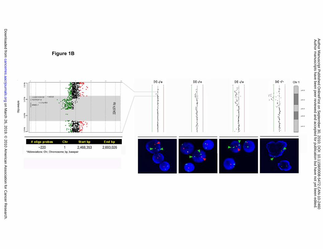
Figure 1B
on March 26, 2019. ©
2010 Am
erican Association for C
ancer Research.
cancerres.aacrjournals.org D
ownloaded from
Author m
anuscripts have been peer reviewed and accepted for publication but have not yet been edited.
Author M
anuscript Published O
nlineFirst on S
eptember 30, 2010; D
OI: 10.1158/0008-5472.C
AN
-10-2460
Cheung et alFigure 2
on March 26, 2019. ©
2010 Am
erican Association for C
ancer Research.
cancerres.aacrjournals.org D
ownloaded from
Author m
anuscripts have been peer reviewed and accepted for publication but have not yet been edited.
Author M
anuscript Published O
nlineFirst on S
eptember 30, 2010; D
OI: 10.1158/0008-5472.C
AN
-10-2460

Overall survival (Kaplan-Meier)
Cheung et alFigure 3A
urvi
ving
No TNFRSF14 mut [n=109]
prop
ortio
n su
TNFRSF14 mut [n=23]
*p<0.001
Cum
ulat
ive
p<0.001+ censored
Time (year)
on March 26, 2019. ©
2010 Am
erican Association for C
ancer Research.
cancerres.aacrjournals.org D
ownloaded from
Author m
anuscripts have been peer reviewed and accepted for publication but have not yet been edited.
Author M
anuscript Published O
nlineFirst on S
eptember 30, 2010; D
OI: 10.1158/0008-5472.C
AN
-10-2460
Disease-specific survival (Kaplan-Meier)
Figure 3B
urvi
ving
No TNFRSF14 mut [n=109]
prop
ortio
n su
TNFRSF14 mut [n=23]
*p=0.004
Cum
ulat
ive
p 0.004+ censored
Time (year)
on March 26, 2019. ©
2010 Am
erican Association for C
ancer Research.
cancerres.aacrjournals.org D
ownloaded from
Author m
anuscripts have been peer reviewed and accepted for publication but have not yet been edited.
Author M
anuscript Published O
nlineFirst on S
eptember 30, 2010; D
OI: 10.1158/0008-5472.C
AN
-10-2460

Overall survival (Kaplan-Meier)
Figure 3C
urvi
ving
prop
ortio
n su
Mut or del [n=61]
No mut/del NA [n=108]
*p=0.005
Cum
ulat
ive
No mut/no del [n=74]
Mut/del [n=8]
p 0.005+ censored
Time (year)
on March 26, 2019. ©
2010 Am
erican Association for C
ancer Research.
cancerres.aacrjournals.org D
ownloaded from
Author m
anuscripts have been peer reviewed and accepted for publication but have not yet been edited.
Author M
anuscript Published O
nlineFirst on S
eptember 30, 2010; D
OI: 10.1158/0008-5472.C
AN
-10-2460
Disease-specific survival (Kaplan-Meier)
Figure 3D
urvi
ving
No mut/del NA [n=108]
prop
ortio
n su
Mut or del [n=61]
*p=0.037
Cum
ulat
ive
No mut/no del [n=74]Mut/del [n=8]
p 0.037+ censored
Time (year)
on March 26, 2019. ©
2010 Am
erican Association for C
ancer Research.
cancerres.aacrjournals.org D
ownloaded from
Author m
anuscripts have been peer reviewed and accepted for publication but have not yet been edited.
Author M
anuscript Published O
nlineFirst on S
eptember 30, 2010; D
OI: 10.1158/0008-5472.C
AN
-10-2460

Published OnlineFirst September 30, 2010.Cancer Res K-John J Cheung, Natalie Johnson, Joslynn Affleck, et al. associated with inferior prognosisAcquired TNFRSF14 mutations in follicular lymphoma are
Updated version
10.1158/0008-5472.CAN-10-2460doi:
Access the most recent version of this article at:
Material
Supplementary
http://cancerres.aacrjournals.org/content/suppl/2010/09/30/0008-5472.CAN-10-2460.DC1
Access the most recent supplemental material at:
Manuscript
Authoredited. Author manuscripts have been peer reviewed and accepted for publication but have not yet been
E-mail alerts related to this article or journal.Sign up to receive free email-alerts
Subscriptions
Reprints and
To order reprints of this article or to subscribe to the journal, contact the AACR Publications
Permissions
Rightslink site. Click on "Request Permissions" which will take you to the Copyright Clearance Center's (CCC)
.http://cancerres.aacrjournals.org/content/early/2010/09/29/0008-5472.CAN-10-2460To request permission to re-use all or part of this article, use this link
on March 26, 2019. © 2010 American Association for Cancer Research.cancerres.aacrjournals.org Downloaded from
Author manuscripts have been peer reviewed and accepted for publication but have not yet been edited. Author Manuscript Published OnlineFirst on September 30, 2010; DOI: 10.1158/0008-5472.CAN-10-2460