1 anatomy and physiology of the circulation of the · pdf file2 1 anatomy and physiology of...
TRANSCRIPT

1 Anatomy and Physiology of the Circulation of the Blood
Blood
Blood can be regarded as a liquid tissue. It circulates in thebody, driven by a pump, the heart. Our blood accounts for7%–8% of our body weight, which in a person of 70 kg(154 lb) body weight amounts to about 4.5–6 L of blood.Blood is made up of blood plasma and blood cells (ery-throcytes, leukocytes, and thrombocytes) (Fig. 1.1).
Red blood cells (erythrocytes) develop like all otherblood cells from pluripotent stem cells in the bonemarrow(Fig. 1.2). Erythrocytes contain hemoglobin, which trans-ports oxygen. They are not motile (i. e., they cannot moveon their own), but are carried along in the bloodstream.
White blood cells (leukocytes) include granulocytes(neutrophilic, basophilic, eosinophilic), lymphocytes,plasma cells, and monocytes.
Thrombocytes are blood platelets, which play an impor-tant part in blood coagulation.
Blood plasma contains dissolved organic and inorganicmolecules. Albumins make up the majority of plasma pro-teins. They are metabolized in the liver and have a role astransporters, e. g., of hormones. Like all plasma proteins,albumins are water soluble and are thus responsible forthe colloid osmotic pressure. The immunoglobulins (alsocalled antibodies) are themolecular front of the body’s de-fense system. They are released into the blood by certainlymphocytes, called plasma cells.
Both blood and lymph contain fibrinogen, which has arole in coagulation. Examples of organic substances foundin blood are lipids, lipid–protein compounds (lipopro-teins), hormones, vitamins, amino acids, and bile pig-ments. (“Organic substances” is the name given collec-tively to allmolecules containing the carbon atomC, exceptfor CO [carbon monoxide] and CO2 [carbon dioxide]).
Examples of inorganic substances are phosphate, iodine(I), iron (Fe), potassium (K), and sodium (Na).
Themain taskof blood is as a transporter. Oxygen is car-ried from the lungs directly to all tissues via the red bloodcorpuscles (erythrocytes), and carbon dioxide is carriedback from the tissues to the lungs. The only structures ex-cluded from this direct exchange are joint cartilage, a smallsection of the bone–tendon connection, and parts of theintervertebral disk. In addition, as a liquid medium, thebloodstream transports nutrients from the intestines tothe tissues and metabolic waste to the organs of excretion.
Red Blood Cells (Erythrocytes)
Erythrocytes, which are non-nucleated, make up 99% ofthe corpuscular components of the blood. Their functionis to transport oxygen,which bonds tohemoglobin, the fer-rous blood pigment in the cell.
Erythrocytes are formed in the bonemarrowand have alife cycle of 120 days. They are broken down in the spleen.Atmaturity theyare 6–7 µm in size,whichmeans that theyare larger than the diameter of the capillaries. Because theycannot move on their own, they have to be very pliable sothat they can be pushed through the capillaries (Fig. 1.3).
White Blood Cells (Leukocytes)
Leukocytes are not a uniform group of cells: their threemain groups comprise such differing cells as lymphocytes,granulocytes, and monocytes.
Granulocytes, which are nonspecific defense cells,make up 60% of leukocytes. They are divided into threegroups (Fig. 1.4):
2 1 Anatomy and Physiology of the Circulation of the BloodA
Theo
reticalB
asicsof
Man
ualLym
phDrainag
e
Blood 4.5–6 L
Cellular components ±45% Plasma ±55%
Erythrocytes Leukocytes Thrombocytes Water ProteinsIons,glucose,enzymes,hormones,creatinine,urea
4.6–6.2 million/mm3
4.2–5.4 million/mm34000–9000/mm3
18 000–32 000/mm3
90% of the plasma
8% of the plasma
2% of the plasma
Fig. 1.1 Solid and liquid blood components.
from: Wittlinger, Dr. Vodder’s Manual Lymph Drainage (ISBN 9783131431912), © 2010 Georg Thieme Verlag KG
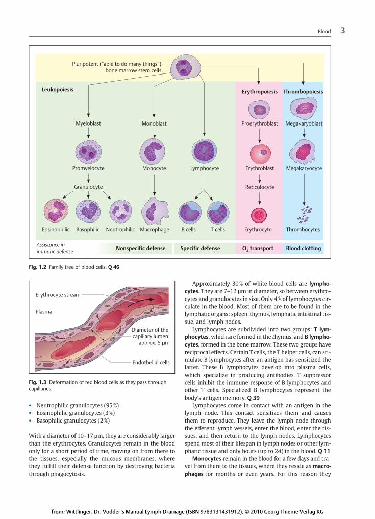
• Neutrophilic granulocytes (95%)• Eosinophilic granulocytes (3 %)• Basophilic granulocytes (2 %)
With a diameter of 10–17 µm, they are considerably largerthan the erythrocytes. Granulocytes remain in the bloodonly for a short period of time, moving on from there tothe tissues, especially the mucous membranes, wherethey fulfill their defense function by destroying bacteriathrough phagocytosis.
Approximately 30% of white blood cells are lympho-cytes. They are 7–12 µm in diameter, so between erythro-cytes and granulocytes in size. Only 4% of lymphocytes cir-culate in the blood. Most of them are to be found in thelymphatic organs: spleen, thymus, lymphatic intestinal tis-sue, and lymph nodes.
Lymphocytes are subdivided into two groups: T lym-phocytes, which are formed in the thymus, and B lympho-cytes, formed in the bone marrow. These two groups havereciprocal effects. Certain T cells, the T helper cells, can sti-mulate B lymphocytes after an antigen has sensitized thelatter. These B lymphocytes develop into plasma cells,which specialize in producing antibodies. T suppressorcells inhibit the immune response of B lymphocytes andother T cells. Specialized B lymphocytes represent thebody’s antigen memory. Q 39
Lymphocytes come in contact with an antigen in thelymph node. This contact sensitizes them and causesthem to reproduce. They leave the lymph node throughthe efferent lymph vessels, enter the blood, enter the tis-sues, and then return to the lymph nodes. Lymphocytesspend most of their lifespan in lymph nodes or other lym-phatic tissue and only hours (up to 24) in the blood. Q 11
Monocytes remain in the blood for a few days and tra-vel from there to the tissues, where they reside as macro-phages for months or even years. For this reason they
3Blood
Pluripotent (“able to do many things”) bone marrow stem cells
Leukopoiesis Erythropoiesis Thrombopoiesis
Myeloblast Monoblast Proerythroblast Megakaryoblast
MegakaryocyteErythroblastLymphocyteMonocytePromyelocyte
Granulocyte Reticulocyte
Eosinophilic Basophilic Neutrophilic Macrophage B cells T cells Erythrocyte Thrombocytes
Assistance in immune defense Nonspecific defense Specific defense O2 transport Blood clotting
Fig. 1.2 Family tree of blood cells. Q 46
Erythrocyte stream
Plasma
Diameter of the capillary lumen:
approx. 5�μm
Endothelial cells
Fig. 1.3 Deformation of red blood cells as they pass throughcapillaries.
from: Wittlinger, Dr. Vodder’s Manual Lymph Drainage (ISBN 9783131431912), © 2010 Georg Thieme Verlag KG
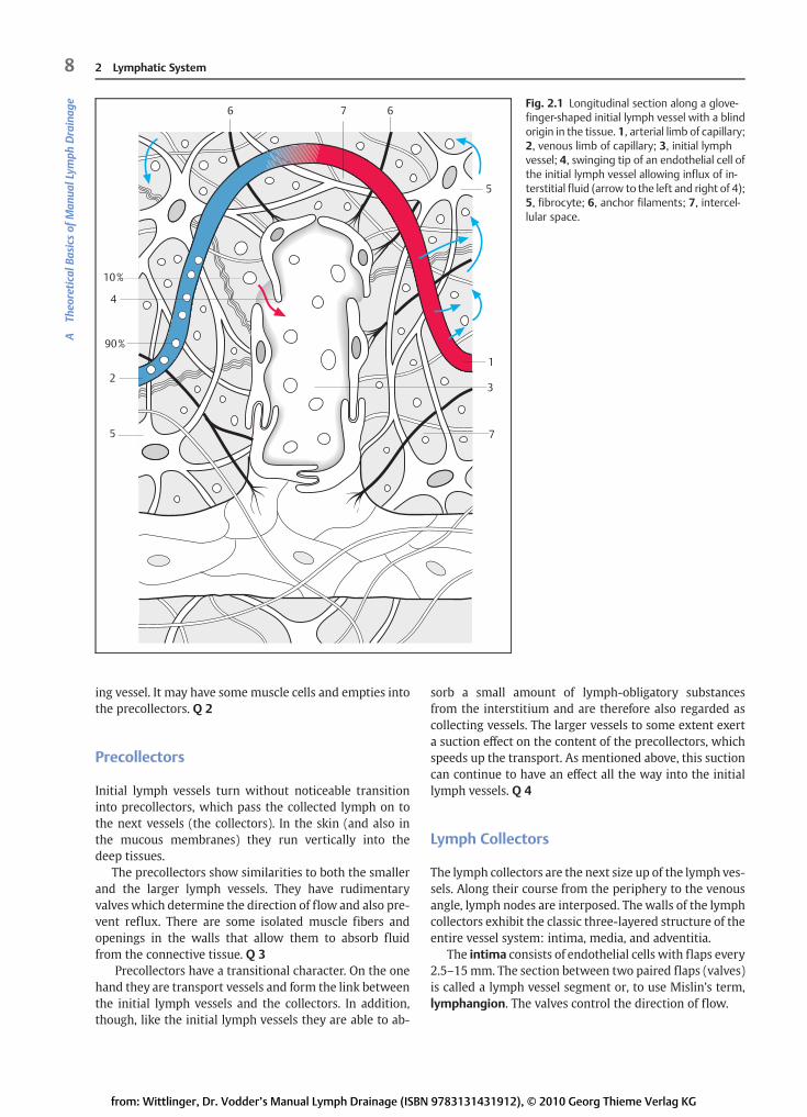
ing vessel. It may have some muscle cells and empties intothe precollectors. Q 2
Precollectors
Initial lymph vessels turn without noticeable transitioninto precollectors, which pass the collected lymph on tothe next vessels (the collectors). In the skin (and also inthe mucous membranes) they run vertically into thedeep tissues.
The precollectors show similarities to both the smallerand the larger lymph vessels. They have rudimentaryvalves which determine the direction of flow and also pre-vent reflux. There are some isolated muscle fibers andopenings in the walls that allow them to absorb fluidfrom the connective tissue. Q 3
Precollectors have a transitional character. On the onehand they are transport vessels and form the link betweenthe initial lymph vessels and the collectors. In addition,though, like the initial lymph vessels they are able to ab-
sorb a small amount of lymph-obligatory substancesfrom the interstitium and are therefore also regarded ascollecting vessels. The larger vessels to some extent exerta suction effect on the content of the precollectors, whichspeeds up the transport. As mentioned above, this suctioncan continue to have an effect all the way into the initiallymph vessels. Q 4
Lymph Collectors
The lymph collectors are the next size up of the lymph ves-sels. Along their course from the periphery to the venousangle, lymph nodes are interposed. The walls of the lymphcollectors exhibit the classic three-layered structure of theentire vessel system: intima, media, and adventitia.
The intima consists of endothelial cells with flaps every2.5–15mm. The section between two paired flaps (valves)is called a lymph vessel segment or, to use Mislin’s term,lymphangion. The valves control the direction of flow.
8 2 Lymphatic SystemA
Theo
reticalB
asicsof
Man
ualLym
phDrainag
e
�
���
���
���
�
��
�
Fig. 2.1 Longitudinal section along a glove-finger-shaped initial lymph vessel with a blindorigin in the tissue. 1, arterial limb of capillary;2, venous limb of capillary; 3, initial lymphvessel; 4, swinging tip of an endothelial cell ofthe initial lymph vessel allowing influx of in-terstitial fluid (arrow to the left and right of 4);5, fibrocyte; 6, anchor filaments; 7, intercel-lular space.
from: Wittlinger, Dr. Vodder’s Manual Lymph Drainage (ISBN 9783131431912), © 2010 Georg Thieme Verlag KG
The media is mainly made up of smooth muscle cells,with a multilayered structure consisting of a medial circu-lar layer and a longitudinal layer. This is a spiral-like plexusthat may include several angions. It also contains somethin collagen fibers. Themuscles are only found in themid-dle section between the valves: the valves themselves arewithout muscles. This gives an impression of constrictionat the valves, leading to a “string of pearls” appearanceon contrast imaging.
The adventitia is the support layer and is linked to theconnective tissue. Q 5
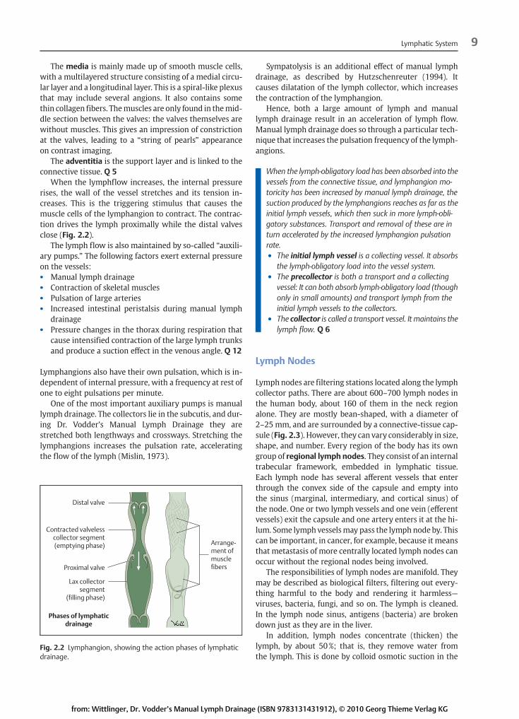
When the lymphflow increases, the internal pressurerises, the wall of the vessel stretches and its tension in-creases. This is the triggering stimulus that causes themuscle cells of the lymphangion to contract. The contrac-tion drives the lymph proximally while the distal valvesclose (Fig. 2.2).
The lymph flow is also maintained by so-called “auxili-ary pumps.” The following factors exert external pressureon the vessels:• Manual lymph drainage• Contraction of skeletal muscles• Pulsation of large arteries• Increased intestinal peristalsis during manual lymph
drainage• Pressure changes in the thorax during respiration that
cause intensified contraction of the large lymph trunksand produce a suction effect in the venous angle. Q 12
Lymphangions also have their own pulsation, which is in-dependent of internal pressure, with a frequency at rest ofone to eight pulsations per minute.
One of the most important auxiliary pumps is manuallymph drainage. The collectors lie in the subcutis, and dur-ing Dr. Vodder’s Manual Lymph Drainage they arestretched both lengthways and crossways. Stretching thelymphangions increases the pulsation rate, acceleratingthe flow of the lymph (Mislin, 1973).
Sympatolysis is an additional effect of manual lymphdrainage, as described by Hutzschenreuter (1994). Itcauses dilatation of the lymph collector, which increasesthe contraction of the lymphangion.
Hence, both a large amount of lymph and manuallymph drainage result in an acceleration of lymph flow.Manual lymph drainage does so through a particular tech-nique that increases the pulsation frequency of the lymph-angions.
When the lymph-obligatory load has been absorbed into thevessels from the connective tissue, and lymphangion mo-toricity has been increased by manual lymph drainage, thesuction produced by the lymphangions reaches as far as theinitial lymph vessels, which then suck in more lymph-obli-gatory substances. Transport and removal of these are inturn accelerated by the increased lymphangion pulsationrate.• The initial lymph vessel is a collecting vessel. It absorbs
the lymph-obligatory load into the vessel system.• The precollector is both a transport and a collecting
vessel: It can both absorb lymph-obligatory load (thoughonly in small amounts) and transport lymph from theinitial lymph vessels to the collectors.
• The collector is called a transport vessel. It maintains thelymph flow. Q 6
Lymph Nodes
Lymph nodes are filtering stations located along the lymphcollector paths. There are about 600–700 lymph nodes inthe human body, about 160 of them in the neck regionalone. They are mostly bean-shaped, with a diameter of2–25 mm, and are surrounded by a connective-tissue cap-sule (Fig. 2.3). However, they can vary considerably in size,shape, and number. Every region of the body has its owngroup of regional lymph nodes. They consist of an internaltrabecular framework, embedded in lymphatic tissue.Each lymph node has several afferent vessels that enterthrough the convex side of the capsule and empty intothe sinus (marginal, intermediary, and cortical sinus) ofthe node. One or two lymph vessels and one vein (efferentvessels) exit the capsule and one artery enters it at the hi-lum. Some lymphvesselsmay pass the lymph node by. Thiscan be important, in cancer, for example, because it meansthat metastasis of more centrally located lymph nodes canoccur without the regional nodes being involved.
The responsibilities of lymph nodes are manifold. Theymay be described as biological filters, filtering out every-thing harmful to the body and rendering it harmless—viruses, bacteria, fungi, and so on. The lymph is cleaned.In the lymph node sinus, antigens (bacteria) are brokendown just as they are in the liver.
In addition, lymph nodes concentrate (thicken) thelymph, by about 50%; that is, they remove water fromthe lymph. This is done by colloid osmotic suction in the
9Lymphatic System
Contracted valvelesscollector segment(emptying phase)
Lax collectorsegment
(filling phase)
Phases of lymphaticdrainage
Proximal valve
Distal valve
Arrange-ment ofmuscle fibers
Fig. 2.2 Lymphangion, showing the action phases of lymphaticdrainage.
from: Wittlinger, Dr. Vodder’s Manual Lymph Drainage (ISBN 9783131431912), © 2010 Georg Thieme Verlag KG

9 Massage Techniques
The Nature of the Massage
The technique ofmanual lymph drainage (MLD) developedby Dr. Vodder is a large-surface massage technique thatcannot be classified among any of the other existing,well-knownmassage techniques. A studyofmanual lymphdrainage will show that the technique is complex and themovements involved require special training. They cannotbe learned from a book.
Allmassage techniques have one thing in common: skincontact is used to stimulate receptors, leading to a particu-lar reaction. Which receptors are stimulated and what ef-fect is achieved is determined by the nature of the skincontact.
To achieve the intended effects with Dr. Vodder’s Man-ual Lymph Drainage, it must be carried out exactly astaught in its original form at the Dr. Vodder Schools in Aus-tria and North America.
Manual lymph drainage consists of four stroke techni-ques as described below. They can be applied in any com-bination during treatment. As the descriptions make clear,Dr. Vodder’s Manual Lymph Drainage is made up of a com-bination of round or oval, small or large, large-area circularmotions that move the skin without sliding over it.
Stationary Circle
“Stationary circles” are primarily applied to the neck andthe face.• Phalangeal and metacarpophalangeal (MCP) joints are
extended; the wrist is rigid and does not move. The cir-cularmovement is created throughmotion of the elbowand shoulder.
• Stationary circles are performedwith both hands and inthe same direction.
• In the starting position (SP), the fingers or whole handsare placed on the skin in the zero phase. Zero phasemeans that there is enough tension in the therapist’shands to extend the phalangeal and MCP joints butthe touch remains very light. We call the touch on theskin: “… as light as a fly… awaspwould be too heavy!”
• After the initial contact (SP) with the flat fingers orhand, the skin is moved with a push-pressure motiontoward the tips of the fingers and the circle is finishedin the direction of lymph flow. The push-pressuremovement increases until it reaches the maximumpush-pressure force.
• While finishing the circle, the skin leads the fingersback to the SP, and the push-pressure decreases untilthe SP is reached (zero phase).
Scoop Technique
The scoop technique is used on the extremities. Thismove-ment is performedwith onehand, or with two hands alter-nating. The scoop technique is learned on the forearm.• The therapist’s hand is placed flat on the palmar side,
phalangeal and MCP joints extended, and the thumbis juxtaposed in opposition to the fingers, similar to alumbrical grip.
• In this SP, the therapist does not apply pressure, becausewhile in this position the hand is in the zero phasewithmaximal skin contact.
• The ideal push-pressure phase is initiated, still withoutpressure, by ulnar abduction of the therapist’s wrist,“wrist forward.”
• The palm is partially lifted off the forearm; only the ul-nar side of the hand remains in contact. We say: “Bringthe wrist forward, perhaps ever so slightly the elbow aswell…”
• During the following increasing push-pressure phase,with a movement that resembles palmar flexion, westretch the skin transversely (transverse push) untilthe majority of the palm is in contact with the skinagain.
• Once the palm is in contact with the forearm again, itspirals in the direction of the index finger, performingdorsal extension and longitudinal push. In this phasethe therapist swings his/her extended fingers from dis-tal to proximal, not sliding over the skin. The stretch(push) is released without lifting the wrist and theskin allowed to return under the hand. At this point,the movement is repeated.
Pump Technique
The pump technique is used on the extremities. Themove-ment is performed with one hand or two hands, alternat-ing or together.• The therapist’s hand is placed flat in dorsal extension on
the front of the thigh. The thumb is again in oppositionto the fingers. Contact is made without pressure (zerophase) but with the entire hand surface. During theSP, the muscles of the hand are not engaged and nopressure is exerted onto the skin.
• The ideal push-pressure phase is initiated, still withoutpressure, by palmar flexion of the therapist’s wrist. Thispalmar flexion continues so that the ulnar side of thepalm remains in contact with the leg. Tip: Make surethat the radius moves forward, not the ulna. In this po-sition, the increasing transverse push takes place withthe MCP joint of the thumb on one side and the MCPjoints of the fingers on the other until the greatest pos-
46 9 Massage TechniquesC
TheTechniqu
eof
Man
ualLym
phDrainag
e
from: Wittlinger, Dr. Vodder’s Manual Lymph Drainage (ISBN 9783131431912), © 2010 Georg Thieme Verlag KG

sible area of contact between the palm and the thigh isreached. The direction of the push is toward the table.
• Maintaining the transverse push, the wrist is lowereduntil the thenar and hypothenar eminences touch thethigh and with a push-pressure movement the skin ofthe front of the thigh is moved proximally (longitudinalpush).
• The transverse and longitudinal push phases are per-formed in one smooth motion.
• This is followed by the phase of decreasing push-pres-sure down to zero, the hand remaining in the greatestpossible contact with the skin. During this movementthe skin of the patient returns beneath the hand ofthe therapist.
Rotary Technique
The rotary technique is used on flat body surfaces such asthe back. The rotary technique is always performed withtwo hands together or alternating.• The therapist places bothhands flat on theback, parallel
to the spine. The finger joints and MCP joints are ex-tended. The thumb is abducted in a 90° angle to the in-dex finger. The hand lies flat and relaxed on the skin inthe zero phase.
• From this SP the hand moves the skin forward (towardthe fingertips) with increasing push-pressure motionand outward (toward the little fingers) in a slightlyoval circle. The oval circle is the “rotation” of the rotarytechnique. This rotation is achieved through slight ulnarabduction and decreases until the zero phase isreached.
• During the zero phase, the hand lies on the skinwithoutexerting pressure and the thumb moves in across theskin toward the index finger.
• Now the palm of the hand is lifted off the skin of theback, but the thumb and tips of the extended fingersmaintain contact with the skin of the back. The finger-tips slide cranially along the spine without exertingpressure. The thumb remains a fixed point and remainswhere it waswhen thewrist was raised. The span of thehand (distance between index finger and thumb) is in-creasing. Thus the hand moves cranially.
• Once the angle between index finger and thumb hasreached approximately 90° the hand is placed flat onthe back again, the thumb moving slightly mediallywithout exerting pressure.
• Thehand has now returned to the SP as described aboveand the push phase starts again.
The fingers are always an extension of the palm of the hand.The work is done not with the palmar aspect of the fingersbut with the palm of the hand. This rule applies to the pumptechnique, scoop technique, and rotary technique.
Thumb Circles
Thumb circles can be used on all parts of the body exceptthe face and neck. Thumb circles are usually applied withtwo hands together or alternating. For practice purposes,thumb circles are done on the back of the hand.• The thumb lies on the back of the hand in the direction
of drainage. It is in the zero phase (SP). One thumb ismoved 90° laterally.
• With increasing transverse push the skin of the back ofthe hand ismoved and at the same time spiraled inwardproximally. This proximal inward spiraling is the longi-tudinal push of the thumb circle. The thumb circle is a90° movement performed by the wrist alone.
• During the zero phase, the skin of the back of the handslides very slightly distally under the thumb.
• Now thewrist moves the thumb back to the SP withoutexerting pressure, and the movement starts again, thistime using the other hand and thumb.
Duration and Intensity of the Massage
There is no general rule for the length of treatment. Inmanycases it is stipulated by the patient’s health insuranceor prescribed by official guidelines (Germany).
The intensity of treatment is determined on the basisof the clinical features of the individual case. This requiresexperience, sensitivity, and intuition on the part of thetherapist.
Experience has taught us that the more precisely thestrokes are performed, the better the desired effects. Theapplication of pressure can vary greatly and depends onthe condition of the tissue. As a general rule it may besaid that the softer the tissue, the lighter themassage pres-sure should be.
Lymphedemas are usually treated with greater pressure.
Creating the Environment for OptimalTreatment
For the best possible treatment, certain requirements aremade of the therapist and the environment:• Avoid conversations during treatment. The patient is
intended to experience your hands. This allows the ef-fects of manual lymph drainage on the autonomic ner-vous system to become more noticeable.
• Avoid interruptions during treatment if possible.• The decisionwhether to accompany the treatment with
music should be left to the patient.• The room should be well insulated against, or located
away from, external noise (telephone, street noise, etc.).
47Creating the Environment for Optimal Treatment
from: Wittlinger, Dr. Vodder’s Manual Lymph Drainage (ISBN 9783131431912), © 2010 Georg Thieme Verlag KG

Knee (Fig. 10.14)
Cauliflower: Pump-push. The fingers and thumb grip likeflat pliers (lumbrical), without pinching. The push is athumb circle with the proximal hand. Hands alternate, 3 ×.Popliteal space: The fingers of both hands are quite flat.The fingertips do not touch. Continuous spirals are appliedacross the back of the knee, counting to 5, from distal toproximal, 3 × (not shown).Patella: The thumbs are placed on theborder of the patella,5 continuous thumb circles are performed progressingproximally along the border, 3 × (not shown).Pump technique:With the inferior hand over theknee, thesuperior hand supports underneath, counting to 5, 3 × (notshown).Pes anserinus:Work the pes anserinus (goosefoot)with al-ternating thumb circles, counting to 6, 3 ×.
Notes:
62 10 Treatments of the Individual Parts of the BodyC
TheTechniqu
eof
Man
ualLym
phDrainag
e
Fig. 10.14a Pump and push on the “cauliflower” during treat-ment of the knee, position 1.
Fig. 10.14b Position 2
Fig. 10.14c Alternating thumb circles on the pes anserinus.
from: Wittlinger, Dr. Vodder’s Manual Lymph Drainage (ISBN 9783131431912), © 2010 Georg Thieme Verlag KG

Lower Leg (Fig. 10.15)
The therapist stands next to the lower leg.
Pump–scoop on the lower leg: Place the foot flat on thetable, the knee flexed. The hand on the shin bone pumps,the hand on the calf scoops, counting to 6 or 8. Boththumbs rest on the lateral side of the lower leg, 3 ×.
The therapist stands at the foot.
Alternating scoop technique on the calf, counting to 6 or8. One thumb is placed on the medial aspect of the lowerleg, the other thumb is placed on the lateral aspect of thelower leg, 3 × (not shown).
Foot (Fig. 10.16)
Achilles tendon: The leg returns to the extended position.Workwith 5 continuous spiralswith 4 fingers of each handproximally. Similar to the technique for the back of theknee, 3 ×.Thumb circles alternating, in several lines over the anklejoint, 3 × each (not shown).Thumb circles alternating, in several lines over the dorsumof the foot, 3 × each (not shown).Lymph sea: Parallel simultaneous thumb circles on thelymph sea; 5 ×, 3 × (not shown).Pressing of the transverse arch, 3 × (not shown).
Final Effleurage
1 × (not shown).
Notes:
63Treatment of the Leg
Fig. 10.15a SP for the alternating pumps and scoops duringtreatment of the right leg; medial view.
Fig. 10.15b Lateral view
Fig. 10.16 Continuous circles on the Achilles tendon in parallelwith two hands.
from: Wittlinger, Dr. Vodder’s Manual Lymph Drainage (ISBN 9783131431912), © 2010 Georg Thieme Verlag KG

Treatment of the Nape of the Neck
The therapist stands at the patient’s left side.The patient is prone.
Effleurage
Rotary technique from the middle of the thoracic verte-brae to the cervical vertebrae, 1 × (not shown).
Profundus to Terminus
Stationary circles from the profundus down the neck tothe terminus, 5 circles per position, 3 × (not shown).
Occiput to Terminus (Fig. 10.17)
Stationary circles from the occiput down the neck to theterminus, 5 circles per position, 3 ×.
Notes:
64 10 Treatments of the Individual Parts of the BodyC
TheTechniqu
eof
Man
ualLym
phDrainag
e
Fig. 10.17aStationary circleson the occiput.
Fig. 10.17b… down the middleof the neck.
Fig. 10.17c… to the terminus.
from: Wittlinger, Dr. Vodder’s Manual Lymph Drainage (ISBN 9783131431912), © 2010 Georg Thieme Verlag KG

Back of the Head (Fig. 10.18)
The therapist stands at the patient’s head.
Stationary circles along the nuchal line, 5 × per position,3 positions, push toward the body, circle toward the littlefinger, 3 × (not shown).Stationary circles in 2 lines on the backof the head, 3 posi-tions per line, then the next line, 3 repetitions per line (notshown).Stationary circles, on the lateral aspect of the back of thehead caudally to the terminus, connecting the end pointsof each of the previous lines. Depending on the anatomyof the patient (length of the neck, size of the head), 5–6 po-sitions are treated, in terminus supinate hands, 3 ×.
Shoulders
Pump technique from lateral to medial, over the deltoidmuscle, counting to 5 (when finished, the thumbs rest inthe termini), 3 × (not shown).
“Rabbit” Technique (Fig. 10.19)
The therapist stands at the patient’s left side.
Pump technique with the cranial hand, alternating withpushing toward the terminus, using the thumb and the fin-gers of the caudal hand, counting to 6, 3 ×.
Notes:
65Treatment of the Nape of the Neck
Fig. 10.18 Lateral stationary circles during neck treatment. Posi-tion midway down the neck.
Fig. 10.19a “Rabbit” technique: SP, caudal hand.
Fig. 10.19b “Rabbit” technique: caudal hand, push phase.
from: Wittlinger, Dr. Vodder’s Manual Lymph Drainage (ISBN 9783131431912), © 2010 Georg Thieme Verlag KG