1) anesthesia and euthanization of rodents
TRANSCRIPT

Optimizing Experimental Research in Respiratory Diseases: an ERS Statement
The online supplement provides complementary and detailed information in the following domains
of the Task Force. These supplements are based on the earlier working documents to generate the
main report. The Task Force members felt strongly that the information shown here could be highly
valuable as reference documents to the research community. Some areas that were covered in the
main report are not covered in the supplement, because the working documents did have a different
draft format and were not suitable for publication.
1) Anesthesia and euthanization of rodents
2) Pre-analytical conditions of tissues/cell harvesting/ collection/storage
3) Large animal models for respiratory diseases
4) Chronic Obstructive pulmonary disease (COPD) including clinically relevant subgroup
of COPD, environmental factors and exacerbations
5) Infection/Pneumonia
1) Anesthesia and euthanization of rodents
The most frequently employed short-term injection anesthesia which can be applied via
intravenous, intramuscular, or intra-abdominal routes to mice and rats as well as dogs, cats, sheep,
and horses is a combined ketamine hydrochloride and xylazine hydrochloride anesthesia, which may
be combined with the dopamine 2 receptor blocker azepromazine (Jaber et al.). Although significant
species-specific differences in dosage regimens exist, for anesthesia in mice, ketamine should be
applied in a concentration range of 50-100 mg/kg body weight (b.w.), while xylazine hydrochloride
should be used at final concentrations of 2.5-8 mg/kg of b.w., as both drugs dose-dependently cause
bradycardia and bradypnea, which can be reversed by yohimbine [1-4]. In case re-dosing of
anesthesia is required, mice should receive either 50 % of the initial ketamine dose, or 25 % of the
initial combined ketamine-xylazine dose as soon as the pedal withdrawal reflex starts to return [5].
Commonly employed volatile anesthetics in laboratory animals include isoflurane (Baxter,
Unterschleissheim, Germany), sevoflurane, and desflurane (Suprane, Baxter) [6]. The advantage of

inhalational over injection anesthesia is its simple titratability of anesthetic depth and its very rapid
recovery, which qualifies it for use for long-term anesthesia, or for repetitive anesthesia within short
time periods [7].
Beside its role as inhalational anesthetic, isoflurane is frequently used for the sacrifice of mice and
rats [8], which according to recommendations of the Federal European Laboratory Animal Science
Association (FELASA), must be accompanied by immediate blood withdrawal from the Vena cava
or cardiac puncture to ensure death of the laboratory animal. Alternatively, due to low costs and ease
of availability, many researchers still make use of carbon dioxide (CO2) to euthanize laboratory
animals. However, compared to volatile anesthetics such as isoflurane, use of CO2 for euthanasia
has several disadvantages, as inappropriate use of CO2 may lead to major hypercapnic excitation of
animals at the end of the experiment. Second, exposure to CO2 leads to a reduction in pH in the
airways and peripheral blood of both rats and mice, which as a consequence adversely affects the
pharmacokinetics of e.g., basic drugs, which is not observed when isoflurane is used for sacrifice
[9]. A most recent report also demonstrates adverse effects of CO2 euthanasia of mice for the
subsequent generation of mouse embryos using in vitro fertilization approaches [10]. These data
imply that wherever possible, available alternatives to CO2 euthanization of laboratory animals
should be used. As another alternative method to euthanize mice, cervical dislocation (CD) is
frequently employed, although more recent studies raised concerns as of its efficacy as successful
method for euthanization of mice [11]. Since CD requires specific training, it is recommended that
CD should only be performed by well-trained lab personnel to ensure its correct execution.
2) Pre-analytical conditions of tissues/cell harvesting/ collection/storage
It is important not to overfix tissue/cytological preparation if immunohistochemistry (revelation of
antigen by specific antibody) is to performed. For histological techniques, the choice of the fixative
and duration of fixation need to be optimised [12, 13]. While in research a combination of 4%
formaldehyde with 0.1% glutaraldehyde in 0.2 M HEPES buffer followed by freeze substitution in
0.5% uranyl acetate in methanol have been recommended [14], other fixatives (such the ones used
in the clinical setting) namely buffered 4% formaldehyde alone is also applicable and fixation
should be kept short (12-24 hours for small fragments, 48 hours max for larger fragments) and
uniformed within the groups studied.
Similarly, cytology preparations either form BAL cytopsins or cell cultures should be fixed either in
10% acetone for 10 minutes, 4% buffered formalin for 5 min, or PBS- Ethyl alcohol for 30 seconds,
depending on what techniques are used subsequently [15].
Cytology slides can be kept at +4 degrees (fridge) for a few days or at -20 degrees for a few months.
Antigenicity (expression of antigens at the surface of the cells) does fade overtime, and on old
stored slides, testing for preserved/retained antigenicity might be needed before applying
immunohistochemistry.
Animal lung tissue after fixation (12-24h max) should undergo “processing” readily as it is best
practice to keep paraffin embedded tissue blocks and only cut slides upon the need for
immunohistochemistry rather than storing cut unstained slides. Tissues are processed in a vacuum
infiltration processor (VIP) automated tissue processor. This is based on tissue being exposed to
increasing concentrations of ethanol (50% to 100% over a few hours), then in xylene/toluene and
finally in liquid paraffin at 60 degrees. Researchers should be encouraged to liaise with their local
hospital histopathology department to streamline this process.
One dehydrated and cleared, the tissue is embedded in melted paraffin, that once hardened, provide
a hard support for tissue slides sectioning on glass slides, usually at 4-5um thin.
If immunohistochemistry (IHC) is being performed, slides should be cut on positively charged
(sialinised) slides to prevent detachment of tissue section from the slide during antigen retrieval and
immunohistochemical protocol. These sialinised slides are either commercially available but glass
slides can also be sialinised using a solution of 3-aminopropyltriethoxysilane (APES).
Cut slides should be put in an oven at 56 degrees for 20 min and then used for
immunohistochemistry of short-term storage in a dry cool dark area for a few months.
Resources such as IHC world [16] are available on line to facilitate and optimise tissue processing.
Studies have demonstrated that the profiles of these molecules can change drastically during
transport and storage thus making a reliable diagnostic or pharmaceutical research unreliable or
even impossible.
Similarly, optimising pre-analytical steps will provide good quality RNA, DNA and protein for
molecular studies. International consortiums such as SPIDA are promoting standardisation of pre-
analytic conditions, quality assurance schemes and innovative pre-analytical tools by establishing
guidelines in sample collection, handling, stabilisation, purification and storage of clinical samples
that should be applied to research samples [17] Reducing or eliminating pre-analytical errors that

lead to inaccurate results can be achieved by integrating and standardizing workflows for the
collection and processing of samples to increase analytical accuracy and laboratory efficiency and
developing automated sample preparation when feasible.
3) Large animal models for respiratory diseases
3.1 Chances and limitations of large animal models in respiratory research
Despite 17 Nobel prizes were awarded to scientists that studied cattle, horses, sheep or poultry as
models for biomedical research [18], the vast majority (about 98-99%) of animal experiments are
currently undertaken with rodents, predominantly mice. Interestingly, animal models based on
domestic animals or livestock have gained increasing attractiveness during the last decade, and are
currently re-introduced as an essential part of biomedical research. Table 6 (in the main document)
summarizes large animal models for non-infectious and infectious respiratory diseases.
In principal, there are three kinds of animal models suitable for biomedical research: natural models,
experimentally induced models, and transgene models:
Natural models are based on naturally occurring pulmonary diseases with similar
pathophysiology in animals and humans. Typical examples are feline asthma, "ski asthma" in
sled dogs, RSV infection in calves, bovine tuberculosis, or naturally acquired Chlamydia
infections. Species-specific peculiarities in the pulmonary vasculature pre-dispose calves as a
model for pulmonary.
Experimental models in pulmonary research can either simulate the natural diseases under
defined conditions or have been introduced for distinct purposes, for example ventilated models
of MRSA-induced pneumonia in sheep or swine.
With steadily increasing possibilities to generate genetically modified large animals, even
transgene models become available in addition to mice models. The pig model of cystic fibrosis
is one example [19-21]. Superior to the mouse model, the pigs lacking CFTR exhibit defective
chloride transport and develop typical signs of CF seen in humans. In addition of serving as in
vivo models of complex pathophysiological functions, genetically modified animals will be a

future source of primary cells for ex vivo examination of biological functions at a cellular level
[22].
Large animal models are more expensive, laborious and time-consuming, and the experimental use
of pet and livestock animals receives lower ethical acceptance compared to laboratory animals.
Nevertheless, the choice of an animal species to be used as a model should be based primarily on the
biological relevance according to the current state of knowledge instead of convenience and reduced
cost.
Large animal models offer unique changes that will complement existing models in rodents while
taking into account the 3R principles (refinement, reduction‚ replacement) as introduced by Russell
& Burch more than 50 years ago [23]:
Chronicity & long-term studies.
Because of their limited life span, rodents are not suitable to study the complex pathogenesis
of chronic diseases with a manifestation that may last for years. In contrast, a considerably
long life-span of large animals supports investigations on chronic diseases and on chronic
infections. Typical examples are naturally occurring chronic obstructive pulmonary diseases
in animals (asthma in cats, chronic obstructive pulmonary diseases in horses) partially share
pathogenetic features with human diseases. Also, chronic infections with so-called
‚atypicals’ (e. g. Chlamydia spp., Mycoplasma spp.) in bovines present suitable animal
models in natural pathogen-host settings. The latter provide strong evidence that persistent
and recurrent chlamydial infections are associated with chronic airway obstructions [24].
Complexity & system biology.
Larger animals offer the great potential to perform long-term functional studies allowing a
simultaneous within-subject approach of functional, inflammatory and morphological
changes. Different samples (for example, blood, BALF, tissue biopsies, exhaled breath) or
biological variables can be perfectly analysed in their interactions if derived from one
subject over time. This is particularly valuable when following system biology approaches.
Biological variability of data sets & sample size.
Parallel assessments of multiple parameters in intra-individual follow-ups over time
minimises the number of experimental animals as well as data variability. In a biological
setting, intra-individual variation accounts for only 25 – 30 % of the total variability of

physiological parameters. By contrast, variability between subjects is generally much higher
(by 3 – 4fold). In consequence, data obtained in intra-individual kinetics are less variable
compared to inter-individual baseline data, and the number of animals required for a
meaningful statistical analysis can be kept much lower than in group comparisons.
Biological relevance.
Significant differences exist between species with respect to the genetically determined
regulation of defence mechanisms. For example, interleukin-8 (IL 8) plays a significant role
in inflammatory processes. However, the il-8 gene is missing in the mouse genome. It does
exist in the genome of dogs, pigs, sheep, and cattle. The protein encoded for IL 8 even
exhibits a high cross reactivity between those species [25, 26]. Intriguingly, the bovine
genome, fully sequenced in 2009, more closely resembles the human genome than it
resembles that of mice and rats. Approximately 80 % of the genes identified in the bovine
also exist in other mammals [27]. Beyond the genetic background, structural and
physiological similarities of the porcine and human airways, e.g. with regards to the local
immune system [28, 29] or the composition of the epithelial surface liquid [30] qualify pigs
as suitable models for human airway diseases. Pigs can be used to study gene transfer events
in the lung in the development of gene therapies for lung diseases as cystic fibrosis [31].
Species lacking collateral airways, and presenting a strong segmental anatomy of the lung (e.
g. pigs, cattle) are particularly suited to mirror pulmonary dysfunctions associated with
airway obstructions [32].
The development of immunological competence during fetal ontogenesis and in the neonate
(immuno-physiology) is similar in larger animals species compared to humans. Thus,
livestock models are predestined to study maternal-fetal interactions and immunological
mechanisms during post-natal development [26]. In this context, the asthma model in dogs is
highly relevant to elucidate maternal influences in the transmission of asthma susceptibility
[33].
3.2 Modes of experimental challenges
Different modes of exposure are available with each presenting significant advantages and
disadvantages. The most frequently used are given in Table S1 with typical examples of application.
As exemplarily shown for lung infections with Chlamydia, large animal models of respiratory

infections strongly support a deeper understanding of the host-pathogen interactions in a complex
pathogenesis, and allow evaluation of clinically relevant treatment options [34-36].
3.3 Read out parameters in vivo
Blood gas analysis and assessment of acid base status
To assess the efficacy of pulmonary gas exchange, arterial blood gas analysis is still the gold
standard. Practically, access to arterial blood depends on the animal species. In horses, arterial blood
can be easily collected from A. carotis while this procedure is difficult or even requires surgery in
ruminants and pigs.
Collecting blood samples in conscious animals by puncture may induce stress or pain, and
consequently hyperventilation. The latter, however, could reduce the actual degree of hypoxaemia
or even could make hypercapnia invisible. Thus, catheterization of an arterial blood vessel is highly
recommended to facilitate repeated sampling of arterial blood in ongoing studies by minimizing
stress for the animals. As an example, catheterization of Aorta abdominalis has been successfully
implemented in respiratory infection studies in calves [37, 38]. Any reduction of stress while
collecting blood from the animals increases the quality of the samples.
When analyzing blood gases or blood pH, one has to be taken into account that analyzers usually
work at 37°C which is the normal body temperature of human beings. Physiologically, all animal
species present significantly higher body temperatures, sometimes above 39°C. A difference of
about one degree in body temperature, however, corresponds with a change of about 0.5 kPa in
partial pressures of blood gases. Therefore, blood gases and pH always need to be corrected for the
actual body temperature measured immediately before or after blood collection.
If there is no access to arterial blood, some valuable variables of acid base status can be obtained
from venous blood as described for pigs calves experimentally infected with viruses or bacteria [39,
40].
Pulmonary function testing
Using a tightly fitting face masks of the appropriate size, all techniques of pulmonary function
testing (PFT) known from human medicine are principally available to be used in an awake,
spontaneously breathing large animal with the exception of active breathing manoeuvres.
Spirometry can be used to assess variables of spontaneous ventilation (i.e., tidal volume Vt,

respiratory rate fR, minute ventilation Vmin). Combined with capnography, i.e. CO2-exhalation
versus exhaled volume, dead space volume (Vd), and the ration Vd/Vt can be assessed, and alveolar
ventilation can be calculated. Re-breathing methods based on multi-breath analyses are helpful to
measure functional residual capacity (FRC) by using Helium-wash-in or –wash-out, or assessing the
diffusion capacity based on CO-transfer [41] [38].
To evaluate respiratory mechanics in large animals, three principal techniques have widely been
used in many species of domestic animals. (i) The classical approach calculates pulmonary
resistance (RL) and dynamic compliance (Cdyn) from measuring oesophageal pressure via
oesophageal balloon while flow and volume derive from pneumotachography. (ii) Forced oscillation
techniques bear the advantage of being non-invasive and are capable of distinguishing between
obstructive and restrictive ventilation alterations but at the same time differentiates between
obstructions of the proximal and peripheral airways by measuring complex respiratory impedance
(Zrs) or its components, i.e. respiratory resistance (Rrs) and respiratory reactance (Xrs) in a
frequency range appropriate to the lung size of the animal: Reports from experimental models of
lung infections are exemplarily available for pigs [42] and calves [38, 40]. (iii)
Bodyplethysmography has been described for piglets [43], sheep [44], cats [45], and dogs [46].
Comparable to the use of this technique in mice or rats, the outcome parameter is ‚Penh’. This
parameter provides rather non-specific information associated with a changing breathing pattern, but
does not allow differentiating restrictive from obstructive ventilatory disorders.
Technical solutions for measuring pulmonary function data strongly depend on the size of the
animal. Taking this aspect into account, equipment of human pneumology is often directly
applicable to animals weighting about 20 to 100 kg, i.e. calves, sheep, goats and pigs to assess
pulmonary functions non-invasively in conscious animals while breathing spontaneously. Measuring
identical parameters as in humans, direct comparisons between data obtained in large animal models
and data obtained in patients become possible. Due to their non-invasive character, they are of
beneficial value with regards to animal welfare protection.
Biological specimens from the respiratory tract
The diagnostic repertoire obtaining biological samples from the lungs of large animals is widely
identical to human medicine, which also allows direct comparisons and translation of results.
Bronchoscopy can be applied to larger animals while being conscious and in standing position

(horses, cattle), but requires anaesthesia in pigs, cats or dogs. Focussing on translational respiratory
medicine, anatomy and bronchoscopy of the porcine lung have been described [47]. Bronchoscopic
sampling techniques include broncho-alveolar lavage, epithelial brushing, and tissue biopsies as
recently described and illustrated for the bovine lung in a video publication [48]. Due to the size of
large animals, all these biological samples may be taken repeatably in sufficient amounts without
having to sacrifice the animal.
Exhaled breath and exhaled breath condensate
Collection of exhaled breath (EB) and/or exhaled breath condensate (EBC) for further analysis is a
current research option directly comparable to its use in human medicine when searching for
exhaled biomarkers. Analysis of volatile organic compounds (VOC) has been implemented in large
animal studies to identify respiratory infections. Methods of VOC analysis applied to large animals
include (i) GC-MS to identify biochemical compounds [49], and (ii) methods of pattern recognition
using DMS/IMS [50] or electronic noses. The high level of standardization in large animal studies
that can never reached in humans with respect to ambient conditions, nutrition etc. qualifies these
models to elucidate physiological background and physiological variability of VOC candidates that
are of interest for future diagnostic purposes [51, 52]. With respect to EBC, studies focussing on
collection and analysis in domestic animals contributed significantly to understand the strengths and
methodological limitations of this diagnostic technique in a general sense – independent of any
species [53, 54].
Imaging techniques
Although ultrasound imaging of lungs is possible in many large animal species, it has been most
frequently applied to pigs and calves. Using pigs as models, lung sonography has been
recommended as a very accurate method in diagnosing even small amounts of intrapleural air in
cases of pneumothoraces [55], or - combined with lung flooding – for successful tumour detection
[56]. Furthermore, the pig has been exploited as a useful model for ultrasound-guiding endoscopic
lung surgery or to evaluate high-intensity focused ultrasound as a potential new strategy for treating
lung cancer [57]. In calves in experimentally induced lung infections, the diagnostic value of
sonography to identify pneumonic lesions was characterized in comparison to pulmonary function

testing and clinical examination [58]. As in clinical pneumology, X-ray, scintigraphy, and computed
tomography [44] are further techniques applicable to large animals.
3.4 Conclusions
Large animal models form an innovative part of modern interdisciplinary biomedical research. For
use in translational medicine and as comparative models, these models offer the distinctive
opportunity to obtain results with high biological relevance and, in various instances, even of dual
use (to improve human as well as animal health). Although in good agreement with the 3R concept
and the requirements of animal welfare, models deploying domestic animals suffer from low ethical
acceptance. In contrast to rodent models, large animal models are more demanding regarding time
and financial resources. However, biologically relevant livestock species models – with the animal
species investigated being selected according to the particular requirements of the scientific
questions to be solved - embedded into the communication and collaboration between several
disciplines from human and veterinary medicine inhere the outmost potential to make progress
according to the ‚One Health’ concept.
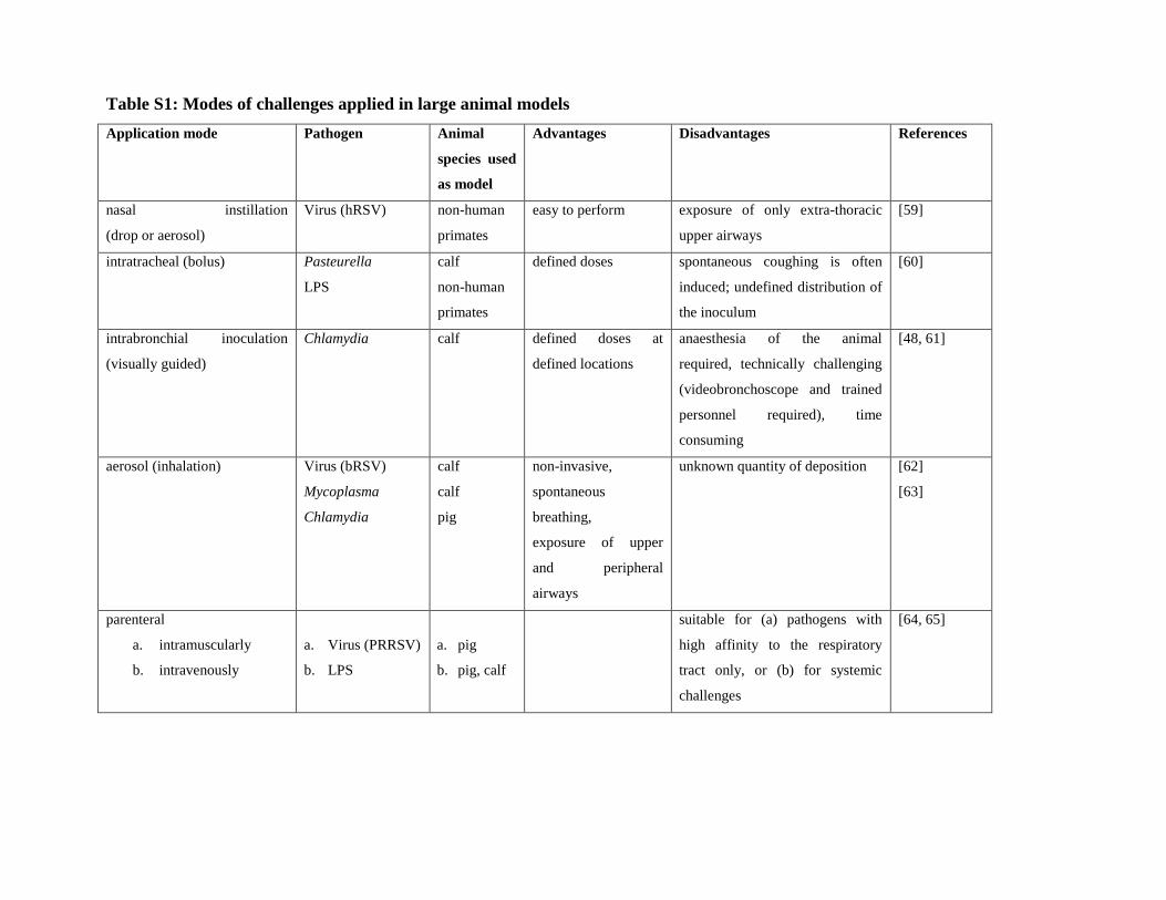
Table S1: Modes of challenges applied in large animal models
Application mode Pathogen Animal
species used
as model
Advantages Disadvantages References
nasal instillation
(drop or aerosol)
Virus (hRSV)
non-human
primates
easy to perform exposure of only extra-thoracic
upper airways
[59]
intratracheal (bolus) Pasteurella
LPS
calf
non-human
primates
defined doses spontaneous coughing is often
induced; undefined distribution of
the inoculum
[60]
intrabronchial inoculation
(visually guided)
Chlamydia calf defined doses at
defined locations
anaesthesia of the animal
required, technically challenging
(videobronchoscope and trained
personnel required), time
consuming
[48, 61]
aerosol (inhalation) Virus (bRSV)
Mycoplasma
Chlamydia
calf
calf
pig
non-invasive,
spontaneous
breathing,
exposure of upper
and peripheral
airways
unknown quantity of deposition [62]
[63]
parenteral
a. intramuscularly
b. intravenously
a. Virus (PRRSV)
b. LPS
a. pig
b. pig, calf
suitable for (a) pathogens with
high affinity to the respiratory
tract only, or (b) for systemic
challenges
[64, 65]

12
4) CHRONIC OBSTRUCTIVE PULMONARY DISEASE (COPD)
4.1 How do we define COPD for experimental research (what do we want to model)?
According to the Global Initiative for Chronic Obstructive Lung Disease, “COPD is
characterized by persistent airflow limitation that is usually progressive and associated with
an enhanced inflammatory response in the airways and lung to noxious particles and/or
gases” [66]. In an individual patient, varying degrees of chronic bronchitis, bronchiolitis, and
emphysema contribute to chronic airflow limitation [67-69]. Exacerbations and comorbidities
contribute to the overall severity of the disease1.
In recent years, findings obtained in large cohort studies have provided longitudinal insight
into the evolution of airway obstructions and its role in the development of COPD. An
accelerated rate of decline in forced expiratory volume in 1 second (FEV1) has long been
regarded as a key feature of COPD [70]. In the ECLIPSE (Evaluation of COPD
Longitudinally to Identify Predictive Surrogate Endpoints) study, the rate of change in FEV1
over 3 years was found to be highly variable, with only 38% of patients showing an elevated
rate of decline of FEV1 of more than 40 mL/ year. In close to a third of all patients, there was
no decline in lung function over the study period [71]. Moreover, in a combined analysis of 3
cohort studies, it was found that approximately half of the patients with COPD had already a
low FEV1 in early adulthood, with a normal decline in FEV1 afterward, despite similar
smoking exposure [72]. This study emphasizes the importance of lung growth and the early
origins of COPD [73]. Last but not least, it has become increasingly clear that current and
former smokers with preserved lung function may still present with respiratory symptoms,
exacerbations, activity limitation, and evidence of airway disease [74].
Attention has been paid to tobacco smoking as a major cause of COPD. Accordingly, the
inflammatory response to tobacco smoke has been well characterized, with involvement of the
innate and adaptive immunity [75-77]. It is widely accepted that COPD results from a gene-
environment interaction [78]; among people with the same smoking history, not all will
develop COPD. The best-documented genetic risk factor to date is alpha-1 antitrypsin
deficiency [79, 80]. Several genome-wide association studies have discovered genes that are
associated with both the presence of the disorder and the severity of airflow obstruction [78].
In recent years, it has become evident that, in major parts of the world, COPD can be caused
by exposure to biomass fuels [81]. Other risk factors that have been implicated in the
pathogenesis of COPD are occupational exposure to dust and gases, respiratory tract

13
infections during childhood, outdoor air pollution and poor socioeconomic status, as well as a
history of pulmonary tuberculosis and chronic asthma [73].
The natural course of COPD is punctuated by periods of disease exacerbation [66, 82, 83].
Clinically, exacerbations are defined as “a sustained worsening of symptoms from the
patient’s stable COPD state, beyond normal day-to-day variations, which necessitates a
change in medications” [84]. Based on the ECLIPSE-study, it appears that some patients
present with frequent exacerbations (i.e. ≥ 2 exacerbations per year) and that this frequent
exacerbation phenotype is relatively stable over a 3 year period [82]. Four distinct biological
clusters have been associated with COPD exacerbations, including bacteria-predominant,
eosinophil-predominant, viral-predominant, and pauci-inflammatory [85].
Comorbidities in patients with COPD have an important impact on disease severity and
patient survival [66, 86]. Different clusters of clinically important and objectively diagnosed
comorbidities have been reported. Cluster analysis revealed five distinct clusters: a cachectic
cluster, a metabolic cluster, a cardiovascular cluster, a psychologic cluster and a subgroup of
patients with significantly less comorbidity [86]. Systemic inflammation was found to be
present in a limited proportion of patients, e.g. 16% of patients with COPD in the ECLIPSE
study [87]. The presence of elevated levels of CRP, fibrinogen and leukocyte counts in
individuals with COPD is associated with an increased risk of having exacerbations [88].
Taken together, COPD is a complex condition, encompassing a spectrum of chronic lung
disorders. It is of utmost importance that these different clinical phenotypes are taken into
consideration when modelling COPD.
4.2 What are the major unmet needs to model and study COPD?
To date, inflammatory processes elicited by tobacco smoke have been widely studied in
animal models [89-100]. Less well understood is how these inflammatory processes
contribute to accelerated decline in lung function, how they persist following smoking
cessation, and why they cause predominant airway versus parenchymal disease in subgroup of
patients. Given the early origin of COPD [73], there is a need to study the contribution of lung
development in the genesis of COPD. There is a further need for models of acute
exacerbations [90, 94, 101-103] and COPD-associated comorbidities [92, 98, 104]. While
these are important research areas, data today are still limited. For example, there is a limited
understanding of inflammatory mechanisms involved in the various types of exacerbations of
COPD [85] and how these processes contribute to the decline in lung function. A further

14
question that is currently not well understood is whether exposures to cigarette smoke,
biomass fuel, and/or outdoor air pollution results in similar or differing pathogenetic
mechanisms and disease phenotypes.
4.3 How can we model specific clinically relevant subgroups of COPD?
The pathogenesis of COPD is the result of a complex system of events perpetrated over
decades; hence, experimental models represent the capacity to examine specific facets of the
disease and study them in isolation from confounding factors. In addition, experimental
studies have access to a broad range of reagents, tissues, and intervention strategies that are
not available or feasible in clinical research. The strengths of these models however are also
analogous to their weaknesses. The reductionist recreation of a subset of pathogenic
components fails to recreate the complexity of the disease. This needs to be taken into
consideration when translating observation from experimental models to the human disease
[89].
4.3.1 Genetic factors, epigenetic alterations, and microRNAs
There is clear evidence that genetic and epigenetic factors influence the susceptibility for
COPD. Of the genetic factors, alpha-1 antitrypsin (AAT) deficiency remains one of the most
common risk factors for the development of emphysema [80]. While circulating AAT in
specific mouse strains is associated with distinct patterns of emphysema [91], studying the
loss of function of AAT in mice has been challenging, as there are multiple encoding genes
(Serpina1a-1e) [105, 106]. Of interest is the report by Alam et al. that transgenic
overexpression of the most common human mutant AAT in mice conferred a pro-
inflammatory phenotype [107]. In addition to AAT, gene association studies have implicated
numerous candidate genes over the past 40 years in the pathogenesis of COPD, although these
studies have been largely inconclusive [108]. In more recent years, information was gained
from applying genome-wide association (GWAS) to large cohorts [78]. While GWAS studies
establish association, functional studies are required to gain insight into biological
mechanisms of genetic determinants of COPD and/or lung function. For example, in vitro and
in vivo experimentations have provided critical mechanistic insights into GWAS loci
associated with COPD, such as HHIP and FAM13A [109-112]. These studies clearly
document the importance of experimental models to study biological mechanisms and
establish causality. Similarly, epigenetic modifications, as well as microRNAs (miRNAs) and
long non-coding RNAs have been implicated in the pathogenesis of COPD [113, 114].
Moreover, there is some albeit limited evidence of epigenetic inheritance in COPD, i.e.

15
transmission of developmental programming across generations that were not exposed to the
initial environment that triggered the effect [115]. Experimental models have and will
continue to play a significant role in developing an understanding of the biological function of
specific genetic factors, epigenetic alterations, and miRNAs in diseases processes associated
with COPD.
4.3.2 Lung development
Emerging evidence suggests that factors in early life that affect lung development are
implicated in the pathogenesis of COPD [73]. For example, two cohorts from mid-childhood
to adulthood were assessed for their lung functions, and those with preterm birth and a history
of bronchopulmonary dysplasia (BPD) had significant lower FEV1 and mid-expiratory flow
than term-born controls [116]. A longitudinal study also found an association between low
quartile of maximal expiratory flow at birth and low FEV1/FVC ratio at 26-32 years of age
[117]. Moreover, intrauterine exposure to harmful stimuli (maternal smoke) and growth
retardation, early childhood lung infection and asthmatic insults are all negatively affecting
lung development and reducing the maximal FEV1 in adulthood [118-121]. In addition,
noxious factors encountered in early life may also accelerate the rate of FEV1 decline in adult
with active smoking [122].
Although growing evidence suggests early origins of COPD in humans, definitive and
mechanistic studies are needed. Studies using animal models have already provided evidences
regarding important developmental genes in the pathogenesis of COPD. For example,
defective TGF-β-Smad3 pathway results in reduced alveolar growth during development that
contributes to emphysema-like pathology in adult mice [123, 124]. Other key developmental
pathways including FGF, Shh/HHIP, ErbB/ERRFI1, and VEGF have been shown to play key
roles in both lung development and emphysema pathogenesis in genetically manipulated mice
[109, 125-129]. Dysregulation of elastin fibre synthesis and degradation has been clearly
demonstrated to affect both developmental alveolar growth and adult alveolar destruction
[130, 131], one of the important mechanisms for COPD. In addition, there is evidence that
prenatal exposure to cigarette smoke predisposes to airspace enlargement in adult mice,
suggesting a role for in utero exposure on the adult development of COPD [132, 133].
Potential mechanisms include epigenetic modifications [134, 135], although further research
is warranted.
4.3.3 Environmental factors

16
Cigarette smoke exposure models
Exposure of animals to cigarette smoke is perceived as one of the most relevant models to
study smoking-associated lung pathologies [89-92, 94]. Of importance, human smoking
behaviour varies substantially geographically and between individuals [136], and no single
experimental smoke exposure system replicates the diversity of human smoking. Moreover, it
is widely acknowledged that human smoking behaviour differs substantially from the
commonly used Federal Trade Commission (FTC) and International Organization for
Standardization (ISO) parameters [136]. As there are no standards available, smoke exposure
parameters, such as mode (nose-only versus whole body exposure; side-stream smoke
included or excluded), frequency, and duration vary profoundly between studies [93]. It is
unsurprising that, in mice, the gene expression profiles differ markedly between studies [137].
It is likely that each experimental system reflects facets of the overall picture, given the
diversity of human smoking behaviour. Complementing in vivo studies, the effects of
cigarette smoke have been extensively modelled in in vitro systems using cigarette smoke
extracts, cigarette smoke-conditioned medium, and air liquid interphase systems [89, 138].
Studies used cell lines and primary cells, as well as 3D culture systems and precision cut lung
slices [89]. When using in vitro systems, levels of cigarette smoke exposure, type of
cigarettes, and cell viability have to be taken into consideration and need to be reported to
allow to compare studies.
Cigarette smoke exposure elicits lung inflammation in mice, rats, guinea pigs, sheep, and dogs
[90, 91]. While cigarette smoke exposure of larger animals, such as rats and guinea pigs
appear to elicit more robust airway response [90, 91], the mouse is the species that is most
frequently used [93]. Advantages of the mouse model are the relative low cost, the rapid
reproductive cycle and large litter sizes, as well as the vast number of molecular tools and the
ability of gene manipulation with the induction of gain or loss of function [139]. In mice,
inflammation is a function of the level of exposure to total particular matter based on studies
by Hodge-Bell et al [140], using well-controlled exposure conditions. Hence, it is of critical
importance that measures of exposure levels, such as total particular matter and cotinine
levels are consistently provided in experimental studies.
While most animal studies are performed in young animals, COPD is a disease of the elderly.
Schuster et al. reported that advanced age increased the susceptibility to cigarette smoke-
induced pathophysiological hallmarks of COPD [141]. Contrasting these findings, no
differences in airspace size were observed between mice exposed to cigarette smoke starting

17
at age 3 or 12 months [142]. Evidence suggests that women may have an increased risk of
developing COPD compared to men, although the underlying mechanisms are not well
understood. Tam et al. reported increased small airway wall remodelling that was associated
with increased distal airway resistance in female compared to male mice [143, 144]. Clearly,
further studies are warranted to assess how aging and gender impact inflammatory processes
and tissue damage in animal models.
Biomass fuel exposure models
There is emerging evidence that exposure to indoor air pollution due to domestic use of solid
biomass fuels (wood, dung, crop residue, charcoal) increases the risk of COPD [145, 146].
This has generated interest in models of biomass combustion-induced models of COPD.
There are currently no comprehensive reviews how to model indoor biomass fuel exposure in
animals. Current approaches include exposure of rats to wood burning smoke [147]. In this
study, Hu and colleagues showed that biomass exposure elicited lung inflammation and
airspace enlargement. Exposure of mice to wood or cow dung particulate matter collected
from rural Indian homes during biomass cooking resulted in pro-inflammatory cytokine
production, neutrophilic inflammation, airway resistance, and hyperresponsiveness [148].
More studies are required to establish models of biomass fuel-induced lung pathologies.
Air pollution
Although air pollution has been associated with worsening of symptoms in patients with
COPD, the cellular and molecular processes are currently not well understood. Like cigarette
smoke, air pollution is a complex mixture of particle matter and gases. The chemical
composition is influenced by the production source, seasonal variations, and weather patterns
[149, 150]. The gaseous components include among others, ozone, carbon dioxide, carbon
monoxide, nitrogen oxides, volatile organic hydrocarbons. Particle matter is categorized by
size into coarse particulate matter (PM10), fine particulate matter (PM2.5), and ultrafine
particulate matter less than 0.1μM. Of note, the different sizes of particulate matter are able to
induce different immune responses in vitro [151] and in vivo [152]. Further complicating
modelling the adverse effect of air pollution is the fact that particulate matter component may
act as a vehicle for biological components including endotoxin, pollen, and fungal spores,
leading to complex multi-exposures [100, 153-155].
Air pollution models exist for mouse, rats, and guinea pigs and typically use particulate
matter, gases (e.g. ozone), or a combination of the two factors (e.g. diesel exhaust) [90].

18
Exposure to air pollution leads to lung inflammation, goblet cell metaplasia, and lung function
alterations. Evidence suggests that repeated exposure of mice to ozone may induce airspace
enlargement [156, 157], although, this observation was not observed in other studies [158]. In
this latter study, ozone had little effect on endpoints that were significantly affected by
cigarette smoke exposure. Animal models of exposure to air pollution alone or in combination
with cigarette smoke exposure will be valuable to explore how this environmental risk factor
impacts the development and exacerbations of COPD.
Exposure to microbial agents.
Lipopolysaccharide (LPS), a glycolipid of the outer membrane of Gram-negative bacteria, is
ubiquitously present as a contaminant in airborne particles, including air pollution, organic
dusts, and cigarette smoke. LPS administration elicits robust neutrophilic inflammation [92].
Long-term LPS instillation in mice results in mucus cell metaplasia, airway wall thickening,
airspace enlargement and altered lung function [159-162]. While this mimics persistent
chronic inflammatory processes and tissue pathologies observed in COPD, glucocorticoids
inhibit LPS-induced inflammation [163, 164]. This is in contrast to the steroid-insensitive
nature of inflammatory processes that are associated with COPD [92].
4.3.4 Airway remodelling
In COPD, airway remodelling is characterized by thickening of the airway wall (including the
smooth muscle layer), increased blood vessel density, enlargement of submucosal glands,
mucus hypersecretion, and squamous and/or goblet cell metaplasia [165-168]. Airway
remodelling is not a unique feature of COPD, but present in other chronic inflammatory lung
diseases, such as asthma and cystic fibrosis. An important, but frequently neglected aspect of
remodelling is that pathological remodelling has to be distinguished from physiological
remodelling [167]. Physiological remodelling refers to structural changes characteristic of
normal lung development and growth, as well as transient changes that occur during an acute
response to injury and/or inflammation. In contrast, pathological airway remodelling refers to
structural changes that result from disturbed lung development and growth, chronic injury,
and/or inflammation that results in persistent altered airway wall structure.
Notably, radiologic studies indicated that terminal bronchioles are reduced by about 90% in
lungs from patients with very severe COPD [169]. Moreover, these data suggest that
destruction of terminal bronchioles begins long before the radiologic manifestation of
emphysema. Notably, multivariate analysis suggested that remodelling of small airways

19
explained more of the variance in the association between structural changes and loss of
function (FEV1) than airway inflammation [170]. The vast majority of experimental studies in
animal models have focussed on the pathogenesis and prevention of emphysematous lesions
and only few approaches were made towards airway remodelling [171, 172]. Airway
remodelling in rodent species exposed to cigarette smoke for a longer period of time typically
presents as a mild form of small airway lesions. Loss of terminal bronchioles has never been
reported in any exposure or genetic model. In general, the distinction between physiological
and pathological remodelling is largely missing in studies of experimental animal models.
4.3.5 Alveolar destruction/emphysema
In human patients, emphysema is being considered as an end-stage phase in the progression of
the disease. Most animal models have used cigarette smoke exposure. Only a few models
have been using exposures related to air pollution particles, dusts, or biomass fuels [97, 173,
174]. Cigarette smoke exposure induces airspace enlargement in mice, rats, hamsters, and
guinea pigs. Cigarette smoke-induced airspace enlargement is more robust in larger animals,
such as rats and guinea pigs [93, 175]. It has been suggested that the development of
emphysema in mice may go through different phases, with early repair and late failure to
repair smoke-induced damage [176]. Hence, timing of intervention may need to be taken into
consideration when applying observations from animal models to the human disease.
The use of transgenic mouse strains has helped to unravel mechanisms of cigarette smoke-
induced emphysema [95-97]. Studies using these models have implicated protease/anti-
protease balance, oxidants/anti-oxidants, apoptosis/proliferation, matrix
destruction/deposition, pro-/anti-inflammatory mediators, accelerated aging, and autoimmune
mechanisms in emphysema formation. At present, it is largely unclear whether the various
mechanisms relate to physiological or pathological remodeling, which poses great difficulties
on drawing the right conclusions. Remarkably, interventional studies designed to prevent or
(better) treat experimental emphysema in animal models frequently reported positive
outcomes, which is very much unlike clinical studies in human emphysema patients [97]. The
reasons for this are currently not well understood. One aspect that may contribute is the fact
that many studies use a single quantitative histopathological parameter to assess emphysema
in animal models, i.e. mean linear intercept length, a parameter that has been controversially
discussed [177]. Today, design-based stereology offers a number of structural parameters that
allow for an unambiguous assessment of the loss of gas-exchange units in an experimental
setting [178, 179].

20
In addition to cigarette smoke-induced emphysema, instillation of elastases, such as human
neutrophil elastase and porcine pancreatic elastase, into the lungs leads to airspace
enlargement [92, 96, 99]. Advantages of the elastase models are the rapid and robust on-set of
the emphysematous lung destruction. Emphysema formation is accompanied by acute
alveolitis, pulmonary edema, hemorrhage, and mucus cell metaplasia. Of note, inflammation
in these models is transient only and does not reflect the progressive and slowly resolving
inflammation associated with COPD [92]. Similarly, administration of a combination of LPS
and elastase once a week for 4 weeks display hallmarks of COPD pathologies, including
widespread lung inflammation, goblet cell metaplasia, increased lung volume, emphysema
and decreased elastic recoil [180, 181].
4.3.6 COPD Exacerbations
Modelling acute exacerbations of COPD in animals has proven challenging due to the clinical
and pathological complexity of the underlying disease and the fact that exacerbations have a
variety of causes and severities [182]. Physiologically, acute exacerbations are characterised
by worsening airflow obstruction and lung hyperinflation [183]. Patients that experience
frequent exacerbations are now recognized as a distinct clinical subgroup, the 'frequent
exacerbator' phenotype [82, 83]. To date, four distinct biologic clusters have been identified,
including bacterial-, viral-, and eosinophilic-predominant exacerbations. The fourth cluster is
termed pauci-inflammatory, as it is associated with limited changes in the inflammatory
profile [85].
Viral and bacterial infections account for approximately 50-75% of all exacerbations [85, 184,
185]. Thus, understanding the underlying pathology of viral and bacterial infections and their
consequences to lung function both in healthy and diseased lungs becomes increasingly
important to study mechanisms associated with COPD exacerbations. For this reason, most
animal models to date are based on models of viral or bacterial infection, and concurrent
cigarette smoke exposure [90, 94, 101]. Importantly, disease severity is an important
determinant of exacerbation frequency; hence, pathological changes characteristic of
advanced disease, such as increased mucus production, thickening of the epithelium, or
changes in epithelial cell integrity, and parenchymal damage may predispose to microbial
infection [186]. While relevant, models that incorporate specific pathological features of
COPD are less common. Finally, there are currently no models available for either eosinophil-
predominant and pauci-inflammatory exacerbations.
Models of bacteria-predominant exacerbations

21
Bacteria most commonly associated with acute exacerbations of COPD include nontypeable
Haemophilus (H.) influenzae, Moraxella (M.) catarrhalis and Streptococcus (S.) pneumoniae,
while invasive bacteria such as Pseudomonas (P.) aeruginosa or Chlamydia (C.) pneumonia
may be isolated in patients with more advanced disease [187, 188]. Numerous studies have
examined the effect of cigarette smoke exposure on responses to bacteria [27, 189-193].
Evidence from several different laboratories suggests that cigarette smoke exacerbates
inflammatory processes elicited by nontypeable H. influenzae. Of note, increased
inflammation is associated with changes in lung function and tissue damage [27, 190, 191,
193, 194]. Similarly, cigarette smoke exacerbates inflammatory processes elicited by S.
pneumoniae and P. aeruginosa [189, 191]. Whether increased inflammation affects
physiological parameters, such as airflow and gas trapping is currently not understood. A
further caveat of these studies is that the models utilize human pathogens in mice. Moreover,
these models utilize instillation of single bacterial strains. This may be at variance with
emerging data on the airway microbiome during acute exacerbations of COPD that highlight
the microbial complexity associated with these events [187]. Clinically, there is a clear
association between bacterial colonization and the frequency, character, and severity of
COPD exacerbations [195]. Bacterial colonization has been an understudied aspect of COPD
in animal models, primarily because it is difficult to select relevant microbial species [196].
Pathogens that colonize experimental animals often lack clinical applicability, while the
clinically relevant pathogens often do not colonize animals. Importantly, bacterial
colonization may impact the susceptibility to secondary viral and bacterial infections, or alter
the ensuing immune-inflammatory responses, through mechanisms that are still poorly
understood. This is an important knowledge gap as there is an association between bacterial
colonization and exacerbation frequency.
Models of viral-predominant exacerbations
Human rhinoviruses (HRV) and respiratory syncytial virus (RSV) are responsible for the
largest fraction of viral exacerbations [185, 197-199], although seasonal influenza has also
been shown to be a significant cause of acute episodes. Most animal studies reported to-date
have utilized influenza virus as the pathogen of choice to examine the consequences of
cigarette smoke to antiviral host defense [16, 101, 103, 197], as influenza virus naturally cross
infects diverse species. In contrast, HRV is highly species specific and does not infect rodents.
This is because HRV uses the human intracellular adhesion molecule 1 (ICAM-1) to infect
cells [200, 201]. The development of a transgenic mouse that expresses chimeric murine-

22
human ICAM-1 will facilitate studies examining the impact of cigarette smoke exposure on
rhinovirus infection [202].
Cigarette smoke exposure exacerbates inflammatory processes elicited by influenza infection
[203-205]. Increased inflammation is associated with increased tissue damage and airspace
enlargement [204], providing experimental evidence that viral exacerbations contribute to
disease progression. Cellular and molecular mechanisms that contribute to excessive
inflammation are still actively investigated, but likely involve members of the IL-1 family,
including IL-1 and IL-18 [17, 204]. Of interest is a recent study documenting that cigarette
smoke silences innate lymphoid cell function and facilitates an exacerbated type I interleukin-
33 dependent response to infection [206]. The consequences of exacerbated inflammation to
airflow limitation, gas exchange, and gas trapping are currently not understood. It is also
unclear how the combination of cigarette smoke and viral infection affects cardiovascular
parameters. Similar to bacterial exacerbation, current models use single viral agents. It is
noteworthy that viral respiratory infections are often associated with bacterial pneumonia
[207]. Moreover, approximately one third of exacerbations appear to be associated with both
viruses and bacteria [185]. This raises a chicken and egg question: Does the viral infection
predispose to bacterial superinfection or vice versa [208].
4.3.7 Comorbidities
Clinical evidence shows that COPD rarely occurs alone; the majority of patients with COPD
are diagnosed with multiple comorbid conditions, with almost half of patients being
diagnosed with four or more conditions [86]. Five comorbidity clusters were identified,
including less comorbidity, cardiovascular, cachectic, metabolic, and psychological. Animal
models may be used to study the pathogenesis of COPD-associated comorbidities [92, 98,
104]. For example, Apolipoprotein E (Apoe)-deficient mice, one of the most commonly used
models in atherosclerosis research [209, 210], develop increased airspace enlargement in the
lungs [211]. Lietz et al. showed that cigarette smoke exposure of Apoe-/- mice results in
significantly increased aortic plaque size compared to room air exposed mice [212]. Cigarette
smoke exposure of mice also led to skeletal muscle dysfunction and hypertension [92]. Hence,
models of cigarette smoke exposure present an opportunity to investigate cellular and
molecular mechanisms that contribute the development of cigarette smoking-associated
comorbidities.

23
4.4 How suitable are the (current) COPD animal models to test and validate the efficacy
of new drugs?
COPD is a complex condition, encompassing a spectrum of chronic lung disorders. While
historically defined by progressive and poorly reversible airflow obstruction, emerging
evidence suggests that both current and former smokers with preserved lung function may still
present with respiratory symptoms and activity limitation [74]. Furthermore, it is estimated
that almost half of the patients with COPD present with low FEV1 in early adulthood,
suggesting that perinatal and postnatal lung development may contribute to disease
development [72]. No single animal model reflects this complexity. Data generated using
experimental models have implicated protease/anti-protease balance, oxidants/anti-oxidants,
apoptosis/proliferation, matrix destruction/deposition, pro-/anti-inflammatory mediators, lung
development (early origin), accelerated aging, and autoimmune mechanisms in the
pathogenesis of COPD. Similarly, animal models of viral or bacterial infection, and
concurrent cigarette smoke exposure have contributed to our understanding of mechanisms of
COPD exacerbations [90, 94, 101-103]. Animal models have also been used to study COPD-
associated comorbidities [92, 98, 104]. Thus, although experimental models have provided
insight into putative pathogenetic mechanisms associated with COPD, successful translation
of these observations from bench to bedside will require a clear understanding of the
relevance of specific experimental models relative to specific COPD phenotypes. Hence,
careful selection of specific model systems is indicated to model aspects of the overall
disease. This requires an in-depth understanding of the research question, the strengths and
limitations of the corresponding models, and selection of clinically relevant endpoints. Drugs
developed using this approach will likely not be suitable for all patients with COPD, but be
effective in well-defined patient subpopulations.
1) No single experimental model reflects the overall complexity of COPD.
2) Current experimental models can be used to study specific clinical phenotypes.
3) Modelling COPD requires in-depth understanding of the research question, the
strengths and limitations of the corresponding models, and selection of clinically
relevant endpoints.

24
5) Infection/Pneumonia
5.1 Introduction
Clinically, lung infections are induced by viable pathogens that spread and replicate in the
lungs and should be distinguished from pathogen colonization of the lung and/or airways.
Pneumonia frequently involves systemic inflammation with distant organ pathology, which
may be related to bacteremia and systemic inflammatory effects of bacteria and/or with local
pulmonary inflammation and spillover of inflammatory mediators. Streptococcus pneumoniae
(S. pneumoniae, the pneumococcus) is the most prevalent pathogen in community-acquired
pneumonia (CAP) in humans [213-216], accounting for more deaths in humans worldwide
than any other single pathogen [217]. Pneumococci may experimentally cause pneumonia not
only in mice and rats, but also in various other mammals, including guinea pigs, dogs, cats,
horses, gorillas, and dolphins [218]. A considerable portion of our current knowledge about
the dynamic host-pathogen interaction in bacterial pneumonia stems from research involving
this pathogen. However, many other pathogens, including bacteria, viruses and fungi also
cause pneumonia and have been investigated in experimental pneumonia models.
Besides living pathogens, specific pathogen products, the so-called pathogen-associated
molecular patterns (PAMPs) stimulate pattern recognition receptors (PRRs), thereby inducing
inflammatory reactions and pathologies in the lung that partly reflect lung infections.
However, the dynamics of the PAMP-induced processes usually differ substantially from lung
infections.
We briefly summarize advantages and shortcomings of current experimental pneumonia
models, focusing on unmet needs, requirements for meaningful experimental protocols and
endpoints regarding therapeutic interventions and the importance of lung-distant organ
involvement.
5.2 How is (infectious) pneumonia defined and what do experimental models need to
reflect?
Pneumonia is an acute infection of the lung parenchyma caused by a pathogen. The most
common pathogen in bacterial community-acquired pneumonia (CAP) is Streptococcus
pneumoniae (S. pneumoniae), followed by Mycoplasma pneumoniae, Legionella spp.,
Haemophilus influenzae, Staphylococcus aureus (S. aureus), Escherichia coli (E. coli),

25
Klebsiella spp. and others. The most frequent pathogens in bacterial hospital-acquired
pneumonia (HAP) are Pseudomonas aeruginosa, S. aureus, Acinetobacter spp., Serratia,
Proteus spp., E. coli and Klebsiella spp. Further, viruses (e.g. influenza, respiratory syncytial
virus) and fungi (e.g. Candida spp., Aspergillus niger) are causative organisms of pneumonia.
In immunocompromised individuals, pneumonia may also be caused by tuberculous and non-
tuberculous Mycobacteria, Pneumocystis jirovecii, Nocardia spp. and Rhodococcus spp. or
others [219-222].
The pathogen, and also the serotype determine the lung phenotype after infection. More than
90 serotypes of S. pneumoniae have been identified so far [223], with major differences in
virulence profiles among and between serotypes. For example, while some pneumococcal
serotypes such as types 19, 23, and 33 are less invasive in mice and humans and primarily
affect patients >70 years with underlying co-morbidities, other serotypes of S. pneumoniae
such as types 2, 3, 4, or 7F are naturally more invasive, causing bacteremia and invasive
pneumococcal disease (IPD) even in younger, immunocompetent patients as well as in mice
[224-226]. Therefore, in pulmonary infection research, the chosen serotype often defines the
developing disease entity.
The most widely employed animal model system of bacterial pneumonia makes use of
laboratory inbred mice, as well as rats, since many features of bacterial pneumonia are
common in humans and rodents, such as early secreted proinflammatory cytokines, as well as
the ensuing bronchoalveolar recruitment and activation of inflammatory leukocytes, including
interstitial and alveolar recruited neutrophils, exudate macrophages, and a delayed
lymphocytic response [227-230]. Lung inflammatory responses to bacterial challenge can, at
least in part, be mimicked in mice by application of purified bacterial toxins such as
lipopolysaccharide from Gram-negative E. coli [227], or pneumolysin from Gram-positive S.
pneumoniae [231, 232], or even in part by defined upstream cytokines, such as Tumor
necrosis factor- (TNF-) or Interleukin-1 (IL-1) [233, 234]. Histopathological hallmarks
of bacterial pneumonia in mice and pneumonia patients include interstitial and alveolar
fibrinous exudates along with a characteristic neutrophilic alveolitis, which is usually
diagnosed by bronchoalveolar lavage [228], as well as purulent bronchopneumonia with or
without the histologic pattern of consolidating pneumonia. Without any need of host
adaptation, clinical isolates of bacterial pathogens can be employed to study the dynamic
host-pathogen interaction in mice, while a major drawback of most viral mouse infection

26
models is the need to use mouse-adapted viruses in order to achieve productive infection and
clinically apparent signs of disease [235]. Numerous transgenic, knockout and knock-in mice
as well as mouse-specific immunological tools are commercially available. Since laboratory
mice are relatively cheap and easy to handle, mice have become the most popular animal
model system in lung bacterial infection research to date.
Major differences between human and murine bacterial pneumonia need to be taken into
account. As one example, major species-specific differences exist in the kinetics of resolving
bacterial pneumonia: Empirical observations suggest that the overall clearance of pneumonic
infiltrates in patients with e.g., pneumococcal pneumonia may take between two to four
weeks, depending on the severity of the disease, with a considerable portion of such patients
being re-hospitalized in the US within 30 days [236], whereas histological examinations in
mice with pneumococcal pneumonia demonstrated substantially accelerated kinetics of
resolving pneumonia in the range of 5-10 days [237, 238], with obvious relevance for
translational infection research.
Several techniques are available to deliver pathogens into the lungs of mice, like intranasal
instillation, oropharyngeal intubation of mice followed by intratracheal aspiration, and
surgical exposure of the trachea followed by direct injection of bacteria into the tracheal
lumen for subsequent fluid aspiration:
Intranasal application of bacteria can either be used to establish nasopharyngeal
colonization of mice, or to induce bacterial pneumonia in mice, which is largely
dependent on the fluid volume applied [239, 240]. In case bacterial pneumonia is
induced via intranasal routes, this application will inevitably also cause
nasopharyngeal pathogen recognition, which depending on the immune status of the
mouse, may possibly impact the disease course of pneumonia. Generally, it requires
lightly anesthetized mice and, for safety reasons, is recommended to be performed
under a laminar flow hood.
Intratracheal instillation of bacteria into the lungs of mice can be achieved by
previous oropharyngeal intubation of a vertically fixed and anesthetized mouse using a
teflon catheter under visual control of the pharynx via illumination of the mouse neck,
and represents a fast and non-invasive as well as secure way of bacterial
administration to lung distal airways [224, 225, 237, 241, 242]. Care must be taken to
correctly intubate the mouse, before bacteria are instilled. Using this method of lung

27
bacterial infection, the applied volume should be limited to 50 µl per mouse lung,
which is well tolerated, but should be applied in small aliquots and not as a bolus.
Filling the syringe with 500 µl of air prior to filling in the bacterial suspension will
allow the investigator to completely empty the syringe as the last aliquot is being
delivered into the lungs. Alternatively, a MicroSprayer™ can be employed, which
enhances homogeneity of fluid distribution in the lung periphery [243-245]. For
infection of one specific lung area (i.e. lobe, segment), miniaturized bronchoscopy can
be used [246].
The third maneuver to deliver bacteria into the lungs of mice involves surgical
exposure of the trachea of an anesthetized mouse, which is fixed in a horizontal
position. The bacterial suspension is injected directly into the trachea [231, 247]. The
advantage of this technique is that the fluid aspiration process itself can be observed
easily under a stereo-microscope, which may be advantageous when oropharyngeal
aspiration is less well tolerated by a given mouse strain, or when using very young
mice. The disadvantage of this application route is that it requires a surgical
preparation of the trachea, and therefore is more time consuming and invasive.
5.3 What are the major unmet needs to model and study pneumonia?
The lungs contain an uncounted number of different cell types; the respiratory epithelium
alone may contain nearly 50 different cell types [248]. For the modelling of human diseases,
cells in culture need to represent their in situ counterparts in appearance, gene expression,
functions and the signaling pathways related to these functions. That cells lose many
properties and functions when removed from their native site has long been known [249] and
the crucial question is whether these cells retain enough functions to make them suitable to
study at least some aspects of complex diseases. Major problems in using cultured cells to
study diseases come from problems with phenotypic representation and stability.
Phenotypic Representation.
To know whether the in vitro system in question represents the in situ situation, we need at
least a gross understanding of the physiological blueprint, which however in the lungs is far
from trivial: (i) The properties of many cells depend on their exact location; for example, the
properties of pulmonary endothelial cells and of bronchial epithelial cells change gradually
along their way. (ii) Even similar-looking cells from the very same region may have different
functions as is suggested by the presence of endothelial pacemaker cells [250] or

28
heterogeneous epithelial cell populations. (iii) The phenotype and the properties of individual
cells in situ may be more fluid than was once believed [251].
Phenotypic Stability.
Once the representative cell has been taken into culture, it must retain its phenotype.
However, the plasticity of many seemingly terminally differentiated cells may be much
greater than once thought possible. For instance, conversions of fibroblasts into skeletal
muscle cells [252] or of B-cells into macrophages [253] have been described and the
dedifferentiation of cells requires only a few transcription factors [254]. In the lungs it was
shown that FGF can reprogram tracheal epithelium into distant lung cells [255]. In addition
there is compelling evidence that primary pulmonary epithelial and endothelial cells change
their gene and protein expression patterns shortly after their removal from the lungs [256,
257]. Therefore, it appears likely that there is a multitude of cues from neighboring cells, from
mediators and from the prevailing physical forces that constantly induce or maintain a given
phenotype.
From this it becomes obvious that the culture of pulmonary cells is fraught with many serious
problems. Hence, it may not be surprising that in those studies that cells in culture
systematically compared with their real counterparts, the degree of resemblance was found to
be at best faint. Pulmonary epithelial type I and type II cells taken into culture changed their
phenoytpe within 6 hours according to gene expression studies [257]. In a study on the
usefulness of in vitro models to predict lung toxicity, human airway 3D models or A549 cells
did not respond different than 3T3 cells, and none of them had strong predictive power [258].
Confluent endothelial cells in culture that are frequently used to study pulmonary edema
respond in many ways different than endothelial cells in the lungs: cells in culture, for
instance, have untypical calcium and NO levels and dynamics, they respond to mediators (e.g.
thrombin, LPS, TNF) that do not cause pulmonary edema directly (they can of course do so
indirectly, through the activation of leukocytes) and they do so by mechanisms that up to date
do not appear relevant in intact lungs (e.g. Rho kinase) [259]. These comprehensive studies
clearly demonstrate that it is not sufficient when cultured cells resemble their counterparts in
terms of provenience, look and the expression of some genes, but that functional resemblance
is also critical. For meaningful models, the addition of infectious agents to cells in vitro even
further increases the complexity of requirements.

29
Therefore, at present there is little comprehensive evidence for any parenchymal lung cell that
would suggest that these cells retain in culture enough properties to give them a high
predictive power for the in situ situation. The current massive interest in 3D models [260,
261], microfluidic systems [262, 263] and mechanical factors [263-265] of cultured
pulmonary cells indicate that these insufficiencies have been recognized and that there is
some hope that these approaches will provide us with better in vitro tools. Because usually the
contact with other cells dramatically alters the behavior of cells [266-268], it may well be that
3D-printing techniques [269] will be required to approach the in vivo situation.
In our opinion, progress in this area also depends on a much deeper understanding of the
programming of lung cells, including epigenetics. The scope of the problem is further
illustrated by findings from developmental biology where it is known that the same factor
(e.g. FGF) can even have opposite effects depending on timing, tissue site, and perhaps its
level of signaling [255]. Taken together, the current technologies for culturing lung cells bear
so many problems that at present caution needs to be advised when extrapolating in vitro
results to the in vivo situation.
Intrinsic host susceptibility
Intrinsic host susceptibility to infection also needs to be taken into account, which is
influenced by gender, age and existing co-morbidities [270, 271]. Studies in mice have
revealed a great diversity in terms of infection susceptibility between most commonly
employed mouse strains [272, 273]. For example, C57BL/6 mice (favoring Th1 responses)
and BALB/c mice (favoring Th2 responses) were found to be more resistant, while DBA/2
and 129S2 mice were found to be more susceptible to challenge with either bacterial or
mycobacterial or viral pathogens [272-277]. After pneumococcal infection (D39) of nine
inbred mice strains, Gingles et al. observed a susceptible phenotype (CBA/Ca mice and SJL
mice) and a resistant phenotype (BALB/c mice) and suggest an association between
susceptibility or resistance and recruitment and/or function of neutrophils [278].
The aspect of host susceptibility to infection must be considered when studying bacterial (or
any other) infections in vivo, or when infection studies using different mouse strains are
compared with each other. Using a highly genetically-diverse mouse resource population, the
so-called Collaborative Cross (CC) mice, a recent study suggested that particularly the host
genetic background defined the risk of morbidity and mortality in a model of P. aeruginosa

30
pneumonia, whereas initial variables such as body weight, age and gender had only a limited
influence on outcome [276]. Nevertheless, despite this important finding, appropriate design
of an experimental infection study means to exclude as much as possible any variables such as
gender and age, as well as differences in the genetic background that might affect readouts.
Actually, most researchers use female mice at the age of 8-12 weeks, rather than males, which
however is most probably simply due to practical rather than scientific considerations, as
males exhibit a more aggressive group behavior compared to females, which in turn makes
their housing more difficult and expensive. However, various reports demonstrate gender
differences in susceptibility to CAP in humans as well as in experimental infection models in
mice: For example, prospective studies from Spain reported higher incidence rates of CAP in
males as compared to females, with a significantly increasing incidence of CAP with ageing
>75 years [279]. As a possible explanation, a recent report suggested that enhanced
pneumococcal killing by alveolar macrophages from female mice and humans, and improved
survival of pneumococcal pneumonia in females as compared to males was due to estrogen-
mediated activation of lung macrophage nitric oxide synthase-3 (NOS3) [280]. Such gender
differences in host infection susceptibility imply that results obtained from infection studies
employing females may not necessarily be applicable to males.
Microbiota
Finally, the microbiota which is influenced by environmental factors (e.g. husbandry
practices) and its significant impact on the outcome of animal experiments [281] has to be
considered. Ma et al. investigated the effects of changes in common husbandry practices (food
bedding, facility, cage) on the gut microbiota over a short time course of five days [282].
They found a transient change in microbiota after a short cross-campus facility transfer, but
did not detect comparable microbiota alterations due to changes in common laboratory food
or bedding, or following an isolated cage change in mice acclimated to their housing facility.
These results highlight the importance of the acclimation period following transfer of mice.
Zhang et al. demonstrated that diet has a dominating role in shaping gut microbiota. They
showed that changes of some key populations may transform the gut microbiota of wildtype
mice into a pathogen-like entity relevant to development of metabolic syndromes, despite a
complete host genome [283].
There is emerging evidence that gene deficiencies influence the gut microbiota leading to
changes in microbiota composition; this influence was demonstrated by numerous studies for
genes involved in host response to pathogen-associated molecular patterns such as TLR and

31
NOD family members [284-286]. In conclusion, controlling basic animal husbandry practices
is crucial to keep the influence of the gut microbiota on the course of pneumonia as low as
possible.
While there is increasing evidence for a lung microbiome, its impact on invading pathogens
and pulmonary immune responses in infections needs to be identified.
5.4 How can we model specific clinically relevant phenotypes (e.g. local vs. systemic
infection) in pneumonia?
Significant experimental research has been done focusing on Streptococcus pneumoniae, the
major bacterial agent of community-acquired pneumonia. The disease entity in mice
following transnasal pneumococcal inoculation is defined by the chosen serotype, thereby
allowing modeling of different clinically relevant phenotypes. While infection of mice with
S. pneumoniae serotype 2 (D39) leads to a primary sepsis-like disease, mice challenged with
serotype 3 pneumococci develop pneumonia with subsequent bacteremia [287]. If left
untreated, mice infected with S. pneumoniae serotype 3 develop lethal ARDS and sepsis
within 72h post infection [288]. Interestingly, reducing the pneumococcal infection dose does
not lead to a mild, self-limited course of pneumonia (unpublished data). Using an LD50, two
distinct clinical courses in infected mice are observed, sick and non-sick animals. But,
infecting mice with a lethal dose of S. pneumoniae (PN36) and treating those when severe
pneumonia was established (24h after infection) with antibiotics to kill pathogens, enables
monitoring of the resolution of infection-induced inflammatory processes [288].
Cardiac and vascular complications are commonly observed in CAP patients and substantially
contribute to the mortality of hospitalized older patients [289]. Recently it was shown that S.
pneumoniae is capable to translocate across the vascular endothelium into the myocardium
and form bacteria-filled microlesions, which may contribute to heart failure during fulminant
invasive pneumococcal disease [290]. To address specific aspects of pneumococcal-induced
microlesion formation in the heart, detailed protocols for the mouse model were recently
published [291].
Mechanical ventilation is the only live-saving intervention in patients with acute respiratory
failure (due to pneumonia), although it may cause ventilator-induced lung injury (VILI). This
clinical scenario can also be studied experimentally in a second-hit model of established

32
pneumococcal pneumonia and mechanical ventilation. In this model it was demonstrated that
mechanical ventilation with moderate tidal volumes aggravated lung injury and promoted
progression of sepsis and multiple organ failure in pneumococcal pneumonia [292].
5.5 How suitable are the currently available models to test and validate the efficacy of
new drugs in pneumonia?
Testing new therapeutic approaches (antibiotics, adjunctive therapies) requires meaningful
experimental protocols that match the clinical setting as good as possible. There are some key
aspects that need to be considered when studying new treatments. Adjunctive therapies should
be tested in infection models with antibiotic treatment to match the clinical setting.
Therapeutic administration of substances should begin after disease symptoms are established
and frequency adapted to the metabolism of the subject employed for the experiment. It
should be noted that pharmaco- and toxicokinetics of substances differ between animals and
humans [293]. Adaptation to the metabolism of the subject employed for the experiment is
necessary, taking substantial changes of metabolism in infected or even septic animals into
account. The application route should be selected according to the properties of the substance
to be tested and the objective of the study. For this, knowledge about the chemical and
physical characteristics of the substance is mandatory [294]. Further, volume of
administration (may alter course of disease) and site of delivery (e.g. resorption from gut or
peritoneum or subcutaneum may be changed due to severe illness) have to be considered
[295]. When substances are applied as solutions/suspensions, vehicles should be carefully
selected to prevent unintentional adverse effects on the animals or disease course [296].
Determining the efficiency of a new treatment requires meaningful endpoints. Currently, the
most valid (common) way is to measure the (approximate) survival of the infected animals
following treatment. There are attempts to identify reliable predictors of severity and outcome
of the infectious disease (earlier, more humane endpoints) to reduce pain/distress of the
animals. “The earlier endpoints should be based on scientifically valid values of variables, not
just honest judgments based on an appreciation of the distress and pain the animal may be
experiencing. For example, animals becoming sick in an infection challenge or an antibiotic
efficacy trial might be euthanized for humane reasons when in fact they could have survived
the challenge, proving that a (new) antibiotic was an effective treatment.” [297]. Body
temperature decrease (e.g. more than 4-6°C) has been shown to indicate deterioration in
animals’ condition in infection experiments [298-300]. Bast et al. demonstrated that changes
in skin temperature (measured by infrared temperature scanners) are predictive of mortality in

33
a murine model of pneumonia that was used to evaluate drug efficiency. Therefore, the
authors suggested that skin temperature might be used as an earlier more humane endpoint
than death without affecting scientific concerns [301]. Olfert et al. recommend using a
predefined amount of weight loss (e.g. 10-20% or 20%) as well as its duration and
consistency as an endpoint for infectious disease animal models [297]. Overall, regular
clinical monitoring including measurements of body weight and rectal temperature according
to an approved pain and distress scoring system with clearly predefined humane endpoints
minimizes pain and distress of the animals.
References
1. Klide AM, Calderwood HW, Soma LR. Cardiopulmonary effects of xylazine in dogs. American journal of veterinary research 1975: 36(7): 931-935. 2. Benson GJ, Thurmon JC, Tranquilli WJ, Smith CW. Cardiopulmonary effects of an intravenous infusion of guaifenesin, ketamine, and xylazine in dogs. American journal of veterinary research 1985: 46(9): 1896-1898. 3. Tranquilli WJ, Thurmon JC, Benson GJ. Alterations in epinephrine-induced arrhythmogenesis after xylazine and subsequent yohimbine administration in isoflurane-anesthetized dogs. American journal of veterinary research 1988: 49(7): 1072-1075. 4. Hsu WH, Bellin SI, Dellmann HD, Hanson CE. Xylazine-ketamine-induced anesthesia in rats and its antagonism by yohimbine. Journal of the American Veterinary Medical Association 1986: 189(9): 1040-1043. 5. Jaber SM, Hankenson FC, Heng K, McKinstry-Wu A, Kelz MB, Marx JO. Dose regimens, variability, and complications associated with using repeat-bolus dosing to extend a surgical plane of anesthesia in laboratory mice. Journal of the American Association for Laboratory Animal Science : JAALAS 2014: 53(6): 684-691. 6. Terrell RC. The invention and development of enflurane, isoflurane, sevoflurane, and desflurane. Anesthesiology 2008: 108(3): 531-533. 7. Joshi GP. Inhalational techniques in ambulatory anesthesia. Anesthesiology clinics of North America 2003: 21(2): 263-272. 8. Lawrance CC, Lucas EA, Clarke SL, Smith BJ, Kuvibidila S. Differential effects of isoflurane and CO2 inhalation on plasma levels of inflammatory markers associated with collagen-induced arthritis in DBA mice. International immunopharmacology 2009: 9(7-8): 807-809. 9. Angus DW, Baker JA, Mason R, Martin IJ. The potential influence of CO2, as an agent for euthanasia, on the pharmacokinetics of basic compounds in rodents. Drug metabolism and disposition: the biological fate of chemicals 2008: 36(2): 375-379. 10. Hazzard KC, Watkins-Chow DE, Garrett LJ. Method of euthanasia influences the oocyte fertilization rate with fresh mouse sperm. Journal of the American Association for Laboratory Animal Science : JAALAS 2014: 53(6): 641-646.

34
11. Carbone L, Carbone ET, Yi EM, Bauer DB, Lindstrom KA, Parker JM, Austin JA, Seo Y, Gandhi AD, Wilkerson JD. Assessing cervical dislocation as a humane euthanasia method in mice. Journal of the American Association for Laboratory Animal Science : JAALAS 2012: 51(3): 352-356. 12. Srinivasan M, Sedmak D, Jewell S. Effect of fixatives and tissue processing on the content and integrity of nucleic acids. The American journal of pathology 2002: 161(6): 1961-1971. 13. Goldstein NS, Hewitt SM, Taylor CR, Yaziji H, Hicks DG, Members of Ad-Hoc Committee On Immunohistochemistry S. Recommendations for improved standardization of immunohistochemistry. Applied immunohistochemistry & molecular morphology : AIMM 2007: 15(2): 124-133. 14. Hsia CC, Hyde DM, Ochs M, Weibel ER, Structure AEJTFoQAoL. An official research policy statement of the American Thoracic Society/European Respiratory Society: standards for quantitative assessment of lung structure. American journal of respiratory and critical care medicine 2010: 181(4): 394-418. 15. Daubeuf F, Frossard N. Performing Bronchoalveolar Lavage in the Mouse. Current protocols in mouse biology 2012: 2(2): 167-175. 16. Donovan C, Bourke JE, Vlahos R. Targeting the IL-33/IL-13 Axis for Respiratory Viral Infections. Trends in Pharmacological Sciences 2016: 37(4): 252-261. 17. Botelho FM, Bauer CMT, Finch D, Nikota JK, Zavitz CCJ, Kelly A, Lambert KN, Piper S, Foster ML, Goldring JJP, Wedzicha JA, Bassett J, Bramson J, Iwakura Y, Sleeman M, Kolbeck R, Coyle AJ, Humbles AA, Stämpfli MR. IL-1α/IL-1R1 expression in chronic obstructive pulmonary disease and mechanistic relevance to smoke-induced neutrophilia in mice. PloS one 2011: 6(12): e28457-e28457. 18. Ireland JJ, Roberts RM, Palmer GH, Bauman DE, Bazer FW. A commentary on domestic animals as dual-purpose models that benefit agricultural and biomedical research. Journal of animal science 2008: 86(10): 2797-2805. 19. Rogers CS, Stoltz DA, Meyerholz DK, Ostedgaard LS, Rokhlina T, Taft PJ, Rogan MP, Pezzulo AA, Karp PH, Itani OA, Kabel AC, Wohlford-Lenane CL, Davis GJ, Hanfland RA, Smith TL, Samuel M, Wax D, Murphy CN, Rieke A, Whitworth K, Uc A, Starner TD, Brogden KA, Shilyansky J, McCray PB, Jr., Zabner J, Prather RS, Welsh MJ. Disruption of the CFTR gene produces a model of cystic fibrosis in newborn pigs. Science 2008: 321(5897): 1837-1841. 20. Hoegger MJ, Fischer AJ, McMenimen JD, Ostedgaard LS, Tucker AJ, Awadalla MA, Moninger TO, Michalski AS, Hoffman EA, Zabner J, Stoltz DA, Welsh MJ. Impaired mucus detachment disrupts mucociliary transport in a piglet model of cystic fibrosis. Science 2014: 345(6198): 818-822. 21. Yan Z, Stewart ZA, Sinn PL, Olsen JC, Hu J, McCray PB, Jr., Engelhardt JF. Ferret and pig models of cystic fibrosis: prospects and promise for gene therapy. Human gene therapy Clinical development 2015: 26(1): 38-49. 22. Bahr A, Wolf E. Domestic animal models for biomedical research. Reproduction in domestic animals = Zuchthygiene 2012: 47 Suppl 4: 59-71. 23. Russell WM. The development of the three Rs concept. Alternatives to laboratory animals : ATLA 1995: 23(3): 298-304. 24. Jaeger J, Liebler-Tenorio E, Kirschvink N, Sachse K, Reinhold P. A clinically silent respiratory infection with Chlamydophila spp. in calves is associated with airway obstruction and pulmonary inflammation. Veterinary research 2007: 38(5): 711-728. 25. Pastoret PP. Handbook of Vertebrate Immunology. Academic Press, 1998. 26. Hein WR, Griebel PJ. A road less travelled: large animal models in immunological research. Nature reviews Immunology 2003: 3(1): 79-84. 27. Nikota JK, Shen P, Morissette MC, Fernandes K, Roos A, Chu DK, Barra NG, Iwakura Y, Kolbeck R, Humbles AA, Stampfli MR. Cigarette smoke primes the pulmonary environment to IL-1α/CXCR-2-dependent nontypeable Haemophilus influenzae-exacerbated neutrophilia in mice. Journal of immunology (Baltimore, Md : 1950) 2014: 193(6): 3134-3145. 28. Pabst R. The respiratory immune system of pigs. Veterinary immunology and immunopathology 1996: 54(1-4): 191-195.

35
29. Pabst R, Binns RM. The immune system of the respiratory tract in pigs. Veterinary immunology and immunopathology 1994: 43(1-3): 151-156. 30. Kozlova I, Vanthanouvong V, Almgren B, Hogman M, Roomans GM. Elemental composition of airway surface liquid in the pig determined by x-ray microanalysis. American journal of respiratory cell and molecular biology 2005: 32(1): 59-64. 31. Cunningham S, Meng QH, Klein N, McAnulty RJ, Hart SL. Evaluation of a porcine model for pulmonary gene transfer using a novel synthetic vector. The journal of gene medicine 2002: 4(4): 438-446. 32. Kirschvink N, Reinhold P. Use of alternative animals as asthma models. Current drug targets 2008: 9(6): 470-484. 33. Barrett EG. Maternal influence in the transmission of asthma susceptibility. Pulmonary pharmacology & therapeutics 2008: 21(3): 474-484. 34. Knittler MR, Berndt A, Bocker S, Dutow P, Hanel F, Heuer D, Kagebein D, Klos A, Koch S, Liebler-Tenorio E, Ostermann C, Reinhold P, Saluz HP, Schofl G, Sehnert P, Sachse K. Chlamydia psittaci: new insights into genomic diversity, clinical pathology, host-pathogen interaction and anti-bacterial immunity. International journal of medical microbiology : IJMM 2014: 304(7): 877-893. 35. Prohl A, Lohr M, Ostermann C, Liebler-Tenorio E, Berndt A, Schroedl W, Rothe M, Schubert E, Sachse K, Reinhold P. Enrofloxacin and macrolides alone or in combination with rifampicin as antimicrobial treatment in a bovine model of acute Chlamydia psittaci infection. PloS one 2015: 10(3): e0119736. 36. Prohl A, Lohr M, Ostermann C, Liebler-Tenorio E, Berndt A, Schroedl W, Rothe M, Schubert E, Sachse K, Reinhold P. Evaluation of antimicrobial treatment in a bovine model of acute Chlamydia psittaci infection: tetracycline versus tetracycline plus rifampicin. Pathogens and disease 2015: 73(1): 1-12. 37. Ostermann C, Schroedl W, Schubert E, Sachse K, Reinhold P. Dose-dependent effects of Chlamydia psittaci infection on pulmonary gas exchange, innate immunity and acute-phase reaction in a bovine respiratory model. Veterinary journal 2013: 196(3): 351-359. 38. Lohr M, Prohl A, Ostermann C, Diller R, Greub G, Reinhold P. Effect of Parachlamydia acanthamoebae on pulmonary function parameters in a bovine respiratory model. Veterinary journal 2016: 213: 9-15. 39. Reinhold P, Hartmann H, Constable PD. Characterisation of acid-base abnormalities in pigs experimentally infected with Chlamydia suis. Veterinary journal 2010: 184(2): 212-218. 40. Ostermann C, Linde S, Siegling-Vlitakis C, Reinhold P. Evaluation of pulmonary dysfunctions and acid-base imbalances induced by Chlamydia psittaci in a bovine model of respiratory infection. Multidisciplinary respiratory medicine 2014: 9(1): 10. 41. Reinhold P, Kirschvink N, Theegarten D, Berndt A. An experimentally induced Chlamydia suis infection in pigs results in severe lung function disorders and pulmonary inflammation. Veterinary research 2008: 39(3): 35. 42. Reinhold P, Jaeger J, Melzer F, Sachse K. Evaluation of lung function in pigs either experimentally or naturally infected with Chlamydiaceae. Veterinary research communications 2005: 29 Suppl 1: 125-150. 43. Halloy DJ, Kirschvink NA, Vincke GL, Hamoir JN, Delvaux FH, Gustin PG. Whole body barometric plethysmography: a screening method to investigate airway reactivity and acute lung injuries in freely moving pigs. Veterinary journal 2004: 168(3): 276-284. 44. Hoffman A, Tsai L, Mazan M, Bellardine C, Bell A, Lutchen K, Ingenito E. Pulmonary function tests versus computed tomography in sheep with experimental emphysema. Experimental lung research 2005: 31(5): 497-512. 45. Kirschvink N, Leemans J, Delvaux F, Snaps F, Marlin D, Sparkes A, Clercx C, Gustin P. Non-invasive assessment of growth, gender and time of day related changes of respiratory pattern in healthy cats by use of barometric whole body plethysmography. Veterinary journal 2006: 172(3): 446-454.

36
46. Talavera J, Kirschvink N, Schuller S, Garreres AL, Gustin P, Detilleux J, Clercx C. Evaluation of respiratory function by barometric whole-body plethysmography in healthy dogs. Veterinary journal 2006: 172(1): 67-77. 47. Judge EP, Hughes JM, Egan JJ, Maguire M, Molloy EL, O'Dea S. Anatomy and bronchoscopy of the porcine lung. A model for translational respiratory medicine. American journal of respiratory cell and molecular biology 2014: 51(3): 334-343. 48. Prohl A, Ostermann C, Lohr M, Reinhold P. The bovine lung in biomedical research: visually guided bronchoscopy, intrabronchial inoculation and in vivo sampling techniques. Journal of visualized experiments : JoVE 2014(89). 49. Bergmann A, Trefz P, Fischer S, Klepik K, Walter G, Steffens M, Ziller M, Schubert JK, Reinhold P, Kohler H, Miekisch W. In Vivo Volatile Organic Compound Signatures of Mycobacterium avium subsp. paratuberculosis. PloS one 2015: 10(4): e0123980. 50. Purkhart R, Kohler H, Liebler-Tenorio E, Meyer M, Becher G, Kikowatz A, Reinhold P. Chronic intestinal Mycobacteria infection: discrimination via VOC analysis in exhaled breath and headspace of feces using differential ion mobility spectrometry. Journal of breath research 2011: 5(2): 027103. 51. Fischer S, Bergmann A, Steffens M, Trefz P, Ziller M, Miekisch W, Schubert JS, Kohler H, Reinhold P. Impact of food intake on in vivo VOC concentrations in exhaled breath assessed in a caprine animal model. Journal of breath research 2015: 9(4): 047113. 52. Fischer S, Trefz P, Bergmann A, Steffens M, Ziller M, Miekisch W, Schubert JS, Kohler H, Reinhold P. Physiological variability in volatile organic compounds (VOCs) in exhaled breath and released from faeces due to nutrition and somatic growth in a standardized caprine animal model. Journal of breath research 2015: 9(2): 027108. 53. Knobloch H, Becher G, Decker M, Reinhold P. Evaluation of H2O2 and pH in exhaled breath condensate samples: methodical and physiological aspects. Biomarkers : biochemical indicators of exposure, response, and susceptibility to chemicals 2008: 13(3): 319-341. 54. Reinhold P, Knobloch H. Exhaled breath condensate: lessons learned from veterinary medicine. Journal of breath research 2010: 4(1): 017001. 55. Oveland NP, Soreide E, Lossius HM, Johannessen F, Wemmelund KB, Aagaard R, Sloth E. The intrapleural volume threshold for ultrasound detection of pneumothoraces: an experimental study on porcine models. Scandinavian journal of trauma, resuscitation and emergency medicine 2013: 21: 11. 56. Lesser TG, Schubert H, Bischoff S, Wolfram F. Lung flooding enables efficient lung sonography and tumour imaging in human ex vivo and porcine in vivo lung cancer model. European journal of medical research 2013: 18: 23. 57. Wolfram F, Boltze C, Schubert H, Bischoff S, Lesser TG. Effect of lung flooding and high-intensity focused ultrasound on lung tumours: an experimental study in an ex vivo human cancer model and simulated in vivo tumours in pigs. European journal of medical research 2014: 19: 1. 58. Reinhold P, Rabeling B, Gunther H, Schimmel D. Comparative evaluation of ultrasonography and lung function testing with the clinical signs and pathology of calves inoculated experimentally with Pasteurella multocida. The Veterinary record 2002: 150(4): 109-114. 59. Grandin C, Lucas-Hourani M, Clavel M, Taborik F, Vabret A, Tangy F, Contamin H, Vidalain PO. Evidence for an intranasal immune response to human respiratory syncytial virus infection in cynomolgus macaques. The Journal of general virology 2015: 96(Pt 4): 782-792. 60. Curths C, Wichmann J, Dunker S, Windt H, Hoymann HG, Lauenstein HD, Hohlfeld J, Becker T, Kaup FJ, Braun A, Knauf S. Airway hyper-responsiveness in lipopolysaccharide-challenged common marmosets (Callithrix jacchus). Clinical science 2014: 126(2): 155-162. 61. Reinhold P, Ostermann C, Liebler-Tenorio E, Berndt A, Vogel A, Lambertz J, Rothe M, Ruttger A, Schubert E, Sachse K. A bovine model of respiratory Chlamydia psittaci infection: challenge dose titration. PloS one 2012: 7(1): e30125. 62. Otto P, Elschner M, Reinhold P, Kohler H, Streckert HJ, Philippou S, Werchau H, Morgenroth K. A model for respiratory syncytial virus (RSV) infection based on experimental aerosol exposure

37
with bovine RSV in calves. Comparative immunology, microbiology and infectious diseases 1996: 19(2): 85-97. 63. Sachse K, Grossmann E, Berndt A, Schutt C, Henning K, Theegarten D, Anhenn O, Reinhold P. Respiratory chlamydial infection based on experimental aerosol challenge of pigs with Chlamydia suis. Comparative immunology, microbiology and infectious diseases 2004: 27(1): 7-23. 64. Wagner J, Kneucker A, Liebler-Tenorio E, Fachinger V, Glaser M, Pesch S, Murtaugh MP, Reinhold P. Respiratory function and pulmonary lesions in pigs infected with porcine reproductive and respiratory syndrome virus. Veterinary journal 2011: 187(3): 310-319. 65. Chalkias A, Spyropoulos V, Koutsovasilis A, Papalois A, Kouskouni E, Xanthos T. Cardiopulmonary Arrest and Resuscitation in Severe Sepsis and Septic Shock: A Research Model. Shock 2015: 43(3): 285-291. 66. Vestbo J, Hurd SS, Agustí AG, Jones PW, Vogelmeier C, Anzueto A, Barnes PJ, Fabbri LM, Martinez FJ, Nishimura M, Stockley RA, Sin DD, Rodriguez-Roisin R. Global strategy for the diagnosis, management, and prevention of chronic obstructive pulmonary disease: GOLD executive summary. American journal of respiratory and critical care medicine 2013: 187(4): 347-365. 67. Hogg JC. Pathophysiology of airflow limitation in chronic obstructive pulmonary disease. The Lancet 2004: 364(9435): 709-721. 68. Niewoehner DE, Kleinerman J, Rice DB. Pathologic Changes in the Peripheral Airways of Young Cigarette Smokers. New England Journal of Medicine 1974: 291(15): 755-758. 69. Jeffery PK. Comparison of the Structural and Inflammatory Features of COPD and Asthma Giles F. Filley Lecture. Chest 2000: 117(5): 251S-260S. 70. Fletcher C, Peto R. The natural history of chronic airflow obstruction. British medical journal 1977: 1(6077): 1645-1648. 71. Vestbo J, Edwards LD, Scanlon PD, Yates JC, Agusti A, Bakke P, Calverley PMA, Celli B, Coxson HO, Crim C, Lomas DA, MacNee W, Miller BE, Silverman EK, Tal-Singer R, Wouters E, Rennard SI, Investigators E. Changes in forced expiratory volume in 1 second over time in COPD. The New England journal of medicine 2011: 365(13): 1184-1192. 72. Lange P, Celli B, Agustí A, Boje Jensen G, Divo M, Faner R, Guerra S, Marott JL, Martinez FD, Martinez-Camblor P, Meek P, Owen CA, Petersen H, Pinto-Plata V, Schnohr P, Sood A, Soriano JB, Tesfaigzi Y, Vestbo J. Lung-Function Trajectories Leading to Chronic Obstructive Pulmonary Disease. The New England journal of medicine 2015: 373(2): 111-122. 73. Postma DS, Bush A, van den Berge M. Risk factors and early origins of chronic obstructive pulmonary disease. The Lancet 2015: 385(9971): 899-909. 74. Woodruff PG, Barr RG, Bleecker E, Christenson SA, Couper D, Curtis JL, Gouskova NA, Hansel NN, Hoffman EA, Kanner RE, Kleerup E, Lazarus SC, Martinez FJ, Paine R, Rennard S, Tashkin DP, Han MK, Group SR. Clinical Significance of Symptoms in Smokers with Preserved Pulmonary Function. New England Journal of Medicine 2016: 374(19): 1811-1821. 75. Brusselle GG, Joos GF, Bracke KR. New insights into the immunology of chronic obstructive pulmonary disease. Lancet (London, England) 2011: 378(9795): 1015-1026. 76. Stämpfli MR, Anderson GP. How cigarette smoke skews immune responses to promote infection, lung disease and cancer. Nature reviews Immunology 2009: 9(5): 377-384. 77. Aspa. The Health Consequences of Smoking—50 Years of Progress: A Report of the Surgeon General, 2014. 78. Hobbs BD, de Jong K, Lamontagne M, Bossé Y, Shrine N, Artigas MS, Wain LV, Hall IP, Jackson VE, Wyss AB, London SJ, North KE, Franceschini N, Strachan DP, Beaty TH, Hokanson JE, Crapo JD, Castaldi PJ, Chase RP, Bartz TM, Heckbert SR, Psaty BM, Gharib SA, Zanen P, Lammers JW, Oudkerk M, Groen HJ, Locantore N, Tal-Singer R, Rennard SI, Vestbo J, Timens W, Paré PD, Latourelle JC, Dupuis J, O'Connor GT, Wilk JB, Kim WJ, Lee MK, Oh Y-M, Vonk JM, de Koning HJ, Leng S, Belinsky SA, Tesfaigzi Y, Manichaikul A, Wang X-Q, Rich SS, Barr RG, Sparrow D, Litonjua AA, Bakke P, Gulsvik A, Lahousse L, Brusselle GG, Stricker BH, Uitterlinden AG, Ampleford EJ, Bleecker ER, Woodruff PG, Meyers DA, Qiao D, Lomas DA, Yim J-J, Kim DK, Hawrylkiewicz I, Sliwinski P, Hardin M, Fingerlin TE, Schwartz DA, Postma DS, MacNee W, Tobin MD, Silverman EK, Boezen HM, Cho MH, Investigators

38
CO, Investigators E, LifeLines I, Group SR, International CGNI, Investigators UKB, International CGC. Genetic loci associated with chronic obstructive pulmonary disease overlap with loci for lung function and pulmonary fibrosis. Nature genetics 2017: 49(3): 426-432. 79. Gooptu B, Ekeowa UI, Lomas DA. Mechanisms of emphysema in 1-antitrypsin deficiency: molecular and cellular insights. European Respiratory Journal 2009: 34(2): 475-488. 80. Laurell C-B, Eriksson S. The electrophoretic α1-globulin pattern of serum in α1-antitrypsin deficiency. 1963. COPD 2013: 10 Suppl 1(sup1): 3-8. 81. Salvi SS, Barnes PJ. Chronic obstructive pulmonary disease in non-smokers. Lancet (London, England) 2009: 374(9691): 733-743. 82. Hurst JR, Vestbo J, Anzueto A, Locantore N, Müllerova H, Tal-Singer R, Miller B, Lomas DA, Agusti A, MacNee W, Calverley P, Rennard S, Wouters EFM, Wedzicha JA, Evaluation of CLtIPSEI. Susceptibility to Exacerbation in Chronic Obstructive Pulmonary Disease. New England Journal of Medicine 2010: 363(12): 1128-1138. 83. Wedzicha JA, Brill SE, Allinson JP, Donaldson GC. Mechanisms and impact of the frequent exacerbator phenotype in chronic obstructive pulmonary disease. BMC Medicine 2013: 11(1): 181-181. 84. Rodriguez-Roisin R. Toward a consensus definition for COPD exacerbations. Chest 2000: 117(5 Suppl 2): 398S-401S. 85. Bafadhel M, McKenna S, Terry S, Mistry V, Reid C, Haldar P, McCormick M, Haldar K, Kebadze T, Duvoix A, Lindblad K, Patel H, Rugman P, Dodson P, Jenkins M, Saunders M, Newbold P, Green RH, Venge P, Lomas DA, Barer MR, Johnston SL, Pavord ID, Brightling CE. Acute Exacerbations of Chronic Obstructive Pulmonary Disease. American Journal of Respiratory and Critical Care Medicine 2011: 184(6): 662-671. 86. Vanfleteren LEGW, Spruit MA, Groenen M, Gaffron S, Van Empel VPM, Bruijnzeel PLB, Rutten EPA, Roodt JOt, Wouters EFM, Franssen FME. Clusters of comorbidities based on validated objective measurements and systemic inflammation in patients with chronic obstructive pulmonary disease. American Journal of Respiratory and Critical Care Medicine 2013. 87. Agustí A, Edwards LD, Rennard SI, MacNee W, Tal-Singer R, Miller BE, Vestbo J, Lomas DA, Calverley PMA, Wouters E, Crim C, Yates JC, Silverman EK, Coxson HO, Bakke P, Mayer RJ, Celli B, Evaluation of CLtIPSEI. Persistent Systemic Inflammation is Associated with Poor Clinical Outcomes in COPD: A Novel Phenotype. PLoS ONE 2012: 7(5): e37483-e37483. 88. Thomsen M, Ingebrigtsen TS, Marott JL, Dahl M, Lange P, Vestbo J, Nordestgaard BG. Inflammatory biomarkers and exacerbations in chronic obstructive pulmonary disease. JAMA 2013: 309(22): 2353-2361. 89. Mercer Paul F, Abbott-Banner K, Adcock Ian M, Knowles Richard G. Translational models of lung disease. Clinical Science 2015: 128(4): 235-256. 90. Jones B, Donovan C, Liu G, Gomez HM, Chimankar V, Harrison CL, Wiegman CH, Adcock IM, Knight DA, Hirota JA, Hansbro PM. Animal models of COPD: What do they tell us? Respirology (Carlton, Vic) 2017: 22(1): 21-32. 91. Ni K, Serban KA, Batra C, Petrache I. Alpha-1 Antitrypsin Investigations Using Animal Models of Emphysema. Annals of the American Thoracic Society 2016: 13 Suppl 4(Supplement_4): S311-316. 92. Stevenson CS, Birrell MA. Moving towards a new generation of animal models for asthma and COPD with improved clinical relevance. Pharmacology & Therapeutics 2011: 130(2): 93-105. 93. Leberl M, Kratzer A, Taraseviciene-Stewart L. Tobacco smoke induced COPD/emphysema in the animal model—are we all on the same page? Frontiers in Physiology 2013: 4: 91-91. 94. Nikota JK, Stämpfli MR. Cigarette smoke-induced inflammation and respiratory host defense: Insights from animal models. Pulmonary Pharmacology and Therapeutics 2012: 25(4). 95. Shapiro SD. The use of transgenic mice for modeling airways disease. Pulmonary Pharmacology & Therapeutics 2008: 21(5): 699-701. 96. Shapiro SD. Animal models for chronic obstructive pulmonary disease: age of klotho and marlboro mice. American journal of respiratory cell and molecular biology 2000: 22(1): 4-7.

39
97. Churg A, Sin DD, Wright JL. Everything prevents emphysema: Are animal models of cigarette smoke-induced chronic obstructive pulmonary disease any use? American Journal of Respiratory Cell and Molecular Biology 2011: 45(6): 1111-1115. 98. Vlahos R, Bozinovski S. Preclinical murine models of Chronic Obstructive Pulmonary Disease. European Journal of Pharmacology 2015: 759: 265-271. 99. Brusselle GG, Bracke KR, Maes T, D'Hulst AI, Moerloose KB, Joos GF, Pauwels RA. Murine models of COPD. Pulmonary pharmacology & therapeutics 2006: 19(3): 155-165. 100. Maes T, Provoost S, Lanckacker EA, Cataldo DD, Vanoirbeek JAJ, Nemery B, Tournoy KG, Joos GF. Mouse models to unravel the role of inhaled pollutants on allergic sensitization and airway inflammation. Respiratory research 2010: 11(1): 7-7. 101. Gaschler G, Bauer C, Zavitz C, Stämpfli M. Animal models of chronic obstructive pulmonary disease exacerbations, 2007. 102. Starkey MR, Jarnicki AG, Essilfie A-T, Gellatly SL, Kim RY, Brown AC, Foster PS, Horvat JC, Hansbro PM. Murine models of infectious exacerbations of airway inflammation. Current Opinion in Pharmacology 2013: 13(3): 337-344. 103. Bauer CMT, Morissette MC, Stämpfli MR. The influence of cigarette smoking on viral infections: Translating bench science to impact COPD pathogenesis and acute exacerbations of COPD clinically. Chest 2013: 143(1). 104. Lo Sasso G, Schlage WK, Boué S, Veljkovic E, Peitsch MC, Hoeng J. The Apoe−/− mouse model: a suitable model to study cardiovascular and respiratory diseases in the context of cigarette smoke exposure and harm reduction. Journal of Translational Medicine 2016: 14(1): 146-146. 105. Paterson T, Moore S. The expression and characterization of five recombinant murine alpha 1-protease inhibitor proteins. Biochemical and biophysical research communications 1996: 219(1): 64-69. 106. Borriello F, Krauter KS. Multiple murine alpha 1-protease inhibitor genes show unusual evolutionary divergence. Proceedings of the National Academy of Sciences of the United States of America 1991: 88(21): 9417-9421. 107. Alam S, Li Z, Atkinson C, Jonigk D, Janciauskiene S, Mahadeva R. Z α <sub>1</sub> -Antitrypsin Confers a Proinflammatory Phenotype That Contributes to Chronic Obstructive Pulmonary Disease. American Journal of Respiratory and Critical Care Medicine 2014: 189(8): 909-931. 108. Silverman EK, Vestbo J, Agusti A, Anderson W, Bakke PS, Barnes KC, Barr RG, Bleecker ER, Boezen HM, Burkart KM, Celli BR, Cho MH, Cookson WOC, Croxton T, Daley D, DeMeo DL, Gan W, Garcia-Aymerich J, Hall IP, Hansel NN, Hersh CP, Kalsheker N, Kiley JP, Kim WJ, Lambrechts D, Lee S-D, Litonjua AA, Lomas DA, London SJ, Nishimura M, Nørdestgaard BG, O'Donnell CJ, Postma DS, Puhan MA, Tesfaigzi Y, Tobin MD, Vogelmeier C, Wilk JB, Wouters E, Young RP, Ziegler-Heitbrock L, MacNee W, Crapo JD. Opportunities and challenges in the genetics of COPD 2010: an International COPD Genetics Conference report. COPD 2011: 8(2): 121-135. 109. Lao T, Jiang Z, Yun J, Qiu W, Guo F, Huang C, Mancini JD, Gupta K, Laucho-Contreras ME, Naing ZZC, Zhang L, Perrella MA, Owen CA, Silverman EK, Zhou X. <i>Hhip</i> haploinsufficiency sensitizes mice to age-related emphysema. Proceedings of the National Academy of Sciences 2016: 113(32): E4681-E4687. 110. Zhou X, Qiu W, Sathirapongsasuti JF, Cho MH, Mancini JD, Lao T, Thibault DM, Litonjua AA, Bakke PS, Gulsvik A, Lomas DA, Beaty TH, Hersh CP, Anderson C, Geigenmuller U, Raby BA, Rennard SI, Perrella MA, Choi AMK, Quackenbush J, Silverman EK. Gene expression analysis uncovers novel hedgehog interacting protein (HHIP) effects in human bronchial epithelial cells. Genomics 2013: 101(5): 263-272. 111. Jiang Z, Lao T, Qiu W, Polverino F, Gupta K, Guo F, Mancini JD, Naing ZZC, Cho MH, Castaldi PJ, Sun Y, Yu J, Laucho-Contreras ME, Kobzik L, Raby BA, Choi AMK, Perrella MA, Owen CA, Silverman EK, Zhou X. A Chronic Obstructive Pulmonary Disease Susceptibility Gene, <i>FAM13A</i> , Regulates Protein Stability of β-Catenin. American Journal of Respiratory and Critical Care Medicine 2016: 194(2): 185-197.

40
112. Zhou X, Baron RM, Hardin M, Cho MH, Zielinski J, Hawrylkiewicz I, Sliwinski P, Hersh CP, Mancini JD, Lu K, Thibault D, Donahue AL, Klanderman BJ, Rosner B, Raby BA, Lu Q, Geldart AM, Layne MD, Perrella MA, Weiss ST, Choi AMK, Silverman EK. Identification of a chronic obstructive pulmonary disease genetic determinant that regulates HHIP. Human Molecular Genetics 2012: 21(6): 1325-1335. 113. De Smet EG, Mestdagh P, Vandesompele J, Brusselle GG, Bracke KR. Non-coding RNAs in the pathogenesis of COPD. Thorax 2015: 70(8): 782-791. 114. Barnes PJ. Targeting the Epigenome in the Treatment of Asthma and Chronic Obstructive Pulmonary Disease. Proceedings of the American Thoracic Society 2009: 6(8): 693-696. 115. Krauss-Etschmann S, Meyer KF, Dehmel S, Hylkema MN. Inter- and transgenerational epigenetic inheritance: evidence in asthma and COPD? Clinical epigenetics 2015: 7(1): 53-53. 116. Vollsæter M, Røksund OD, Eide GE, Markestad T, Halvorsen T. Lung function after preterm birth: development from mid-childhood to adulthood. Thorax 2013: 68(8): 767-776. 117. Martinez FD. Early-Life Origins of Chronic Obstructive Pulmonary Disease. New England Journal of Medicine 2016: 375(9): 871-878. 118. Cook DG, Strachan DP, Carey IM. Health effects of passive smoking. 9. Parental smoking and spirometric indices in children. Thorax 1998: 53(10): 884-893. 119. Sears MR, Greene JM, Willan AR, Wiecek EM, Taylor DR, Flannery EM, Cowan JO, Herbison GP, Silva PA, Poulton R. A Longitudinal, Population-Based, Cohort Study of Childhood Asthma Followed to Adulthood. New England Journal of Medicine 2003: 349(15): 1414-1422. 120. Chan JYC, Stern DA, Guerra S, Wright AL, Morgan WJ, Martinez FD. Pneumonia in Childhood and Impaired Lung Function in Adults: A Longitudinal Study. PEDIATRICS 2015: 135(4): 607-616. 121. Suresh S, O'Callaghan M, Sly PD, Mamun AA. Impact of Childhood Anthropometry Trends on Adult Lung Function. Chest 2015: 147(4): 1118-1126. 122. Guerra S, Stern DA, Zhou M, Sherrill DL, Wright AL, Morgan WJ, Martinez FD. Combined effects of parental and active smoking on early lung function deficits: a prospective study from birth to age 26 years. Thorax 2013: 68(11): 1021-1028. 123. Bonniaud P, Kolb M, Galt T, Robertson J, Robbins C, Stampfli M, Lavery C, Margetts PJ, Roberts AB, Gauldie J. Smad3 null mice develop airspace enlargement and are resistant to TGF-beta-mediated pulmonary fibrosis. Journal of immunology (Baltimore, Md : 1950) 2004: 173(3): 2099-2108. 124. Chen H, Sun J, Buckley S, Chen C, Warburton D, Wang X-F, Shi W. Abnormal mouse lung alveolarization caused by Smad3 deficiency is a developmental antecedent of centrilobular emphysema. American journal of physiology Lung cellular and molecular physiology 2005: 288(4): L683-691. 125. Chuang P-T, Kawcak TN, McMahon AP. Feedback control of mammalian Hedgehog signaling by the Hedgehog-binding protein, Hip1, modulates Fgf signaling during branching morphogenesis of the lung. Genes & development 2003: 17(3): 342-347. 126. Hokuto I, Perl A-KT, Whitsett JA. Prenatal, but Not Postnatal, Inhibition of Fibroblast Growth Factor Receptor Signaling Causes Emphysema. Journal of Biological Chemistry 2002: 278(1): 415-421. 127. Jakkula M, Le Cras TD, Gebb S, Hirth KP, Tuder RM, Voelkel NF, Abman SH. Inhibition of angiogenesis decreases alveolarization in the developing rat lung. American journal of physiology Lung cellular and molecular physiology 2000: 279(3): L600-607. 128. Jin N, Cho SN, Raso MG, Wistuba I, Smith Y, Yang Y, Kurie JM, Yen R, Evans CM, Ludwig T, Jeong JW, DeMayo FJ. Mig-6 is required for appropriate lung development and to ensure normal adult lung homeostasis. Development 2009: 136(19): 3347-3356. 129. Tang K, Rossiter HB, Wagner PD, Breen EC. Lung-targeted VEGF inactivation leads to an emphysema phenotype in mice. Journal of Applied Physiology 2004: 97(4): 1559-1566. 130. Mäki JM, Sormunen R, Lippo S, Kaarteenaho-Wiik R, Soininen R, Myllyharju J. Lysyl oxidase is essential for normal development and function of the respiratory system and for the integrity of elastic and collagen fibers in various tissues. The American journal of pathology 2005: 167(4): 927-936.

41
131. Neptune ER, Frischmeyer PA, Arking DE, Myers L, Bunton TE, Gayraud B, Ramirez F, Sakai LY, Dietz HC. Dysregulation of TGF-beta activation contributes to pathogenesis in Marfan syndrome. Nature genetics 2003: 33(3): 407-411. 132. Drummond D, Baravalle-Einaudi M, Lezmi G, Vibhushan S, Franco-Montoya M-L, Hadchouel A, Boczkowski J, Delacourt C. Combined Effects of in Utero and Adolescent Tobacco Smoke Exposure on Lung Function in C57Bl/6J Mice. Environmental health perspectives 2017: 125(3): 392-399. 133. Xiao R, Perveen Z, Paulsen D, Rouse R, Ambalavanan N, Kearney M, Penn AL. <i>In Utero</i> Exposure to Second-Hand Smoke Aggravates Adult Responses to Irritants. American Journal of Respiratory Cell and Molecular Biology 2012: 47(6): 843-851. 134. Meyer KF, Krauss-Etschmann S, Kooistra W, Reinders-Luinge M, Timens W, Kobzik L, Plösch T, Hylkema MN. Prenatal exposure to tobacco smoke sex dependently influences methylation and mRNA levels of the Igf axis in lungs of mouse offspring. American journal of physiology Lung cellular and molecular physiology 2017: ajplung.00271.02016-ajplung.00271.02016. 135. Singh SP, Gundavarapu S, Smith KR, Chand HS, Saeed AI, Mishra NC, Hutt J, Barrett EG, Husain M, Harrod KS, Langley RJ, Sopori ML. Gestational exposure of mice to secondhand cigarette smoke causes bronchopulmonary dysplasia blocked by the nicotinic receptor antagonist mecamylamine. Environmental health perspectives 2013: 121(8): 957-964. 136. Marian C, O'Connor RJ, Djordjevic MV, Rees VW, Hatsukami DK, Shields PG. Reconciling Human Smoking Behavior and Machine Smoking Patterns: Implications for Understanding Smoking Behavior and the Impact on Laboratory Studies. Cancer Epidemiology Biomarkers & Prevention 2009: 18(12): 3305-3320. 137. Dvorkin-Gheva A, Vanderstocken G, Yildirim AÖ, Brandsma C-A, Obeidat Me, Bossé Y, Hassell JA, Stampfli MR. Total particulate matter concentration skews cigarette smoke's gene expression profile. ERJ Open Research 2016: 2(4): 00029-02016. 138. Thorne D, Adamson J. A review of in vitro cigarette smoke exposure systems. Experimental and Toxicologic Pathology 2013: 65(7-8): 1183-1193. 139. Gardi C, Stringa B, Martorana PA. Animal models for anti-emphysema drug discovery. Expert Opinion on Drug Discovery 2015: 10(4): 399-410. 140. Hodge-Bell KC, Lee KM, Renne RA, Gideon KM, Harbo SJ, McKinney WJ. Pulmonary inflammation in mice exposed to mainstream cigarette smoke. Inhalation toxicology 2007: 19(4): 361-376. 141. John-Schuster G, Günter S, Hager K, Conlon TM, Eickelberg O, Yildirim AÖ. Inflammaging increases susceptibility to cigarette smoke-induced COPD. Oncotarget 2016: 7(21): 30068-30083. 142. Zhou S, Wright JL, Liu J, Sin DD, Churg A. Aging does not Enhance Experimental Cigarette Smoke-Induced COPD in the Mouse. PLoS ONE 2013: 8(8): e71410-e71410. 143. Tam A, Churg A, Wright JL, Zhou S, Kirby M, Coxson HO, Lam S, Man SFP, Sin DD. Sex Differences in Airway Remodeling in a Mouse Model of Chronic Obstructive Pulmonary Disease. American Journal of Respiratory and Critical Care Medicine 2016: 193(8): 825-834. 144. Tam A, Bates JHT, Churg A, Wright JL, Man SFP, Sin DD. Sex-Related Differences in Pulmonary Function following 6 Months of Cigarette Exposure: Implications for Sexual Dimorphism in Mild COPD. PLOS ONE 2016: 11(10): e0164835-e0164835. 145. Kurmi OP, Semple S, Simkhada P, Smith WCS, Ayres JG. COPD and chronic bronchitis risk of indoor air pollution from solid fuel: a systematic review and meta-analysis. Thorax 2010: 65(3): 221-228. 146. Po JYT, FitzGerald JM, Carlsten C. Respiratory disease associated with solid biomass fuel exposure in rural women and children: systematic review and meta-analysis. Thorax 2011: 66(3): 232-239. 147. Hu G, Zhou Y, Hong W, Tian J, Hu J, Peng G, Cui J, Li B, Ran P. Development and systematic oxidative stress of a rat model of chronic bronchitis and emphysema induced by biomass smoke. Experimental Lung Research 2013: 39(6): 229-240. 148. Sussan TE, Ingole V, Kim J-H, McCormick S, Negherbon J, Fallica J, Akulian J, Yarmus L, Feller-Kopman D, Wills-Karp M, Horton MR, Breysse PN, Agrawal A, Juvekar S, Salvi S, Biswal S. Source of

42
Biomass Cooking Fuel Determines Pulmonary Response to Household Air Pollution. American Journal of Respiratory Cell and Molecular Biology 2014: 50(3): 538-548. 149. Becker S, Dailey LA, Soukup JM, Grambow SC, Devlin RB, Huang Y-CT. Seasonal variations in air pollution particle-induced inflammatory mediator release and oxidative stress. Environmental health perspectives 2005: 113(8): 1032-1038. 150. Hetland RB, Cassee FR, Låg M, Refsnes M, Dybing E, Schwarze PE. Cytokine release from alveolar macrophages exposed to ambient particulate matter: heterogeneity in relation to size, city and season. Particle and fibre toxicology 2005: 2(1): 4-4. 151. Schwarze PE, Øvrevik J, Hetland RB, Becher R, Cassee FR, Låg M, Løvik M, Dybing E, Refsnes M. Importance of size and composition of particles for effects on cells in vitro. Inhalation toxicology 2007: 19 Suppl 1(sup1): 17-22. 152. Li N, Wang M, Bramble LA, Schmitz DA, Schauer JJ, Sioutas C, Harkema JR, Nel AE. The adjuvant effect of ambient particulate matter is closely reflected by the particulate oxidant potential. Environmental health perspectives 2009: 117(7): 1116-1123. 153. Ormstad H, Namork E, Gaarder PI, Johansen BV. Scanning electron microscopy of immunogold labeled cat allergens (Fel d 1) on the surface of airborne house dust particles. Journal of immunological methods 1995: 187(2): 245-251. 154. Ormstad H, Johansen BV, Gaarder PI. Airborne house dust particles and diesel exhaust particles as allergen carriers. Clinical and experimental allergy : journal of the British Society for Allergy and Clinical Immunology 1998: 28(6): 702-708. 155. Alexis NE, Lay JC, Zeman K, Bennett WE, Peden DB, Soukup JM, Devlin RB, Becker S. Biological material on inhaled coarse fraction particulate matter activates airway phagocytes in vivo in healthy volunteers. The Journal of allergy and clinical immunology 2006: 117(6): 1396-1403. 156. Triantaphyllopoulos K, Hussain F, Pinart M, Zhang M, Li F, Adcock I, Kirkham P, Zhu J, Chung KF. A model of chronic inflammation and pulmonary emphysema after multiple ozone exposures in mice. AJP: Lung Cellular and Molecular Physiology 2011: 300(5): L691-L700. 157. Wiegman Coen H, Li F, Clarke Colin J, Jazrawi E, Kirkham P, Barnes Peter J, Adcock Ian M, Chung Kian F. A comprehensive analysis of oxidative stress in the ozone-induced lung inflammation mouse model. Clinical Science 2014: 126(6): 425-440. 158. March TH, Barr EB, Finch GL, Nikula KJ, Seagrave JC. Effects of concurrent ozone exposure on the pathogenesis of cigarette smoke-induced emphysema in B6C3F1 mice. Inhalation toxicology 2002: 14(12): 1187-1213. 159. Vernooy JHJ, Dentener MA, van Suylen RJ, Buurman WA, Wouters EFM. Long-Term Intratracheal Lipopolysaccharide Exposure in Mice Results in Chronic Lung Inflammation and Persistent Pathology. American Journal of Respiratory Cell and Molecular Biology 2002: 26(1): 152-159. 160. Stolk J, Rudolphus A, Davies P, Osinga D, Dijkman JH, Agarwal L, Keenan KP, Fletcher D, Kramps JA. Induction of emphysema and bronchial mucus cell hyperplasia by intratracheal instillation of lipopolysaccharide in the hamster. The Journal of pathology 1992: 167(3): 349-356. 161. Savov JD, Brass DM, Berman KG, McElvania E, Schwartz DA. Fibrinolysis in LPS-induced chronic airway disease. American Journal of Physiology - Lung Cellular and Molecular Physiology 2003: 285(4): L940-L948. 162. Brass DM, Hollingsworth JW, Cinque M, Li Z, Potts E, Toloza E, Foster WM, Schwartz DA. Chronic LPS inhalation causes emphysema-like changes in mouse lung that are associated with apoptosis. American journal of respiratory cell and molecular biology 2008: 39(5): 584-590. 163. Birrell MA, McCluskie K, Wong S, Donnelly LE, Barnes PJ, Belvisi MG. Resveratrol, an extract of red wine, inhibits lipopolysaccharide induced airway neutrophilia and inflammatory mediators through an NF-kappaB-independent mechanism. FASEB journal : official publication of the Federation of American Societies for Experimental Biology 2005: 19(7): 840-841. 164. Önnervik PO, Lindahl M, Svitacheva N, Stämpfli M, Thim K, Smailagic A, Virtala R, Taylor JD. The role of the CCR1 receptor in the inflammatory response to tobacco smoke in a mouse model. Inflammation Research 2010: 59(10).

43
165. Hogg JC, Timens W. The Pathology of Chronic Obstructive Pulmonary Disease. Annual Review of Pathology: Mechanisms of Disease 2009: 4(1): 435-459. 166. Hirota N, Martin JG. Mechanisms of airway remodeling. Chest 2013: 144(3): 1026-1032. 167. Jeffery PK. Remodeling in Asthma and Chronic Obstructive Lung Disease. American journal of respiratory and critical care medicine 2001: 164(4): S28-S38. 168. Jones RL, Noble PB, Elliot JG, James AL. Airway remodelling in COPD: It's not asthma! Respirology (Carlton, Vic) 2016. 169. McDonough JE, Yuan R, Suzuki M, Seyednejad N, Elliott WM, Sanchez PG, Wright AC, Gefter WB, Litzky L, Coxson HO, Paré PD, Sin DD, Pierce RA, Woods JC, McWilliams AM, Mayo JR, Lam SC, Cooper JD, Hogg JC. Small-airway obstruction and emphysema in chronic obstructive pulmonary disease. The New England journal of medicine 2011: 365(17): 1567-1575. 170. Hogg JC, Chu F, Utokaparch S, Woods R, Elliott WM, Buzatu L, Cherniack RM, Rogers RM, Sciurba FC, Coxson HO, Paré PD. The nature of small-airway obstruction in chronic obstructive pulmonary disease. The New England Journal of Medicine 2004: 350(26): 2645-2653. 171. Plopper CG, Hyde DM. The non-human primate as a model for studying COPD and asthma. Pulm Pharmacol Ther 2008: 21(5): 755-766. 172. Churg A, Sin DD, Wright JL. Everything Prevents Emphysema. American Journal of Respiratory Cell and Molecular Biology 2011: 45(6): 1111-1115. 173. Wright JL, Cosio M, Churg A. Animal models of chronic obstructive pulmonary disease. AJP: Lung Cellular and Molecular Physiology 2008: 295(1): L1-L15. 174. Fricker M, Deane A, Hansbro PM. Animal models of chronic obstructive pulmonary disease. Expert Opin Drug Discov 2014: 9(6): 1-17. 175. Wright JL, Churg A. A model of tobacco smoke-induced airflow obstruction in the guinea pig. Chest 2002: 121(5 Suppl): 188S-191S. 176. Churg A, Wright JL. Testing Drugs in Animal Models of Cigarette Smoke-induced Chronic Obstructive Pulmonary Disease. Proceedings of the American Thoracic Society 2009: 6(6): 550-552. 177. Knudsen L, Weibel ER, Gundersen HJG, Weinstein FV, Ochs M. Assessment of air space size characteristics by intercept (chord) measurement: an accurate and efficient stereological approach. Journal of applied physiology (Bethesda, Md : 1985) 2010: 108(2): 412-421. 178. Hsia CCW, Hyde DM, Ochs M, Weibel ER. An official research policy statement of the American Thoracic Society/European Respiratory Society: Standards for quantitative assessment of lung structure. American Journal of Respiratory and Critical Care Medicine 2010: 181(4): 394-418. 179. Mühlfeld C, Ochs M. Quantitative microscopy of the lung: a problem-based approach. Part 2: stereological parameters and study designs in various diseases of the respiratory tract. American journal of physiology Lung cellular and molecular physiology 2013: 305(3): L205-221. 180. Ganesan S, Faris AN, Comstock AT, Chattoraj SS, Chattoraj A, Burgess JR, Curtis JL, Martinez FJ, Zick S, Hershenson MB, Sajjan US. Quercetin prevents progression of disease in elastase/LPS-exposed mice by negatively regulating MMP expression. Respiratory research 2010: 11(1): 131-131. 181. Sajjan U, Ganesan S, Comstock AT, Shim J, Wang Q, Nagarkar DR, Zhao Y, Goldsmith AM, Sonstein J, Linn MJ, Curtis JL, Hershenson MB. Elastase- and LPS-exposed mice display altered responses to rhinovirus infection. AJP: Lung Cellular and Molecular Physiology 2009: 297(5): L931-L944. 182. Burge S, Wedzicha JA. COPD exacerbations: definitions and classifications. 2003. 183. Parker CM, Voduc N, Aaron SD, Webb KA, O'Donnell DE. Physiological changes during symptom recovery from moderate exacerbations of COPD. European Respiratory Journal 2005: 26(3). 184. Soler-Cataluña JJ, Martínez-García MA, Román Sánchez P, Salcedo E, Navarro M, Ochando R. Severe acute exacerbations and mortality in patients with chronic obstructive pulmonary disease. Thorax 2005: 60(11): 925-931. 185. Papi A, Bellettato CM, Braccioni F, Romagnoli M, Casolari P, Caramori G, Fabbri LM, Johnston SL. Infections and airway inflammation in chronic obstructive pulmonary disease severe exacerbations. American journal of respiratory and critical care medicine 2006: 173(10): 1114-1121.

44
186. Puchelle E, Zahm JM, Tournier JM, Coraux C. Airway Epithelial Repair, Regeneration, and Remodeling after Injury in Chronic Obstructive Pulmonary Disease. Proceedings of the American Thoracic Society 2006: 3(8): 726-733. 187. Huang YJ, Erb-Downward JR, Dickson RP, Curtis JL, Huffnagle GB, Han MK. Understanding the role of the microbiome in chronic obstructive pulmonary disease: principles, challenges, and future directions. Translational Research 2017: 179: 71-83. 188. Sethi S, Murphy TF. Bacterial Infection in Chronic Obstructive Pulmonary Disease in 2000: a State-of-the-Art Review. Clinical Microbiology Reviews 2001: 14(2): 336-363. 189. Drannik AG, Pouladi MA, Robbins CS, Goncharova SI, Kianpour S, Stämpfli MR. Impact of cigarette smoke on clearance and inflammation after Pseudomonas aeruginosa infection. American Journal of Respiratory and Critical Care Medicine 2004: 170(11). 190. Gaschler GJ, Skrtic M, Zavitz CCJ, Lindahl M, Onnervik PO, Murphy TF, Sethi S, Stämpfli MR. Bacteria challenge in smoke-exposed mice exacerbates inflammation and skews the inflammatory profile. American Journal of Respiratory and Critical Care Medicine 2009: 179(8). 191. Harvey CJ, Thimmulappa RK, Sethi S, Kong X, Yarmus L, Brown RH, Feller-Kopman D, Wise R, Biswal S. Targeting Nrf2 signaling improves bacterial clearance by alveolar macrophages in patients with COPD and in a mouse model. Science translational medicine 2011: 3(78): 78ra32-78ra32. 192. Phipps JC, Aronoff DM, Curtis JL, Goel D, O'Brien E, Mancuso P. Cigarette Smoke Exposure Impairs Pulmonary Bacterial Clearance and Alveolar Macrophage Complement-Mediated Phagocytosis of Streptococcus pneumoniae. Infection and Immunity 2010: 78(3): 1214-1220. 193. Ganesan S, Comstock AT, Kinker B, Mancuso P, Beck JM, Sajjan US. Combined exposure to cigarette smoke and nontypeable Haemophilus influenzae drives development of a COPD phenotype in mice. Respiratory Research 2014: 15(1): 11-11. 194. Gaschler GJ, Skrtic M, Zavitz CCJ, Lindahl M, Onnervik P-O, Murphy TF, Sethi S, Stämpfli MR. Bacteria challenge in smoke-exposed mice exacerbates inflammation and skews the inflammatory profile. American journal of respiratory and critical care medicine 2009: 179(8): 666-675. 195. Patel IS, Seemungal TAR, Wilks M, Lloyd-Owen SJ, Donaldson GC, Wedzicha JA. Relationship between bacterial colonisation and the frequency, character, and severity of COPD exacerbations. Thorax 2002: 57(9): 759-764. 196. Haste L, Hulland K, Bolton S, Yesilkaya H, McKechnie K, Andrew PW. Development and Characterization of a Long-Term Murine Model of Streptococcus pneumoniae Infection of the Lower Airways. Infection and Immunity 2014: 82(8): 3289-3298. 197. Del Vecchio AM, Branigan PJ, Barnathan ES, Flavin SK, Silkoff PE, Turner RB. Utility of animal and in vivo experimental infection of humans with rhinoviruses in the development of therapeutic agents for viral exacerbations of asthma and chronic obstructive pulmonary disease. Pulmonary pharmacology & therapeutics 2015: 30: 32-43. 198. Camargo CA, Ginde AA, Clark S, Cartwright CP, Falsey AR, Niewoehner DE. Viral pathogens in acute exacerbations of chronic obstructive pulmonary disease. Internal and Emergency Medicine 2008: 3(4): 355-359. 199. Roos AB, Sethi S, Nikota J, Wrona CT, Dorrington MG, Sandén C, Bauer CMT, Shen P, Bowdish D, Stevenson CS, Erjefält JS, Stampfli MR. IL-17A and the promotion of neutrophilia in acute exacerbation of chronic obstructive pulmonary disease. American Journal of Respiratory and Critical Care Medicine 2015: 192(4). 200. Greve JM, Davis G, Meyer AM, Forte CP, Yost SC, Marlor CW, Kamarck ME, McClelland A, Poses AD, Mosser AG, Rueckert RR, Sherry B, Vriend G, Stott EJ, Tyrrell DAJ. The major human rhinovirus receptor is ICAM-1. Cell 1989: 56(5): 839-847. 201. Tomassini JE, Graham D, DeWitt CM, Lineberger DW, Rodkey JA, Colonno RJ. cDNA cloning reveals that the major group rhinovirus receptor on HeLa cells is intercellular adhesion molecule 1. Proceedings of the National Academy of Sciences of the United States of America 1989: 86(13): 4907-4911. 202. Bartlett NW, Walton RP, Edwards MR, Aniscenko J, Caramori G, Zhu J, Glanville N, Choy KJ, Jourdan P, Burnet J, Tuthill TJ, Pedrick MS, Hurle MJ, Plumpton C, Sharp NA, Bussell JN, Swallow DM,

45
Schwarze J, Guy B, Almond JW, Jeffery PK, Lloyd CM, Papi A, Killington RA, Rowlands DJ, Blair ED, Clarke NJ, Johnston SL. Mouse models of rhinovirus-induced disease and exacerbation of allergic airway inflammation. Nature Medicine 2008: 14(2): 199-204. 203. Robbins CS, Bauer CMT, Vujicic N, Gaschler GJ, Lichty BD, Brown EG, Stämpfli MR. Cigarette smoke impacts immune inflammatory responses to influenza in mice. American Journal of Respiratory and Critical Care Medicine 2006: 174(12). 204. Kang M-J, Lee CG, Lee J-Y, Dela Cruz CS, Chen ZJ, Enelow R, Elias JA. Cigarette smoke selectively enhances viral PAMP- and virus-induced pulmonary innate immune and remodeling responses in mice. The Journal of clinical investigation 2008: 118(8): 2771-2784. 205. Gualano RC, Hansen MJ, Vlahos R, Jones JE, Park-Jones RA, Deliyannis G, Turner SJ, Duca KA, Anderson GP. Cigarette smoke worsens lung inflammation and impairs resolution of influenza infection in mice. Respiratory Research 2008: 9(1): 53-53. 206. Kearley J, Silver JS, Sanden C, Liu Z, Berlin AA, White N, Mori M, Pham T-H, Ward CK, Criner GJ, Marchetti N, Mustelin T, Erjefalt JS, Kolbeck R, Humbles AA. Cigarette smoke silences innate lymphoid cell function and facilitates an exacerbated type I interleukin-33-dependent response to infection. Immunity 2015: 42(3): 566-579. 207. Kash JC, Taubenberger JK. The Role of Viral, Host, and Secondary Bacterial Factors in Influenza Pathogenesis. The American Journal of Pathology 2015: 185(6): 1528-1536. 208. Hussell T, Williams A. Ménage à trois of bacterial and viral pulmonary pathogens delivers coup de grace to the lung. Clinical & Experimental Immunology 2004: 137(1): 8-11. 209. Plump AS, Smith JD, Hayek T, Aalto-Setälä K, Walsh A, Verstuyft JG, Rubin EM, Breslow JL. Severe hypercholesterolemia and atherosclerosis in apolipoprotein E-deficient mice created by homologous recombination in ES cells. Cell 1992: 71(2): 343-353. 210. Libby P, Lichtman AH, Hansson GK. Immune effector mechanisms implicated in atherosclerosis: from mice to humans. Immunity 2013: 38(6): 1092-1104. 211. Goldklang M, Golovatch P, Zelonina T, Trischler J, Rabinowitz D, Lemaître V, D'Armiento J. Activation of the TLR4 signaling pathway and abnormal cholesterol efflux lead to emphysema in ApoE-deficient mice. American journal of physiology Lung cellular and molecular physiology 2012: 302(11): L1200-1208. 212. Lietz M, Berges A, Lebrun S, Meurrens K, Steffen Y, Stolle K, Schueller J, Boue S, Vuillaume G, Vanscheeuwijck P, Moehring M, Schlage W, De Leon H, Hoeng J, Peitsch M. Cigarette-smoke-induced atherogenic lipid profiles in plasma and vascular tissue of apolipoprotein E-deficient mice are attenuated by smoking cessation. Atherosclerosis 2013: 229(1): 86-93. 213. Marrie TJ. Pneumococcal pneumonia: epidemiology and clinical features. Semin Respir Infect 1999: 14(3): 227-236. 214. Torres A, Blasi F, Peetermans WE, Viegi G, Welte T. The aetiology and antibiotic management of community-acquired pneumonia in adults in Europe: a literature review. Eur J Clin Microbiol Infect Dis 2014: 33(7): 1065-1079. 215. Welte T, Torres A, Nathwani D. Clinical and economic burden of community-acquired pneumonia among adults in Europe. Thorax 2010: 67(1): 71-79. 216. Garau J, Calbo E. Community-acquired pneumonia. Lancet 2008: 371(9611): 455-458. 217. Ostroff SM. Continuing challenge of pneumococcal disease. Lancet 1999: 353(9160): 1201-1202. 218. van der Linden M, Al-Lahham A, Nicklas W, Reinert RR. Molecular characterization of pneumococcal isolates from pets and laboratory animals. PLoS One 2009: 4(12): e8286. 219. Aliberti S, Amir A, Peyrani P, Mirsaeidi M, Allen M, Moffett BK, Myers J, Shaib F, Cirino M, Bordon J, Blasi F, Ramirez JA. Incidence, etiology, timing, and risk factors for clinical failure in hospitalized patients with community-acquired pneumonia. Chest 2008: 134(5): 955-962. 220. Chalmers JD, Taylor JK, Singanayagam A, Fleming GB, Akram AR, Mandal P, Choudhury G, Hill AT. Epidemiology, antibiotic therapy, and clinical outcomes in health care-associated pneumonia: a UK cohort study. Clin Infect Dis 2011: 53(2): 107-113.

46
221. Cilloniz C, Ewig S, Polverino E, Marcos MA, Esquinas C, Gabarrus A, Mensa J, Torres A. Microbial aetiology of community-acquired pneumonia and its relation to severity. Thorax 2011: 66(4): 340-346. 222. Cilloniz C, Torres A, Polverino E, Gabarrus A, Amaro R, Moreno E, Villegas S, Ortega M, Mensa J, Marcos MA, Moreno A, Miro JM. Community-acquired lung respiratory infections in HIV-infected patients: microbial aetiology and outcome. Eur Respir J 2014: 43(6): 1698-1708. 223. Park IH, Pritchard DG, Cartee R, Brandao A, Brandileone MC, Nahm MH. Discovery of a new capsular serotype (6C) within serogroup 6 of Streptococcus pneumoniae. J Clin Microbiol 2007: 45(4): 1225-1233. 224. Bischof A, Brumshagen C, Ding N, Kirchhof G, Briles DE, Gessner JE, Welte T, Mack M, Maus UA. Basophil expansion protects against invasive pneumococcal disease in mice. J Infect Dis 2014: 210(1): 14-24. 225. Maus UA, Backi M, Winter C, Srivastava M, Schwarz MK, Ruckle T, Paton JC, Briles D, Mack M, Welte T, Maus R, Bohle RM, Seeger W, Rommel C, Hirsch E, Lohmeyer J, Preissner KT. Importance of phosphoinositide 3-kinase gamma in the host defense against pneumococcal infection. Am J Respir Crit Care Med 2007: 175(9): 958-966. 226. Sjostrom K, Spindler C, Ortqvist A, Kalin M, Sandgren A, Kuhlmann-Berenzon S, Henriques-Normark B. Clonal and capsular types decide whether pneumococci will act as a primary or opportunistic pathogen. Clin Infect Dis 2006: 42(4): 451-459. 227. Ulich TR, Watson LR, Yin SM, Guo KZ, Wang P, Thang H, del Castillo J. The intratracheal administration of endotoxin and cytokines. I. Characterization of LPS-induced IL-1 and TNF mRNA expression and the LPS-, IL-1-, and TNF-induced inflammatory infiltrate. Am J Pathol 1991: 138(6): 1485-1496. 228. Maus U, Rosseau S, Knies U, Seeger W, Lohmeyer J. Expression of pro-inflammatory cytokines by flow-sorted alveolar macrophages in severe pneumonia. Eur Respir J 1998: 11(3): 534-541. 229. Li XC, Miyasaka M, Issekutz TB. Blood monocyte migration to acute lung inflammation involves both CD11/CD18 and very late activation antigen-4-dependent and independent pathways. J Immunol 1998: 161(11): 6258-6264. 230. Kemp K, Bruunsgaard H, Skinhoj P, Klarlund Pedersen B. Pneumococcal infections in humans are associated with increased apoptosis and trafficking of type 1 cytokine-producing T cells. Infect Immun 2002: 70(9): 5019-5025. 231. Maus UA, Srivastava M, Paton JC, Mack M, Everhart MB, Blackwell TS, Christman JW, Schlondorff D, Seeger W, Lohmeyer J. Pneumolysin-induced lung injury is independent of leukocyte trafficking into the alveolar space. J Immunol 2004: 173(2): 1307-1312. 232. Witzenrath M, Gutbier B, Hocke AC, Schmeck B, Hippenstiel S, Berger K, Mitchell TJ, de los Toyos JR, Rosseau S, Suttorp N, Schutte H. Role of pneumolysin for the development of acute lung injury in pneumococcal pneumonia. Critical care medicine 2006: 34(7): 1947-1954. 233. Ulich TR, Guo K, Yin S, del Castillo J, Yi ES, Thompson RC, Eisenberg SP. Endotoxin-induced cytokine gene expression in vivo. IV. Expression of interleukin-1 alpha/beta and interleukin-1 receptor antagonist mRNA during endotoxemia and during endotoxin-initiated local acute inflammation. Am J Pathol 1992: 141(1): 61-68. 234. Yi ES, Ulich TR. Endotoxin, interleukin-1, and tumor necrosis factor cause neutrophil-dependent microvascular leakage in postcapillary venules. Am J Pathol 1992: 140(3): 659-663. 235. Thangavel RR, Bouvier NM. Animal models for influenza virus pathogenesis, transmission, and immunology. Journal of immunological methods 2014: 410: 60-79. 236. File TM, Jr., Marrie TJ. Burden of community-acquired pneumonia in North American adults. Postgrad Med 2010s: 122(2): 130-141. 237. Winter C, Herbold W, Maus R, Langer F, Briles DE, Paton JC, Welte T, Maus UA. Important role for CC chemokine ligand 2-dependent lung mononuclear phagocyte recruitment to inhibit sepsis in mice infected with Streptococcus pneumoniae. J Immunol 2009: 182(8): 4931-4937.

47
238. Witzenrath M, Schmeck B, Doehn JM, Tschernig T, Zahlten J, Loeffler JM, Zemlin M, Muller H, Gutbier B, Schutte H, Hippenstiel S, Fischetti VA, Suttorp N, Rosseau S. Systemic use of the endolysin Cpl-1 rescues mice with fatal pneumococcal pneumonia. Critical care medicine 2009: 37(2): 642-649. 239. Staff PO. Correction: Host glycan sugar-specific pathways in Streptococcus pneumonia: galactose as a key sugar in colonisation and infection. PLoS One 2015: 10(4): e0127483. 240. Paixao L, Oliveira J, Verissimo A, Vinga S, Lourenco EC, Ventura MR, Kjos M, Veening JW, Fernandes VE, Andrew PW, Yesilkaya H, Neves AR. Host glycan sugar-specific pathways in Streptococcus pneumonia: galactose as a key sugar in colonisation and infection. PLoS One 2015: 10(3): e0121042. 241. Knippenberg S, Ueberberg B, Maus R, Bohling J, Ding N, Tort Tarres M, Hoymann HG, Jonigk D, Izykowski N, Paton JC, Ogunniyi AD, Lindig S, Bauer M, Welte T, Seeger W, Guenther A, Sisson TH, Gauldie J, Kolb M, Maus UA. Streptococcus pneumoniae triggers progression of pulmonary fibrosis through pneumolysin. Thorax 2015: 70(7): 636-646. 242. Steinwede K, Henken S, Bohling J, Maus R, Ueberberg B, Brumshagen C, Brincks EL, Griffith TS, Welte T, Maus UA. TNF-related apoptosis-inducing ligand (TRAIL) exerts therapeutic efficacy for the treatment of pneumococcal pneumonia in mice. The Journal of experimental medicine 2012: 209(11): 1937-1952. 243. Gutbier B, Kube SM, Reppe K, Santel A, Lange C, Kaufmann J, Suttorp N, Witzenrath M. RNAi-mediated suppression of constitutive pulmonary gene expression by small interfering RNA in mice. Pulmonary pharmacology & therapeutics 2010: 23(4): 334-344. 244. Lafontaine ER, Zimmerman SM, Shaffer TL, Michel F, Gao X, Hogan RJ. Use of a safe, reproducible, and rapid aerosol delivery method to study infection by Burkholderia pseudomallei and Burkholderia mallei in mice. PLoS One 2013: 8(10): e76804. 245. Prakash H, Becker D, Bohme L, Albert L, Witzenrath M, Rosseau S, Meyer TF, Rudel T. cIAP-1 controls innate immunity to C. pneumoniae pulmonary infection. PLoS One 2009: 4(8): e6519. 246. Dames C, Akyuz L, Reppe K, Tabeling C, Dietert K, Kershaw O, Gruber AD, Meisel C, Meisel A, Witzenrath M, Engel O. Miniaturized bronchoscopy enables unilateral investigation, application, and sampling in mice. American journal of respiratory cell and molecular biology 2014: 51(6): 730-737. 247. Srivastava M, Jung S, Wilhelm J, Fink L, Buhling F, Welte T, Bohle RM, Seeger W, Lohmeyer J, Maus UA. The inflammatory versus constitutive trafficking of mononuclear phagocytes into the alveolar space of mice is associated with drastic changes in their gene expression profiles. J Immunol 2005: 175(3): 1884-1893. 248. Berube K, Prytherch Z, Job C, Hughes T. Human primary bronchial lung cell constructs: the new respiratory models. Toxicology 2010: 278(3): 311-318. 249. Freshney RI. Culture of Animal Cells: A Manual of Basic Technique. Wiley-Liss, New York, 2000. 250. Pries AR, Kuebler WM. Normal endothelium. Handbook of experimental pharmacology 2006(176 Pt 1): 1-40. 251. Borok Z, Whitsett JA, Bitterman PB, Thannickal VJ, Kotton DN, Reynolds SD, Krasnow MA, Bianchi DW, Morrisey EE, Hogan BL, Kurie JM, Walker DC, Radisky DC, Nishimura SL, Violette SM, Noble PW, Shapiro SD, Blaisdell CJ, Chapman HA, Kiley J, Gail D, Hoshizaki D. Cell plasticity in lung injury and repair: report from an NHLBI workshop, April 19-20, 2010. Proceedings of the American Thoracic Society 2011: 8(3): 215-222. 252. Weintraub H, Tapscott SJ, Davis RL, Thayer MJ, Adam MA, Lassar AB, Miller AD. Activation of muscle-specific genes in pigment, nerve, fat, liver, and fibroblast cell lines by forced expression of MyoD. Proceedings of the National Academy of Sciences of the United States of America 1989: 86(14): 5434-5438. 253. Xie H, Ye M, Feng R, Graf T. Stepwise reprogramming of B cells into macrophages. Cell 2004: 117(5): 663-676. 254. Takahashi K, Yamanaka S. Induction of pluripotent stem cells from mouse embryonic and adult fibroblast cultures by defined factors. Cell 2006: 126(4): 663-676.

48
255. Domyan ET, Sun X. Patterning and plasticity in development of the respiratory lineage. Developmental dynamics : an official publication of the American Association of Anatomists 2011: 240(3): 477-485. 256. Durr E, Yu J, Krasinska KM, Carver LA, Yates JR, Testa JE, Oh P, Schnitzer JE. Direct proteomic mapping of the lung microvascular endothelial cell surface in vivo and in cell culture. Nature biotechnology 2004: 22(8): 985-992. 257. Gonzalez R, Yang YH, Griffin C, Allen L, Tigue Z, Dobbs L. Freshly isolated rat alveolar type I cells, type II cells, and cultured type II cells have distinct molecular phenotypes. Am J Physiol Lung Cell Mol Physiol 2005: 288(1): L179-189. 258. Sauer UG, Vogel S, Hess A, Kolle SN, Ma-Hock L, van Ravenzwaay B, Landsiedel R. In vivo-in vitro comparison of acute respiratory tract toxicity using human 3D airway epithelial models and human A549 and murine 3T3 monolayer cell systems. Toxicology in vitro : an international journal published in association with BIBRA 2013: 27(1): 174-190. 259. Uhlig S, Yang Y, Waade J, Wittenberg C, Babendreyer A, Kuebler WM. Differential regulation of lung endothelial permeability in vitro and in situ. Cellular physiology and biochemistry : international journal of experimental cellular physiology, biochemistry, and pharmacology 2014: 34(1): 1-19. 260. Nichols JE, Niles JA, Vega SP, Argueta LB, Eastaway A, Cortiella J. Modeling the lung: Design and development of tissue engineered macro- and micro-physiologic lung models for research use. Experimental biology and medicine 2014: 239(9): 1135-1169. 261. Alepee N, Bahinski A, Daneshian M, De Wever B, Fritsche E, Goldberg A, Hansmann J, Hartung T, Haycock J, Hogberg H, Hoelting L, Kelm JM, Kadereit S, McVey E, Landsiedel R, Leist M, Lubberstedt M, Noor F, Pellevoisin C, Petersohn D, Pfannenbecker U, Reisinger K, Ramirez T, Rothen-Rutishauser B, Schafer-Korting M, Zeilinger K, Zurich MG. State-of-the-art of 3D cultures (organs-on-a-chip) in safety testing and pathophysiology. Altex 2014: 31(4): 441-477. 262. Benam KH, Dauth S, Hassell B, Herland A, Jain A, Jang KJ, Karalis K, Kim HJ, MacQueen L, Mahmoodian R, Musah S, Torisawa YS, van der Meer AD, Villenave R, Yadid M, Parker KK, Ingber DE. Engineered in vitro disease models. Annual review of pathology 2015: 10: 195-262. 263. Wong KH, Chan JM, Kamm RD, Tien J. Microfluidic models of vascular functions. Annual review of biomedical engineering 2012: 14: 205-230. 264. Schmitt S, Hendricks P, Weir J, Somasundaram R, Sittampalam GS, Nirmalanandhan VS. Stretching mechanotransduction from the lung to the lab: approaches and physiological relevance in drug discovery. Assay and drug development technologies 2012: 10(2): 137-147. 265. Waters CM, Roan E, Navajas D. Mechanobiology in lung epithelial cells: measurements, perturbations, and responses. Comprehensive Physiology 2012: 2(1): 1-29. 266. Majety M, Pradel LP, Gies M, Ries CH. Fibroblasts Influence Survival and Therapeutic Response in a 3D Co-Culture Model. PLoS One 2015: 10(6): e0127948. 267. West AR, Zaman N, Cole DJ, Walker MJ, Legant WR, Boudou T, Chen CS, Favreau JT, Gaudette GR, Cowley EA, Maksym GN. Development and characterization of a 3D multicell microtissue culture model of airway smooth muscle. Am J Physiol Lung Cell Mol Physiol 2013: 304(1): L4-16. 268. Franzdottir SR, Axelsson IT, Arason AJ, Baldursson O, Gudjonsson T, Magnusson MK. Airway branching morphogenesis in three dimensional culture. Respiratory research 2010: 11: 162. 269. Horvath L, Umehara Y, Jud C, Blank F, Petri-Fink A, Rothen-Rutishauser B. Engineering an in vitro air-blood barrier by 3D bioprinting. Scientific reports 2015: 5: 7974. 270. Esposito AL, Clark CA, Poirier WJ. An assessment of the respiratory burst and bactericidal activity of alveolar macrophages from adult and senescent mice. J Leukoc Biol 1988: 43(5): 445-454. 271. Marrie TJ. Community-acquired pneumonia in the elderly. Clin Infect Dis 2000: 31(4): 1066-1078. 272. Shin DL, Hatesuer B, Bergmann S, Nedelko T, Schughart K. Protection from severe influenza infections in mice carrying the Mx1 influenza resistance gene strongly depends on genetic background. J Virol 2015.

49
273. De Simone M, Spagnuolo L, Lore NI, Rossi G, Cigana C, De Fino I, Iraqi FA, Bragonzi A. Host genetic background influences the response to the opportunistic Pseudomonas aeruginosa infection altering cell-mediated immunity and bacterial replication. PLoS One 2014: 9(9): e106873. 274. Alberts R, Srivastava B, Wu H, Viegas N, Geffers R, Klawonn F, Novoselova N, do Valle TZ, Panthier JJ, Schughart K. Gene expression changes in the host response between resistant and susceptible inbred mouse strains after influenza A infection. Microbes Infect 2010: 12(4): 309-318. 275. Leepiyasakulchai C, Ignatowicz L, Pawlowski A, Kallenius G, Skold M. Failure to recruit anti-inflammatory CD103+ dendritic cells and a diminished CD4+ Foxp3+ regulatory T cell pool in mice that display excessive lung inflammation and increased susceptibility to Mycobacterium tuberculosis. Infect Immun 2012: 80(3): 1128-1139. 276. Lore NI, Iraqi FA, Bragonzi A. Host genetic diversity influences the severity of Pseudomonas aeruginosa pneumonia in the Collaborative Cross mice. BMC Genet 2015: 16(1): 106. 277. Srivastava B, Blazejewska P, Hessmann M, Bruder D, Geffers R, Mauel S, Gruber AD, Schughart K. Host genetic background strongly influences the response to influenza a virus infections. PLoS One 2009: 4(3): e4857. 278. Gingles NA, Alexander JE, Kadioglu A, Andrew PW, Kerr A, Mitchell TJ, Hopes E, Denny P, Brown S, Jones HB, Little S, Booth GC, McPheat WL. Role of genetic resistance in invasive pneumococcal infection: identification and study of susceptibility and resistance in inbred mouse strains. Infect Immun 2001: 69(1): 426-434. 279. Gutierrez F, Masia M, Mirete C, Soldan B, Rodriguez JC, Padilla S, Hernandez I, Royo G, Martin-Hidalgo A. The influence of age and gender on the population-based incidence of community-acquired pneumonia caused by different microbial pathogens. J Infect 2006: 53(3): 166-174. 280. Yang Z, Huang YC, Koziel H, de Crom R, Ruetten H, Wohlfart P, Thomsen RW, Kahlert JA, Sorensen HT, Jozefowski S, Colby A, Kobzik L. Female resistance to pneumonia identifies lung macrophage nitric oxide synthase-3 as a therapeutic target. Elife 2014: 3. 281. Bleich A, Hansen AK. Time to include the gut microbiota in the hygienic standardisation of laboratory rodents. Comparative immunology, microbiology and infectious diseases 2012: 35(2): 81-92. 282. Ma BW, Bokulich NA, Castillo PA, Kananurak A, Underwood MA, Mills DA, Bevins CL. Routine habitat change: a source of unrecognized transient alteration of intestinal microbiota in laboratory mice. PloS one 2012: 7(10): e47416. 283. Zhang C, Zhang M, Wang S, Han R, Cao Y, Hua W, Mao Y, Zhang X, Pang X, Wei C, Zhao G, Chen Y, Zhao L. Interactions between gut microbiota, host genetics and diet relevant to development of metabolic syndromes in mice. The ISME journal 2010: 4(2): 232-241. 284. Caricilli AM, Picardi PK, de Abreu LL, Ueno M, Prada PO, Ropelle ER, Hirabara SM, Castoldi A, Vieira P, Camara NO, Curi R, Carvalheira JB, Saad MJ. Gut microbiota is a key modulator of insulin resistance in TLR 2 knockout mice. PLoS biology 2011: 9(12): e1001212. 285. Natividad JM, Verdu EF. Modulation of intestinal barrier by intestinal microbiota: pathological and therapeutic implications. Pharmacological research 2013: 69(1): 42-51. 286. Petnicki-Ocwieja T, Hrncir T, Liu YJ, Biswas A, Hudcovic T, Tlaskalova-Hogenova H, Kobayashi KS. Nod2 is required for the regulation of commensal microbiota in the intestine. Proceedings of the National Academy of Sciences of the United States of America 2009: 106(37): 15813-15818. 287. Orihuela CJ, Gao G, McGee M, Yu J, Francis KP, Tuomanen E. Organ-specific models of Streptococcus pneumoniae disease. Scandinavian journal of infectious diseases 2003: 35(9): 647-652. 288. Muller-Redetzky HC, Wienhold SM, Berg J, Hocke AC, Hippenstiel S, Hellwig K, Gutbier B, Opitz B, Neudecker J, Ruckert J, Gruber AD, Kershaw O, Mayer K, Suttorp N, Witzenrath M. Moxifloxacin is not anti-inflammatory in experimental pneumococcal pneumonia. The Journal of antimicrobial chemotherapy 2015: 70(3): 830-840. 289. Griffin AT, Wiemken TL, Arnold FW. Risk factors for cardiovascular events in hospitalized patients with community-acquired pneumonia. International journal of infectious diseases : IJID : official publication of the International Society for Infectious Diseases 2013: 17(12): e1125-1129.

50
290. Brown AO, Mann B, Gao G, Hankins JS, Humann J, Giardina J, Faverio P, Restrepo MI, Halade GV, Mortensen EM, Lindsey ML, Hanes M, Happel KI, Nelson S, Bagby GJ, Lorent JA, Cardinal P, Granados R, Esteban A, LeSaux CJ, Tuomanen EI, Orihuela CJ. Streptococcus pneumoniae translocates into the myocardium and forms unique microlesions that disrupt cardiac function. PLoS pathogens 2014: 10(9): e1004383. 291. Brown AO, Orihuela CJ. Visualization of Streptococcus pneumoniae within Cardiac Microlesions and Subsequent Cardiac Remodeling. Journal of visualized experiments : JoVE 2015(98). 292. Muller-Redetzky HC, Will D, Hellwig K, Kummer W, Tschernig T, Pfeil U, Paddenberg R, Menger MD, Kershaw O, Gruber AD, Weissmann N, Hippenstiel S, Suttorp N, Witzenrath M. Mechanical ventilation drives pneumococcal pneumonia into lung injury and sepsis in mice: protection by adrenomedullin. Critical care 2014: 18(2): R73. 293. Hartung T. Food for thought... on animal tests. Altex 2008: 25(1): 3-16. 294. Shimizu S. Routes of Administration. The Laboratory Mouse, 2004; pp. 527-542. 295. Turner PV, Brabb T, Pekow C, Vasbinder MA. Administration of substances to laboratory animals: routes of administration and factors to consider. J Am Assoc Lab Anim Sci 2011: 50(5): 600-613. 296. Turner PV, Pekow C, Vasbinder MA, Brabb T. Administration of substances to laboratory animals: equipment considerations, vehicle selection, and solute preparation. J Am Assoc Lab Anim Sci 2011: 50(5): 614-627. 297. Olfert ED, Godson DL. Humane endpoints for infectious disease animal models. ILAR journal / National Research Council, Institute of Laboratory Animal Resources 2000: 41(2): 99-104. 298. Kort WJ, Hekking-Weijma JM, TenKate MT, Sorm V, VanStrik R. A microchip implant system as a method to determine body temperature of terminally ill rats and mice. Lab Anim 1998: 32(3): 260-269. 299. Soothill JS, Morton DB, Ahmad A. The HID50 (hypothermia-inducing dose 50): an alternative to the LD50 for measurement of bacterial virulence. International journal of experimental pathology 1992: 73(1): 95-98. 300. Wong JP, Saravolac EG, Clement JG, Nagata LP. Development of a murine hypothermia model for study of respiratory tract influenza virus infection. Laboratory animal science 1997: 47(2): 143-147. 301. Bast DJ, Yue M, Chen X, Bell D, Dresser L, Saskin R, Mandell LA, Low DE, de Azavedo JC. Novel murine model of pneumococcal pneumonia: use of temperature as a measure of disease severity to compare the efficacies of moxifloxacin and levofloxacin. Antimicrobial agents and chemotherapy 2004: 48(9): 3343-3348.