1 by dr. zahoor. 2 1 answer 1 right middle lobe pneumonia (abnormal whiteness in the right lung) 3
TRANSCRIPT

1
X-RAY INTERPRETATION
By Dr. Zahoor

2
1

3
Answer 1
Right middle lobe pneumonia (abnormal whiteness in the right lung)

4
2

5
Answer 2
Right Pneumothorax Treatment by aspiration

6
3

7
Answer 3
Left pleural effusion

8
4

9
Answer 4
Pulmonary edema(Bilateral white lung fields)
10
5

11
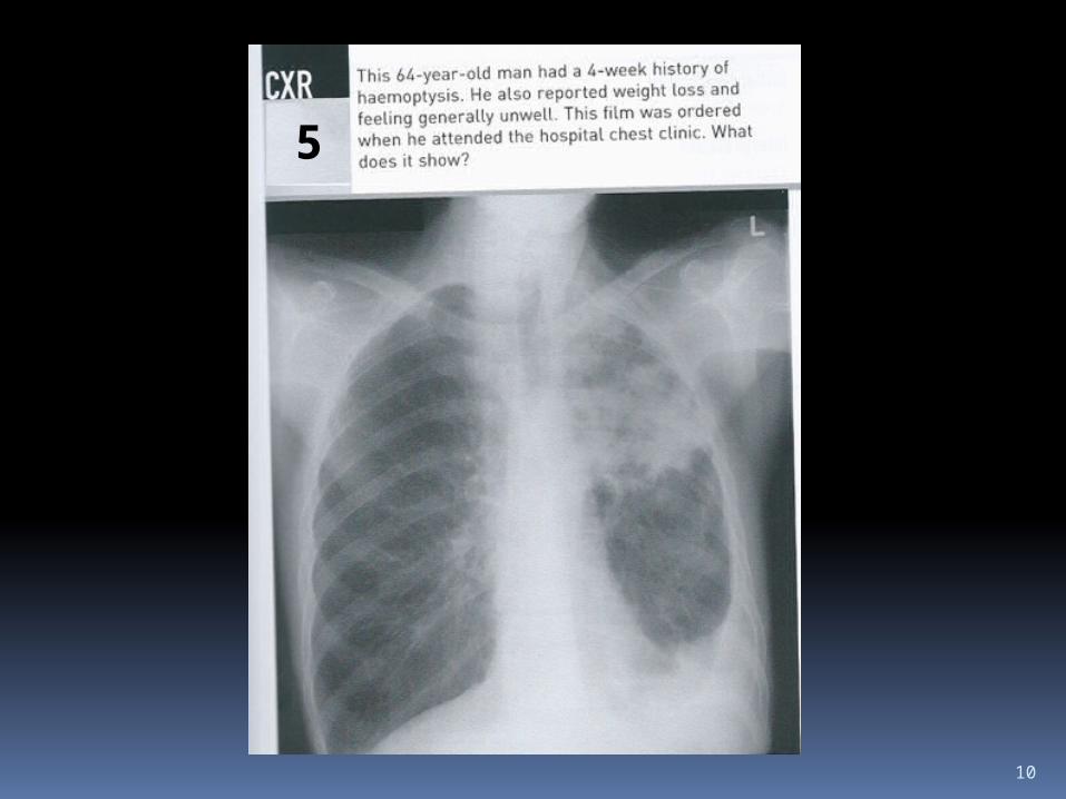
Answer 5
Pulmonary tuberculosis
12
6

13
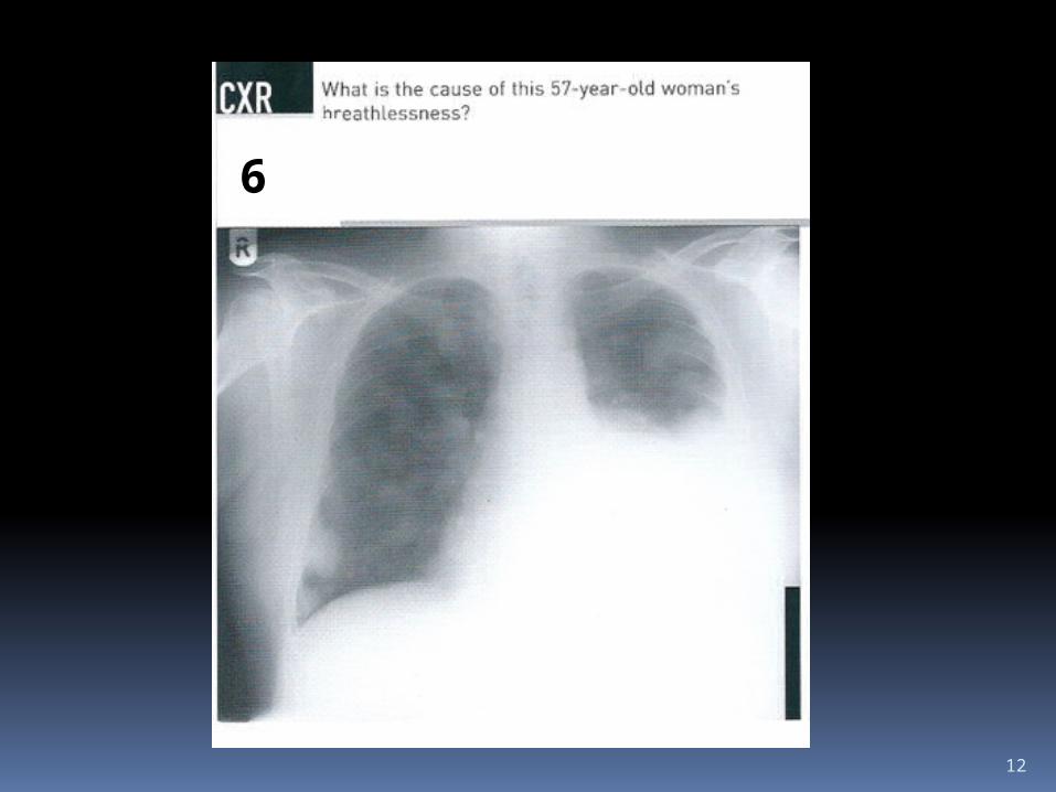
Answer 6
Malignant pleural effusion with pulmonary metastases

14
7

15
Answer 7
Consolidation left lung

16
8

17
Answer 8
Right pleural effusion Next investigation - Aspiration of this effusion and sent for cytology, microbiology and chemistry

18
9

19
Answer 9
Hodgkin’s lymphoma Mediastinal and right supraclavicular lymphadenopathy is present. Confirm by lymph node biopsy

20
10

21
Answer 10
Patient has pulmonary edema secondary to mitral and aortic valve disease

22
1111

23
Answer 11
Pericardial effusionThere is enlargement of heart shadow

24
12

25
Answer 12
Bilateral hilar lymphadenopathy Likely cause with this clinical history - SARCOIDOSIS

26
13

27
Answer 13
Chronic obstructive pulmonary disease (COPD)(Bilateral black lungs)

28
14
29
Answer 14
Chronic obstructive pulmonary disease (COPD)(Bilateral black lungs)
30
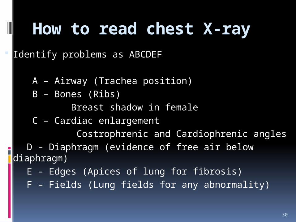
How to read chest X-ray Identify problems as ABCDEF A – Airway (Trachea position) B – Bones (Ribs) Breast shadow in female C – Cardiac enlargement Costrophrenic and Cardiophrenic angles D – Diaphragm (evidence of free air below diaphragm) E – Edges (Apices of lung for fibrosis) F – Fields (Lung fields for any abnormality)

31
CHEST X-RAY White Lung Field1. Pleural effusion2. Consolidation3. Collapse4. Cavitating lung lesion5. Left ventricular failure6. Fibrosis7. Pneumonectomy 8. Asbestos plaques9. Bronchiectasis10.Miliary shadowing

32
CHEST X-RAY
Black Lung Field1. Chronic obstructive pulmonary
disease2. Pneumothorax3. Tension Pneumothorax4. Pulmonary embolus

33
Thank you