1. cell structure 2014-15 upload
TRANSCRIPT

8/11/2019 1. Cell Structure 2014-15 Upload
http://slidepdf.com/reader/full/1-cell-structure-2014-15-upload 1/20
Topic A: Cell Structure
Bi ology AS Level 2014-15 1Notes by Adeel Ahmad Khokhar KSA
Notes
CELL STRUCTURE
Content
• The microscope in cell studies
• Cells as the basic units of living organisms
• Detailed structure of typical animal and plant cells, as seen using the electron
microscope
• Outline functions of organelles in plant and animal cells
• Characteristics of prokaryotic and eukaryotic cells
Learning Outcomes
Candidates should be able to:
(a) [PA] use an eyepiece graticule and stage micrometer scale to measure cells and be
familiar with units (millimetre, micrometre, nanometre) used in cell studies;
(b) explain and distinguish between resolution and magnification (see section 5), with
reference to light microscopy and electron microscopy;
(c) describe and interpret drawings and photographs of typical animal and plant cells,
as seen using the electron microscope, recognising the following: rough endoplasmic
reticulum and smooth endoplasmic reticulum, Golgi body (Golgi apparatus or Golgi
complex), mitochondria, ribosomes, lysosomes, chloroplasts, cell surface membrane,nuclear envelope, centrioles, nucleus, nucleolus, microvilli, cell wall, the large
permanent vacuole and tonoplast (of plant cells) and plasmodesmata. (knowledge that
ribosomes occurring in the mitochondria and chloroplasts are 70S (smaller) than those
in the rest of the cell (80S) should be included. The existence of small circular DNA in
the mitochondrion and chloroplast should be noted);
(d) outline the functions of the structures listed in (c);
(e) [PA] compare the structure of typical animal and plant cells;
(f) [PA] draw and label low power plan diagrams of tissues and organs (including a
transverse section of stems, roots and leaves);
(g) [PA] calculate linear magnification of drawings and photographs;
(h) [PA] calculate actual sizes of specimens from drawings and photographs;
(i) outline key structural features of typical prokaryotic cells (including: unicellular, 1-
5 μ m diameter, peptidoglycan cell walls, lack of membrane-bound organelles, naked
circular DNA, 70S ribosomes) and compare and contrast the structure of prokaryotic
cells with eukaryotic cells (reference to mesosomes should not be included);
(j) use the knowledge gained in this section in new situations or to solve related
problems.

8/11/2019 1. Cell Structure 2014-15 Upload
http://slidepdf.com/reader/full/1-cell-structure-2014-15-upload 2/20
Topic A: Cell Structure
Bi ology AS Level 2014-15 2Notes by Adeel Ahmad Khokhar KSA
Magnification
Magnification is the size of an image of an object compared to the actual size. It is
calculated using the formula M = I/A (M is magnification, I is the size of the image and
A is the actual size of the object, using the same units for both sizes). This formula can
be rearranged to give the actual size of an object where the size of the image andmagnification are known: A = I/M.
e.g., if a cell is 10m in diameter, and a microscope produces an image of it which is
1mm (1000m) in diameter, than the microscope has magnified the image 100 times.
(x100)
Magnification Calculations
Microscope drawings and photographs (micrographs) are usually magnified. There are
two ways of doing this:
1. Using a Magnification Factor
Sometimes the image has a magnification factor on it. The formula for the
magnification is:
For example if this drawing of an object is 40 mm long and
the magnification is x 1000, then the object's actual length is:
Answer has to be converted into appropriate units, usually μm for cells and organelles.
Sometimes you have to calculate the magnification. For
example if this drawing of an object is 40 mm long and its
actual length is 25 μm, the magnification of the drawing is:
Remember, the image and actual length must be in the same units. Magnifications can
also be less than one (e.g. x 0.1), which means that the drawing is smaller than the
actual object.
2. Using a Scale Bar
Sometimes the picture has a scale bar on it. The formula for calculating the actual
length is:
8/11/2019 1. Cell Structure 2014-15 Upload
http://slidepdf.com/reader/full/1-cell-structure-2014-15-upload 3/20
Topic A: Cell Structure
Bi ology AS Level 2014-15 3Notes by Adeel Ahmad Khokhar KSA
The image size and bar length must be measured in the same units (usually mm), and
the actual size will come out in the same units as the bar scale.
For example if this drawing of an object is 40 mm long and
the 5 μm scale bar is 10 mm long, then the object's actual
size is:
It's good to have a rough idea of the size of objects, to avoid silly mistakes. A
mitochondrion is not 30 mm long! Scale bars make this much easier than
magnification factors.
The magnification produced by a light microscope depends on the strengths of the
objective lens and the eyepiece lens. If you are using a ×40 objective lens and a ×10
eyepiece lens, then the image is being magnified 400 times.
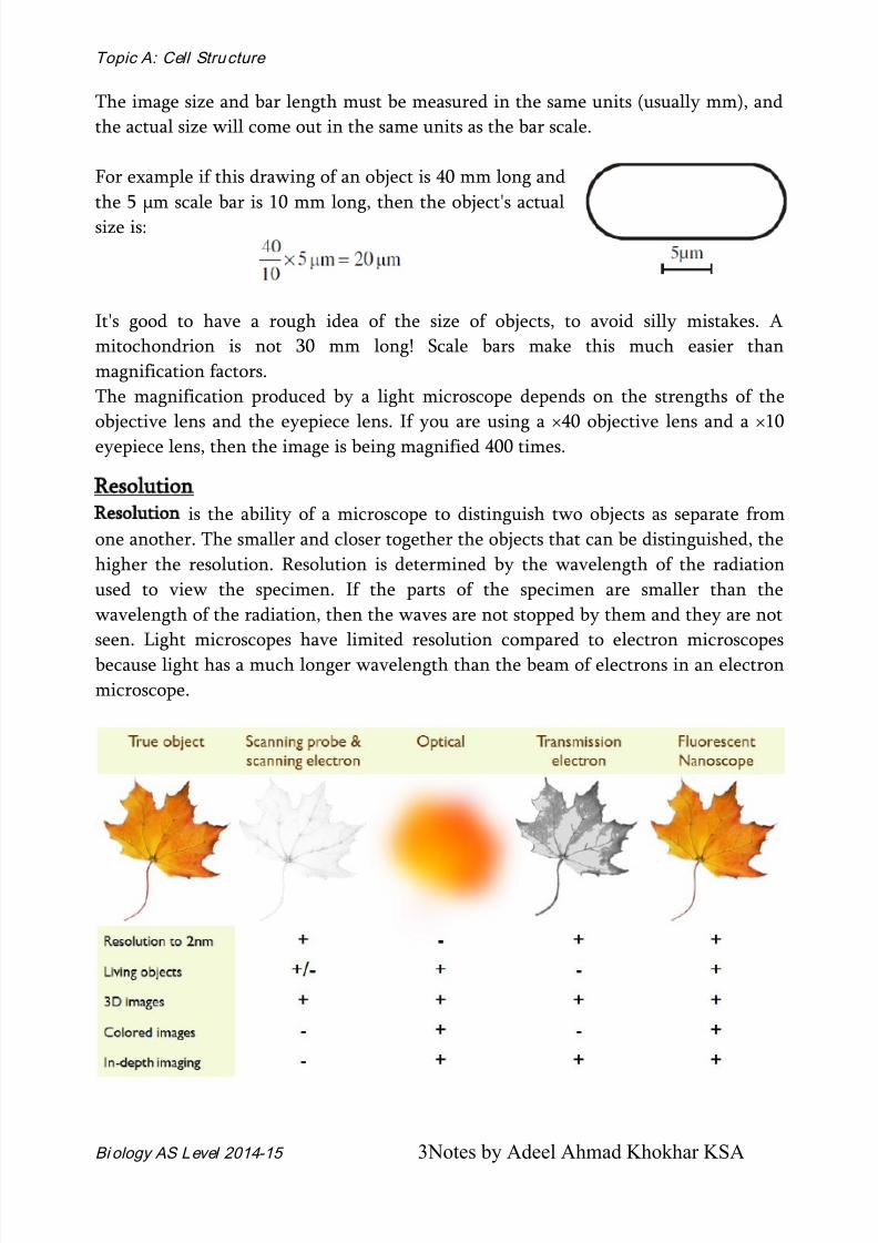
Resolution
Resolution is the ability of a microscope to distinguish two objects as separate from
one another. The smaller and closer together the objects that can be distinguished, the
higher the resolution. Resolution is determined by the wavelength of the radiation
used to view the specimen. If the parts of the specimen are smaller than the
wavelength of the radiation, then the waves are not stopped by them and they are not
seen. Light microscopes have limited resolution compared to electron microscopes
because light has a much longer wavelength than the beam of electrons in an electron
microscope.

8/11/2019 1. Cell Structure 2014-15 Upload
http://slidepdf.com/reader/full/1-cell-structure-2014-15-upload 4/20
Topic A: Cell Structure
Bi ology AS Level 2014-15 4Notes by Adeel Ahmad Khokhar KSA
The limit of resolution depends upon the wavelength of light. The resolution limit is
about 0.45 times the wavelength. Shorter wavelengths give the best (smallest)
resolution. The shortest wavelength light which we can see is blue light, which has a
wavelength of about 450 nm. This gives a resolution of about 0.45 x 450 nm, which is
close to 200 nm. Any objects smaller than this or any points less than 200 nm apart
will either be invisible or appear as blurs.
Electron beams, however, have a much shorter wavelength than light; so much better
resolutions can be achieved. Electron microscopes have a maximum resolution around
400 times better than that of light microscopes, being able to separate objects as little
as 0.5 nm apart.
Microscopy
Microscopy is the use of microscopes to study the structural details of organisms and
the organelles within the cell by magnifying the image.
Different kinds of Microscope.
1.
Light Microscope.
This is the oldest, simplest and most widely-used form of
microscope.
Specimens are illuminated with light, which is focused using glass lenses and viewedusing the eye or photographic film. Specimens can be living or dead, but often need to
60 m 30 m 15 m
3.7 m7.5 m 1.6 m

8/11/2019 1. Cell Structure 2014-15 Upload
http://slidepdf.com/reader/full/1-cell-structure-2014-15-upload 5/20
Topic A: Cell Structure
Bi ology AS Level 2014-15 5Notes by Adeel Ahmad Khokhar KSA
be coloured with a coloured stain to make them visible. Many different stains are
available that stain specific parts of the cell such as DNA, lipids, cytoskeleton, etc. All
light microscopes today are compound microscopes, which mean they use several
lenses to obtain high magnification.
Light microscopy has a resolution of about 200 nm, which is good enough to see
tissues and cells, but not the details of cell organelles. There has been a recent
resurgence in the use of light microscopy, partly due to technical improvements,
which have dramatically improved the resolution far beyond the theoretical limit. For
example fluorescence microscopy has a resolution of about 10 nm, while interference
microscopy has a resolution of about 1 nm.
2. Electron Microscope. This uses a beam of electrons, rather than
electromagnetic radiation, to "illuminate" the specimen. This may seem strange,
but electrons behave like waves and can easily be produced (using a hot wire),
focused (using electromagnets) and detected (using a phosphor screen or
photographic film).

8/11/2019 1. Cell Structure 2014-15 Upload
http://slidepdf.com/reader/full/1-cell-structure-2014-15-upload 6/20
Topic A: Cell Structure
Bi ology AS Level 2014-15 6Notes by Adeel Ahmad Khokhar KSA
A beam of electrons has an effective wavelength of less than 1 nm, so can be used to
resolve small sub-cellular ultrastructure. The development of the electron microscope
in the 1930s revolutionised biology, allowing organelles such as mitochondria, ER and
membranes to be seen in detail for the first time.
There are several problems with the electron microscopy: the electron beam is scattered by air molecules, so to avoid this there is a
vacuum inside an electron microscope, so it can't be used for living organisms.
specimens must be very thin, so are embedded in plastic for support, so can't be
manipulated under the microscope.
specimens can be damaged by the electron beam, so delicate structures and
molecules can be destroyed.
specimens are usually transparent to electrons, so must be stained with an
electron-dense chemical (usually heavy metals like osmium, lead or gold).
Initially there was a problem of artefacts (i.e. observed structures that were dueto the preparation process and were not real), but improvements in technique
have eliminated most of these.
There are two kinds of electron microscope. The transmission electron microscope
(TEM) works much like a light microscope, transmitting a beam of electrons through a
thin specimen and then focusing the electrons to form an image on a screen or on
film. This is the most common form of electron microscope and has the best
resolution. The scanning electron microscope (SEM) scans a fine beam of electron
onto a specimen and collects the electrons scattered by the surface. This has poorer
resolution, but gives excellent 3-dimentional images of surfaces.
Measurements used in Microscopy
1 centimeter (cm) = 1/100 meter = 0.4 inch = 10-2 m
1 millimeter (mm) = 1/1,000 meter = 1/10 cm = 10-3 m
1 micrometer (m) = 1/1,000,000 meter = 1/10,000 cm = 10-6 m
1 nanometer (nm) = 1/1,000,000,000 meter = 1/10,000,000 cm = 10-9 m
1 angstrom (A) = 1/10,000,000,000 meter = 1/100,000,000 cm = 10-10 m
1 meter = 102 cm = 103 mm = 106 m = 109 nm = 1010 A
3 a) Convert the following. All the answers are to be written in standard form.
0.00254 micrometer into millimeter
1.0665x10⁻⁵ nanometer into centimeter
6.211 x10⁻⁵ millimeter into nanometer
2449.88 micrometer into nanometer
b)
Calculate the magnification of a drawing 22 mm long of an object having an
actual length of 0.06 micrometer.
c)
What is the actual length of an organelle 4mm long shown in a drawing with amagnification of x4000.

8/11/2019 1. Cell Structure 2014-15 Upload
http://slidepdf.com/reader/full/1-cell-structure-2014-15-upload 7/20
Topic A: Cell Structure
Bi ology AS Level 2014-15 7Notes by Adeel Ahmad Khokhar KSA
Comparison between the Light microscope Electron Microscope
Light Microscope Electron Microscope
1. Source of radiation Uses light rays of
wavelength between 400to 700 nm
Uses electron beams of
wavelength 0.005 nm
2. Lenses Eyepiece and objective
lenses made of glass
Electromagnets
3. Stains used Dyes with suitable colours Heavy metals such as lead
4. Maximum
magnification
5. Maximum resolution
6. Depth of field
Disadvantages
It only magnifies objects to
an extent of 1500 times.
200 nm
The depth of field is
restricted.
Advantages
It is able to magnify objects
over 250,000 times (for
TEM) and over 100,000
times (SEM)
0.5 nm
The depth of field possible
is greater.
1.
Price and operation
2.
Portability
3.
Type of specimen
4.
Magnetic fields
5. Preparation of
specimen
6.
Image produced
Advantages
Inexpensive to purchase
and easier to operate.
Easy and small.
Living or non-living
organisms.
Magnetic fields have no
effect on it.
Preparation of specimen is
simple and fast
Coloured, with the naturalcolour of specimen or dye
Disadvantages
Expensive to purchase and
operation requires
expertise.
Bulky and less portable.
Only non-living organisms.
Magnetic fields have an
effect on it.
Preparation of specimen is
complex and needs
considerable time and
experience.
Black and white only

8/11/2019 1. Cell Structure 2014-15 Upload
http://slidepdf.com/reader/full/1-cell-structure-2014-15-upload 8/20
Topic A: Cell Structure
Bi ology AS Level 2014-15 8Notes by Adeel Ahmad Khokhar KSA
The Cell
All living things are made of cells, and cells are the smallest units that can be alive.
There are thousands of different kinds of cell, but the biggest division is between the
cells of the prokaryote kingdom (the bacteria) and those of the other four kingdoms
(animals, plants, fungi and protoctista), which are all eukaryotic cells. Prokaryotic
cells are smaller and simpler than eukaryotic cells, and do not have a nucleus.
Prokaryote = without a nucleus
Eukaryote = with a nucleus
These two kinds of cell are being examined in detail, based on structures seen in
electron micrographs.
These show the individual organelles inside a cell.
Euakryotic Cells
Cytoplasm (or Cytosol).
This is the solution within the cell membrane. It contains enzymes for
glycolysis (part of respiration) and other metabolic reactions together with sugars,
salts, amino acids, nucleotides and everything else needed for the cell to function.

8/11/2019 1. Cell Structure 2014-15 Upload
http://slidepdf.com/reader/full/1-cell-structure-2014-15-upload 9/20
Topic A: Cell Structure
Bi ology AS Level 2014-15 9Notes by Adeel Ahmad Khokhar KSA
Membrane Systems and Organelles:
Endoplasmic Reticulum (ER)
This is a series of membrane channels involved in synthesising and transporting
materials. Rough Endoplasmic Reticulum (RER) is studded with numerous ribosomes,
which give it its rough appearance. The ribosomes synthesise proteins, which are
processed in the RER (e.g. by enzymatically modifying the polypeptide chain, or
adding carbohydrates), before being exported from the cell via the Golgi Body.
Smooth Endoplasmic Reticulum (SER) does not have ribosomes and is used to process
materials, mainly lipids, needed by the cell.
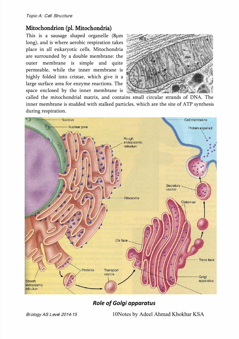
Golgi Apparatus
Another series of flattened
membrane vesicles, formed from
the endoplasmic reticulum. Its
job is to transport proteins from
the RER to the cell membrane
for export.
Parts of the RER containing
proteins fuse with one side of
the Golgi body membranes,
while at the other side small
vesicles bud off and movetowards the cell membrane, where they fuse, releasing their contents by exocytosis.
8/11/2019 1. Cell Structure 2014-15 Upload
http://slidepdf.com/reader/full/1-cell-structure-2014-15-upload 10/20
Topic A: Cell Structure
Bi ology AS Level 2014-15 10Notes by Adeel Ahmad Khokhar KSA
Mitochondrion (pl. Mitochondria)
This is a sausage shaped organelle (8μm
long), and is where aerobic respiration takes
place in all eukaryotic cells. Mitochondria
are surrounded by a double membrane: the
outer membrane is simple and quite
permeable, while the inner membrane is
highly folded into cristae, which give it a
large surface area for enzyme reactions. The
space enclosed by the inner membrane is
called the mitochondrial matrix, and contains small circular strands of DNA. The
inner membrane is studded with stalked particles, which are the site of ATP synthesis
during respiration.
Role of Golgi apparatus

8/11/2019 1. Cell Structure 2014-15 Upload
http://slidepdf.com/reader/full/1-cell-structure-2014-15-upload 11/20
Topic A: Cell Structure
Bi ology AS Level 2014-15 11Notes by Adeel Ahmad Khokhar KSA
Ribosomes
These are the smallest and
most numerous of the cell
organelles, and are the sites of
protein synthesis. They arecomposed of protein and
RNA, and are manufactured
in the nucleolus of the
nucleus.
Ribosomes are either found
free in the cytoplasm, where
they make proteins for the cell's own use, or they are found attached to the rough
endoplasmic reticulum, where they make proteins for export from the cell. All
eukaryotic ribosomes are of the larger, "80S", type.
Lysosomes
These are small membrane-bound vesicles formed from the RER containing a cocktail
of digestive enzymes. They are used to break down unwanted chemicals, toxins,
organelles or even whole cells, so that the materials may be recycled. They can also
fuse with a feeding vacuole to digest its contents.
Role of Lysosome in a cell

8/11/2019 1. Cell Structure 2014-15 Upload
http://slidepdf.com/reader/full/1-cell-structure-2014-15-upload 12/20
Topic A: Cell Structure
Bi ology AS Level 2014-15 12Notes by Adeel Ahmad Khokhar KSA
It is very important that the enzymes contained within Lysosomes are isolated from
the rest of the cell inside the Lysosomes membrane, otherwise their release would
result in self-digestion of the cell. In fact, this sometimes happens in certain tissues,
such as the tadpole’s tail when it is changing into a frog; this process is called apoptosis
or programmed cell death
Centrioles
This is a special pair of short cytoskeleton
fibres involved in cell division. They initiate
the spindle that organises and separates the
chromosomes.
Cell Surface Membrane
The cell surface membrane is the boundary
between the cell and its environment. It has
little mechanical strength but plays a vital
role in controlling which materials pass in and out of the cell.
Although basically a double layer of phospholipids molecules, arranged tail to tail, cellsurface membrane is a complex structure, studded with proteins. These can be
embedded in the membrane or they can penetrate the bilayer forming pores through
which molecules can pass.
Centrioles

8/11/2019 1. Cell Structure 2014-15 Upload
http://slidepdf.com/reader/full/1-cell-structure-2014-15-upload 13/20
Topic A: Cell Structure
Bi ology AS Level 2014-15 13Notes by Adeel Ahmad Khokhar KSA
Chloroplasts
Bigger and fatter than mitochondria, chloroplasts are where photosynthesis takes
place, so are only found in photosynthetic organisms (plants and algae).
Like mitochondria they are enclosed by a double membrane, but chloroplasts also
have a third membrane called the thylakoid membrane. The thylakoid membrane is
folded into thylakoid disks, which are then stacked into piles called grana. The space
between the inner membrane and the thylakoid is called the stroma. The thylakoid
membrane contains chlorophyll and chloroplasts also contain starch grains, ribosomes
and circular DNA.
Large Permanent Vacuole and Tonoplast (of Plant Cells):
Vacuoles are membrane-bound sacs within the cytoplasm of a cell that function in
several different ways. In mature plant cells, vacuoles tend to be very large and are
extremely important in providing structural support, as well as serving functions such
as storage, waste disposal, protection, and growth. Many plant cells have a large, single
central vacuole that typically takes up most
of the room in the cell (80 percent or more).
Vacuoles in animal cells, however, tend to be
much smaller, and are more commonly used
to temporarily store materials or to transport
substances.
The central vacuole in plant cells is enclosed
by a membrane termed the tonoplast, an
important and highly integrated componentof the plant internal membrane network

8/11/2019 1. Cell Structure 2014-15 Upload
http://slidepdf.com/reader/full/1-cell-structure-2014-15-upload 14/20
Topic A: Cell Structure
Bi ology AS Level 2014-15 14Notes by Adeel Ahmad Khokhar KSA
(endomembrane) system. This large vacuole slowly develops as the cell matures by
fusion of smaller vacuoles derived from the endoplasmic reticulum and Golgi
apparatus. Because the central vacuole is highly selective in transporting materials
through its membrane, the chemical palette of the vacuole solution (termed the cell
sap) differs markedly from that of the surrounding cytoplasm.
Plasmodesmata:
Plasmodesmata (singular, plasmodesma) are small channels that directly connect the
cytoplasm of neighboring plant cells to each other, establishing living bridges between
cells. Similar to the gap junctions found in animal cells, the plasmodesmata, which
penetrate both the primary and secondary cell walls, allow certain molecules to pass
directly from one cell to another and are important in cellular communication.
Nucleus:
This is the largest
organelle. It is
surrounded by a
nuclear envelope,
which is a doublemembrane with
nuclear pores–large
holes containing
proteins that
control the exit of
substances such as
RNA and ribosomes
from the nucleus. The interior is called the nucleoplasm, which is full of chromatin –
the DNA/protein complex.

8/11/2019 1. Cell Structure 2014-15 Upload
http://slidepdf.com/reader/full/1-cell-structure-2014-15-upload 15/20
Topic A: Cell Structure
Bi ology AS Level 2014-15 15Notes by Adeel Ahmad Khokhar KSA
During cell division the chromatin becomes condensed into discrete observable
chromosomes.
The nucleolus is a dark region of chromatin, involved in making ribosomes.
Euchromatin and Heterochromatin
The DNA in the nucleus exists in two forms that reflect the level of activity of the cell.
Heterochromatin appears as small, darkly staining, irregular particles scattered
throughout the nucleus or accumulated adjacent to the nuclear envelope.
Euchromatin is dispersed and not readily stainable. Euchromatin is prevalent in cells
that are active in the transcription of many of their genes while heterochromatin is
most abundant in cells that are less active or not active.
Functions of Nucleus
To control all cellular activities.
To produce RNA.
To control the synthesis of proteins, including enzymes, in the cell, and so
control the cell’s activities.
To undergo nuclear division in the start of cell division, ensuring that the
daughter cells have exact copies of the cell’s genetic material in their
chromosomes.
To assemble ribosomes (function of the nucleolus).
8/11/2019 1. Cell Structure 2014-15 Upload
http://slidepdf.com/reader/full/1-cell-structure-2014-15-upload 16/20
Topic A: Cell Structure
Bi ology AS Level 2014-15 16Notes by Adeel Ahmad Khokhar KSA
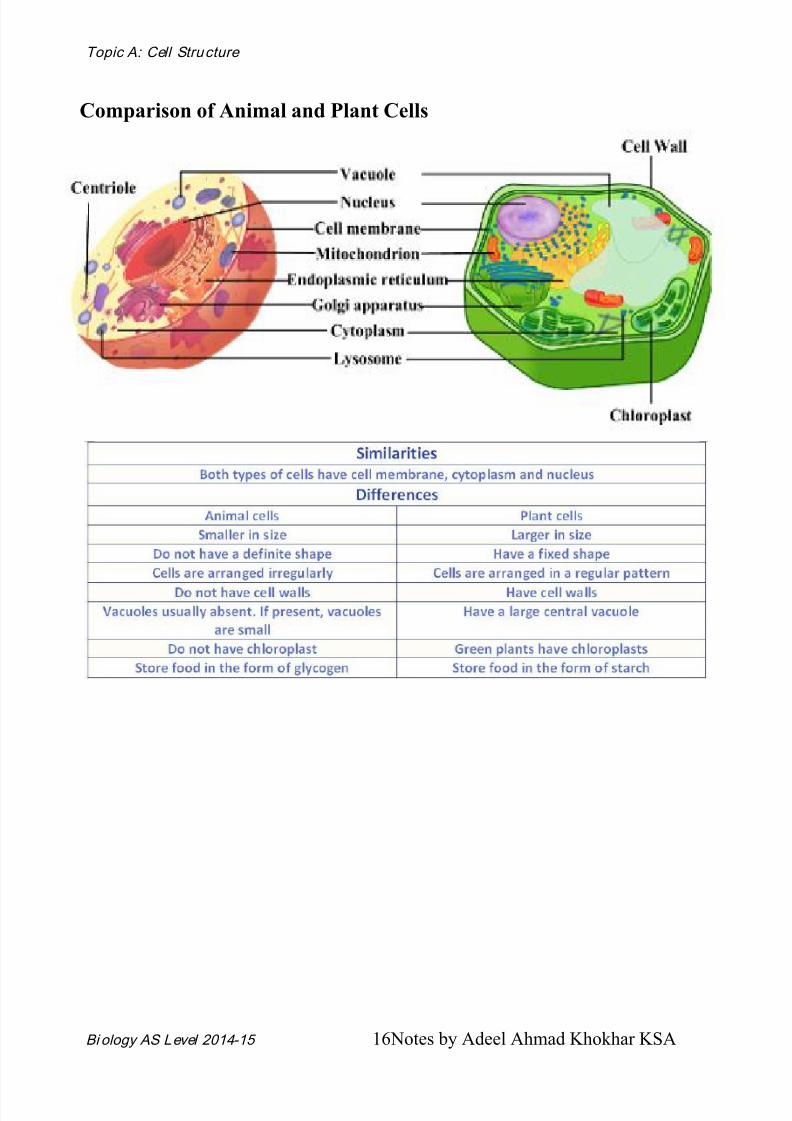
Comparison of Animal and Plant Cells

8/11/2019 1. Cell Structure 2014-15 Upload
http://slidepdf.com/reader/full/1-cell-structure-2014-15-upload 17/20
Topic A: Cell Structure
Bi ology AS Level 2014-15 17Notes by Adeel Ahmad Khokhar KSA
Double membranous
organelles
Single membranous
organelles
Non-membranous
organelles
Nucleus Endoplasmic reticula ribosome
Chloroplast Golgi apparatus
Mitochondria Lysosome
Organelle Location and
occurrence in cell
Size Function
Nucleus Usually one per cell in
cytoplasm
10 - 20m Contains the hereditary material
(DNA) which codes for synthesis of
proteins in the cytoplasmNucleolus One to several in
nucleus
1 - 2m Synthesizes ribosomal RNA and
manufactures ribosomes
Rough
endoplasmic
reticulum
Throughout cytoplasm Membranes
about 4nm
thick,
enclosing
Transport of proteins synthesized
on ribosomes
Smooth
endoplasmic
reticulum
In cytoplasm. Extent
depends on types of
cell
Cisternae of
varying
diameter
Synthesis of lipids
Ribosomes Attached to rough
endoplasmic reticulum
or free in cytoplasm
20 – 25 nm Site of protein synthesis
Golgi
apparatus
In cytoplasm Variable Synthesis of glycoproteins,
packaging of proteins.
Lysosomes In cytoplasm 100nm Digestion of unwanted materials
and worn-out organelles
Mitochondria In cytoplasm. Severalto thousands per cell.
1 m wideand up to 10
m in
length
Production of energy by aerobicrespiration
Centrioles Pair, in cytoplasm,
usually near nucleus
0.5 m X 0.2
m
Form the spindle fibres during cell
division of animal and fungal cells
Chloroplasts In cytoplasm of some
plant cells
2 – 10 m in
diameter
Site of photosynthesis
Summary of Cell Organelles

8/11/2019 1. Cell Structure 2014-15 Upload
http://slidepdf.com/reader/full/1-cell-structure-2014-15-upload 18/20
Topic A: Cell Structure
Bi ology AS Level 2014-15 18Notes by Adeel Ahmad Khokhar KSA
Prokaryotic Cells
1. The word prokaryotes means ‘before nucleus’. This describes the main
difference between eukaryotic and prokaryotic cells: prokaryotes have no
nucleus or nuclear membrane. Their DNA is therefore not separated from the
cytoplasm, but forms a single circular loop, sometimes called a bacterialchromosome.
2. Their DNA is not associated with proteins, unlike eukaryotic chromosomes.
Bacteria also have smaller loops of DNA in the cytoplasm, called plasmids.
3. Prokaryotic cells are much smaller than eukaryotic ones, and much simpler in
their structure.
4. They lack endoplasmic reticulum and membrane-bound organelles like
mitochondria and chloroplasts and any complex structures such as Golgi bodies,
cytoskeleton or lyososmes.
Structure of Bacterium (A Prokaryotic Cell)
Prokaryotic cells are smaller than
eukaryotic cells and do not have a
nucleus or indeed any membrane-
bound organelles. All prokaryotes are
bacteria. Prokaryotic cells are much
older than eukaryotic cells and they
are far more abundant (there are tentimes as many bacteria cells in a
human than there are human cells).
The main features of prokaryotic
cells are:
Cytoplasm. Contains all the
enzymes needed for all metabolic
reactions, since there are no
organelles
Ribosomes. The smaller (70S) type,
all free in the cytoplasm and never attached to membranes. Used for protein synthesis.
Nuclear Zone (or Nucleoid). The region of the cytoplasm that contains DNA. It is
not surrounded by a nuclear membrane.
DNA. Always circular (i.e. a closed loop), and not associated with any proteins to
form chromatin. Sometimes confusingly referred to as the bacterial chromosome.

8/11/2019 1. Cell Structure 2014-15 Upload
http://slidepdf.com/reader/full/1-cell-structure-2014-15-upload 19/20
Topic A: Cell Structure
Bi ology AS Level 2014-15 19Notes by Adeel Ahmad Khokhar KSA
Plasmid. Small circles of DNA, separate from the main DNA loop. Used to exchange
DNA between bacterial cells, and also very useful for genetic engineering.
Cell membrane. Made of phospholipids and proteins, like eukaryotic membranes.
Mesosome. A tightly-folded region of the cell membrane containing all themembrane-bound proteins required for respiration and photosynthesis. Can also be
associated with the nucleoid.
This is now thought to be an artefact of the electron microscope and not real
structure.
Cell Wall. Made of murein (not cellulose), which is a glycoprotein (i.e. a
protein/carbohydrate complex, also called peptidoglycan).
Capsule. A thick polysaccharide layer outside of the cell wall. Used for sticking cells
together, as a food reserve, as protection against desiccation and chemicals, and as
protection against phagocytosis. In some species the capsules of many cells fuse
together forming a mass of sticky cells called a biofilm. Dental plaque is an example of
a biofilm.
Flagellum. A rigid rotating helical-shaped tail used for propulsion. The motor is
embedded in the cell membrane and is driven by a H+ gradient across the membrane.
Anticlockwise rotation drives the cell forwards, while clockwise rotation causes a
chaotic spin.

8/11/2019 1. Cell Structure 2014-15 Upload
http://slidepdf.com/reader/full/1-cell-structure-2014-15-upload 20/20
Topic A: Cell Structure
Bi ology AS Level 2014-15 20Notes by Adeel Ahmad Khokhar KSA
Differences between eukaryotic and prokaryotic cells
Eukaryotic Cells Prokaryotic Cells
1. True nucleus surrounded by a nuclear
envelop.
No true nucleus
2. Linear DNA associated with histone
proteins forming true chromosomes.
Circular DNA not associated with
proteins. Separate loops of DNA called
plasmids.3. Cell wall, if present, made of cellulose
(plants and algae) or chitin (fungi).
Cell wall containing peptidoglycan
4. Endoplasmic reticulum present. No endoplasmic reticulum or associated
organelles such as Golgi apparatus.
5. Membrane–bound organelles such as
mitochondria and chloroplast (in plants
and algae).
No membrane-bound organelles.
Mesosomes and thylakoids present in
some bacteria.
6. Large (80S) ribosomes attached to
endoplasmic reticulum.
Small (70S) ribosomes scattered in the
cytoplasm.
7. If present, flagella have (9+2)
arrangement of microtubules.
If present, flagella are made of a single
microtubule.
8. Cells are large, typically 10-100m in
diameter, some cells can be up to 400m.
Cells are small, typically 0.5-3m in
diameter, Volume may be as little as
1/10,000th of eukaryotic cell.