1 chapter 19 lecture outline see powerpoint image slides for all figures and tables pre-inserted...
TRANSCRIPT

11
Chapter 19Chapter 19
Lecture OutlineLecture Outline
See PowerPoint Image SlidesSee PowerPoint Image Slides
for all figures and tables pre-inserted intofor all figures and tables pre-inserted into
PowerPoint without notes.PowerPoint without notes.
Copyright (c) The McGraw-Hill Companies, Inc. Permission required for reproduction or display.

22
Circulatory System: The HeartCirculatory System: The Heart
Gross anatomy of the heartGross anatomy of the heart Overview of cardiovascular systemOverview of cardiovascular system Cardiac conduction system and cardiac Cardiac conduction system and cardiac
musclemuscle Electrical and contractile activity of heartElectrical and contractile activity of heart Blood flow, heart sounds, and cardiac Blood flow, heart sounds, and cardiac
cyclecycle Cardiac outputCardiac output

33
Circulatory System: The HeartCirculatory System: The Heart
Circulatory system Circulatory system heart, blood vessels and bloodheart, blood vessels and blood
Cardiovascular system Cardiovascular system heart, arteries, veins and capillariesheart, arteries, veins and capillaries
Two major divisions:Two major divisions: Pulmonary circuit - right side of heartPulmonary circuit - right side of heart
carries blood to lungs for gas exchangecarries blood to lungs for gas exchange Systemic circuit - left side of heartSystemic circuit - left side of heart
supplies blood to all organs of the bodysupplies blood to all organs of the body

44
Cardiovascular System CircuitCardiovascular System Circuit

55
Position, Size, and ShapePosition, Size, and Shape Located in Located in
mediastinum, between mediastinum, between lungslungs
Base - broad superior Base - broad superior portion of heartportion of heart
Apex - inferior end, tilts Apex - inferior end, tilts to the left, tapers to to the left, tapers to pointpoint

66
Heart PositionHeart Position

77
PericardiumPericardium Allows heart to beat without friction, room to expand and Allows heart to beat without friction, room to expand and
resists excessive expansionresists excessive expansion Parietal pericardiumParietal pericardium
outer, tough, fibrous layer of CTouter, tough, fibrous layer of CT Pericardial cavity Pericardial cavity
filled with pericardial fluidfilled with pericardial fluid Visceral pericardium (a.k.a. epicardium of heart wall)Visceral pericardium (a.k.a. epicardium of heart wall)
inner, thin, smooth, moist serous layer inner, thin, smooth, moist serous layer covers heart surfacecovers heart surface

88
Pericardium and Pericardium and Heart WallHeart Wall
Pericardial cavity contains 5-30 ml of pericardial fluidPericardial cavity contains 5-30 ml of pericardial fluid

99
Heart WallHeart Wall Epicardium Epicardium (a.k.a. visceral pericardium)(a.k.a. visceral pericardium)
serous membrane covers heartserous membrane covers heart MyocardiumMyocardium
thick muscular layerthick muscular layer fibrous skeleton - network of collagenous and fibrous skeleton - network of collagenous and
elastic fiberselastic fibers• provides structural support and attachment for cardiac provides structural support and attachment for cardiac
musclemuscle• electrical nonconductor, important in coordinating electrical nonconductor, important in coordinating
contractile activitycontractile activity
Endocardium - smooth inner liningEndocardium - smooth inner lining

1010
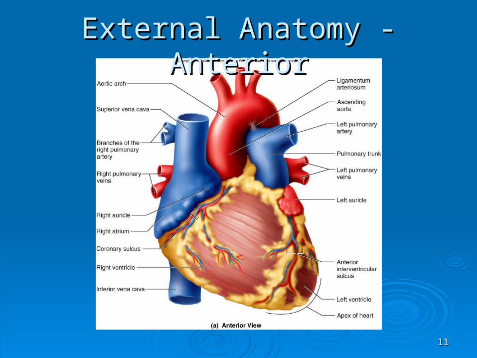
Heart ChambersHeart Chambers 4 chambers4 chambers
right and left atria right and left atria • two superior, posterior two superior, posterior
chamberschambers• receive blood returning receive blood returning
to heartto heart right and left right and left
ventricles ventricles • two inferior chamberstwo inferior chambers• pump blood into pump blood into
arteriesarteries
1111
External Anatomy - AnteriorExternal Anatomy - Anterior

1212
External Anatomy - PosteriorExternal Anatomy - Posterior

1313
Heart Chambers - InternalHeart Chambers - Internal
Interatrial septumInteratrial septum wall that separates atriawall that separates atria
Pectinate musclesPectinate muscles internal ridges of myocardium in right atrium internal ridges of myocardium in right atrium
and both auriclesand both auricles Interventricular septumInterventricular septum
wall that separates ventricleswall that separates ventricles Trabeculae carneaeTrabeculae carneae
internal ridges in both ventriclesinternal ridges in both ventricles

1414
Internal Anatomy - AnteriorInternal Anatomy - Anterior

1515
Heart ValvesHeart Valves Atrioventricular (AV) valvesAtrioventricular (AV) valves
right AV valve has 3 cusps (tricuspid valve)right AV valve has 3 cusps (tricuspid valve) left AV valve has 2 cusps (mitral, bicuspid valve)left AV valve has 2 cusps (mitral, bicuspid valve) chordae tendineae - cords connect AV valves to chordae tendineae - cords connect AV valves to
papillary muscles (on floor of ventricles)papillary muscles (on floor of ventricles) Semilunar valves - control flow into great Semilunar valves - control flow into great
arteriesarteries pulmonary: right ventricle into pulmonary trunkpulmonary: right ventricle into pulmonary trunk aortic: from left ventricle into aortaaortic: from left ventricle into aorta

1616
Heart ValvesHeart Valves

1717
Heart ValvesHeart Valves
1818
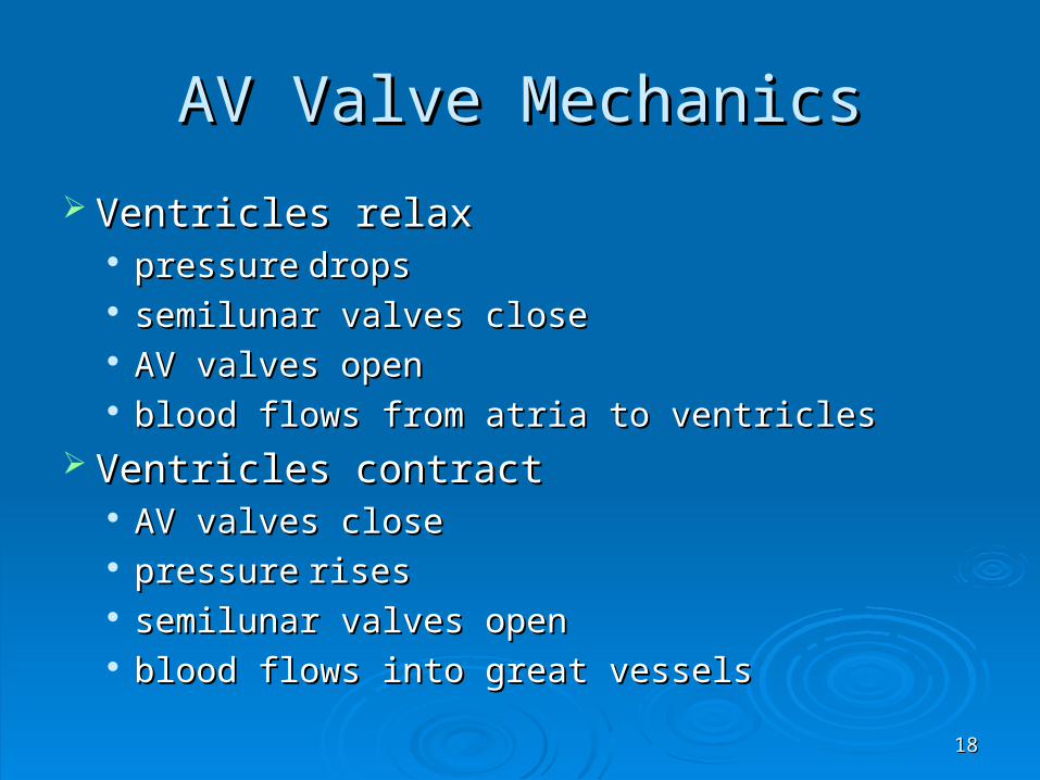
AV Valve MechanicsAV Valve Mechanics
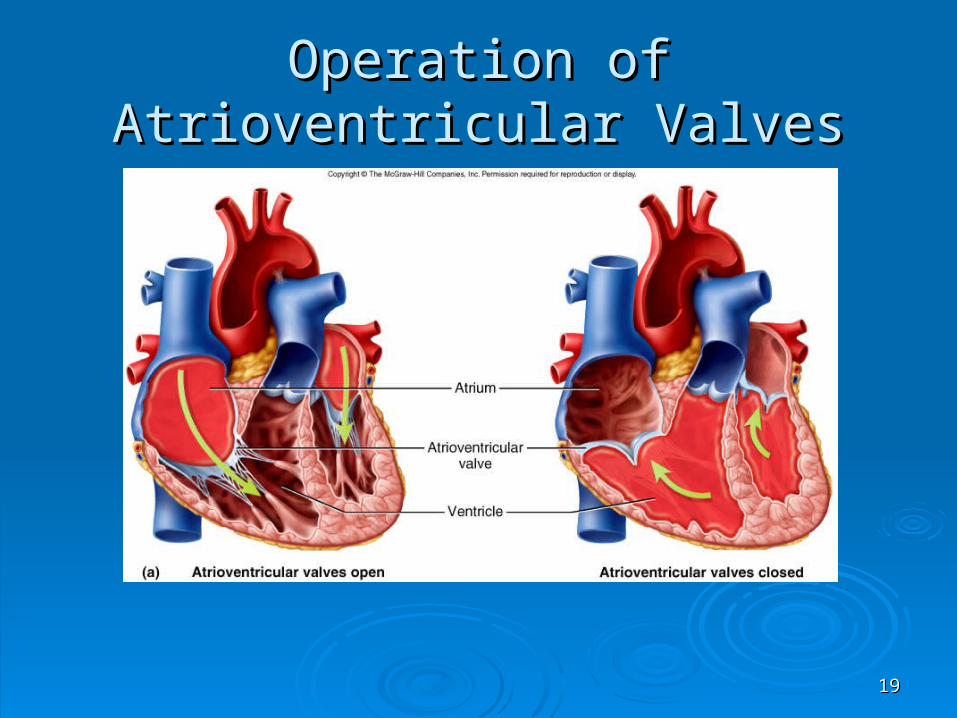
Ventricles relaxVentricles relax pressurepressure dropsdrops semilunar valves closesemilunar valves close AV valves openAV valves open blood flows from atria to ventriclesblood flows from atria to ventricles
Ventricles contractVentricles contract AV valves closeAV valves close pressurepressure risesrises semilunar valves opensemilunar valves open blood flows into great vesselsblood flows into great vessels
1919
Operation of Atrioventricular ValvesOperation of Atrioventricular Valves

2020
Operation of Semilunar ValvesOperation of Semilunar Valves

2121
Blood Flow Through HeartBlood Flow Through Heart

2222
Coronary CirculationCoronary Circulation Left coronary artery (LCA)Left coronary artery (LCA)
anterior interventricular branchanterior interventricular branch• supplies blood to interventricular septum and anterior supplies blood to interventricular septum and anterior
walls of ventricleswalls of ventricles circumflex branchcircumflex branch
• passes around left side of heart in coronary sulcus, passes around left side of heart in coronary sulcus, supplies left atrium and posterior wall of left ventricle supplies left atrium and posterior wall of left ventricle
Right coronary artery (RCA)Right coronary artery (RCA) right marginal branch right marginal branch
• supplies lateral R atrium and ventriclesupplies lateral R atrium and ventricle posterior interventricular branchposterior interventricular branch
• supplies posterior walls of ventriclessupplies posterior walls of ventricles

2323
Coronary Vessels - AnteriorCoronary Vessels - Anterior

2424
Venous Drainage of HeartVenous Drainage of Heart
20% drains directly into all 4 chambers via the 20% drains directly into all 4 chambers via the thebesian veinsthebesian veins
80% returns to right atrium via:80% returns to right atrium via: great cardiac vein great cardiac vein
• blood from anterior interventricular sulcusblood from anterior interventricular sulcus middle cardiac vein middle cardiac vein
• from posterior sulcusfrom posterior sulcus left marginal veinleft marginal vein coronary sinus coronary sinus
• collects blood and empties into right atrium collects blood and empties into right atrium

2525
Coronary Vessels - PosteriorCoronary Vessels - Posterior

2626
Angina and Heart AttackAngina and Heart Attack
Angina pectoris Angina pectoris partial obstruction of coronary blood flow can partial obstruction of coronary blood flow can
cause chest pain cause chest pain pain caused by ischemia, often activity pain caused by ischemia, often activity
dependent dependent Myocardial infarction Myocardial infarction
complete obstruction causes death of cardiac complete obstruction causes death of cardiac cells in affected areacells in affected area
pain or pressure in chest that often radiates down pain or pressure in chest that often radiates down left armleft arm

2727
Nerve Supply to HeartNerve Supply to Heart
Sympathetic nerves from Sympathetic nerves from upper thoracic spinal cord, through sympathetic upper thoracic spinal cord, through sympathetic
chain to cardiac nerveschain to cardiac nerves directly to ventricular myocardiumdirectly to ventricular myocardium can raise heart rate to 230 bpmcan raise heart rate to 230 bpm
Parasympathetic nervesParasympathetic nerves right vagal nerve to SA noderight vagal nerve to SA node left vagal nerve to AV nodeleft vagal nerve to AV node vagal tone – normally slows heart rate to vagal tone – normally slows heart rate to
70 - 80 bpm70 - 80 bpm

2828
Cardiac Conduction SystemCardiac Conduction System PropertiesProperties
myogenic - heartbeat originates within heartmyogenic - heartbeat originates within heart autorhythmic – regular, spontaneous depolarizationautorhythmic – regular, spontaneous depolarization
ComponentsComponents next slidenext slide
2929
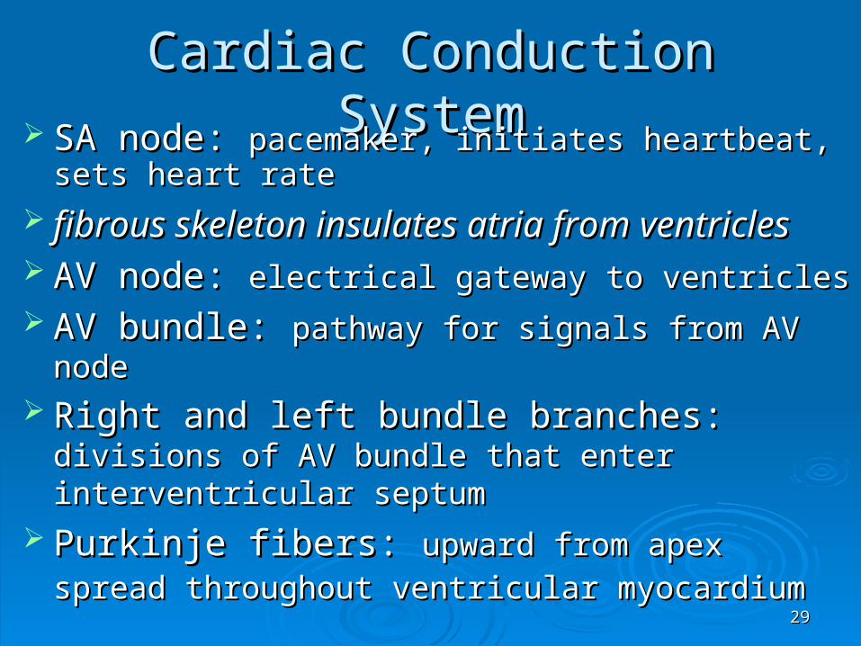
Cardiac Conduction SystemCardiac Conduction System SA node: SA node: pacemaker, initiates heartbeat, sets heart pacemaker, initiates heartbeat, sets heart
raterate
fibrous skeleton insulates atria from ventriclesfibrous skeleton insulates atria from ventricles AV node: AV node: electrical gateway to ventricleselectrical gateway to ventricles AV bundle: AV bundle: pathway for signals from AV nodepathway for signals from AV node Right and left bundle branches: Right and left bundle branches: divisions of AV divisions of AV
bundle that enter interventricular septumbundle that enter interventricular septum Purkinje fibers: Purkinje fibers: upward from apex spread upward from apex spread
throughout ventricular myocardiumthroughout ventricular myocardium

3030
Cardiac Conduction SystemCardiac Conduction System

3131
Structure of Cardiac MuscleStructure of Cardiac Muscle Short, branched cells, one central nucleusShort, branched cells, one central nucleus Sarcoplasmic reticulum, large T-tubules Sarcoplasmic reticulum, large T-tubules
admit more Caadmit more Ca2+2+ from ECF from ECF Intercalated discs Intercalated discs join myocytes end to endjoin myocytes end to end
interdigitating folds - interdigitating folds - surface area surface area mechanical junctions tightly join myocytesmechanical junctions tightly join myocytes electrical junctions - gap junctions allow ions to electrical junctions - gap junctions allow ions to
flowflow

3232
Structure of Cardiac Muscle CellStructure of Cardiac Muscle Cell

3333
Metabolism of Cardiac MuscleMetabolism of Cardiac Muscle
Aerobic respirationAerobic respiration Rich in myoglobin and glycogenRich in myoglobin and glycogen Large mitochondria Large mitochondria Organic fuels: fatty acids, glucose, Organic fuels: fatty acids, glucose,
ketonesketones Fatigue resistant Fatigue resistant

3434
Cardiac RhythmCardiac Rhythm
Systole – ventricular contractionSystole – ventricular contraction Diastole - ventricular relaxationDiastole - ventricular relaxation Sinus rhythmSinus rhythm
set by SA node at 60 – 100 bpmset by SA node at 60 – 100 bpm adult at rest is 70 to 80 bpm (vagal inhibition)adult at rest is 70 to 80 bpm (vagal inhibition)

3535
Depolarization of SA NodeDepolarization of SA Node SA node - no stable resting membrane potentialSA node - no stable resting membrane potential Pacemaker potential Pacemaker potential
gradual depolarization gradual depolarization from -60 mVfrom -60 mV, slow influx of Na, slow influx of Na++
Action potential Action potential occurs at threshold of occurs at threshold of -40 mV-40 mV depolarizing phase depolarizing phase to 0 mVto 0 mV
• fast Cafast Ca2+2+ channels open, (Ca channels open, (Ca2+ 2+ in) in) repolarizing phaserepolarizing phase
• KK++ channels open, (K channels open, (K++ out) out)• atat -60 mV-60 mV K K++ channels close, pacemaker potential starts over channels close, pacemaker potential starts over
Each depolarization creates one heartbeatEach depolarization creates one heartbeat SA node at rest fires at 0.8 sec, about 75 bpmSA node at rest fires at 0.8 sec, about 75 bpm

3636
SA Node PotentialsSA Node Potentials

3737
Impulse Conduction to MyocardiumImpulse Conduction to Myocardium
SA node signal travels at SA node signal travels at 1 m/sec1 m/sec through atria through atria AV node slows signal to AV node slows signal to 0.05 m/sec0.05 m/sec
thin myocytes with fewer gap junctionsthin myocytes with fewer gap junctions delays signal 100 msec, allows ventricles to fill delays signal 100 msec, allows ventricles to fill
AV bundle and purkinje fibersAV bundle and purkinje fibers speeds signal along at speeds signal along at 4 m/sec4 m/sec to ventricles to ventricles
Ventricular systole begins at apex, progresses Ventricular systole begins at apex, progresses upup spiral arrangement of myocytes twists ventricles spiral arrangement of myocytes twists ventricles
slightlyslightly

3838
Contraction of MyocardiumContraction of Myocardium
Myocytes have stable resting potential of -90 mVMyocytes have stable resting potential of -90 mV Depolarization Depolarization (very brief)(very brief)
stimulus opens voltage regulated Nastimulus opens voltage regulated Na++ gates, (Na gates, (Na++ rushes rushes in) membrane depolarizes rapidlyin) membrane depolarizes rapidly
action potential peaks at +30 mV action potential peaks at +30 mV NaNa++ gates close quickly gates close quickly
Plateau - 200 to 250 msec, sustains contractionPlateau - 200 to 250 msec, sustains contraction slow Caslow Ca2+2+ channels open, Ca channels open, Ca2+2+ binds to fast Ca binds to fast Ca2+2+
channels on SR, releases channels on SR, releases CaCa2+2+ into cytosol: into cytosol: contractioncontraction Repolarization - CaRepolarization - Ca2+2+ channels close, K channels close, K++ channels channels
open, rapid Kopen, rapid K++ out returns to resting potential out returns to resting potential

3939
Action Potential of MyocyteAction Potential of Myocyte
1) Na1) Na++ gates open gates open
2) Rapid 2) Rapid depolarizationdepolarization
3) Na3) Na++ gates close gates close
4) Slow Ca4) Slow Ca2+2+ channels openchannels open
5) Ca5) Ca2+2+ channels channels close, Kclose, K++ channels openchannels open

4040
Electrocardiogram (ECG)Electrocardiogram (ECG) Composite of all action potentials of nodal and Composite of all action potentials of nodal and
myocardial cells detected, amplified and recorded by myocardial cells detected, amplified and recorded by electrodes on arms, legs and chestelectrodes on arms, legs and chest

4141
ECGECG P waveP wave
SA node fires, atrial depolarizationSA node fires, atrial depolarization atrial systoleatrial systole
QRS complexQRS complex ventricular depolarizationventricular depolarization (atrial repolarization and diastole - signal (atrial repolarization and diastole - signal
obscured)obscured) ST segment ST segment - ventricular systole- ventricular systole
T waveT wave ventricular repolarizationventricular repolarization

4242
Normal Electrocardiogram Normal Electrocardiogram (ECG)(ECG)

4343
1) atrial depolarization 1) atrial depolarization beginsbegins
2) atrial depolarization 2) atrial depolarization complete (atria complete (atria contracted)contracted)
3) ventricles begin to 3) ventricles begin to depolarize at depolarize at apex; atria apex; atria repolarize (atria repolarize (atria relaxed)relaxed)
4) ventricular 4) ventricular depolarization depolarization complete complete (ventricles (ventricles contracted)contracted)
5) ventricles begin to 5) ventricles begin to repolarize at repolarize at apexapex
6) ventricular 6) ventricular repolarization repolarization complete complete (ventricles (ventricles relaxed)relaxed)
Electrical Activity of MyocardiumElectrical Activity of Myocardium

4444
Diagnostic Value of ECGDiagnostic Value of ECG
Invaluable for diagnosing abnormalities Invaluable for diagnosing abnormalities in conduction pathways, MI, heart in conduction pathways, MI, heart enlargement and electrolyte and enlargement and electrolyte and hormone imbalanceshormone imbalances

4545
ECGs, Normal and AbnormalECGs, Normal and Abnormal

4646
Cardiac CycleCardiac Cycle
One complete contraction and One complete contraction and relaxation of all 4 chambers of the heartrelaxation of all 4 chambers of the heart
Atrial systole, Ventricle diastoleAtrial systole, Ventricle diastole Atrial diastole, Ventricle systoleAtrial diastole, Ventricle systole Quiescent periodQuiescent period

4747
Resistance opposes flowResistance opposes flow great vessels have positive great vessels have positive
blood pressureblood pressure ventricular pressure must ventricular pressure must
rise above this resistance rise above this resistance for blood to flow into great for blood to flow into great vesselsvessels
Principles of Pressure and FlowPrinciples of Pressure and Flow Pressure causes a fluid to flow Pressure causes a fluid to flow
pressure gradient - pressure difference between two pointspressure gradient - pressure difference between two points

4848
Heart SoundsHeart Sounds
Auscultation - listening to sounds made Auscultation - listening to sounds made by bodyby body
First heart sound (SFirst heart sound (S11), louder and longer ), louder and longer
“lubb”, occurs with closure of AV valves“lubb”, occurs with closure of AV valves Second heart sound (SSecond heart sound (S22), softer and ), softer and
sharper “dupp” occurs with closure of sharper “dupp” occurs with closure of semilunar valves semilunar valves
4949
Phases of Cardiac CyclePhases of Cardiac Cycle Quiescent periodQuiescent period
all chambers relaxedall chambers relaxed AV valves open and blood flowing into ventriclesAV valves open and blood flowing into ventricles
Atrial systoleAtrial systole SA node fires, atria depolarizeSA node fires, atria depolarize P wave appears on ECGP wave appears on ECG atria contract, force additional blood into atria contract, force additional blood into
ventriclesventricles ventricles now contain end-diastolic volume ventricles now contain end-diastolic volume
(EDV) of about 130 ml of blood(EDV) of about 130 ml of blood

5050
IsovolumetricIsovolumetric Contraction of Ventricles Contraction of Ventricles
Atria repolarize and relaxAtria repolarize and relax Ventricles depolarizeVentricles depolarize QRS complex appears in ECGQRS complex appears in ECG Ventricles contractVentricles contract Rising pressure closes AV valves - heart Rising pressure closes AV valves - heart
sound S1 occurssound S1 occurs No ejection of blood yet (no change in No ejection of blood yet (no change in
volume)volume)

5151
Ventricular EjectionVentricular Ejection
Rising pressure opens semilunar valvesRising pressure opens semilunar valves Rapid ejection of bloodRapid ejection of blood Reduced ejection of blood (less pressure)Reduced ejection of blood (less pressure) Stroke volume: amount ejected, 70 ml at Stroke volume: amount ejected, 70 ml at
restrest End-systolic volume: amount left in heartEnd-systolic volume: amount left in heart

5252
Ventricles-Ventricles- Isovolumetric Isovolumetric RelaxationRelaxation
T wave appears in ECGT wave appears in ECG Ventricles repolarize and relax (begin to Ventricles repolarize and relax (begin to
expand)expand) Semilunar valves close Semilunar valves close (dicrotic notch of (dicrotic notch of
aortic press. curve) - haortic press. curve) - heart sound Seart sound S22 occurs occurs
AV valves remain closedAV valves remain closed Ventricles expand but do not fill (no Ventricles expand but do not fill (no
change in volume)change in volume)

5353
Major Events of Cardiac Major Events of Cardiac CycleCycle Quiescent Quiescent
periodperiod
Ventricular Ventricular fillingfilling
Isovolumetric Isovolumetric contractioncontraction
Ventricular Ventricular ejectionejection
Isovolumetric Isovolumetric relaxationrelaxation

5454
Events of the Cardiac CycleEvents of the Cardiac Cycle

5555
Cardiac Output (CO)Cardiac Output (CO) Amount ejected by ventricle in 1 minuteAmount ejected by ventricle in 1 minute Cardiac Output = Heart Rate x Stroke VolumeCardiac Output = Heart Rate x Stroke Volume
about 4 to 6L/min at restabout 4 to 6L/min at rest vigorous exercise vigorous exercise CO to 21 L/min for fit person CO to 21 L/min for fit person
and up to 35 L/min for world class athleteand up to 35 L/min for world class athlete Cardiac reserve: difference between a persons Cardiac reserve: difference between a persons
maximum and resting COmaximum and resting CO with fitness, with fitness, with disease with disease

5656
Heart RateHeart Rate Pulse = surge of pressure in arteryPulse = surge of pressure in artery
infants have HR of 120 bpm or moreinfants have HR of 120 bpm or more young adult females avg. 72 - 80 bpmyoung adult females avg. 72 - 80 bpm young adult males avg. 64 to 72 bpmyoung adult males avg. 64 to 72 bpm HR rises again in the elderlyHR rises again in the elderly
Tachycardia: resting adult HR above 100Tachycardia: resting adult HR above 100 stress, anxiety, drugs, heart disease or stress, anxiety, drugs, heart disease or body body
temp.temp. Bradycardia: resting adult HR < 60Bradycardia: resting adult HR < 60
in sleep and endurance trained athletesin sleep and endurance trained athletes

5757
Inputs to Cardiac CenterInputs to Cardiac Center Higher brain centers affect HRHigher brain centers affect HR
cerebral cortex, limbic system, hypothalamus cerebral cortex, limbic system, hypothalamus • sensory or emotional stimuli sensory or emotional stimuli (rollercoaster, IRS audit)(rollercoaster, IRS audit)
Proprioceptors Proprioceptors inform cardiac center about changes in activity, inform cardiac center about changes in activity,
HR HR before metabolic demands arise before metabolic demands arise Baroreceptors signal cardiac centerBaroreceptors signal cardiac center
aorta and internal carotid arteries aorta and internal carotid arteries • pressure pressure , signal rate drops, cardiac center , signal rate drops, cardiac center HR HR• if pressure if pressure , signal rate rises, cardiac center , signal rate rises, cardiac center HR HR

5858
Inputs to Cardiac CenterInputs to Cardiac Center
ChemoreceptorsChemoreceptors sensitive to blood pH, COsensitive to blood pH, CO22 and oxygen and oxygen aortic arch, carotid arteries and medulla aortic arch, carotid arteries and medulla
oblongata oblongata primarily respiratory control, may influence HRprimarily respiratory control, may influence HR COCO22 (hypercapnia) causes (hypercapnia) causes H H++ levels, may levels, may
create acidosis (pH < 7.35)create acidosis (pH < 7.35) Hypercapnia and acidosis stimulates cardiac Hypercapnia and acidosis stimulates cardiac
center to center to HR HR

5959
Stroke Volume (SV)Stroke Volume (SV)
Governed by three factors:Governed by three factors:1.1. preloadpreload
2.2. contractility contractility
3.3. afterloadafterload
ExampleExample preload or contractility causes preload or contractility causes SV SV afterload causes afterload causes SV SV

6060
PreloadPreload
Amount of tension in ventricular myocardium Amount of tension in ventricular myocardium before it contractsbefore it contracts
preload causes preload causes force of contraction force of contraction exercise exercise venous return, stretches myocardium venous return, stretches myocardium ((
preloadpreload)) , myocytes generate more tension during , myocytes generate more tension during contraction, contraction, CO matches CO matches venous return venous return
Frank-Starling law of heart - SVFrank-Starling law of heart - SV EDV EDV ventricles eject as much blood as they receiveventricles eject as much blood as they receive
• more they are stretched more they are stretched (( preload) preload) the harder they the harder they contractcontract

6161
AfterloadAfterload
Pressure in arteries above semilunar Pressure in arteries above semilunar valves opposes opening of valvesvalves opposes opening of valves
afterload afterload SV SV any impedance in arterial circulation any impedance in arterial circulation
afterload afterload Continuous Continuous in afterload (lung disease, in afterload (lung disease,
atherosclerosis, etc.) causes hypertrophy atherosclerosis, etc.) causes hypertrophy of myocardium, may lead it to weaken of myocardium, may lead it to weaken and failand fail

6262
Exercise and Cardiac OutputExercise and Cardiac Output
ProprioceptorsProprioceptors HR HR at beginning of exercise due to signals at beginning of exercise due to signals
from joints, musclesfrom joints, muscles Venous returnVenous return
muscular activity muscular activity venous return causes venous return causes SV SV HR and HR and SV cause SV cause COCO Exercise produces ventricular hypertrophyExercise produces ventricular hypertrophy
SV allows heart to beat more slowly at restSV allows heart to beat more slowly at rest cardiac reservecardiac reserve