1 chapter 4 embryological development of cns chris rorden university of south carolina norman j....
Post on 22-Dec-2015
218 views
TRANSCRIPT
1
Chapter 4 Embryological Development of CNS
Chris RordenUniversity of South CarolinaNorman J. Arnold School of Public HealthDepartment of Communication Sciences and DisordersUniversity of South Carolina
2
MCQ
The parasympathetic system:
a) Conserves and restores energy
b) Facilitates digestion and absorption of nutrients
c) Facilitates excretion of waste products
d) All of the above
The sympathetic division typically functions in actions requiring quick responses.
The parasympathetic division functions with actions that do not require immediate reaction.
The main actions of the parasympathetic nervous system are summarized by the phrase “rest and digest"
(in contrast to the "fight-or-flight" of the sympathetic nervous system). A useful acronym used to summarize the functions of the parasympathetic nervous system is SLUDD (salivation, lacrimation [production of tears], urination, digestion and defecation).

3
MCQ
The hypothalamus is involved in regulation of:
a) Food consumption
b) Body heat
c) Water intake
d) All of above

4
MCQ
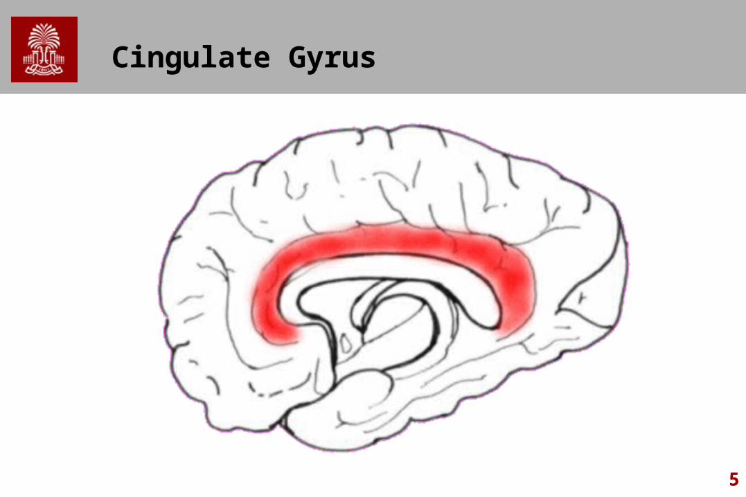
The cingulate gyrus a) Is a medial structure of
the cortex.
b) Is located in the brain-stem
c) Is located in the cerebellum
d) Is a lateral structure of the cortex
5
Cingulate Gyrus
6
MCQ
Functions of the brainstem include a) Swallowing, respiration, and blood pressure
regulation
b) Vision, language, and muscle coordination
c) Emotional memory, executive function, and visual processing
d) Calculation, reading, and writing
7
MCQ
The Colliculi a) Are located on the
anterior brainstem
b) Are located on the posterior brainstem
c) Are located on the ventral frontal lobe
d) Are located in the insula
8
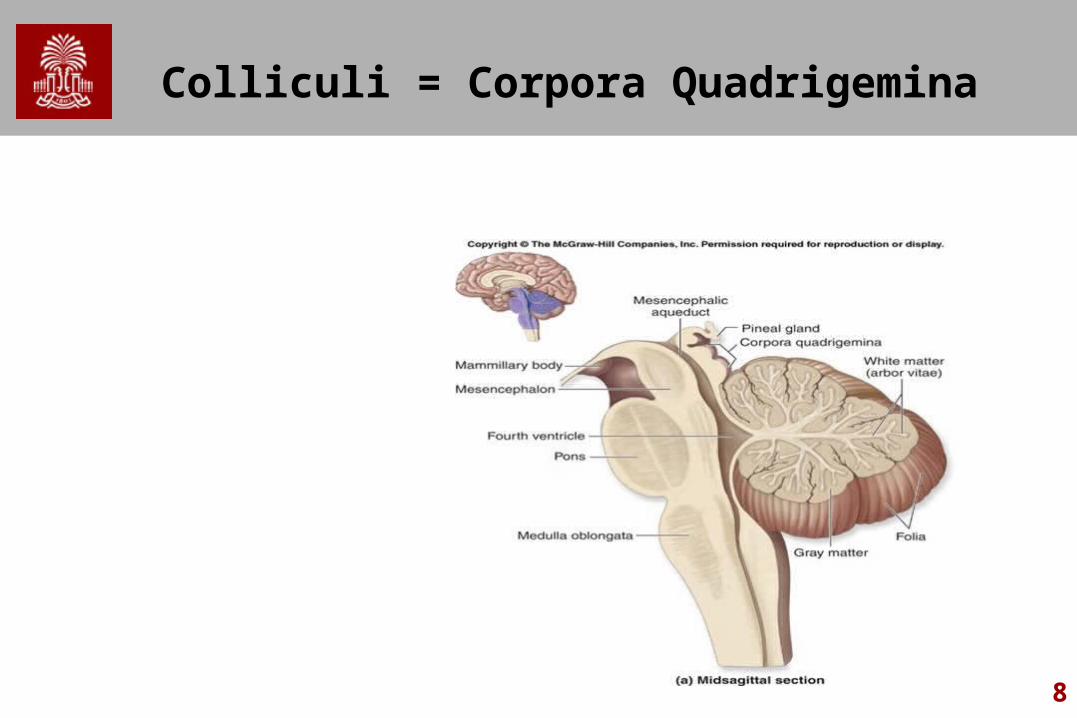
Colliculi = Corpora Quadrigemina

9
MCQ
Which is part of the cortical spinal tract?
a) Internal Capsule
b) Lateral Geniculate Nucleus
c) Dura mater
d) Cerebellum

10
Internal Capsule
11
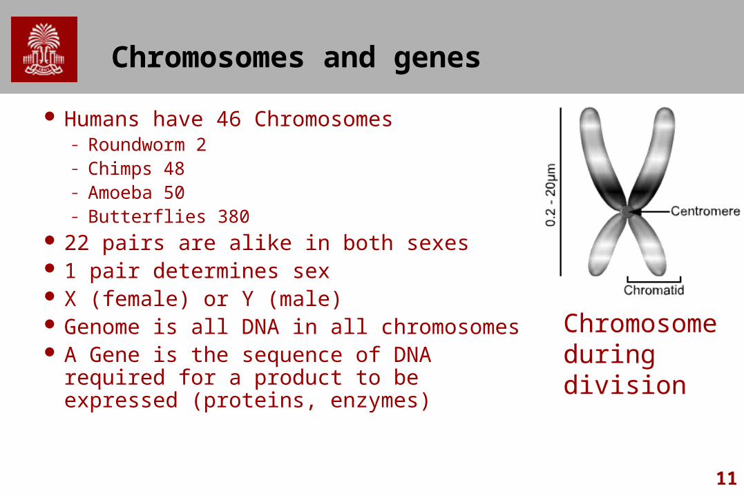
Chromosomes and genes
Humans have 46 Chromosomes– Roundworm 2– Chimps 48– Amoeba 50– Butterflies 380
22 pairs are alike in both sexes 1 pair determines sex X (female) or Y (male) Genome is all DNA in all chromosomes A Gene is the sequence of DNA required for a
product to be expressed (proteins, enzymes)
Chromosome during division
12
Types of Division
Mitosis– For general body growth and function– Regularly occurring for much of our body during
our entire lifeMeiosis
– Special division during reproduction
13
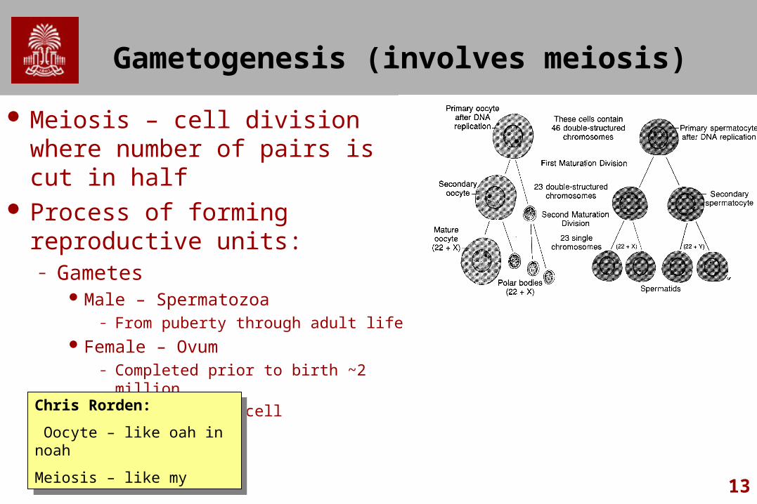
Gametogenesis (involves meiosis)
Meiosis – cell division where number of pairs is cut in half
Process of forming reproductive units:– Gametes
Male – Spermatozoa– From puberty through adult life
Female – Ovum– Completed prior to birth ~2 million– Oocyte = germ cell
Chris Rorden:
Oocyte – like oah in noah
Meiosis – like my
Chris Rorden:
Oocyte – like oah in noah
Meiosis – like my
14
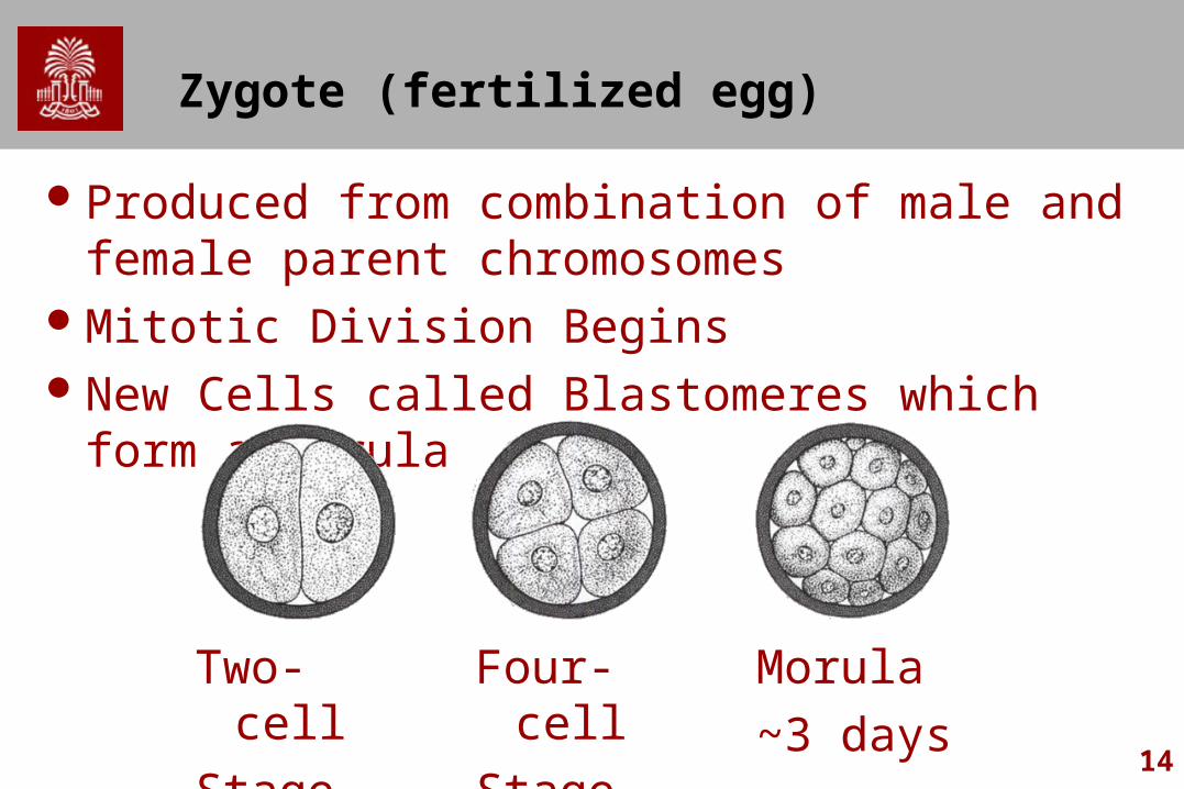
Zygote (fertilized egg)
Produced from combination of male and female parent chromosomes
Mitotic Division BeginsNew Cells called Blastomeres which form a Morula
Two-cell
Stage
Four-cell
Stage
Morula
~3 days

15
Morula
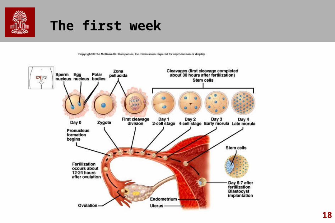
Morula develops central cavity called Blastocyst
Blastocyst attaches to uterine wallOne week from fertilization to implantation in
uterine wall– Allows blastocyst to get nutrients and excrete
waste products
16
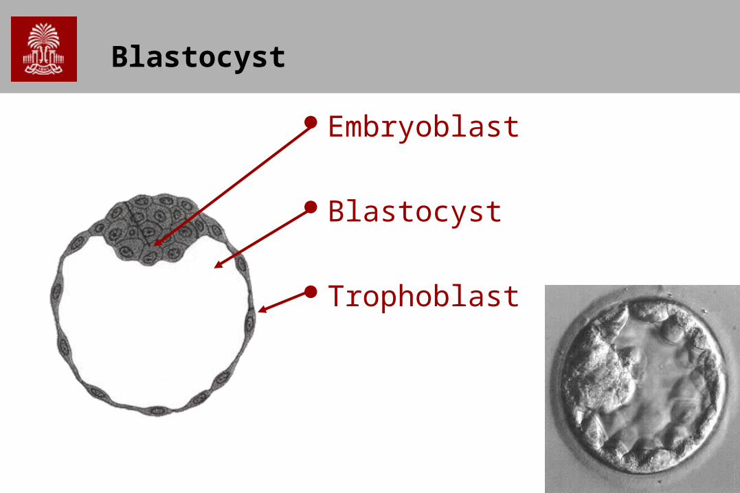
Blastocyst
Embryoblast
Blastocyst
Trophoblast

17
Blastocyst
Embryoblast
Blastocyst cavity
Uterine stroma
Trophoblast cells
18
The first week

19
The second week: Bilaminar Embryo
Cytotrophoblast
EpiblastHypoblast
Amniotic Cavity
Primary Yolk Sac
Exocoelomic Membrane
Embryo has two primary layers: Epiblast & Hypoblast
20
When does life begin?
British Warnock Committee (1984) suggested experimentation on the human embryo within the first 14 days of its development.
1. Because before this time implantation in the uterus is not complete;
2. Because only after this time do the embryo cells lose their so-called ‘totipotency’: Because after the 14th day there no longer exists the possibility that monozygotic twins could be formed from a single embryo.
3. Appearance of ‘primitive streak’ considered as ‘the sign’ of a ‘new’ human subject
21
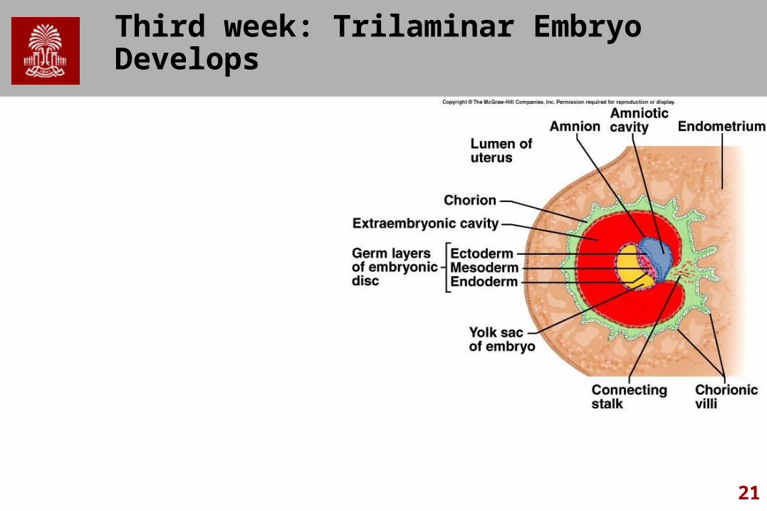
Third week: Trilaminar Embryo Develops
22
Week 3
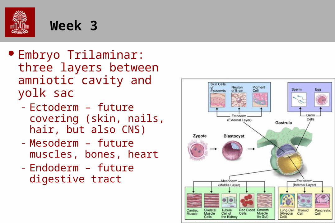
Embryo Trilaminar: three layers between amniotic cavity and yolk sac– Ectoderm – future
covering (skin, nails, hair, but also CNS)
– Mesoderm – future muscles, bones, heart
– Endoderm – future digestive tract
23
Week 3
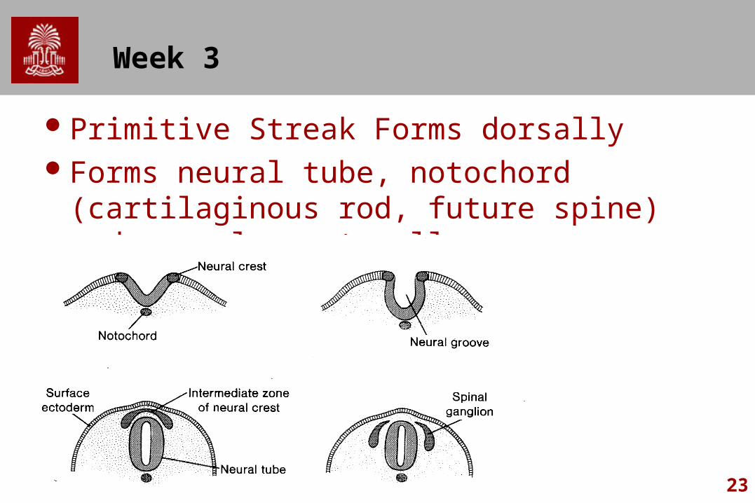
Primitive Streak Forms dorsallyForms neural tube, notochord (cartilaginous
rod, future spine) and neural crest cells
24
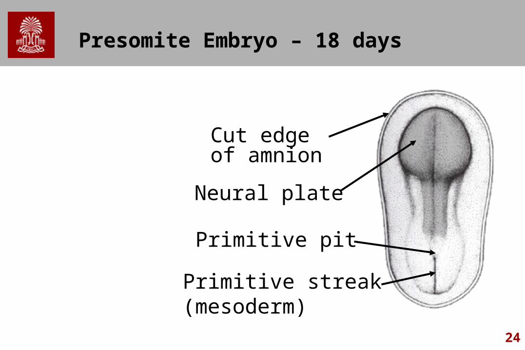
Presomite Embryo – 18 days
Neural plate
Primitive pit
Primitive streak (mesoderm)
Cut edge of amnion
25
Early Highlights
Day 18 - Neural plate invaginates (encloses) to form neural groove
Day 22 - Neural Tube Forms – Becomes brain and spinal cord
About the same time, Neural Crest Forms– Becomes cranial and spinal nerve ganglia
26
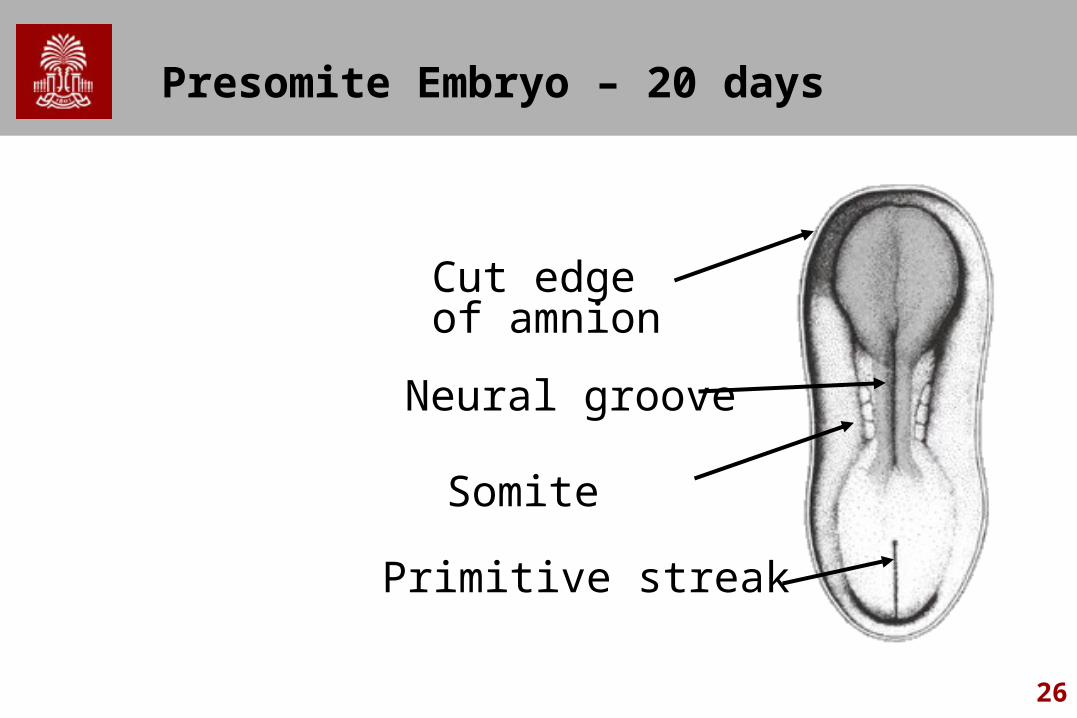
Presomite Embryo – 20 days
Neural groove
Somite
Primitive streak
Cut edge of amnion

27
Neural Tube
Anterior 2/3 will form brain Caudal 1/3 will form spinal cord Day 25 - Cranial opening closes Brain has 3 sections
– Prosencephalon– Mesencephalon– Rhombencephalon
Day 27 - Caudal end closes Problems cause neural tube defects

28
Human Embryo – 22 days
Optic placode
Somite
Cut edge of amnion
Neural foldsomites are masses of mesoderm that will eventually become skin, skeletal muscle , and vertebrae.
29
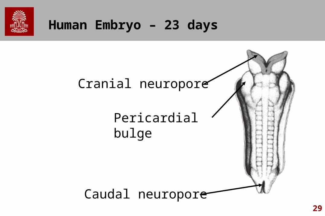
Human Embryo – 23 days
Pericardial bulge
Cranial neuropore
Caudal neuropore
30
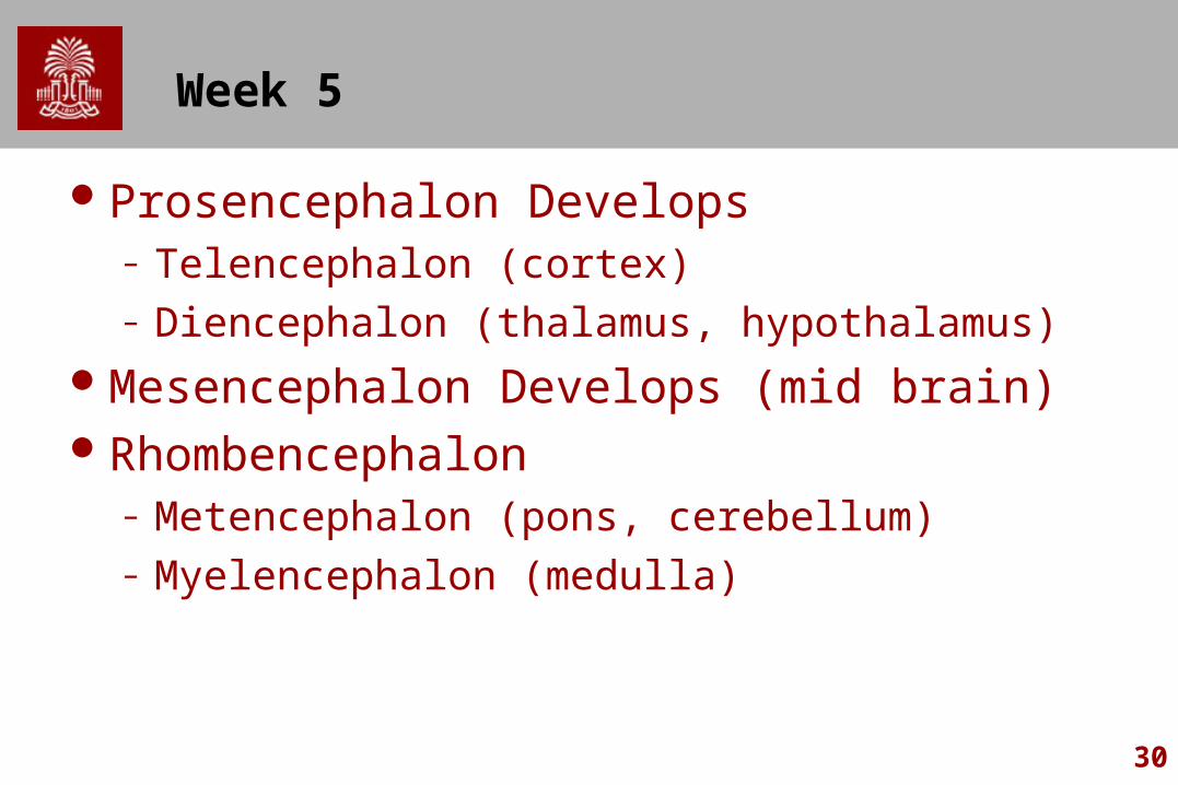
Week 5
Prosencephalon Develops– Telencephalon (cortex)– Diencephalon (thalamus, hypothalamus)
Mesencephalon Develops (mid brain)Rhombencephalon
– Metencephalon (pons, cerebellum)– Myelencephalon (medulla)

31
Telencephalon
Optic Vessels - retinae, optic nerveCerebral Hemispheres - Lateral VentricleMedial Connection – Corpus CallosumOlfactory LobeCorpus Striatum
– (Caudate N. & Lenticular N.)Cerebral Cortex
– Very primitive though 20 weeks
32
Third Trimester
All structures present at birthAll structures become more distinct in Third
TrimesterCommissures develop
33
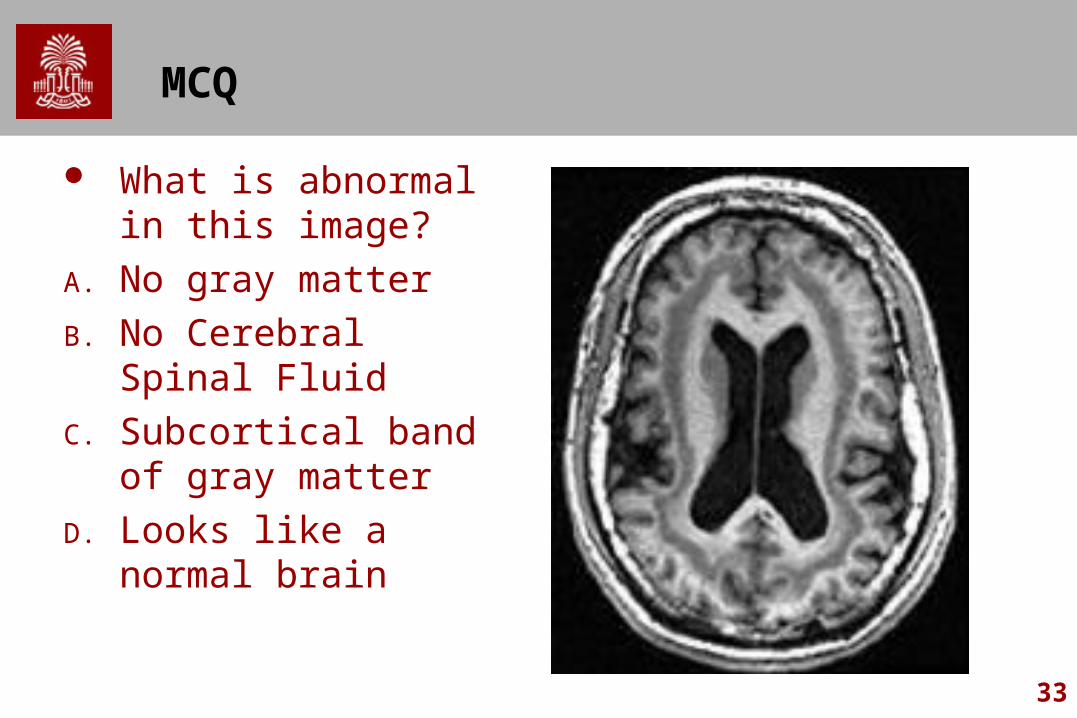
MCQ
What is abnormal in this image?
A. No gray matter
B. No Cerebral Spinal Fluid
C. Subcortical band of gray matter
D. Looks like a normal brain

34
Seven Steps of CNS Development
1. Production of initial neurons and glial cells2. Migration of cells to definitive location3. Selective gathering of cells to functional group4. Cytodifferentiation (axon, dendrite, synaptic
patterns)5. Selective death of some cells in groups (Apoptosis)6. Outgrowth of axons to specific target cells and
establishment of connections7. Elimination of certain connections and functional
stabilization of others
35
Maturation of CNS
At birth, all neurons you will ever have present.– Only a few exceptions (neurons involved w smell)
Process of myelination signals onset of mature function– Slow process
Partially completed completed by age 7 Axons and dendrites not until teens Some areas continue to age 70
Some cells have programmed cell death (Apoptosis)– tadpoles lose their tails and pigeons' feet become unwebbed.
Crucial in brain Note: not all developmental language disorders present at
birth.
36
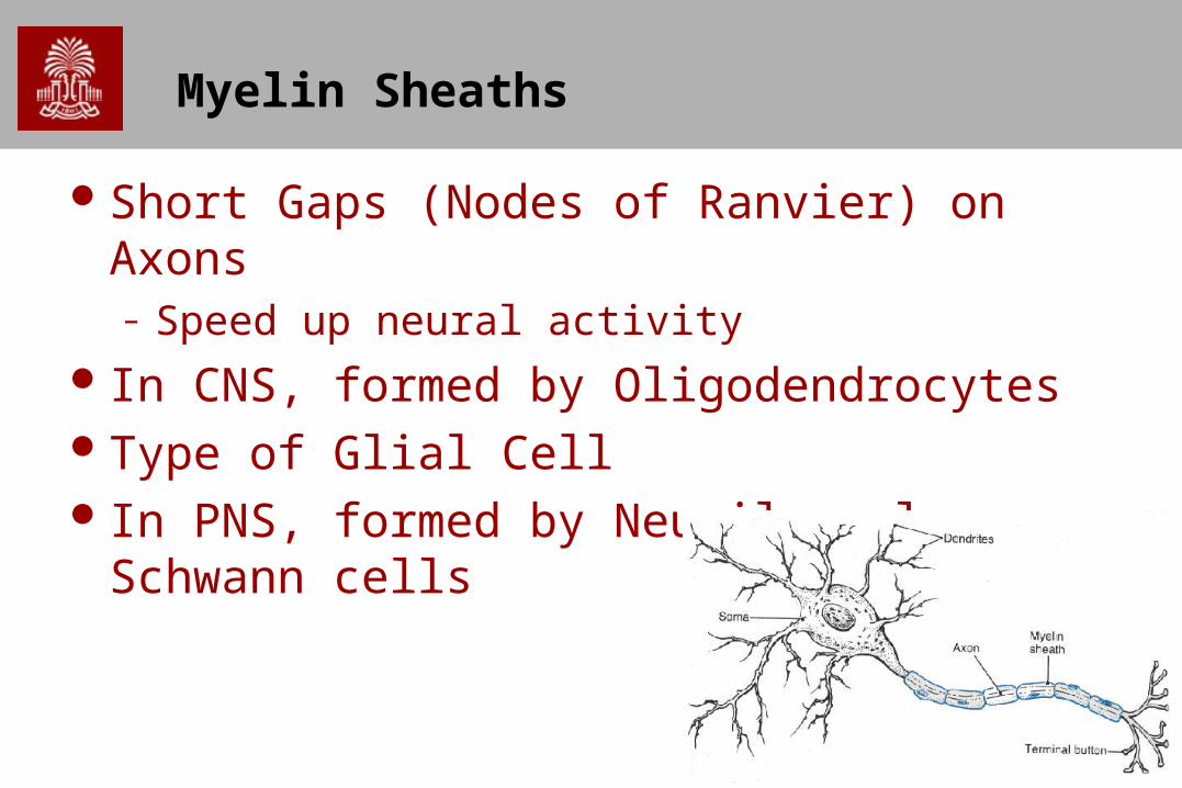
Myelin Sheaths
Short Gaps (Nodes of Ranvier) on Axons– Speed up neural activity
In CNS, formed by OligodendrocytesType of Glial Cell In PNS, formed by Neurilemmal or Schwann
cells
37
Rate of Myelination Varies
Spinal tract completed by 9th monthMajor motor tracts by 2 yearsCerebrum and Cerebellum into the teens
38
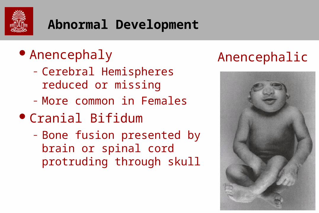
Abnormal Development
Anencephaly– Cerebral Hemispheres reduced
or missing– More common in Females
Cranial Bifidum– Bone fusion presented by brain
or spinal cord protruding through skull
Anencephalic
39
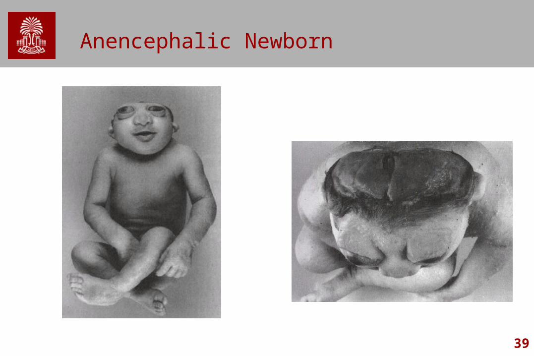
Anencephalic Newborn
40
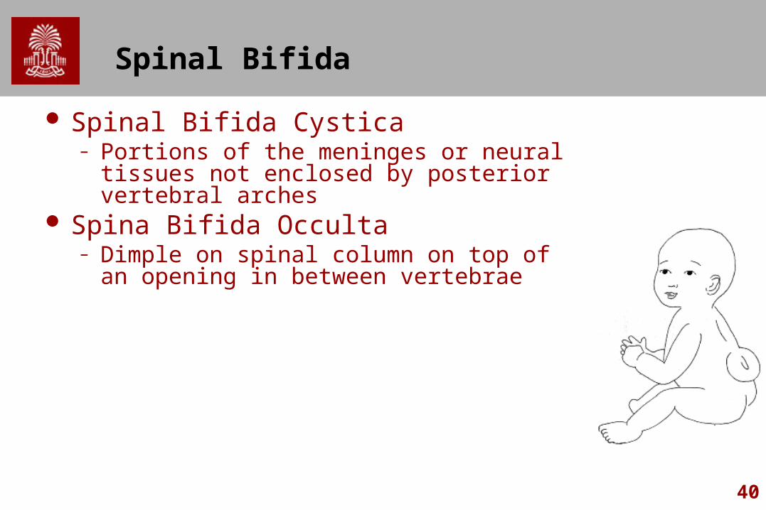
Spinal Bifida
Spinal Bifida Cystica– Portions of the meninges or neural tissues not
enclosed by posterior vertebral arches Spina Bifida Occulta
– Dimple on spinal column on top of an opening in between vertebrae

41
Other Developmental Conditions
Hydrocephaly– Enlarged head, brain atrophy mental deficiency– Excessive production of CSF or obstruction of
drainage pathways
– http://neurosurgery.seattlechildrens.org/conditions_treated/hydrocephalus.asp

42
Causes of hydrocephalus
43
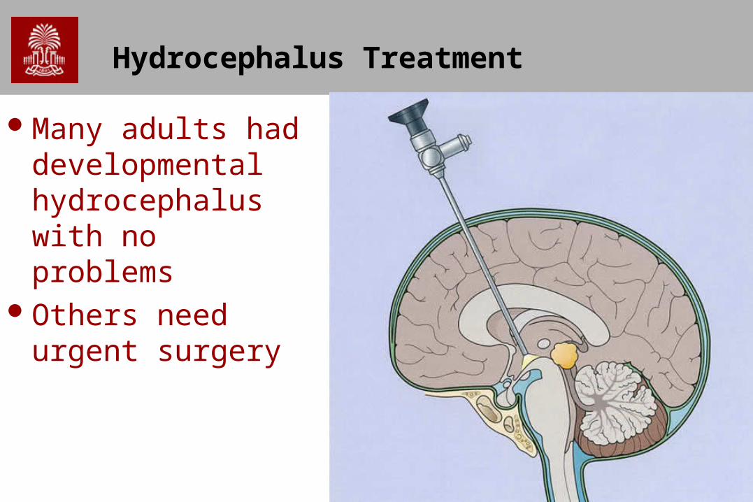
Hydrocephalus Treatment
Many adults had developmental hydrocephalus with no problems
Others need urgent surgery
44
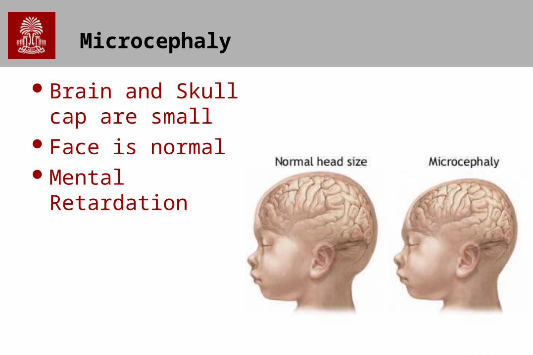
Microcephaly
Brain and Skull cap are small
Face is normalMental Retardation