1 co 2 physiology. 2 what is carbon dioxide? capnos comes from the greek word for “smoke”...
TRANSCRIPT

1
CO2 Physiology

2
What is Carbon Dioxide?
• Capnos comes from the Greek word for “smoke”– smoke from the fire of metabolism– a natural waste product of cellular activity
• CO2 is a compound molecule
– 1 element of carbon and 2 elements of oxygen
– colorless and heavier than air
– green plants clean up after our exhaled CO2

3
Physiology of CO2
• CO2 produced by cellular metabolism diffuses across the cell membrane into the circulating blood.
• The blood transports the CO2 to the lungs.• Then it diffuses from the blood into the lungs.• CO2 is eliminated with alveolar ventilation on
exhalation.

4
Physiology of CO2
• Carbon Dioxide is transported in the blood in three (3) principle forms:– 5 to 10% as gas & reflected by the PCO2
– 20 to 30% is bound to blood proteins, the major one being hemoglobin
– 60 to 70% is carried as bicarbonate (HCO3)

5
Physiology of CO2
• About 5-10% of CO2 is eliminate through exhalation only.
• The rest is recycled in the body through the circulatory and renal systems.
• The heart and lungs would have to increase their work 10 times if they were required to eliminate all the CO2 the body produces!

6
Ventilation & EtCO2 Monitoring
• Endtidal CO2 (EtCO2) is the CO2 measured at the end of expiration.
• EtCO2 concentration provides a clinical estimate of the PaCO2, if ventilation and perfusion are appropriately matched.
• EtCO2 monitoring allows for a breath by breath assessment of ventilation.

7
Capnography—
The continuous measurement and graphic display (waveform) of the CO2 concentration in the patient’s airway during the respiratory cycle.
Normal waveform:

Respiratory Cycle
O2 CO2
CO2 O2

9
Respiratory Cycle
• Oxygenation = oxygen → lungs→ alveoli→ blood
Monitored by a Pulseoximeter
• Metabolism = oxygen is converted to energy + CO2
Monitored by a Metabolic Computer
• Hymodynamic Parameter
• Monitored by ECG, IPB, NIBP, Temperature
• Ventilation = CO2 → blood→ lungs→ exhalation
Monitored by a Capnograph

10
Normal conditions: • EtCO2 is between 35 – 45 mmHg
• PaCO2 & EtCO2 will be very close
• EtCO2 is most 2 - 5 mmHg less with normal physiology
Widening of this difference can be caused by:• Incomplete alveolar emptying• Ventilation-perfusion abnormalities• Poor sampling
Capnography monitoring of Critically ill patient may alert clinicians to underlying conditions
The relationship – EtCO2 and PaCO2
11
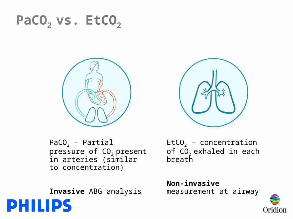
PaCO2 vs. EtCO2
PaCO2 – Partial pressure of CO2 present in arteries (similar to concentration)
Invasive ABG analysis
EtCO2 – concentration of CO2
exhaled in each breath
Non-invasive measurement at airway

12
A-B: Baseline = no CO2 in breath
B-C: Rapid rise in CO2
D-E: Inhalation
C-D: Alveolar plateau
D
D: End expiration (EtCO2)
Normal waveform
D

13
Normal waveform - 35-45 mmHg

14
Hypoventilation

15
Hypoventilation with shallow breathing

16
Relationship between EtCO2 and RR
Breath-to-breath measure of ventilatory status

17
Hyperventilation

18
Some Definitions
• Capnometer• Capnography

19
Capnometer
A Capnometer provides only a numerical measurement of carbon dioxide in mmHg or kPa or Vol.-%

Capnograpy
Capnography provides the CO2 value and the waveform of carbon dioxide over time

21
Capnography—The Ventilation Vital Sign™
• Earliest sign that something is going wrong
• Breath by breath assessment of ventilation

22
CapnographyCapnography
An EtCO2 value of e.g. 38 mm/Hg
without a
Time
5040302010
0
it´s like a heart rate of e.g. 80 without an

23
CO2 Measurement Technology
General:
• CO2 measurement technologyInfrared absorption
• Technique of airway gas sampling
Main stream vs. side stream vs. Microstream

24
Sampling Technology
• Mainstream sampling - CO2 analysis chamber is in-line between the patient airway and the ventilator circuit
• Sidestream sampling - CO2 analysis chamber is within the device. The patient’s expired gas is sucked from the airway and drawn to that chamber through a sampling line.

25
Conventional main stream technology
Monitor
Fresh gas
InspirationExpiration

26
Conventional side stream technology
Sample line
(Monitor)

27
Unique solutions for Capnography= Microstream®

28
Microstream® CO2
• A combination of a unique CO2 sidestream measurement technology and;
• FilterLine (proprietary sampling lines) - for single patient use
• Only system providing accurate EtCO2 readings for non-intubated patients that receive supplemental O2 and switch between oral and/or nasal breathing

29
Microstream® CO2— Major benefits
• Ease of Use
• Reliable Technology
• Flexible for all patient types
• Versatile for all environments

30
Microstream® advantages
• Superior moisture handling of liquids, secretions and humidity
• CO2 specificity – no cross-sensitivity to anesthetic gases
• Rugged – no moving parts in sensor
• Long-term monitoring
Reliable technology

• 50 ml/min flow rate supports entire patient population – including neonates (Competition at 3 – 5 times the flow rate)
• Does not compete for Neonate tidal volume
• The lower the flow, the less moisture to be handled
Flexible for all patient populations – solution for monitoring Neonates
Microstream® advantages

32
Microstream® advantages
• No expensive sensors to replace• Yearly calibration – done in 5 minutes• Warm up time –
45 seconds from ON until first waveform and number appears
• One-piece Plug & Play consumables
Ease of use

33
Light source Micro sample cell 15 µL
Light source housing
1 Eurocent
Microstream® advantages

34
Microstream® Core Technology
Sensor Housing
I.R Source
Optic Block
(Micro Sample Cell)
I.R Detectors

35
Microstream® advantages
• Fast response time
• 1 mm micro bore tubing reduces delay time
• Crisp waveform – longitudinal filter maintains laminar flow
• Build-in water trap – don't clean and re-use any FilterLine – it destroys the inline filter
Reliable Technology

36
Microstream® advantages
Flexible
• Both intubated and nonintubated applications
• Alternating mouth and nose breathing
• Oxygen delivery (low flow O2 solution; solution for high flow O2 delivery)
• Adult, pediatric, and neonates

37
Microstream® advantages
Versatile
All clinical environments:
• Critical Care
• Sedation Procedures
• EMS/ED
• Operating Room

38
Unique solutions for CapnographyFilterLine® patient interfaces

39
FilterLine® solutions for all applications
FilterLine®
Sets
IntubatedNon-Intubated
Smart Solutions NIV-Line
CapnoLine H
Smart CapnoLine / Smart CapnoLine O2

40
Smart Solutions for nonintubated patients
• Continuous sampling from both
mouth and nose
• Special oral-piece design optimally
samples from mouth - Increased surface area provides greater sampling accuracy in the presence of low tidal volume (adult/intermediate size)
“Microstream® technology allows the accurate measurement of EtCO2 in the absence of an endotracheal tube.”*
*ASA 2001 Jay Brodsky, MD Professor of Anesthesia, Stanford University Medical Center, CA USA

41
Smart Solutions for nonintubated patients
Smart CapnoLine™ Plus /Smart CapnoLine™ Plus O2
nasal cannula for CO2 measurement and O2 delivery
• Uni-junction sampling method ensures optimal waveform and ultra-fast response time
• Unique O2 delivery method reduces CO2 sampling dilution (up to 5l/min)
• Solution for high flow O2 delivery (works effectively under oxygen delivery mask)

42
Solutions for non-intubated patientsSolutions for non-intubated patients
CapnoLine H*™ / CapnoLine H O2
• Enables continuous EtCO2 monitoring in high humidity environments (i.e. ICU)
• Can be used up to 72 hours
* = Humidity
Piece of Nafion

43
Small pin holes deliver pillow of oxygen around both nose and mouth
Nasal and Oral Sampling
Microstream®—A Unique Solution For Non-intubated PatientsCO2 sampling / O2 delivery for non-intubated patients (up to 5 L/min.)
Uni-junction™ of sampling ports prevents dilution from non-breathing source
Increased surface area provides greater sampling accuracy in the presence of low tidal volume

44
• Easily handles moisture and secretions without water traps
• Able to measure in any position
• Nafion® tubing allows for long-term monitoring without moisture build up
• Easily switches to non-intubated monitoring without re-calibration of monitor
• Low add. dead space (0,4 cc) to use on neonates
FilterLine® Sets - Solutions for intubated patients

45
FilterLine® recommendations: Sedation Areas; GI Lab, Cath Lab, EP Lab
Is the Patient on Oxygen?
YES NO
Smart CapnoLine Plus O2 Smart CapnoLine Plus

46
Do not try to dry the FilterLine® - this will damage the filter
Ensure there are no kinks in the sampling line
Do not cut the oral flange on the Smart CapnoLine
Do not cover the Nafion®
Do not instill medications through the airway adapter
Never pass a suction catheter or stylus through the airway adapter
Change the FilterLine® or the Set if a “Blockage” message appears on the monitor screen or if the readings become extremely erratic
FilterLine® information to avoid problems

47
Latex free
Single-patient use
Not sterile
FilterLine® answers for the most FAQ´s:

48
Sedation Procedures
“Monitoring of exhaled carbon dioxide should be considered for all patients receiving deep sedation and for patients whose ventilation cannot be directly observed during moderate sedation.”*
*Practice Guidelines for Sedation and Analgesia by Non-Anesthesiologists, Developed by the American Society of Anesthesiologists Task Force on Sedation and Analgesia by Non Anesthesiologists: Anesthesiology 2002; 96:1004

49
• Assesses -
patent airway (airway obstruction)
protective reflexes
response to verbal/physical stimuli
• Respiratory changes can immediately be assessed
• Microstream® allows for continuous respiratory monitoring with no nuisance alarms in procedural sedation environments where currently there is minimal usage of monitoring
Microstream® solutions during Sedation Procedures
Benefits and Uses

50
Microstream® solutions during Sedation Procedures
• Cardiac Cath. Lab
• GI lab
• Pulmonary lab
• Emergency Department
• Hyperbaric medicine
• Dental Clinics
• Radiology
Applications

51
Moderate – procedural sedation

52
How can capnography make a difference in how you care for the sedated patient?
• What you do will not change
• When you do it will!
Capnography and sedation
Early detection of potential patient compromise

53
Protocol during procedural sedation
• E.g. after 12 hours NPO (nothing by mouth) = EtCO2• Know the respiratory rate, waveform, and EtCO2 numeric
value before drug administration
Baseline Ventilatory Assessment
• RR, ETCO2 value…changes from baseline (trends)
• Changes in the Waveform…Earliest indicator of potential problems. (size, shape)
Continuous monitoring throughout case and recovery
Early intervention

54
Changes from baseline
• Change in EtCO2 value > 10 mmHg
• Significant waveform change
Becomes erratic
Flatlines

55
• Remember the ABC’s (airway, breathing, circulation)
• Assess the patient
• Follow your normal protocol, which may include:
Changes from baseline - action
Ensure open airway
Stimulate patient if necessary
Check the cannula positioning
Stop drug delivery
Inform M.D. / pause procedure if necessary
Administer reversal agents as prescribed

56
• Requires higher vigilance in ventilatory monitoring
• Maintain patent airway
• Potential dead space ventilation
• Chest moves up and down
Deep sedation
• Inadequate respiratory effort to clear dead space

57
Assessing for changes from baseline
Hypoventilation with shallow respirations

58
Nursing interventions
• Continue to monitor
• Ask patient to take a deep breath

59
Abnormal waveforms
Possible causes
• Partial airway obstruction caused by:
• Tongue
• Position of head
Absent alveolar plateau indicates incomplete alveolar emptying or loss of airway integrity

60
Assessing for changes from baseline
• Poor head and neck alignment
• Draping near the airway
• Shallow breathing – not clearing
dead space
Rebreathing often results from:

61
Assessing for changes from baseline
• Chest movement
• Little – to no air movement in and
out of lungs
Dead space ventilation

62
Abnormal waveforms
Possible causes
• Partial airway obstruction caused by:
– Tongue
– Position of head
Absent alveolar plateau indicates incomplete alveolar emptying or loss of airway integrity

63
Nursing interventions
• Assess patient
• Ask patient to take a deep breath
• Adjust patient’s head position, if
necessary
• Adjust cannula position, if
necessary

64
Putting it all together
• The transition from conscious sedation to unconscious/anesthesia is very subtle and can be undetected until oxygenation is impaired
• You must be prepared to monitor a patient at a level deeper than intended
• “Respiratory frequency and adequacy of pulmonary ventilation are continually monitored”
• Only capnography provides an immediate notification of a ventilatory event