1 dilated cerebral ventricles radiology
TRANSCRIPT

1 Dilated Cerebral Ventricles
CLINICAL IMAGAGINGAN ATLAS OF DIFFERENTIAL DAIGNOSIS
EISENBERG
DR. Muhammad Bin Zulfiqar PGR-FCPS III SIMS/SHL

• Fig SK 1-1 Level of the foramen of Monro. (A) Bilateral enlargement of the frontal horns with a normal-sized third ventricle in a patient with a hyperdense colloid cyst (c). (B) Unilateral enlargement of the left frontal horn caused by a tiny hypodense unilateral tumor (arrow).
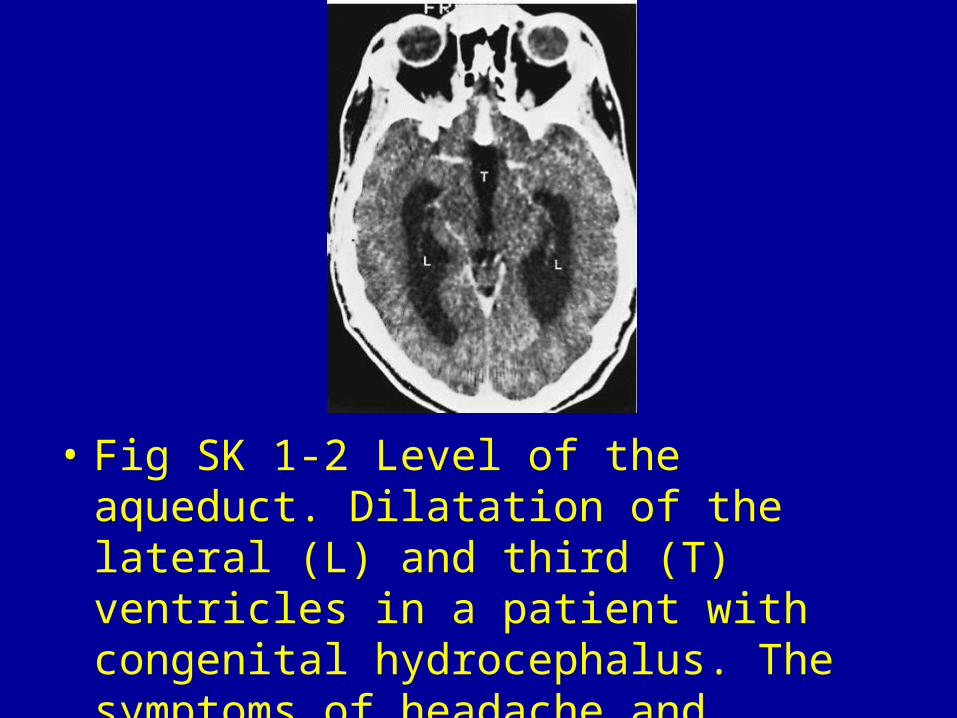
• Fig SK 1-2 Level of the aqueduct. Dilatation of the lateral (L) and third (T) ventricles in a patient with congenital hydrocephalus. The symptoms of headache and papilledema were resolved after ventricular shunting.
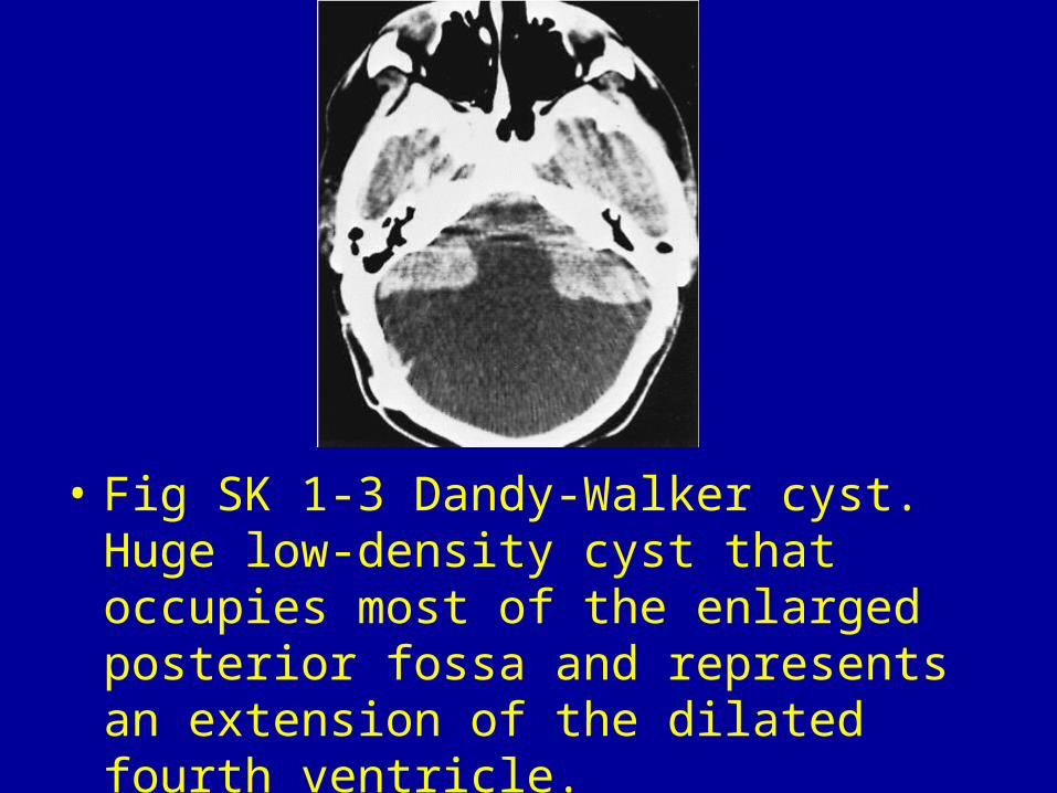
• Fig SK 1-3 Dandy-Walker cyst. Huge low-density cyst that occupies most of the enlarged posterior fossa and represents an extension of the dilated fourth ventricle.

• Fig SK 1-4 Communicating hydrocephalus. Generalized ventricular enlargement in a 69-year-old patient with ataxia, dementia, and incontinence. Note the absence of the dilated sulci in the obstructive hydrocephalus.1

• Fig SK 1-5 Choroid plexus papilloma. Enhancing ventricular mass (arrow) causing pronounced generalized enlargement of the ventricular system.
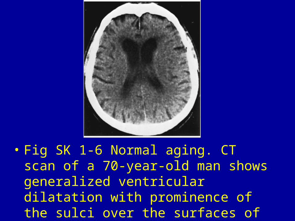
• Fig SK 1-6 Normal aging. CT scan of a 70-year-old man shows generalized ventricular dilatation with prominence of the sulci over the surfaces of the cerebral hemispheres.

• Fig SK 1-7 Alzheimer's disease. Noncontrast scan of a 56-year-old woman with progressive dementia shows generalized enlargement of the ventricular system and sulci.
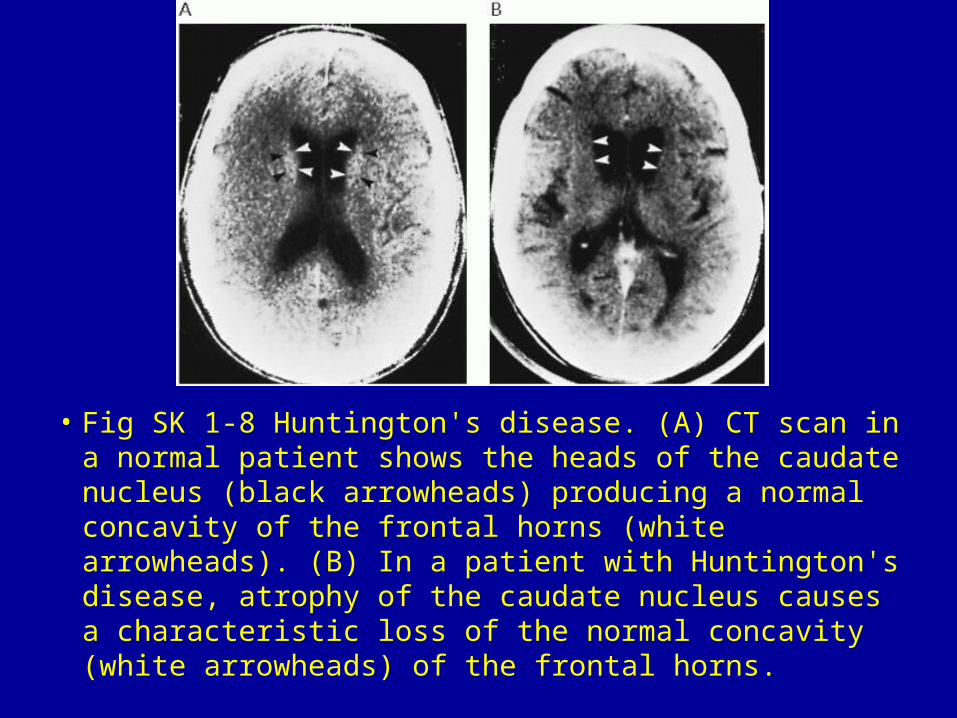
• Fig SK 1-8 Huntington's disease. (A) CT scan in a normal patient shows the heads of the caudate nucleus (black arrowheads) producing a normal concavity of the frontal horns (white arrowheads). (B) In a patient with Huntington's disease, atrophy of the caudate nucleus causes a characteristic loss of the normal concavity (white arrowheads) of the frontal horns.
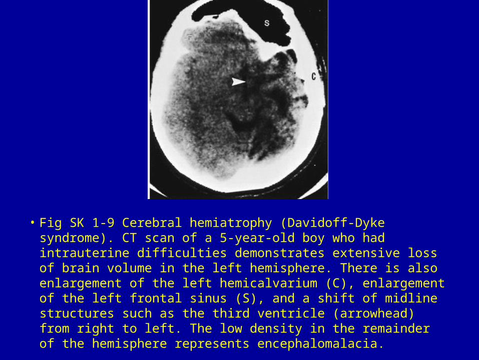
• Fig SK 1-9 Cerebral hemiatrophy (Davidoff-Dyke syndrome). CT scan of a 5-year-old boy who had intrauterine difficulties demonstrates extensive loss of brain volume in the left hemisphere. There is also enlargement of the left hemicalvarium (C), enlargement of the left frontal sinus (S), and a shift of midline structures such as the third ventricle (arrowhead) from right to left. The low density in the remainder of the hemisphere represents encephalomalacia.
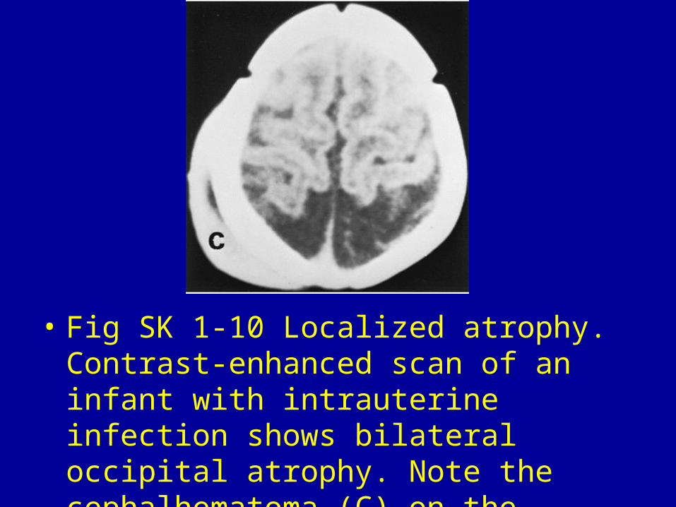
• Fig SK 1-10 Localized atrophy. Contrast-enhanced scan of an infant with intrauterine infection shows bilateral occipital atrophy. Note the cephalhematoma (C) on the right.
