1 essentials of human anatomy essentials of human anatomy the skeletal system 4 joints of the...
TRANSCRIPT
1
Essentials of Human AnatomyEssentials of Human Anatomy
The Skeletal System 4
Joints of the Skeletal System
Chapter 5
Dr Fadel NaimAss. Prof. Faculty of Medicine
IUG
Joints of the Skeletal System
• Articulations• Functional junctions between bones• Bind parts of skeletal system together• Make bone growth possible• Permit parts of the skeleton to change shape during childbirth• Enable body to move in response to skeletal muscle contraction

Naming of Joints
• Usually derived from the names of the articulating bones.

Classification of Joints
• Fibrous Joints• dense connective tissues connect bones• between bones in close contact
• Cartilaginous Joints• hyaline cartilage or fibrocartilage connect bones
• Synovial Joints• most complex• allow free movement
• synarthrotic• immovable
• amphiarthrotic• slightly movable
• diarthrotic• freely movable

Fibrous Joints
3 Types• Syndesmosis• Suture• Gomphosis
Syndesmosis •a sheet or bundle of fibrous tissue connects bones• amphiarthrotic• lies between tibia and fibula

Fibrous Joints
Suture• between flat bones• synarthrotic• thin layer of connective tissue connects bones
Gomphosis• cone-shaped bony process in a socket• tooth in jawbone• synarthrotic

Cartilaginous Joints
2 Types• Synchondrosis• Symphysis
Synchondrosis• bands of hyaline cartilage unite bones• epiphyseal plate (temporary)• between manubrium and first rib• synarthrotic
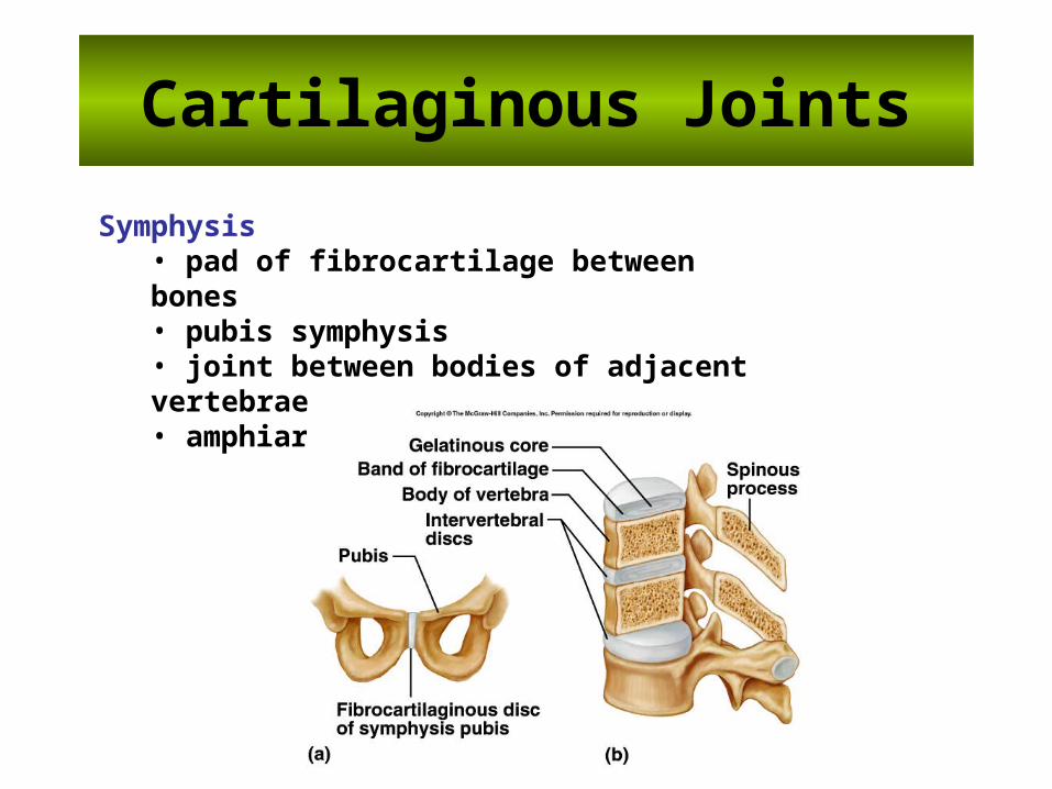
Cartilaginous Joints
Symphysis• pad of fibrocartilage between bones• pubis symphysis• joint between bodies of adjacent vertebrae• amphiarthrotic

General Anatomy of Synovial Joints
• Basic features: – articular capsule– joint cavity – synovial fluid– articular cartilage – ligaments– nerves– blood vessels

General Anatomy of Synovial Joints – Accessory Structures
• Bursae – fibrous, saclike structure that contains synovial fluid
and is lined by a synovial membrane• Fatpads
– often distributed along the periphery of a synovial joint – act as packing material and provide some protection
for the joint – fill the spaces that form when bones move and the
joint cavity changes shape• Tendons
– attaches a muscle to a bone – help stabilize joints


Types of Synovial Joints
• Classified by the shapes of their articulating surfaces • Types of movement they allow
– uniaxial if the bone moves in just one plane– biaxial if the bone moves in two planes– multiaxial (or triaxial) if the bone moves in
multiple planes

Types of Synovial Joints
• From least movable to most freely movable, the six specific types of synovial joints are:
• planar (gliding) joints • hinge joints • pivot joints• condyloid (ellipsoid) joints • saddle joints • ball-and-socket joints

Types of Synovial Joints
Ball-and-Socket Joint• hip• shoulder
Condyloid Joint• between metacarpals and phalanges

Types of Synovial Joints
Gliding Joint• between carpals• between tarsals
Hinge Joint• elbow• between phalanges

Types of Synovial Joints
Pivot Joint• between proximal ends of radius and ulna
Saddle Joint• between carpal and metacarpal of thumb

Types of Synovial Joints

Mobility and Stability in Joints
• Motion permitted ranges from none to various extensive motions.
• Structure determines both its mobility and its stability. – more mobile = less stable

Types of Joint Movements
• abduction/adduction• dorsiflexion/plantarflexion• flexion/extension/hyperextension

Types of Joint Movements
• rotation/circumduction• supination/pronation

Types of Joint Movements
• eversion/inversion• protraction/retraction• elevation/depression
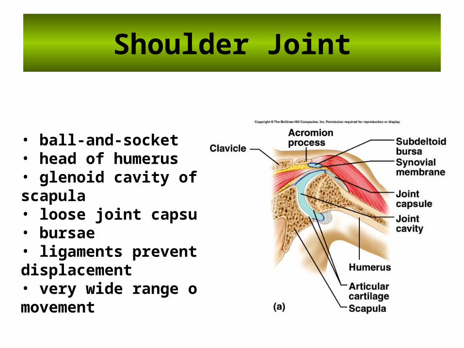
Shoulder Joint
• ball-and-socket• head of humerus• glenoid cavity of scapula• loose joint capsule• bursae• ligaments prevent displacement• very wide range of movement

Shoulder Joint

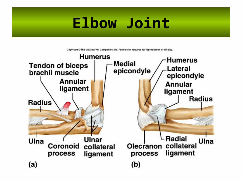
Elbow Joint
• hinge joint• trochlea of humerus• trochlear notch of ulna
• gliding joint• capitulum of humerus• head of radius
• flexion and extension• many reinforcing ligaments• stable joint
Elbow Joint

Hip Joint
• ball-and-socket joint• head of femur• acetabulum of coxa• heavy joint capsule• many reinforcing ligaments• less freedom of movement than shoulder joint

Hip Joint
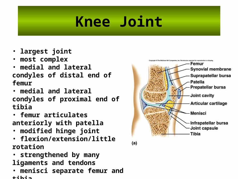
Knee Joint
• largest joint• most complex• medial and lateral condyles of distal end of femur• medial and lateral condyles of proximal end of tibia• femur articulates anteriorly with patella• modified hinge joint• flexion/extension/little rotation• strengthened by many ligaments and tendons• menisci separate femur and tibia• bursae

Knee Joint

Life-Span Changes
• Joint stiffness is an early sign of aging•Fibrous joints first to change; can strengthen over a lifetime• Changes in symphysis joints of vertebral column diminish flexibility and decrease height • Synovial joints lose elasticity• Disuse hampers the blood supply• Activity and exercise can keep joints functional longer
Cycle of Life: Articulations
• Bone development and the sequence of ossification between birth and skeletal maturity affect joints– Fontanels between cranial bones disappear– Epiphysial plates ossify at maturity
• Older adults– ROM decreases– Changes in gait occur
• Skeletal diseases manifest as joint problems– Abnormal bone growth (lipping)—influences joint motion– Disease conditions can be associated with specific
developmental periods

Clinical Application
Joint DisordersSprains• damage to cartilage, ligaments, or tendons associated with joints• forceful twisting of joint
Bursitis• inflammation of a bursa• overuse of a joint
Tendonitis
•An inflammation of the tendon
•Caused by excessive use.

Arthritis
• A group of inflammatory or degenerative diseases of joints that occur in various forms. – swelling of the joint– pain – stiffness
• Most prevalent crippling disease in the United States. – gouty arthritis
– osteoarthritis :A condition which results when the articular cartilage is enlarged or as deteriorated this results is a decrease in the range of movement of the joint.
– rheumatoid arthritis: An inflammation of a joint which is the result of an autoimmune disease


THE END