1 imaging findings of bacterial pneumonia in aids
TRANSCRIPT

1 Imaging findings of bacterial pneumonia in AIDS
1.1 Introduction
The incidence of AIDS patients with opportunistic infections is related with the vir-ulence of pathogenic bacteria and the patient’s immune level. The level of CD4+ cell in the peripheral blood (Table 1.1) is the best reflection of the immune status, many opportunistic infections will arise when CD4+ cell counts declines. The incidence of pulmonary infection is the highest in the opportunistic bacterial infections. AIDS with bacterial pneumonia accounted for more than 30 % of HIV/AIDS with pulmo-nary infection. It can be occurred at each stage, especially in the early stage (i. e. the count of CD4+ is relatively high). The incidence of AIDS patients with pulmonary bacterial infections is five times that of HIV-negative people. The Staphylococcus is the main pathogens, followed by Streptococcus pneumniae, Haemophilus influenzae, Pseudomonas aeruginosa and so on.
The main clinical features are recurrent fever, cough, expectoration, fatigue and weight loss, and some patients with chest pain, diarrhea and superficial lymph nodes enlargement.
Table 1.1: HIV infection classification and AIDS diagnostic criteria revised by U.S. Centers for Disease Control in 1993
HIV infection Classification
CD4+ T cell number
Clinical classification
A asymptomatic and acute HIV infection or PGL
B people who have symptoms, but different from A or C
C disease which has AIDS features
≤500/μl A1 B1 C1
200–499/μl A2 B2 C2
<200/μl (T cell which has AIDS indications)
A3 B3 C3
PGL: persistent generalized lymphadenopathy.

2 1 Imaging findings of bacterial pneumonia in AIDS
1.2 Imaging findings
Case 1.1 (Fig. 1.1 a–g)A 25-year-old male patient. Fever occurred two weeks ago before hospitalized, up to 40 °C no obvious chills, cough, expectoration or sore throat. Lumbago occurred one week ago and gradually get worse, accompanied by the generalized muscle aches. Admission body temperature was 38.8 °C. Pharynx mild congestive, bilateral tonsil no enlargement. Bilateral lung breath sounds coarse, no dry or moist rales. Left lower extremity showed multiple 0.5 × 0.5 cm pustules. Superficial lymph nodes no enlarge-ment. CD4+ cell count was 28/μl. Bronchoalveolar lavage fluid (BALF), blood, marrow and pustular fluid culture: Staphylococcus aureus positive. The diagnosis was AIDS with Staphylococcus aureus pneumonia.
(a)
(c)
(b)
(d)
Fig. 1.1 a–g: Staphylococcus aureus pneumonia.

1.2 Imaging findings 3
Chest X-ray showed multiple and various size of nodules in bilateral lung, with blur-ring edge, and low-density area was seen in some lesions (a). CT showed multiple nodules in bilateral lung, mainly located in subpleural area, edge blur, cavity can be seen in most nodule lesions, with uneven thickness cavity wall, and air-fluid level was seen in some cavities (b–g).
Case 1.2 (Fig. 1.2 a–g)A 25-year-old male patient. Fever and chill occurred two months ago, highest body temperature up to 40.0 °C, accompanied by cough and expectoration with some blood stained sputum, getting worse for a month. Admission body temperature was 37.2 °C. Physician examination: throat congestion, no tonsil enlargement. Dry rales were heard at expiratory in bilateral lung. The scattered rash was seen in the whole body, and with some ulcer and eschar. No superficial lymph nodes enlargement. CD4+ cell count was 4/μl. Bronchoalveolar lavage fluid (BALF), blood and marrow culture: Staphylococcus aureus positive. The diagnosis was AIDS (C3) accompanied by Staphylococcus aureus pneumonia and septicemia.
(e)
(g)
(f)
Fig. 1.1: (continuing)

4 1 Imaging findings of bacterial pneumonia in AIDS
(a)
(c)
(e)
(b)
(d)
(f)
Fig. 1.2 a–g: Staphylococcus aureus pneumonia.

1.2 Imaging findings 5
The chest radiograph showed multiple small cystic lesions with thin walls in the right upper lung, the nodular opacity in the left lower lung and the heart shadow extending flask-shaped (a). After 15 days treatment, the lesions were absorbed in the right lung and the nodules shrank in the right lower lung and heart postzone as showed in the chest radiograph (b). Chest CT scans with lung window showed multiple nodules in bilateral lung and small cavity in the left lower lung lesions (c, d). The CT scans with mediastinal window showed mediastinal lymph node enlargement (e) and pericar-dial effusion (f). After 25 days treatment, lesions were absorbed in the bilateral lung, and normal heart shadow was showed in the chest radiograph (g).
Case 1.3 (Fig. 1.3 a–j)A 42-year-old male patient. Persistent fever occurred one week ago, body tempera-ture fluctuated at 38–39 °C, accompanied by cough with a little yellow white thick phlegm and slight chills. The symptoms got worse two day ago, with severe obvious cough. Admission body temperature was 37.8 °C. Respiratory smooth, bilateral lung breath sounds coarse, a little moist rale could be heard. The scattered rash was seen in the whole body skin, and with some ulcer and eschar. No superficial lymph nodes enlargement. CD4+ cell count was 388/μl. Bronchoalveolar lavage fluid (BALF), and blood culture: Staphylococcus aureus positive. The diagnosis was AIDS (B2) accompa-nied by Staphylococcus aureus pneumonia and septicemia.
(g) Fig. 1.2: (continuing)

6 1 Imaging findings of bacterial pneumonia in AIDS
(a)
(c)
(e)
(b)
(d)
(f)
Fig. 1.3 a–j: Staphylococcus aureus pneumonia.

1.2 Imaging findings 7
The chest radiograph showed multiple lumpy and patchy high-density opacities of various sizes, blurring edges, and thickened fuzzy lung-markings in the bilateral lung (a). After 4 days treatment, the lesions were worsened with many small cavities in them and moderate pleural effusions in the right side as showed in the chest radio-graphs (b). The CT scans with lung window showed multiple nodules of various sizes in bilateral lung, with cavities in some nodules, and the ground-glass-like image in the bilateral upper lobes (c, d). The CT scans with mediastinal window showed mul-tiple opacities in some nodules, pleural effusion in the right lung and mediastinal lymph nodes enlargement (e, f). After 17, 29, 38, 73 days treatment, lesions in the both lungs and pleural effusion in the right lung were gradually absorbed as showed in the chest radiographs (g–j).
(g) (h)
(i) (j)
Fig. 1.3: (continuing)

8 1 Imaging findings of bacterial pneumonia in AIDS
Case 1.4 (Fig. 1.4 a–b)A 59-year-old male patient. Fever occurred one week ago, body temperature fluctu-ated at 38–39 °C, persistent fever, accompanied by cough with a little yellow white thick phlegm and slight chills. The symptoms got worse two day ago, with obvious cough. Admission body temperature was 36.3 °C. Respiratory smooth, bilateral lung breath sounds clear, no dry or moist rales. Enlarged bilateral cervical and supracla-vicular lymph nodes can be touched. CD4+ cell count was 39/μl. Bronchoalveolar lavage fluid (BALF) culture: Staphylococcus epidermidis positive. The diagnosis was AIDS (C3) accompanied by Staphylococcus epidermidis pneumonia.
The chest radiograph showed the scattered small patchy and blotchy high-density opacity in the bilateral lung, with blurring edge (a). After 58 days treatment, the chest radiograph showed the evidently absorbed lesions in the bilateral lung (b).
Case 1.5 (Fig. 1.5 a–b)A 16-year-old male patient. Recurrent fever, cough and expectoration, accompanied by weight loss for 6 months. Admission body temperature was 36.5 °C. A little wheez-ing rales and moist rales were heard in left lower lung. The bilateral buccal visible red rash, which can fade after pressing. Soybeans size lymph nodes in submandibular and cervical can be touched. CD4+ cell count was 13/μl. Sputum culture: Streptococ-cus pneumoniae positive. The diagnosis was AIDS (C3) accompanied by Streptococcus pneumoniae.
(a) (b)
Fig. 1.4 a–b: Staphylococcus epidermidis pneumonia.

1.2 Imaging findings 9
The chest radiograph showed the high-density inhomogeneous opacities and the blurring edge in the left lower lung (a). After 21 days treatment, the lesions were evi-dently absorbed in the left lower lung as showed in the chest radiograph (b).
Case 1.6 (Fig. 1.6 a–b)A 29-year-old male patient. Cough without obvious incentive occurred two months ago before admission, with a little white thin sputum. Fever occurred six days ago, body temperature up to 39 °C, cough and expectoration got worse. Admission body temper-ature was 37.6 °C. Lung breath sounded weaken, no dry or moist rales. Enlarged lymph nodes in submandibular and bilateral axillary and inguinal can be touched. CD4+ cell count was 3/μl. Sputum culture: Pseudomonas aeruginosa positive. The diagnosis was AIDS (C3) accompanied by Pseudomonas aeruginosa pneumonia.
(a)
Fig. 1.6 a–b: Pseudomonas aeruginosa pneumonia.
(a) (b)
Fig. 1.5 a–b: Streptococcus pneumonia.
(b)

10 1 Imaging findings of bacterial pneumonia in AIDS
The chest radiographs showed the patchy high-density opacity with the blurring edge in the heart postzone of the left lower lung and around the left porta pulmonis (a, b).
Case 1.7 (Fig. 1.7)A 37-year-old male patient. Fever occurred one month ago before admission, body temperature fluctuated at 37–39 °C, accompanied by cough and a little yellow white thin sputum and slight chills. Admission body temperature was 37.5 °C. The bilateral lower lung percussion was dullness, bilateral lung breath sounds coarse, bilateral lower lung breath sounds weaken, no dry or moist rales. Superficial lymph nodes no enlargement. CD4+ cell count was 52/μl. Bronchoalveolar lavage fluid (BALF), blood and marrow culture: Pseudomonas aeruginosa positive. The diagnosis was AIDS (B3) accompanied by Pseudomonas aeruginosa pneumonia.
The chest radiograph showed the multiple patchy opacities with blurring edge in the bilateral lower lung.
Case 1.8 (Fig. 1.8 a–d)A 51-year-old male patient. Fever occurred five days ago, body temperature up to 42 °C, accompanied by chills, cough and a little yellow thick phlegm. Admission body temperature was 38 °C. Bilateral lung breath sounded weaken, the tiny moist rales can be heard in right lower lung. Superficial lymph nodes no enlargement. CD4+ cell count was 1/μl. Sputum culture: Pseudomonas aeruginosa positive. The diagnosis was AIDS (C3) accompanied by Pseudomonas aeruginosa pneumonia.
Fig. 1.7: Pseudomonas aeruginosa pneumonia.

1.2 Imaging findings 11
The chest radiographs showed the multiple high-density patchy opacities of various size in the right lung and the blurring edge (a, b). After 8 days treatment, the lesions were partially absorbed in the right lung as showed in the chest radiographs (c, d).Imaging features
Focal consolidation is the most common pulmonary manifestation of AIDS complicated by bacterial pneumonia, and typically it is distributed in the lobes or segments. Imageologically, it presents with high-density multiple patchy opacities. Notably, pulmonary cavitary lesions are another common manifestation of AIDS with bacterial pneumonia.
(c) (d)
Fig. 1.8 a–d: Pseudomonas aeruginosa pneumonia.
(a) (b)

12 1 Imaging findings of bacterial pneumonia in AIDS
The characteristic imaging manifestations of Staphylococcus aureus pneumonia in AIDS patient include: the little patches grow fast into larger size of opacities and in the center are honeycomb opacities or emphysema, and pleural effusion or bullae. The primary Staphylococcus aureus pneumonia presents with its focal consolida-tions in the lobe or segment and by imaging study it manifests with patchy opacities with visible opacities in them. The hematogenous Staphylococcus aureus pneumonia present with pulmonary multiple nodules in the bilateral lung, mainly located in pul-monary peripheral or subpleural areas, with cavities of different sizes and even some air-liquid interfaces. The most common complication is empyema, pneumothorax secondly.
The typical features of AIDS with Pseudomonas aeruginosa pneumonia present with bilateral multiple scattered patchy opacities or nodules, which grow fast into larger patchy and fuzzy opacities.
AIDS complicated with bacterial infections may be at high risk of being concom-itant with other pathogens. For the affirmative diagnosis, therefore, it is suggestive to take other etiological tests by way of cultures of blood, alveolar wash fluid and sputum and even fiberoptic bronchoscopy biopsy, if necessary.
Editors: Songfeng Jiang, Haolan He, Xiaoping Tang,Yi Liang

2 Imaging findings of AIDS with pulmonary Rhodococcus equi disease
2.1 Introduction
Rhodococcus equi (Color Figure 2.0), called Corynebacterium equi, is a Gram stain pos-itive bacteria. The case of human with Rhodococcus equi infection was first reported in 1967. It mainly affects immunocompromised patients, especially the AIDS patients. The Rhodococcus equi secondary infection in AIDS patients is a severe complication. Rhodococcus equi, commonly considered as a pathogen only to horses, pigs and cattle, can often reside in nasal passages, throats, external auditory canals, eye conjunc-tiva, vulva and skin of humans and animals. Rhodococcus equi infection in human is rarely reported. In recent years, however, the cases of human respiratory diseases and septicemia caused by Rhodococcus equi grow in number due to the increase of AIDS patients. Rhodococcus equi infection, anyway, remains to be a rare opportunistic infection.
Rhodococcus equi grows slow, produces orange-red pigment and never breaks down any sugar or alcohol. In most cases, it is catalase positive, and oval and club-like in shape, but polymorphic sometimes. As an intracellular facultative parasite, the in vitro infectivity is restricted in the monocyte-macrophage lineage. It is patho-genic seemingly due to its sustained destroy to the alveolar macrophages by way of its fusing with lysosomes to result in the absence of pahgosome. Its pathogenic capacity may depend on the host and microbes. In addition, acid ester of sugar contained in the cell wall of is also associated with its virulence.
When AIDS complicated with pulmonary Rhodococcus equi infection always manifests with subacute pneumonia and bacteremia. It was reported that pathogen diffusion in the lung was slow in the early infection stage, and its infection presents with mild fever which may distract physician’s attention, therefore, makes the early diagnosis very difficult. Early clinical symptoms usually present a low-grade fever which often does not attract attention. When it progresses to pneumonia, it presents with such obvious clinical symptoms as fever, cough, expectoration and chest pain.The CD4+ lymphocyte count of AIDS patients with pulmonary Rhodococcus equi infection is often less than 50/μl.

14 2 Imaging findings of AIDS with pulmonary Rhodococcus equi disease
2.2 Imaging findings
Case 2.1 (Fig. 2.1 a–j)A 41-year-old male patient. Cough and hemoptysis occurred three months ago, occa-sional fever. Admission body temperature was 38.3 °C. The dry and moist rales can be heard in right lung at the bottom. The superficial lymph nodes showed no enlarge-ment. CD4+ cell count was 77/μl. BALF and blood culture: Rhodococcus equi positive. The diagnosis was AIDS (C3) accompanied by Rhodococcus equi pneumonia).
Fig. 2.0: Rhodococcus equi (pure culture), Gram stain,×400.
(a) (b)
Fig. 2.1 a–j: Rhodococcus equi pneumonia in the lingular segment of left upper lobe.

2.2 Imaging findings 15
(c)
(e)
(g)
(d)
(f)
(h)
Fig. 2.1: (continuing)

16 2 Imaging findings of AIDS with pulmonary Rhodococcus equi disease
The chest radiographs showed the high-density patchy opacity in the lingular segment of left upper lobe the blurring edge, the small low-density uneven-distributed patchy areas in the lesions, and the thickened left pleura (a, b). The CT scans confirm consol-idated opacities in the lingular segment of left upper lobe along the lung segment, the low-densitypatchy opacities and the small cavities in the lesions, and the high-den-sity spotty opacities in the left lower lobe (c–f). After 13 days treatment, the left lung lesions was significantly reduced, with sharp edge and the spread low-density areas in the lesions as showed in the chest radiograph (g). After 5.6 months, the lesions in left lung were further absorbed and the funicular high-density opacity was seen in the localities as showed in the chest radiographs (h, i). After 10 months, the lesions in the left lung were radically absorbed and a little funicular high-density opacity with sharp edge was still seen in the localities, as showed in the radiograghs (j).
Case 2.2 (Fig. 2.2 a–r)A 28-year-old male patient. Fever with no obvious incentive occurred four months ago. Cough occurred one month ago, and a white thick phlegm with blood streak. Accompanied by back pain. Diarrhea occurred seven days ago, with nausea and vomit. Admission body temperature was 36.9 °C. Bilateral lung breath sounded coarse, no dry or moist rales. The superficial lymph nodes showed no enlargement. CD4+ cell count was 6/μl. Bone marrow culture: Rhodococcus equi positive. BALF culture: Rhodococcus equi and non-tuberculous mycobacteria positive. The diagnosis was AIDS (C3) accompanied by Rhodococcus equi pneumonia and non-tuberculous mycobacteria lung disease.
(i) (j)
Fig. 2.1: (continuing)

2.2 Imaging findings 17
The chest radiographs showed a huge rounded opacity in the right lung, with sharp edge and cavity with liquid-air interface in the lesion, the high-density patchy opacity with blurring edges around the lesion and thickened markings of the bilateral lung
(a)
(c)
(e)
(b)
(d)
(f)
Fig. 2.2 a–r: Rhodococcus equi pneumonia and non-tuberculous mycobacteria pulmonary disease in the right lung.

18 2 Imaging findings of AIDS with pulmonary Rhodococcus equi disease
(a, b). The CT scans with the lung window showed a huge high-density opacity in the posterior segment of upper lobe and the dorsal segment of lower lobe in the right lung with many cavities inside the lesion, and the small high-density spotty or patchy opacities with blurring edge around the lesion (c–f). The CT scans with mediastinal window showed a huge opacity in the right lung with low-density patchy lesions and cavities with liquid-air interfaces and bronchial air sign inside the lesion (g, h).
(i)
(k)
(j)
(l)
Fig. 2.2: (continuing)
(g) (h)
2.2 Imaging findings 19
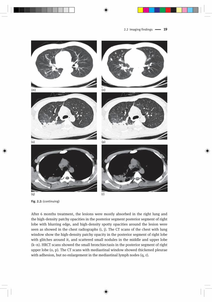
After 6 months treatment, the lesions were mostly absorbed in the right lung and the high-density patchy opacities in the posterior segment posterior segment of right lobe with blurring edge, and high-density spotty opacities around the lesion were seen as showed in the chest radiographs (i, j). The CT scans of the chest with lung window show the high-density patchy opacity in the posterior segment of right lobe with glitches around it, and scattered small nodules in the middle and upper lobe (k–n). HRCT scans showed the small bronchiectasis in the posterior segment of right upper lobe (o, p). The CT scans with mediastinal window showed thickened pleurae with adhesion, but no enlargement in the mediastinal lymph nodes (q, r).
(m)
(o)
(n)
(p)
(q) (r)
Fig. 2.2: (continuing)

20 2 Imaging findings of AIDS with pulmonary Rhodococcus equi disease
Case 2.3 (Fig. 2.3 a–d)A 31-year-old male patient. Cough with no obvious incentive occurred three months ago, with a white thin phlegm. Fever occurred one month ago, body temperature at 38.39 °C, and become obvious afternoon, accompanied by sweating and blood streak phlegm. Gasp occurred two weeks ago. Admission body temperature was 37 °C. The tofu-like objects was seen on the surface of tongue. The hairy leukoplakia was seen in the side of tongue. The right lower lung breath sounded slightly weaken. No dry or moist rales. The superficial lymph nodes showed no enlargement. CD4+ cell count
(a)
(c)
(b)
(d)
Fig. 2.3 a–d: Rhodococcus equi pneumonia