1 modern methods for the expression of proteins in ... · manding, metabolic pathways. in a...
TRANSCRIPT

1.1Introduction
The introduction of stable isotopes into proteins has significantly reduced the time re-quirements for structure elucidation of biomolecules. Moreover, structural studies of pro-teins with molecular weights exceeding the 10 kDa limit are usually not possible withoutuniform isotope labeling because of severe resonance overlap and inefficient coherencetransfer along the rather small 3J 1H-1H couplings. Nowadays, efficient expression of re-combinant proteins is a prerequisite for many techniques used in structural biology, butthe requirement for isotope labeling in particular often precludes NMR (nuclear mag-netic resonance) structure determination of proteins isolated from natural sources. Speci-fically, proteins that have been uniformly labeled with 15N and/or 13C are commonly re-quired for NMR spectroscopy, especially for backbone chemical shift assignment proce-dures, which are greatly facilitated by the use of a series of rather sensitive multi-dimen-sional triple-resonance NMR experiments (see Chapt. 4) [1], in a process that can also beautomated with good success [2]. Moreover, the random replacement of nonexchangeableprotons by deuterons reduces 1H-1H dipolar interactions and scalar couplings, thereby re-ducing peak line widths considerably and allowing structure elucidation of proteins ex-ceeding 30 kDa [3, 4]. Random fractionally deuterated protein samples also permit theuse of longer mixing times in NOESY (nuclear Overhauser effect spectroscopy) experi-ments, since spin-diffusion pathways are largely eliminated. In addition, transverse-re-laxation optimized spectroscopy (TROSY [5], see also Chapt. 10), which has been usedfor molecules larger than 100 kDa [6], benefits dramatically from deuteration. This stemsfrom the fact that the TROSY component that is narrowed by the DD-CSA compensationis broadened by dipolar interactions with nearby protons.
Besides uniform labeling approaches, stable isotopes can also be introduced at specificsites in proteins in order to simplify the assignment process and to isolate spectral infor-mation from the region of interest. For example, biosynthetically-directed fractional 13Clabeling offers the possibility of making stereospecific assignments of all isopropylmethyl groups of Val and Leu residues [7]. In another approach often used for solid-stateNMR termed residue-specific labeling, isotope labels are introduced at single sites in a pro-tein, as described in another chapter of this volume (Chapt. 11). A related scheme, calledamino acid-type labeling, is accomplished by expression in an amino acid-based medium
1
1
Modern Methods for the Expression of Proteinsin Isotopically Enriched FormHeiko Patzelt, Natalie Goto, Hideo Iwai, Kenneth Lundstrom,
and Erhard Fernholz
BioNMR in Drug Research. Edited by Oliver ZerbeCopyright © 2002 Wiley-VCH Verlag GmbH & Co. KGaA
ISBNs: 3-527-30465-7 (Hardback); 3-527-60066-3 (Electronic)

where only targeted amino acids contain isotope labels [8]. Preparing a series of sampleswith different isotopically enriched single amino acid types provides a useful approachfor the assignment of large systems not accessible by traditional methods (for an exam-ple see Ref. [9]). However, labeling specificity and yield can suffer from isotope scram-bling arising from metabolic conversion of amino acids. This problem can be circum-vented through the use of specially engineered amino acid-type specific auxotrophicstrains. An interesting alternative that can be used to achieve residue and/or amino acidtype-specific labeling is presented by the in vitro cell-free expression systems. These sys-tems are additionally advantageous when expression products display cell toxicity.
Another labeling strategy geared toward the study of large proteins combines the favor-able relaxation properties conferred by extensive deuteration with site-specific strategiesfor introducing protons. For example, methyl groups and/or aromatic amino acids can betargeted for protonation in otherwise fully deuterated proteins. An alternative approachfor the study of large proteins features segmental labeling methods based on protein spli-cing methodology. Consequently, longer stretches of protein are isotopically enriched,leaving the remainder unlabeled. Isotope editing will remove all signals from unlabeledsegments of the proteins, thereby largely reducing resonance overlap and facilitating as-signment. Protein splicing methods can also be used to introduce non-natural aminoacids or chemical modifications into the sequence. Therein, a protein segment contain-ing an unnatural residue can be chemically synthesized and then ligated to a recombi-nantly produced (isotopically enriched) segment. Protein splicing can also be used to pro-duce proteins which have high potential for cytotoxicity [10] or to stabilize proteinsthrough cyclization.
This chapter will mainly focus on recent developments in protein labeling methodolo-gy. For an introduction to methods involved in the generation and yield optimization ofprotein samples labeled with 13C and/or 15N by recombinant methods in E. coli the inter-ested reader is referred to excellent reviews published in the literature [8, 11–18]. Herewe first describe the use of labeled algal hydrolyzates for the production of labeled pro-teins in E. coli or other organisms. We then review methods used for the introduction ofisotope labels into specific sites, and this is followed by a section on segmental labelingapproaches. Subsequently, we will summarize recombinant protein expression methodsin hosts other than E. coli that have proven to be especially suitable for post-translation-ally modified proteins and membrane proteins, before concluding with an introductionto cell-free expression systems.
1.2Isotope-Labeled Proteins from Hydrolyzates of the Green Alga Scenedesmus obliquus
Although alternative expression systems have been successfully adapted for the produc-tion of isotope-labeled proteins (see Sect. 1.5), heterologous expression in E. coli often re-mains the method of choice for NMR sample preparation. There is a fundamental differ-ence, however, with respect to the kind of medium in which the cells are cultivated. In aso-called “chemically defined” or “minimal” medium only one or a very limited numberof carbon sources is provided, e.g. glucose or glycerol. All bacterial metabolites have tobe biosynthesized by the cells through the various, sometimes lengthy and energy-de-
1 Modern Methods for the Expression of Proteins in Isotopically Enriched Form2

manding, metabolic pathways. In a “complex” or “rich” medium, the cells grow, as thename suggests, on a complex mixture of amino acids and/or carbohydrates. Amino acidinterconversions, and thus the potential for isotope scrambling in selectively labeled sam-ples, are here reduced to a minimum. Unlabeled fermentations are usually performed incomplex media (i. e. yeast extract-containing LB for E. coli), since protein yield and celldensity are here considerably higher than in minimal media. The same is desirable forisotope-labeled fermentations, but the limited availability and/or high price of commer-cial amino acid/sugar mixtures in the required isotope composition often impose fermen-tations on a single carbon source.
Several companies are meanwhile supplying labeled amino acid/sugar mixtures of ac-ceptable quality. However, especially if larger-scale or repeated preparations of labeledproteins are envisaged, investment of some extra time for the in-house production of la-beled complex growth media for E. coli or other host cells clearly becomes advantageous,especially from the point of view of financial considerations.
The most frequently employed source for complex amino acid/sugar mixtures, labeledin any combination of 2H,13C and/or 15N in E. coli continues to be a phototrophic greenalga. Scenedesmus obliquus was introduced for that purpose in 1972 by Crespi and Katz[19]. In recent years, the original protocols have been modified by several groups, leadingto improvements in the yield and purity of the algal amino acid mixtures, thereby en-hancing protein labeling efficiency and expression levels in the hosts [20–22].
Depending on the desired labeling pattern, 2H2O, 13CO2 and/or 15NH4Cl or Na15NO3
are used as exclusive isotope sources during the algal fermentation. All of the above havebecome commercially available at affordable rates. For the preparation of random par-tially isotope-labeled amino acid/sugar mixtures, unlabeled water, carbon dioxide or nitro-gen salts are simply admixed to the labeled starting material in the appropriate propor-tions.
Thus, the three basic steps required for the preparation of a uniformly labeled proteinfor NMR experiments are
1. production of isotope-labeled algal hydrolyzates,2. adaptation of the protein overproducing organism (usually, but not always, E. coli) to
growth on the algal medium, and3. preparation and purification of the isotope-labeled protein on a preparative scale.
If specific amino acid-type labeling is required, the labeled amino acid is added to the fer-mentation of the expression host (topic 1 above, see Sect. 1.2.3). In this case, a thoroughisotope analysis of the expressed protein is advisable prior to NMR spectroscopic investi-gations. This is preferentially achieved by GC-MS analyses of the hydrolyzed amino acidsfrom the protein product.
1.2.1Production of Isotope-Labeled Algal Hydrolyzates
Cultures of S. obliquus can easily be grown photoautotrophically in two-tier flasks in aninorganic medium. A stepwise replacement of H2O by 2H2O leads to deuterated cultures,and a replacement of CO2 (the sole carbon source) by 13CO2 and/or the replacement of
1.2 Isotope-Labeled Proteins from Hydrolyzates of the Green Alga Scenedesmus obliquus 3

the nitrogen-containing salts by their 15N-isotopomers produces 13C- and/or 15N-labeledcultures. These are used as inoculi for larger fermentations in Fernbach flasks or stirredor airlifting fermenters. All fermentations are continuously illuminated with standardplant light bulbs or fluorescent tubes. In the last stage of 2H2O fermentations, all saltscontaining crystal water are repeatedly dissolved in small amounts of 2H2O and lyophi-lized before addition to the medium. If the algal cells are harvested under sterile condi-tions, the recovered medium can be re-inoculated for up to four further fermentations,and only phosphate is supplemented when the growth rate declines [22]. The recycling ofthe media cuts isotope costs by about 90%.
The algal cell mass is then purified from low-molecular-weight metabolites and hydro-lyzed in HCl (2HCl in the case of a deuterated fermentation). After neutralization andlyophilization, a white powder (typically 2–2.5 g L–1 of medium and fermentation cycle)is obtained, containing around 50% amino acids (for composition see Ref. [23]), 30% su-gars (composition in Ref. [24]) and 20% NaCl. This amino acid/sugar mixture for com-plex microbial growth media can be produced with any combination of 2H, 13C and/or15N, including random fractional label distributions. Used as carbon source it enables thesimple and quick preparation of isotope-labeled, complex microbial growth media for theproduction of labeled proteins.
1.2.2Adaptation of the Protein Overproducer to the Algal Medium
The described algal hydrolyzate contains amino acids and sugars in a physiological com-position, i. e. in a relative composition similar to that required by most host cells. Aminoacid biosynthesis, interconversion, and thus the potential for isotope scrambling, areminimized. When all potentially inhibitory low-molecular-weight compounds are re-moved by extraction prior to hydrolysis, most organisms grow well in media containingtheir typical salt and trace element composition, with the exception of NaCl, which is in-troduced as part of the algal hydrolyzate. Only the carbon sources are substituted by thealgal amino acid/sugar mixture (e. g. yeast extract is replaced by algal hydrolyzate withthe required isotope composition). Examples of the production of isotope-labeled proteinsin Bacteria [25], Archaea [23, 26] and Eucarya [27] can be found in the recent literature.
Since changing the carbon source may influence bacterial growth and expression char-acteristics, a series of unlabeled test experiments is recommended in order to establishthe minimum hydrolyzate concentration required, as well as the reproducibility of pro-tein expression levels.
While expression in 13C- and/or 15N-labeled media is usually straightforward, most or-ganisms need to be adapted in 3 to 4 steps to growth in 2H2O (e.g. 50, 75, 90, 100%). Inaddition, while formulating any deuterated medium, it is important to recall that thereading on pH meters equipped with normal glass electrodes is about 0.4 units lower in2H2O than in H2O with the same hydrogen/deuterium ion concentration [28]. Because ofthe different physical properties of 2H2O, growth may be slower than usual, and the tim-ing for induction of protein expression may require adjustment. The extent of deutera-tion depends on the type of experiments that will be performed. For investigations of in-ternal dynamics using 15N relaxation or for backbone assignment with triple-resonance
1 Modern Methods for the Expression of Proteins in Isotopically Enriched Form4
spectroscopy, 100% deuteration at the nonexchangable carbon sites maximizes signal sen-sitivity and resolution. On the other hand, structural information traditionally relies ondistance restraints derived from 1H,1H NOEs. Nietlispach et al. have calculated and mea-sured the effects of various levels of random fractional deuteration and found 50–70%deuteration most useful for larger proteins [29].
1.2.3Preparation of Homogenously Isotope-Labeled Protein by Fermentation on Algal Media
Production of isotope-labeled proteins on a larger scale from the optimized test condi-tions is typically routine as long as the physical growth parameters (reactor type, aera-tion, etc.) are not changed significantly. However, the purification of deuterated proteinsmay require some adjustments, depending on the techniques utilized. For example, be-cause of their considerably higher density, centrifugation gradients must be adapted.Also, the chromatographic properties of deuterated proteins may display differences rela-tive to their unlabeled (1H at natural abundance) counterparts, reflecting potential shiftsin isoelectric point, stronger intramolecular hydrogen bonds and weaker van der Waalsinteractions. A final consideration is the re-introduction of protons at the exchangeableamide sites. Since the quantitative exchange of amide protons from the protein core canbe extremely slow, it is sometimes necessary to expose the sample to denaturing condi-tions, followed by refolding in H2O, if possible.
1.2.4Amino Acid-Type Specific Labeling
The principal difficulty associated with the preparation of amino acid-type specific la-beled proteins is the suppression of metabolic scrambling of the label into other aminoacid types through the common metabolic pathways in the host cell. The use of a com-plex amino acid/sugar mixture, such as the one present in the algal hydrolyzates, re-duces this danger greatly compared to fermentations on a single carbon source. In fact,the activity of many enzymes responsible for amino acid interconversions appears to below or absent in bacteria grown under these conditions. For example, for the productionof a fully deuterium-labeled protein containing 1H-Trp, the host cell is grown on fully2H-labeled algal hydrolyzate in 2H2O to which unlabeled Trp is admixed. The individualamount of the differentially labeled amino acid to be added to the fermentation may varyfor the different residues and depends on the biosynthetic origin of the amino acid [30]as well as on its background concentration in the algal hydrolyzate [22]. Labels in the bio-synthetically central amino acids Asx and Glx show a pronounced tendency for biosynthe-tically-directed isotope relocation, since these molecules may be used as metabolic precur-sors for a number of different downstream amino acids (e.g. Met, Lys, Thr, and Ile orPro and Arg, for Asx and Glx, respectively). The metabolically peripheral amino acids canusually be labeled specifically with much higher isotope purity.
For every new amino acid, a small series of test experiments is normally sufficient to es-tablish a compromise between the minimum concentration required for high specific label-ing (usually at least ten times the amount introduced with the algal hydrolyzate) and the
1.2 Isotope-Labeled Proteins from Hydrolyzates of the Green Alga Scenedesmus obliquus 5

toxicity limit for the respective amino acid. Before a large-scale fermentation is attempted,the isotope composition downstream from the labeled amino acid should be analyzed. Themost probable sites of undesired isotope incorporation are found in the same biosyntheticgroup [30]. Labeled Tyr, for instance, may be found after media supplementation with la-beled Phe, whereas Cys and Gly labeling may result from the addition of labeled Ser.
1.2.5Mass Spectrometric Analysis of the Labeled Amino Acids
For amino acid analysis the labeled protein needs to be hydrolyzed and derivatized. Mostcommonly the hydrolysis is performed in 6 M HCl, and the amino acids are convertedinto their isopropyl ester and pentafluoropropanamide derivatives (Fig. 1.1) before GC/MS analysis. The molecular ion is not always visible after standard electron impact (EI)ionization, and the fragment after loss of the carboisopropoxy group is the highest obser-vable peak. This leaves m/e= 175 plus the mass of the amino acid side chain, fromwhich the degree of labeling can be directly deduced.
Fig. 1.2 shows illustrative mass spectra for derivatized Phe with various isotope pat-terns. In unlabeled Phe (Fig. 1.2a), the highest observable peak at m/e= 266 accounts forthe aforementioned fragment of 175 plus the mass of the benzyl group (C7H7 = 91). Thestrongest peak is due to the tropylium cation (C7H7) at m/e= 91. Complete deuteration(Fig. 1.2d) takes molecular ion to m/e= 274 (175 + C7
2H7), and the tropylium signal(C7
2H7) accordingly to m/e 98. As expected, the spectrum of 13C,15N-labeled Phe (Fig.1.2e) shows the corresponding signals at m/e= 275 (175 + 2 + 13C7H7) and 98 (13C7H7).
The signals in the spectra of the partially deuterated amino acid (Figs. 1.2 b–d and 1.2 f)show a statistical distribution of the masses around the calculated values for all fragmentsand are thus an unambiguous proof of a complete random distribution of the labels.
1.3Selective Labeling Schemes
While general labeling strategies relying on expression of proteins using hydrolyzates ofalgae are useful for uniform or amino acid-specific labeling, expression can also be per-formed in minimal media in a cost-effective manner. These types of expression media
1 Modern Methods for the Expression of Proteins in Isotopically Enriched Form6
Fig. 1.1 Most common derivatization of aminoacids for GC-MS analysis. The fragment withoutthe carboisopropoxy group normally produces the
highest observable peak, which is used for the de-termination of the isotope composition.

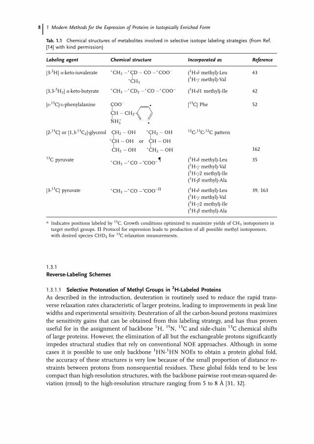
typically use glucose and ammonium salts as the sole carbon and nitrogen sources, re-spectively, and include D2O as the medium base when deuteration is required. Thesegrowth conditions are easily adapted for uniform or random fractional isotope labeling.In addition, modifications to this general formula can be made to introduce isotope la-bels into specific sites, for example, by supplemention of the expression media with spe-cifically labeled amino acids or amino acid precursors. In the following section we will re-view a number of methods used for different patterns of selective labeling in E. coli. Re-agents used in these different strategies, with corresponding literature references, aresummarized in Tab.1.1.
1.3 Selective Labeling Schemes 7
Fig. 1.2 EI mass spectra of derivatized phenylalanine, isolated from S. obliquus; a unlabeled, b random50% 2H-, c random 70% 2H-, d 100% 2H-, e 13C,15N-, and f 13C,15N/random 75% 2H-labeled.
1.3.1Reverse-Labeling Schemes
1.3.1.1 Selective Protonation of Methyl Groups in 2H-Labeled ProteinsAs described in the introduction, deuteration is routinely used to reduce the rapid trans-verse relaxation rates characteristic of larger proteins, leading to improvements in peak linewidths and experimental sensitivity. Deuteration of all the carbon-bound protons maximizesthe sensitivity gains that can be obtained from this labeling strategy, and has thus provenuseful for in the assignment of backbone 1H, 15N, 13C and side-chain 13C chemical shiftsof large proteins. However, the elimination of all but the exchangeable protons significantlyimpedes structural studies that rely on conventional NOE approaches. Although in somecases it is possible to use only backbone 1HN-1HN NOEs to obtain a protein global fold,the accuracy of these structures is very low because of the small proportion of distance re-straints between protons from nonsequential residues. These global folds tend to be lesscompact than high-resolution structures, with the backbone pairwise root-mean-squared de-viation (rmsd) to the high-resolution structure ranging from 5 to 8 Å [31, 32].
1 Modern Methods for the Expression of Proteins in Isotopically Enriched Form8
Tab. 1.1 Chemical structures of metabolites involved in selective isotope labeling strategies (from Ref.[14] with kind permission)
Labeling agent Chemical structure Incorporated as Reference
[3-2H] �-keto-isovalerate �CH3 ��CD � CO ��COO���CH3
(1H-� methyl)-Leu(1H-� methyl)-Val
43
[3,3-2H2] �-keto-butyrate �CH3 ��CD2 ��CO ��COO� (1H-�1 methyl)-Ile 42
[�-13C]-l-phenylalanine COO�� [13C] Phe 52
CH � CH2��NH�
3
[2-13C] or [1,3-13C2]-glycerol CH2 � OH �CH2 � OH� ��CH � OH or CH � OH� �CH2 � OH �CH2 � OH
12C-13C-12C pattern
162
13C pyruvate �CH3 ��CO ��COO�� (1H-� methyl)-Leu(1H-� methyl)-Val(1H-�2 methyl)-Ile(1H-� methyl)-Ala
35
[3-13C] pyruvate �CH3 ��CO ��COO�� (1H-� methyl)-Leu(1H-� methyl)-Val(1H-�2 methyl)-Ile(1H-� methyl)-Ala
39, 163
* Indicates positions labeled by 13C. Growth conditions optimized to maximize yields of CH3 isotopomers intarget methyl groups. � Protocol for expression leads to production of all possible methyl isotopomers,with desired species CHD2 for 13C relaxation measurements.
For the purpose of increasing the number of protons in the protein core while main-taining the benefits of extensive deuteration, it is possible to re-introduce protons using a“reverse isotope” labeling approach. In some of the original approaches, side chains oftarget amino acid types were selectively protonated in deuterated proteins by adding pro-tonated forms of these amino acids to the D2O growth medium (see, for example, Refs.[33, 34]). In a variation of this theme, Rosen and coworkers developed a protocol to selec-tively incorporate protons at the methyl positions of Ala, Val, Leu and Ile �2 [35]. Methylgroups are enriched in protein hydrophobic cores and hence are attractive targets for se-lective protonation [36]. In addition, NMR spectroscopic properties of methyl groups arefavorable owing to reasonably well-resolved 13C-1H correlations and rapid rotation aboutthe methyl symmetry axis that reduces peak line widths [37].
The original protocol for the production of 15N, 13C, 2H, 1Me (protonated methyl)-proteinsutilized 13C,1H-pyruvate as the exclusive source of carbon in 100% D2O minimal media [35].Pyruvate can be diverted into the tricarboxylic acid (TCA) cycle to produce many of the in-termediates used in the biosynthesis of amino acids [38], but can also be directly incorpo-rated into amino acids either by transamination (Ala), or reactions with threonine (Ile), py-ruvate (Val) or both pyruvate and acetyl CoA (Leu). As was observed, pyruvate that is directlyincorporated into these amino acids will largely retain the methyl protons, while those ami-no acids synthesized indirectly via intermediates will be highly deuterated. However, theincorporation of protons at each methyl site tends to be variable, with the result that methylisotopomers such as 13CHD2 and 13CH2D are also produced [35]. The additive deuteriumisotope effect on both carbon and proton chemical shifts produces upfield shifts relativeto the 13CH3 peak by 0.02 and 0.3 ppm per 2H atom in the 1H and 13C dimensions respec-tively. As a result, 13C-1H correlation spectroscopy of the methyl region shows three peaksfor every methyl group labeled in this way, translating into resolution and sensitivity prob-lems. Nonetheless, through the use of 2H-purging pulse schemes during the acquisition ofcarbon chemical shift, it is possible to remove the peaks arising from methyl groups con-taining 2H [32]. In addition, it should be noted that other pyruvate-based labeling schemeshave also found great utility in the measurement of side-chain dynamics involving thesemethyl-containing side chains [39–41].
More recently, the yield and uniformity of methyl group protonation was enhancedthrough the use of �-ketoisovalerate in combination with �-ketobutyrate to produce 15N,13C, 2H-labeled proteins with protons introduced at the methyl positions of Leu, Val andIle (�1) [42, 43]. Proteins expressed in D2O/13C, 2H-glucose/15NH4Cl minimal media canbe supplemented with 13C, [3, 2H] �-ketoisovalerate for the selective protonation of theVal and Leu methyl groups and [3,3-2H2], 13C �-ketobutyrate for Ile �1 methyl group la-beling. Using this strategy, labeling efficiency of the target methyl groups was shown toexceed 90%, while high levels of deuteration were maintained at other sites without pro-duction of methyl group isotopomers containing deuterium. Since the selectively deuter-ated form of these amino acid precursors can be obtained by base-catalyzed proton ex-change in aqueous buffer, the protonated, commercially available forms of these precur-sors are straightforwardly adapted to this labeling scheme.
1.3 Selective Labeling Schemes 9

1.3.1.2 Structure Determination of Selectively Methyl Protonated ProteinsHigh levels of deuteration combined with selective methyl protonation using one of theschemes outlined above permits the measurement of 1HN-1HN, 1HN-methyl and methyl-methyl NOEs. Global folds can be determined using this subset of NOEs, where the qual-ity of these structures is a reflection of protein topology, secondary structure content, andthe location and distribution of methyl groups in the protein [32]. In the case of a 30 kDacell adhesion fragment from intimin, for example, intradomain backbone rmsd values ofthe ensemble of structures ranged between 1.5 and 1.8 Å from the mean [44]. In con-trast, MBP structure quality was lower, with intradomain backbone rmsds between NMRand crystal structures of 3.1–3.8 Å [45]. Although structures produced by this methodolo-gy are often of a preliminary quality, they can nonetheless be useful in the localization ofligand or protein interaction sites and the identification of homologous proteins (e.g.Ref. [46]). Global folds can also be used as a structural stepping stone in the generationof high-resolution structures, since the assignment of additional NOEs from random frac-tionally deuterated samples or fully protonated molecules is facilitated by the use of apreliminary structure [47, 48]. Further improvements in the quality of structures can alsobe obtained through the incorporation of additional restraints such as dipolar couplings([45]) or homology modeling (e.g. Ref. [49]).
1.3.1.3 Introducing 1H,12C Aromatic Residues into Otherwise 13C UniformlyLabeled Proteins
Alternative schemes involving selective protonation have also been developed to increase thenumber of side-chain distance restraints that can be obtained in highly deuterated proteins.For example, 1H, 12C Phe and Tyr can be directly incorporated into an otherwise uniformly13C-labeled protein expressed in minimal media [50]. Since these amino acids are also pre-ferentially located in the hydrophobic cores of proteins, as well as at ligand binding inter-faces, distance restraints involving these residues can be very valuable. This labeling strat-egy was shown to be useful for proteins under 30 kDa with relatively few Phe and Tyr suchas a 24 kDa Dbl homology domain [48] and the 25 kDa antiapoptotic protein Bcl-xL [51]. Incases where overlap in the aromatic spectrum becomes problematic, a synthetic strategy hasbeen introduced to produce Phe that is 13C labeled only at the epsilon position [52]. An il-lustration of the utlility of this approach is provided by the structure determination of a 21kDa Dbl homology domain containing seven phenylalanine residues [47].
1.3.1.4 Backbone-Labeled ProteinsProtocols for selective isotope labeling of protein backbone atoms are also being developed,since the prevention of 13C incorporation at the C� site circumvents resolution problemsassociated with homonuclear 1JC�C� coupling. Toward this end, syntheses of backbone-la-beled amino acids have been described for ten different amino acids starting from 15N,13C2-glycine [53, 54]. While original demonstrations of backbone labeling utilized a CHOcell expression system to prevent isotope scrambling [53], bacterial cell expression systemshave also proven amenable to this strategy [54]. In this case, the expression medium mustcontain the full complement of amino acids, which are then replaced with those that are 13C�,13CO, 15N, and 1H� (or 50% 2H�) labeled just prior to induction of protein expression. As wasdemonstrated for ubiquitin backbone-labeled with a subset of amino acids, sensitivity and
1 Modern Methods for the Expression of Proteins in Isotopically Enriched Form10

resolution in HNCA-type experiments is enhanced, and couplings can be readily measuredfrom IPAP 1H-13C HSQC (heteronuclear single-quantum correlation) spectra [54, 55].
1.3.2Selective 13C Methyl Group Labeling
To reduce the expense of producing selectively methyl-labeled proteins, it is possible touse 13C-methyl iodide to synthesize �-ketobutyrate and �-ketoisovalerate containing 13Conly at the methyl sites [56]. A larger than 20-fold reduction in the cost of precursor mol-ecules can be achieved using this synthetic strategy in place of the commercially availableuniformly 13C-labeled isotopomers. Although these compounds can be adapted to the se-lective methyl protonation scheme described above, they can also be used to produce pro-teins that only contain 13C at the methyl positions of Val, Leu, and Ile �1, with 12C at allother sites. The reduced cost, spectral simplification and sensitivity enhancement of 13Cmethyl-labeled proteins over uniformly 13C-labeled samples facilitates the use of chemicalshift mapping in the search for potential lead compounds in the drug discovery process.If, on the other hand, selective methyl protonation is required, the 1H, 13C methyl-la-beled �-ketoisovalerate and �-ketobutyrate would be added to D2O expression media con-taining 2H, 13C-labeled glucose and 15N-labeled ammonium salt. However, compared tothe uniformly 13C-labeled selectively protonated samples described previously, structuredetermination is less straightforward since backbone carbon atoms for isoleucine and va-line are derived from the nonmethyl portion of the �-ketobutryate and �-ketoisovalerate,respectively, and would therefore contain the 12C isotope. Nonetheless, once assignmentsare made, NOE measurements involving these methyl groups benefit from eliminationof one-bond 13C-13C couplings leading to narrow carbon line widths without the require-ment for constant-time evolution periods [51].
1.4Intein-Based Protein Engineering for NMR Spectroscopy
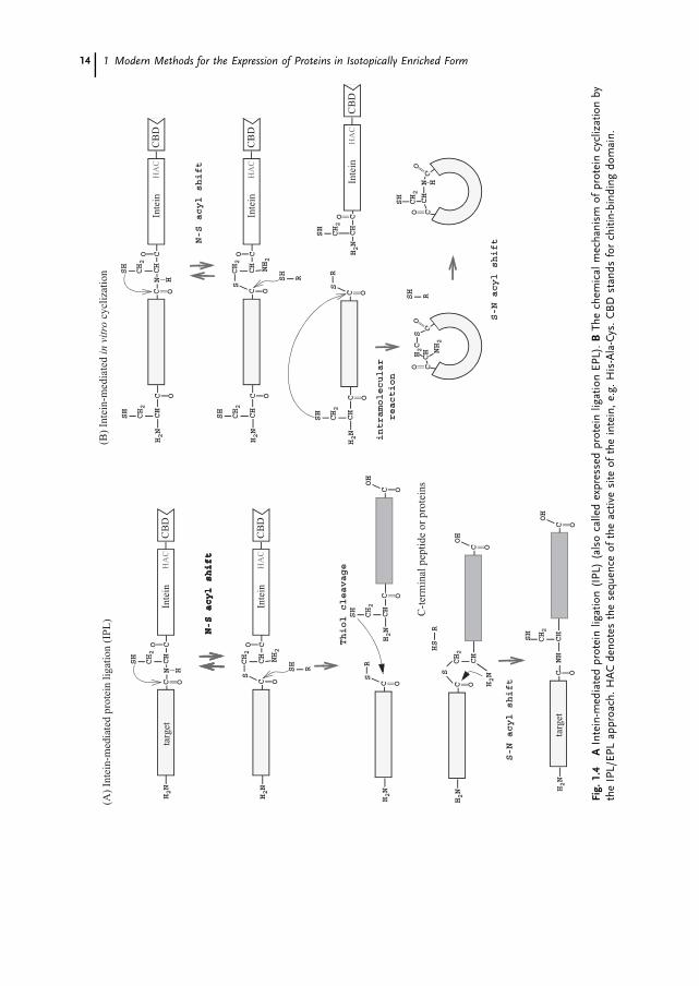
Recently, new advances in biochemistry have opened up a novel approach for protein en-gineering, which utilizes a protein-splicing domain. Protein splicing is a post-transla-tional chemical modification discovered in nature, which catalyzes the excision of an in-tervening polypeptide (internal protein, intein) while simultaneously ligating both the N-and C-terminal flanking polypeptide chains (Fig. 1.3, 1.5A), analogous to RNA splicing[57, 58]. This unique enzymatic process has been used in various biochemical and bio-technological applications such as protein purification, protein ligation, backbone cycliza-tion and C-terminal modifications. In particular, protein ligation using inteins hasopened up a new way for the production of segmentally labeled proteins, thereby reduc-ing the complexity of NMR spectra. For larger proteins or multimeric proteins, it may benecessary to combine selective labeling and segmental isotope-labeling approaches. More-over, it will be useful in cases where information is desired only for a small part of alarge protein, e. g. to characterize interactions with known binding sites or to detect con-formational changes in a specific region. Another potentially useful application of inteinsfor NMR is backbone cyclization to enhance the stability of proteins.
1.4 Intein-Based Protein Engineering for NMR Spectroscopy 11

1 Modern Methods for the Expression of Proteins in Isotopically Enriched Form12
Fig.
1.3
The
curr
ently
acce
pted
chem
ical
mec
hani
smof
prot
ein
splic
ing.
1N
-S(O
)ac
ylsh
ift,
2tr
anse
ster
ifica
tion,
3cl
eava
geby
succ
inim
ide
form
atio
n,4
S(O
)-N
acyl
shift
.

1.4.1Segmental Labeling of Proteins
Although more than 100 protein splicing domains have been found in nature [59], only ahandful have been used for segmental labeling purposes, namely SceVMA (PI-SceI), PI-PfuI and PI-PufII. The currently accepted reaction mechanism for protein splicing con-sists of the following four steps, namely, (i) N-S(O) acyl shift, (ii) transesterification, (iii)succinimide formation, and (iv) S(O)-N acyl rearrangement (Fig. 1.3). Either a subset ofthese four steps or the entire reaction process can be used for protein ligation. For exam-ple, the intein-mediated protein ligation (IPL) approach utilizes a subset of these reac-tions by using a modified intein as described in Sect. 1.4.1.1. On the other hand, thesplit intein approach requires all four reaction steps. In this case the success of the reac-tion depends on refolding properties of the split intein (Sect. 1.4.1.2).
1.4.1.1 Intein-Mediated Protein Ligation (IPL)/Expressed Protein Ligation (EPL)using the IMPACT System
The IMPACT (Intein-Mediated Purification with an Affinity Chitin-binding Tag) systemwas originally developed as a novel purification method by New England Biolab. Itmakes use of a modified intein, SceVMA, in which the active site was mutated from His-Asn-Cys to His-Ala-Cys, so that the usual cleavage due to succinimide formation involv-ing the side-chain of Asn is no longer possible (Figs. 1.4 A and 1.6A). Since a chitin-bind-ing domain (CBD) is fused to the C-terminus of the intein, this protein can be immobi-lized to a chitin column, providing a convenient tool for purification [60]. The desiredprotein segment is fused at the N-terminus of the intein, which can be liberated fromthe intein-CBD portion of the fusion protein by addition of nucleophiles such as dithiol-threitol (DTT), ethanethiol, 2-mercaptoethane sulfonic acids (MESNA), hydroxylamine orcysteine. The IMPACT system provides a good opportunity to expand the application ofnative chemical ligation, which was originally developed by the Kent group, to a varietyof protein targets, because the C-terminus of the N-terminal fusion polypeptide can beconverted easily into a thioester group by the intein-mediated cleavage [61]. For nativechemical ligation, an N-terminal peptide segment containing a C-terminal thioester ischemoselectively ligated to a C-terminal peptide segment that has an N-terminal cysteinein aqueous solution, without protecting any functional groups in the peptides. With theintein-based E. coli expression system, it is now possible to produce larger protein seg-ments with a C-terminal thioester group easily, which can subsequently be used for na-tive chemical ligation. Using this approach, Xu et al. have demonstrated the domain-se-lective 15N labeling of the SH2 domain of the Abl-kinase SH domain [62]. In this experi-ment, the 15N-labeled SH2 domain containing an N-terminal cysteine capped with a spe-cific proteolytic sequence, which can be removed, was expressed and purified in 15N-la-beled media. The protective N-terminal sequence was subsequently removed by proteoly-sis in order to create an N-terminal thionucleophile (N-terminal cysteine). The N-termi-nal segment of the SH3 domain was separately produced as the intein-fusion protein inunlabeled media and eluted with ethanethiol to form the C-terminal thioester. The unla-beled SH3 domain and 15N-labeled SH2 domain were ligated in aqueous solution at pH7 in the presence of thiophenol and benzyl mercaptan, achieving a yield of 70%.
1.4 Intein-Based Protein Engineering for NMR Spectroscopy 13
1 Modern Methods for the Expression of Proteins in Isotopically Enriched Form14
Fig.
1.4
AIn
tein
-med
iate
dpr
otei
nlig
atio
n(I
PL)
(als
oca
lled
expr
esse
dpr
otei
nlig
atio
nEP
L).
BTh
ech
emic
alm
echa
nism
ofpr
otei
ncy
cliz
atio
nby
the
IPL/
EPL
appr
oach
.H
AC
deno
tes
the
sequ
ence
ofth
eac
tive
site
ofth
ein
tein
,e.
g.H
is-A
la-C
ys.
CB
Dst
ands
for
chiti
n-bi
ndin
gdo
mai
n.

It has also been demonstrated that multiple ligation steps can be performed with theIPL/EPL approach, thereby illustrating its potential use in central-segment labeling [63].
One of the intrinsic limits of the IPL/EPL system is the requirement for a cysteine re-sidue at the site of protein ligation, which will replace the thioester group. Recently, itwas shown that this requirement could be avoided by using a cleavable thiol-containingauxiliary group. Low et al. demonstrated protein ligation by introducing a cleavable thiol-containing auxiliary group, 1-phenyl-2-mercaptoethyl, at the alpha-amino group of a che-mically synthesized peptide, which is removed upon protein ligation [64]. Unfortunately,this modification at the N-terminus could be difficult to introduce into proteins whichare prepared from bacterial expression systems, and hence its use could be limited to sit-uations where the C-terminal peptide can be chemically synthesized. Nevertheless, the re-moval auxiliary approach presents an opportunity for segmental isotope labeling regard-less of the primary sequence.
A second approach that can be adopted to overcome the intrinsic requirement for cyste-ine at the N-terminus of C-terminal fragment utilizes the enzyme subtiligase, a doublemutant of subtilisin, which is able to join two unprotected peptides. Thioester-modifiedproteins were shown to present good substrates of subtiligase [65]. However, althoughthis approach could be potentially useful for general isotope labeling, the efficiency ofthis process remains to be proven.
It is noteworthy that there is another limiting factor in the choice of amino acid typesat the junction sites which affect the enzymatic process of the intein. For example, in thecase of SceVMA (also called PI-SceI) from the IMPACT system, proline, cysteine, aspara-gine, aspartic acid, and arginine cannot be at the C-terminus of the N-terminal targetprotein just before the intein sequence. The presence of these residues at this positionwould either slow down the N-S acyl shift dramatically or lead to immediate hydrolysisof the product from the N-S acyl shift [66]. The compatibility of amino acid types at theproximal sites depends on the specific inteins and needs to be carefully considered dur-ing the design of the required expression vectors. The specific amino acid requirementsat a particular splicing site depends on the specific intein used and is thus a crucialpoint in this approach.
1.4.1.2 Reconstitution of Split InteinsIt has been demonstrated that an intein can be split into two fragments and reconsti-tuted in vitro as well as in vivo to form an active intein capable of trans splicing [67–69].This trans-splicing activity can be directly used for protein ligation as an alternative to thenative chemical ligation step, which requires additional thionucleophile groups. Yamazakiet al. have applied trans-splicing to the segmental labeling of RNA polymerase subunit �by splitting an intein from Pyrococcus furiosus, PI-PfuI (Fig. 1.5 B) [70]. Each half of thesplit intein fused to the N- (or C)-extein was produced separately, one in isotopically la-beled and the other in unlabeled medium. The independently prepared protein frag-ments were expressed as inclusion bodies and purified under denaturing conditions. Thetwo independently prepared fragments were reassembled and refolded in aqueous so-lution in order to form a functional intein, resulting in a ligated extein fragment and aspliced intein. The splicing reaction was found to be efficient at elevated temperature(70 �C), presumably because PI-PfuI is a thermophilic enzyme. However, the general use
1.4 Intein-Based Protein Engineering for NMR Spectroscopy 15

1 Modern Methods for the Expression of Proteins in Isotopically Enriched Form16
Fig.
1.5
AN
atur
alpr
otei
nsp
licin
g,B
Tran
ssp
licin
gw
itha
split
inte
in.
The
two
frag
men
tsca
nbe
prep
ared
sepa
rate
lyan
dre
as-
sem
bled
invi
tro
tofo
rman
activ
ein
tein
dom
ain
for
prot
ein
liga-
tion.
CC
entr
alse
gmen
tla
belin
gus
ing
two
diff
eren
tsp
litin
tein
s.
Res
idue
sin
volv
edin
the
prot
ein
splic
ing
reac
tion
are
show
nby
asi
ngle
char
acte
rco
deof
amin
oac
ids.
N-
and
C-t
erm
inia
rein
di-
cate
dby
NH
2an
dC
OO
Hre
spec
tivel
y.
B)
tran
s-sp
licin
gw
ith
split
inte
in

of elevated temperatures may often be unfavorable, because many proteins denature athigher temperatures. Therefore, conditions for refolding and ligation such as tempera-ture, pH, additives like glycerol etc. must be carefully optimized for each protein system.On the other hand, the split intein approach does not require any additional thionucleo-phile, in contrast to the IPL/EPL approach.
Remarkably, this method can even be extended to joining three segments by using twodifferent inteins, PI-PfuI and PI-PfuII (Fig. 1.5 C). Otomo et al. have successfully demon-strated this approach by isotope labeling a central segment of the 370-residue maltose-binding protein [71]. Three protein fragments were constructed for the two ligation reac-tions. The first fragment was a fusion protein of the N-terminal domain of the split tar-get protein and the N-terminal split intein-1. The second segment consisted of the C-ter-minal split intein-1 (PI-PfuI), the central part of the split target protein fragment and theN-terminal split intein-2 (PI-PfuII). The third segment contained the C-terminal split in-tein-2 fused to the C-terminal fragment of the split target protein. Ligation of the firstand the second fragment was facilitated by intein-1, while the second and the third frag-ments were ligated by intein-2. These ligation reactions were highly specific because twodifferent inteins were used. The second fragment was prepared in isotopically enrichedmedium, and hence the ligated protein was isotopically labeled only in the central part.
The intrinsic limitation of this approach, as in the case of the IPL/EPL approach, isthe requirement for specific proximal residues near or at the ligation sites. In inteins, theresidues at the junction where ligation occurs are highly conserved because of the chemi-cal mechanism and typically require either cysteine, serine, or threonine [72]. Therefore,at least the N-terminal amino acid of the C-extein must be one of these residues, depend-ing on which intein is used. In addition, the peptide sequence preceding the intein aswell as the sequence following the ligation site play an important role for the efficiencyof the protein-splicing reaction. For example, in the case of the DnaE intein, the five resi-dues preceding the intein N-terminus and the three residues following the ligation sitemust seem to be native extein residues in order to achieve efficient splicing [73]. How-ever, these sequential and structural determinants are presently not well understood forall known inteins. Otomo et al. have speculated that the flexibility of the junction regioncould be one of the important factors for ligation with PI-PfuI. Such requirements wouldrestrict the position of ligation sites, probably to (flexible) linker regions.
One advantage of the split intein over the IPL/EPL approach is the direct use of inteinsplicing activity, eliminating the requirement for additional thionucleophiles such as thio-phenol. An another potential advantage is the ability to perform multiple ligations in aone-pot process, greatly simplifying the reaction procedure for the ligation of several frag-ments. In contrast, the IPL/EPL approach requires ligation reactions to be performed se-quentially for multiple fragment ligations.
Although the use of split inteins for segmental isotope labeling has great potential, thenumber of inteins which have been adapted to this purpose is currently limited. Addi-tional difficulties arise from the fact that the determinants influencing the success of anintein splicing reaction are not well understood. Moreover, the refolding requirements ofsplit inteins could hinder its use as a general method for the ligation of protein frag-ments. Hence, further biochemical characterization of inteins is required for the advanceof intein-mediated protein ligation methods.
1.4 Intein-Based Protein Engineering for NMR Spectroscopy 17
1.4.2Stabilizing Proteins by Intein-Mediated Backbone Cyclization
Limited protein stability often hampers successful structure elucidation by X-ray crystallo-graphy and/or NMR spectroscopy. Relaxation properties are usually improved at elevatedtemperatures, and multidimensional NMR experiments require sample lifetimes to ex-tend over several days to weeks in order to acquire all the necessary data. In addition, theactivity of contaminating proteases that are sometimes present in purified samples canbe significant at the experimental temperatures. Therefore, the stability of a target pro-tein can be a concern, in particular for expensive isotope-labeled proteins.
There have been many attempts to improve protein stability and protein properties, uti-lizing methods such as random mutagenesis, directed evolution, and rational protein de-sign approaches. In general, these methods are far from straightforward and can betime-consuming. In addition, the stabilization of proteins without loss of function is nota trivial problem.
One new approach to stabilizing proteins without changing the primary sequence is tointroduce backbone cyclization [74]. No mutations in the primary sequence are intro-duced by this method, although it might be necessary to insert a flexible linker compris-ing several residues to join the termini [74–76]. Polymer theory by Flory predicts an im-provement in protein stability upon cyclization, because the entropy of the unfoldedstates should be reduced [77]. Backbone cyclization has long been used for small pep-tides to reduce the accessible conformational space. Recent advances in intein technologyhave opened up a new avenue for the cyclization of large proteins, because these pro-teins can be produced with recombinant techniques in bacterial expression systems [74,76]. Cyclized proteins can be produced either in vitro or in vivo, as discussed in the fol-lowing two sections. Statistical analysis of the structure database reveals that more than30% of all known proteins might have termini in relatively close proximity, and hencethe use of backbone cyclization to stabilize proteins has a good chance of success even incases where the structure is not yet known [78].
1.4.2.1 In vitro Cyclization of ProteinsThe IPL/EPL method described in Sect. 1.4.1.1 can be used for cyclization of the back-bone polypeptide chains of proteins simply by introducing a nucleophilic thiol group atthe N-terminus of the protein (Fig. 1.4 B, 1.6A). This can be achieved either by creatingan N-terminal cysteine by removing residues at the front of the cystein by specific proteo-lysis or by introducing a cysteine right after the methionine start codon, which is then re-moved enzymatically in vivo [74, 76]. Another method is to use the so-called TWIN sys-tem developed by New England Biolab, in which the target protein is fused into the mid-dle of two different modified inteins (Fig. 1.6 B). The N-terminal nucleophilic cysteine isproduced by an intein fused to the N-terminus of the target protein. At the same timethe C-terminus can be transformed into the corresponding thioester by another inteinfused to the C-terminus of the target protein [79]. This system circumvents the require-ment for a specific proteolytic site in order to create the N-terminal cysteine, thereby sim-plifying the cyclization procedure.
1 Modern Methods for the Expression of Proteins in Isotopically Enriched Form18

1.4 Intein-Based Protein Engineering for NMR Spectroscopy 19
Fig.
1.6
AIP
L/EP
Lcy
cliz
atio
nw
ithth
eIM
PAC
Tsy
stem
,B
back
bone
cycl
izat
ion
usin
gth
eTW
INsy
stem
,C
back
bone
cycl
izat
ion
with
split
inte
ins.
CB
Dst
ands
for
chiti
n-bi
ndin
gdo
mai
n.N
-ter
min
iar
ein
dica
ted
byN
H2.

The biggest problems associated with in vitro cyclization methods using the IPL/EPLor the TWIN system are competing intermolecular reactions such as polymerization andhydrolysis, which complicate purification as well as reduce yields [74, 79].
1.4.2.2 In vivo CyclizationThe principle of in vivo cyclization is based on the circular permutation of precursor pro-teins containing an intein (Fig. 1.6C) [74, 75, 80, 81]. A naturally occurring split intein,DnaE from Synechocystis sp. PCC6803, was first successfully used for cyclization. How-ever, similarly to the IPL/EPL approach, a mixture of linear and circular forms is ob-tained, presumably because of hydrolysis of an intermediate [73, 75]. On the other hand,artificially split inteins such as PI-PfuI, DnaB, and the RecA intein have been success-fully applied for in vivo cyclization, and only circular forms were observed [80–82], sug-gesting that the circular permutation approach is more suitable for cyclization. Com-pared to the IPL/EPL or the TWIN system, in vivo cyclization does not require any exter-nal thiol group for cyclization, similarly to protein ligation with split inteins. Moreover,there are no undesired products, such as linear forms or polymers, originating from in-termolecular reactions.
1.4.2.3 Stability Enhancement by Backbone CyclizationThe effect of backbone cyclization was originally tested on BPTI, but no stabilization ef-fects were observed, presumably because the three disulfide bridges reduce entropicgains [83]. Nevertheless, intein-mediated backbone cyclization has opened the way to astudy of cyclization effects on protein stability (including membrane proteins) in moredetail. Experimentally improved thermal and/or chemical stability has been shown toresult from backbone cyclization of a range of proteins, including �-lactamase, DHFR,E. coli IIAGlc, a destabilized mutant of SH3 domain and the N-terminal domain of DnaB[74, 75, 81, 82, 84]. An additional advantage imparted by backbone cyclization is essen-tially complete resistance to exopeptidases.
1.5Alternatives to E. coli Expression Systems
Structural biology and the structural understanding of the genome, popularly calledstructural genomics, play an increasingly important part in drug discovery today. Fastand reliable protein expression tools are therefore of prime importance. To this end, thechoice of protein expression systems has become increasingly important. While in thenot so distant past, only Escherichia coli-based expression was used, today a variety of ex-pression systems have been developed ranging from Archaebacteria to mammalian ex-pression vectors. Needless to say, there is no universal expression system, and hence it isoften necessary to balance various parameters to achieve optimal expression. For in-stance, considering the cost of isotopically enriched media, it can be advantageous to sa-crifice some native characteristics of a recombinant protein in order to benefit from thehigher yields that can be achieved in a more basic expression system. In contrast, specif-ic modifications of the target protein (e.g. glycosylation) predominantly occur in certaincell types, which therefore require the development of special expression vectors.
1 Modern Methods for the Expression of Proteins in Isotopically Enriched Form20

Here, we describe the various alternatives to the use of E. coli expression hosts for het-erologous gene expression. Advantages and disadvantages for the different expression sys-tems are discussed, and practical aspects of expression technologies are also described.The feasibility of isotope labeling of recombinantly expressed proteins and their potentialuse for NMR is also discussed, since the costs and quantities of recombinant proteinsproduced depend on the system being used.
1.5.1Expression Vectors
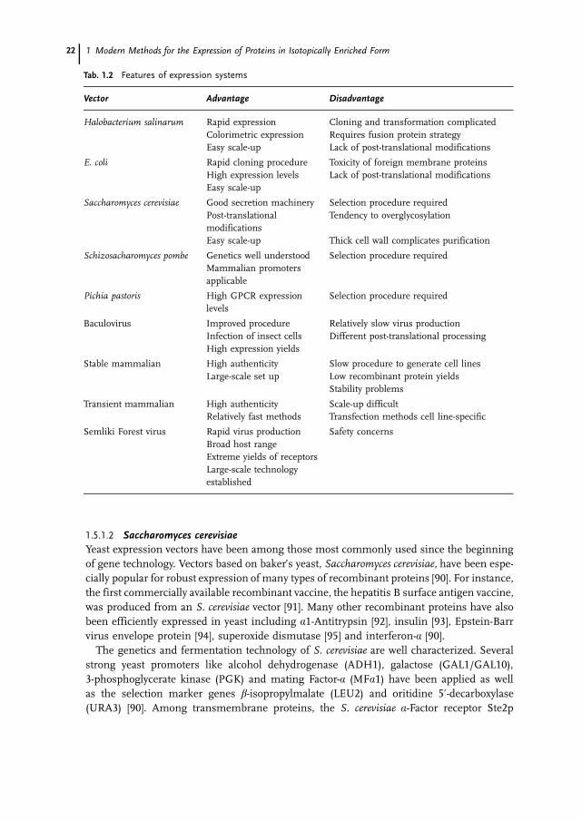
Traditionally, prokaryotic expression, especially employment of E. coli-based vectors, hasbeen the system of choice. However, bacteria are unable to provide many vital compo-nents required for post-translational modifications including various forms of glycosyla-tion or lipid attachment and protein processing, all of which can also be important forproper protein folding. For this reason, it is not surprising that much time and effort hasbeen dedicated to the development of alternative systems, summarized in Tab.1.2.
1.5.1.1 Halobacterium salinarumArchaea are interesting organisms in the sense that they represent a phylogenetically dis-tinct group of Prokarya, which is as distantly related to Eubacteria as to Eukarya [85]. H.salinarum, the best characterized Archaeon harbors a purple-colored plasma membraneconsisting of a complex of one protein, bacterio-opsin (Bop) and its chromophore retinalin a 1:1 ratio [86]. The complex was named bacteriorhodopsin, and it forms typicallyhighly ordered two-dimensional structures in the purple membrane, which allowed itspurification and the determination of a high-resolution structure [87]. Recently, a systemfor heterologous gene expression was constructed for H. salinarum [88]. Fusion con-structs between the Bop gene and heterologous sequences have been introduced into theH. salinarum expression vectors as follows: (i) C-terminally tagged bacteriorhodopsin [88];C-terminal fusion with (ii) the catalytic subunit of E. coli aspartate transcarbamylase [88],(iii) the human muscarinic M1 receptor [88], (iv) the human serotonin 5-HT2c receptor[88], (v) the yeast �-mating factor Ste2 receptor [88]. The Bop-transcarbamylase fusionwas well expressed, generating yields of 7 mg receptor per liter of culture. However, in-troduction of tags at the C-terminus of the Bop gene significantly reduced its expressionlevels. This was partly because of a decrease in Bop-fusion protein mRNA levels com-pared to the wild-type Bop. More dramatically, expression studies of fusion constructs be-tween the Bop gene and mammalian GPCRs (G protein-coupled receptors, human mus-carinic M1 receptor, platelet-activating receptor and angiotensin-1 receptor) failed to de-tect fusion protein expression detected by Western blotting [89]. In this case, coding re-gion swaps between Bop and GPCRs improved RNA yields and resulted in detectable lev-els of Ste2 receptor. These results suggest that H. salinarum can be considered as a po-tentially interesting alternative. The simple and rapid large-scale culture technology is at-tractive; however, improvements are still required concerning heterologous gene expres-sion. In addition, questions related to codon usage and fusion construct optimizationneed to be properly addressed.
1.5 Alternatives to E. coli Expression Systems 21
1.5.1.2 Saccharomyces cerevisiaeYeast expression vectors have been among those most commonly used since the beginningof gene technology. Vectors based on baker’s yeast, Saccharomyces cerevisiae, have been espe-cially popular for robust expression of many types of recombinant proteins [90]. For instance,the first commercially available recombinant vaccine, the hepatitis B surface antigen vaccine,was produced from an S. cerevisiae vector [91]. Many other recombinant proteins have alsobeen efficiently expressed in yeast including �1-Antitrypsin [92], insulin [93], Epstein-Barrvirus envelope protein [94], superoxide dismutase [95] and interferon-� [90].
The genetics and fermentation technology of S. cerevisiae are well characterized. Severalstrong yeast promoters like alcohol dehydrogenase (ADH1), galactose (GAL1/GAL10),3-phosphoglycerate kinase (PGK) and mating Factor-� (MF�1) have been applied as wellas the selection marker genes �-isopropylmalate (LEU2) and oritidine 5�-decarboxylase(URA3) [90]. Among transmembrane proteins, the S. cerevisiae �-Factor receptor Ste2p
1 Modern Methods for the Expression of Proteins in Isotopically Enriched Form22
Tab. 1.2 Features of expression systems
Vector Advantage Disadvantage
Halobacterium salinarum Rapid expression Cloning and transformation complicatedColorimetric expression Requires fusion protein strategyEasy scale-up Lack of post-translational modifications
E. coli Rapid cloning procedure Toxicity of foreign membrane proteinsHigh expression levels Lack of post-translational modificationsEasy scale-up
Saccharomyces cerevisiae Good secretion machinery Selection procedure requiredPost-translationalmodifications
Tendency to overglycosylation
Easy scale-up Thick cell wall complicates purification
Schizosacharomyces pombe Genetics well understood Selection procedure requiredMammalian promotersapplicable
Pichia pastoris High GPCR expressionlevels
Selection procedure required
Baculovirus Improved procedure Relatively slow virus productionInfection of insect cells Different post-translational processingHigh expression yields
Stable mammalian High authenticity Slow procedure to generate cell linesLarge-scale set up Low recombinant protein yields
Stability problems
Transient mammalian High authenticity Scale-up difficultRelatively fast methods Transfection methods cell line-specific
Semliki Forest virus Rapid virus production Safety concernsBroad host rangeExtreme yields of receptorsLarge-scale technologyestablished
has been expressed with C-terminal FLAG and His6 tags [96]. Ste2p belongs to the familyof GPCRs with a 7-transmembrane topology. Yields of up to 1 mg of almost homologousreceptor were obtained, and the purified receptor was reconstituted into artificial phos-pholipid vesicles. However, restoration of ligand-binding activity required the addition ofsolubilized membranes from an Ste2p negative yeast strain. Also, the human dopamineD1A receptor was expressed with C-terminal FLAG and His6 tags in S. cerevisiae, whichallowed for purification and reconstitution of receptor [97].
1.5.1.3 Schizosaccharomyces pombeAnother yeast strain that has received much attention as an expression host is the fissionyeast Schizosaccharomyces pombe. In contrast to S. cerevisiae, no budding occurs, and theyeast only reproduces by means of fission and by spores [98]. Two types of expressionvectors have been developed for S. pombe. The chromosomal integration type of vectormaintains the foreign gene stably in the chromosome [99], and the episomal vector repli-cates autonomously in yeast cells [100]. Some mammalian promoters like the humanchorionic gonadotropin and CMV promoters are functional in S. pombe [101]. The fissionyeasts possess many similar features to mammalian cells. S. pombe has a signal transduc-tion system similar to the mammalian G protein-coupling system [102], and the mamma-lian endoplasmatic reticulum retention signal KDEL is also recognized [103]. The glycosy-lation pattern for S. pombe is also different from that of S. cerevisiae and other yeast spe-cies.
A wide range of mammalian proteins have been expressed in S. pombe. In a successfulexample, the human lipocortin I comprised 50% of the total soluble proteins in yeastcells and showed high activity, indicating that the post-translational modifications weremammalian-like [104]. Membrane proteins including cytochrome P450 were expressed atten times the levels of those in other yeast systems [105]. Also, GPCRs have been ex-pressed in S. pombe, where the human dopamine D2 receptor was correctly inserted intothe yeast cell membrane and demonstrated expression levels three times those of S. cere-visiae [106].
1.5.1.4 Pichia pastorisThe methylotrophic yeasts including Pichia pastoris, Hansenula polymorpha and Kluyvero-myces lactis have become potentially attractive expression hosts for various recombinantproteins [107]. In addition to the relative ease with which molecular biology manipula-tions can be carried out, P. pastoris has demonstrated a capacity for performing manypost-translational modifications such as glycosylation, disulfide bond formation and pro-teolytic processing [108]. P. pastoris utilizes the tightly methanol-regulated alcohol oxidase1 (AOX1) promoter, and the vector is integrated as several copies into the yeast host ge-nome. When human insulin was expressed in P. pastoris, the secretion was comparableto that obtained for S. cerevisiae. Peptide mapping and mass spectrometry confirmedidentical processing of human insulin in yeast and mammalian cells. P. pastoris has alsobeen used as a host for expression of GPCRs [109]. The mouse 5-HT5A receptor and thehuman �2-adrenergic receptor were fused to the prepropeptide sequence of the S. cerevi-siae �-factor, which enhanced the expression levels by a factor of three. Multiple chromo-somal integrations further improved the expression twofold. In the case of the �2-adre-
1.5 Alternatives to E. coli Expression Systems 23

nergic receptor, addition of the antagonist alprenol to the culture medium increased thenumber of specific binding sites. A similar but weaker effect was seen for the 5-HT5A re-ceptor after addition of yohimbine. The binding activity for the �2-adrenergic receptorand the 5-HT5A receptor were 25 pmol and 40 pmol, respectively, per milligram of mem-brane protein. The pharmacological profiles assayed by ligand-displacement analysis weresimilar to those obtained from receptors expressed in mammalian cells.
1.5.1.5 BaculovirusHeterologous gene expression has been studied to a great extent in insect cells with theaid of baculovirus vectors. The popularity of the baculovirus system is mainly due to thehigh expression levels obtained for various recombinant proteins resulting from the useof strong viral promoters [110]. Generally, heterologous genes are expressed from thepolyhedrin promoter of Autographa californica in several insect cell lines such as Spodop-tera frugiperda (Sf9), Trichoplusia ni (Tn), Mamestra brassicae and Estigmene acrea [111].Although baculovirus vectors have been used for expression of various mammalian re-combinant proteins, a limitation has been the differences in the N-glycosylation pathwaybetween insect and mammalian cells. However, Estigmene acrea cells can produce a simi-lar glycosylation pattern as occurs in mammalian cells [112]. Moreover, modifications ofbaculovirus vectors by replacing the polyhedrin promoter with a CMV promoter made itpossible to carry out expression studies in mammalian cell lines [113]. Using this so-called BacMam system, milligram quantities of a cellular adhesion protein (SAF-3) couldbe produced in CHO cells [114]. Baculovirus vectors have been used extensively to ex-press GPCRs and ligand-gated ion channels [115]. Expression levels up to 60 pmol recep-tor per milligram have been obtained, which has led to relatively efficient purificationprocedures. In attempts to further enhance the expression level of the �2-adrenergic re-ceptor, an artificial sequence was introduced, which resulted in approximately double thereceptor levels in insect cells [116].
1.5.1.6 Transient Mammalian ExpressionSeveral approaches have been taken to develop efficient transient mammalian expressionsystems. The most straightforward process has been to engineer expression vectors withstrong promoters. Relatively high expression levels for cytoplasmic and even some trans-membrane proteins have been obtained in adherent cells on a small scale. However, amajor problem arises in the scale-up of these growth procedures, which are also rela-tively expensive [117]. In spite of this difficulty, transient transfection experiments usinga modified calcium-phosphate coprecipitation method have been carried out in HEK293EBNA cells adapted to suspension cultures grown on a 100 L scale [118]. More than 0.5 gof a monoclonal antibody was produced from this system, although similar methodshave yet to be developed for receptor expression.
1.5.1.7 Stable Mammalian ExpressionGeneration of various cell lines (BHK, CHO and HEK293) with the target gene inserteddownstream of a strong promoter into the genome is a common approach to achieve over-expression in mammalian hosts. However, one drawback is the time-consuming procedureinvolved in the establishment of stable cell lines, which generally requires 6–8 weeks. Other
1 Modern Methods for the Expression of Proteins in Isotopically Enriched Form24
problems associated with this approach are related to the relatively low expression levels andthe instability of generated cell lines. These issues have been addressed by engineering in-ducible expression systems, which are usually based on tetracyclin-based regulation (Tet on-off systems) [119]. A highly interesting development has been the generation of a cold-in-ducible expression system based on the Sindbis virus replicon [120]. Because of a point mu-tation in one of the replicase genes, the viral replicase complex is totally inactive at 37 �C,whereas a shift in temperature below 34 �C results in high replication activity and high lev-els of heterologous gene expression. Using this approach, the serotonin transporter gene,characterized by its low expression levels in any system tested, generated reasonable yields(approximately 250,000 copies per cell) [121].
1.5.1.8 Viral VectorsThe two common features that have made viral vectors attractive for recombinant proteinexpression are their high infection rates for a broad range of mammalian cell lines andtheir strong promoters. Adenovirus vectors have shown high expression levels in, for in-stance, human embryonic kidney (HEK293) cells, but their use has been to some extentrestricted by the fairly complicated virus generation procedure [122]. Another potentiallyuseful class of viruses are the poxviruses. Recombinant gene expression of herpes sim-plex virus thymidine kinase (TK) has been established for vaccinia virus vectors [123].Moreover, the engineering of a hybrid bacteriophage-vaccinia virus vector by applying theT7 promoter has simplified and broadened the use of pox virus-based expression systems[124]. However, vaccinia vectors are still quite complicated to use for rapid recombinantprotein expression, and they have instead found applications in the field of vaccine devel-opment. Alphavirus vectors have proven to be highly efficent for heterologous gene ex-pression. Both Semliki Forest virus- (SFV) [125] and Sindbis virus-based [126] expressionvectors have been engineered to rapidly generate high-titer recombinant particles, whichare susceptible to a broad range of mammalian cell lines and primary cells in culture[127]. Typically, both GPCRs and ligand-gated ion channels have been expressed at ex-treme levels, i. e. up to 200 pmol receptor per milligram protein [128]. Large-scale SFVtechnology has been established, which has generated large quantities of, for instance,mouse serotonin 5-HT3 receptor, purified to homogeneity and subject to structural char-acterization [129]. Moreover, several GPCRs have been expressed at levels of 5–10 mg re-ceptor yields per liter suspension culture of mammalian host cells [130], which has pro-vided material for large-scale purification.
1.5.2 Comparison of Expression SystemsComparison between different systems for transmembrane protein expression are alwaysdifficult to make, and they obviously reflect individual needs and are strongly influencedby personal experience. It is, however, important to define the usefulness of each systemby taking into account different aspects such as ease of handling, expression levels, timeand labor requirements, safety, costs, and the quality of the produced recombinant pro-tein (Tab.1.3).
Obviously, prokaryotic systems are easy to use, the costs for their large-scale applica-tions are low, and no safety risks are involved. The drawbacks are their limited capacityfor post-translational modifications and generally low yields of complex mammalian
1.5 Alternatives to E. coli Expression Systems 25
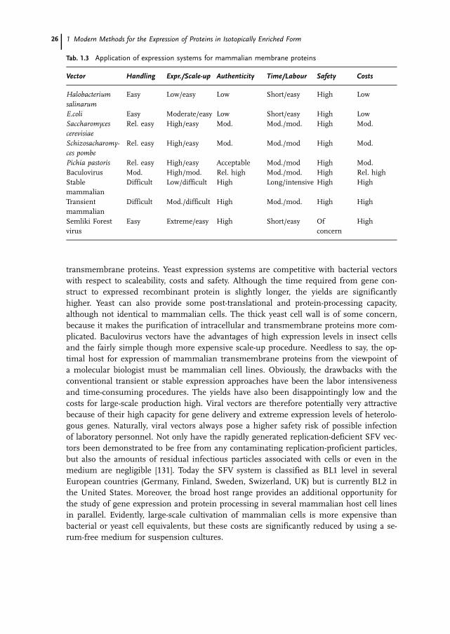
transmembrane proteins. Yeast expression systems are competitive with bacterial vectorswith respect to scaleability, costs and safety. Although the time required from gene con-struct to expressed recombinant protein is slightly longer, the yields are significantlyhigher. Yeast can also provide some post-translational and protein-processing capacity,although not identical to mammalian cells. The thick yeast cell wall is of some concern,because it makes the purification of intracellular and transmembrane proteins more com-plicated. Baculovirus vectors have the advantages of high expression levels in insect cellsand the fairly simple though more expensive scale-up procedure. Needless to say, the op-timal host for expression of mammalian transmembrane proteins from the viewpoint ofa molecular biologist must be mammalian cell lines. Obviously, the drawbacks with theconventional transient or stable expression approaches have been the labor intensivenessand time-consuming procedures. The yields have also been disappointingly low and thecosts for large-scale production high. Viral vectors are therefore potentially very attractivebecause of their high capacity for gene delivery and extreme expression levels of heterolo-gous genes. Naturally, viral vectors always pose a higher safety risk of possible infectionof laboratory personnel. Not only have the rapidly generated replication-deficient SFV vec-tors been demonstrated to be free from any contaminating replication-proficient particles,but also the amounts of residual infectious particles associated with cells or even in themedium are negligible [131]. Today the SFV system is classified as BL1 level in severalEuropean countries (Germany, Finland, Sweden, Swizerland, UK) but is currently BL2 inthe United States. Moreover, the broad host range provides an additional opportunity forthe study of gene expression and protein processing in several mammalian host cell linesin parallel. Evidently, large-scale cultivation of mammalian cells is more expensive thanbacterial or yeast cell equivalents, but these costs are significantly reduced by using a se-rum-free medium for suspension cultures.
1 Modern Methods for the Expression of Proteins in Isotopically Enriched Form26
Tab. 1.3 Application of expression systems for mammalian membrane proteins
Vector Handling Expr./Scale-up Authenticity Time/Labour Safety Costs
Halobacteriumsalinarum
Easy Low/easy Low Short/easy High Low
E.coli Easy Moderate/easy Low Short/easy High LowSaccharomycescerevisiae
Rel. easy High/easy Mod. Mod./mod. High Mod.
Schizosacharomy-ces pombe
Rel. easy High/easy Mod. Mod./mod High Mod.
Pichia pastoris Rel. easy High/easy Acceptable Mod./mod High Mod.Baculovirus Mod. High/mod. Rel. high Mod./mod. High Rel. highStablemammalian
Difficult Low/difficult High Long/intensive High High
Transientmammalian
Difficult Mod./difficult High Mod./mod. High High
Semliki Forestvirus
Easy Extreme/easy High Short/easy Ofconcern
High

1.5.3Isotope Labeling and NMR
Recent developments in technologies within structural biology should also play an impor-tant part for transmembrane proteins. The potential to incorporate stable isotopes wouldfacilitate structure determination by NMR techniques. Although NMR technologies werelong considered to be applicable only to smaller proteins, the development of transverserelaxation-optimized spectroscopy (TROSY) has made it possible to use NMR for largerproteins also [5], even integral membrane proteins. For example, the OmpX and OmpAintegral membrane proteins of E. coli were labeled with 13C/15N/2H isotopes and overex-pressed as inclusion bodies in bacterial cells. After solubilization in 6 M Gdn-HCl and re-constitution in detergent micelles, solution NMR techniques could be used to identifyregular secondary structural elements [132].
Proteins that require non-E.coli expression systems are generally too large in size to beused for NMR studies without uniform 15N, 13C and and sometimes 2H labeling. Hence,when using the expression systems described in this chapter, it is important to ensurethat cells can be grown on defined, isotopically enriched media. This fact at the momentexcludes, for example, the use of fetal calf serum. However, special isotope-enriched de-fined media are available from commercial suppliers which present fully rich, serum-free(protein-free) media at reasonable costs containing labeled amino acids and carbohy-drates and which can be used to effectively express heterologous proteins in insect cellsapplying the baculovirus vector system [133]. Similarly, rich media are also available forexpression in S. cerevisiae [134]. Conversely, the methylotropic yeast P. pastoris can begrown on minimal media, facilitating its use as a potential host for isotope labeling. Infact, there have been a number of successful examples where P. pastoris was used to pro-duce isotopically enriched samples for solution NMR studies, including a cysteine-richglycosylated domain of thromobomodulin [135], a glycosylated EGF module [136], do-mains from rat calretinin [137], and tick anticoagulant peptide [138]. Even more impor-tantly, it was shown that expression in yeast enables the production of heterologous pro-teins in deuterated form [139], which would be very difficult or even impossible toachieve in mammalian expression systems because of the cell toxicity of deuteratedwater. Similarly, H. salinarum can easily be grown in D2O and on the above-described al-gae hydrolyzates. Moreover, residue type-specific labeling is possible in this host [26]. Ex-amples of proteins expressed in this host in isotopically labeled form can be found in theliterature, e.g. Refs. [25] and [140]. In order to obtain proteins with more native-like gly-cosylation patterns, CHO mammalian cell expression systems have also been developedfor NMR sample generation [141–143]. However, the requirement for rich media inmammalian cell-based expression systems combined with difficulties associated with gen-erating high expression levels have to date prevented a more widespread utilization forNMR. Nonetheless, considering the range of proteins that may be inaccessible to E. coli-based expression systems as well as the potential information that can be gained bystudying the native-like post-translationally modified forms of proteins, the adaptation ofheterologous protein expression systems to the purpose of isotope labeling for solutionNMR clearly requires further development.
1.5 Alternatives to E. coli Expression Systems 27
Reconstitution of GPCRs in appropriate membrane mimetics or membrane prepara-tions is still very difficult, and success requires efficient expression systems to yield en-ough protein material. As large quantities of an increasing number of recombinant pro-teins become available, it will be possible to develop techniques for solubilization, purifi-cation and reconstitution in a high-throughput format. The first global structural geno-mics project for membrane proteins, MePNet (Membrane Protein Network) was recentlyinitiated with the aim of comparing the overexpression of 100 GPCRs in three systemsbased on E. coli, P. pastoris and SFV vectors [144]. The goal of this three year program isto verify the expression levels for the 100 targets and establish platforms for solubiliza-tion, purification and crystallization technologies which should form a solid base for ob-taining novel high-resolution structures of GPCRs. Technological developments arisingfrom this initiative should also benefit the field of solution and solid-state NMR.
1.5.4Target Proteins
The choice of expression system is to a large extent dictated by the type of target protein.In general, many soluble proteins, which are fairly easy to express and have a low molec-ular weight, are efficiently expressed from bacterial vectors. As a rule of thumb one cansuggest that E. coli-based expression should be used whenever possible. However, E. colivectors are not suitable for expression of many authentic mammalian transmembraneproteins. Additionally, the near-completion of the human genome sequence has revealeda multitude of genes as potential targets for structural analyses. Many of these includetransmembrane proteins, whose properties quite often make them more difficult to ex-press. Many of these transmembrane proteins, including receptors and channels, are im-portant targets for drug discovery. GPCRs alone stand for approximately 50% of drug tar-gets today. Moreover, a quarter of the top 200 drugs in the United States are based onGPCRs, and the annual sales in 2000 reached more than 18.5 billion US$ [145]. It istherefore understandable that so much interest has been focused on transmembrane pro-teins today. At present, it is possible to express complicated transmembrane receptors inseveral vector systems at reasonably high levels, cost-effectively in a near-to-native state.As more recombinant proteins become available, the solubilization, purification and crys-tallization for NMR technology can also be developed, which will contribute to theachievement of rates of structure determination similar to those common for soluble pro-teins today. However, the determination of high-resolution structures of membrane pro-teins, and particularly mammalian ones, has been modest compared to soluble proteins(Tab.1.4). Recently, methodological advances in NMR spectroscopy have significantlyraised the size limit amenable to NMR investigations, and hence membrane proteinstructures determined by NMR have also appeared in the literature. The outer mem-brane protein A from E. coli was reconstituted in DPC micelles and the structure deter-mined by heteronuclear multidimensional NMR [146]. Similarly, the structure of the out-er membrane protein OmpX was elucidated in DHPC micelles [147]. Both proteins wereexpressed in E. coli. However, in the case of OmpA, a series of selectively 15N-labeledsamples was prepared, whereas in the case of OmpX, spectroscopy was performed on asingle triply 2H,13C,15N-labeled sample. The best successes have been achieved when it
1 Modern Methods for the Expression of Proteins in Isotopically Enriched Form28

was possible to isolate proteins from their natural sources instead of using recombi-nantly expressed forms. The results have been particularly poor for GPCRs, and the onlyexample of a mammalian 7-transmembrane protein for which a high-resolution structurehas been obtained is bovine rhodopsin [148].
1.6The Use of Cell-Free Protein Expression for NMR Analysis
The expression of proteins usually requires optimization by trial and error, since condi-tions leading to production of high yields of active protein are difficult to predict. There-fore, additional time-consuming steps are often involved that may require changes in thechoice of expression system, of reaction conditions, and of tests for the expression of pro-tein sub-domains. In structural genomics programs this problem was rapidly identifiedas a major bottleneck, since high-throughput production of soluble proteins in milligramamounts is essential for success. Hence, systems that allow rapid, productive expression,are easy to manipulate, and can be run in a parallel format are highly desired in order torapidly screen for the best conditions.
Until recently, cell-free protein expression (also sometimes erroneously named in vitroprotein expression) did not exhibit the productivity required for preparation of NMR sam-ples, especially considering the high cost of using isotopically labeled starting material.Rather, it was exclusively used as an analytical tool that served to verify correct cloning orto study promotor sites. Because of the very low yields, detection of the expressed prod-uct usually required incorporation of a radioactive label (usually via 35S-methionine).
This situation changed fundamentally when Spirin published his substantial improve-ments in 1988 that resulted in much higher product yields [149]. He developed a set-upin which the coupled transcription/translation reaction could be continuously suppliedwith all the essential low-molecular-weight components (nucleotides, amino acids, energycomponents) while maintaining a constant reaction volume by continuously removingthe product through a membrane (continuous-flow cell-free system, CFCF). Under theseconditions, the system would remain active for more than 1 day, in contrast to the usualupper limit of 2 h which was observed in the conventional batch system. The prolongedexpression period indicated that sufficient amounts of all factors necessary for translationwere still available in the system, although the membrane clearly allowed the leakage ofat least some of them. Soon, these findings were used to run the reaction in a more ro-bust dialysis mode, keeping the reaction volume constant while feeding the system withall necessary low-molecular-weight consumables.
In the following years the productivity of the method was tremendously improved, sothat milligram amounts of protein per milliliter of reaction mixture could be obtained[150–154]. This finally opened the door to cell-free protein expression to be used for theproduction of isotopically enriched proteins suitable for NMR analysis. However,although widely considered to be a promising method for labeling proteins at specific po-sitions and therefore facilitating the process of chemical shift assignment as well as re-ducing spectral overlap [155, 156], in fact it was used in very few laboratories worldwide.This was mainly due to the fact that the preparation of the lyzate was tedious and the ex-pression levels of each batch varied from lot to lot.
1.6 The Use of Cell-Free Protein Expression for NMR Analysis 29

Recently, a system for cell-free protein production has became commercially available(the Rapid Translation System, RTS) [157]. In the following sections this system will bedescribed, and advantages as well as limitations will be discussed.
1.6.1The Cell-Free Protein Expression System RTS
The RTS system includes two different technology platforms for cell-free protein expres-sion as well as a number of tools for finding optimal conditions (Scheme 1.1). All expres-sion systems use the T7-polymerase for transcription and an E. coli lyzate with reducednuclease and protease activity for translation. The conditions are optimized for a coupledtranscription/translation reaction so that the DNA can be directly used as the template.
The first platform (RTS 100 HY) is designed as a screening tool. It uses the batch for-mat, so that the reaction time does not exceed 3 h. In particular, in the RTS 100 HY sys-tem the exonuclease activity is reduced, so that the direct use of PCR-generated DNAtemplates is possible. To facilitate the generation of the PCR templates there is a specialproduct available (linear template kit), which introduces all regulatory elements [T7-pro-motor, gene10 enhancer sequence and the Shine-Dalgarno (RBS) sequence]. Conse-quently, RTS 100 HY can be used for the rapid evaluation of the best template, withoutspending time with cloning, and for optimization of the reaction conditions (e.g. tem-perature, choice of additives like detergents, chaperones etc.). In addition, a bioinformatictool (the program ProteoExpert) facilitates the process of designing the optimal templateby analyzing and improving the secondary structure of the corresponding mRNA (with-out changing the amino acid sequence of the protein).
1 Modern Methods for the Expression of Proteins in Isotopically Enriched Form30
Scheme 1.1 From gene to protein structure via cell-free protein expression.
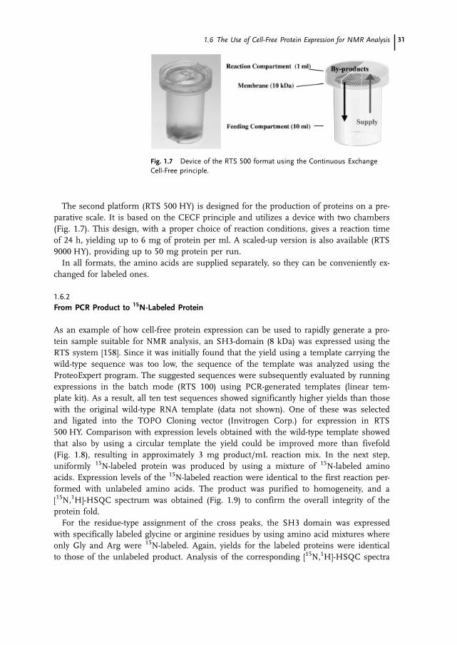
The second platform (RTS 500 HY) is designed for the production of proteins on a pre-parative scale. It is based on the CECF principle and utilizes a device with two chambers(Fig. 1.7). This design, with a proper choice of reaction conditions, gives a reaction timeof 24 h, yielding up to 6 mg of protein per ml. A scaled-up version is also available (RTS9000 HY), providing up to 50 mg protein per run.
In all formats, the amino acids are supplied separately, so they can be conveniently ex-changed for labeled ones.
1.6.2From PCR Product to 15N-Labeled Protein
As an example of how cell-free protein expression can be used to rapidly generate a pro-tein sample suitable for NMR analysis, an SH3-domain (8 kDa) was expressed using theRTS system [158]. Since it was initially found that the yield using a template carrying thewild-type sequence was too low, the sequence of the template was analyzed using theProteoExpert program. The suggested sequences were subsequently evaluated by runningexpressions in the batch mode (RTS 100) using PCR-generated templates (linear tem-plate kit). As a result, all ten test sequences showed significantly higher yields than thosewith the original wild-type RNA template (data not shown). One of these was selectedand ligated into the TOPO Cloning vector (Invitrogen Corp.) for expression in RTS500 HY. Comparison with expression levels obtained with the wild-type template showedthat also by using a circular template the yield could be improved more than fivefold(Fig. 1.8), resulting in approximately 3 mg product/mL reaction mix. In the next step,uniformly 15N-labeled protein was produced by using a mixture of 15N-labeled aminoacids. Expression levels of the 15N-labeled reaction were identical to the first reaction per-formed with unlabeled amino acids. The product was purified to homogeneity, and a[15N,1H]-HSQC spectrum was obtained (Fig. 1.9) to confirm the overall integrity of theprotein fold.
For the residue-type assignment of the cross peaks, the SH3 domain was expressedwith specifically labeled glycine or arginine residues by using amino acid mixtures whereonly Gly and Arg were 15N-labeled. Again, yields for the labeled proteins were identicalto those of the unlabeled product. Analysis of the corresponding [15N,1H]-HSQC spectra
1.6 The Use of Cell-Free Protein Expression for NMR Analysis 31
Fig. 1.7 Device of the RTS 500 format using the Continuous ExchangeCell-Free principle.

(Fig. 1.9) verified that the numbers of cross peaks for the 15N-Gly as well as for the 15N-Arg labeled protein were identical to the predicted ones (10 for the 10 Gly and 6 for the3 Arg, respectively). Importantly, no scrambling of the 15N-labels could be detected. Allcross peaks were contained in the [15N,1H]-HSQC spectrum of the uniformly labeled pro-tein (Fig. 1.9) and no additional signals occurred. Therefore, by simply overlaying theHSQC spectra, the cross peaks belonging to the glycine and the arginine residues couldbe readily assigned.
1 Modern Methods for the Expression of Proteins in Isotopically Enriched Form32
Fig. 1.8 Western blot analysis (via His6-tag) of the expression of the SH3 do-main using the wild-type DNA-sequence (lanes 1 and 2) or the optimized DNA-sequence (lanes 3 and 4). On lanes 1 and 3 0.25 �L DNA were loaded on thegel, while on lanes 2 and 4 0.125�L were applied.
Fig. 1.9 [15N,1H]-HSQC spectrum of 15N-uniformly labeled SH3 domain (A) and of a sample selectivelylabeled with only 15N-Gly (B) or 15N-Arg (C).

1.6.3Discussion and Outlook
This example demonstrates how cell-free protein expression can be used to rapidly opti-mize the reaction conditions as well as to shorten the time for spectral assignments byproducing individually labeled proteins.
To discuss in general the applicabilities of cell-free protein expression technology [159],two features are most valuable. First, only the protein of interest is produced, because thehighly active T7-polymerase is used for translation. Consequently, labeled amino acidsare almost exclusively incorporated into the newly produced protein. Since the labeledamino acids can be supplied as a mixture or added individually, the time for assigningthe cross peaks to the particular amino acid(s) can be significantly reduced. Thisapproach will enable partial assignments to be made in molecules that are far too largeto allow spectral assignments from uniformly labeled protein by classical methods.
The second important feature is that cell-free protein expression can be considered asan “open system”, meaning that no lipid membrane barriers are present. Consequently,chemicals, proteins (e.g. chaperones) as well as PCR-generated templates can be addeddirectly to the reaction solution. Even major changes of the reaction conditions are possi-ble (e.g. using a redox system to produce active proteins containing correctly formed dis-ulfide bonds [160]). These features, as well as the ease of sample handling, dramaticallyreduce the time for optimizing the expression conditions (in fact, pipetting robots can bedesigned to run the reactions). Moreover, proteins can be synthesized which display celltoxicity and which therefore can hardly be expressed in classical systems.
However, certain limitations do exist that need to be considered. Although enzymesnecessary for post-translational modifications can be added, in principle there is currentlyno productive system available for the preparation of glycosylated proteins, althoughsome interesting results have already been obtained [161]. Also, the expression of func-tional membrane proteins in quantities necessary for structural analysis will be a chal-lenging task for the future.
Nonetheless, the speed and flexibility of this emerging technology could provide thekey to meeting the demands of high-throughput structure determinations.
1.6 The Use of Cell-Free Protein Expression for NMR Analysis 33

1 Modern Methods for the Expression of Proteins in Isotopically Enriched Form34
1.7References
1 Bax, A.; Grzesiek, S. Acc. Chem. Res. 1993,26, 131–138.
2 Zimmerman, D.E.; Kulikowski, C.A.;
Huang, Y. P.; Feng, W. Q.; Tashiro, M.; Shi-
motakahara, S.; Chien, C.Y.; Powers, R.;
Montelione, G.T. J. Mol. Biol. 1997, 269,592–610.
3 Yamazaki, T.; Lee, W.; Arrowsmith, C.H.;
Muhandiram, D.R.; Kay, L.E. J. Am. Chem.Soc. 1994, 116, 11655–11666.
4 LeMaster, D.M.; Richards, F.M. Biochemis-try 1988, 27, 142–150.
5 Pervushin, K.; Riek, R.; Wider, G.;
Wüthrich, K. Proc. Natl. Acad. Sci. USA1997, 94, 12366–12371.
6 Salzmann, M.; Pervushin, K.; Wider, G.;
Senn, H.; Wüthrich, K. J. Am. Chem. Soc.2000, 122, 7543–7548.
7 Neri, D.; Szyperski, T.; Otting, G.; Senn,
H.; Wüthrich, K. Biochemistry 1989, 28,7510–7516.
8 Muchmore, D.C.; McIntosh, L.P.; Russel,
C.B.; Anderson, D.E.; Dahlquist, F.W. InMethods Enzymol.; Oppenheimer, N. J.,
James, T. L., Eds.; Academic Press: San Die-go, 1989; Vol. 177, pp 44–73.
9 Patzelt, H.; Simon, B.; Ferlaak, A.; Kess-
ler, B.; Kühne, R.; Schmieder, P.; Oester-
helt, D.; Oschkinat, H. Proc. Natl. Acad.Sci. USA 2002, 99, 9765–9770.
10 Evans, T.C., Jr.; Benner, J.; Xu, M.Q. Pro-tein Sci. 1998, 7, 2256–2264.
11 Pardi, A. Curr. Opin. Struct. Biol. 1992, 2,832–835.
12 Markley, J. L.; Kainosho, M. In NMR ofMacromolecules: A Practical Approach; Ro-
berts, G. C.K., Ed.; Oxford University Press:Oxford, 1993; pp 101–152.
13 LeMaster, D.M. Progr. NMR Spectrosc. 1994,26, 371–419.
14 Goto, N.K.; Kay, L.E. Curr. Opin. Struct. Biol.2000, 10, 585–592.
15 Bolton, P.H. Progr. NMR Spectrosc. 1990, 22,423–452.
16 Kainosho, M. Nature Struct. Biol. 1997, 4Suppl., 858–861.
17 Lian, L.-Y.; Middleton, D.A. Prog. NMRSpectrosc. 2001, 39, 171–190.
18 Makrides, S. C. Microbiol. Rev. 1996, 60,512–538.
19 Crespi, H.L.; Katz, J. J. Methods Enzymol.1972, 26, 627–637.
20 Brodin, P.; Drakenberg, T.; Thulin, E.;
Forsen, S.; Grundstroem, T. Protein Eng.1989, 2, 353–357.
21 Sorensen, P.; Poulsen, F.M. J. Biomol.NMR 1992, 2, 99–101.
22 Patzelt, H.; Kessler, B.; Oschkinat, H.;
Oesterhelt, D. Phytochemistry 1999, 50, 215–217.
23 Patzelt, H.; Ulrich, A.S.; Egbringhoff,
H.; Dux, P.; Ashurst, J.; Simon, B.; Oschki-
nat, H.; Oesterhelt, D. J. Biomol. NMR1997, 10, 95–106.
24 Takeda, H. Phytochemistry 1996, 42, 673–675.25 Schweimer, K.; Marg, B.-L.; Oesterhelt, D.;
Rösch, P.; Sticht, H. J. Biomol. NMR 2000,16, 347–348.
26 Reat, V.; Patzelt, H.; Ferrand, M.; Pfister,
C.; Oesterhelt, D.; Zaccai, G. Proc. Natl.Acad. Sci. U. S. A. 1998, 95, 4970–4975.
27 Celeste, S. G.; Jay, G.D.; Brenda, G. S.; En-
gin, S.H. Arch. Biochem. Biophys. 1995, 319,203–210.
28 Glasoe, P. K.; Long, F.A. J. Phys. Chem.1960, 64, 188–190.
29 Nietlispach, D.; Clowes, R. T.; Broadhurst,
R. W.; Ito, Y.; Keeler, J.; Kelly, M.; Ashurst,
J.; Oschkinat, H.; Domaille, P. J.; Laue,
E.D. J. Am. Chem. Soc. 1996, 118, 407–415.30 For more information see: http://
www.genome.ad.jp/kegg/kegg2.html31 Venters, R. A.; Metzler, W.J.; Spicer, L.D.;
Mueller, L.; Farmer, B.T. J. Am. Chem. Soc.1995, 117, 9592–9593.
32 Gardner, K. H.; Rosen, M.K.; Kay, L.E. Bio-chemistry 1997, 36, 1389–1401.
33 Crespi, H.L.; Rosenberg, R. M.; Katz, J. J.
Science 1968, 161, 795–796.34 Crespi, H.L.; Katz, J. J. Nature 1969, 224,
560–562.35 Rosen, M.K.; Gardner, K. H.; Willis, R. C.;
Parris, W.E.; Pawson, T.; Kay, L.E. J. Mol.Biol. 1996, 263, 627–636.
36 Janin, J.; Miller, S.; Chothia, C. J. Mol.Biol. 1988, 204, 155–164.
37 Kay, L.E.; Bull, T. E.; Nicholson, L. K.;
Griesinger, C.; Schwalbe, H.; Bax, A.;
Torchia, D.A. J. Magn. Reson. 1992, 100,538–558.

1.7 References 35
38 Gottschalk, G. Bacterial Metabolism; 2 ed.;Springer Verlag: New York, 1986.
39 Lee, A. L.; Urbauer, J. L.; Wand, A. J. J. Bio-mol. NMR 1997, 9, 437–440.
40 Ishima, R.; Louis, J. M.; Torchia, D.A.
J. Biomol. NMR 2001, 21, 167–171.41 Mulder, F.A.; Hon, B.; Mittermaier, A.;
Dahlquist, F.W.; Kay, L.E. J. Am. Chem.Soc. 2002, 124, 1443–1451.
42 Gardner, K. H.; Kay, L.E. J. Am. Chem. Soc.1997, 119, 7599–7600.
43 Goto, N.K.; Gardner, K.H.; Mueller, G. A.;
Willis, R.C.; Kay, L. E. J. Biomol. NMR1999, 13, 369–374.
44 Kelly, G.; Prasannan, S.; Daniell, S.; Flem-
ing, K.; Frankel, G.; Dougan, G.; Conner-
ton, I.; Matthews, S. Nat. Struct. Biol. 1999,6, 313–318.
45 Mueller, G.A.; Choy, W.Y.; Yang, D.; For-
man-Kay, J. D.; Venters, R. A.; Kay, L.E.
J. Mol. Biol. 2000, 300, 197–212.46 Berardi, M.J.; Sun, C.H.; Zehr, M.; Abild-
gaard, F.; Peng, J.; Speck, N.A.; Bushwel-
ler, J. H. Struct. Fold. Des. 1999, 7, 1247–1256.
47 Liu, X.; Wang, H.; Eberstadt, M.; Schnu-
chel, A.; Olejniczak, E. T.; Meadows, R.P.;
Schkeryantz, J. M.; Janowick, D.A.; Har-
lan, J. E.; Harris, E.A.; Staunton, D.E.;
Fesik, S.W. Cell 1998, 95, 269–277.48 Aghazadeh, B.; Zhu, K.; Kubiseski, T. J.;
Liu, G.A.; Pawson, T.; Zheng, Y.; Rosen,
M.K. Nat. Struct. Biol. 1998, 5, 1098–1107.49 DeRose, E.F.; Li, D.W.; Darden, T.; Harvey,
S.; Perrino, F.W.; Schaaper, R. M.; Lon-
don, R. E. Biochemistry 2002, 41, 94–110.50 Vuister, G. W.; Kim, S. J.; Wu, C.; Bax, A.
J. Am. Chem. Soc. 1994, 116, 9206–9210.51 Medek, A.; Olejniczak, E.T.; Meadows,
R. P.; Fesik, S. W. J. Biomol. NMR 2000, 18,229–238.
52 Wang, H.; Janowick, D. A.; Schkeryantz,
J. M.; Liu, X.H.; Fesik, S. W. J. Am. Chem.Soc. 1999, 121, 1611–1612.
53 Coughlin, P. E.; Anderson, F.E.; Oliver,
E. J.; Brown, J.M.; Homans, S.W.; Pollak,
S.; Lustbader, J. W. J. Am. Chem. Soc. 1999,121, 11871–11874.
54 Giesen, A. W.; Bae, L.C.; Barrett, C.L.; Chy-
ba, J. A.; Chaykovsky, M.M.; Cheng, M.C.;
Murray, J. H.; Oliver, E. J.; Sullivan, S.M.;
Brown, J.M.; Dahlquist, F.W.; Homans,
S.W. J. Biomol. NMR 2001, 19, 255–260.
55 Giesen, A. W.; Bae, L.C.; Barrett, C.L.; Chy-
ba, J. A.; Chaykovsky, M.M.; Cheng, M.C.;
Murray, J. H.; Oliver, E. J.; Sullivan, S.M.;
Brown, J.M.; Homans, S.W. J. Biomol. NMR2002, 22, 21–26.
56 Hajduk, P. J.; Augeri, D. J.; Mack, J.; Men-
doza, R.; Yang, J. G.; Betz, S. F.; Fesik, S. W.
J. Am. Chem. Soc. 2000, 122, 7898–7904.57 Hirata, R.; Ohsumk, Y.; Nakano, A.; Kawa-
saki, H.; Suzuki, K.; Anraku, Y. J. Biol.Chem. 1990, 265, 6726–6733.
58 Kane, P. M.; Yamashiro, C.T.; Wolczyk,
D.F.; Neff, N.; Goebl, M.; Stevens, T. H.
Science 1990, 250, 651–657.59 Perler, F.B. Nucleic Acids Res 2002, 30, 383–
384.60 Chong, S.; Montello, G. E.; Zhang, A.;
Cantor, E. J.; Liao, W.; Xu, M.Q.; Benner, J.
Nucleic Acids Res 1998, 26, 5109–5115.61 Dawson, P. E.; Kent, S. B. Annu. Rev. Bio-
chem. 2000, 69, 923–960.62 Xu, R.; Ayers, B.; Cowburn, D.; Muir, T. W.
Proc. Natl. Acad. Sci. USA 1999, 96, 388–393.63 Cotton, G. J.; Ayers, B.; Xu, R.; Muir, T.W.
J. Am. Chem. Soc. 1999, 121, 1100–1101.64 Low, D.W.; Hill, M.G.; Carrasco, M.R.;
Kent, S. B.; Botti, P. Proc. Natl. Acad. Sci.USA 2001, 98, 6554–6559.
65 Welker, E.; Scheraga, H.A. Biochem. Bio-phys. Res. Commun. 1999, 254, 147–151.
66 Chong, S.; Mersha, F.B.; Comb, D. G.;
Scott, M.E.; Landry, D.; Vence, L.M.; Per-
ler, F.B.; Benner, J.; Kucera, R. B.; Hirvo-
nen, C.A.; Pelletier, J. J.; Paulus, H.; Xu,
M.Q. Gene 1997, 192, 271–281.67 Mills, K.V.; Lew, B.M.; Jiang, S.; Paulus,
H. Proc. Natl. Acad. Sci. USA 1998, 95, 3543–3548.
68 Wu, H.; Xu, M. Q.; Liu, X. Q. Biochim. Bio-phys. Acta 1998, 1387, 422–432.
69 Southworth, M.W.; Adam, E.; Panne, D.;
Byer, R.; Kautz, R.; Perler, F.B. EMBO J.1998, 17, 918–926.
70 Yamazaki, T.; Otomo, T.; Oda, N.; Kyogoku,
Y.; Uegaki, K.; Ito, N.; Ishino, Y.; Naka-
mura, H. J. Am. Chem. Soc. 1998, 120, 5591–5592.
71 Otomo, T.; Teruya, K.; Uegaki, K.; Yamaza-
ki, T.; Kyogoku, Y. J. Biomol. NMR 1999, 14,105–114.
72 Paulus, H. Annu. Rev. Biochem. 2000, 69,447–496.

1 Modern Methods for the Expression of Proteins in Isotopically Enriched Form36
73 Evans, T.C., Jr.; Martin, D.; Kolly, R.;
Panne, D.; Sun, L.; Ghosh, I.; Chen, L.;
Benner, J.; Liu, X. Q.; Xu, M.Q. J. Biol.Chem. 2000, 275, 9091–9094.
74 Iwai, H.; Plückthun, A. FEBS Lett. 1999,459, 166–172.
75 Scott, C.P.; Abel-Santos, E.; Wall, M.;
Wahnon, D.C.; Benkovic, S. J. Proc. Natl.Acad. Sc. USA 1999, 96, 13638–13643.
76 Camarero, J. A.; Muir, T.W. J. Am. Chem.Soc. 1999, 121, 5597–5598.
77 Flory, P. J. J. Am. Chem. Soc. 1956, 78, 5222–5235.
78 Trabi, M.; Craik, D. J. Trends Biochem. Sci.2002, 27, 132–138.
79 Evans, T.C., Jr.; Benner, J.; Xu, M.Q. J. Biol.Chem. 1999, 274, 3923–3926.
80 Iwai, H.; Lingel, A.; Plückthun, A. J. Biol.Chem. 2001, 276, 16548–16554.
81 Williams, N.K.; Prosselkov, P.; Liepinsh,
E.; Line, I.; Shapiro, A.; Littler, D.R.; Cur-
mi, P.M.; Otting, G.; Dixon, N. E. J. Biol.Chem. 2002, 277, 7790–7798.
82 Siebold, C.; Flukiger, K.; Beutler, R.;
Erni, B. FEBS Lett. 2001, 504, 104–111.83 Goldenberg, D.P.; Creighton, T.E. J. Mol.
Biol. 1983, 165, 407–413.84 Camarero, J. A.; Fushman, D.; Sato, S.;
Giriat, I.; Cowburn, D.; Raleigh, D.P.;
Muir, T. W. J. Mol. Biol. 2001, 308, 1045–1062.
85 Woese, C.R.; Fox, G.E. Proc. Natl. Acad. Sci.USA 1977, 74, 5088–5090.
86 Murata, K.; Mitsuoka, K.; Hirai, T.; Walz,
T.; Agre, P.; Heymann, J.B.; Engel, A.; Fu-
jiyoshi, Y. Nature 2000, 407, 599–605.87 Luecke, H.; Schobert, B.; Richter, H.T.;
Cartailler, J. P.; Lanyi, J. K. J. Mol. Biol.1999, 291, 899–911.
88 Turner, G. J.; Reusch, R.; Winter-Vann,
A. M.; Martinez, L.; Betlach, M.C. ProteinExpr. Purif. 1999, 17, 312–323.
89 Winter-Vann, A.M.; Martinez, L.; Bartus,
C.; Levay, A.; Turner, G. J. In Perspectives onSolid State NMR in Biology; Kühne, S. R., de
Groot, H. J. M., Eds.; Kluwer: Netherlands,2001; pp 141–159.
90 Hinnen, A.; Buxton, F.; Chaudhuri, B.;
Heim, J.; Hottiger, T.; Meyhack, B.; Poh-
lig, G. In Gene expression in recombinant Mi-croorganisms; Smith, A., Ed.; Marcel Dekker:New York, 1994; pp 121–193.
91 Valenzuela, P.; Medina, A.; Rutter, W. J.;
Ammerer, G.; Hall, B.D. Nature 1982, 298,347–350.
92 Cabezon, T.; De Wilde, M.; Herion, P.;
Loriau, R.; Bollen, A. Proc. Natl. Acad. Sci.USA 1984, 81, 6594–6598.
93 Emini, E.A.; Ellis, R. W.; Miller, W. J.;
McAleer, W.J.; Scolnick, E.M.; Gerety, R. J.
J. Infect. 1986, 13 Suppl. A, 3–9.94 Schultz, L.D.; Tanner, J.; Hofmann, K. J.;
Emini, E.A.; Condra, J. H.; Jones, R.E.;
Kieff, E.; Ellis, R. W. Gene 1987, 54, 113–123.
95 Hallewell, R. A.; Mills, R.; Tekamp-Olson,
P.; Blacher, R.; Rosenberg, S.; Otting, F.;
Masiarz, F.R.; Scandella, C. J. Biotechnol.1987, 5, 363–366.
96 David, N. E.; Gee, M.; Andersen, B.; Nai-
der, F.; Thorner, J.; Stevens, R.C. J. Biol.Chem. 1997, 272, 15553–15561.
97 Andersen, B.; Stevens, R. C. Protein Expr.Purif. 1998, 13, 111–119.
98 Giga-Hama, Y.; Kumagai, H. Biotechnol.Appl. Biochem. 1999, 30, 235–244.
99 Grallert, B.; Nurse, P.; Patterson, T. E.
Mol. Gen. Genet. 1993, 238, 26–32.100 Maundrell, K.; Hutchison, A.; Shall, S.
EMBO J. 1988, 7, 2203–2209.101 Toyama, R.; Okayama, H. FEBS Lett. 1990,
268, 217–221.102 Xu, H.P.; White, M.; Marcus, S.; Wigler,
M. Mol. Cell Biol. 1994, 14, 50–58.103 Hildebrandt, V. In Foreign Gene Expression
in Fission Yeast Schizosaccharomyces pombe;Giga-Hama, Y., Kumagai, H., Eds.; SpringerVerlag: Berlin, 1997; pp 97–110.
104 Giga-Hama, Y.; Tohda, H.; Okada, H.; Owa-
da, M.K.; Okayama, H.; Kumagai, H. Bio-technology 1994, 12, 400–404.
105 Yasumori, T. In Foreign Gene expression inFission Yeast Schizosaccharomyces pombe;Giga-Hama, Y., Kumagai, H., Eds.; SpringerVerlag: Berlin, 1997; pp 111–121.
106 Sander, P.; Grunewald, S.; Reilander, H.;
Michel, H. FEBS Lett. 1994, 344, 41–46.107 Kjeldsen, T.; Pettersson, A.F.; Hach, M.
Biotechnol. Appl. Biochem. 1999, 29, 79–86.108 Cereghino, J.L.; Cregg, J.M. FEMS Micro-
biol. Rev. 2000, 24, 45–66.109 Weiss, H.M.; Haase, W.; Michel, H.; Rei-
lander, H. Biochem. J. 1998, 330, 1137–1147.110 Luque, T.; O‘Reilly, D. R. Mol. Biotechnol.
1999, 13, 153–163.

1.7 References 37
111 Possee, R.D. Curr. Opin. Biotechnol. 1997, 8,569–572.
112 Ogonah, O.W.; Freedman, R. B.; Jenkins,
N.; Patel, K.; Rooney, B.C. Nat. Biotechnol.1996, 14, 197–202.
113 Condreay, J. P.; Witherspoon, S.M.; Clay,
W. C.; Kost, T. A. Proc. Natl. Acad. Sci. USA1999, 96, 127–132.
114 Payne, L.A.; Fornwald, J.A.; Kane, J. F.;
McNulty, D.E.; Trill, J. J.; Ramos, L. In Ani-mal Cell Technology: From Target to Market;Lindner-Olsson, E., Chatissavidou, N., Lil-
lau, E., Eds.; Kluwer: Netherlands, 2001; pp94–100.
115 Mazina, K.E.; Strader, C.D.; Fong, T. M.
J. Recept. Res. 1994, 14, 63–73.116 Guan, X.M.; Kobilka, T. S.; Kobilka, B.K.
J. Biol. Chem. 1992, 267, 21995–21998.117 Gu, H.; Wall, S. C.; Rudnick, G. J. Biol.
Chem. 1994, 269, 7124–7130.118 Girard, P.; Derouazi, M.; Baumgartner,
G.; Bourgeois, M.; Jordan, M.; Wurm, F.
M. In Animal Cell Technology: From Target toMarket; Lindner-Olsson, E., Chatzissavi-
dou, N., Lillau, E., Eds.; Kluwer AcademicPublishers: Netherlands, 2001; pp 37–42.
119 Fux, C.; Moser, S.; Schlatter, S.; Rimann,
M.; Bailey, J. E.; Fussenegger, M. NucleicAcids Res. 2001, 29, E19.
120 Boorsma, M.; Nieba, L.; Koller, D.; Bach-
mann, M.F.; Bailey, J.E.; Renner, W. A. Nat.Biotechnol. 2000, 18, 429–432.
121 Tate, C.G. FEBS Lett. 2001, 504, 94–98.122 Graham, F.L. Immunol. Today 2000, 21, 426–
428.123 Panicali, D.; Paoletti, E. Proc. Natl. Acad.
Sci. USA 1982, 79, 4927–4931.124 Ward, G. A.; Stover, C.K.; Moss, B.; Fuerst,
T.R. Proc. Natl. Acad. Sci. USA 1995, 92,6773–6777.
125 Liljestrom, P.; Garoff, H. Biotechnol. 1991,9, 1356–1361.
126 Xiong, C.; Levis, R.; Shen, P.; Schlesinger,
S.; Rice, C.M.; Huang, H. V. Science 1989,243, 1188–1191.
127 Lundstrom, K. J. Recept. Signal. Transduct.Res. 1999, 19, 673–686.
128 Lundstrom, K. In Perspectives on Solid-StateNMR in Biology; Kühne, S. R., de Groot,
J. J.M., Eds.; Kluwer: The Netherlands, 2001;pp 131–139.
129 Hovius, R.; Tairi, A.-P.; Blasey, H.; Ber-
nard, A.R.; Lundstrom, K.; Vogel, H. J.Neurochem. 1998, 70, 824–834.
130 Lundstrom, K.; Schweitzer, C.; Rotmann,
D.; Hermann, D.; Schneider, E.M.; Ehren-
gruber, M.U. FEBS Lett. 2001, 504, 99–103.131 Lundstrom, K.; Rotmann, D.; Herrmann,
D.; Schlaeger, E.-J. Cytotechnology 2001, 35,213–221.
132 Fernandez, C.; Hilty, C.; Bonjour, S.;
Adeishvili, K.; Pervushin, K.; Wüthrich,
K. FEBS Lett. 2001, 504, 173–178.133 Bellizzi, J. J.; Widom, J.; Kemp, C.W.; Clar-
dy, J. Structure 1999, 7, R263–267.134 Bushnell, D. A.; Cramer, P.; Kornberg,
R. D. Structure 2001, 9, R11–R14.135 Wood, M.J.; Komives, E.A. J. Biomol. NMR
1999, 13, 149–159.136 Morgan, W. D.; Birdsall, B.; Frenkiel,
T.A.; Gradwell, M.G.; Burghaus, P.A.;
Syed, S. E.H.; Uthaipibull, C.; Holder,
A. A.; Feeney, J. J. Molec. Biol. 1999, 289,113–122.
137 Palczewska, M.; Groves, P.; Kuznicki, J.
Protein Expr. Purif. 1999, 17, 465–476.138 Laroche, Y.; Storme, V.; Demeutter, J.;
Messens, J.; Lauwereys, M. Bio-Technology1994, 12, 1119–1124.
139 Morgan, W. D.; Kragt, A.; Feeney, J. J. Bio-mol. NMR 2000, 17, 337–347.
140 Helmle, M.; Patzelt, H.; Ockenfels, A.;
Gartner, W.; Oesterhelt, D.; Bechinger,
B. Biochemistry 2000, 39, 10066–10071.141 Wyss, D.F.; Withka, J. M.; Knoppers, M.H.;
Sterne, K.A.; Recny, M.A.; Wagner, G. Bio-chemistry 1993, 32, 10995–11006.
142 Hansen, A. P.; Petros, A. M.; Mazar, A. P.;
Pederson, T.M.; Rueter, A.; Fesik, S.W. Bio-chemistry 1992, 31, 12713–12718.
143 Lustbader, J. W.; Birken, S.; Pollak, S.;
Pound, A.; Chait, B.T.; Mirza, U.A.; Ram-
narain, S.; Canfield, R. E.; Brown, J. M.
J. Biomol. NMR 1996, 7, 295–304.144 See http://www.mepnet.org for further infor-
mation145 Bailey, W.J.; Vanti, W. B.; George, S. R.; Le-
vins, R.; Swarminathan, S.; Bonini, J. A.;
Smith, K.E.; Weinshank, R.L.; O’Dowd,
B.F. Expert Opin. Ther. Patents 2001, 11,1861–1887.
146 Arora, A.; Abildgaard, F.; Bushweller,
J. H.; Tamm, L. K. Nat. Struct. Biol. 2001, 8,334–338.

1 Modern Methods for the Expression of Proteins in Isotopically Enriched Form38
147 Fernandez, C.; Adeishvili, K.; Wüthrich,
K. Proc. Natl. Acad. Sci. USA 2001, 98, 2358–2363.
148 Palczewski, K.; Kumasaka, T.; Hori, T.;
Behnke, C.A.; Motoshima, H.; Fox, B.A.;
Le Trong, I.; Teller, D.C.; Okada, T.; Sten-
kamp, R.E.; Yamamoto, M.; Miyano, M.
Science 2000, 289, 739–745.149 Spirin, A.S.; Baranov, V. I.; Ryabova, L.A.;
Ovodov, S.Y.; Alakhov, Y.B. Science 1988,242, 1162–1164.
150 Kim, D.M.; Kigawa, T.; Choi, C.Y.; Yokoya-
ma, S. Eur. J. Biochem. 1996, 239, 881–886.151 Kigawa, T.; Yabuki, T.; Yoshida, Y.; Tsutsui,
M.; Ito, Y.; Shibata, T.; Yokoyama, S. FEBSLett. 1999, 442, 15–19.
152 Madin, K.; Sawasaki, T.; Ogasawara, T.;
Endo, Y. Proc. Natl. Acad. Sci. USA 2000, 97,559–564.
153 Kim, D.M.; Swartz, J.R. Biotechnol. Prog.2000, 16, 385–390.
154 Kim, D.M.; Swartz, J.R. Biotechnol. Bioeng.2001, 74, 309–316.
155 Kigawa, T.; Muto, Y.; Yokoyama, S. J. Bio-mol. NMR 1995, 6, 129–134.
156 Yabuki, T.; Kigawa, T.; Dohmae, N.; Takio,
K.; Terada, T.; Ito, Y.; Laue, E. D.; Cooper,
J. A.; Kainosho, M.; Yokoyama, S. J. Biomol.NMR 1998, 11, 295–306.
157 See www.proteinexpression.com for more in-formation.
158 Fernholz, E.; Besir, H.; Kühlewein, A.;
Mayr, D.; Schmitt, R.; Schwaiger, M. un-published results 2002.
159 Spirin, A.S. Cell-Free Translation Systems;Springer Verlag: Berlin, 2002.
160 Fernholz, E.; Zaiss, K.; Besir, H.; Mutter,
W. In Cell–Free Translation Systems; Spirin,
A. S., Ed.; Springer Verlag: Berlin, 2002; pp175–179.
161 Tarui, H.; Imanishi, S.; Hara, T. J. Biosci.Bioeng. 2000, 90, 508–514.
162 LeMaster, D.M.; Kushlan, D. M. J. Am.Chem. Soc. 1996, 118, 9255–9264.
163 Ishima, R.; Louis, J. M.; Torchia, D.A. J.Am. Chem. Soc. 1999, 121, 11589–11590.
164 Toyoshima, C.; Nakasako, M.; Nomura, H.;
Ogawa, H. Nature 2000, 405, 647–655.165 Hunte, C.; Koepke, J.; Lange, C.; Rossma-
nith, T.; Michel, H. Struct. Fold. Des. 2000,8, 669–684.
166 Yoshikawa, S.; Shinzawa-Itoh, K.; Naka-
shima, R.; Yaono, R.; Yamashita, E.; Inoue,
N.; Yao, M.; Fei, M. J.; Libeu, C. P.;
Mizushima, T.; Yamaguchi, H.; Tomizaki, T.;
Tsukihara, T. Science 1998, 280, 1723–1729.167 Stock, D.; Leslie, A. G.; Walker, J. E.
Science 1999, 286, 1700–1705.168 Lancaster, C.R.; Kroger, A.; Auer, M.;
Michel, H. Nature 1999, 402, 377–385.169 Unger, V. M.; Kumar, N. M.; Gilula, N. B.;
Yeager, M. Science 1999, 283, 1176–1180.170 Fu, D.; Libson, A.; Miercke, L. J.; Weitz-
man, C.; Nollert, P.; Krucinski, J.;
Stroud, R. M. Science 2000, 290, 481–486.171 Kolbe, M.; Besir, H.; Essen, L.O.; Oester-
helt, D. Science 2000, 288, 1390–1396.172 Auer, M.; Scarborough, G. A.; Kühl-
brandt, W. Nature 1998, 392, 840–843.173 Chang, G.; Spencer, R. H.; Lee, A. T.; Bar-
clay, M.T.; Rees, D. C. Science 1998, 282,2220–2226.
174 Williams, K.A. Nature 2000, 403, 112–115.175 Miyazawa, A.; Fujiyoshi, Y.; Stowell, M.;
Unwin, N. J. Mol. Biol. 1999, 288, 765–786.176 Lancaster, C.R.; Michel, H. J. Mol. Biol.
1999, 286, 883–898.177 Zouni, A.; Witt, H.T.; Kern, J.; Fromme,
P.; Krauss, N.; Saenger, W.; Orth, P. Na-ture 2001, 409, 739–743.
178 Meyer, J. E.; Hofnung, M.; Schulz, G. E.
J. Mol. Biol. 1997, 66, 761–765.179 Doyle, D. A.; Morais Cabral, J.; Pfuetzner,
R. A.; Kuo, A.; Gulbis, J.M.; Cohen, S.L.;
Chait, B. T.; MacKinnon, R. Science 1998,280, 69–77.