1,* , murli dhar mitra 1, shubhangi shukla 1 and roger j
TRANSCRIPT

biosensors
Communication
Organotrialkoxysilane-Functionalized Noble MetalMonometallic, Bimetallic, and Trimetallic NanoparticleMediated Non-Enzymatic Sensing of Glucose by ResonanceRayleigh Scattering
Prem C. Pandey 1,* , Murli Dhar Mitra 1, Shubhangi Shukla 1 and Roger J Narayan 2,*
�����������������
Citation: Pandey, P.C.; Mitra, M.D.;
Shukla, S.; Narayan, R.J
Organotrialkoxysilane-
Functionalized Noble Metal
Monometallic, Bimetallic, and
Trimetallic Nanoparticle Mediated
Non-Enzymatic Sensing of Glucose
by Resonance Rayleigh Scattering.
Biosensors 2021, 11, 122. https://
doi.org/10.3390/bios11040122
Received: 24 February 2021
Accepted: 13 April 2021
Published: 15 April 2021
Publisher’s Note: MDPI stays neutral
with regard to jurisdictional claims in
published maps and institutional affil-
iations.
Copyright: © 2021 by the authors.
Licensee MDPI, Basel, Switzerland.
This article is an open access article
distributed under the terms and
conditions of the Creative Commons
Attribution (CC BY) license (https://
creativecommons.org/licenses/by/
4.0/).
1 Department of Chemistry, Indian Institute of Technology (BHU), Varanasi 221005, India;[email protected] (M.D.M.); [email protected] (S.S.)
2 Joint Department of Biomedical Engineering, University of North Carolina, Chapel Hill, NC 27599-7575, USA* Correspondence: [email protected] (P.C.P.); [email protected] (R.JN.)
Abstract: Organotrialkoxysilanes like 3-aminopropyltrimethoxysilane (3-APTMS)-treated noblemetal cations were rapidly converted into their respective nanoparticles in the presence of 3-glycidoxypropylytrimethoxysilane (3-GPTMS). The micellar activity of 3-APTMS also allowed usto replace 3-GPTMS with other suitable organic reagents (e.g., formaldehyde); this approach hassignificant advantages for preparing bimetallic and trimetallic analogs of noble metal nanoparticlesthat display efficient activity in many practical applications. The formation of monometallic gold,silver, and palladium nanoparticles, bimetallic Ag-Pd, and Au-Pd nanoparticles at various ratios ofnoble metal cations, and trimetallic Ag-Au-Pd nanoparticles were studied; their biocatalytic activityin non-enzymatic sensing of glucose based on monitoring synchronous fluorescence spectroscopy(SFS) was assessed. Of these nanoparticles, Au-Pd made with an 80:20 Au:Pd ratio displayed excellentcatalytic activity for glucose sensing. These nanoparticles could also be homogenized with Nafion toenhance the resonance Rayleigh scattering (RRS) signal. In this study, the structural characterizationof noble metal nanoparticles as well as bi- and tri-metallic nanoparticles in addition to their use innon-enzymatic sensing of glucose are reported.
Keywords: organotrialkoxysilane; bimetallic and trimetallic nanoparticles; resonance Rayleighscattering; synchronous fluorescence spectroscopy; glucose sensing
1. Introduction
Noble metal nanoparticles with surface functionalization by a organotrialkoxysilane(e.g., 3-aminopropyltrimethoxysilane (3-APTMS) and 3-glycidoxypropylytrimethox ysilane(3-GPTMS)) have potential use in catalytic applications [1–15]. The use of 3-APTMS and 3-GPTMS to synthesize noble metal nanoparticles has previously been demonstrated [1–13].It has been reported that 3-APTMS capped gold ions are converted into nanoparticlesin the presence of reducing agents such as 3-GPTMS, cyclohexanone, tetrahydrofuranhydroperoxide, and formaldehyde. The controlled conversion of gold cations to goldnanoparticles within one minute was enabled by the reducing functionality of 3-APTMS,3-GPTMS, cyclohexanone, and formaldehyde. We demonstrated that 3-APTMS in thepresence of cyclohexanone allows for the conversion of potassium ferricyanide to Prussianblue; these studies showed the reducing capability of cyclohexanone in the presenceof 3-APTMS [14,15] to convert Fe+3 to Fe+2. Uppal et al. found that cyclohexanoneenables gold cations to be converted into nanoparticles [16]. We have demonstrated thatAuNPs that were prepared with cyclohexanone undergo rapid agglomeration, whichcan be controlled by the presence of 3-APTMS; this method allow gold or palladiumcations to be converted into nanoparticles [15]. The micellar behavior of 3-APTMS enablesthe conversion of hydrophilic cations to stable AuNPs in the presence of hydrophobic
Biosensors 2021, 11, 122. https://doi.org/10.3390/bios11040122 https://www.mdpi.com/journal/biosensors
Biosensors 2021, 11, 122 2 of 12
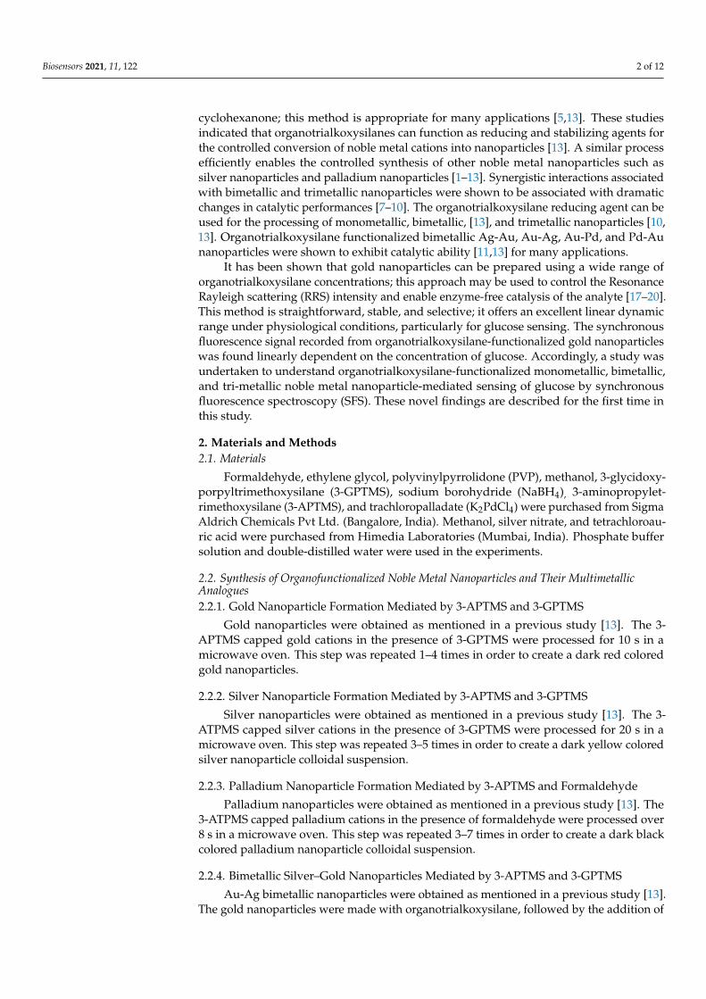
cyclohexanone; this method is appropriate for many applications [5,13]. These studiesindicated that organotrialkoxysilanes can function as reducing and stabilizing agents forthe controlled conversion of noble metal cations into nanoparticles [13]. A similar processefficiently enables the controlled synthesis of other noble metal nanoparticles such assilver nanoparticles and palladium nanoparticles [1–13]. Synergistic interactions associatedwith bimetallic and trimetallic nanoparticles were shown to be associated with dramaticchanges in catalytic performances [7–10]. The organotrialkoxysilane reducing agent can beused for the processing of monometallic, bimetallic, [13], and trimetallic nanoparticles [10,13]. Organotrialkoxysilane functionalized bimetallic Ag-Au, Au-Ag, Au-Pd, and Pd-Aunanoparticles were shown to exhibit catalytic ability [11,13] for many applications.
It has been shown that gold nanoparticles can be prepared using a wide range oforganotrialkoxysilane concentrations; this approach may be used to control the ResonanceRayleigh scattering (RRS) intensity and enable enzyme-free catalysis of the analyte [17–20].This method is straightforward, stable, and selective; it offers an excellent linear dynamicrange under physiological conditions, particularly for glucose sensing. The synchronousfluorescence signal recorded from organotrialkoxysilane-functionalized gold nanoparticleswas found linearly dependent on the concentration of glucose. Accordingly, a study wasundertaken to understand organotrialkoxysilane-functionalized monometallic, bimetallic,and tri-metallic noble metal nanoparticle-mediated sensing of glucose by synchronousfluorescence spectroscopy (SFS). These novel findings are described for the first time inthis study.
2. Materials and Methods2.1. Materials
Formaldehyde, ethylene glycol, polyvinylpyrrolidone (PVP), methanol, 3-glycidoxy-porpyltrimethoxysilane (3-GPTMS), sodium borohydride (NaBH4), 3-aminopropylet-rimethoxysilane (3-APTMS), and trachloropalladate (K2PdCl4) were purchased from SigmaAldrich Chemicals Pvt Ltd. (Bangalore, India). Methanol, silver nitrate, and tetrachloroau-ric acid were purchased from Himedia Laboratories (Mumbai, India). Phosphate buffersolution and double-distilled water were used in the experiments.
2.2. Synthesis of Organofunctionalized Noble Metal Nanoparticles and Their MultimetallicAnalogues2.2.1. Gold Nanoparticle Formation Mediated by 3-APTMS and 3-GPTMS
Gold nanoparticles were obtained as mentioned in a previous study [13]. The 3-APTMS capped gold cations in the presence of 3-GPTMS were processed for 10 s in amicrowave oven. This step was repeated 1–4 times in order to create a dark red coloredgold nanoparticles.
2.2.2. Silver Nanoparticle Formation Mediated by 3-APTMS and 3-GPTMS
Silver nanoparticles were obtained as mentioned in a previous study [13]. The 3-ATPMS capped silver cations in the presence of 3-GPTMS were processed for 20 s in amicrowave oven. This step was repeated 3–5 times in order to create a dark yellow coloredsilver nanoparticle colloidal suspension.
2.2.3. Palladium Nanoparticle Formation Mediated by 3-APTMS and Formaldehyde
Palladium nanoparticles were obtained as mentioned in a previous study [13]. The3-ATPMS capped palladium cations in the presence of formaldehyde were processed over8 s in a microwave oven. This step was repeated 3–7 times in order to create a dark blackcolored palladium nanoparticle colloidal suspension.
2.2.4. Bimetallic Silver–Gold Nanoparticles Mediated by 3-APTMS and 3-GPTMS
Au-Ag bimetallic nanoparticles were obtained as mentioned in a previous study [13].The gold nanoparticles were made with organotrialkoxysilane, followed by the addition of
Biosensors 2021, 11, 122 3 of 12
silver cations as mentioned earlier [13].The resulting mixture was incubated over 10 s ina microwave oven. This step was repeated 3–7 times in order to create a dark yellowish-orange colored bimetallic (Au@Ag) colloidal suspension.
2.2.5. Trimetallic Au-Ag-Pd Nanoparticle Formation Mediated by 3-APTMS, 3-GPTMS,and Formaldehyde
Au-Ag-Pd trimetallic nanoparticles were obtained as mentioned in a previousstudy [13]. The mixture of bimetallic (Au-Ag) and cations of palladium was mixed understirring. The mixture was incubated for over 20 s in a microwave oven. This step wasrepeated 2–7 times in order to create a dark yellowish-orange colored trimetallic Au-Ag-Pdcolloidal suspension.
2.2.6. Bimetallic Au-Pd Nanoparticle Formation Mediated by 3-APTMS, 3-GPTMS, andFormaldehyde
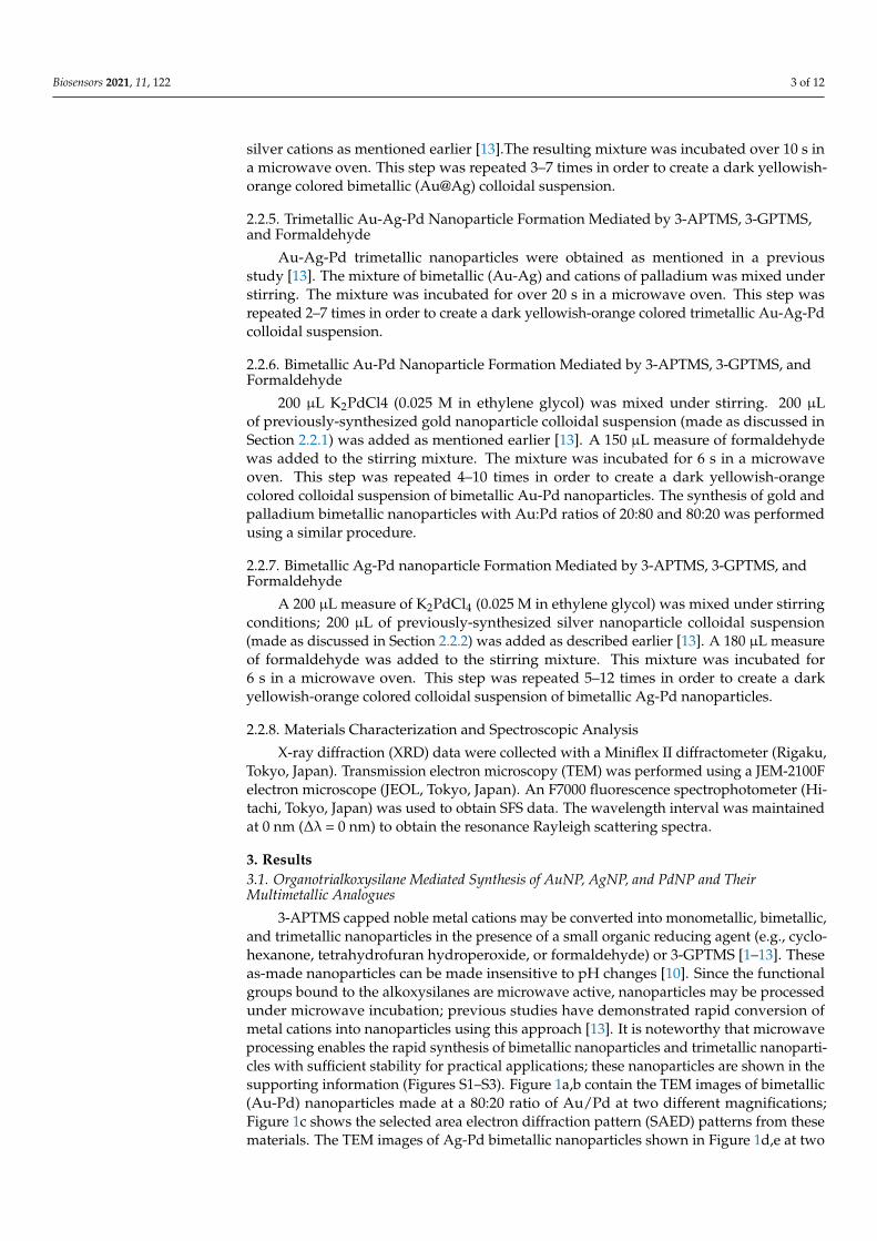
200 µL K2PdCl4 (0.025 M in ethylene glycol) was mixed under stirring. 200 µLof previously-synthesized gold nanoparticle colloidal suspension (made as discussed inSection 2.2.1) was added as mentioned earlier [13]. A 150 µL measure of formaldehydewas added to the stirring mixture. The mixture was incubated for 6 s in a microwaveoven. This step was repeated 4–10 times in order to create a dark yellowish-orangecolored colloidal suspension of bimetallic Au-Pd nanoparticles. The synthesis of gold andpalladium bimetallic nanoparticles with Au:Pd ratios of 20:80 and 80:20 was performedusing a similar procedure.
2.2.7. Bimetallic Ag-Pd nanoparticle Formation Mediated by 3-APTMS, 3-GPTMS, andFormaldehyde
A 200 µL measure of K2PdCl4 (0.025 M in ethylene glycol) was mixed under stirringconditions; 200 µL of previously-synthesized silver nanoparticle colloidal suspension(made as discussed in Section 2.2.2) was added as described earlier [13]. A 180 µL measureof formaldehyde was added to the stirring mixture. This mixture was incubated for6 s in a microwave oven. This step was repeated 5–12 times in order to create a darkyellowish-orange colored colloidal suspension of bimetallic Ag-Pd nanoparticles.
2.2.8. Materials Characterization and Spectroscopic Analysis
X-ray diffraction (XRD) data were collected with a Miniflex II diffractometer (Rigaku,Tokyo, Japan). Transmission electron microscopy (TEM) was performed using a JEM-2100Felectron microscope (JEOL, Tokyo, Japan). An F7000 fluorescence spectrophotometer (Hi-tachi, Tokyo, Japan) was used to obtain SFS data. The wavelength interval was maintainedat 0 nm (∆λ = 0 nm) to obtain the resonance Rayleigh scattering spectra.
3. Results3.1. Organotrialkoxysilane Mediated Synthesis of AuNP, AgNP, and PdNP and TheirMultimetallic Analogues
3-APTMS capped noble metal cations may be converted into monometallic, bimetallic,and trimetallic nanoparticles in the presence of a small organic reducing agent (e.g., cyclo-hexanone, tetrahydrofuran hydroperoxide, or formaldehyde) or 3-GPTMS [1–13]. Theseas-made nanoparticles can be made insensitive to pH changes [10]. Since the functionalgroups bound to the alkoxysilanes are microwave active, nanoparticles may be processedunder microwave incubation; previous studies have demonstrated rapid conversion ofmetal cations into nanoparticles using this approach [13]. It is noteworthy that microwaveprocessing enables the rapid synthesis of bimetallic nanoparticles and trimetallic nanoparti-cles with sufficient stability for practical applications; these nanoparticles are shown in thesupporting information (Figures S1–S3). Figure 1a,b contain the TEM images of bimetallic(Au-Pd) nanoparticles made at a 80:20 ratio of Au/Pd at two different magnifications;Figure 1c shows the selected area electron diffraction pattern (SAED) patterns from thesematerials. The TEM images of Ag-Pd bimetallic nanoparticles shown in Figure 1d,e at two

Biosensors 2021, 11, 122 4 of 12
different magnifications; the SAED pattern from this material is shown in Figure 1f. FiguresS1–S3 contain the TEM images of the monometallic, bimetallic, and trimetallic nanoparti-cles that were obtained from the organotrialkoxysilane-mediated conversion of the metalcations. Figure S1a,c show the TEM images of the AuNPs and AgNPs, respectively. TheSAED patterns from the AuNPs and AgNPs are provided in Figure S1b,d, respectively.Figure S2a shows the TEM image of the Au-Ag NPs; a higher magnification image ofthe Au-Ag NPs is shown in Figure S2b. A SAED pattern from the Au-Ag NPs is shownin Figure S2c,d shows a TEM image of the PdNPs; a higher magnification image of thePdNPs is shown in Figure 2e. A SAED pattern from the PdNPs is shown in Figures S2fand S3a shows a TEM image of the Au-Ag-Pd NPs; a higher magnification TEM imageof the Au-Ag-Pd NPs is shown in Figure S3b. A SAED pattern from the Au-Ag-Pd NPsis shown in Figure S3c. The mechanism of acid-base adduct mediated reduction of noblemetal cations is shown in the supporting information (Figure S4).
Biosensors 2021, 11, x FOR PEER REVIEW 4 of 12
microwave processing enables the rapid synthesis of bimetallic nanoparticles and trime-
tallic nanoparticles with sufficient stability for practical applications; these nanoparticles
are shown in the supporting information (Figures S1–S3). Figure 1a,b contain the TEM
images of bimetallic (Au-Pd) nanoparticles made at a 80:20 ratio of Au/Pd at two different
magnifications; Figure 1c shows the selected area electron diffraction pattern (SAED) pat-
terns from these materials. The TEM images of Ag-Pd bimetallic nanoparticles shown in
Figure 1d,e at two different magnifications; the SAED pattern from this material is shown
in Figure 1f. Figures S1–S3 contain the TEM images of the monometallic, bimetallic, and
trimetallic nanoparticles that were obtained from the organotrialkoxysilane-mediated
conversion of the metal cations. Figure S1a,c show the TEM images of the AuNPs and
AgNPs, respectively. The SAED patterns from the AuNPs and AgNPs are provided in
Figure S1b,d, respectively. Figure S2a shows the TEM image of the Au-Ag NPs; a higher
magnification image of the Au-Ag NPs is shown in Figure S2b. A SAED pattern from the
Au-Ag NPs is shown in Figure S2c,d shows a TEM image of the PdNPs; a higher magni-
fication image of the PdNPs is shown in Figure 2e. A SAED pattern from the PdNPs is
shown in Figures S2f and S3a shows a TEM image of the Au-Ag-Pd NPs; a higher magni-
fication TEM image of the Au-Ag-Pd NPs is shown in Figure S3b. A SAED pattern from
the Au-Ag-Pd NPs is shown in Figure S3c. The mechanism of acid-base adduct mediated
reduction of noble metal cations is shown in the supporting information (Figure S4).
Figure 1. TEM images (a,b) and corresponding selected area electron diffraction patter (SAED)patterns of bimetallic (Au-Pd) nanoparticles (NPs) (c). TEM images (d,e) and corresponding SAEDpatterns of bimetallic (Au-Pd) NPs (f).
Biosensors 2021, 11, 122 5 of 12
Biosensors 2021, 11, x FOR PEER REVIEW 5 of 12
Figure 1. TEM images (a,b) and corresponding selected area electron diffraction patter (SAED) pat-
terns of bimetallic (Au-Pd) nanoparticles (NPs) (c). TEM images (d,e) and corresponding SAED pat-
terns of bimetallic (Au-Pd) NPs (f).
These findings confirm that the organotrialkoxysilane efficiently allows the con-
trolled reduction of noble metal cations into nanoparticles in a variety of configurations
to yield noble metal nanoparticles and multimetallic nanoparticles. These nanoparticles
have been further characterized by XRD analysis. Figure 2 shows the XRD analysis of
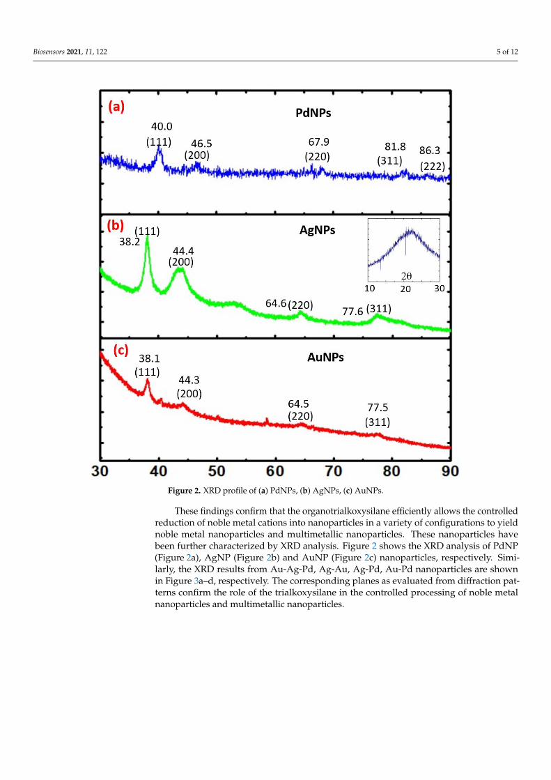
PdNP (Figure 2a), AgNP (Figure 2b) and AuNP (Figure 2c) nanoparticles, respectively.
Similarly, the XRD results from Au-Ag-Pd, Ag-Au, Ag-Pd, Au-Pd nanoparticles are
shown in Figure 3a–d, respectively. The corresponding planes as evaluated from diffrac-
tion patterns confirm the role of the trialkoxysilane in the controlled processing of noble
metal nanoparticles and multimetallic nanoparticles.
Figure 2. XRD profile of (a) PdNPs, (b) AgNPs, (c) AuNPs. Figure 2. XRD profile of (a) PdNPs, (b) AgNPs, (c) AuNPs.
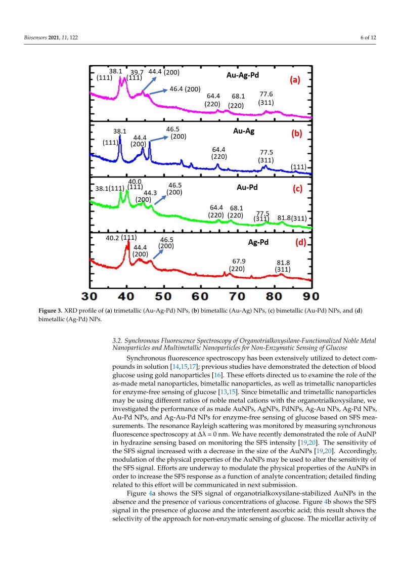
These findings confirm that the organotrialkoxysilane efficiently allows the controlledreduction of noble metal cations into nanoparticles in a variety of configurations to yieldnoble metal nanoparticles and multimetallic nanoparticles. These nanoparticles havebeen further characterized by XRD analysis. Figure 2 shows the XRD analysis of PdNP(Figure 2a), AgNP (Figure 2b) and AuNP (Figure 2c) nanoparticles, respectively. Simi-larly, the XRD results from Au-Ag-Pd, Ag-Au, Ag-Pd, Au-Pd nanoparticles are shownin Figure 3a–d, respectively. The corresponding planes as evaluated from diffraction pat-terns confirm the role of the trialkoxysilane in the controlled processing of noble metalnanoparticles and multimetallic nanoparticles.
Biosensors 2021, 11, 122 6 of 12Biosensors 2021, 11, x FOR PEER REVIEW 6 of 12
Figure 3. XRD profile of (a) trimetallic (Au-Ag-Pd) NPs, (b) bimetallic (Au-Ag) NPs, (c) bimetallic
(Au-Pd) NPs, and (d) bimetallic (Ag-Pd) NPs.
3.2. Synchronous fluorescence Spectroscopy of Organotrialkoxysilane-Functionalized Noble
Metal Nanoparticles and Multimetallic Nanoparticles for Non-Enzymatic Sensing of Glucose
Synchronous fluorescence spectroscopy has been extensively utilized to detect com-
pounds in solution [14,15,17]; previous studies have demonstrated the detection of blood
glucose using gold nanoparticles [16]. These efforts directed us to examine the role of the
as-made metal nanoparticles, bimetallic nanoparticles, as well as trimetallic nanoparticles
for enzyme-free sensing of glucose [13,15]. Since bimetallic and trimetallic nanoparticles
may be using different ratios of noble metal cations with the organotrialkoxysilane, we
investigated the performance of as made AuNPs, AgNPs, PdNPs, Ag-Au NPs, Ag-Pd
NPs, Au-Pd NPs, and Ag-Au-Pd NPs for enzyme-free sensing of glucose based on SFS
measurements. The resonance Rayleigh scattering was monitored by measuring synchro-
nous fluorescence spectroscopy at Δλ = 0 nm. We have recently demonstrated the role of
AuNP in hydrazine sensing based on monitoring the SFS intensity [19,20]. The sensitivity
of the SFS signal increased with a decrease in the size of the AuNPs [19,20]. Accordingly,
modulation of the physical properties of the AuNPs may be used to alter the sensitivity
of the SFS signal. Efforts are underway to modulate the physical properties of the AuNPs
in order to increase the SFS response as a function of analyte concentration; detailed find-
ing related to this effort will be communicated in next submission.
Figure 4a shows the SFS signal of organotrialkoxysilane-stabilized AuNPs in the ab-
sence and the presence of various concentrations of glucose. Figure 4b shows the SFS sig-
nal in the presence of glucose and the interferent ascorbic acid; this result shows the se-
lectivity of the approach for non-enzymatic sensing of glucose. The micellar activity of the
Figure 3. XRD profile of (a) trimetallic (Au-Ag-Pd) NPs, (b) bimetallic (Au-Ag) NPs, (c) bimetallic (Au-Pd) NPs, and (d)bimetallic (Ag-Pd) NPs.
3.2. Synchronous Fluorescence Spectroscopy of Organotrialkoxysilane-Functionalized Noble MetalNanoparticles and Multimetallic Nanoparticles for Non-Enzymatic Sensing of Glucose
Synchronous fluorescence spectroscopy has been extensively utilized to detect com-pounds in solution [14,15,17]; previous studies have demonstrated the detection of bloodglucose using gold nanoparticles [16]. These efforts directed us to examine the role of theas-made metal nanoparticles, bimetallic nanoparticles, as well as trimetallic nanoparticlesfor enzyme-free sensing of glucose [13,15]. Since bimetallic and trimetallic nanoparticlesmay be using different ratios of noble metal cations with the organotrialkoxysilane, weinvestigated the performance of as made AuNPs, AgNPs, PdNPs, Ag-Au NPs, Ag-Pd NPs,Au-Pd NPs, and Ag-Au-Pd NPs for enzyme-free sensing of glucose based on SFS mea-surements. The resonance Rayleigh scattering was monitored by measuring synchronousfluorescence spectroscopy at ∆λ = 0 nm. We have recently demonstrated the role of AuNPin hydrazine sensing based on monitoring the SFS intensity [19,20]. The sensitivity ofthe SFS signal increased with a decrease in the size of the AuNPs [19,20]. Accordingly,modulation of the physical properties of the AuNPs may be used to alter the sensitivity ofthe SFS signal. Efforts are underway to modulate the physical properties of the AuNPs inorder to increase the SFS response as a function of analyte concentration; detailed findingrelated to this effort will be communicated in next submission.
Figure 4a shows the SFS signal of organotrialkoxysilane-stabilized AuNPs in theabsence and the presence of various concentrations of glucose. Figure 4b shows the SFSsignal in the presence of glucose and the interferent ascorbic acid; this result shows theselectivity of the approach for non-enzymatic sensing of glucose. The micellar activity of
Biosensors 2021, 11, 122 7 of 12
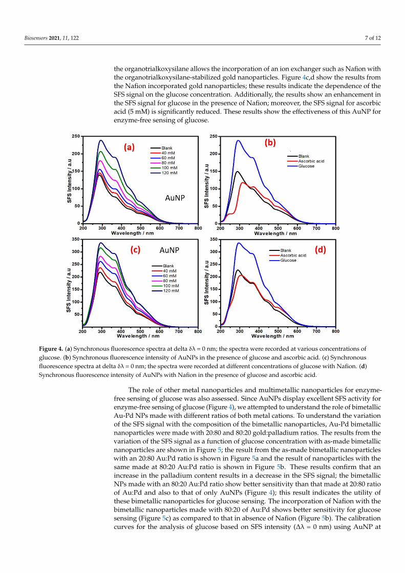
the organotrialkoxysilane allows the incorporation of an ion exchanger such as Nafion withthe organotrialkoxysilane-stabilized gold nanoparticles. Figure 4c,d show the results fromthe Nafion incorporated gold nanoparticles; these results indicate the dependence of theSFS signal on the glucose concentration. Additionally, the results show an enhancement inthe SFS signal for glucose in the presence of Nafion; moreover, the SFS signal for ascorbicacid (5 mM) is significantly reduced. These results show the effectiveness of this AuNP forenzyme-free sensing of glucose.
Biosensors 2021, 11, x FOR PEER REVIEW 7 of 12
organotrialkoxysilane allows the incorporation of an ion exchanger such as Nafion with
the organotrialkoxysilane-stabilized gold nanoparticles. Figure 4c,d show the results from
the Nafion incorporated gold nanoparticles; these results indicate the dependence of the
SFS signal on the glucose concentration. Additionally, the results show an enhancement
in the SFS signal for glucose in the presence of Nafion; moreover, the SFS signal for ascor-
bic acid (5 mM) is significantly reduced. These results show the effectiveness of this AuNP
for enzyme-free sensing of glucose.
Figure 4. (a) Synchronous fluorescence spectra at delta δλ = 0 nm; the spectra were recorded at various concentrations of
glucose. (b) Synchronous fluorescence intensity of AuNPs in the presence of glucose and ascorbic acid. (c) Synchronous
fluorescence spectra at delta δλ = 0 nm; the spectra were recorded at different concentrations of glucose with Nafion. (d)
Synchronous fluorescence intensity of AuNPs with Nafion in the presence of glucose and ascorbic acid.
The role of other metal nanoparticles and multimetallic nanoparticles for enzyme-
free sensing of glucose was also assessed. Since AuNPs display excellent SFS activity for
enzyme-free sensing of glucose (Figure 4), we attempted to understand the role of bime-
tallic Au-Pd NPs made with different ratios of both metal cations. To understand the var-
iation of the SFS signal with the composition of the bimetallic nanoparticles, Au-Pd bime-
tallic nanoparticles were made with 20:80 and 80:20 gold:palladium ratios. The results
from the variation of the SFS signal as a function of glucose concentration with as-made
bimetallic nanoparticles are shown in Figure 5; the result from the as-made bimetallic na-
noparticles with an 20:80 Au:Pd ratio is shown in Figure 5a and the result of nanoparticles
with the same made at 80:20 Au:Pd ratio is shown in Figure 5b. These results confirm that
an increase in the palladium content results in a decrease in the SFS signal; the bimetallic
NPs made with an 80:20 Au:Pd ratio show better sensitivity than that made at 20:80 ratio
of Au:Pd and also to that of only AuNPs (Figure 4); this result indicates the utility of these
bimetallic nanoparticles for glucose sensing. The incorporation of Nafion with the bime-
tallic nanoparticles made with 80:20 of Au:Pd shows better sensitivity for glucose sensing
(Figure 5c) as compared to that in absence of Nafion (Figure 5b). The calibration curves
for the analysis of glucose based on SFS intensity (Δλ = 0 nm) using AuNP at wavelength
Figure 4. (a) Synchronous fluorescence spectra at delta δλ = 0 nm; the spectra were recorded at various concentrations ofglucose. (b) Synchronous fluorescence intensity of AuNPs in the presence of glucose and ascorbic acid. (c) Synchronousfluorescence spectra at delta δλ = 0 nm; the spectra were recorded at different concentrations of glucose with Nafion. (d)Synchronous fluorescence intensity of AuNPs with Nafion in the presence of glucose and ascorbic acid.
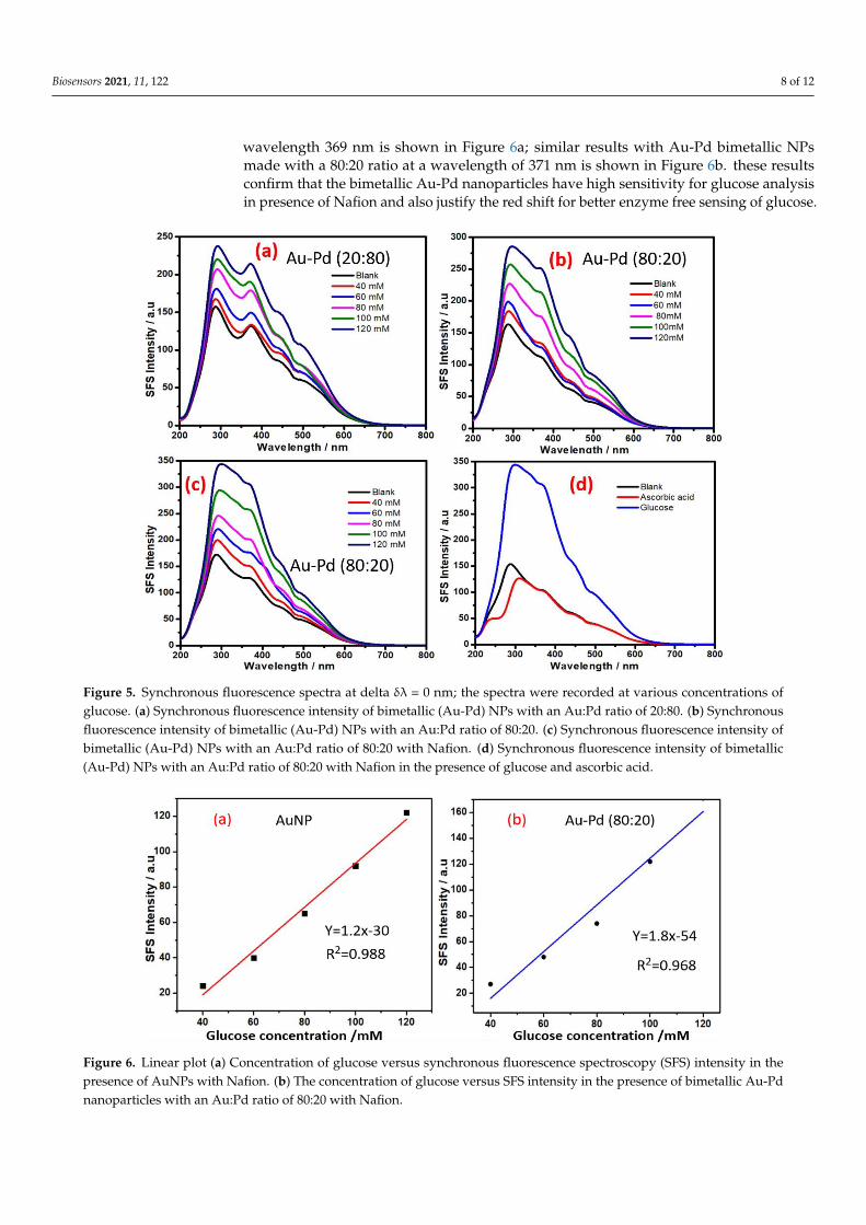
The role of other metal nanoparticles and multimetallic nanoparticles for enzyme-free sensing of glucose was also assessed. Since AuNPs display excellent SFS activity forenzyme-free sensing of glucose (Figure 4), we attempted to understand the role of bimetallicAu-Pd NPs made with different ratios of both metal cations. To understand the variationof the SFS signal with the composition of the bimetallic nanoparticles, Au-Pd bimetallicnanoparticles were made with 20:80 and 80:20 gold:palladium ratios. The results from thevariation of the SFS signal as a function of glucose concentration with as-made bimetallicnanoparticles are shown in Figure 5; the result from the as-made bimetallic nanoparticleswith an 20:80 Au:Pd ratio is shown in Figure 5a and the result of nanoparticles with thesame made at 80:20 Au:Pd ratio is shown in Figure 5b. These results confirm that anincrease in the palladium content results in a decrease in the SFS signal; the bimetallicNPs made with an 80:20 Au:Pd ratio show better sensitivity than that made at 20:80 ratioof Au:Pd and also to that of only AuNPs (Figure 4); this result indicates the utility ofthese bimetallic nanoparticles for glucose sensing. The incorporation of Nafion with thebimetallic nanoparticles made with 80:20 of Au:Pd shows better sensitivity for glucosesensing (Figure 5c) as compared to that in absence of Nafion (Figure 5b). The calibrationcurves for the analysis of glucose based on SFS intensity (∆λ = 0 nm) using AuNP at
Biosensors 2021, 11, 122 8 of 12
wavelength 369 nm is shown in Figure 6a; similar results with Au-Pd bimetallic NPsmade with a 80:20 ratio at a wavelength of 371 nm is shown in Figure 6b. these resultsconfirm that the bimetallic Au-Pd nanoparticles have high sensitivity for glucose analysisin presence of Nafion and also justify the red shift for better enzyme free sensing of glucose.
Biosensors 2021, 11, x FOR PEER REVIEW 8 of 12
369 nm is shown in Figure 6a; similar results with Au-Pd bimetallic NPs made with a 80:20
ratio at a wavelength of 371 nm is shown in Figure 6b. these results confirm that the bi-
metallic Au-Pd nanoparticles have high sensitivity for glucose analysis in presence of
Nafion and also justify the red shift for better enzyme free sensing of glucose.
Figure 5. Synchronous fluorescence spectra at delta δλ = 0 nm; the spectra were recorded at various concentrations of
glucose. (a) Synchronous fluorescence intensity of bimetallic (Au-Pd) NPs with an Au:Pd ratio of 20:80. (b) Synchronous
fluorescence intensity of bimetallic (Au-Pd) NPs with an Au:Pd ratio of 80:20. (d) Synchronous fluorescence intensity of
bimetallic (Au-Pd) NPs with an Au:Pd ratio of 80:20 with Nafion. (d) Synchronous fluorescence intensity of bimetallic
(Au-Pd) NPs with an Au:Pd ratio of 80:20 with Nafion in the presence of glucose and ascorbic acid.
Figure 6. Linear plot (a) Concentration of glucose versus synchronous fluorescence spectroscopy (SFS) intensity in the
presence of AuNPs with Nafion. (b) The concentration of glucose versus SFS intensity in the presence of bimetallic Au-Pd
nanoparticles with an Au:Pd ratio of 80:20 with Nafion.
Figure 5. Synchronous fluorescence spectra at delta δλ = 0 nm; the spectra were recorded at various concentrations ofglucose. (a) Synchronous fluorescence intensity of bimetallic (Au-Pd) NPs with an Au:Pd ratio of 20:80. (b) Synchronousfluorescence intensity of bimetallic (Au-Pd) NPs with an Au:Pd ratio of 80:20. (c) Synchronous fluorescence intensity ofbimetallic (Au-Pd) NPs with an Au:Pd ratio of 80:20 with Nafion. (d) Synchronous fluorescence intensity of bimetallic(Au-Pd) NPs with an Au:Pd ratio of 80:20 with Nafion in the presence of glucose and ascorbic acid.
Biosensors 2021, 11, x FOR PEER REVIEW 8 of 12
369 nm is shown in Figure 6a; similar results with Au-Pd bimetallic NPs made with a 80:20
ratio at a wavelength of 371 nm is shown in Figure 6b. these results confirm that the bi-
metallic Au-Pd nanoparticles have high sensitivity for glucose analysis in presence of
Nafion and also justify the red shift for better enzyme free sensing of glucose.
Figure 5. Synchronous fluorescence spectra at delta δλ = 0 nm; the spectra were recorded at various concentrations of
glucose. (a) Synchronous fluorescence intensity of bimetallic (Au-Pd) NPs with an Au:Pd ratio of 20:80. (b) Synchronous
fluorescence intensity of bimetallic (Au-Pd) NPs with an Au:Pd ratio of 80:20. (d) Synchronous fluorescence intensity of
bimetallic (Au-Pd) NPs with an Au:Pd ratio of 80:20 with Nafion. (d) Synchronous fluorescence intensity of bimetallic
(Au-Pd) NPs with an Au:Pd ratio of 80:20 with Nafion in the presence of glucose and ascorbic acid.
Figure 6. Linear plot (a) Concentration of glucose versus synchronous fluorescence spectroscopy (SFS) intensity in the
presence of AuNPs with Nafion. (b) The concentration of glucose versus SFS intensity in the presence of bimetallic Au-Pd
nanoparticles with an Au:Pd ratio of 80:20 with Nafion.
Figure 6. Linear plot (a) Concentration of glucose versus synchronous fluorescence spectroscopy (SFS) intensity in thepresence of AuNPs with Nafion. (b) The concentration of glucose versus SFS intensity in the presence of bimetallic Au-Pdnanoparticles with an Au:Pd ratio of 80:20 with Nafion.
Biosensors 2021, 11, 122 9 of 12
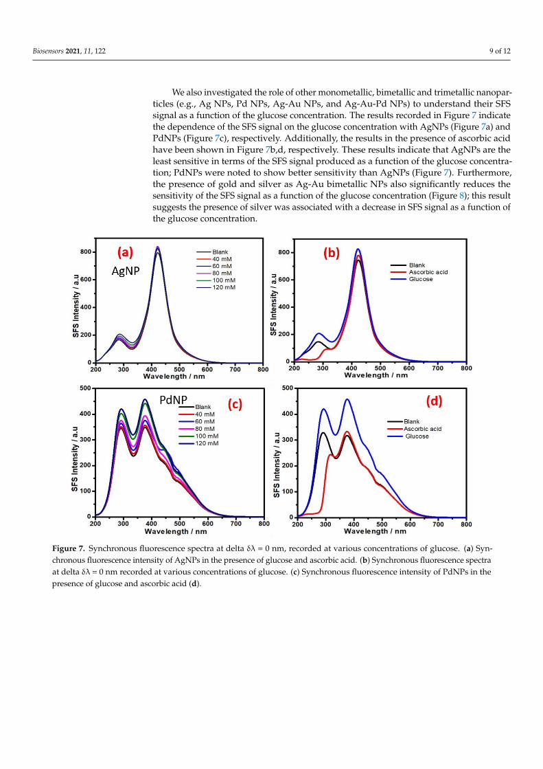
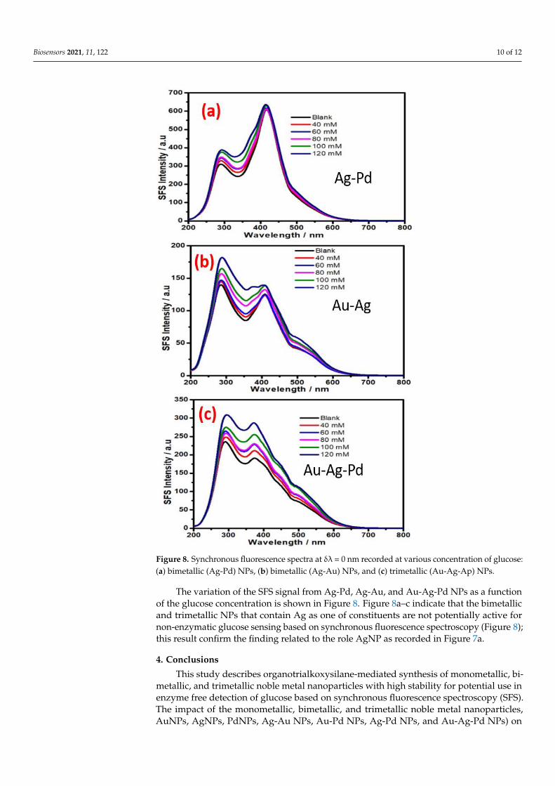
We also investigated the role of other monometallic, bimetallic and trimetallic nanopar-ticles (e.g., Ag NPs, Pd NPs, Ag-Au NPs, and Ag-Au-Pd NPs) to understand their SFSsignal as a function of the glucose concentration. The results recorded in Figure 7 indicatethe dependence of the SFS signal on the glucose concentration with AgNPs (Figure 7a) andPdNPs (Figure 7c), respectively. Additionally, the results in the presence of ascorbic acidhave been shown in Figure 7b,d, respectively. These results indicate that AgNPs are theleast sensitive in terms of the SFS signal produced as a function of the glucose concentra-tion; PdNPs were noted to show better sensitivity than AgNPs (Figure 7). Furthermore,the presence of gold and silver as Ag-Au bimetallic NPs also significantly reduces thesensitivity of the SFS signal as a function of the glucose concentration (Figure 8); this resultsuggests the presence of silver was associated with a decrease in SFS signal as a function ofthe glucose concentration.
Biosensors 2021, 11, x FOR PEER REVIEW 9 of 12
We also investigated the role of other monometallic, bimetallic and trimetallic nano-
particles (e.g., Ag NPs, Pd NPs, Ag-Au NPs, and Ag-Au-Pd NPs) to understand their SFS
signal as a function of the glucose concentration. The results recorded in Figure 7 indicate
the dependence of the SFS signal on the glucose concentration with AgNPs (Figure 7a)
and PdNPs (Figure 7c), respectively. Additionally, the results in the presence of ascorbic
acid have been shown in Figure 7b,d, respectively. These results indicate that AgNPs are
the least sensitive in terms of the SFS signal produced as a function of the glucose concen-
tration; PdNPs were noted to show better sensitivity than AgNPs (Figure 7). Furthermore,
the presence of gold and silver as Ag-Au bimetallic NPs also significantly reduces the
sensitivity of the SFS signal as a function of the glucose concentration (Figure 8); this result
suggests the presence of silver was associated with a decrease in SFS signal as a function
of the glucose concentration.
The variation of the SFS signal from Ag-Pd, Ag-Au, and Au-Ag-Pd NPs as a function
of the glucose concentration is shown in Figure 8. Figure 8a–c indicate that the bimetallic
and trimetallic NPs that contain Ag as one of constituents are not potentially active for
non-enzymatic glucose sensing based on synchronous fluorescence spectroscopy (Figure
8); this result confirm the finding related to the role AgNP as recorded in Figure 7a.
Figure 7. Synchronous fluorescence spectra at delta δλ = 0 nm, recorded at various concentrations of glucose. (a) Synchro-
nous fluorescence intensity of AgNPs in the presence of glucose and ascorbic acid. (b) Synchronous fluorescence spectra
at delta δλ = 0 nm recorded at various concentrations of glucose. (c) Synchronous fluorescence intensity of PdNPs in the
presence of glucose and ascorbic acid (d).
Figure 7. Synchronous fluorescence spectra at delta δλ = 0 nm, recorded at various concentrations of glucose. (a) Syn-chronous fluorescence intensity of AgNPs in the presence of glucose and ascorbic acid. (b) Synchronous fluorescence spectraat delta δλ = 0 nm recorded at various concentrations of glucose. (c) Synchronous fluorescence intensity of PdNPs in thepresence of glucose and ascorbic acid (d).
Biosensors 2021, 11, 122 10 of 12Biosensors 2021, 11, x FOR PEER REVIEW 10 of 12
Figure 8. Synchronous fluorescence spectra at δλ = 0 nm recorded at various concentration of glu-
cose: (a) bimetallic (Ag-Pd) NPs, (b) bimetallic (Ag-Au) NPs, and (c) trimetallic (Au-Ag-Ap) NPs.
4. Conclusions
This study describes organotrialkoxysilane-mediated synthesis of monometallic, bi-
metallic, and trimetallic noble metal nanoparticles with high stability for potential use in
enzyme free detection of glucose based on synchronous fluorescence spectroscopy (SFS).
The impact of the monometallic, bimetallic, and trimetallic noble metal nanoparticles,
AuNPs, AgNPs, PdNPs, Ag-Au NPs, Au-Pd NPs, Ag-Pd NPs, and Au-Ag-Pd NPs) on the
variation of the SFS signal for non-enzymatic sensing of glucose was demonstrated. The
finding predicts that bimetallic Au-Pd NPs made with an 80:20 Au:Pd ratio display excel-
lent results for glucose sensing. The micellar activity of the as-made nanomaterials can be
effectively explored for making Nafion-metal nanoparticles colloidal suspensions for non-
enzymatic detection of glucose.
Figure 8. Synchronous fluorescence spectra at δλ = 0 nm recorded at various concentration of glucose:(a) bimetallic (Ag-Pd) NPs, (b) bimetallic (Ag-Au) NPs, and (c) trimetallic (Au-Ag-Ap) NPs.
The variation of the SFS signal from Ag-Pd, Ag-Au, and Au-Ag-Pd NPs as a functionof the glucose concentration is shown in Figure 8. Figure 8a–c indicate that the bimetallicand trimetallic NPs that contain Ag as one of constituents are not potentially active fornon-enzymatic glucose sensing based on synchronous fluorescence spectroscopy (Figure 8);this result confirm the finding related to the role AgNP as recorded in Figure 7a.
4. Conclusions
This study describes organotrialkoxysilane-mediated synthesis of monometallic, bi-metallic, and trimetallic noble metal nanoparticles with high stability for potential use inenzyme free detection of glucose based on synchronous fluorescence spectroscopy (SFS).The impact of the monometallic, bimetallic, and trimetallic noble metal nanoparticles,AuNPs, AgNPs, PdNPs, Ag-Au NPs, Au-Pd NPs, Ag-Pd NPs, and Au-Ag-Pd NPs) on
Biosensors 2021, 11, 122 11 of 12
the variation of the SFS signal for non-enzymatic sensing of glucose was demonstrated.The finding predicts that bimetallic Au-Pd NPs made with an 80:20 Au:Pd ratio displayexcellent results for glucose sensing. The micellar activity of the as-made nanomaterialscan be effectively explored for making Nafion-metal nanoparticles colloidal suspensionsfor non-enzymatic detection of glucose.
Supplementary Materials: The following are available online at https://www.mdpi.com/article/10.3390/bios11040122/s1, Figure S1: TEM images (a,c) of the AuNPs and AgNPs, respectively. Selectedarea electron diffraction patterns (c,d) of the AuNPs and AgNPs, respectively, Figure S2: TEM images(a–b) and corresponding SAED patterns of the bimetallic (Au-Ag) NPs (c). TEM images (d–e) andcorresponding SAED patterns of the PdNPs (f), Figure S3: TEM images (a–b) and correspondingSAED pattern of the trimetallic (Au-Ag-Pd) NPs (c) and Figure S4: Acid-base adduct mediatedreduction of noble metal cations.
Funding: The authors would like to acknowledge support from the Department of Science andTechnology’s VAJRA (Visiting Advanced Joint Research) Faculty award through Grant #VJR-2017-000034 to Roger J. Narayan, which facilitated interaction between Indians and US institutions andDRDO for Grant #LSRB-316.
Institutional Review Board Statement: The study was conducted according to the guidelines of theDeclaration of Indian Institute of Technology (BHU), and approved by the Institutional Review Board.
Informed Consent Statement: Informed consent was obtained from all subjects involved in the study.
Data Availability Statement: Data supporting reported results can be found in the laboratory ofProf. Prem C Pandey of IIT(BHU).
Conflicts of Interest: The authors declare no conflict of interest.
References1. Pandey, P.C.; Chauhan, D.S. 3-Glycidoxypropyltrimethoxysilane mediated in situ synthesis of noble metal nanoparticles:
Application to hydrogen peroxide sensing. Analyst 2011, 137, 376–385. [CrossRef] [PubMed]2. Pandey, P.C.; Pandey, A.K.; Pandey, G. Functionalized alkoxysilane mediated controlled synthesis of noble metal nanoparticles
dispersible in aqueous and non-aqueous medium. J. Nanosci. Nanotechnol. 2014, 14, 6606–6613. [CrossRef] [PubMed]3. Pandey, P.C.; Pandey, G. Tunable functionality and nanogeometry in tetrahydrofuran hydroperoxide and 3-aminopropyl-
trimethoxysilane mediated synthesis of gold nanoparticles; functional application in glutathione sensing. J. Mater. Chem. B 2014,2, 3383–3390. [CrossRef] [PubMed]
4. Pandey, P.C.; Singh, R. Controlled synthesis of functional silver nanoparticles dispersible in aqueous and non-aqueous me-dium.J. Nanosci. Nanotechnol. 2015, 15, 5749–5759. [CrossRef] [PubMed]
5. Pandey, P.C.; Panday, D.; Pandey, G. 3-Aminopropyltrimethoxysilane and organic electron donors mediated synthesis offunctional amphiphilic gold nanoparticles and their bioanalytical applications. RSC Adv. 2014, 4, 60563–60572. [CrossRef]
6. Pandey, P.C.; Shukla, S.; Pandey, Y. 3-Aminopropyltrimethoxysilane and graphene oxide/reduced graphene oxide-inducedgeneration of gold nanoparticles and their nanocomposites: Electrocatalytic and kinetic activity. RSC Adv. 2016, 6, 80549–80556.[CrossRef]
7. Pandey, P.C.; Singh, R.; Pandey, A.K. Tetrahydrofuran hydroperoxide and 3-Aminopropyltrimethoxysilane mediated controlledsynthesis of Pd, Pd-Au, Au-Pd nanoparticles: Role of Palladium nanoparticles on the redox electrochemistry of ferrocenemonocarboxylic acid. Electrochim. Acta 2014, 138, 163–173. [CrossRef]
8. Pandey, P.C.; Pandey, G. One-pot two-step rapid synthesis of 3-aminopropyltrimethoxysilane-mediated highly catalyticAg@(PdAu) trimetallic nanoparticles. Catal. Sci. Technol. 2016, 6, 3911–3917. [CrossRef]
9. Pandey, P.C.; Shukla, S. Solvent dependent fabrication of bifunctional nanoparticles and nanostructured thin films by selfassembly of organosilanes. J. Sol-Gel Sci. Technol. 2018, 86, 650–663. [CrossRef]
10. Pandey, P.C.; Pandey, G. Synthesis of gold nanoparticles resistant to pH and salt for biomedical applications; functional ac-tivityof organic amine. J. Mater. Res. 2016, 31, 3313–3323. [CrossRef]
11. Pandey, P.C.; Mitra, M.D.; Tiwari, A.K.; Singh, S. Synthetic incorporation of palladium-nickel bimetallic nanoparticles withinmesoporous silica/silica nanoparticles as efficient and cheaper catalyst for both cationic and anionic dyes degrada-tion. J. Environ.Sci. Health Part A 2021, 56, 1–13. [CrossRef] [PubMed]
12. Pandey, P.C.; Katyal, N.; Pandey, G.; Narayan, R.J. Synthesis of self-assembled siloxane–polyindole–gold nanoparticle poly-mericnanofluid for biomedical membranes. MRS Commun. 2020, 10, 482–486. [CrossRef]
13. Pandey, P.C.; Mitra, M.; Pandey, A.K.; Narayan, R.J. Organotrialkoxysilane mediated rapid and controlled synthesis metalnanoparticles in both homogeneous and heterogeneous phase and their catalytic applications. MRS Adv. 2021, 1–11. [CrossRef]
Biosensors 2021, 11, 122 12 of 12
14. Pandey, P.C.; Upadhyay, B.C. Studies on differential sensing of dopamine at the surface of chemically sensitized ormo sil-modifiedelectrodes. Talanta 2005, 67, 997–1006. [CrossRef]
15. Pandey, P.C.; Singh, R. Controlled synthesis of Pd and Pd–Au nanoparticles: Effect of organic amine and silanol groups onmorphology and polycrystallinity of nanomaterials. RSC Adv. 2015, 5, 10964–10973. [CrossRef]
16. Uppal, M.A.; Kafizas, A.; Ewing, M.B.; Parkin, I.P. The room temperature formation of gold nanoparticles from the reaction ofcyclohexanone and auric acid; a transition from dendritic particles to compact shapes and nanoplates. J. Mater. Chem. A 2013, 1,7351–7359. [CrossRef]
17. El Kurdi, R.; Patra, D. Tuning the surface of Au nanoparticles using poly (ethylene glycol)-block-poly (propylene gly-col)-block-poly (ethylene glycol): Enzyme free and label free sugar sensing in serum samples using resonance Rayleigh scatteringspectroscopy. Phys. Chem. Chem. Phys. 2018, 20, 9616–9629. [CrossRef] [PubMed]
18. Samokhvalov, A. Analysis of various solid samples by synchronous fluorescence spectroscopy and related methods: A review.Talanta 2020, 216, 120944. [CrossRef] [PubMed]
19. Pandey, P.; Pandey, G.; Narayan, R. Polyethylenimine-mediated controlled synthesis of Prussian blue-gold nanohybrids forbiomedical applications. J. Biomater. Appl. 2020. [CrossRef] [PubMed]
20. Pandey, P.C.; Mitra, M.D.; Shukla, S.; Narayan, R.J. Organotrialkoxysilane-functionalized mesoporous Pd–Ni nanocatalyst forselective hydrazine decomposition and sensing. MRS Commun. 2021, 1–8. [CrossRef]