1 relationships between bacteria and clinical features of disease 4-c the bacteria and various pages...
TRANSCRIPT

1
Relationships Between Bacteria and Clinical Features of Disease
4-c The Bacteria4-c The BacteriaAnd various pages in additional chapters
2
Differences exist between human (eukaryotic) and bacterial (prokaryotic) cells
Structure/Function RelationshipsStructure/Function Relationships
Many of the differences account for disease pathogenesis
And, allow exploitation of the differences to develop chemotherapy (antibiotics)
3
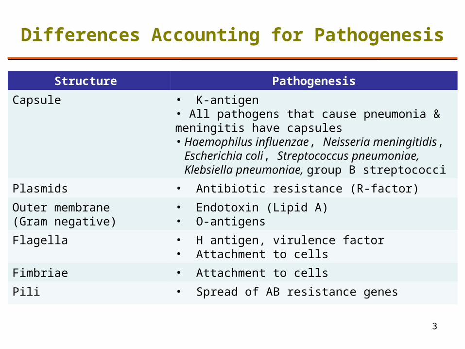
Structure Pathogenesis
Capsule • K-antigen• All pathogens that cause pneumonia & meningitis have capsules • Haemophilus influenzae, Neisseria meningitidis,
Escherichia coli, Streptococcus pneumoniae, Klebsiella pneumoniae, group B streptococci
Plasmids • Antibiotic resistance (R-factor)
Outer membrane (Gram negative)
• Endotoxin (Lipid A)• O-antigens
Flagella • H antigen, virulence factor• Attachment to cells
Fimbriae • Attachment to cells
Pili • Spread of AB resistance genes
Differences Accounting for Pathogenesis
4
Toxins Toxins
Poisonous substances produced by microbes
– Transported in blood
– Produce fever, cardiovascular disturbances, diarrhea, shock
– Inhibit protein synthesis, destroy blood cells & vessels, disrupt nervous system
5
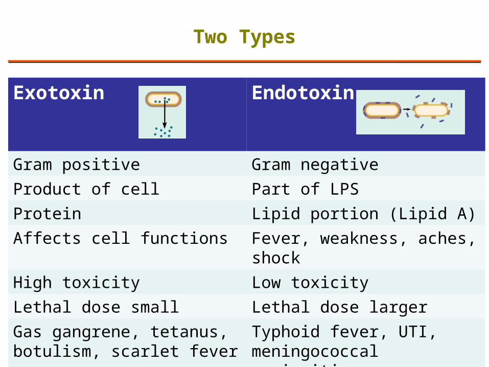
Exotoxin Endotoxin
Gram positive Gram negative
Product of cell Part of LPS
Protein Lipid portion (Lipid A)
Affects cell functions Fever, weakness, aches, shock
High toxicity Low toxicity
Lethal dose small Lethal dose larger
Gas gangrene, tetanus, botulism, scarlet fever
Typhoid fever, UTI, meningococcal meningitis
Two Types

6
Alcaligenes faecalis
Azotobacter
Bacillus
Bacillus anthracis
Bacillus cereus
Bacillus megaterium
Bacillus subtilis
Borrelia burgdorferi
Clostridium
Clostridium botulinum
Clostridium perfringens
Clostridium sporogenes
Clostridium tetani
Corynebacterium diphtheria
Corynebacterium xerosis
Enterobacter
Enterobacter aerogenes
Enterobacter sakazaki
Enterobacteriaceae
Escherichia
Escherichia coli
Gram negative
Gram positive
Klebsiella
Klebsiella pneumonia
Lactobacillus plantarum
Micrococcus luteus
Mycobacteria
Mycobacterium leprae
Mycobacterium smegmatis
Mycobacterium tuberculosis
Neisseria gonorrhoea
Nocardia
Proteus
Proteus vulgaris
Pseudomonas aeruginosa
Salmonella
Salmonella typhimurium
Serratia marcescens
Shigella dysenteriae
Spirillum
Staphylococcus
Staphylococcus aureus
Staphylococcus epidermidis
Streptococcus
Streptococci mutans
Streptococci pneumoniae
Streptococcus viridans
Treponema pallidum
Vibrio cholera
7
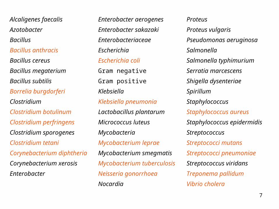
Alcaligenes faecalis
Azotobacter
Bacillus
Bacillus anthracis
Bacillus cereus
Bacillus megaterium
Bacillus subtilis
Borrelia burgdorferi
Clostridium
Clostridium botulinum
Clostridium perfringens
Clostridium sporogenes
Clostridium tetani
Corynebacterium diphtheria
Corynebacterium xerosis
Enterobacter
Enterobacter aerogenes
Enterobacter sakazaki
Enterobacteriaceae
Escherichia
Escherichia coli
Gram negative
Gram positive
Klebsiella
Klebsiella pneumonia
Lactobacillus plantarum
Micrococcus luteus
Mycobacteria
Mycobacterium leprae
Mycobacterium smegmatis
Mycobacterium tuberculosis
Neisseria gonorrhoea
Nocardia
Proteus
Proteus vulgaris
Pseudomonas aeruginosa
Salmonella
Salmonella typhimurium
Serratia marcescens
Shigella dysenteriae
Spirillum
Staphylococcus
Staphylococcus aureus
Staphylococcus epidermidis
Streptococcus
Streptococci mutans
Streptococci pneumoniae
Streptococcus viridans
Treponema pallidum
Vibrio cholera
8
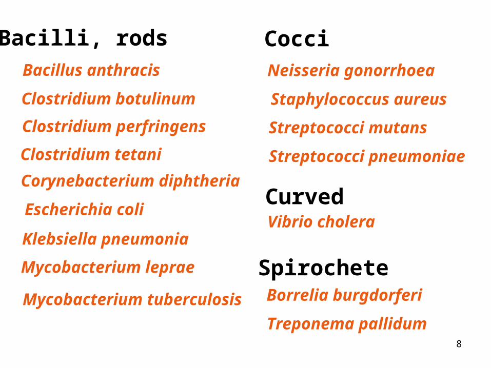
Bacillus anthracis
Borrelia burgdorferi
Escherichia coli
Klebsiella pneumonia
Mycobacterium leprae
Staphylococcus aureus
Neisseria gonorrhoea
Vibrio cholera
Bacilli, rods Cocci
Curved
Spirochete
Clostridium botulinum
Clostridium perfringens
Clostridium tetani
Corynebacterium diphtheria
Mycobacterium tuberculosis
Streptococci mutans
Streptococci pneumoniae
Treponema pallidum
9
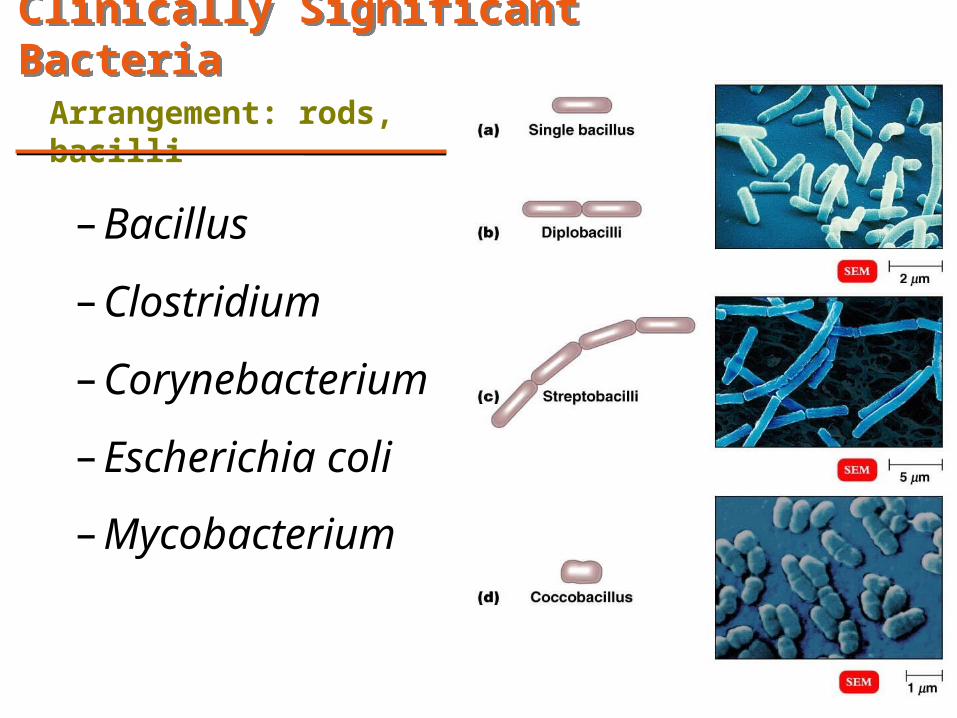
Clinically Significant BacteriaClinically Significant Bacteria
Arrangement: rods, bacilli
– Bacillus
– Clostridium
– Corynebacterium
– Escherichia coli
– Mycobacterium

10
Bacillus anthracis
• Gram positive, rods• Spore forming• Aerobe or facultative • Found in soil
Disease:
• Anthrax – Pulmonary – Cutaneous– GI
• Biological warfare– Ch 23, p 680-1
Ch / Pg
1) 3 / 712) 4 / 96-973) 11 / 3314) 23 / 679-681, 7025) LM / 18-9
Fig. 11.17b Bacillus germinating
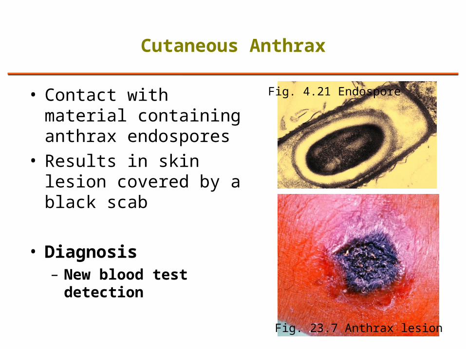
• Contact with material containing anthrax endospores
• Results in skin lesion covered by a black scab
• Diagnosis– New blood test
detection
11
Cutaneous Anthrax
Fig. 4.21 Endospore
Fig. 23.7 Anthrax lesion
12
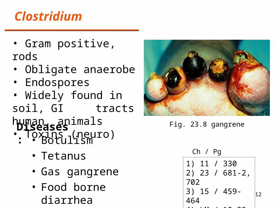
Clostridium
• Gram positive, rods• Obligate anaerobe • Endospores• Widely found in soil, GI
tracts human, animals• Toxins (neuro)
Diseases: • Botulism• Tetanus• Gas gangrene• Food borne diarrhea
1) 11 / 3302) 23 / 681-2, 7023) 15 / 459-4644) LM / 18-20
Ch / Pg
Fig. 23.8 gangrene
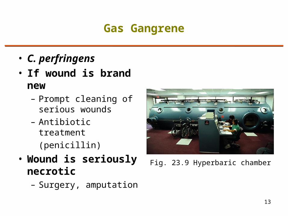
• C. perfringens• If wound is brand
new– Prompt cleaning of
serious wounds– Antibiotic treatment
(penicillin)
• Wound is seriously necrotic– Surgery, amputation
13
Gas Gangrene
Fig. 23.9 Hyperbaric chamber
14
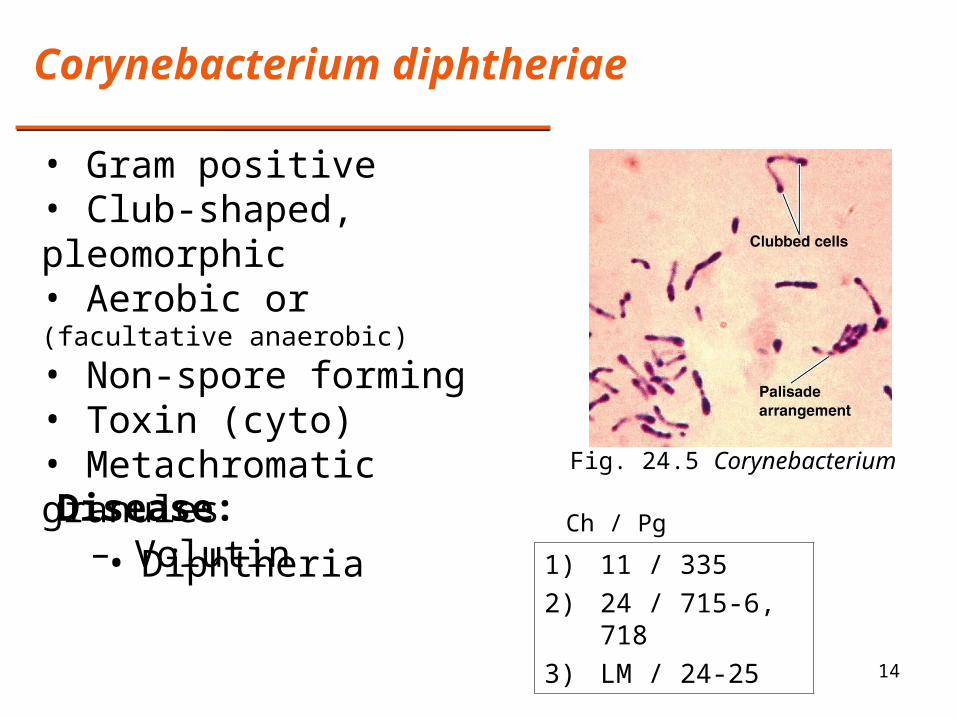
Corynebacterium diphtheriae
• Gram positive• Club-shaped, pleomorphic • Aerobic or (facultative anaerobic)
• Non-spore forming• Toxin (cyto)• Metachromatic granules
– VolutinDisease:
• Diphtheria 1) 11 / 335
2) 24 / 715-6, 718
3) LM / 24-25
Ch / Pg
Fig. 24.5 Corynebacterium
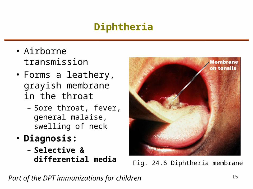
• Airborne transmission• Forms a leathery,
grayish membrane in the throat– Sore throat, fever,
general malaise, swelling of neck
• Diagnosis:– Selective &
differential media
15
Diphtheria
Fig. 24.6 Diphtheria membrane
Part of the DPT immunizations for children
16
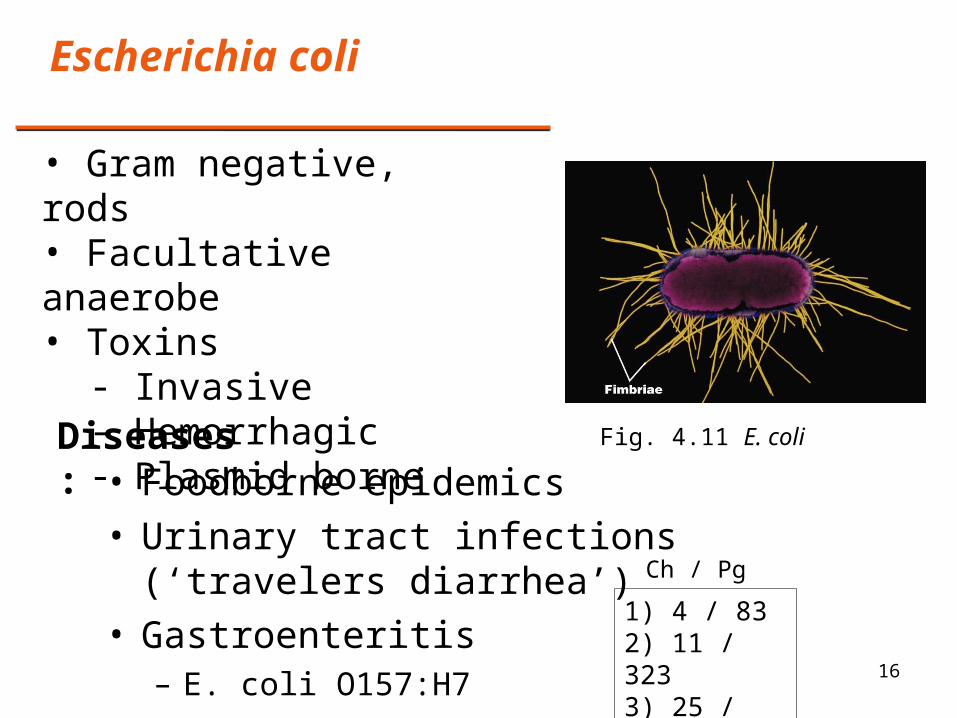
Escherichia coli
Fig. 4.11 E. coli
• Gram negative, rods• Facultative anaerobe• Toxins
- Invasive- Hemorrhagic- Plasmid borne
Diseases: • Foodborne epidemics• Urinary tract infections (‘travelers diarrhea’)• Gastroenteritis
– E. coli O157:H71) 4 / 832) 11 / 3233) 25 / 758-9
Ch / Pg
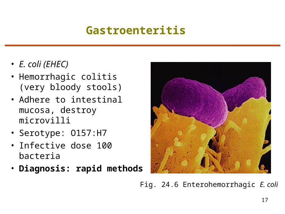
• E. coli (EHEC)
• Hemorrhagic colitis (very bloody stools)
• Adhere to intestinal mucosa, destroy microvilli
• Serotype: O157:H7
• Infective dose 100 bacteria
• Diagnosis: rapid methods
17
Gastroenteritis
Fig. 24.6 Enterohemorrhagic E. coli

18
Mycobacterium
• Acid fast, slender rod• Obligate aerobe• Non-spore forming
Disease:
• Tuberculosis• Leprosy (Hanson’s Disease)
Fig. 24.9 Mycobacterium
1) Mycobacterium tuberculosis2) M. leprae
Ch / Pg
1) 3 / 70-12) 11 / 334-53) 22 / 651-2, 6654) 24 / 719-7235) LM / 21-22

• Inhale the bacillus– Lodge in the lung
alveoli – Can progress to a lung
damaging inflammation
• Diagnosis:– TB skin test– Sputum smears– Chest X ray, CT
19
Tuberculosis
Fig. 24.2 Lower Respiratory System
Fig. 24.11 TB skin test

• Invades myelin sheath, peripheral nervous system– Cause nerve damage– 12 day generation time– Never grown in culture
• Necrosis of tissue• Diagnosis:
– Detect acid-fast rods in patient fluids
– Lepromin test 20
Leprosy
Fig. 22.9 Leprosy
Also called Hansen’s disease
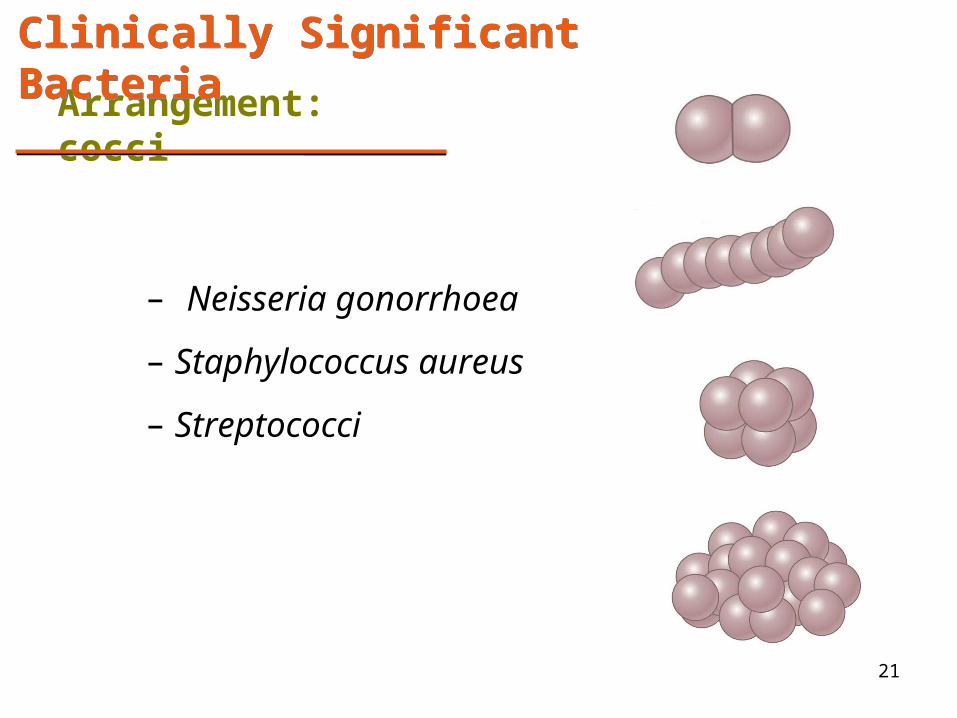
21
Arrangement: cocci
Clinically Significant BacteriaClinically Significant Bacteria
– Neisseria gonorrhoea
– Staphylococcus aureus
– Streptococci

22
Neisseria gonorrhoea
• Gram negative • Diplococcus • Obligate aerobe• Non-spore forming
Disease:
• STD: Gonorrhea• Meningitis Ch / Pg
1) 22 / 645-62) 26 / 790-793
Fig. 22.4 Neisseria meningitis, pharynx
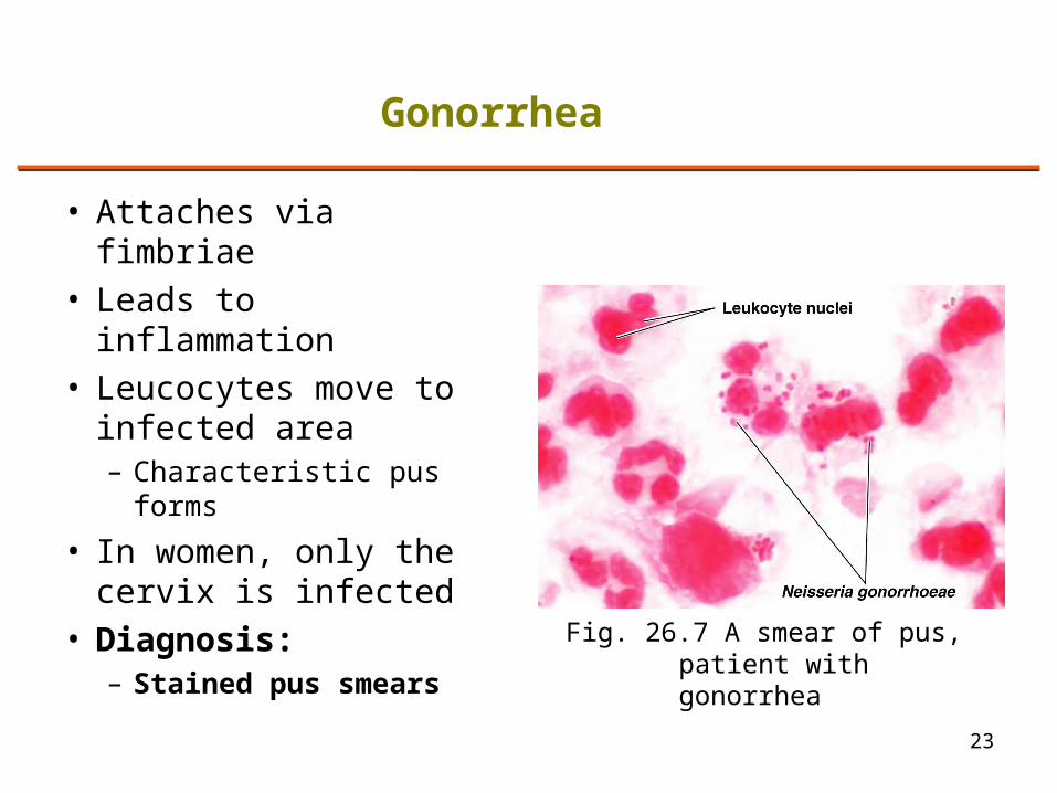
• Attaches via fimbriae• Leads to inflammation• Leucocytes move to
infected area– Characteristic pus
forms
• In women, only the cervix is infected
• Diagnosis:– Stained pus smears
23
Gonorrhea
Fig. 26.7 A smear of pus, patient with gonorrhea
24
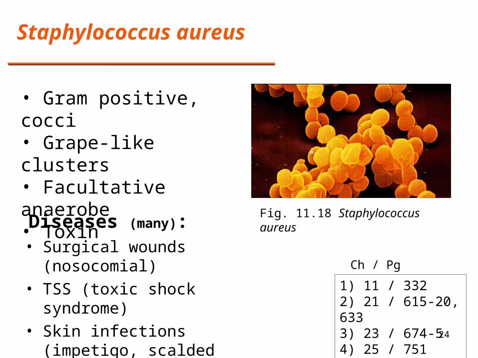
Staphylococcus aureus
Diseases (many):
1) 11 / 332 2) 21 / 615-20, 6333) 23 / 674-54) 25 / 751
Ch / Pg
• Gram positive, cocci• Grape-like clusters• Facultative anaerobe• Toxin
Fig. 11.18 Staphylococcus aureus
• Surgical wounds (nosocomial)• TSS (toxic shock syndrome)• Skin infections (impetigo,
scalded skin)• Endocarditis
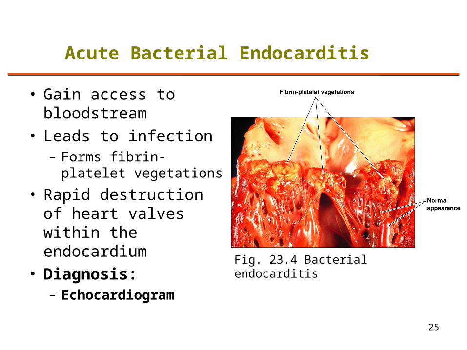
• Gain access to bloodstream
• Leads to infection– Forms fibrin-platelet
vegetations
• Rapid destruction of heart valves within the endocardium
• Diagnosis:– Echocardiogram
25
Acute Bacterial Endocarditis
Fig. 23.4 Bacterial endocarditis
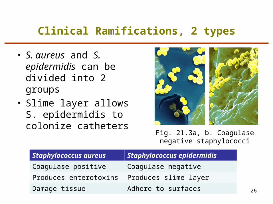
• S. aureus and S. epidermidis can be divided into 2 groups
• Slime layer allows S. epidermidis to colonize catheters
26
Clinical Ramifications, 2 types
Fig. 21.3a, b. Coagulase negative staphylococci
Staphylococcus aureus Staphylococcus epidermidis
Coagulase positive Coagulase negative
Produces enterotoxins Produces slime layer
Damage tissue Adhere to surfaces
27
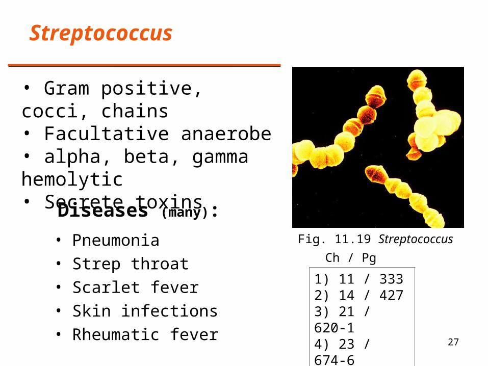
Streptococcus
Diseases (many):
1) 11 / 333 2) 14 / 4273) 21 / 620-14) 23 / 674-65) 25 / 751
Ch / Pg
• Gram positive, cocci, chains• Facultative anaerobe• alpha, beta, gamma hemolytic• Secrete toxins
Fig. 11.19 Streptococcus• Pneumonia• Strep throat• Scarlet fever• Skin infections• Rheumatic fever
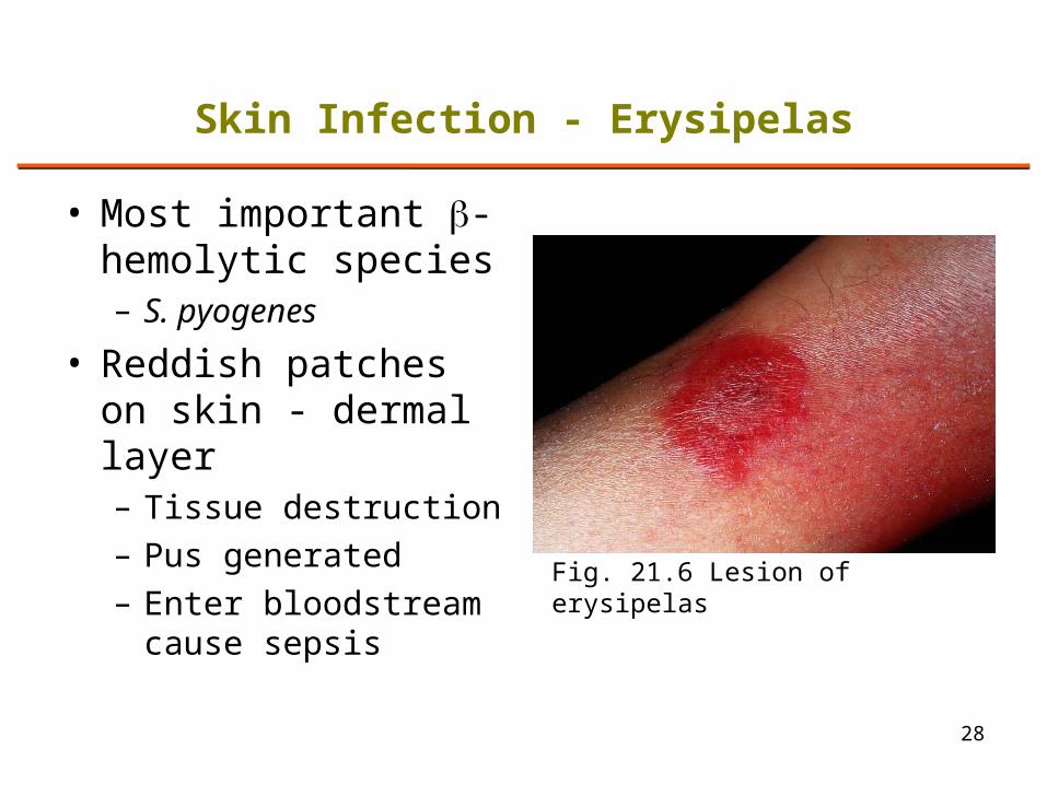
• Most important -hemolytic species– S. pyogenes
• Reddish patches on skin - dermal layer– Tissue destruction– Pus generated– Enter bloodstream
cause sepsis
28
Skin Infection - Erysipelas
Fig. 21.6 Lesion of erysipelas
29
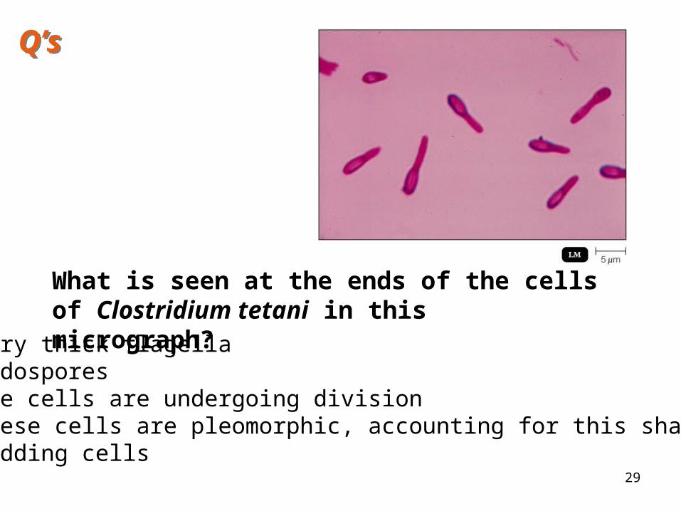
What is seen at the ends of the cells of Clostridium tetani in this micrograph?
1. Very thick flagella2. Endospores3. The cells are undergoing division4. These cells are pleomorphic, accounting for this shaped cell5. Budding cells
Q’sQ’s
30
1. Which of these is NOT correctly matched?
1. Escherichia coli – gastroenteritis
2. Staphylococcus aureus – TSS
3. Clostridia tetani – gangrene
4. Bacillus anthracis – anthrax
2. Which of following is not true about fimbriae?
1. They are composed of protein
2. They may be used for attachment
3. They are composed of pilin4. They may be used for motility
5. They are found on Gram negative cells
Q’sQ’s
31
1. The formation of a tough grayish membrane in the throat is characteristic of:
1. Tuberculosis
2. Scarlet fever
3. Diphtheria
4. Streptococcal pharyngitis
2. A positive tuberculin skin test indicates that an individual:
1. Has an active case of tuberculosis
2. May have been vaccinated with BCG
3. Has immunity to TB due to an earlier infection
4. Any of the above is possible
Q’sQ’s
32
2. An acid-fast stain of a patient’s sputum reveals acid-fast rods. This indicates infection with:
1. Which of these is an infection of the lower respiratory tract:
1. Botulism
2. Diphtheria
3. Tuberculosis
4. Streptococcal pharyngitis
1. Staphylococcus aureus
2. Mycobacterium tuberculosis
3. Corynebacterium diphtheria
4. Any of the above is possible
Q’sQ’s
33
1. Which of these diseases is also known as Hanson’s disease:
1. Botulism
2. Leprosy
3. Tuberculosis
4. Gangrene
5. Anthrax
2. Staphylococcus aureus is responsible for all of the following except:
1. Acne
2. Impetigo
Q’sQ’s
3. Toxic shock syndrome
4. Scalded skin syndrome
34
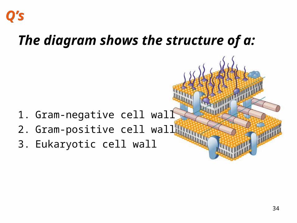
The diagram shows the structure of a:
1. Gram-negative cell wall
2. Gram-positive cell wall
3. Eukaryotic cell wall
Q’sQ’s
35
The antibiotic Gentamicin binds the 30S ribosome subunit. This will interfere with:
1. Protein synthesis in eukaryotic cells
2. Protein synthesis in prokaryotic cells
3. Cell division in eukaryotic cells
4. Cell division in prokaryotic cells
Q’sQ’s
36
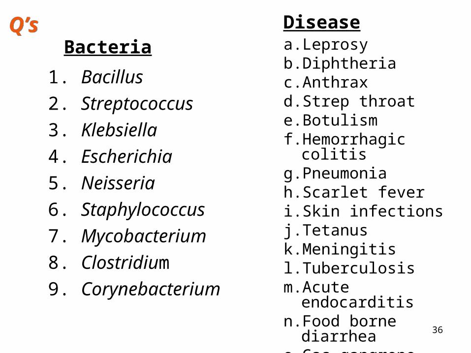
a. Leprosyb. Diphtheriac. Anthraxd. Strep throate. Botulismf. Hemorrhagic colitisg. Pneumoniah. Scarlet fever i. Skin infectionsj. Tetanusk. Meningitisl. Tuberculosism.Acute endocarditisn. Food borne diarrheao. Gas gangrenep. UTI
Disease
1. Bacillus
2. Streptococcus
3. Klebsiella
4. Escherichia
5. Neisseria
6. Staphylococcus
7. Mycobacterium
8. Clostridium
9. Corynebacterium
BacteriaQ’sQ’s