1 - royal holloway, university of london · inactivation of malate 5 dcpip reduction in both...
TRANSCRIPT

— 1 ""
EFFECTS OF LIGHT ON PIGNENTE3 AND NON-PÏGiîEMTSD STRAINS OF S\RCIN\ LUï'RA
A thesis presented by
ABU NUIIAU'IAD SIIANSUL BUDA
in part fulfilment of the requirements for the degree of Doctor of Philosophy in the University of London
1070
Department of Biochemistry, Bedford College,London, N.W.l*

ProQuest Number: 10098177
All rights reserved
INFORMATION TO ALL USERS The quality of this reproduction is dependent upon the quality of the copy submitted.
In the unlikely event that the author did not send a complete manuscript and there are missing pages, these will be noted. Also, if material had to be removed,
a note will indicate the deletion.
uest.
ProQuest 10098177
Published by ProQuest LLC(2016). Copyright of the Dissertation is held by the Author.
All rights reserved.This work is protected against unauthorized copying under Title 17, United States Code.
Microform Edition © ProQuest LLC.
ProQuest LLC 789 East Eisenhower Parkway
P.O. Box 1346 Ann Arbor, Ml 48106-1346

*"• 2 ■»
Abstract
A comparative study of the photosensitlKation of isolated membranes of pigmented and non-pigmented Sarciiia lutea in the presence end absence of an exogenous photosensitizer has been made* Illumination of membranes at lov light intensities with toluidino blue resulted in photoinactivation of malate, succinate, lactate and aecorbote oxidases* Methylene blue and DCPIP reductions, cytochrome c and ascorbate—TtTPD oxidase activities also showed photoinactivations* Photoinactivation was always greater in non-pigmented than in pigmented membranes* 20 minutes* illumination caused reversible inactivation, while 00 minutes* illumination caused irreversible photoinactivation of malate oxidase activity in the non-pigrrjentcd membranes,
5 minutes* illumination at high light intensity, without exogenous photosensitizer, caused reversible photoinactivation of malate and succinate oxidases and malate s DCPI? and succinate i DCPIP reductions in pigmented membranes* Non-pigmented membranes showed irreversible photoinactivation of these enzyme activities* Photoinactivation of malate : vitcunin K reductase was reversible in all membranes*
15 minutes’ illumination irreversibly photoinactivated malate and succinate oxidases, but reversibly photoinactivated malate and succinate t DCPIP reductions in the pigmented membranes. Kon- pignented membranes showed irreversible photoinactivation of these enzyme activities* Malate and succinate : vitaiîîin K reductases of both strains showed reversible photoinactivation*
25 minutes* illumination with ultraviolet-filtered light showed no photoinactivation of PMS or methylene blue reductions, but showed reversible photoinactivation of malate oxidase activity in the pigmented strain and irreversible photoinactivation in non- pigmented cells and membranes. There was irreversible photo- inactivation of malate 5 DCPIP reduction in both strains* Permanent photoinactivation of NADIÎ oxidase activity was observed in pigmented membranes, while NADîl : DCPIP reduction showed no photoinactivation*

— 3 —’
Malate ; vitamin îC reductase activity in both strains showed reversible photoinactivation. The malate oxidase and malate : vitamin K reductase activities of two other non-pignented mutants showed identical properties.
It is concluded that caroteneid reduces photoinactivation generally in the presence of an exogenous photosensitizer, but protects tt specific site in the respiratory chain in the absence of added dye.

— 4 —
AbstractContents
ContentsPage Number
2 4
Chapter 1. Section 1. Section 2.
Section 8.
Section 4.
Section G,
Section G. Section T* Section 8. Section 9. Section 10.
Tilstortcol Introduction General Introduction Carotenoids Q.« Generalb. Carotenoid chemistryc. Bacterial caroteneidsPhotosensitization and bacterial
carotonoidsPhotodynamic action in Photosynthetic
bacteriaPhotodyncuaic action in Non-Photo synthetic
bacteriaPhotooxidation of carotenoidaPt oto induction of carotenoid synthesisBacterial cell membraneProperties of bacterial membranesConclusion
10
12131314
IG
IT
252G272933
Chapter 2. Section 1• Section 2,
Section 3. Section 4*
Section 0.
Materials and Methodsdiemicals 35The strain of bacteria used in the 35
present studyCulture and maintenance 35Production of mutantsA. Mutation by ultraviolet light 38B. Mutation by base analogue 38
5—brosiK>urac ilCm Mutation by ethyl in ethanesulphonate 37D# Morphological and biochemical tests 37
for the mutantsGrowth curve for Sarcina lutea (wild type) 33

Pago
Section 0.
Section 7, Section 8. Section 9#
Section 10.
Section 11.
Section 12,
- 5 -
Preparation of cell suspensions forexperiments using standard manometric techniques and the oxygon electrode
Preparation of membranesExperiments on cell viabilityIllumination of cell suspensions and
membranes prior to enzyme assayMaoometric assay methodsI, Malate oxidaseII, Succinate oxidaseIII, Ascorbate oxidase TSf, Lactate oxidaseV, Ascorbate-TMPD cytochrome oxidaseVI, Malate : methylene blue oxido-
reductaseVII, ASCOrbato-cytochrome c oxidase activity Polarographic assay methodsA, Oxygen electrode apparatusB, Calibration of apparatus(I). Daily calibration of oxygen electrode(II). Absolute calibration of 0^ electrode(III).Method I(IV), Method IIC,
Number
D.
Assay of malate, sucdinate, lactate and ascorbate oxidases
Maiate/succinate : methylene blue oxidoreductases
E. Malate dehydrogenaseF. Malate/auccinate j vitamin S reductasesG. A SCO rbato-cyto chrome c oxidaseII, NADÎÎ oxidaseSpectrophotoraetric assay methodsA, Malate : dichloroplieuol indophenol,
succinate t dichlorophenol, indophenol and NADU s dichloro— phenol indophenol reductions
38
334042
43 43 434344 44
44
45
4747474743
49
494950 50
50

— G ""
Section 13.
B, NADh oxidase C# ProteinExtraction, estimation of pigment
concentration;!and analysis of carotenoida of Sarcinfi lutoa
Page Number 515152
Chapter 3. Section 1, Section 2.
Section 3.
Section 4.
Section 5*
Section 6.
Section 7.
Section B.
Section 9.
Experiments with Toloidine BlueIntroductionOxidation of m l ate, sncciimte, lactate
and ascorbate by whole cells and membranes of pigmented S. lutea and a Cttrotenoidless mutant
Effect of exposure of pigmented and corotenoidless whole cells and membranes to light from tungsten lamps (G20 lumens/sq.ft,) on malate oxidation
Viability of illuminated pigmentedSorcina livtea and its a.v.—induced white mutfMkt in the presence of 2.5pH toluidioe blue
Effect of illumination with various concentrations of toluidine blue (ijjH, 2#5uM and 5pT!) on the malate oxidase activity of whole cells of pigmented and white S. lotea
Effect of illumination in tlie presence of E.GpM toluidine blue on succinoxidase and lactate oxidase activities of white and pigmented cells
Effect of various concentrations of toluidine blue in the light on malate oxidation by cell membranes of the white mutant of S. 1utea
Effect of light with toluidine blue and histidine on malate oxidation by the membranes of pigmented and white S, Iwte^
Effect of variouSydurations of illumination with 10”^M toluidine blue on the malate oxidase activity of membranes of the white mutant
5354
50
59
59
61
67
70
71

- 7 ~
Section 10.
Section 11•
Section 12.
Section 10.
Section 14,
Section 15.
Pgf?e NnnbcrEffect o^ light with toluidine blue 75
(lO”'Bi) on malate oxidation o.nd methyleno blue reduction by pigmented and white membranes
Effect of light with toluidine blue 73(2.5pM) on malate oxidation, methylene blue reduction, 2, 6—Dichlorophenol indophenol (BCPIP) reduction and cytochrome c oxidation by pigmented and non-pigmented membranes
Effect of light with toluidine blue 31on the ascorbate-TlIPD
cytochrome oxidase activities of the membranes of pigmented and white mutant of S. lutoa
Effect of light with 2.5pM toluidine blue 84on the malate s vitamin K reductase activity of membranes of the pigmented and white mutant of S. lutea
Effect of illumination for 00 minutes 33with 2.5p-M toluidine blue on the malate dehydrogenase and malate ; vitamin S reductase activities of the membranes of pigmented and the white mutant of S. lotea
Summary ' 83
Chapter 4.
Section 1. Section 2,
Section 3,
Section 4.
Experiments without an exogenous photo» sensitizerIntroductionEffect of high light intensity on the
malate oxidase activity and the activities of segments of the respiratory chain in pigmented and non-pigmonted mcabranes of S. Intea
02
03Effect of a short exposure to illumination at high light intensity on the malate oxidase and succinate oxidase activities,and activities of segments of the respiratory chain, in pigmented and Don-pigmented membranes of S. lotea
Effect of 15 minutes' illumination at high light intensities on the malate
100

- 8
Pago Number
Section 5.
Section 0,
Section 7.
lection 8.
Section 9*
Section 10#
Section II.
Section 12*
Section 13. Chapter 5.
and succinate oxidase activities, and the activities of segments of the respiratory chain in pigmented and noJt>- pigmented membranes of S. lutea
Effect of 25 minutes* illumination with an OY 13 and OY 10 filter on malate oxidase activity and on methylene blue redaction by pigmented membranes
Effect of illumination with various colour filter glasses on methylene blue reduction by membranes of pigmented S. lutea
Effect of 25 minutes’ illumination with OY 13 colour filtered light on NADIl oxidase activities and NADU : DCPIP reduction activities by membranes of pigmented and iion-pigmcnted S. Ixitec.
Effect of 25 minutes* illumination with the OY 13 filter at high light intensity on the malate s vitamin £ reductase activity of pigmented and non-pigmented membranes
Effect of 25 minutes* illumination with OY 13-filtcred light on the malate oxidase activity of pigmented and non-pigmented cell suspensions
Effect of 23 minutes* illumination with OY 13-filtered light at high intensity (1395 lumens/sq.ft.) on the malate oxidase and malate dehydrogenase activities of membranes of pigmented S. IIIteg
Effect of 25 minutes* illuiination with GY 13-filtered light at 4®C and 20^0 on tlie malate oxidase activity of pigmented membranes of S, lutea
Effect of 25 minutes* illumination with OY 13-filtered light on malate oxidase, malate ; vitamin K reductase and malate t methylene blue oxido- reductase activities of two other mutant membranes
SummaryCarotonoids
114
123
125
123
131
134
134
137
140143

— G —
Pp.,g© NagborChanter 6. DiscussionSection 1. Malic dehydrogenase of S, lutea 147Section 2. Reversible damage to malic oxidase in
the presence of toluidine blue143
Section 3. Sites of light inactivation in the presence of toluidine blue
149
Section 4. Action of toluidine blue and light 149Section 5. Endogenous photosensitizer 150Section 6. Effect of light on flavoproteins 151Section 7. Vitamin K reductase 152Section 8. DCPl? reduction 153Section G. Sites of light inactivation 154Section 10, Relationship between photoinactivation
with exogenous and endogenous photosoijsitizers
155
Section 11. Endogenous photosensitizers 155Section 12, Hole of carotenoids in the protection 156
against pliotodynoaiic actionTransmission characteristics of Chance colour filters (2 EKa thicimcss)
IGO
References 162
A c knowledgen ent s 163

• 10
CHAPTER 1
n 1ST on TC AL INT1> ODUCT ION
1, General introduction
Snrcing Intea Schroeter, a non—photosynthetic, non-nathogenic, coranon nir-contorainant, forras coarsely granular, yellowish pigmented, circular colonies when grown on nutrient agar. The yellow compounds belong to the group carotenoids (l). The carotenoid pigments most commonly found in bacteria belong to the following groups:(i) hydrocarbons or carotenes such as betacarotcne, (ii) alcohols or santhophylls, (iii) esters and (iv) carotenoid acids. Carotenoid pifwments are rather widely distributed in the microbial world. They are present invariably in all photosynthetic organisms but are also produced by some fungi and non—photosynthetic bacteria. These pigments have been widely studied during the last few decades owing to their interesting chemical structure and also to their possible biological and physiological importance.
However, at the present time comparatively little is known about the precise physiological functions of tiio carotenoids other than as precursors of vitamin A in animal systems. In the photo- synthetic bacteria the carotenoid pigments have been found to protect these organisms against lethal photooxidations in wiiich the intracellular bacteriochlorophyll is the photosensitizer (2, 3). It has been found that the carotenoid pigments of non-photosynthetic bacteria such as Sarcim lutea or Corynebacteriun nolnaettiae have a similar protective function (4, 5). Unlike the photosynthetic bacteria, photodynamic killing of carotenoidless mutants of these organisms normally occurred at low light intensities only in the presence of an added photosensitizer and oxygen, while pigmented strains were unaffected. At high light intensities (e.g. direct sunlight) and in the absence of an exogenous photosensitizer, carotenoidless kautants wore killed in the presence of oxygen while the pigmented strains escaped photodynaraic action (6). These results suggest the presence

- Il -
within the cells of suhstahccs which act as photosensitizers at high light intensities in the photodynamic killing of carotenoidless colls* The carotenoid pigments prevent the lethal action of the exogenous as well as of endogenous photosensitizers* The chemical nature of the endogenous photosensitizer is, however, not known*
The role which carotenoids play in bacterial photosynthesis, other than their protective one, is far from being understood* Owing to the ubiquitous association of carotenoids with all photosynthetic systems, their direct participation In photosynthesis has been regarded as one of their principal functions. Carotenoids participate in photosynthesis as auxiliary absorbers of radiant ener^ which, in turn, may be transferred to chlorophyll and converted to chemical energy. The efficiency of this energy trapping, however, varies with different carotenoids and organisms (7). Chlorophyll plays an essential role in photosynthesis since, in its absence, pliotosyntliesis cannot take place. By contrast, the role of carotonoids is of a more supplementary nature since photosynthesis can proceed in their absence (8). Carotenoids may also play other roles in photosynthesis. There is a probability that they might participate in the transport of electrons in photosynthesis. Electrons could be transferred from photoexcited chlorophyll to a carotenoid, with the latter then playing some essential role in "mediating” an electroa-transfer from a donor to an acceptor molecule (O)* In this connection several workers have observed absorption changes during photosynthesis believed to be due to carotenoids (lO).
Oxygon-evolving phototrophs usually contain epoxidic carotenoids, and Blass et al. (ll) have advanced the hypothesis that reversible removal of epoxidic oxygen may be part of the oxygen evolution sequence in photosynthesis. In photosynthetic bacteria, where epoxidic carotenoids are not present, oxygen evolution does not occur during photosynthesis* Krinslry et al. (12) have pointed out that the epoxidic carotenoid, neoxanthin, is virtually absent in dark grown Euglena gracilis, and that formation of neoxanthin upon illumination of the same organism, previously grown in dark, paralleled the development of photosynthetic competence* However, no direct

- 12 -
evidence for this hypothesis has been obtained*As noted above, carotenoids play on important role in the
protection of cells against photodynociic killing* However, the precise way in which this protective function is perforated is still unknown* Since these pigments are found in the membranes (13, 14) the present study was planned to investigate the properties of the cell membrane in relation to the carotenoid pigments of Sarcina. lutea. The study comprises preliminary experiments with cell-suspensions of wild and a non-pigmented mutant of S. lutea which were subjected to illumination in the presence and absence of an external photosensitizer, toluidine blue. Membranes from pigmented and non-pigraented S. lutea were illuminated initially in the presence of toluidine blue and finally without any added photosensitizer* These latter experiments were designed to investigate the properties of endogenous photo- sensitizers. So far no investigations have been made on the protective function of carotenoids using isolated cell membranes* This study of the role of carotenoids in S. lutea involves work with isolated membranes and provides an opportunity to gain a fuller understanding of the functions of these pigments in a non-photosynthetic bacterium*
(a) General*Carotenoids have attracted the curiosity of scientists since
the beginning of organic chemistry. In the classical period the structural determinations of the carotenoids, derived chiefly from plant sources, were carried out by semi-micro methods, means of chemical degradation* Various types of oxidative degradations, catalytic hydrogenation, determination of side-methyl groups etc* were used, and the structure of many of the common C—40 skeleton of carotenoids was established* This early work was elegantly reviewed by Karrcr and Jucker (15) in 1943. Physical methods were partly used at that time, but the subsequent period has been characterized by development of improved micro-techniques and new methods, particularly spectroscopic and chromatographic ones. In the last ten to

- 13 -
fifteen years, considerable progress has also been made in the field of biosynthesis of the coloured 40-carbon pigments»
(b) Carotenoid chemistry.Carotenoida consist of a long chain of conjugated hydrocarbons
substituted at certain positions by methyl groups and generally terminated by two partially saturated rings. They may be regarded as composed of isoprene residues, usually eight, arranged in such a way that nearest to the centre of the molecule two methyl groups are present in 1:G positions, while the lateral methyl groups occupy ls5 positions with respect to each other (see Pig. l)#
Fig. 1. Isoprene units in y3-carotene.
Many of the physical and chemical properties of carotenoids result from characteristic features of their molecular structure.In particular the delocalization of the ^/-electrons over the entire carbon chain readily permits polarization of the molecule (IG), and the energetic values of the highest occupied and lowest empty molecular orbitals suggest that these compoands should be excellent electron donors and acceptors (17).
(c) Bacterial carotenoids,Carotenoid pigments are widely distributed in the microbial
world. They are invariably present in all photosynthetic microorganisms and are also present in a variety of non-photosynthetic bacteria (13). Early work by Sobin and Stolily (l) sliowed Sarcina lutea to contain only two carotenoids, while S. flava had only one,

14- —
identical with one of those found In S. lutea, Tliirkell and Strang(19) re investi got oil pigments in S. lutea and S, f I ava and isolated seven fractions, all apparently with the same cliromophoric group.These fractions were hydrocarbons or polar carotenoids. There was no evidence for any simple corotenoid esters. Strang (20) studied the spectral characteristics of the carotenoids of S, lutea and S, fI ova and found six pigments, Most carotenoids reported to occur in bacteria have 40 carbon atoms, but Jensen and Qfeeka (21) reported a carotenoid in F1 ovobe.cteriurn dehydrogenans. Thirkell et al,(22) have now reported the existence of on apparently identical carotenoid in S, flava and suggested the possible occurrence of the same in S, lutea,
3, Photosensitization o.nd bacterial carotenoids
In a biological system exogenous dyes can bring about damage or even complete destruction when subjected to artificial light of low intensity* Similar damage or destruction could be produced at very high light intensities (e.g, direct sunlight) without any added photosensitizer. In the latter case the endogenous pignents in the cells (e,g. cytochromes, flavins) presumably act as the photosensitizing agent. It is well known that some of the wnino acid residues in a protein, such as histidine, tryptophan, metliionine and tyrosine, can specifically be oxidised by irradiation with visible light in the presence of a suitable photosensitizer (23), It has also been found in vitro that when solutions of a number of proteins were irradiated with visible light in the presence of various dyes like methylene blue (24) and proflavin (25), oxygen was taken up and oxidative destruction of histidine, tyrosine and tr)g)tophan and oxidation of side-chains containing sulphur took place* The precise nature of this photodynamic action, however, is not known. Blum (26) defined photo- dynamic action as "the sensitization of a biological system to light by a substance which serves as a light absorber for photochemical reactions in which molecular oxygen takes part." The dye appears to act catalytically and only undergoes destruction in side reactions.

- 15 -
The commonly proposed mechanisms for photodynamic action found in the biological literature may be summarised as follows (27):
(I) 3 + hv S*-^ s'5 S'+ S + RII*; Rllg + 0^-» products
(II) s + hv s*-^ s ' I S' + sn^ + r; sn. + s + n*o*(i Imt 6 Ù «W «Z
(ill) S + hv — ^ S ^ S;S + S + 0( 5 RHfj 4* Og—^ products
(IV) S + hv S*--4 s' } s'+ 0^-4 SOO; ERg + SOO products 4 S
(V ) S 4 FJÎ -4- S?dî«$ SRIL 4 hv -4 SRII%-4 SRR^} SPJî' 4 0^-4- S 4 producttw «M Ii* t- W V&»*Inhere S is the sensitizing dye, hv is a quantum of light, S is the
first excited singlet state of the dye, S' is the triplet state of the dye, îîlî is the substrate, RH^ is an excited form of the substrate,<Ù4 I.*"products" represents the oxidised form(s) of the substrate, R is anoxidised (dehydrogenated) form of the substrate, is a photo-* *" reduced form of the dye, 0 , is an excited or reactive form of oxygen,SOO is a reactive dye-oxygea complex (regarded as a "moloxide," or asa free radical, the oxyradical etc.), SBUg is a dye-substrate complex,
* /and SEÏI and SRÎÎ are excited forms of the dye-substrate complex.It is to be noted that the ground state sensitizer is ultimately
regenerated in all the proposed reaction schemes. In mechanisms (l) and (ll), substrate la the primary reactant with the excited sensitizer, while in mechanisms (ill) and ( IV ) oxygen is the primary reactant. Mechanism (V ) requires a binding of sensitizer to substrate prior to light absorption. Although a number of reaction mechanisms have been proposed to account for photodynamic action, it must be admitted that tlie mechanisms involved, especially in more complex systfôns, are little understood. More than one mechanism can probably occur with a given substrate, depending on the dye and the reaction conditions used.^

- 10 -
4. PhotodynriCTic action tn r h o t o i c bacteria
Carotenoid pigoents of the bacterial cells can act as protective agents against lethal photosensitized oxidations. This was first demonstrated in photosynthetie bacteria by Griffiths et al# (S)#Their observations were based on studies with a blue—green carotenoid— less mutant strain of the non-sulphur purple bacterium Ehodonsendononaa Sphereides. In this mutant, the coloured carotenoids were replaced by an accumulation of the colourless polyene, phytoene, which led to changes in the absorption spectrum of the organism both in the blue region of the spectrum and in the infrared# The major change, however, was noticed when these blue-green organisms were grown under aerobic conditions in the light. Under these conditions, the wild strain continued to grow at the same rate as it had under anaerobic conditions, although there was an Inhibition of pigment formation.Under aerobic conditions there was cessation of bacteriochlorophyll and carotenoid synthesis, which was resumed on establishing anaerobic conditions. In the mutant strain, growth proceeded at its normal rate under either anaerobic conditions in the light or under aerobic conditions in the dark. However, when mutant cells were exposed to aerobic conditions in the presence of light, there was cessation of growth and decrease in the concentration of bacteriochlorophyll. Subsequently there was massive death of the bacterial cells as was demonstrated by a decrease in viable counts. Therefore, as neither light nor air alone caused death of the mutant cell, destruction was
J thought to be due to a photodyhamie oxidation. Using suitable filters they further domonstrated that the light which caused the death of the blue-green mutant strain was light absorbed by bacteriochlorophyll. Since the carotenoid-containing strain of the same organism did not sliow such a photosensitization, they concluded that the presence of visibly coloured carotenoid within the cell prevented the killing of the cell when exposed simultaneously to oxygen and light, and this killing was produced by light absorbed by the bacteriochlorophyll in the absence of coloured carotenoids.

- 17 -
Coîiei -Bazire and Stanier (2) produced carotenoidless cells of Fhodosnirillum rubriim by using dinhenylofaine (d p a), a compound which selectively inhibits carotenoid biosynthesis* The diphenylomine is considered to act by inhibiting oxidation (dehydrogenation) of the colourless carotenoids (nhytofluene etc*)^which then accumulate*They observed similar photodynamic killing of the mutant as above and formulated a hypothesis that the carotenoid pigments acted as che deal buffers against lethal photooxidations*
The study of the blue—green mutant of R. soheroides was continued by SiStrom et al* (8), who also used DPA to inhibit carotenoid biosynthesis, and demonstrated that either genetic or physiological elimination of carotenoid pigments renders the cell «stremely susceptible to lethal photooxidation by its own bacteriochlorophyll*
Feldman and Lindstrom (23) studied the effect of carotenoid pi;pnents on photooxidations of some photosynthetic bacteria. The photooxidase activity of normal and carotenoidless mutants of R. snhoroides and E. rubrtim were compared* In Eliodopseiidomonaacarotenoids functioned as accessory pigments for photooxidations* lihen carotenoids were absent, more of the photooxidant was apparently available for indophenol oxidations* It seemed that the carotenoids could function as biological buffers for erccess oxidising power*With R-hodo spir i II urn * carotenoids apparently did not function as accessory pigments in photooxidations, and absence of carotenoids did not increase tlic specific activity of indophenol photooxidations. This apparent difference in carotenoid function probably reflected a difference in carotenoid concentration and/or in membrane structure.
5. Photodynnriic action in non—nhotosynthetic bacteria
In the noi>-pho to synthetic bacteria the presence of endogenous photosensitizers has not been readily observable unless very high light intensities are used* Most workers have used exogenous photosensitizers to demonstrate a photodynamic effect normally in the absence of carotenoid pigments. The photodynamic effect has occasionally been an inhibition of growth, but if the process of

— 13 —
pîiotosonsltizatlon is prolonged, ultimately it leads to lethal photooxidations. It is essential to mention that not all of tîie studies to be discussed here fall under Blum*s (26) definition of photodynamic action, which requires a role for molecular 0^* There are several photosensitizers (both natural and artificial) which apparently do not operate via an 0^- requiring step but nevertheless cause damage to the cell. This was pointed out by Mathews (29) who compared lethal ^)hotosensitisation in S. lutea using either toluidine blue or the plant furocoumarine, 8—laethoxypsoralen (8-MO?)* In her study, the pigmented and carotenoidless mutants of S, lutea were killed at essentially the same rate when the cell suspensions were exposed to long-wave ultraviolet light (330 - 430 nanometers in the presence of 8-MOP* did not cause any damage to thecell in the dark* Further, the degree of photokilling was less in the presence of air (or oxygen) than in the presence of helium*The apparent protective function of oxj^en may have resulted from some destruction of psoralen by oxygen. By contrast, cells treated with toluidine blue only suffer lethal effects of light in the presence of oxygen, and carotenoid protects these cells from photo- oxidation* It has been suggested by Mathews and Sistroia that the anaerobic pliotosensitizatioa of S. lutea by 3-MOP does not cause ory damage to cellular protein as does toluidine blue in air, but rather causes damage to cellular deoxyribonucleic acid (DMA)* This suggestion is supported by the mutagenic effects of 8-HOP in the presence of light*
Griffiths et al* (3) and Sistrom et al* (3), who studied photosensitization of photosynthetic bacterium H. snheroides* suggested that the hypothesis tliat carotenoids serve as protective agents against potentially lethal photosensitized oxidations might be extended to nonn-photosynthetic bacteria, where the carotenoid pigments are often present in cells frequently exposed to light and oxygen under natural growth conditions*
Kunisawa and Stanier (4) working with the non-photo synthetic bacterium Corynebacteriom poinsettice used a wild strain containing coloured carotenoid pigments and a white mutant obtained by ultra-

— 19 —
violet irradiation. C. poinsettioe is believed to form four principal carotenoid pigments: lycoxantîiin, lycopene, cryptoxanthia and Bpirilloxanthia (80), althou/^h the presence of spirilloxanthin has since been denied (81), Non-pigmented cells in which the normal carotenoids were largely replaced by the colourless C-40 polyene, phytoene, were obtained by two methods; isolation of a white mutant and cultivation of the wild type in the presence of diplienylamine* The effect of visible light on dye-sensitizedpigmented and non—pigmented cells showed that the non-pigmented cells could be killed rapidly by exposures which were without any effect on pigmented cells* Photosensitivity was produced by both physiological and genetic suppression of pi^^ient synthesis* The rate of killing by ultraviolet light was the same in the noj^-pigmonted mutant as in the pigmented wild strains, indicating that the carotenoid-protected photosensitivity was specific for visible light.
The protective function of carotenoid pigments against photo- dynamic killing of Sarcina lutea was studied by Mathews and Sistrom (5, 6)* They were unable to show any killing of the mutant S. lutea, in tlie absence of added photosensitizer, after exposure for 4 hours to light from either tungsten lamps (3,COO ft* candles) or fluorescent lamps (1200 ft* candles)* However, in the presence of toluidine blue, light and air, the colourless mutant was rapidly killed while the wild type was unaffected, as was the mutant in the absence of either light or oxygen. They further studied the effect of direct sunlight (12,000 ft* candles) on the wild tyyie and carotenoidless mutant* The result of their experiment showed that after an exposure for 2 hours, more than 99 percent of the mutant cells were killed; in the some period the pigmented cells were not affected*The mutant cells were not killed in the absence of oxygen, thus demonstrating that the carotenoids of non—photosynthetic bacteria do protect the cells against a lethal photodjuiamic action which can occur in the absence of exogenous photosensitizer at high light intensities* These results indicated the presence within the cell of substances which can act as photosensitizers in the photodynamic killing of carotenoidless cells* The carotenoid pigments prevented

— 20 —
the lethal action of these endogenous, as well as exogenous, photo- sensitizers* The chemical nature of the endogenous photosensitizers, however, is not known*
Since it haa been shown that the carotenoids are located in the cell membranes (14) it may be supposed that the membrane is the site of photodynanic action. Hence, it was possible that cellular death was associated with changes in tîio permeability of the cells* Mathews and Sistrom (6) used two methods to detect changes in cell permeability* In the first, the release of material absorbing at 2G0 nm was taken as an idea of change in permeability which took place at the some time as, or shortly after, the onset of killing due to photodynamic action* Release of the material from the pigmented strain was less than 3 percent of that from the some amount of noo-pigmented cells ex%)osed to light* The second method for the detection of changes in permeability made use of the properties of a dye, sodium-3-anilino—l—naphthalene sulphonate (AMSA), which is not fluorescent until conjugated to proteins* The mutant cells showed stronger fluorescence than the pigmented strain when illuminated in the presence of toluidine blue and AMSA* They suggested that a breakdown in the control of permeability might be the cause of photodynamic death* To substantiate this xu>tion, the cells were killed by polymixin, an agent known to cause death by disrupting the permeability barrier of the cell. The increase in permeability caused by polymixin was 4 to 5 times greater than that caused by photodynamic action. Mathews and Sistrom concluded tliat these results did not support the suggestion that the primary letlial event in photodynamic killing was a change in permeability, although some changes in membrane permeability due to photodynamic action occurred*
It is possible that the primary lethal event in photodynamic action is inactivation of cellular enzymes* Succinate dehydrogenase and nicotinamide adenine dinucleotide oxidase were nearly totally inactivated when carotenoidless cells were exposed to ligîit and air in the presence of toluidine blue* The rates of inactivation were temperature-independent* In the pigmented cells these enzymes

— 21 —
wore not affected by this treatment (C)^In a comparative ftady of lethal photosensitization of S, loteg
(20) lïy 8-raethoxypsoralen (8-4Î0P) and by toluidine blue, Mathews showed that succinate dehydrogenase, reduced nicotinamide adenine dinucleotide oxidase and adenosine deaminase activities of the extracts of both the pigmented and colourless strains were not appreciably reduced by irradiation of cell suspensions in the presence of 8-MOP, However, the enzyme activities in the toluidine blue treated ones were greatly reduced in the colourless mutant only* Similarly, the suecinoxidase and the pyruvic oxidase activities of pigmented and colourless mutant cell suspensions were not affected by 8-MOP treatment, while in the colourless mutant were greatly diminished by toluidine blue treatnent. It is to be noted that the adenosine deaminase and pyruvic oxidase are the two enzymes located in the soluble fraction of the cell. She explained her findings by postulating that the permeability barrier of pigmented cells treated with toluidine blue is intact^and that not enough dye is allowed to enter the cells to photosensitize the enzymes* On the other hand, the permeability barrier of the dye-treated colourless cells is destroyed, and dye enters the cell and destroys the soluble enzymes. It may further be noted that the membrane-associated adenosine triphosphatase activity of the colourless Mycoplasma laidlawii was also appreciably destroyed when the cell suspension was exposed to visible light in the presence of toluidine blue (32)» The carotenoid-containing pigmented cells exposed to light under the same conditions lost only a small amount of enzyjo activity,
Roth (nde Mathews) (33) studied the effect of 2.5 x 10 acridine orange, an organic dye capable of photosensitizing cells, to determine the cellular site of lethal photosensitization and the protection offered by the carotenoida to the bacterium S. lutea.After irradiation of cell suspensions with visible light (2000 ft. candles) for 2.5 hours, cell extracts were prepared and assayed for reduced nicotinamide adenine dinucleotide (NABH^) oxidase and adenosine deaminase activities, the former enzyme being associated with the cell membrane and the latter with the cytoplasm (14).

- 22 -
The NâDïÎ oxidase of noa-pigmentod cells was destroyed, whereasthat of the pigmented cells was unharmed. There was destruction ofthe soluble enzyme, adenosine deaminase, to approximately the samedegree in both colourless end pigmented cells. Exposure of colourlessand pigmented cells to light from a tungsten lamp (lOOO ft, candles)
—5in the presence of 2,5 x 10 M acridine orange resulted in no difference in the rate of killing of the colourless mutant as compared with the wild type# However, when the pigmented wild type was exposed to acridine orange and light, it was found that colourless mutants appeared in numbers much greater than the spontaneous mutation rate. The results of the experiment indicated that the carotenoid pigments were capable of preventing the destruction of membrane enzymes by acridine orajoge in the presence of light, even though tliey failed to prevent the lethal action of the dye. The exposure to acridine orange and light resulted in changes in the DNA of the exposed cells.
Thus, the effects of acridine orange on cells appear to be similar to those of toluidine blue: destruction of both solubleand membrane-associated proteins as evidenced by loss of enzyme activity. It also causes destruction of DNA as indicated by increased mutation rate. The primary mode of action of toluidine blue, as found above, appears to be destruction of membrane proteins and enzymes, but with acridine orange, however, the effect on protein seems to be a minor one, Carotenoida are capable of preventing the protein destruction and lethal effect of toluidine blue, but are incapable of preventing cell death from photosensitization by acridine orange, which is possibly due to alteration of cellular DNA, It is, however, interesting to note that, although the carotenoids failed to afford protection against both the lethal effect of acridine orange on the DNA and the destruction of soluble proteins, nevertheless they could prevent destruction of membrane protein as in toluidine blue photo- sensitization.
The effect of temperature on the protection by carotenoids against photoseusitizations in S, lutea was studied by Mathews (34), Both pigmented and non-pigmented bacteria, in the presence of

— 23 —
toluidine blue and air, were exposed to light (lOOO ft, candles) for 8 hours at 4®C and 34^C, The carotenoids of the pigmented S, lutea offered less protection at 4^C than they did at 34^G#The colourless organism was killed at the same rate at both temperatures, as had been reported previously (6), Mathews further showed experimentally that the pigmented cells of S, lutea, when exposed to high light intensities (16,000 ft, candles) without any photosensitizor, offered less protection at 4^C than they did at 34^C, However, at this light intensity, even in pigmented cells without photosensitizer, photokilling did occur after a delay of about 5 hours at 4^C and 8 hours at 34^C* The onset of photokilling in tlie carotenoidless cello was also delayed.
These photodynamic effects, which are modified the presence of carotenoids, have been shown by Mathews and Krinsky (35) to be unrelated to tlie effects of ionizing radiations end ultraviolet light. Subjecting both the radiation resistant Micrococcus and its colourless mutant to gamma radiation, they found identical killing curves, With n«v« light similar results were obtained,Wlien left exposed to visible light in the presence of photo sensitizer toluidine blue, the pigmented wild type remained unaffected whereas there was photokilling of the colourless mutants. Similar results have been obtained with S, lutea. It was concluded that the carotenoid pigments had limited effects in protecting cells from radiation damage by visible light, and that they had no effect at all during damage to cells by ionizing radiation (gam:m rays and x-rays) and u.v, light.
A. purely photochemical process, photolysis of dark grown stationary—phase cells of Myxococcus xai^thus, has been reported by Burchard and Dworkin (36), This process was a typical photodynamic one which involved no diffusional or enzymic steps. The cells grown in the light developed an orange caroteno idj»whila the cells grown in the dark did not develop any carotenoid. Dark-grown cells were lysed by a relatively low light intensity but only during the stationary-phase. This lysis was temperature-independent and oxygen-dependent. They found protoporphyrin IX to be the natural

- 84 -
endogenous photoacnsitizer.Mathews (37) observed that Mycobacterium whose
carotenoid synthesis was promoted by light (see page 26 ), was f>rotectod by the presence of carotenoid against lethal photo- oxidations mediated by the dye toluidine blue*
In the non-photosynthetic bacteria there is no direct evidence of how carotenoid pigments protect cells against lethal photo- oxidations* However, Srinsky (33) studied the role of c&rotenoid pignents as protective agents, against photosensitized oxidation in chloroplasts, and suggested that the carotenoid pair antlieroxanthin and zeaxanthin could act as "chemical buffers" to protect cells against lethal photooxidations, Epoxidatioa of zeaxanthin took place only in light and oxygen when conditions were established which could lead to photosensitization. The reductive de- epoxidation of entheraxaathia to zeaxanthin was a dark enzymic reaction. Those two reactions constituted the "ef>oxide cycle" by which carotenoida could prevent lethal photosensitization of cells* The role of the epoxide cycle against lethal photosensitized oxidations was suggested to be as follows: chlorophyll, excited by light, isusually deactivated by the process of photosynthesis* Some of the excited chlorophyll could occasionally combine with molecular oxygen leading to photosensitized oxidations* This potentially letlial combination, however, would normally be inactivated by a carotenoid such as zeaxanthin, which would itself be oxidised to its epoxide derivative, anthoraxanthin. An enzymic mechanism such as aathera- Xanthin do-epoxidase would then regenerate the protective substrate zeaxanthin. By this mechanism the epoxide cycle could protect cells against lethal photosensitized oxidations*
In Mycobacteriun phlei it has been shown by Brodie et al.(39) and Brodie and Ballantine (40) that exposure of electron transport particles to light (360 nm) resulted in loss of the natural naphthoquinone and the ability to carry out oxidative pliosphorylation, together with NAD oxidase and suecinoxidase activities* Restoration of both oxidation and phosphorylation occurred with substrates linked to nicotinamide adenine dinucleotide (NAD^) by the addition

wG
of vitamin Tn controat, neitîter tbonninone nor lïon^oqoinonesrestored oxidative phosphorylation with succinate as substrate, indicating that a light-sensitive factor other than the known quinones is required for electron transport from succinate (41, 42), Since knowledge of the coraponents and sequence of carriers of the respiratory chain is necessary for an understanding of the bio—energetic process, a study was undertaken by Murti and frodio (43) to find the nature and role of this new light-sensitive factor required for succinoxidase activity in Mycobacterium ^hlei. Light daiiiage caused by irradiation of the particles differed in two pathways. The suecinoxidase pathway was more prone to damage by irradiation than tlie nicotinamide adenine dinucleotide pathway. Addition of a thermostable, water- soluble extract obtained from elthér nhlei or from rat liver mitochondria resulted in GO to 60 percent restoration of succinoxidase activity. Addition of other known co-factors, such as flavin adenine dinucleotide, flavin mononucleotide, benso-and naphthoquinones and sulphydril agents, however, failed to restore succinoxidase activity after irradiation. The site of action of the water-soluble material from M. phlei appeared to be between the flavoprotein and the cytochrome b on the succinoxidase pathway. The succinoxidase factor failed to restore NA33H oxidation, whereas GO to 60 percent restoration of NADII oxidase activity could be achieved the addition of vitamin Kj, which in the case of irradiated particles failed to restore succinoxidase activity. Whether oxygen or carotenoid are involved in this light effect is not known,
6, Photooxidation of carotenoids
Wright and Rilling (44) worked with a Mycobacterium sp. and demonstrated photokilling of the caroteneidless dark-grown cells using only endogenous photosensitizers and very bright light (7400 — 10,000 ft, candles]• Death did not occur under nitrogen. They also £>und that the carotenoids of Mycobacterium s-p, were rapidly bleached under the conditions of high light intensity which they used. Using S. lutea as a test organism, Roth and Krinsky (45)

— 20 —
observed general destruction or disappearance of carotenoid pigments during photodynamic killing. They were unable to demonstrate specific light-driven reactions among the carotenoids and suggested that the carotenoid pigments might play a physical role in the protection of non-photo synthetic bacteria against the harmful effects of liglit and oxygen, such as stabilization of the cell wall membrane complex and not a chemical or biochemical role.
Photosensitized bleaching of /S-carotene witli li^t was demonstrated by Hasegawa et al. (4G)# Their results showed that irradiation with a mo no chromât i c beam of light (632.8 era) from a continuous-wave gas laser did not affect carotene. In the presence of photosensitizing dye toluidiae blue 0 (4 x 10"*' M), however, numerous changes took place in/3-carotene. A considerable decrease in the absorbance of/3- carotene was noted initially which was accompanied by small shifts of the absorption maxima to shorter wave lengths as well as the formation of two new peaks at 400 and 3T5 nm. Finally tliey observed a conr^lete bleaching of the solution. Using column and thin-layer chromatography, up to 13 different compounds were sliown to be formed by the photo sensitization of the -carotene. All the reactions were oxygon-dependent.
7* Photo induction of carotenoid synthesis '
Some organisms are photochromogenic, i.e. they will synthesize pigments only when exposed to light. The kinetics of carotenoid synthesis of two species of Mycobacteria have been studied ty Batra and Rilling (47). The action spectrum of plio to induction for pigment formation of Mycobacterium sn. had maxima at 3G5 and 400 rsa separated by a minimum at 305 nm. However, the action spectrum of photoinduction of another species M. morinnm. had a principal maximum at 404 nm and two lesser maxima at 405 and 577 mi* Flavin was suggested to be the likely photoreceptor for the Mycobacterium sp. end a porj)hyrin for I!, marl num. The photo induction of carotene— genesis In Mycobacterium sp. was found to be depressed at an acid pH. A Mycobacterium species that synthesizes carotenoids only

**• 27 —
after light stimulation, has been used by Rilling (43) to study the inhibition of carotenoid syntliesis by a variety of compounds* la addition to diphenylamine, other compounds such as benzophenone, acridine oran,go, methylene blue, neutral red, toluidiae blue, proflavine, acridine and 9, 10-dihydroacridine were found to inhibit the formation of the more highly unsaturated carotenoids* In all cases accumulation of phytofluene occurred when the formation of more highly unsaturated carotenoids was inliibited* Under increased oxygen tension all of these compounds, except diplienylaîaine and benzophenone, no longer inliibited formation of carotenoid pigments* Using molecular models, it could be demonstrated that the dihydro derivatives of beuzophenona and diphenylamine had a close structural similarity to that of the relatively saturated carotenoids iu the region where subsequent dohydrogenatioa occurs* Thus they %)Ostulated that these compounds could bind to the carotenoid dehydrogenases subsequently inhibiting carotenogenosis*
It appears from the work so far reviewed that the cell membranes are the primary sites of lethal |diotodynamic effects* The carotenoids are located exclusively in the membranes and they play an important role in the protection of the cells from the lethal effects of photo- oxidations* Therefore, some aspects of the bacterial cell membrane will now be discussed*
8* Bacterial cell membrane
The development of a limiting mcmibrane is, in all probability, a fairly early phenomenon in the evolution of cells which are capable of showing growth and multiplication* Cell membranes possess a degree of biological universality enjoyed by genetic structures and the apparatus for the synthesis of protein, together with Some energy yielding mechanisms*
Besides providing the cell with a protective and functional (in the permeability sense) barrier, the cell membranes may confer

— 23 —
surface specificity, a property which may be of considerable importance to the cell in the determination of its response to the environment* However, Salton (49) is of the opinion that, in the Gram-positive bacteria, the surface specificity is a property of capsules or the cell wall polysaccharides or teiehoic acids* In the majority of bacteria, the plasma membrane appears to be much less conspicuous as a carrier of surface specificity than does the outer cell wall* Robertson (50) studied thin sections of cell membranes under the electron microscope and considered them to be "unit membranes" as seen in animal, plant and other microbial cells with an overall thickness of about 75 S.
Murray (51) and Schlessinger et al* (52) showed that a lower degree of intracellulor differentiation was one of the distinctive features of bacteria which were characterized by the absence of a nuclear membrane, of organized mitochondrial structures, and of a membranous type of reticulum carrying the polyribosomes* Thus the bacterial cell possesses fewer membrane bound organelles, and in this respect it offers a simpler system for investigation of membrane properties and functions. Anatomical studies of the bacterial cells reveal the presence of a single limiting plasma membrane, and in many cases a well developed internal membrane system which Fitz-James (53) referred to as "mesosomcs" and van Iterson (54) as "chondroids.” Salton (55) describes the mesosom© as vesicular, lamellar or tubular packets of membrane formed Ty the invagination of the plasma membrane* When the presence of mesosomes became evident during the electron microscopic study of bacterial cells, many functions were attributed to them,
Fitz-James (50) showed that the mesosomes vary from species to species by their overall appearance in thin sections. Broadly speaking three types can be recognised, namely a Imacllar type, a packed sac type or a loi^ tubular type* It is not known at the present time what determines the type of mesosoEie seen in a particular organism (55)* There is still insufficient direct experimental evidence to enable one to distinguish tlie functions of the plasma membrane from those of a mesosomal meiabrano* Salton (55) considers

- 29 -
roesosomea to bo anatomically reminiscent of mitochondria# Van Iterson and Leeiie (67) provided evidence supporting the idea that mesosomes are mitochondrial equivalents* They hoticed a greater concentration of tellurium deposition in the region of mesosomes in Bacillus subtills, indicating more sites active in reduction of tellurite* However, Salton has pointed out that as the total surface area of the membrane in the mesosome is greater than that of the plasma membrane, heavier deposits of tellurium are to be expected in this region*
Bladen et al* (58) have shown that phosphotungstate freely penetrated into the mesosomes or intracellular membrane-intrusions, which suggested the absence of any permeability barrier enveloping these structures. Salton (55) is of the opinion that much still remains to be done on mesosomes before we can come to any conclusion about their function or their relationship to the rest of the cell*
Thus^membranes detected in bacteria appear to be limited to one or at the best two ty iess the plasma membrane, and its invaginations in the form of the mesosome* In photosynthetic bacteria Drews (59), Cohen-Bazire and Kunisawa (GO) and Holt et al* (61) have shown that the membranes containing the photosynthetic pigments may, In some cases, be regarded as a special type of membrane invogination which constitutes the intracellular site of the photo- synthetic apparatus and from which chromatophores arise following mechanical breakage of the cell*
9* Properties of bacterial membranes
feibull (62), using lysozymo to digest the cell wall, obtained fractions from lysed protoplasts of Bacillus megateriuni wîiich he referred to as "ghosts*" These ghosts were assumed to represent the plasma membranes of the bacterial cell# Recently Salton end Chapman (63) found that conditions normally employed for the isolation of cytoplasmic membranes from a lysozyme-sensitive organism such as Micrococcus lysodeikticus produced a total membrane preparation of plasma and mesosome membranes*

•* 30 *#
The most popular and probably the moat reliable method used for the isolation of bacterial membranes involves the muralytic or cell wall degrading enzymes, followed by the separation of the membranes from the cytoplasm by differential centrifugation. A wide variety of muralytic enzymes such as lysozytae, phage muralysins, lysostapins etc. are available for the removal of the cell wall before isolation of the cell membrane. Membranes may be recovered from either totally lysed cells in dilute buffers,or by osmotic lysis of stabilized protoplasts formed by lysozyrae treatment in isotonic media. Salton and Chapman (63), isolated membrane preparations by both methods from M. lysodeikticus and were unable to find any difference wiien viewed under the electron microscope.It has been concluded by Salton and Ehtisham—ud-din (64)| Salton and Freer (65); and Salton and Schmidt (66), that if proper conditions are used it is quite possible to wash the membranes without leaching out any of the more conspicuous markers such as carotenoids, menaquinones and cytochromes. However, it should be noted that Shah and King (67) have found that the succinate dehydrogenase in the succinic oxidase system in M. lysodeikticns was inhibited when the cell was lysed with lysozyme, but was hardly inhibited at all if ultrasound was used to disrupt the cell. On treating the ultrasonic fragments with lysozyme, they observed inhibition of succinate oxidation*
The chemical composition of isolated membranes has been the subject of numerous investigations. Salton and Freer (65); Kates (63); Gilby et al* (13); Weibull and Bergstrom (69); Brown (70); Macfarlane (71), and Yudkin (72) found that the composition is very similar to that of other membrane systems in that they are composed of about 50 to 75 percent protein and 20 to 30 percent lipid.Varying amounts of carbohydrate, RNA and BNA have been reported in cell membrane preparations. At the moment it cannot be said precisely the extent to which some of the additional components are part of the membrane structure or covalently attached to membrane constituents.
A great deal is now known about the nature of the membrane

— 31 —
lipids* Eazin et al* (73) studied the membranes from Grom—positive bacteria and found that they were devoid of cholesterol* This distinguishes these membranes from those of higher microorganisms, plants, animals and pathogenic mycoplaaas which possess steroids*
Mathews and Sistrom (14) found the carotenoid pigments of the pigmented S* lutea to be located exclusively in the cell membrane, and suggested their occurrence in the form of a pigment-protein complex* Gilby et al, (13) have suggested that the carotenoid pigments in the related organism M* lysodeikticus wore also bound to the cell membrane* The carotenoid from S* flava has been shown to be an integral part of the structure of the membrane-protein fraction from wîiich it may be isolated in the form of a carotenoid- glycoprotein complex (74), However, the presence or absence of carotenoids from the membrane has not been reported to have any effect on its structure.
The ultrastructure of non-photosynthetic caroteneid-containing bacteria was studied by Mathews (75)* Her intention was to see whether any change in intracellular structure was associated with the development of carotenoid pigments in two bacteria, M* marinum and S. lutea. Examination of many tliin sections showed no difference in the intracellular structure between cells of M. marinum grown in the dark (non-pigmented) and those grown in the light (pigmented). Neither was any difference found between cells of S* lutea wild type (pigmented) and cells of colourless mutant. She concluded that no new structures were made to accommodate carotenoid pigments in non-ï5Îioto synthetic carotonoid-containing bacteria. Thus the pigment was either built into the membrane as an integral part of it, as in S* lutea, or was added on to pre-existing membranes, as in M. martnun*
Most bacteria possess a membrane-bound respiratory chaih (an electron transport chain), i.e, a system of spatially organised dehydrogenases and cytochromes which bring about electron transport.In general the respiratory chain is regarded as a multienzyme complex which catalyzes the dehydrogenation of substrates and the transfer of reducing equivalents to oxygen. In the mammalian

— 32 —
respiratory chain the dehydrogenases are connected to cytochromes by hydrogen-transferring co-enzymes, N.\l> and ubiquinone, which then pass on the electrons to the cytochromes of groups A, B and C. The bacterial chain essentially contains the same sequence of enzyme complexes but with some differences, la bacteria, the initial stage in the oxidation of substrates often involves flavin enzymes rather than NAD-1 inked enzymes. Despite tLieir similar functions, the cytochromes of groups A, B and C differ from mitochondrial cytochromes by having different absorption spectra. In addition a cytochrome 0 oxidase is known to occur. Very little is known about the actual pathways of electron transport or any relationship between the respiratory system and the carotenoids in S» lutea. Erickson and Parker (TG) investigated the membrane-bound electron transport system of Micro coccus liiteus (S. lutea). They found cytochromes^593» ^562» ^557* ®554> ®549> & menaquinone (MK-3), carotenoids,NADI!, malate and succinate dehydrogenases. Sensitivities to different inhibitors indicated the difference between the bacterial and mammalian systems. The M. luteus system was found to be insensitive to antimycin A and showed very low sensitivity to roteuone.Effective inhibition was shown 1^ cyanide, pericidin A, dicouoarol, S-n-heptyl-4-hydroxyquinoline-N-oxide and ultraviolet irradiation at GGO nm. The carotenoidless mutant exhibited identical properties to the parent organism except that no measurable succinate oxidase activity was found and there was an 8-fold increase in the malate oxidase activity of the electron transport particles. There was no difference in the action of inhibitors. They suggested the following scheme for electron transport in M. luteus:
Substrate —> Pp -> MK-8, Cyt Cyt ? *^0^
=554' cy* =549]-^ [* y* *593' ' y “a]-* °z
This appears to be the only study of the electron transport system in this organism.

- 33 -
10, Conclusion
The non-photosynthetic bacterium Sarctna lutea contains carotenoid pigments and these pigments afford protection to photo- dynamic killing \yy very high light intensities (sunlight) without any exogenous photosensitizer or Î3y low light intensities (artificial light) with an exogenous photosonsitizer* Although the carotenoid pigments play a protective role against aerobic photosensitivity, there is no direct spectral or chemical evidence suggestive of carotenoid involvement* Several investigations involving studies of viability, permeability, enzyme lability, ciutogenicity, growth, temperature effects, gas effects, light effects and ultrastructure etc* have been made in 8, lutea, but at present nothing is known about the mechanism by which carotenoids protect cells under photosensitizing conditions, nor about the mechanisms involved when photodynamic death commences. The carotenoid-containing microorganism, S, lutea, though subjected to direct sunlight in nature, is not being killed, while the carotenoidless mutants of the same microorganism could be killed. Nature seems to protect these microorganisms from the deleterious effects of sunlight for the perpetuation of the species. However, the mechanism of carotenoid participation in tlie protective function is far from being completely understood,
Carotenoid pigments in S. lutea have been located exclusively in cell membranes in the form of a carotenoid-glycoprotein complex. Like most other bacteria, S, lutea has a respiratory chain (electron transport system) which is present in the cell membranes. Other enzyme activities are also located in the cell membrane. It is possible that the primary lethal event in photodynamic killing could involve tfie enzymes of the respiratory chain, many of which absorb visible light,
I have, therefore, chosen to study the effect of visible light on tlie respiratory chain of isolated membranes of S, lutea, using both low intensity H0it with a photo sensitizer and high intensity light without a photosensitizer. These investigations have been

— 34 —
particularly aimed at determining a more specific role for carotenoids in protecting membrane enzymes from photodynamic action. As a member of the carotenoid-containing cocci, S, lutea is very easy to grow in the laboratory, and the study of the cell membrane was chosen because it is relatively easy to obtain membranes by a gentle method using lysozyme treatment which does not appear to cause ouch damage to the membrane enzyme activities.

- 35 -
CHAPTER 2
MAT]!TU\I.8 iND METHODS
1, Cliomical m
Antiaycin A, 5-brorao-2, 4-dihydroxypyrid.ine (5-bromouracil), cytochrome c, menadione (vitamin , NAD and NADU were obtained from Sigma Chemicals Company, St. Louis, Missouri, U.S.A. Toluidine blue and methylene blue were obtained from George T. Gurr Ltd., London, England. Ethyl methanesulphonate and lysozyme chloride ex egg-white cryst. were obtained from Koch Light Laboratories Ltd., Coliibrook, Bucks., England. Nutrient broth and nutrient agar were obtained from Oxoid, London, Great Britain. All other chemicals were obtained from British Drug House Chfôuicals Ltd.,Poole, England, and were the finest grade obtainable, Manometric experiments were done with distilled water, whereas experiments on the oxygen electrode were done with deionized distilled water. Chaace-Pilkiagtoa colour filter glasses were obtained from Precision Optical Instruments (Fulham) Ltd,, 158 Fulham Palace Road, London W,8, England,
2, The strain of bacteria used in the present study.
Only one strain of Sarcina lutea (Micrococcus luteus) andthree mutants of the same were used in the present study. Thestrain was obtained from Dr, A.11. Dadd, Imperial College, London, and was No» 3 in the Imperial College collection,
3, Culture and mmlntenance.
All strains of Sorcina lutea used in this study were maintained on nutrient agar slopes, which were stored at 5^C in dark. As it

— 33 —
grows luxuriantly in nutrient broth mediuiti, the same modiam was used throughout the course of the study, A loopful from a nutrient agar slope wo.3 used to inoculate lOOol. of the medium (13 g./l, nutrient brotli, autoclaved at 15 lbs* pressure per square inch for 15 minutes) in a 250ml, conical flask, and the bacteria were grown with shaking in a reciprocating shaker incubator having 200 movements r?er minute q.t 2G^C to late log phase, 5ml, of this pilot culture were used to inoculate 500ml, of the modiuia in a 1 litre Pyres culture flask, end grown to late log phase at 2G^C as above.
4* Production of mutonta.
For production of the mutants of Sercina lutea, the following three methods were employed?A, Mutation by ultraviolet light* 0$2ml, aliquots of an overnight
culture of the wild strain of Soreinn lutea were plated on sterile agar plates and irradiated with ultraviolet light, A 12 in., 7 W,, mercury quartz discharge tube with an ozone— reducing filter was used to give monochromatic radiation at 253*7 nm, (The tube was No, 01GS/k obtained from Engelhard Industries Limited, Ilanovia Lamp Division, Slougîi, England)*The plates were irradiated for a period of 6 — 10 minutes at a distance of 6 inches in a sterile cabinet* The irradiated plates were left in the incubator at 2G*C for 3 to 4 days, after which the mutants were selected as colourless colonies on the agar plates. Suspected mutants wore examined under the phase contrast microscope for shape, size and Grom staining, and sulw culturod on agar slopes for confirmatory tests,
B.Mutation by the Imse-analogue 5-bromouracil* 0*5^ 5-bromouracilwith 1^ bacteriological peptone was prepared in 10ml* of 10 times diluted nutrient broth and autoclaved at 20 lbs,/in*for 30 minutes, 0,5mi* of a thickly grown overnight culture of wild Sarcing, lutea was added to 10ml* of tlie 5—bromouracil solution in a flask, and incubated for 5 hours at 23^C with shaking,

- 37 -
after which cell counts were made in a counting chamber. Thesuspension was then diluted with nutrient broth to give a cell
2density of approximately 2 x 10 bacteria per ml, G.Stal, aliquots of the diluted cell suspension, containing approximately 40 bacteria, were plated out and the plates incubated at 2G^C for 2 to 3 days. The resultant white colonies, suspected of being mutants, were examined under the phase contrast microscope to check cell shape, size and Oro i staining, and subcultured on agar slopes for furtîier confirmatory tests.
C, Mutation by ethyl methanesulphonate. This mutagenic agent removes bases from DNA by acting specifically on guanine at the Ny position, labilising the deoxyriboside linkage so that 7— alkylguaniiie is released from the BNA, The missing guanines might be replaced by any one of the four bases.
Saturated solutions of ethyl methanesulphouate were made iu 1 ml. of sterile 0,211—sodium phosphate buffer (pH 7,2) in duplicate test tubes, 1 ml, of the overnight culture of wild Sarcina lutea was added to one tube. Both tubes were incubated at 26^C for 20 minutes, after which 0,2îd1, aliquots of two dilutions (50 and 100 times) of the bacterial suspension were plated out in triplicate, 0,2ml, aliquots from the control tube without bacteria were also plated out in triplicate, without dilution, and the plates left in the incubator at 26* C for 4 to 6 days. vHiite colonies, believed to be non-pigmanted mutants, wore examined and subcultured as above*
D, Morphological and biochemical tests for the mutajats.Morphological comparisons as regards size, shape and motility
of the mutants were made under the phase contrast microscope* Biochemical tests including reaction to litmus milk, gelatin liquefaction, gas production and Gram’s stain were performed to confirm the properties of the mutants. All morphological and biochemical tests were carried out in comparison with the wild Sarcina lutea. These testa were primarily aimed at ensuring that contaminants were distinguished from genuine mutants.

— 3S —
5, . Growth curvo for Sarcino, lutea (wild ty >e),
An overnight culture of Sarcing lutea was obtained by the method described above, and a 1 litre flask containing medium was inoculo.ted with 5ral* of the inoculum. A zero time measurement of the turbidity of the medium was made on a nepholomoter. The flask was incubated with shaking at ami, at SO minute intervals,samples were taken for turbidity estimations. The percentage of turbidity increased after a lag phase of 30 minutes, and 10 such readings were taken. The flask was then left in the incubator overnight and the percentage of turbidity recorded on the following morning. The Dopholometer was calibrated for cell density using a bacterial counting chamber. Fig, 2 gives the growth curve for Sarcina lutea under the conditions used for experiments described in this tliesis,
G, Préparation of cell suspensions for ex^erbT?ents using standardmanometric tecltntques end the oxygen electrode.
The bacterial cells grown by the methods described in section 3 above were harvested at room temperature by spinning at GOOD r.p.m, for 5 minutes on a bench centrifî^ge, collected in cold OLOSH-phosphate buffer (pn 7.2), washed four times with cold buffer, and finally suspended in the same buffer. The suspension was made uniform by means of a glass horaogenizer,
7# Preparation of no'^ibranes.
An overnight culture of wild or mutant Sarcina lutea was obtained in the usual way. The cells wore harvested at room temperature by centrifugation at 6000 r.p.m, for 5 minutes on a BTL bench centrifuge, and the pellet of cells suspended in a medium composed of cold sucrose (1,2'J), NaCl (O.OKâ) and phosphate buffer (0,06M) pil 6,8. Using a glass homogonlzer, the cells were mixed very gently to give a uniform suspension, Lysozyme chloride

-59-
9.40
9.20
9.00
"S 8.80
_ûo 8.60_û
^8.40
8.20
8.00Time (hours)
Fig.2. Standard growth curve for Sarcina lutea (wild type).

« 4o -
(15 ml. ) wa.3 oxlded to the homogonate which was incubated atroom temperature for 15 minutes with occasional stirring. After incubation the protoplasts were spun down at 20,000 g, at 5^# The cell protoplasts were suspended in cold phosphate buffer (O.OGIî) pH 6.8 containing NaCl (l^), and occasionally stirred gently to dislodge the clumped cell protoplasts for 10 minutes, keeping the tube in ice. The suspension was esaînined under the phase contrast microscope to ensure that ghost membranes had been obtained. The suspension was spun at 2,500 r.p.m. for 5 minutes to get rid of residual whole cells which did not undergo osmotic lysis. The supernatant fraction containing the cell membranes looking like ghost cells under the phase contrast microscope was collected in a cold plastic tube and spun at 20,000 g. for SO minutes. The membranes were obtained as a pellet which was resuspended in phosphate buffer (0.02H) pH 7.2 containing 0.0005ÎÎ NgCl^, and a uniform suspension was made by gently homogenizing the pellet with a glass homogoaizer. The homogenate was examined under the phase contrast microscope and,wlien found to contain more than 95/â membranes, experiments were carried out with this material.A comparison of the appearances of whole cells and membranes prepared by these methods may be made by reference to Plate 1.
In the earlier experiments with membrane preparations the lysozymo incubation time was 45 minutes. Due to prolonged incubation with lysozyme the succinoxidase activity of the membranes was very low. Shah and Xing (67) have also found that lysozyme incubation for a long time affected the succinoxidase activity of bacteria. Therefore, in later experiments the period of lysozyme incubation was shortened from 45 minutes to 15 minutes, wîiich gave on increased succinoxidase activity in membrane preparations.
8* Experiments on cell viability.
Overnight (late logarithmic phase) cultures of wild Sarcina lutea and the carotenoidless mutant (bV^) were obtained as described earlier. Two 250ml. conical flasks containing 100ml. each of

>41-
. >1:.mm€ •' # 4»
Whole cells
WÊ Æ tKS # I
/ v ':^
Membranes
Plate 1. Comparison of the appearances of whole cells and membranes of Sarcina lutea. ( Phase c ont r as t x 3 9 0 0 )

«— 42 —
sterile nutrient broth were inoculated with 5ml. of wild andmutant strains of Sarcina lutea and incubated with shaking in thedark for 2 hours. Wîien the cells were in the logarithmic phaseof their growth after 00 minutes, measured amounts of the pigmentedculture were put in one sterile glass cuvette while aliquots of thenon-pigmented culture were placed in the other. The cuvettes weremaintained at 25^C and their contents were continuously stirred bybubbling sterile air throu^%h the cultures. Sterile toluidine blue
—Gsolution was added to give a final concentration of 2.5 x 10 M. Sampl08 were taken from each cuvette at zero time end were diluted in sterile Na^hPO^-Noîî^jPO^ (0.02lî) buffer pîî 7.2. 0#2ml, aliquotsof the diluted samples were plated out in duplicate and incutmtod at 2G^C for 3 days for a viable colony count. The cuvettes were placed 9 inches away from four 100 W. tui^sten lamps giving a light intensity of 4C0 lumens/sq.ft. at the front of the culture. A water filter was interposed between the cuvettes and tlie light source. At 15 minute intervals, tîiree samples were collected, diluted in sterile buffer, and 0.2ml. aliquots were plated out in duplicate and left in the incubator at 26^C for 3 days, after which colony counts were made. In the case of the mutant, dilutions of the culture were increased to give a more concentrated preparation to allow for cell death caused by illumination# At the end of tlie experiments the temperature of the culture remaining in the cuvettes was 27*C.
9. Illumination of cell suspensions and monbranes ^rior to enzyme essay.
The bacterial preparations were placed in two Perspex cuvettes Jacketted by constant temperature water circulated from a water beth< The illuminated samples were incubated in one cuvette, while the dark sample was incubated in a second cuvette covered with blackened aluminium foil and kept away from natural and artificial light as far as practicable. Before illuminating a sample the light intensity was measured with a meter and the bacterial preparation

• 43 •
was placed in the cuvettes# The uniformity of suspension of the bacterial preparation was achieved by means of two stirrers which continuously kept the contents of the cuvettes stirred at a fixed speed, When the contents of the cuvettes attained the temperature of the water bath, control samples from the cuvette covered with foil were analysed# The sample to be illuminated was subjected to light from a Prinz bowline GOO W. aircooled projector, from which the focusing Ions had been removed. After illumination the cuvette was covered with blackened aluminium foil to cut off further light. Under our experimental conditions a slight amount of laboratoi^ ceiling light could not be screened from the top of the cuvettes.
10* Manornetric assay tpethods.
Various dehydrogenase and oxidase activities were assayed by standard manometric techniques for the measurement of oxygen uptake. The activities were expressed as microlitres of oxygen utilized per minute.I. Malate oxidase.
Oxygen uptake was assayed raanometrically at 23^C in 2ml. of medium composed of 150 mlî-sodium-hydrogen malatein0.02ll-p!iospliate buffer (pll 7.2) and O.OOOSlï-MgCl^.
II. Succinate oxidase.Oxygen uptake was assayed manometrically at 23^C in 2ml. of
medium composed of 150 mM-sodium succinateiw 0.02IÏ—phosphate buffer (pH 7.2) and 0*0005^j-MgCl^.
III. Ascorbate oxidase.Oxygen uptake was assayed manometrically at 23^C in 2ml, of
medium composed of 150 mH-ascorbic acid neutralized with NaOIî, 0.02H-phosphate buffer (pH 7.2) and 0.0005M—MgCl^*
IV. Lactate oxidase.Oxygen uptake was assayed manometrically at 23®C in a medium
composed of 150 mM-sodiun-lactateiw0,02M-phosp!iate buffer (pH 7.2) and O.OOOSMgClg.

w 44 «*
V. Ascorbate—T ïPD cytochrome oxidase.The ascorbate—tetrumothyI—para—phonylenodiaoine (TîûPB)
cytocliro ne oxidase activity was assayed by a manoiaetric method at 23^C in a median» composed of 0.05*!—ascorbic acid (neutralized), 10" .!-TViPD, O.OSvI-phosphate buffer 7.2) and O.OOOaj-MgClg. Control flasks recording; the oxidation of substrate without enzyme gave significant oxygen uptakes.
VI. Malate : methylene blue oxidoredactase.Methylene blue reduction was assayed manoraetrically at 23®C
by means of the reoxidation of reduced methylene blue by oxygen.The assay mediuta was composed of IGOnM—sodioia hydrogen malate,1 M-KCN (neutralized), 5 x 10"^M-NaN^, 0.002^ methylene blue,0.02?,'-phosphate buffer (pH 7.2) and O.OûOSM-lîgClg in 2ml.
VII. Ascorbate-cytochromo c oxidase activity.The ascorbate—cytochrome c oxidase activity of the membranes
of pigmented and white mutant S. lutea was assayed by a ©anometric method at 23^C in a medium composed of 1.0ml* of membrane preparation, ISOmM ascorbate (neutralized with NaOîî), 0.5 » cytochrome c and 0.02M phosphate buffer (%)II 7.2) and 0.0005M—MgClg in 2.0ml. Control flasks recording the oxidation of substrate without enzyme gave significant oxygen uptalces.
Substrates were nut in the side arm of the Warburg flasks, while O.&Bl. of 20fo KOIl together with a strip of chromatography poper were placed in the centre compartiment. 1.0ml. of a bacterial suspension or membrane preparation was placed in the main compartment, together with any additions (photosensitizer, electron acceptors, cofactors ond inhibitors). The reaction was started by tipping the substrate into the main compartment. To ensure a uniform suspension of whole cells or membranes before pipetting into tlie flasks, homogeneity of the preparations was maintained by shaking the tube containing the bacterial preparation. To compensate for variations in the amount of preparation added, the initial rate of oxidation was recorded at intervals of 5 minutes for 30 minutes at 28*C preceding illumination.
A Perspex rotary Warburg apparatus (from B. Braun, Melsungen. West Gennony) was illuminated by 14 x 40 W. tungsten bulbs, producing a light intensity of C60 lumens/sq. ft. at tlie base of et%ch Warburg flask. Following tlie initial 30 minutes* assay, tlie flasks were

m 45 "
either illuminated or covered in blackened aluiDiniw; foil to exclude all light, and the manometers were read at intervals of 10 minutes.The control and the experiinental flasks were in roost cases duplicated.
11. Polarogrardn.c assay nethods.
A. Oxygen electrode apparatus.The malate dehydrogenase, malate oxidase, succinate and
mal8,to j vitamin K reductase, ascorbate-Cytochrome c oxidase, NADIÎoxidase, succinate oxidase, lactate oxidase and ascorbate oxidaseactivities were also assayed on a Bectonan 89550 0^ electrodeassembly* The oxygen sensor was designed specifically for themeasurement of oxygen dissolved in samples using the Beclanaa
TM100301 Fieldlab " Oxygen Analyzer. The analyzer was connected to a Bryans chart recorder, model 27000 with variable chart speeds, via a zero suppressor and scale expander.
The essential features of the oxygen electrode and the experimental arrangement for its use in this investigation are given in Fig. 3. JThe electrode consisted of a sensor (a) consisting of a rhodium cathode and a silver anode. The anode was connected electrically to the cathode by a film of potassium chloride solution (n). A gas-permeable Teflon membrane (C) separated tlie cathode- anode assembly from tlie sample. The membrane fitted finely against the cathode surface and made the electrode virtually indifferent to the composition of the medium except as the composition influenced the oxygon concentration. The tip of the sensor slipped inside a plastic sensor cap (D), which entered the reaction vessel (E), at the neck of which there was a rublier 0 ring (F). The reaction vessel was made of Perspex and had a central cavity accommodating 1.23ml. of fluid when stoppered. The vessel was fixed inside a Perspex water jacket (G) 44 x 44 mm., through which water from a constant temperature bath at 20^C was circulated, and tlio whole was mounted on a Gallenkomp magnetic stirrer with an external rheostat. A magnetic stirring bar (lî) coated with PTFE was placed at the bottom of the reaction chamber. This bar, when rotating, maintained a constant flow rate past the sensor* The apparatus was Calibrated with distilled water at 2Q^C. The analyzer scale between 5 and 8.33 ppm 0^ was expanded to give full scale deflection on the chart recorder.

-46-
10mm
A — — — —
B —
F — — G
Fig. 3» The oxygen electrode.
A - sensor; B - saturated KCl; C - teflon membrane;D - plastic cap; E - reaction vessel; F - rubber 0 ring;G - perspex water jacket; H - magnetic stirring bar;I - perspex capillary plug for opening for reactant additions;J - rubber 0 ring.

— 47 •*
B, Calibration of apparatus.(i) Daily calibration of oxygen electrode*
Distilled water was saturated with oxygen by mixing thoroughly on a Uliirliraixer and the reaction vessel filled with this and stoppered* The scale was calibrated at 0.33 p.p.m. (i.e., 8.33 parts of 0^ in 1 million parts of fluid) at 2G^C, the solubility of 0^ at 2G^C.By using the zero suppressor, the scale between 5 p.p.m. and3.33 p.p.m. was expanded so that a full scale deflection of 0 to 20 cm. on the chart recorder was equivalent to approximately 5 to3.33 p.p.m.
(ii) Absolute calibration of 0^ electrode.The oxygen electrode was calibrated by two independent methods.The first method made use of the îaïown solubility of 0^ in water at a given temperature. The second method was a modification of that used by Chappell (77). Chappell used a comparison of the total amount of oxidised in a apectrophotometric assay, with thetotal amount of 0 , consumed in the oxidation of that NABîï as measured in an oxygen electrode apparatus. I have used a comparison of the rate of IhlDHg oxidation measured spectrophotometrically, witli the rate of 0^ uptake associated with the oxidation of that MADII .
(iii) Method I.From the daily calibration of the 0^ electrode as mentioned above, the difference from 5 to 8.33 p.p.m. of was equivalent to full scale deflection ( « 20 cm.) of the chart recorder. The chart recorder was operated at a speed of 1 cm./min.
Since the voluae of the reaction vessel was 1.23ml. and the known solubility of 0^ in water at 2G^C is 3.33 p.p.m., a chart slope of 1 _
_ O.lCa^x _1\%1 jig,aton,3
or, 0.0135 pg.atoms O^ml./min.
(iv) Method II.This depends on the assay of two identical preparations using NADÎÎ

o* 48 *»
oxidase:(a) A measure of the rate of liMill oxidation in the spectre photo-
meter at 340 im.(b) A measure of the rate of O, uptake of a duplicate preparation
on the oxygen electrode.
The rate of oxidase activity by spectropliotometric assay s 0.170 O.D. units/min.
The rate of oxidase activity by oxygen electrode assay :2.00 units/min.
(N.B. These electrode units are arbitrary, being defined as 1/20 of the full scale deflection of the chart recorder per minute as described abrove).
0 2 /has an extinction (E) of 0.22 x 10 cm./mole, i.e.0.22 X 10 O.D. units for 1 mole/ml. with a 1 cm. light path.
Hence, an O.D. change of 0,170/min. _ 0 .170 golc/ml^/mia.6.22 X 10^
Since 1 mole MDII^ = i mole 0^,
atoms O^ml./min.,Ü # ww ws 0*0274 pg,atoms O^yml./sin,
This is equivalent to a slope of 2*00/iaia. on the chart recorder, nence, a slon. of I - 0j|274 ^^.atoma O/ml./min.
«8 0.0137 pg.atoms O^ml./min.Therefore, a mean value of 0.0133 has been used to calculate the rate of oxygon uptake in enzyme assays.
C. Assay of malate, succinate, lactate and ascorbate oxidases.1*0 ml. of membrane preparation or cell suspensioh was added
to 1.0 ml. of substrate and other additions in 0.02M-phosphate buffer (pll 7.2) in a test tube. The final substrate concentrations
— 49 •
were 150 niM in a final concentration of 0*021—phosphate buffer (pH 7.2). The tube was put in a constant temperature water bath at 2G^C for two minutes to attain the temperature of the assay.After thorough aeration in a V»hirlir.iixer, l*23rol. was placed in the electrode vessel. The oxygen uptake was followed on the chart recorder for 2 to 5 minutes. The activities were expressed as pg.atoms 0, uptake/ol./min./mg. membrane protein, or as pg*atoms 0^ uptake/ml./win. for cell suspensions.
D* Malate/Succinate i methylene blue oxidoroductases.Methylene blue reductions were assayed by means of the
reoxidation of reduced methylene blue by oxygen, using the oxygen electrode. The assay mixture contained 1,0 ml. of membrane preparation, 0,20 ml. of l*5M-malate or succinate, 0,10 ml. of IM-ICCN (neutralized) or 0,20 ml. of 0.05‘î-sodiuHi azide, 0.20 ml, of 0.02^ methylene blue in 0.02’! phosphate buffer (pII 7,2) to give a final volume of 2.0 ml. The reduction was followed polarographically on the Of) electrode as already described. The activity was expressed as pg.atoms uptake/ml./rain./mg. membrane protein.
E. Malate dehydrogenase.The malate dehydrogenase activity was assayed as above on the
0^ electrode by reduction ofptienazine methosulphate (PMS), The method was a modification of the manometric method of King (73) with a constant level of phenazine methosulphate. The assay mixture contained 1.0 ml. of the membrane preparation, 0.10 ml. of0.009M-PMS in buffer (stored frozen in dark and avoiding undue exposure to light), 0.10 ml. of IM-KCN (neutralized), 0,20 ml. of1.DIt-malate and 0*60 ml, of phosphate buffer (O.OS î) 7.2 to give a final volume of 2,0 ml. The enzyme assay was carried out as already mentioned. The activity was expressed as pg.atoms 0^ uptake/ial./min,/rog. membrane protein.
P. Malate/euccinate-vitamin K reductase.The malate/succinate vitamin K reductase activities were

- 50 -
assayed by reoxidisiug reduced vitamiu K in the 0^ electrode* The assay medium was composed of 1.0 ml* of the membrane preparation,0.10 ml. of IM-KCN (neutralized) or 0.20 ml. of O.GDIî-NaN^, 0.20 ml. of 1.5U malate or succinate and 0.10 ml. of 1 m:.I-vitamin (menadione). Phosphate buffer (O.Û2Lî) pU 7.2 was added to give a final volume of 2*0 ml. to the assay medium, and the activity was assayed on the 0^ electrode as mentioned before. The activity was expressed as
atoms 0^ uitake/ml./suin./mg. membrane protein.
G. Ascorbate-cytochrome c oxidase.The oscorbate-cytochrome c oxidase activity was assayed on
Og electrode. The assay mixture contained 1.0 ml. of membrane preparation, 0.20 ml. of O.COOl ll-ascorbate (neutralized), 0.10 ml. of 2 X 10*^ M-cytochrome c and 0.70 ml# of phosphate buffer (0*02?j) plî 7.2 to give a final volume of 2.0 ml. The activity was assayed as described above and expressed as jug.atoms 0^ uptake/ml./min./mg. membrane protein.
II. NAOII oxidase.The NADil oxidase activity was measured in the 0^ electrode.
The assay medium was composed of 0.50 ml. of the membrane preparation in buffer, 0.20 ml. of O.ljS NA3II solution in buffer (used within 4 hours of preparation) and 1.SO ml. of phosphate buffer (0.02 1) pU 7.2. The activity was assayed as mentioned before and was expressed as jPv".atoms 0, uptake/ml./min./mg. membrane protein.
12# S^ectroVnotometric assay methods.
k Beckman model D.3. spectrophotometer was used with cuvettes having a 1 cm. light path. The reactants were equilibrated at 20® before mixing and the spectrophotometer water jacket was maintained at 20®#
A. Malate t dichloropheaol Indophenol, succinate t dichiorophenol

- 51 -
indophenol, and NADIÎ ; dichloropheaol indophenol reduction.Reduction of 2, G-dichlorophenol indophenol (BCPIP) was
followed spectrophotoraetrically at COO am. The reaction mixture contained 0.00 ml. of 1.CM—malate or succinate, or 0.00 ml. of O.lÿ M.U)H, in phosphate buffer (0.02lf) pll 7.2 containing 0.0005;î- MgCl%, 0.00 ml. of 0.00050M-rC?I? in buffer, 0.50 ml. of membranes in buffer and 0.15 ml. of IM-KCN (neutralized) or O.SO ml. of 0.05vl-NaN^ in buffer. The final volume was made up to 3.00 ml. with buffer. The final concentrations of phosphate buffer and -gClg were 0.02M and 0.0Ü05M respectively. The reaction was started by addition of enzyme (0.50 ml. of membrane preparation) to the cuvette. The absorbance reading of the experimental cuvette was set at about 0.0, against a blank which contained all the components except the enzyme. Spectrophotometric readings began at about 5 seconds after adding the membrane preparation to the mixture in the experimental cuvette. The reaction was linear with time for at least 3 minutes. The reduction of DCPIP was followed by the decrease of absorbance at 600 nm. The activity was expressed as p. moles DCPIP reduced/ml./rain./mg. membrane protein.
B. NABII oxidase.The NADIl oxidase activity was assayed spectrophotometrically.
The assay medium, containing 0.50 ml. of membrane preparation in buffer and 0.30 ml. of 0.1^ HADIî solution in buffer (used within 4 hours of preparation), was placed in a cuvette of 1 cm. optical path, and 2.20 ml. of phosphate buffer (0.02M) pH 7.2 containing 0.0005H-MgCl^ was added to give a final volume of 3.00 ml. The blank was prepared without enzyme, and the oxidase activity was assayed by measuring a decrease in absorption at 340 nm. The activity was expressed as pg.atoms 0^ uptake/ml./min./mg. membrane protein.
C. Protein.The protein content of the membrane preparations was estimated
by the colorimetric method of Folin and Lowry (79).

•• 52 “
13. Ibctractio», estimation of pigment concentration, and analysis of carotenoids of Snrcina lutoa.
Sp,rcii>g lu tea was grown in 5 litres of nutrient broth as described earlier, and harvested by centrifuging at 6000 g. for 10 minutes. The pellet of whole cells was suspended in distilled water and the volume made ui to lOOal. 3.0ml. aliquots of the whole coll suspension wore dried in an oven at 00® overnight to obtain an estimate of the dry weight of the suspension.
The remainder of the suspension of whole cells was spun to obtain a pellet, and repeatedly extracted with acetone until no further appreciably coloured extract was obtained. The volume of the coloured extract was noted, and the absorption spectruia of the extract was recorded.
This acetone solution was exhaustibly extracted with light petroleum (b.p. 40 - GO®), and finally with ether to remove the more polar pigments. The petroleui^ and ether extracts were combined, washed with water, dried over anhydrous Na^SO^, and evaporated to small volume under nitrogen.
The evaporated material was dissolved in light petroleum and analysed by chromatography on a zinc carbonate column containing two parts of zinc carbonate and one part of ”hyflo-supercel.”The series of elating solvents for fractions 1, 2 and 3 were 1( ether in light petroleum, 2 — 1(^ acetone in light petroleum, and 5/# ethanol in light petroleum spirit respectively. The volumes of the different fractions were noted, and the absorption spectra, with their maxima, were recorded. The percentage of carotenoids in each fraction was calculated using an value of 2500 assuggested by Davies (80)#

•m 53 ■»
CH APTER 3
Ex'ieriments with Toluidino Bltio
1, Introduction
The work so far reviewed in the first chapter discussed some aspects of photodynamic action in the noMf-photosynthetic bacterium S. lotoa. Workers mostly used an external photosensitizer and demonstrated photodynaoic action by cellular death, which was evidenced by viable colony count. Studies on some enzyme activities have demonstrated enzyme lability in the presence of exogenous photo- sensitizers and artificial light* Damage to cell permeability has been suggested as one of the causative factors in cellular death due to photodynamic action. In all these studies the assay of enzyme activities was performed after subjecting whole cells to photosensitizing conditions, and obtaining membrane fragments afterwards.
In this section, apart from the study of cellular death, some enzyme activities of cell suspensions, and cell membranes obtained after lysozyme treatment, wore studied by using low light intensities (artificial light) with an exogenous photosensitizer, toluidiae blue. The question of permeability is thus ruled out altogether in studies using cell membranes. Various oxidative systems of the respiratory chain have been assayed using substrates like malate, succinate, lactate and cytochrome c. Unlike previous work, the enzyme activities of cell suspensions and cell membranes were assayed during the photosensitizing conditions rather than at the end of the illumination period. The study includes enzyme assays of the entire electron transport chain, and of isolated segments of the chain* Different electron acceptors, in conjunction with inîiibitors, have been used in an attempt to study the entire sequence of cellular death from the onset of damage to the terminal stages of photo- dynamic action. Both manoraetric and polarographic 0^ assay methods

— 54 —
have been employed. Unlike other workers vÆio worked mostly on whole cells, the present studies were carried out mainly on cell membranes where the carotenoids and the various enzymes are located.
2. Oxidation of malate, succinate, lactate and ascorbate by whole cells and membranes of pigmented S. lutea and a cerotenoidlesa mutant•
Initial experiments with whole cells and membranes of Snrcina lutea wore designed to find a concentration of substrate which would be sufficient for manoraetric experiments lasting 3 to 4 hours. A suitable concentration of malate, lactate, succinate and ascorbate for oxidation studies was found to be 150 mM. Using 150 bM substrate, the oxidation of malate, succinate, lactate and ascorbate by whole cells and cell membranes of the pigiaented strain of Sarcina lutea was tested. Malate, lactate and succinate are readily oxidised by whole cells.
The result showed that in pigmented membranes, while there was good oxidation of malate and moderately good oxidation of lactate, the oxidation of succinate was obviously low (see Fig.4)* The membrane preparations were made by lysozyme treatment as mentioned on page 33 • The results of tlie experiment conform with the observations of Shah and King (G7) who found that the succinate oxidase system of M. lysodeikticus was inhibited by lysozyme treatment.
In later experiments, shorter periods of lysozyme treatment yielded membranes with moderately good succinoxidase activities. Unfortunately, initial experiments were performed with maabranes obtained by lysozyme treatment for 45 minutes, while in the subsequent experiments the lysozyme treatment was only for 15 minutes.
Membranes of the pigmented strain and the caroicnoidless white mutant of S. lutea were compared for their ability to oxidise malate and aseorbate-cytochrome c (mammalian cytochrome c)•

-55-
M a l a t e100
80
CL
60_Q
O
40CL
20L a c t a t e
" A s cor bat e - Succinate j Endogenous
0 15 30 45Time [minutes)
Fig. 4. Oxidation of malate, lactate, eiscorbate and succinate by membranes of the pigmented strain of S. lutea.
For details of experimental procedures and malate, lactate, ascorbate and succinoxidase assays see pages 43 and 44,

■*53 *•
The results (Fig. 5) showed that the malate oxidase system of both the pigmented and the wîxite mutant was very active. As the rate of oxidation in both the pigmented and caroteneidless mutant was almost the some, it seems that the presence or absence of carotenoids does not influence the oxidative processes. Of the three substrates (malate, lactate and succinate), malate oxidation was the most active, and as such it was decided to use malate as the substrate for enzyme assays for most of the later experiments. There was poor oxidation of ascorbate-cytochromc c. Therefore, higher concentrations of ascorbate (150 oM) were needed which resulted in high rates of autooxidation of ascorbate. Thus assays involving ascorbate were necessarily insensitive.
3. Kffect of exposure of pignented and carotenoidless whole cells and membranes to light from tungsten Inmps (620 lumens/sq.ft.) on malate oxidation.
Six experiments were performed to show the effect of exposure to light (G30 lumens/sq.ft.) on the malate oxidase activities of pigmented and non-pigmented whole cells and membranes. The result of a representative experiment using an oxygen electrode appears in Table 1.
There was only a ssnall and very variable depression of malate oxidation in either pigmented or white whole cells or nusabranes when subjected to illumination for 180 minutes. When Mathews and Sistrora (5) studied the protective function of carotenoid pigments in cell suspensions of the pigmented and the white mutant of S. lutea by illuminating at a higher light intensity (3000 ft. candles) for 4 hours, they showed that there was no photodynamic killing of either the pigmented or the white mutant. Therefore, the use of on exogenous photosensitizer seemed to be essential to demonstrate a photodynamic action at a lower light intensity. The remaining experiments of this chapter, therefore, have been performed in the presence of an exogenous photosensitizer, toluidine blue.

-57-
150 p i g m e n t e d stra i n
M a l a t e w h i t e m u t a n t
50A s c o r b a t e +
Cyt ochrome C
15 30 450Time (minutes)
Fig. 5* Oxidation of malate and ascorbate-cytochrome c by membranes of the pigmented and white strains of S. lutea.
150mM malate, 150mM ascorbate and 0.5% cytochrome c were added to the respective flasks. The auto-oxidation of ascorbate in the blanks were subtracted from the experimental results. For details of experimental procedures and assays of malate and ascorbate- cytochrome c oxidase activities see pages 43 and 44.

- 58
I<D*>•Hin«0•H1 +>§O•d
TS011n d@ *0
« +* g d
"d *3
u o
s 8O <H iH d
I .O +)§"] •H dt! cd -rt
1 3 v>M 44 W O
t4«m4
0> <f»
0 © d90 d »0 n*ri
O)3."d X 4»© 0o 9 9 0>3 9 iH•H'94» d •Ha3 d 4» ©n413 3 UX 9> « r4« o Q
44 5 k0 Q
d© 4>4> •H3 >if4 •i44>d Uc d•H© ©k 9d dto •3TJ X 0) 0t .o *»8>-3•H (8a. s
mI
i !JF!I ^ *«*
•a
I03 d
® *3
9 Qt
ΕH
i ls +*
t 4 CO 01t4 Q t4o o
t4 .d* •#t4 t4 <Ho o o o
m K\ lar4 t4 r4O O Oo o o o o o o o c
r\ r\ r4 t4 t4O O O o o oCN irvt4 t4O O
o o o o o o o oo
o o
OJ
M m .d«»4 r4 »4 t4o o o o
v4 K\ o; r\V4 v4 v4 T4o o o o o o o
o o o o o o
K\ K\ m t4 t4 t4O O O o o o
o o o o o o o o o o
CO IV h - CO CO 01 CO CO0 3 0 3 0 3 0 ) 0 3 0 3 0 1 0 1 O O O O O O
o o o o o o
CO t r v X V O J K H v O O V O 0 \ 030 3 K \ K \ K \ K \ K \ K \ m C 3 K \ O O O O O O O O O O • # # # • • • • • •O O O O O O O O O O
o\ o 8 '. 9 9 9 9 O O o o o
S 8 S 8 8 ^ S 8 % 8o o o o o o o o o oo o o o o o o o o o
® a s s s 8 s s s s▼4 t4 r< rl »4
<H0§■0}
S 8 ? » *
1 | lI 8-v. 33 O M Ci _ "08^8 ! V3 O O C w O9 9 "HS'S 5 3 I b5
34»9
II
Jp4 iH ©2 S.| «goo
O 4» 9 9 "H »"4C k C y o Oi «H d •H ^
85A ©© i4-I." 5 § X
1*
k E(H O ©
•O S ®©AM 4) © Md k ©S3 c a O
; S'"
4>
I
■S
3k <5 « •§ 8 1 8 » *0 . 0
gS
■3 5' «
•3 ©9 9 k © g>c •H 05 ® do d 9 A
•H9
f4r4 2 9
3 G •H A ©© 1 a
k
X 9A9 1
9
3 © C 99 O A © ©
lA © •HM t4 *0 Ji 4>«H © 44© "3 9 9 >Ü 5 9 d •H
d © JH 4»9 U O
«"4 O A 4> do X **4x: Ô © 9
01 9t4 s HI
9 © r4X3 4» © 9 CH d X3 O ©

- 59 -
4* Viability of illuminated Sarcina latea and itsU.V.—indncoA white mutant in tho presence of 2»5 pM-tolaidine blue.
It has been demonstrated that the carotenoids afford protection against photo dynamic killing in S, Intea. llatheiys and Sistrom (5) observed that carotcnoidless matants died either at very high ligîit intensities such as sunlight and air in the absence of an added photosensitizer, or at low light intensities such as artificial light ill the presence of an added photosensitizer toluidine blue. Prior to investigations of effects of light on membrane enzj Tses, experiments were performed to reproduce the saoe type of light effect on the viability of S. lutea. W!ien pigmented S. lutea and the white mutant were illuminated at 620 lumens/sq.ft. light intensity in the presence of 2,5 /iM-toluidino blue, the caroteneidless mutants showed decreased viability with the increase of duration of illumination, whereas tlie pigmented variety showed no cell death at all (Table 2)• This suggests that the carotenoids afforded protection against photo-» killing in these strains of S. lutea. The number of viable cells of the pigmented strain showed an increase due to slight growth as the illuiüination progressed.
5, Effect of illumination with various concentrations of toluidine blue (l yd!, 2.5 pM and 5 piî) on the malate oxidase activity of whole cells of pigmented and vltite S. lutea.
It has been shown, both by other workers and above, that low light intensity with an external phatoscasitizer, toluidine blue, causes cell death in the caroteaoidless mutant of S. lutea. Four experiments with tîiree different concentrations of toluidine blue were performed with a view to finding the effect of illumination at 660 lumens/sq.ft. on the malate oxidase activity of the two strains of S. lutea.
The light sensitivity of malate oxidase activity of whole cells of both the pigmented and the mutant strains increased (evidenced by a depressed oxidation rate) with the increase of toluidine blue

mm 00
Table 9. Viability of illuxniiiated pigmented Snrcina lutea ami qqu.v*-iaduced white mutant of tho same in the presence of 2.5 jaM toluidine blue.
/ iIllumination time Viable cella/al. x 10(mins.) Pigmented strain Wliite mutant
0 80 84015 09 34030 103 11745 180 43
For details of the experimental procedures and illumination see page 40.

- 61 -
concentration, but the extent of damage to the malate oxidase activity was found to be more in the carotcno idless mutant than in the pigmented strain (Figs. G a, b, c). These results suggested that an external photosensitizer would cause some damage to the malate oxidase system in both strains of S. lutea, though the carotenoids in the pigmented strain afforded some protection to tho bacterium (evidenced by lesser depression of oxidation rate) which was lacking In the carotcnoidless white mutant. However, the protection afforded by carotenoids did not last long.
6. Effect of illumination in the presence of S.5 pSf-toluidine blue on succinoxldaso and lactate oxidase activities of white and pigmented cells.
The effect of illumination (GGO lumens/sq.ft.) with the external photosensitizer toluidine blue (2.5 jiM) on the succinoxidase and lactate oxidase activities of whole cells of the pigmented and wîiite strains of S. lutea is shown in Fig. 7 and Fig* Ô respectively.With succinoxidahe, depressed oxidation rates due to illumination were found in both tlio pigmented and white mutant strains as with malate oxidase, thouf h the extent of damage was more in the mutant than in the pigmented strain. Here also the carotenoids in tîîe pigmented strain afforded some protection to the damage caused by light with photosensitizer, toluidine blue. Experiments on the lactate oxidase activity of whole cells of the pigmented and non^ pigmented strains of S. lutea showed similar results to those above.
The results of illumine,tion at low light intensities (660 lumens/sq.ft.) with an external photosensitizer, toluidine blue, on the malate, succinate and lactate oxidase activities of the pigmented and while whole cells, suggested that there was more damage in the carotcnoidless mutants than in the pigmented strain. Mathews and Sistrom (6) also found damage caused by light with photosensitizer to occur more in the white mutant than in the pigmented strain, and they suggested the site of damage to be cell

—62—
P i gme nt ed strain400— — W h i t e mut ant D a r k
300
Light
100
1 |j M to lu id in e blue
20 3Time hours)
Fig, 6a. Effect of exposure to illumination (65O lumens/sq ft) with 1|lM toluidine blue on the malate oxidase activity of whole cells of the pigmented and white strains of S , lutea.
For details of experimental procedures, illumination and assay of malate oxidase activity see pages 45 and 44,

.63-
400 Pi gmented strain
— — W h i t e mutant
D a rk
300
L ig h t
0)OQ. 200O
100
2 . 5 |j M to l uid i ne blue
20 1 3Time (h o u r s )
Fig. 6b. Effect of exposure to illumination (65O lumens/sq ft) with 2.5viM toluidine blue on the malate oxidase activity of whole cells of the pigmented and white strains of S. lutea.
For details of experimental procedures, illumination and assay of malate oxidase activity see pages 43 and 44.

-64-
400 - ' P i g m e n t e d strain
•— — W h i t e m u t an t
D a r k
300
0)oa3 200CNo
L i g h t
100
5 |j M to lu id in e blue
20 3T im e ( h o u r s )
Fig. 6c. Effect of exposure to illumination (65O lumens/sq ft) with 5p,M toluidine blue on the malate oxidase activity of the whole cells of the pigmented and white strains of S . lutea.
For details of experimental procedures, illumination and assay of malate oxidase activity see pages 43 and 44.

-65-
+ TB, D a r k
P i g m e n t e d s tr a i n
W h i t e m u t a n tLi ght
+ TB, Light
D a r k400
300
d)PQ.
/ D a r k
/ + T B , D a r k
CNoy L ig ht
/ / ^200+ TB, L i g h t
/ //
# '/•100
30 2T i m e hours
Fig. 7» Effect of exposure to illumination (660 lumens/sq ft) with 2 .5pM toluidine blue on the succinoxidase activity of whole cells of the pigmented and white strains of S. lutea.
150mM sodium succinate euid 2.5pM toluidine blue were added to flasks together with cell suspensions in buffer. No toluidine blue was added to the control flasks. For details of the experimental procedures, illumination and assay of succinoxidase activity see pages 43 and 44.

-66-
1000— P i g m e n t e d strain
“ " W h i t e mut ant
/ D a r k
/ +T B, D a r k
/ L ight
800 D a r k
600+ TB, L i g h t
CL
400
200
0 1T im e (hours)
3
Fig, 8, Effect of exposure to illumination (660 lumens/sq ft) with 2,3]iM toluidine blue on the lactate oxidase activity of whole cells of he pigmented and white strains of S, lutea.
l^OmM sodium lactate and 2,5|iM toluidine blue were added to flasks together with cell suspensions in buffer. No toluidine blue was added to the control flasks. For details of the experimental procedures, illumination and assay of lactate oxidase activity see pages 43 and 44.

— 67 -•
mcrabranes# As carotenoids are located in the cell membranes, and most of the respiratory enzyme systems are also located in the membrane, it seemed quite probable that these enzyme activities wore damaged by illumination with an external photosensitizer which, if prolonged, could bring about complete inactivation of the respiratory activity leading to cell death. Carotenoids seemed to delay the onset of cell death by affording some protection from photohilling in the case of the pigjaented strain, while the carotenoidless mutants were more vulnerable to damage. Therefore, this work was directed towards cell membranes which were obtained by lysozyme treatment. Previous workers (6) had irradiated whole cells and considered the possibility that a permeability effect was the primary cause of cellular death; this they later discounted. The study of isolated membranes made it further possible to eliminate any permeability effect.
7# Effect of various concentrations of toluidine blue in the light on malate oxidation by cell membranes of the white mutant of S. lutea.
m mumnin m im m i iii ■ — m—iw i ii ■■■ >iBU h—■ wma i mih iwip ii ■m» im ■■■isiw ■■■ in iii ■■iii niumiiiwi m
Experiments were performed to find the effect of illumination (600 lumens/sq.ft.) on the malate oxidase activity of the membranes from the white mutant. Various concentrations of the external photosensitizer, toluidine blue, were used. Fig. 9 sltows that the carotenoidless membranes were also sensitive to toluidine blue in the presence of light, like the whole cells. The sensitivity increased (evidenced by depressed oxidation rate) with the increase in the concentration of photosensitizer.
Having observed the effects of light with various concentrations of toluidine blue on the malate oxidase activities of carotenoidless membranes, malate oxidation in the light of both pigmented and white membranes was compared. The concentration of toluidine blue used was that employed by Mathews and Sistrom (C), namely 2.5 jiM. The results of a typical experiment are shown in Fig. 10.

-68-
800 D a r kc on t r o
m 600a
CL
O 200
2 30
T ol u i d in e blue concn. ( p M )
2 . 5
5
10
5 0
Time (hours)
Fig.9. Effect of various concentrations of toluidine blue onmalate oxidation in the light (660 lumens/sq ft) by membranes of the white strain of S. lutea.
For experimental procedures, illumination and assay of malate oxidase activity see pages 43 and 44.

••69“
500—— Pi gme nte d strain
— — W h i t e m ut a nt
400
300
L i g h t
D a r kOCO200
L i g h t
100
0 2 3T i m e (hours)
Fig. 10. Effect of 2.5p.M toluidine blue on malate oxidation in the light (660 lumens/sq ft) by the membranes of the pigmented and white strains of S. lutea.
For details of experimental procedures, illumination and assay ofmalate oxidase activity see pages 4,3 and 44.

- 70 -
The Eialate oxidase activity of the pigmented strain of S, lutea was slightly sensitive to light with 2#5 jiM-toluidino blue (evidenced by less depressed rate of malate oxidation), but the mutant variety showed a much greater sensitivity* The results also suggested that there was some protection from light damage in the carotenoid containing strain, whereas protection was lacking in the carotenoidless white mutant*
Therefore, it appeared that in tho process of illumination at a toluidine blue concentration between and 10 thesensitivities of the pigmented and non-pigmented membranes increased with the increase in the sensitizer concentration, but the nonpigment ed membranes showed much greater light sensitivity with toluidine blue particularly at higher concentrations*
3* Effect of light with toluidine blue and exogenous histidine on malate oxidation by the membranes of pigmented and white S. lutea.
It is well known that some of the amino acid residues such as histidine, tryptophan, methionine and tyrosine in a protein were specifically oxidised by irradiation with visible light in tîie presence of a suitable photosensitizer (23), and that such a reaction did not cause cleavage of the peptide bond (31)* Various reagents have been used for the oxidative modifications of proteins, but most of them showed a low degree of specificity* They generally attacked sulphydryl and disulphide groups, thio ether, indole,phenol, imidazole and other groups* It has also been found that when solutions of a number of proteins were irradiated with visible light in the presence of various dyes like methylene blue (24) and proflavin (25), oxygen was taken up, and oxidative destruction of histidine, tyrosine and tryptophan nuclei and oxidation of side chains containing sulphur took place* The precise nature of the products formed during the reactions was, however, unloaown*
In the present experiment toluidine blue was used* It seemed probable that prolonged illumination of membranes with higher concentrations of toluidine blue (lO ’M) might cause damage to

— 71 —
histidine residues in the proteins of the membranes* Further experiments were planned to see if there was any protection of tlie histidine residues from photooxidation with toluidine blue* The results of one of the four experiments performed is shown in Fig* 11*
In the irradiated white mutant, damage to the malate oxidase activity commenced after 30 minutes* Illumination, and inactivation continued progressively* By the addition of exogenous histidine to the mutant, the onset of light daînage to the malate oxidase activity was delayed by one hour, after which the damaging action continued progressively, suggesting that exogenous histidine afforded some protection. In the pigmented membranes, the onset of light inactivation was slightly more delayed than in the noa-pigraented mutant membranes, but in the pigmented membrane the malate oxidase activity was fully protected from inactivation by the addition of exogenous histidine. The results suggested that while light with toluidine blue caused irreversible damage to the mutant, damage to the pigmented strain could be protected fully by the addition of exogenous histidine*
The results of this experiment led to the idea that toluidine blue normally used at a higher concentration (lO ^m) in the presence of light would probably attack amino acid residues in membrane proteins* If illumination was allowed to continue for up to 3 - 4 hours, irreversible changes occurred rendering almost complete inactivation of the membranes* Therefore it was decided to see whether a shorter period of illumination with toluidine blue could produce a partial and reversible inactivation of tlie membranes in the carotenoidless mutant strain of S. lutea*
—59* Effect of various durations of illumination with 10 il-'toluidine blue on the malate oxidase activity of membranes of the white mutant.
Four experiments were performed to see the effect of 10, 20,30, 60 and 210 minutes* Illumination at GGO lumens/sq.ft., followed by a period of dark incubation, on the malate oxidase activity of the membranes of the white mutant of S* lutea* The results of one

—72—
300D a r kP i gme nt ed strain
W h i t e m u t a n t
+ Hist. , L ig ht
D a r k
c<uoÛ.2000)co L i g h t_QE0)E
O)EooCO
L i g h t
(DOg- 100 cT
30T im e (h o u r s )
Fig. 11. Effect of light (65O lumens/sq ft) with 10 M toluidine blue and exogenous histidine on malate oxidation by membranes of the pigmented and white strains of S . lutea.
5xl0'” M histidine hydrochloride (Hist.) was added to the respective flasks together with 10 M toluidine blue, l^OmM malate and the membrane preparations. For experimental procedures, illumination and assay of malate oxidase activity see pages 43 and 44.

- 73 -
experiment are shown in Fig. 12.Short Illumination of 10 minutes did not cause any damage to
the malate oxidase system as compared with the dark control. 20 minutes* illumination caused slight damage to the oxidase activity (evidenced by slight depression of malate oxidation) which recovered almost completely when kept in the dark after illumination. GO minutes* illumination caused more damage whichj when kept in the dark, recovered about 50 percent of its activity. Illumination for longer than 60 minutes produced almost permanent inactivation, rendering inactivation of the oxidase completely irreversible.Thus, longer illumination with toluidine blue at higher concentration caused almost persnanent inactivation to the malate oxidase activity in the membranes of the white mutant of S. lutea, while illumination for less than GO minutes caused slight inactivation which recovered completely when kept in the dark.
The results of experiments done so far suggested that the malate, succinate and lactate activities of both the pigmented and the Don-pigmented strains of S. lutea were damaged by light with toluidine blue, though the damage was less in the former than in the latter which lacked in carotenoids. Mathews and Sistrom (5,14) suggested that light with toluidine blue caused doznage to the protein structure in carotenoidless mutants, while some amount of protection from photodynamic action was afforded to the pigmented strain by the carotenoids. Mathews (20) studied the lethal photosensitization of S. lutea with toluidine blue, and suggested that the primary site of photokilling was the protein of the cell membrane as manifested by the destruction of the membrane enzyme activity and the regulation of permeability. However, no-one has demonstrated experimentally the exact site of dcuaage by light with a photosensitizer. Since the pigments of the respiratory chain might act as photosensitizing agents in the absence of an exogenous photo- sensitizer, it seemed probable that they could be the site of carotenoid protection as well.

—7^—
500
400
2 300_Q
O)
Q.
100
0 32 4
I l lumination t im e (mins)
0,10
20
3 0
6 0
210
T im e (ho urs )
Fig. 12. Effect of various durations of illumination at 660 lumens/ sq ft with 10 M toluidine blue on malate oxidation by membranes of the white strain of S. lutea.
For experimental procedures, illumination and assay of malate oxidaseactivity see pages 43 and 44. /

- 75 -
An electron can enter the respiratory chain through various dehydrogenases udiicli are firmly bound to the whole enzyme complex of the chain* Thus, the first stage in the oxidation of some substrates is not necessarily their dehydrogenation to reduced nicotinamide adenine dinucleotide (NADîî); the process can start with the flavin enzymes which transfer an electron directly to the respiratory chain*
Light with toluidino blue caused damage to the protein and membranes of bacteria, which may include damage to the respiratory apparatus which is located la the membranes. It was therefore decided to study the electron transport chain of S* liitea end its mutants, with a view to finding on exact site of damage caused by light with the photosensitizer, toluidino blue, and to investigate the particular role played by the carotcnoids in affording protection against photokilling. The flavoproteln enzymes transfer electrons to the cytochromes in bacteria* Therefore the next step in the study of the electron transport chain was to see whether the flavo-* proteins were affected. Malote could be oxidised anaerobically at the expense of artificial electron acceptors such as methylene blue* The enzyme is malate t methylene blue oxidoreductase. Therefore experiments were designed to see the effect of light with toluidino blue (lO** M) on the malate oxidation and methylene blue reduction by membranes of pigmented and non-pigmeated S* luteg*
10* Effect of light with toluidino blue (lO ^M) on malate oxidation and metliylene blue reduction by pigmented and white membranes.
Six manometric experiments were performed to observe the effect of light with toluidino blue (10"* M) on malate oxidation and methylene blue reduction by pigmented and non-pigmented mfôabranes, The results (see Figs. 13a and 13b ) showed that the rate ofreduction of methylene blue was depressed by light with toluidine blue in both the pigmented and the non-pigraented strains of S. lutea almost to the some extent, suggesting equal damage to the flavo- proteins in both strains. The carotenoid pigments failed to afford

«-76-
Da rk
300 + M B , + A z i d e , D a r k
L i g h t
_QE 200CD
+ MB, + A z i d e , LightCO
CNCN
- 100
+ A z i d e , Da rk
+ A z i d e , Light
0 32T im e (hours)
1
Fig. 13a. Effect of light (65O lumens/sq ft) with 10 M toluidine blue on malate oxidation and methylene blue (MB) reduction by membranes of the pigmented strain of S. lutea.
For details of experimental procedures, illumination and assays ofmalate oxidase and malate-methylene blue oxidoreductase activitiessee pages 43 and 44.

-77-
Dark400
L ig ht
+ MB, + A z i d e , D o rk300
CLOcO
_nEQJE
^200onCO\0)DQ.3cT
+ M B, + A z i d e ,
100
+ A z i d e , Do rk
+ A z i d e , Light
2T im e ( h o u r s )
30
Fig, 13b. Effect of light (63O lumens/sq ft) with 10 M toluidine blue on malate oxidation and methylene blue ( M B ) reduction by membranes of the white strain of S. lutea.
For details of experimental procedures, illumination and assays of malate oxidase and malate-methylene blue oxidoreductase activities see pages 43 and 44.

- 73 -
any protection to the pigmented strain* Therefore it was evident that light with toluidine blue caused some damage to the early pert of the bacterial electron transport chain — the flavoproteins*
Having found one site of dauage by light with toluidine blue at the flavoprotein levelj experiments were planned to examine the electron transport chain of %)igmented and white mutant of S, lutea beyond the flavoproteins» 2 6-I)ichlorophenol indophenol (DCPIP) probably accepts electrons at the cytochrome c level# Therefore, if there is any dmaoge by light with toluidine blue between the flavoprotein and tlie cytochrome c, there will be a greater depression of the rate of BCPIP reduction tlian of methylene blu^ reduction*
11# Effect of light with toluidine blue (2,5 on malate oxidation, methylene blue reduction, 2, G—Diehloropheool indophenol (DCPIP) reduction and cytochrome c oxidation by pigmented and non-pigmented membranes.
Four experiments were performed to compare the effects of light with toluidino blue on malate oxidation, methylene blue reduction and cytochrome c oxidation by membranes of pigmented and the colourless mutant of S» lutea*
The results of one of the experiments (Table 3) showed that the rates of malate oxidation were depressed in both the pigmented and non-pigmented membranes after GO minutes* illumination# The depression in the non-pigmeated membranes was more than in the pigmented membranes* Similarly the rates of reduction of metliylene blue and dichloropheaol indophenol were also depressed after illumination, but again the effect was more in the white mutant than in t ie pigmented membranes* The magnitude of depression of reduction of dichlorophenol indophenol was more than that of methylene blue#This eu<^gests the possibility of another light effect on the electron transport chain beyond the flavoprotein and up to the cytochrome c

- 79 -
mX
(ܻ3(Cc
•H
§V)«M0
1I5a
« »-«(U «H4* IH *3 *P
n -p
^ M r-i
" > J o
a a
h I£
gCQ S
i l1 3
| 3«
•o 4ia i
132
0
$ ^« «P k Ol e•e ^
a i
s • 2 | g0 d 44 "3 +)3 k ° * gO
CN
IT»CM
SC3K\
8 R
R
<5
M* %
•oao
s3ASiA
ÜU ’§I «H
ilo Ai §
•H44 .£3 O U
W 449 ®II| 1e
5 S%4 32 §
I
OII:!44 0o 3« *3iH3
I
0 O
. 8
l i
ftO•PlA
IA0If4
g•H+»3 OC E
w O ft S

— 80 ■*
level* The oxidation of ascorbate-cytoclirome c was also depressed more in the \diite mutant than in the pigmented membranes* However, the rate of oxidation of ascorbate-cytochrome c was very low (0.00224 jag# atoms 0^ uptake/ml#/min./mg, membrane protein). The cytochrome c used in t!iese experiments was a mamcmlian cytochrome c. It has been shown by several workers (82, 83, 84, 85) that the oxidases of a few bacterial species oxidise soluble mammalian cytochrome c rapidly, but most do not oxidise it at all or do so at a very low rate. The oxidases of S. lutea appear to oxidise mammalian cytochrome c at a very low rate* These results showed inactivation of up to three sites of the respiratory chain in both strains by light with toluidine blue, firstly at the flavoprotein, secondly between the flavoprotein and cytochrome c, and thirdly at the oxidase region. The results using polarographic assay of oxygen uptake showed slight differences as regards the reduction of methylene blue from those shown in the previous experiment using manometric technique# These differences, which could not bo observed readily in manometric experiments, were apparently more obvious here due to the greater sensitivity of the polarographic method#
In studies of the respiratory chain, artificial electron donors have long been used# By this means segments of the respiratory chain can be isolated for sequential transfer of electrons* The site of action of the artificial electron donors is often not specific, and therefore results obtained by these means must be interpreted with care# On the findings of the experiments done before, it was thought that some damage could be caused by light with toluidino blue to the cytochromes of the respiratory chain. For studies of the isolated segments of respiratory chain between cytochrome à and oxygen, Jacobs and Saaadi (86) used reduced cytochrome c# With the introduction of N, N, N', N^-tetroniethyl-para-phenyleao diamine (TUFD) as a mediator of electrons from the ascorbate, an efficient electron donor was

— SI -•
available for the cytochrome c etoge (87, 88). It was therefore decided to observe the effect of light with toluidine blue on the ascorbate—TIÎPD cytochrome oxidase system of the membranes of pigmented and white mutant of S# lutea.
12. Effect of light with toluidine blue (2.5 pM) on the ascorbate— Tyj’D cytochrome oxidase activities of the membranes of pigmented and white mutant of S. lutea.
’ Several experiments were performed and the results of two of the experiments ere represented by Figs. 14 and 15 « The rate ofoxidation of a,scorbate-T’!PD was depressed by light with 2.5 ;aiî toluidine blue in both the pigmented and the white mutant of S. lutea* though depression in the pigmented membrane was less tîian in the white mutant membranes. Thus it appears that the carotenoid pigments were able partially to protect the cytochrome oxidase system from photo inactivation. The experiments with TMPD were not carried further because of the high level of autooxidation of ascorbate-TüîPD, especially at high concentrations. Lower concentrations gave very little oxidation.
Naphthoquinones and ubiquinones are found in bacterial cells. Bishop et al. (80) investigated the quinones in bacteria and showed that Gram-positive bacteria contained only naphthoquinones, while the Gram-negative bacteria contained both naphthoquinones and ubiquinones (mainly vitamin K^). The role of ubiquinones and naphthoquinones in the bacterial respiratory chain has been intensely studied. k3Q.no and Brodie (42) have made an extensive study of the electron transport systems in bacterial particles. The bacterial particles oxidised malate, succinate and NADIÎ, and the oxidations of all three substrates wore stopped by irradiation with light at S60 nm. By subsequent addition of vitamin K the oxidation of malate and NABII could be restored. The agent responsible for the restoration was regarded as a possible participant in the particular

—82-
Dark
200
L ig ht
150
_Û
es
m 100CL
50
30 2oursTime
Fig, l4. Effect of light (660 lumens/sq ft) with toluidineblue on the ascorbate-TMPD cytochrome oxidase activity of the membranes of the pigmented strain of S. lutea.
-4150mM ascorbate with 10 M tetraraethyl-p-phenylene diamine (TMPD) were added to flasks containing 1ml of membrane preparation in 2,5p.M toluidine blue. The autooxidation of ascorbate-TMPD of blanks without membranes were subtracted from the experimental results. For details of experimental procedures, illumination and assays see page 44.

-83-
100
D a r k
75
L i g h t
OCO
CL
25
0 31 2T im e ( h o u r s )
Fig* 15, Effect of light (560 lumens/sq ft) with 2*5^M toluidine blue on the ascorbate-TMPD cytochrome oxidase activity of membranes of the white strain of S» lutea. 1
150mM ascorbate with 10 tetramethy1-p-pheny1ene diamine (TMPD)were added to flasks containing 1ml of membrane preparation in 2*3p,M toluidine blue* The autooxidation of ascorbate-TMPD of blanks without membranes were subtracted from the experimental results*For details of experimental procedures, illumination and assay see page 44*

- 34 -
respiratory chain, especially if the activatiix^ quinono wo,a similar to the natural one*
In addition to the firmly hound dehydrogenases irhicli are not dependent on N.U) for their activities, bacteria Contain weakly homid dehydrogenases such as malate : vitamin K reductase* In the bacterial respiratory chain, vitamin K could be reduced not only lay the action of several dehydrogenase components of the chain, but also by the specific enzymes — malate $ vitamin K reductase and NA.KÎ j vitamin K reductase* Those enzymes are bound weakly with the respiratory chain and easily pass into the soluble state*M* T>hlei has a special malate ; vitamin K reductase which introduces on electron from malate into the respiratory chain (42, 90, 91)#Prom their study, Asano and Brodie postulated that the electron transport from malate dehydrogenase and ILIBIÎ dehydrogenase was effected through vitamin K. to the cytochromes. It was therefore decided to see whether the malate ; vitcunin K reductase activity was destroyed by irradiation in the presence of toluidine blue*
13* Effect of light with 2*5 p!l-toluidine blue on the malate ; vitamin K reductase activity of membranes of the pigmented and white mutant of lutea.
One of four experiments performed is represented by Tables 4 and 5, Prolonged illumination for 90 minutes by light (775 lumens/ sq*ft«) with 2*5 jiM-toluidine blue of membranes of pigmented and white strains caused depressed malate ; vitamin K reductase activities in both strains, although the extent of damage to the pigmented strain was less than in the caroteneidless mutant strain* Reduction of DCPIP was similarly affected.
Results of another experiment wiiere illumination with toluidine blue was carried on for 120 minutes (see Table 6) also showed similar results to the above*
It cannot necessarily be asserted that tliis irradiation caused damage to the vitamin K in the presence of toluidine blue* Damage to the flavoproteins could effect a failure to reduce
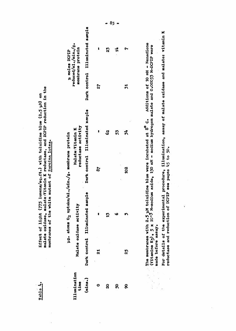
- 85 -
i!lA +» # OM 3 w *0 0$ L
3 5•HS 1 <e
9) *»0 - 34J <11 fH
5l g»| £ 4*«H « <Ha* cill«H « O tf\ ^S ’S
,4» * A° 9 Ü•H « <Hr4 *0 O«H *S « 0 0 04# O 8
all34 a 6
I
ft'glmW *4III« 8 M
o!IIo”
*s
CB
i?TJ1«
I
I«■gI•Hr-tf-t
1IIQ*â
g -g4» 3> O M9 9
"% O• 3 5% P
gIIA
1I
I
8
NCM (A
S A
& 8
I KN VO IA
44 tr\CM CM
hUPIA O 3AW O
•H 4)«CJ 441IO%1fi A1 1 44 «H O "O"§ 8c I
♦H
gRII•HS s3 1II• o
CM T4
p s ,II
fi*rt
O
%*3a
'd
9"SS«HO
s*0)
I•H
1•HRO
+>-4 lA"H -d<I*oIIII«HO
g
g
o mo4»
, 4» OÆ 2 «j H w a
•oI 9r-H■êH
I4>d 9e B ° 8 R g

— 66 *
A
A
H I|li4 10 SM
ts01MM
Ia
%
Oa
A
01
s iB 4»44>
IIo
38
*d01
O M0 - 9S gg1 i t
2IIOA
I•H
OVVO eo8
T4CO R
I 44 CO
4> 0 O 44O 0 %a <wM S 0
0»
t3•ao0"S1 I CO MCM CM
CM
Q §
ml
•ik*
II Is a
M
° 8 R 8 a s l j

— 87 ""
Table 6* Effect of illumination (775 lumens/sq*ft*) for 120 minutes with 2*5 pi»! toluidine blue on the malate : vitamin K roductu.se activity of membranes of the pigmented and white strain of S. lutea.
Illumination time Malate s vitamin K reductase activity(mins*) pg.atoms 0^ uptake /ml./min./mg* membrane protein
PlCTjeiitod strain Wliito mutantbark Illuminated Darkcontrol sample control
Illuminatedsample
0153045GO7590105120
0*396
0.27 9
G *252 0*255 0,255 0*239 0.255 0.231 G.191 0.207 0*473
0.4420*3060.3050.3310,3050.2900.1830,183
The membranes with 2,5 toluidine blue were illuminated at 8®C*Prior to assay, additions of 10 mM menadione (vitmnin £«) >
—35 X 10 M-NoN^, and 150 bM sodium hydrogen malate were made.For details of illumination, experimental procedure and assay of malate % vitamin K reductase activity, see pages 45 to 49,

«* 83 *»
vitamin K, which in tarn reflected the depressed malate t vitamin K reductase activity in both the pigmented and non-pigmented membranes. Thus a possible second site of inactivation of the membrane enzymes due to illumination with toluidine blue could not be suggested#
14, Effect of illiioination for 90 minutes with 2.5 pM toluidine blue on the malate dehydrogemse and malate i vitamin K reductase activities of the membranes of pigmented and the white mutant of S# lutea.
Prolonged illumination in the presence of toluidine blue caused damage to the malate ; vitamin K reductase activities in both the pigmented and the nonr-pigîiîented membranes of S. lutea. Initially, malate dehydrogenase passes electrons to vitamin K reductase, which in turn reduces the cytochromes. Therefore the effect of prolonged illan)ination with toluidine blue on the malate dehydrogenase and the malate : vitamin K reductase activities of the membranes of pigmented and non-pigracnted S. lutea wore compared#
Four experiments were performed and the results of one of the experiments are shown in Tables 7 and 8. The malate dehydrogenase activities, assayed by reduction of phenazine meüîosulphate (PMS), and malate t vitamin K reductase activities, were depressed to the some extent due to prolonged irradiation in the presence of toluidine blue in both the pigmented and the white mutant of S. lutea#
Thus the results of these experiments suggest only one place in the vitamin K reductase system which is affected by illumination with toluidine blue, namely the flavoprotein.
15, Summary
1* Illumination of whole cells of pigmented and non^pigmented

•* 80 ""
Table 7* Effect of illumination for 00 minutes (775 lumens/sq.ft.)with 2.5 ;a?J toluidine blue on malate dehydrogenase and malate s vitamin K reductase activities of the membranes of pigmented S. lutea.
Illumination time ;ug,atoms 0^ uptake /ml./min./mg. membrane protein(mins. ) Malate dehydrogenase
activityDarkcontrol
Illuminatedsample
Malate t vitamin K reductase activityDark Illuminatedcontrol sample
0153045007500
0.151 0.454
0.151
0.1080.0070.0300.0G50.0430.033 0.407
0.4320,3390,3030.2430.1730.167
The membranes with 2.5 pM toluidine blue were illuminated at 8 C* O.OOOM-phenazinc methosulphate (PMS), 5 x lO^^M-NoÜ , 150 mM-malate and 10 mM menadione (vitamin K^) were added prior to the assay of enzyme activities. For details of illumination, experimental procedure, and assay of malate dehydrogenase and malate t vitamin % reductase activities see pages 45 to 49.

— 90 —
Table 8. Effect of illui^inatlon for 00 minutes (775 lumens/sq.ft.)with 2*5 ;alî toluidine blue on malate dehydrogenase and malate t vitomin K reductase activities of the membranes of white mutant of S. lutea.
Illuî^ination time (oias*)
pg.atoms 0, uptake/ml,/min,/mg. membrane proteinMalate dehydrogenase activity
Darkcontrol
Illuminatedsample
Malate s vitamin K reductase activity
Dark Illuiainatedcontrol sample
0153045GO7500
0.233 0.840
0.259
0.1510.1240.0730.0390.0090.031 0.622
0.5750.4970.3110.2490.1370.109
The membranes with 2.5 pH-i toluidine blue were illuminated at 8 C. O.OOC'J-PMS, 5 X 150 mM malate and 10 mM menadione(vitamin K^) were added prior to the assay of enzyme activities. Details of experimental procedures, illumination, and assay of malate dehydrogenase and malate t vitamin K reductase activities appear on pages 45 to 49.

- 01 -
Sarcina lutea at low light intensities in the presence of an exogenous photosonsitizer, toluidine blue, sîiowed decreased viability in the carotenoidless wiiite mutant, while the pigmented strain escaped nhotodynamic killing.ii. ïïhen the cell membranes were irradiated at low light intensities in the presence of a photosensitizer, toluidine blue, there was widespread inactivation of the enzyme activities of the membrane—bound respiratory chain. The malate oxidase activities of cell suspensions and membranes of both the pigmented and non—pigmented strains were depressed. The depression was more in the carotenoidless mutant than in the pigmented strain.iii# The succinoxidase, lactate oxidase and ascorbate oxidase activities were also affected in the same manner as above.iv. In the study of the electron transport chain of isolated membranes, it was found that visible light with toluidine blue caused damage to the early part of the electron transport chain — the flavoproteins. Both methylene blue reduction and vitamin K reduction showed a depressed rate in both strains, which was more in the carotenoidless mutant than in the wild type.V . The rate of reduction of dichloropheaol indophenol was also depressed by light with toluidine blue but the inactivation appeared to be greater.V i . Cytochrome c oxidase and ascorbate-TMPD cytochrome oxidase activities were also similarly affected, suggesting a further site at idle cytochrome c—cytochrome oxidase level which is affected by visible light.vii. Illumination of shorter duration (10 minutes) with toluidine blue, followed by dark incubation, in the white mutant membranes caused no damage to the malate oxidase activity as compared with the dark control. 20 minutes* illumination caused slight damage which recovered almost completely after dark incubation. Illumination of more than 60 minutes produced permanent damage, rendering the oxidase activity completely irreversible.

— 02 •»
CHAPTER 4
Expérimenta without an exogenous nhotosensitlzer
1# Introduction.
Results of the experiments described so far suggest/^d several places in the electron transport chain of S. lutea which were affected by illumination in the presence of the exogenous photo- sensitizer, toluidine blue. The first inactivation of the respiratory chain was at the flavoprotein level, the second was between the flavoprotein and cytochrome c, and the third in the cytochrome oxidase region. In these experiments it was evident that the damage by light to the enzyme activities was complicated by the presence of toluidine blue, Toluidine blue at any of the concentrations used did not affect the oxidation rate unless it was subjected to illumination. It is not known with absolute certainty the precise mode of action of toluidine blue, but it was evident from the results so far obtained that toluidine blue caused widespread damage to a variety of enzymes, possibly 1^ attacking the histidine residues of the proteins or some other amino acid residues in the membrane. Besides, there are pigments in the mfônbranes such as flavins, cytochromes etc., which could also act as endogenous photosensitizers in the presence of visible light, and could produce photooxidative damage. It was, therefore, decided to omit toluidine blue from further experiments, and to use a much higher light intensity of the order of 1000 to 1500 lumens/sq.ft. All experiments involving the assay of 0^ uptakedescribed in this chapter were performed with an oxygen electrode.
2. Effect of high light intensity on the malate oxidase activity and the activities of segments of the reaoiratorr chain in nicmented aiwl non-nig^ented membranes of 8. lutea*

— 93 ""
Four expérimenta were performed to esaaine the effect of high light intensity, without the photosensitizer toluidino blue, on the nalato oxidase activities of membranes of the pigmented and non— pigmented strains of S. lutea. Results of one of the experiments are represented by Table 9,
Illumination for 60 minutes at higher light intensities without toluidine blue caused damage to the malate oxidase system (evidenced by depressed oxidation rate of malate) in both the pigmented and the white mutant of S» lutea. The depression was more in the white mutant than in the pigmented strain. This suggested that, in the absence of the exogewus photo sensitizer toluidine blue, some photooxidation took place due to the presence of endogenous photosensitizers which acted at higher light intensities. The carotenoid pigments offered some protection against photo— Inactivation in the pigmented membranes which was lacking in the carotenoidless membranes.
Having found that the malate oxidase activity could be damaged by illumination at high light intensities in the absence of an exogenous photosensitizer, toluidine blue, it was decided to examine which segments of the electron transport system of S. lutea could be damaged at higher light intensities without any exogenous photo- sensitizer* Comparative studies were also made to observe whether the effect of illumination on the succinoxidase system differed from that on malate oxidase*
Four experiments were performed to compare the effect of high light intensities (1240 lumens/sq.ft.) without toluidine blue on the malate % vitamin K. reductase activity, succinate i vitamin K reductase activity, and malate and succinate oxidase activities of membranes of the white mutant of S. lutea. Table 10 represents the results of one of the experiments.
The malate % vitamin Z reductase and succinate t vitamin £

— 94 —
Table 9. Effect of high light intensity (1240 Inaens/sq.ft.)without toluidine blue on the rmlate oxidase activity of the membranes of pigmented and white strains of S* lutea*
Illuiaination time Malate oxidase activity(mina*) pg*atoms 0^ uptake/ml./aln./mg. membrane protein
Pigmented strain white mutantDark Illuminated Dark Illuminatedcontrol sample control sample
0 0.080 - 0.07615 0.067 0.04600 0.047 0.03145 0.033 C.02360 0.073 0.023 0.075 0.021
The membranes were illuminated at 8 C and, prior to assay of the malate oxidase activity, 150 mM malate was added to the membranes. For experimental details, illumination, and malate oxidase assay see pagea 45 to 43.

- 95 -
Table 10. Effect of high light Intensity (1240 Iubgbs/sq.ft.)without toluidine blue on the malate : vitamin £ reductase and succinate ; vitamin £ reductase activities, and malate and succinate oxidase activities of membranes of the white mutant of S* lutea.
Illumination time pg.atoms 0^ uptake/ml./mln./mg. membrane protein(, i.is,) Malate : vitamin £ Succinate ; vitamin K
reductase activity reductase activityDarkcontrol
Illuminated smapl ©
Darkcontrol
Illuminatedsample
01530456090
0.039
0.203
0.1620.1620.1620.175
0.039
0.0870.0850.0780.070
pg.atoms 0^ uptake/ml./mii,v./mg. membrane proteinMalate oxidase activity Succinate oxidase activity
Dark Illuminated Dark Illuminatedcontrol sample control sannle
70 0.083 o.oi; 0.057 0.012
Membranes were illuminated at 8 C and additions of 10 mM menadione (vitmain Kg), 5 x lO"* M-NaHg, 150 mU~oalate and 150 mM-succinate were made prior to assay. For details of experimental procedure, Illumination and assay of malate and succinate : vitamin K reduction activities, malate and succinate oxidase activities, see pages 45 to 49.

03 -
reductase activities were depressed by illumination, and the malate and succinate oxidase activities were also depressed. The malate oxidase system seemed to bo more active than the succinate oxidase system. Those results were similar to those obtained in the presence of an exogenous photosensitizcr, toluidine blue. Therefore, in the absence of any exogenous pliotosensitizer, endogenous pigments appeared to act as photosensitizers in the membranes at higher light intensities.
The effects of high light intensities (1240 lumens/sq.ft.) on DCPIP reduction by the membranes of pigmented and wîiite S. lutea are shown in Table 11. A depressed rate of reduction of DCPIP caused by prolonged illumination of up to 120 minutes was observed in both the pigmented and non—pigmented strains, and the extent of damage to both seemed to be equal in most experiments. These results suggest some damage to the malate-cytochrome c reductase activity of the respiratory chain.
It appears, from the results of the experiments done so far without any exogenous photosensitizer, that various partial reactions of the electron transport chain are damaged by high light intensities. It is, however, not known what causes this damage, nor whether, as in the toluidine blue system (see page 71 ), reversible changes precede irreversible changes brought about by illumination at high light intensities. Therefore,it was decided to perform some ^periments at high light intensities without any photosensitizer for a variable duration of time, to test for the reversibility of various enzyme activities.
3# Effect of g short exposure to illumination at high light intensity on the malate oxidase and succinate oxidase activities, and activities of segments of the respiratory chain, in pigmented and noD—pigmented membranes of S. lutea.
Four experiments were performed to see the effect of short

— 97 —
Table 11. Effect of high light intensity (1240 lumens/sq.ft.)without toluidine blue on DCPIP reduction by the membranes of pigmented and white strains of S. lutea.
Illumination time p, moles DCPIP reduced/ml./min./mg.membrane protein (mine.) Pigmented strain
Darkcontrol
Illuminatedsample
liiliitc mutantDarkcontrol
Illuminatedsample
0,067 0.05415GO43GO7590105120 0.005
.060
.055
.047
.047
.047
.043
.037
.030 0.057
0.0430.0460.0370.0360.0370.0370.0330.025
The membranes were illuminated at 8^0, and C,00053M-DCPlf,5 3C 10"^M NaNg and 150 mM-malate wore added prior to assay.For details of experimental procedures, illumination and assays of enzyme activity see page 60.

— 93 —
exposure to illumination at high light intensity (050 lumens/sq.ft.) on malate oxidase activity and on BCPIP redaction by the membranes of pif^entod S. lutea. The results of one esperiscnt, represented by Table 12, showed that exposure to illumination for 5 minutes caused slight depression of the enzyme activities, which reversed completely daring the following 45 minutes* incubation in the dark at 8^# There is no apparent difference between the effects on oxidase activity and on DCPIP reduction.
lilion these experiments were repeated using non-pigmeated membranes (see Table 13), similar results were obtained* However, the reversibility of malate oxidase activity and the reduction of DCPiP wore not complete even after one hour of dark incubation.
Four experiments were performed to compare the effects of short exposures (5 minutes) to illmnlnation at high light intensities on malate oxidase activity and DCPIP reduction by membranes of pigmented S. lutea* The results of one experiment are shown in Table 14*
The results showed that exposures to illumination for 5 minutes caused depressed malate oxidase activity, which reversed completely during the following 75 minutes* incubation in the dark at 8^*The malate t DCPI? and succinate t DCPIP reductions also showed similar effects, and there was no apparent difference between the oxidase activities and the DCPIP reduction activities. The recovery time was slightly longer than la the previous experiment because of the higher light intensity used, which caused slightly more damage to the system.
The effects of 5 minutes* Illumination on the malate and succinate oxidase activities and on DCPIP reduction by the membranes of non-pigmented S. lutea are shown in Table 15. The results showed similar depressions in all the enzyme activities assayed due

- 99 -
Table 12. Effect of 5 mlns. exposure to illumination at highintensity (950 lumens/sq# ft.) on tho malate oxidase activity and DCPIP reduction by membranea of pigmented S. lutea.
Time(rains.)
Malate oxidase activity;ig.atoms 0^ uptake/ml./min./ rag. membrane proteinDarkcontrol
Illuminatedsample
DCPIP reductionp moles DCPIP reduced/ml./min./ rag. mejnbrano proteinDarkcontrol
Illuminatedsample
05153045607590
0*106 0*070
0,106
0.0900.1010.1010*1060.1060.1060.106 0,067
0.0020.0620.0670.0700.0700.0630.067
The membranes were irradiated at S^C by ligîit (950 lumens/sq. ft.) for 5 minutes and then kept in the dark away from the light source by covering with blackened aluminium foil. Additions of 150 raM- malate, 0.00053M-BCPIP and 5 x 10""^M-NaN^, were made prior to assay. For further details of experimental procedure, illumination and assays of malate oxidase and DCPIP reduction see page s 43 to 50»

- 100 -
Table 13. Effect of 5 ctinutes* exposure to illu’ination at high iîîtewsity (1240 luoens/sq* ft#) on the malate oxidase activity and DCPIP reduction by membranes of the white outcmt of S. lutea.
Tirae Malate oxidase activity(uins.) 0, uptake/ml#/min./
mg. membrane proteinBarkcontrol
Illuminatedsample
BCPI? reduction;i moles DC?IP reduced/ml./lain./ mg. membrane proteinDarkcontrol
Illuminatedsample
0515004560
0.117 0.070
0.123
0*1020.0970.0970.1170.117 0.030
0.0690*0760.0760.0730.073
The membranes were irradiated at 8^C by light (1240 lumens/sq.ft.) for 5 minutes and then kept away from light source covered by blackened aluminiimD foil. Additions of 150 mH-malate, Ô.G0053M— DC?IP and 5 x 10"" M NoN^ were made prior to assay. For details of experimental procedures, illumination and assay of palate oxidase and DCPIP reduction, see pages 45 to 50.

- 101 -
Table 14. Effect of 5 minutes* illumination at high light intensity (1240 Imaens/sq.ft.) on palate oxidase activity and DCPIP reduction by membranes of pigmented S, lutea*
Tirao Halate oxidase activity(t.ina) 0,, uptake/ml./min./
mg. membrane protein
05153045CO7390
Darîccontrol0.130
0.134
Illuminatedsaiimle
0.1700.1770.1730.170 0.177 0,187 0.134
Halate t DCPIP reductionji moles DCPIP reduced/ml./min./ mg. membrane proteinDarkcontrol0.143
0.1G7
Illuminatedsample
0.127 0.143 0.143 0.139 0.139 0.167 0 167
Time(mins.)
0515GO45GO7590
Succinate t DCPIP reduction p. moles DCPIP reducod/ml./min./mg. membrane protein Dark control Irradiated sample0.003
0.0500.0570.0590.0G30.0000.000
0,003 0.063
The membranes were irradiated at 8^C by light (1240 1-omens/sq.ft) for 5 minutes and then kept away from light source covered by blackened almuinium foil. Additions of 150mM mal ate and ISOnjlî- succinate, 0.0005oJ-DCPIP and 5 x 10"*^H-NaNg, wore made prior to assay. For details of experimental procedures, illumination and assays of enzyme activities see pages 45 to SO,

102
Table 15. Effect of 5 minutes* illumination at liigb light intensity (1240 lumens/sq.ft.) on malate and succinate oxidase activities, and DCPIP reduction, Isy membranes of the white mutant of S. lutea.
Time(mins.)
05153045GOGO
Maiate oxidase activitypg.atoms Of, uptake/ml./min./ pg. fijonibroiio proteinDarkcontrol0.176
0.220
Illuminatedsamnle
0.1670.1570.1830.1730.1730.133
Malate ; DCPIP reductionp moles DCPIP reduced/ml./min./ m]. membrane proteinDark 111uainatcdcontrol sam%)le0.107
0.0950.0950.0970.1050.107
0.126 0.108
Tirao(oins.)
05
1580456090
Succinate oxidase activitypg.atoms 0, uptake/ml./min./ rag. membrane proteinDarkcontrol
0.057
Illuminatedsample
0.05:
Succinate : DCPIP reductionp moles DCP.ÏP reduced/ml ./min./ mg. membrane proteinDarkcontrol
0.052
0.070 0.053 0.052
Illuninatedsample
0.047 0.052 0.044 0.044 0.044 0.042
The membrane8 were irradiated at 8®C by light (1240 luoens/sq.ft.) for 5 minutes and then kept away from light source covered by blackened almninium foil. Additions of 150raM-malate ojîd 150aM- succinate, 0.0005oM-DC?I? and 5 x 10*"% NoJf were made prior to assay. For details of experimental procedures, illumination and assays of malate and succinate oxidase activities and reduction of DCPIP see page®45 to 50*

103
to illoiîiination for 5 minutes. However, the reversibility of nalate and succinate oxidase activities, and of DCPIP reduction, were not co npTete even after 90 minutes of dark incubation#
Vïîien the suecinoxidase and malate oxidase activities were compared using pigmented membranes of S. lutea after illuminating at high light intensities for 5 minutes, there were depressed oxidase activities with both malate and sucèinate (see Table IG), which reversed completely after dark incubation.
The effects of short exposure to illumination at high light intensities on the malate oxidase and malate : vitamin K reductase activities by the membranes of pigmented S. lutea are shown in Table 17# Both the malate oxidase and malate : vitamin K reductase activities were depressed by illumination of short duration, end the activities were completely restored during the following 75 minutes* incubation in the dark at 8®*
The results of all experiments done so far without any exogenous photosensitizer showed that short illumination for 5 minutes at high light intensities caused some damge to several enzyme activities, such as malate oxidase, succinate oxidase, DCPIP reduction and malate s vitamin K reductase activity, in pigmented and non-pigmented S. lutea. The inactivation of enzyme activities, which was caused by illumination of the pigmented membranes, was slight and all reversed completely when incubated in the dark for periods which varied according to the intensities of light* The higher the light intensity, the longer was the time required to achieve complete restoration of activities* la the case of the carotenoidless mutants, the depressions in ensyr’e activities were slightly more in comparison with the pigmented strain, end a complete restoration of enzyme activities could not be achieved. These results suggested that the onset of the damaging effect was fairly rapid, but permanent inactivation was not achieved during

- 104
Table 16. Effect of 5 minutes* illumination at high lightintensity (1240 lumens/sq.ft.) on malate oxidase and succinate oxidase activities of membranes of pigmented S. Intea.
Time Malate oxidase activity(mins.) 0^ uptake/ml#/min./
mg. moinbrano proteinBarkcontrol
Illuminatedsampl e
Succinate oxidase activity;xg.atoras 0^ uptake/ml./ndn./ mg. membrane proteinBarkcontrol
Illuminatedsample
0515253045007590105135150165
0.167 0.003
0.162
0.152
0.1520.1520.1570.1570.157
0.1540.1620.162
0.001
0.064
0.066 0.066
The membranes were irradiated at 8^C by light (1240 lumens/sq.ft.) for 5 minutes and then kept away from the light source, covered by blackened aluminiuin foil. Additions of 150mM malate and 150mM succinate were made prior to assay. For details of experimental procedures, illumination and assay of enzyme activities, see pages 45 to 43*

« 105 -
Table 17. Effect of 5 minutes* illumination at high lightintensities (1240 lumens/sq.ft.) on malate oxidase and malate j vitamin K reductase activities by tlie membranes of nigmonted S, lutea.
Time(nins.)
Jig.atoms 0^ uptalce/ml./min./mg. membrane proteinMalate oxidase activity Malate : vitamin K reductase
activityDarkcontrol
Illuminatedsample
Darkcontrol
Illuminatedsample
0 0.211 0.3415 0.193 0.32015 0.106 0.82030 0.206 0.82045 0.206 0.33060 0.209 0.32375 0.211 0.34190 0.211 0.211 0.341 0.341
The membranes were irradiated at 8^C by light (1240 lumens/sq,ft*) for 5 minutes, after which they were kept in the dark away from the light source by covering with blackened almninium foil. Additions of 150mM malate, lOralî menadione (vitamin K, ) and 5 x 10 M NaN^ were made prior to enzyme assays. For details of experimental procedures, illumination and assay of enzyme activities, see ptiges 45 to 49.

- 106 -
that short period* Therefore furtlier experiments were performed to see the effects of a longer duration of illumination at high light intensities.
4* Effect of 15 rainutes* illumination at high light intensities on the oo.late and succinate oxidase activities and the activities ofscf^pcnts of the rosnir&tory Chain in nigirsonted and non-pigtqentedmoobranos of S. lutea.
Four experiments were performed to see the effect of 15 minutes* illumination at high light intensity (1240 lumens/sq.ft.) on the malate and succinate oxidase activities and on DCPIP reduction ly tlie pigmented meinbranoa of S. lutea. Table 13 represents the results of one experiment.
The results showed depressed malate and succinate oxidase activities, but these did not recover during dark incubation for the following SO minutes. The succinoxidase activity seemed to be loss affected than the malate oxidase activity. The DCPI? reduction was also depressed }jj illumination, but the extent of inactivation seemed to be less than in the malate and succinate oxidase systeriîs. There was complete restoration of DCPIP reduction activity daring the following 90 minutes of incubation in the dark at S^, This suggested the possibility of some irreversible damage to the cytochrome oxidase system if it was assuined tliat DCPIP received its electrons from cytochrome c.
i/hen these experiments were repeated using noîv-pigmented membranes (see Table 19), similar results were obtained. The magnitude of inactivation was more in tlie non-pigmented membranes than in the pigmented membranes, end the malate oxidase system was more affected than the succinate oxidase system. Co&splete restoration of activities was not achieved in any case, and damage to the DCPIP-reducing system seemed to be less than to tlie malate and succinate oxidase systems.

- 107 -
Table 13. Effect of 15 minutes* illumination at high lightintensity (1240 lumens/sq.ft.) on malate and succinate oxidase activities, and DCPIP redaction, by membroJios of pigmented S. lutea.
Time(mins.)
Jig.atoms 0^ uptake/ml./min,/mg. membrane proteinMalate oxidase activity Succinate oxidase activity
Darkcontrol
Illuminatedsarapl 0
Darkcontrol
Illuminatedsample
0 0.161 0.04715 0,130 0.045GO 0.111 0.03945 0.136 0.022GO 0.124 0.02275 0.124 0.04500 0.143 0.124 0.052 0.045
.molies.
p moles DCPIP reduced/ml Maiat© I DCPIP reduction
./min./mg. membrane protein Succinate î DCPIP reducti
Darkcontrol
Illuminatedsample
Darkcontrol
Illuminatedsample
0 0.134 0.05913 0.121 0.05200 0.123 0.05045 0.121 0.050GO 0 .143 0.05975 0.144 0.003GO Û.1G3 0.133 0.0G3 0.003
The D^jbranes were irradiated at S^C by light (1240 lisnens/sq.ft.) for 15 minutes, after which they were kept in the dark away from the light source by covering with blackened aluminium foil. Additions of 150i,3iJ malate, 150dM succinate, 0.00053M-IÏCPIP and 5 x lO^'^M-NaNg were made prior to assay. For details of experimental procedures, illumination and assay of enzyme activities see page g 45 to 50*

- 103 -
Table 10. Effect of 15 minutes* illumination at high lightintensity (1243 lumens/sq.ft*) on malato and succinate oxidase activities, and DCFI? reduction, by membranes of the white mutant of 3, lutea.
Time(mine.)
pg.atoms 0 , uptake/inl./min./mg. membrane protein
0153045607590
Darkcontrol0.118
Malate oxidase activityIlluminated sample
0.118
0.0730.0630.0710.0550.0630.063
Succinate oxidase activityBark Illuminatedcontrol sample0.051
0.055
0.0470.0400.0470.0510.0510.050
Time(mins.)
pi moles DCPIP reduced/ml./min./mg, membrane proteinMalate t DCPIP reduction
0153045607590
Darkcontrol
0.264
Illuminatedsamnle
0.1740.1430.1350*1950.2330.105
Succinate s DCPiP reductionDarkcontrol0.037
0.100
Illuminatedsoiimle
0.0790.0370.0900.0900.0900.090
The membranes were irradiated at 8®C by light (1240 lujTseas/sq.ft.) for 15 minutes, after which they were kept in the dark away from the light source by covering with blackened aluminium foil. Additions of 150m?!-malate, ISOoM-succinate, 0.00053M-BCFIP and 5 x 10**%-HaNg were made prior to assay. For details of experimental procedures, illumination and assay of enzyme activities see pages 45 to 50.

— X09
The effects of 15 minutes* illumination at 1240 lumens/sq.ft. on the malate oxidase and malate s vitamin K reductase activities of pigmented membranes are shown in Table 20.
The malate oxidase activity was depressed by illumination, and complete restoration of activity did not occur even after 105 minutes* incubation in the dark following illamination. There wasalso damage to the vitamin K reductase activity, but complete restoration of activity was achieved during the following 90 minutes* incubation in the dark at 8^#
Table 21 represents one of four experiments performed to observe the effect of 15 minutes* illumination by light (1240 lumens/ sq.ft.) on the succinate oxidase and succinate : vitamin K reductase activities by pigmented membranes.
The succinoxidase activity and the succinate : vitamin K reductase activity were both depressed by illumination, and complete restoration of succinate : vitamin K reductase activity was achieved following 90 minutes* incubation in the dark at 3^. However, the succiaoxidase activity did not reverse completely.
Similar experiments were performed to compare the malate oxidase, malate : vitamin K reductase and succinate : vitamin K reductase activities in the non-pigmented membranes of S. lutea.The results showed (see Table 22) depressed malate oxidase activity, which did not show complete restoration after 90 minutes* incubation in the dark following illumination. However, the malate t vitamin H reductase and succinate : vitamin K reductase activities were also depressed, but complete restoration of the activities was achieved after 90 minutes* incubation in the dark following illumination.
Therefore, the results of experiments done so far to find the effects of short illumination of 5 Eîimites showed that the malate oxidase activity of the pigmented membranes of S. lutea was depressed,

— lie —
Table 20, Effect of 15 minutes* Illumination at high lightIntensity (1240 lumens/sq.ft.) on the malate oxidase and malate s vitamin S reductase activities of the membranes of pijjmented S, lutea.
Time(mins.)
Jig.atom8 0^ uptake/ml./min./mg. membrane protein Malate oxidase activity
Porkcontrol
0 0.05415 30 45 60 75 00105 0.051
Illuminatedsaonl©
0.0290,00.026 0.032 0.034 0.034 0 .034
Malate j vitamin K reductase activityBarkcontrol0.032
0.056
Illuminatedsample
0.061 0.056 0.051 0.051 0.051 0 .056 0.056
The membranes were irradiated at 8^C by light (1240 livaens/sq.ft.) for 15 minutes, after which they were kept away from the light source covered 1^ blackened aluminium foil. Additions of ISOr M— malate, lOmll-menaclione (vitemin K^) and 5 x 10""%-3ZoNg were made prior to assay. For details of experimental procedures, illumination and assay of enzyme activities see pag% 45 to 49.

- Ill -
Table 21. Effect of 15 minutes* illumination at high lightintensity (1240 lumens/sq.ft.) oa succinate oxidase and succinate % vitamin K reductase activities of the membranes of pigmented S. lutea.
Time (mins. )
0153045607590
pg.atoms Oj, uptake/ml./min./mg. membrane protein Succinate oxidase activity
BaricControl0.C15
0.014
Illuminatedstunpl e
0.0130.0130,0130.0140.0140.014
Succinate : vitamin % reductase activity
Darkcontrol0.034
0.031
Illuminatedsaianle
0.0290.0240.0240.0290.0290.031
The membranes were irradiated at 3^0 by light (1240 lumens/sq.ft.) for 15 minutes, after which t2iey were kept away from the light source covered by blackened aluminium foil. Additions of 15ômlî- succinate, lOmM-menadione and 5 x 10"" were made prior toassay. For details of experimental procedures, illumination and assay of enzyme activities see pagea 45 to 40,

— 112 »
5 51 5•P P
C «^ 3d 4»
|S
A
?1
“ I«M .H8 -PJ a ? «
&
11
fî C9 ÇN g 3ifS ifS îA s5 O No o o o o o• « * • • •o o o o o o
I
s â s s so o o o o ^• « • • • •o o o o o o
A n@ 1-4•HO # 04»k 0 e0 4» "d
x - a« B fet
55 8 5*52 m% <s •
44 d ok 0 -ü*0 04» M 4* 0^ 1 k
4*•H O lA
<► 4» •H -3*» «H k 8)0 g A m4» "d Ç)3 < 4) dc P A•H dG S d# d
lA M « mt4 *H k0 dk (H > %P _ d
o <P*s s*3 *«X usc Tt
IaeA
•d ►»
0 *p5 a1
■sI#4gItA
I I
I SrO
-gfr*
III I4* wl - s S ï

- 113 -
and complete restoration took place after 45 minutes of dark incubation* DCPIP redaction was also depressed, and complete restoration of activity was achieved after 45 minutes of dark incubation (Table 12). Similar experiments on noa-pigmeated membranes showed loss of malate oxidase activity ©aid DCPIP reduction activity, but there traa no complete restoration of activity even after one hour of dark incubation (Table 18). Succinoxidase activities showed the same effects of illumination as did malate oxidase. Malate oxidase activity and malate t vitamin K reductase activity showed depression in pigmented membranes, which recovered completely after 75 minutes of incubation in the dark. These results suggested the possibility of two damaging effects of light OH the respiratory system of S. luteat one at the beginning of the respiratory systom, and the other at the cytochrome oxidase system.
kilien the illuiaination time was prolonged to 15 minutes there was damage to tlie malate and succinate oxidase systems in pigmented membranes, which did not reverse completely (Table 18). The DCPIP reduction activities, however, reversed after 60 minutes of incubation in the dark, suggesting that there was an irreversible inactivation outside the region between malate and the site of DCPIP reduction.The sawe experiment on non-pig:i?ented membranes showed more damage to the malate oxidase, succinate oxidase, malate % DCPIP reduction and succinate t DCPIP reduction activities (Table 19), but all of them were damaged irreversibly, Malate s vitamin K reductase and succinate ; vitamin K reductase activities were also depressed in both the pigmented and non-pigiaented membranes, but there was complete restoration of activities after 90 minutes* incubation in the dark (Tables 20, 21 and 22), suggesting that inactivation of vitamin £ reductase activities was completely restored later.This suggested the possibility that the initial damaging effect on the respiratory chain, possibly the flavoprotein, was reversible even in non-pigmented membranes.

— 114 —
la view of tlie several possible sites for types of light damage oa the respiratory chain of the bacterioa S» lotea< at least one in the flavoprotein region and possibly one in the cytochrome oxidase region, it was thought possible that the various sites of photodynamic action might bo inactivated by different wave lengths of visible light. It was therefore decided to filter out certain wave lengths from tungsten light by using colour filter glasses, and experiments were planned to observe the effects of filtered light on the enzyme activities of S. lutea. As the light intensities were diminished due to insertion of colour filter glasses, the duration of illumination was increased from 15 minutes to 25 minutes.
5. Effect of 25 minutes* illumination with an OY 13 and OY 10 fiIter on malate oxidase activity, and on methylone blue reduction^ by pigmented membranes.
Four experiments were performed to observe the effect of 25 minutes’ illumination with an OY 13 yellow filter glass at high light intensity (142G lumens/sq.ft.) on malate oxidase activity and on redaction of methylene blue by the membranes of pigmented S. lutea. The results of one of the experiments are shown in Table 23.
The malate oxidase activity was depressed after 25 minutes’ illumination with the filtered light, but activity was completely restored in the following period of dark incubation. However, the malate t methylene blue oxidoreductase activity was not affected at all by the illumination.
The OY 13 light orthochromatic colour filter glass gives Oÿ transmission at 400 nm and a peak transmission (T5^) from 500 nm; it thus cuts off the ultraviolet and violet regions of white light from the tungsten lamp (so© page i6o)# Since the flavoproteins absorb light in üie near ultraviolet and blue regions of the spectrum, it was presumed that when the OY 13 filter was used, the flavoproteins would be unaffected. As the artificial electron acceptor methylene blue presumably accepts electrons from the flavo-* proteins, the malate : methylene blue oxidoreductase is not

- 115 -
Table 23. Effect of 25 minutes* illumination with an OY 13 colour filter glass at high light intensity (1248 lumens/sq.ft.) on malate oxidase activity and methylene blue reduction in the membranes of pigmented S. lutea.
Time jmg.atoms 0^ uptake/ml./min./n?g. membrane protein(mins.)
Malate oxidase activity Methylene blue reductionDark Illuminated Dark IlluiTîinatedcontrol somplo control ©ample
0 0.032 0.02925 0.013 0.02945 0.014GO 0.012T5 0.020GO 0.021105 0.013120 0.020135 0.021150 0.029105 0.033130 0.030ISO 0.029220 0.030 0.030
The membranes were irradiated at 20^C by light (1426 lumens/sq.ft.) with OY 13 colour glass filter. Before inserting the colour filter glass, the light intensity was measured by the light meter, and after 25 minutes* Illumination the membranes were kept in the dark away from the light source covered by blackened aluminium foil. Additions of lOOraMwaalate, 6.001^ methylene blue and 1 M-KCN (neutralized) were mad© prior to assay. For details of experimental procedures, illuminations end assay of enzyme activities see pageg 45 to 49.

•“ IIG **
inactivated by the OY 13 filtered light. Hence a second light reaction, sensitive to OY 13 filtered li«^t, is demonstrated in the assay of malate oxidase activity* This inactivation appears to be reversible*
VHien these experiments were performed with membranes from the white mutant (see Table 24), the inactivation, of malate oxidase was not completely reversible* Methylene blue reduction again did not show any light effect at all after 25 minutes of illumination*
Those results suggested that the flavoproteins in both the pigmented and non-pigmented membranes escaped the damaging effect of filtered light, and the early part of the electron transport chain was unaffected* The malate oxidase activity of the pigmented membranes sliowed reversible damage, while that of the non-pigmented membranes showed irreversible damage* Therefore, it could be assumed that the flavoproteins are damaged by tlie ultraviolet and violet regions of white light, while a second site beyond the flavoproteins is damaged by visible light from 500 im onwards.The carotenoid pigments appeared to be associated with complete recoveiy of the malate oxidase activity in the pigmented membranes, while there was irreversible inactivation of the malate oxidase activity in the mutant membranes which lacked carotenoids.
The effect of 25 minutes* illumination with OY 13 filtered light on DCPIP reduction was compared with tlie effect on malate oxidase activity in pigmented membranes, as shown in Table 25,
There was complete restoration of the malate oxidase activity after irradiation, but not of the DCPIP reduction, suggesting permanent damage to the cytochrome system by OY 13-filtered light.
By comparison, in the wiiito mutant membranes OY 13-filtered light produced Inactivation of the malate oxidase and the DCPIP reduction which was irreversible (see Table 20).

- 117 -
Table PA» Effect of 25 minutes* illumination with an OY 13 colour filter glass at high light intensity (1395 luiaens/eq.ft.) on malate oxidase activity and methylene blue reduction in membranes of the wliite mutant of S. lutea.
Time(mins.
02545GÜ7500105120105150165130190220
pg.atoms 0^ uptake/ml./min./mg, membrane proteinMalate oxidase activity
Barkcontrol0.032
0.030
Illuminatedsample
0.0130.0170.0150.0210.0190.0240.0230.0100.0230.0240.019
Ketîiylene blue reductionDarkcontrol0.033
Illuminatedsample
0.040
0.040 0.040
The mesjibranes were irradiated at 20^C by light (1335 lumens/sq.ft.) with the OY 13 colour glass filter. Before inserting the colour filter glass, the light intensity was measured by the light meter, and after 25 minutes* illuuination the membranes were kept in the dark away from the light source covered by blackened aluminium foil. Additions of ISOmM-malate, 0.00^^ methylene blue and 1 M-KCN (neutralized) were made prior to assay. For details of experimental procedures, illumination and assay of enzyme activities see pages 45 to 49.

113 —
Table 25» Effect of 25 minutes* illumination with an OY 13 colour filter glass at high light intensity (1395 lumens/sq.ft.) on malate oxidase activity and DCPIP reduction by the membranes of pigjiented S. lutea.
Time jig.atoms 0,, uptake/ml#/iain./ (mins.) tag. membrane protein
Malate oxidase activityDarkcontrol
0 0.187G254550GO7590100105120135150
0.137G
0.2067
0.137G
Illuminatedsample
0.1G450,1377
0.14920,16340.1539
0.17000.133G0.13700.1870
p moles DCPIP reduced/ml./ min./mg, membrane protein
Darkcontrol0.6314
DCPiP reductionIlluminatedsample
0.5904
0.7380
0.59040.01320.5904
The membranes were irradiated at 20^0 by light (1395 lumens/sq.ft.) with an OY 13 colour glass filter. Before inserting the colour filter glass the li^it intensity was measured by the light meter, and after 25 minutes* illumination the membranes were kept in the dark away from the light source covered by blackened almainiuia foil. Additions of IDOmM-taalate, 0.00053H-DCPIP, and 1 M-CCN (neutralized) were made prior to assay. For details of eîiperinieatal procedures, illumination and assay of enzyme activities see pag®s 45 to 50,

- 119
Table EG. Effect of 25 minutes* illuminatiou with an OY 13 colour filter glass at high light intensity (13G4 Imnens/sq.ft.) on malate oxidase activity and DCPI? reduction by membranes of the #iite mutant of S. lutea.
Time jig.atoms 0 uptake/ml./min,/(mins.) mg, membrane protein
Malate oxidase activityBark Illmniaatcdcontrol stuaple
02545GO7590105120135150165170130190
0.034
0,091
0.0710.009O.OG50.0670.0540.0650.0730.0710.0710.065
0,000
ji moles DCPIP reduced/ml./ min./mg, membrane protein
DCPIP reductionDarkcontrol0.130
Illuminatedsample
0.155
0.1620.032 0.201
The membranes were irradiated at 20®C by light (1364 lumens/sq.ft.) with the OY 13 colour filter glass. Before inserting the colour filter glass, the light intensity was measured by the light meter, and after 25 minutes* illumination the membranes were kept in the dark away from the light source covered by blackened aliminim? foil* Additions of ISOmM-rjalate, 0.00055M-DCPIP and 1 K-£CN were made prior to assay. For details of experimental procedures, illumination and assay of enzyme activities see pogeg 45 50

120 -
Therefore, the résulta of the above experiments showed that photoinactivation of the malate oxidase activity was completely reversible in the membranes of pigmented S. lutea after 25 minutes of illuïnination with OY 13 colour filter glass at high liglit intensities. In the non-pigmented mutant, inactivation of the □date oxidase activity was not reversible. Methylene blue reduction was completely unaffected by light in both pigmented and non-pigraentcd strains. The inactivation of DCPIP redaction was not reversible in either strain, suggesting some more permanent damage to the cytochrome system by the filtered light#
An OY 10 (haze) filter was available, giving 0^ transmission at 350 nm and peak transmission (90^) from 450 nm over tlie remainder of the visible spectrum (see page 160 )# In view of the success gained with the use of the OY 13 filter, it was thought worth while to test the effect of illumination with the OY 10 filter at high light intensities on malate and NADU oxidase activities, metiiylene blue reduction and DCPIP reduction by the membranes of pigmented S. lutog#
Four experiments were performed to test the effect of 25 minutes* illumination with the OY 10 filter at high light intensities on malate and NAD3 oxidase activities, methylene blue reduction and DCPIP reduction by the membranes of pigmented S. lutea. Tables 27a and 27b show the results of one of the experiments.
There was considerable damage by light with the OY 10 filter to all the enzyme activities assayed# Malate and NAIJîî oxidase, methylene blue reduction and DCPIP reduction all showed photo- inactivation in the pigmented strain. Thus, the flavoproteins were affected by light transmitted by this filter# As the DCPIP reduction showed irreversible inactivation, this suggested some damage beyond flavoproteins and up to the cytochrome c level in the electron transport chain of S. lutea.

• 121 —
0X p4i M•H x>c 00 C** 0a•H XÆ4> 4»xi OO) G•Hf4 a0x:a> 03•H sx: 3
a %0 0)•HOi4»a •Ha >iH•H0> 4»k 34» 0iH•HX •0L .3 0 d0 0fH K 4J0 9 30 < m4zo d*4 cg ♦HX 3 Üo Jg
0c 0 t/53 dJ3 h4 04> X 4>"H 0 C> 0
e04> 1)
o « •H•H «"4 A4» CO2 5 Xc 0•H 0Æ «4» 0iH
r4 c §*H 0 u
« 10 04» 4» s0 Xe 0•H O* j3s «lA 0 Xca G
X 00
04*iH 3
*>U lA P0 ON 3<M lA "0<H T4 0(4 k
«•**IIIJII
CD
•H>•H
i
*0*xoIss
n,
«c•H
§•HI§.Û MI MII *>
«A
X+»•H.5•P0T»10tjI
•g1•H
£IIQA
g4>
II
lA tA cn 00 09 o\O »A lA -d* «4* tAO O O O O • « • • • •o o o o o o
N SÙONo
T4
M lA r- tN. ts. !>•CO lA kO 0\ c\ 0\w cq 0Î C3 ca • • • • • •o o o o o o
o\3 SlA
T< lA t4 VO CJlA tA tA »A Cvi WO O O O O O• * • • • ♦O O O O O Q
01
§5 ~g § gS 4* S
M° R S S R R SV4 *4 *4

— 12B —
Table 27b, Effect of 23 minutes* illumination with an OY 10 colour glass filter at high light intensity (1393 lumens/sq.ft.) oa DCPIP reduction by the membranes of pigmented S. lutea
Time(mins.)
p moles DCPIP reduced/ml./min./mg. mcmbraao proteinMalate : DCPIP redaction NADU t DCPIP redaction
Darkcontrol
Illuminatedsaj:iiple
Darkcontrol
Iliumsampl
0 0.743 1.3925 0.027 1.0060 0.342 1.0490 0.342 1.15120 0.056 1.04150 0.372 1.04130 0.723 0.336 1.34 1.03
The rambranes were irradiated at 20®C by light (1395 lumens/sq.ft.) with an OY 10 colour filter glass. Before inserting the colour filter glass, tho light intensity was measured, and after 25 minutes* illumination the membranes were kept in tlie dark away from the light source covered by blackened aluminium foil. Additions of 150oM— malate, 0.C0053M-IiCPlP, 0.1/a NAKl and 1 H-KGN (neutralized) were made prior to assay. For details of experimental procedures, illumination ond assay of enzyme activities see pages 45 to 50*

— 123 —
The use of the OY 13 and OY 10 filters gave very interesting results with reactions involving the flavoproteins; and, in view of this, the effect of illumination with various other colour filter glasses filtering near ultraviolet and blue violet light was tested on methylene blue reduction ly the membranes of pigmented 8. lutea» As the light intensity was much higher than in the previous experiments (1433 lumens/sq.ft.), the illumination time after insertion of the colour filter glass was shortened to 15 minutes instead of 25 minutes#
6* Effect of illumination with various colour filter glasses on methylene blue reduction by nunabranes of pigr^ented S» lutea#
Four experiments were performed to observe the effect of 15 minutes* illumination with different colour filters at high li^t intensities on methylene blue reduction by membreues of pigmented S. lutea. The results of one experiment are shown in Table 28.The OGr 3 green and OB 14 colour filter glasses produced a light effect (evidenced by a depressed rate of methylene blue reduction) while the OY 13 light ortliochromatic, OS 1 ultraviolet and OV 1 purple colour filter glasses did not produce any light effect at all#
Tlie OX 1 ultraviolet colour filter gives O/a tran^ission at 230 nm and a peak transmission (T /û) at 350 nm. The CV 1 purple colour filter glass gives 0^ transmission at 340 na and a peak transmission (72?») between 370 and 330 nm (see page IGl), Light from these two filters did not produce any inactivation of methylene blue reduction, and consequently may be presumed to have no effect on the flavoproteins.
The 03 14 colour filter glass gives 0^ transmission at 270 nm, and the peak transmission (85/o) is between 350 and 400 nm# The OGr 3 green colour filter glass gives 0^ transmission at 400 xm and a peak transmission (7Sfo) between 500 and 550 nm (see page 100).The results suggested that 03 14 and OGr 3 filtered-light could produce some inactivation of flavoproteins, though the inactivation of the former was more than that of the latter# Other colour

- 124 -
s4»
•sa oG fH •H A
§ IIo
S 5o o
- # X 9 @ o oX 0 X X 0X 0 k XX X X ,n p lAc X X0 _ k . p VO vDX 0 O ^ k O VDc c O k X O OX X 0 CQ 0 o OX k UÆ fflCT c/3XX T» •30 0JS X XO C 0 « N XX 0 k c fH N Njü a 0 X A O O5 C X § GX X X X 9 % ô O0 A 0 X X 0X A Xa X 0 M0 0 k X0 A Xm 0 > , p vO vD0 0 X C 1
o 1 1 t OA 0 3 c • *U k Q 0 O ok Xi JP Q0 g CX 0 0X E X aX X *0X 0 o • 0Æ 3 o Xk X *0 6 0 0 X XS ^ 0 k C X r- N0 X k ■ • 0 X A o O
0'° 0 CX XX I § ô •Oo G 3 S X X 00 X \ k X0 X n ' • M3 X X X0 O 0 wfi XX 3 c X , P VD vOk tf 0 0 O X k V3 O0 0 X -3 k X O O> k X 0 « C m ■ •X Q 0 O oJS 0 X A . OX 3 0 3X X £> Xi cao •dC 0 00 c m XX 0 0 0 o VOX X k G X VD VO0 X X 0 X A O O.5 5 0 XfH § 1 «O O§ g •TO k X 0 XX A XX e #AX 0 X X, 0 vo VO0 0^ X X k VD VO0 • o k X O OX X « C #3 X Q 0 O oc • OX O's 0«A 0 *0X 2 00 XX s 0 3 k 0 0C fH R 5X 0 X A O oXÜ co XX § § •O o0 03 X X 0X .d* k XX X MM N-f X X I I
. g 10 *r1 «H Xa -H X Oflî X (H X « O U «o »Hh U a0 k X+> *H 0 .4 • naX 0
lio lAS u•H 0 X X" "3
& &
&0S
l•o X
IIsoXs
x: •OX TOX 0 O 0 0k •3 AX 3 kX 0 X a 0X 3 0» 0 •o 0 0a 0 kk 0 m0 0 > 0X 0 > XX > 0 X* o *0 Xc k 0 ►0 X 0 N XX u X Xs 0C ^ *3 ë0 0 o kX 0 X 0C 3 sX X X 0 kX C Nco X ra w Cco X X 0-j* TO X zX X u XX © X 0
X 1X 0 X k.3 0TO X a X 0X p 5X k *3 00 X Ck 0 3 *3X 0 k so 0 o c>8 k0 fH •H•d <w
f4 O*3 *o k "ff4 00 X
. I g_ X0 X
1 ®II= g0 Xë<S
M b 1 g'I•0 0 C
X X XC S X^ »X ca » A o 0!:itu «• uM l1 1 ^
M l 1 - 0 t

- 125 -
filters failed to demonstrate any damage to the flavoproteias# Therefore,it appears that the flavoproteins may be sensitive mainly to 050 — 400 nm, and slightly sensitive to beyond 400 nia of the visible spectrnm* It is possible that the total energy transmitted by OV 1 is inadequate to produce an effect on the flavoproteias.
Weber et al* (92) have studied the possible role of vitamin K in electron transport, and their data indicate that vitamin £ transfers electrons between and the cytochromes* It wastherefore decided to examine the NâB:i oxidase system of S* lutea.
T* Effect of 25 minutes* illumination with OY 13 colour filtered light on NADII oxidase activities and on NADÎI s BCI’I? reduction activities by membranes of pigmented and non-pigmented S, lutea*
Four experiments were performed to test the effect of 25 minutes* illuraination with OY 13 filtered light on NADII oxidase activity and on NADII t BCPIF reduction by the membranes of pigmented S. lutea. The results of one of the experiments are represented by Table 29. There was inactivation by illuniination of the NADII oxidase system (evidenced by depressed rate of NADII oxidation), but no inactivation of the NADII i DCPIP reduction activity* The damage to the NADU oxidase appeared to be permanent.
Similar results were obtained when the white membranes were examined for these enzyme activities (see Table SO).
Therefore, illumination with the OY 13 filter at high light intensities showed inactivation of the NADII oxidase systems of both the pigmented and non-pigmented strains of S, lutea, but no inactivation was evident in the NADII % BCD ID reduction activities in either strains* By contrast it is interesting to note that malate oxidase activity in the pigmented membranes showed complete reversibility, while the malate : DCPI? reduction activity did not

- 126 -
W
.8 «P*
.-I•H **IIs S
îi•H a
o> -P
• c
f t11 Mcu *oys
§ : •H 0) <P +>U IS2 Ia S
srv
H
g II
g
CO l>. CO fl HVO IT\ VO VO vO VO VOO O O O O O O• • • • • • •o o o o o o o
«M M
K\ 6•35
I O o
0» g
► *0 t4 r< O v O v Ov N OvK\ r\ M CJ M CM CMO O O C O O O« • • • • • •Q O O O O O O S B §
■3 RO I 3
Ov|05
"o (3

- 127 -
•o «0. Xi
lAÔ
COir\
QUO w<H 00) wP o m oA C4» 0 a»•H M*4* •Hm «i4 dm o xt 4»A •H •0 0)rH 4» < •0Ü> ctS
C uU •H 0o k,+> M•H m# •H•H r4 0«M •H «M •
>1U n G d0 @ S «
0» 4*
X4»4»
11•H
Q CM CM tr\ ^ ir\ mCM (M v4 M %4 *4t4 tM t4 t4 t4 <H t4• • • • • • •o o o o o o o
.sc«HG
A•SIIo"|I ®"ëo>
*o1I
3
tstp
«A
■sI
I ITNU> 5
ir\ m o o N r, r\VO so »A IfN KS -ÿ «iÿo o o o o o o• • • • • • •O O O O O O O
«A «» «Z I iy %W A
"O r-t •rt WX
0"ë1 s1oto
ov
o8o
•4* volA tf\9 9o o
RI
•ëH
I I §fi t4 T-l tH
«I *ë "ë

•* X2G —•
reverse# la the aoi^-pignentcd membranes, neither the malate oxidase activity nor the malate : PCPIP reduction activity showed complete reversibility# The NilDN oxidase system was inactivated, while there was no inactivation of NADÎI i DCPIP reduction activity in the mutant membranes# Therefore it seemed that while there was irreversible NADII oxidase photoinactivation in both strains, there was DO inactivation of the NADU ; DC?IP reduction at all in either strain. This suggested that there was some irreversible damage to the NADU oxidase system beyond the site from where DCPIP receives its electrons# It was now decided to observe the effect of illmnination with OY 13 filter on vitamin K reductase activity,
8# Effect of 25 minutes* illumination with the OY 13 filter at high light intensity on the malate t vitamin K reductase activity of pigmented and non—pigmented membranes.
Four experiments were performed to see the effect of 25 minutes* illumination with the OY 13 filter on malate oxidase and malate i vitamin K reductase activities the pigmented membranes of S. lutea. Tlie results of one of the experiments are shown in Table 31# Both the malate oxidase and malate ; vitamin £ reductase activities showed inactivation by GY 13 filtered light, and both showed complete reversibility, suggesting that tlie damage caused to the oxidase system and the vitamin S reductase activity was not permanent*
£hexi these activities, together with malate : methylene blue oxidareductaso activity, were examined In non-pigmented membranes, there was similar inactivation of the malate oxidase and malate ; vitamin K reductase activities, but there was complete recovery of the vitamin K reductase activity while the malate oxidase activity did not recover completely (see Table 32)# The methylene blue reduction, however, was not affected at all* The complete reversibility of the inactivation of malate t vitamin K reductase activity, even in the non-pigmented membranes, suggested that there

— X 29 —
Table 31» Effect of 25 minutes* illumination with an OY 13 colour filter glass at higli light intensity (1428 lumens/sq.ft.) on the malate oxidase and malate 3 vitamin K reductase activities of membranes of pigmented S. lutea.
Time(mins.)
43GO90120150180
pg*atoms Og uptake/ml./min$/mg, membrane proteinilalato oxidase activity Malate t vitamin K reductase
activityBarkcontrol
0 0.050
0.050
0.043
0.045
Illuminated SGmol e
0.0450.0410.0470.0410.0470.0370.045
Barkcontrol0.112
0.074
0.056
0.031
Illuminatedsample
0.0540.0450.0870.0870.08S
0.081
The membranes wore irradiated at 20^C ly light (1428 lumens/sq.ft.) with an OY 13 colour filter glass. Before inserting this, the light intensity was measured, and after 25 minutes* illumination the membranes were kept in the dark away from the light source covered by blackened aluminium foil. Additions of 150mM—malote, IrcM-tnenadione (vitcuain K^) and 1 M-KCN (neutralized) were made prior to assay. For details of experimental procedures, illumination and assay of enzyme activities see page ®

- 130 -
*H •4->
s §
à
•i
Xi
c•H«
IIEc
•H.G
ia
to*
<8 *d% d>*4
"ë
raP-
0A
§•H
I§ rH3 MI I4
I
X4i>
•H+»U
•o
3cë3
sII§•H•P •<8 0 «C s c•H♦H •H■P Br4r4M
so gQ
IQ
«
A
« «& OG > ëp> 0 MS 3 - *ë O <H
&oo
o
oo o o o o o o o
o6
o o Ro
a
A
■S1
CM CO O Q ^ lAU \ lA c3 VO A" VOO O O O O O• • • • • •o o o o o o
0\ *ACO COO O
Ia g
•H <M 44 O
$ S
*H lA
à -
« $ "S dS A
I0u
tA
S
s
i8*H >
tj
If•H *d .a »■»
•p%I<HO&«
«Ô»IX +» •i4
^ Iw O
o
aë
s s §1c
O O -P
it|n o s8 M -P «H
•H O • iH O « • 0r . i
.111 ■•I-W «S *H§ p w•o -» oë1i
h
&-p .10 *i4•C -O•p d
Ï
dII44O
« ë 3
g|
3II
ÏI §R &«

- 131 -
was some dociago between the flavoproteins and the cytochromes due to illumination with the OY 13 filtered light, which reversed completely when incubated in the dark. This suggests another site in the electron transport chain between the flavoproteins and the cytochromes which is attacked by OY 13 filtered light.
It is possible that the effects of light described above were due to some mechanical injury done while obtaining the membranes by lysing the cells osootically. If this was not true, then the above results should be reproduced with whole cells. Therefore , the effect of OY 18 filtered light on malate oxidase activities in whole cells of the pigmented and noa-pigmented strains was examined.
9. Effect of 25 minutes* illumination with OY 13 filtered light on the malate oxidase activity of pigmented and non—pigmented cell suspensions.
Four experiments were performed to investigate the effect of OY 13 filtered light at high intensity on the malate oxidase activity of whole cells of pigmented S. lutea. The results of one of the experiments are represented in Table 33, where complete reversal of photoinactivation of malate oxidase activity in whole cells may be seen.
In the white mutant, irreversible photoinactivation of malate oxidase activity was obtained (see Table 34).
These results on malate oxidase activities of the pigmented and non-pigmentcd whole cells of S. lutea showed exactly the same type of light effect as was observed in the cases of the pigmented and non—pigmented membranes of S. lutea. Tnerefore,whatever damage was caused to the membranes was not duo to any injury done while preparing the membranes.
Further, it was decided to confirm the effect of 25 minutes*

— 132 —
Table 33. Effect of 25 minutes* Illumination with an OY 13 colour filter glass at high light intensity (1395 lumens/sq.ft.) on malate oxidase activity of whole cells of pigmented S. lutea.
Time (mina.)
pg.atoms 0^ uptake/ml,/min. Malate oxidase activity
Darkcontrol
Illuminatedsample
0 0#032G25 0.027245 0.0190GO 0.0231 0.023175 0.014990 0.0231195 0.0253120 0.0272 0.0253135 C.02G1 0.0217
The whole cell suspension was irradiated at 20^C by light (1395 lumens/sq.ft.) with an OY 13 colour filter glass. Before inserting the colour filter glass the light intensity was measured, and after 25 minutes* illumination tîie cell suspension was kept in the dark away from the light source covered by blackened aluminium foil# Addition of ISOmH-raalate was made prior to assay# For details of experimental procedures, illumination and assay of enzymeactivity see pages 45 to 48#

- V
Tp.bl.e 34. Effect of 25 minutes' illumination with an OY 13 colour glass filter at high light intensity (1457 lumens/s<}.ft.) on malate oxiJa.se activity of whole cells of the white mutant of S.lutea.
Time(mins.)
02545GO30100120140ICO130
pg.atoms 0^ uptake/ml./min. Malate oxidase activity
Barkcontrol0.014
0.014
0.011
0.009
Illuminatedsample
0.0030.0050.0100.0100.0090.0030.0030.0050.005
The whole cell suspension was irradiated at 20^C by light (1457 lumens/sq.ft.) with an OY 13 colour filter glass. Before inserting the colour filter glass the light intensity was measured, and after 25 minutes* illumination the cell suspension was kept in the dark away from the light source covered by blackened aluminium foil. Addition of IGOuM-iaalate was made prior to assay. For details of experimental procedures, illumination and assay of ensyme activity see pages 45 to **3»

134 ~
illumination with OY 13 filtered light on tlio malate dehydrogenase activity of pigmented membranes of S. lutea, using plionaziae oetliosulphate in place of methylene blue#
10# Effect of 25 minutes* illiiudnation with OY 13-filtered light at high intensity (1395 lumens/sq,ft#) on the malate oxidase and malate dehydrogenase activities of membranes of pigmented S. lutea.
The effect of 25 minutes* illumination with OY 13-filtcred light on the malate oxidase and malate dehydrogenase activities of pigmented membranes of S# lutea are shown in Table 35# The malate dehydrogenase activity was assayed by reduction of pheoazine methosulphate (PMS)#
There was complete reversal of the inactivation of malate oxidase, while there was no inactivation of malate dehydrogenase activity at all# This confirmed that the flavoproteins were not damaged at all by the OY 13-filtered light. However, there was expected damage to the malate oxidase system which reversed completely.
11. Effect of 25 minutes* illumination with OY 13 filtered light at 4®C and 20^C on the malate oxidase activity of pigmented membranes of S. lutea.
Mathews (34) performed experiments in which pigmented and non—pigmented strains of S. lutea, in the presence and absence of toluidine blue, were exposed to light at 4^C and 34 C for 3 hours. The results of those experiments indicated that the ability of the carotenoid pigments to protect cells from lethal photooxidations, by both exogenous and endogenous photosensitizers, was decreased at low temperatures. Some experiments with membrane preparations of pigmented S. lutea were performed to examine the effect of 25 minutes* illumination with OY 13 filtered light at 4 C and 20 C at higli intensity (1420 lumens/sq.ft.) on the malate oxidase activity of the bacterium.
The results of one experiment are shown in Table GO. There

- 105 -
T.:J.»lo 05» Effect of 25 minutes* illumination with an OY 13 colour filter glass at high light intensity (1395 lumens/sq.ft.) on malate oxidase and malate dehydro,genaso activities in the membranes of pigmented S» lutea.
Time(mina.)
jag.atoms 0^ uptake/ml./min./mg. proteinMalate oxidase activity
Dariccontrol
0 0.05725GO90 0.001120 150180 0.07G
Illuminatedsamole
0.0810.0480.0920.105
PMS redactionDarkcontrol0.204
Illuminatedsample
0.204
0.070 0.187 0.187
The membranes were irradiated at 20^C by light (1395 lumens/sj.ft.) with an OY 13 colour filter glass. Before inserting this the light intensity was measured, and after 25 minutes* illumination the membranes were kept in the dark away from the light source covered by blackened aluminium foil. Additions of ISOmM-malate, 0.009M— phenazine mothosulphate (PMS) and 1 li-KCN (neutralized) were made prior to assay. For details of experimental procedures, illumination and assay of enzyme activities see pages 45 to 49.

— ISO ■*
Table 3G# Effect of 25 minutes* ilJ i?::iiuitio2i with on OY 13 colour filter glass at 4^C and 20^C by high light intensity (I42Q lonens/sq.ft.) on malate oxidase activity ofmembranes of pigmented S. lutea.
Time atoms 0^ uptake/ml./min./mg# membrane protein(mins.) , , . , , .balate oxiaaso activity
20*C 4*CDark Illuminated Dark Illuminatedcontrol scfople control sample
0 0.072 0.07325 0.050 0.05445 0.047 0.043GO O.OOG Ü.0G5 0.054 0.04175 0.053 0.05290 0.053 0.052105 0.053 0.052120 0.003 0.053 0.052 0.057105 0.053 0.059150 0.053 O.OOG105 0.053 0.059180 0.055 0.055 0.032 0.054
The membranes were irradiated at 20*C and 4^C by light (142G lumens/ sq.ft.) with «a OY 13 colour filter glass. Before inserting the colour filter glass the light intensity was measured, and after 25 minutes* illumination the membranes were kept in the dark away from the light source covered by blackened aluminium foil. Addition of 150m!M-raalato was made prior to assay. For details of experimental procedure, Illumination and assay of malate oxidase activity see pages 45 to 40#

- 137 -
was no appreciable difference between the two temperatures in the protection afforded hy caroteneids to the bacterium. There was complete reversal of activities at both temperatures.
12. Effect of 25 minutes* illumination with OY 13-filtered light on malate oxidase, malate : vitamin K reductase and malate t methylene bluo oxidoreductase activities of two other mutant membranes.
The experiments performed so far were with wild Sarcina lutea containing carotenoid pigments, and an ultraviolet induced white mutant (UV^) which was carotenoidless. The ultraviolet irradiation caused a block in the synthesis of carotenoid pigments. The difference between the results obtained with the carotenoid- containing wild strain and with the carotenoidless mutant strain of S. lutea could be accounted for in terms of the presence and absence of carotenoid pigments. If the presence or absence of carotenoid pigments did matter in the differences of the activities so far assayed, then it could be expected that, in any mutant of Sardno lutea where carotenoid pigments were absent, the activities in response to illumination would be the same. Mutants of S. lutea were produced by three different methods (see page 35 )• All three mutants, namely; UV^, 5BU^ and looked similars »on-pigiaonted and white. As there m s no carotenoid pigment in any of the three mutants, it was expected that they would behave similarly in response to illumination. Therefore,it was decided to ex cun ine the membrane properties of the other two mutants.
Pour experiments for each of the two mutants of S. lutea -»5BU^ and - were performed. Tables 37 and 33 represent theresults of one of the experiments on each strain.
The results sliowed complete reversibility of the malate % vitamin K reductase activities. There was irreversible inactivation of the malate oxidase activity, and the malate s methylene blue oxidoreductase activities were not affected at all by OY 13 filtered light. Therefore,all three mutants of Sgrcina lutea - ITV ,

— 133 —
il4*C QB •H 0 •H'3>*>•H *H M -PJS ë09•*4 0Si 0
i I goe p
^ ë i h i SM ëS-3*
g S Ms a 5 ? i•H o X)
03
f 15 *|1 Ia JS* 3 : §•H O «H3 - g g % 2O*ja 2S30Vt g O
: ^ iÜ \0 « CM Æ Jrt -4» 4* «W ® ta w g
a«ACM
M
■êH
S
11IIr
e•H®PI
I §5c *H ëI :IIo’nIo>p.
S•ono
SII0A
11
® M
i*
IQ
§•H ^Isa‘g p s
i
so
9MAa(3n•0®paG
S •Hp•p> t>4•H tHP MO«
MOk o\P t4G O0O Ô
tQ
K\OlAO
i
N N M glA lA v£> vOO O O O ROG o o o o
&
S § S 9 S «O O O O O O• » • • • •o o o o o o
■9o
R Sv4 P
® Xi <9 (S
s S -Oca 0
a> 0s
® g O O p 3 *H pm* 1-4 p •H iH «H 1#<H *H -O O #
^ | . | 5O >r4 <H a lA^ b m a ^t4 lf\ <W ® i - “ ■
o «M «rt *0(9 S @ ® X! 3 H «P •• r4 «P •H *13 ® «H #• Sn, 2 9
® P P• *0 C 3 *H
S £ J § 9• 3 ü w po* ® 3 ü m 3 M z ®ï«&i I
u 0s A 0
VO P CM *H^ 05P Cw ®I -•H P
§ ï® r4 aI IM X: a ® gC3 a^ M ë .3 G09 P O O ® *H ® P
o £ -
*. a*'8 §3 a s • iHM o o M •o O» I# P« «H
S
3 «
2akà“ 9 l8 ®
^ «3 p 6 £aS^? à® O ® w> * 3 .3 I 5 c 2 'I3 c o. a 3î?l» s ; ?

® np ®
9 3•HP ►JSO) P •H ÜX! 0 a (D •P (S;iqj >0a £•aei «1 =
lA
0 «ë
I®p♦H
JSp
<HOs a n05ÎS111•P IP ®|S5I P 3 g 9
pa Ü® • 3 P P *o
§ % £ «P o*
O O *?i9 3 5<H P ®M w a
00*A
ë
s 2 § S3 9 SÆ «
r
=fS9 a
" .I £
0
0A
*00
I1
£ItA
A
- 139 -
I
ItA
0A
Xt 0I•O ►» P
p G
> P3O« P M3 aI £IgPë 0 99
3O I
COK\O
CM CO lA lA vO-3* »A lA VD -#O O O O OO O O
ROo o
p& Ro
*A N P P NW P CM CM PO O O O O« • • • •o o o o o
8
0 P P 0 0 p
llR
aà
s0 P Ad p P dp p 0 aC» 3 a 3u p £0 i 0 0p 3 P pp P P•H P P k ' •P P -0 0 C\*0p -d*k a < k3 A 0O p p
P 3 00 £ "d (Au p p d -d*
E •H EtA o aP lA P 0 ®ca U 0»>4 0 dO k > A
p£ p £ a3 p P •d a
d 0 aXJ sp p p•H> 1 0
ëa0
•d k p® p p
•d £ 3 pP <D 0 ® >P k X £ p• 3 u pa* â d ga p Z d
x> ua E ià «s X 1 a0 a X) z
gS1 •d pp ® «_ svD p3
U O
S3
«HO
a-a
3
i!0» p o® p ® p
5 " = 9 - 5 ^ 9
a : 8 # ■ â £ ° 344 »
p £
ï- =P P op x; a
c®X P P
Ü
8ë■Së•Ha£
I* «'© I *-
Ia1I
h iO *0
C A
« >
I®
!ë
1I44O

— 14r0
5BUy and DÏLîSjp. — showed similar properties.
13, Surino.ry
I. Illumination at high light intensities without any exogenous pliotosensitizer caused damage to malate oxidase activity. Damage was greater in the non-pigmented than in the pigmented membranes.II# The malate and succinate 5 vitamin K reductase activities, end the succinoxidase activity of non-pigmented membranes, were also damaged.III, The malate ; DCPIP reduction activity was doinaged equally in both the pigmented and the non-pigraented strains,IV, 5 minutes* illumination followed by incubation in the dark caused partial inactivation of malate oxidase, succinoxidase, malate : DCPIP reduction and succinate : DCPIP reduction in the pigmented membranes. There was complete reversibility of the inactivation after 45 minutes* dark incubation. No difference was apparent between malate oxidase and malate % DC?I? reduction.In the non-pigmented membranes, however, there was inactivation of the enzyme activities, which seemed to be permanent as there was only partial recovery after CO - 90 minutes* dark incubation.The malate 1 vitamin K reductase activity in pigmented membranes was partially inactivated, but there was complete recovery after 75 minutes* dark incubation.V, 15 minutes* illnaination caused permanent inactivation of malate and succinate oxidase activities in the pigmented membranes. The inoctivation was slightly less in succinoxidase activity than in malate oxidase activity. There was partial inactivation of malate t DCPIP redaction and succinate 1 DCPIP reduction which reversed completely after 90 minutes* dark incubation.
In the non^pigmentod membranes the above activities were permanently inactivated, and the magnitude of damage was more than that of the pigmented membranes. Complete recovery was not seen in the activities even after 90 minutes* dark incubation.

— 141 —
The malaio i vitoiain Ik reductase ami the succinate ; vitamin S reductase activities of both the pigmented and the non—pigmented membranes were depressed after illumination, but there was complete reversibility of the activity after 90 minutes* dark incubation,VI, 25 minutes* illumination with a yellow (OY 10-filtered) light showed reversible inactivation of malate oxidase activity of the pigmented membranes. There was no inactivation at all of malate ; methylene blue oxidoreductase activity or phenazine methosulphate reduction activity.
In the non-pigiiontod membranes the malate oxidase activity showed irreversible inactivation, while there was again no effect on methylene blue reduction.
The malate t DCPIP reduction activity in both the pigmented and the non—pigmented membranes showed irreversible inactivation,VII, Illui^ination with ultraviolet-filtered (OY lO) light showed irreversible inactivation of malate and NADII oxidase activities, methylene blue reduction and DCPIP reduction activities in the pi(pnented membranes.
Illumination with various filters showed inactivation of malate s methylene blue oxidoreductase activities of pigmented membranes only with OB 14 and OGr 3 filters,VIII, 25 minutes' illuiiilnation with OY 13-filtered light showed inactivation of NADII oxidase activity in the pigmented membranes which did not reverse completely, but the NADII t DCPIP reduction activity showed no inactivation at all,
Malate s vitamin K reductase activity in both tlio pigmented and the non-pigmented strains showed inactivation, but there was complete reversibility in both strains,II, 25 minutes* illumination with OY 13 filtered light on the malate oxidase activity of whole cells of pigmented S. lutea showed inactivation which reversed completely. The noa-pigmented cells, however, showed irreversible inactivation of malate oxidase activity.

- 142 -
No differences were observed between illumination at 4^Cand 20*C.XI. Two further white mutants, and EîîS , behaved identicallyto UV^ in illumination experiments involving assay of malate oxidase and palate : vitamin K reductase activities.

— 143 —'
CüAPTEa 5
Carotonolcla
1 number of noa-photosynthetic bacteria synthesize carotenoids which are usually of a highly polar nature (93). Chargaff and nieryck (94) suggested that S» lutea contained two carotenoids, from the evidence of partition between 99/ methanol and light petroleum (b. p# GO — 80*)* They assumed that the epioliase consisted of one compound which they considered to be a hydrocarbon (sarcinene)•The presence of a more polar hypophasic carotenoid (probably a santhophyll) was inferred by tliem# Nakamura (95) suggested that a single esterified pigment was present in 8. lutea whereas Ta'ioda and üta (9G), from the same bacterium, obtained a crystalline coDir>ound which they considered to be a xanthophyll (sarcinaxanthin) • Sobin and Stoîily (l) reported that S. lutea contained two carotenoids (carotenols), They could find no esters or carotenoid acids in tlie bacterium. Since these results were inconclusive, Thirkell and Strang (19) reinvestigated the pisgments of S. lutea and found seven fractions which they designated hydrocarbons, or polar materials.The bacterium appeared to synthesize pigments which could be complexod in vivo to different proteins or to different amounts of protein. I, therefore, have extracted the carotenoids from the strain of S. lutea discussed here, and noted their spectral characteristics.
Extraction of carotenoids from S. Ig-tea grown on nutrient broth medium yielded a yellow solution with a maximum absorption in
•irfacetone at 440 nm. Assuming an value of 2500, the carotenoidcontent of this strain of S. lutea was found to be 12.55 pg./g. dry weight of bacteria.
Chromatography of the extracted pigment on a zinc carbonate column with varying eluting solvents gave three fractions listed in

— 144 —
Table 39 • ïîie absorption masina of the individual pigment fractions were recorded, and the ooount of carotenoid in each fraction, together with their percentages, were calculated. The absorption spectrao of fraction 2 is shown in Fig*16 • The spectra of all three fractions were almost identical, and these spectra were similar to those obtained by Thirkell and Strang (IG).
There is insufficient information to correlate the fractions obtained here with the pigments identified by Thirkell and Strang (19). Nevertheless, from the absorption maxima, it seems that almost identical pigments have been obtained and that this strain is probably not dissimilar in its pigments from other strains of >S. lutea and may well have the carotenoids as suggested by Thirkell et al# {22)* The last two or three pigments could not be extracted in my experiment because of the difference in the eluting solvents.
No coloured carotenoids could be extracted from the ultraviolet- induced mutant ,

i
ë
ëNI
<M yo 0JS
* » i s î
g
- 145 -
» îî S
10 (H tJ 4* S S 03I O «H g • • •o 0 0 CM VO or* T hf IIO
t/) A w
« H O *3 lA VO lA“ g S S S S■g ®3 fi c vp |s. eou 0 o ÎA K\ *A•H ij« -f «4*O ^•H A # • »0 k CM CM ÏA^ S 5 i 5ll•â"oë «H o4» M -P G 4*5 %4 C S C»O ® Jq O 4J M XX ! « k o JS4* _ 4» C94*+i V a - en «H fi *H «H *Hk o « H k •ri U M k
» “■ g § % g s i 5 i
| i " §■** i l I I “3 10 g « © “k o pi ÆO C k o -PO *H «H M « _ ^4» fi _«H _ 4» D+»
y os il«•a 11* fg.
c 'S %o ë §
sR go 4» n CM »A
&

-l46-
0.5
0.4
uO.3_û
0.2_û
0.0470420370 520
W a v e l e n g t h ( n m )
Fig. l6. Absorption spectrum of carotenoids of fraction No.2 in 3% acetone in petroleum spirit (0.80 mg carotenoids per ml)•
— 147 “
CHiPTEn 6
Discussion
1* ?U>Iic (lc!iydro* cnase of S, lutoa
Bacteria can oxidise an enormous number of organic and inorganic compounds to furnish energy* Many microorganisms can mana ;e with only one compound as a substrate in the growth medium. This siii;*gests that among the different bacterial species there exists a wide variety of dehydrogenases. The majority of these are soluble enzymes in the cytoplasm of the bacterial cell and ere connected with the respiratory chain only through NAD, the universal hydrogen carrier. In addition to NADII dehydrogenase and succinate dehydrogenase, bacteria may contain numerous dehydrogenases which are firmly bound to the membrane. It appears that none of the membrane—bound dehydrogenases reduce pyridine nucleotides. There is evidence that those of NADU, malate, succinate, lactate and formate are flevoproteins in bacteria (42, 97, Ds). Only the membrane—bound dehydrogenases are directly active in electron transport, and in the absence of an added cofactor can reduce a number of dyes and ferricyanide (97).
A characteristic feature of many bacteria is the simultaneous presence of two dehydrogenases which effect dehydrogenatioa of the same substrate. One of them is soluble and requires NAD for its activity; the other is firmly bound in the respiratory chain and does not require NAD. The malic dehydrogenases of Micrococcus lysodeihticus, which is closely related to S. lutea, provide a good example of this ( 99 > lOo)#
Sarclna lutea seemed to have membrane-bound flavoproteias (dehydrogenases) which did not require N,AD for their activity.In preliminary experiments with whole cells and membranes of pignented S. lutea it has been observed tîiat malate is very actively

143 -
oxidised. Succinate and lactate are also moderately oxidised.On addition of NAD to membrane preparations there was no change whatsoever in the rate of oxidation of malate, suggesting that the membrane-bound dehydrogenases in 3. lutea# like those of M. lysodoikticua, are not NAD linked* Moreoverj the N/IDII oxidase pathway differs from that of the malate oxidase pathway in the presence of illuinination. Hence malate is not oxidised via NAD,
2. Reversible damgn-e to malic oxidase in the presence of toluidine blue.
In experiments where reversibility of the inactivation of malate oxidase activity of membranes of the white mutant was demonstrated after illumination in the presence of 2.5 toluidine blue (Fig. 12)> it was observed that illumination of short duration (10 — 20 minutes) produced very little permanent inactivation of the enzyme activity. 30 minutes* illumination caused more permanent damage to the oxidase activity, which recovered only up to 50/* wîiea incubated in the dark following irradiation. There was permanent inactivation of the enzyme activity beyond 60 minutes* illamination.
There are two possible explanations of these observations. Firstly, it is possible that the onset of photodynamic action was a slow process which proceeded until complete inactivation was achieved at a single site. Illuiiîination of shorter duration produced a depression in the rate of oxidation, but complete restoration of activity was achieved wlien the photosensitizing conditions ceased.
Alternatively, it is possible that initially some reversible photoinactivation takes place at one part of the electron transport chain, while prolonged illumination produces a second inactivation, which is irreversible, at another site in tlie electron transport chain. Therefore, it could be the photolability of different segments of the chain which really accounts for these observations. The second alternative is supported by the existence of several sites of photoinactivation as discussed below.

- 149 -
8# Sites of light Inactivation in the prcaence of toluidioe blue.
The results of experiments on methylene blue (Figs. 13a and 18b) suggest that illumination caused damage to the early part of the electron transport chain — the flavoproteins — in both pigmented and non—r>igmented membranes of S. lutea. I» manoraetric experiments the co.ro teno ids did not show much protection against nho to dynamic action. This was possibly because the roanometric technique of assay is less sensitive than the polarcgraphic one. However, experiments on methylene blue and DCPIP redactions and on cytochrome c oxidation in both pigmented and non-plamented membranes measured by polaro-* graphic methods (Table 3) showed interesting results. There was a difference in the rate of reduction of methylene blue between the pigmented and tîie non—pigmented membranes; the rate was depressed nore in the latter than in the former. This reflected some protective role of carotenoids ar;ainst photodynemic action on the flavoproteins. DCPIP reduction showed a greater depression of rate than methylene blue reduction. This increased damage to DCPIP reduction rate can be interpreted not only in terms of inactivation of the flavoproteins in the early part of the chain, but else in terms of another effect of light between the flavoproteins and cytochrome c. The oxidation of cytochrome c was also depressed by illumination, suggesting some dama,ge at the cytochrome oxidase level. Further, results of the experiments on ascorbate-THPD cytochrome oxidase activity (Figs. 14 and 15 ) support the view that there is a site of damage by light at the cytochrome c-cytochrome oxidase level. Therefore, illumination with an exogenous photosensitizer, toluidine blue, causes damage to the flavoproteins, to the cytochrome c-cytoclirome oxidase level, and to the cytochrome oxidase level of the electron transport chain of S. lutea.
4* Action of toluidine blue and light
The use of dyestuffs like methylene blue, toluidine blue etc., as hydrogen acceptors is marred by the fact that they may exert

- 150 -
toxic effects on enzyjiJ© systems. They may also lack the ability to combine in an appropriate manner at the enzyme centre so that reduction will occur* Toluidine blue is a basic dye. It is known that the basic dyes may be easily reduced by enzyme systems (lOl, 102). The acid dyes of similar redox potential, however, may bo reduced very slowly. This is due to tlie fact that the basic dyes are easily accessible to, or easily adsorbed by, the active centres of the enzymes, whereas the acid dyes are not. The adsorption of the basic dyestuff may be so large that irreversible inactivation of the enzymes may take place (103). This has been noted with hydrogen acceptors such as methylene blue and toluidine blue. In the alcove experiments the enzyme activities were assayed after addition of the dyes to the bacteria, but these systems were not subjected to illumination. In my experiments the dye, toluidine blue, was added to the bacterial preparation at a lower concentration than was used in the above work, and was then subjected to Illumination. Under these conditions, addition of toluidine blue did not alter the rate of oxidation before the systems were subjected to illumination.
It is also well known that some of the amino acid residues such as histidine, tryptophan, methionine and tyrosine, in a protein are specifically oxidised by irradiation with visible light in the presence of a suitable photosensitizer (23), and such a reaction does not cause cleavage of the peptide bond (81). The results of the experiment with exogenous histidine (Fig. 11) suggest some damage to the histidine residues and possibly to some other amino acid residues in the protein due to illumination in the presence of toluidine blue. Therefore, in the experiments with toluidine blue, it is reasonable to suppose that the amino acid residues of membrane proteins have suffered photooxidation. The finding that all segments of tlie electron transport chain examined show some damage would support this view.
6# Endogenous ^hotomo^sitizer.
Evidence exists that there are pigments in tlie membranes such

- 151 -
flavins, cytochromes etc., which could act also as endogenous nhotoscnsitizers in the presence of visible light (C). At very high light intensities (e.g. direct sunlight), and in the absence of any exogenous photosensitizer, these pigments could produce photooxidative datnogie. This photo oxidative damage could be prevented if the caroteneid pigments were present. The results in chapter 4 suggest that there are endogenous photosensitizers present in the membranes of S. lutea which could act as agents to bring about photooxidative damage to the membrane—bound enzyme activities of the bacterium. The light intensity in these experiments was relatively lower than that used by other workers (6). The inalate oxidase, succinate oxidase and NADII oxidase activities are all affected by illumination at relatively high light intensities without any exogenous photosensitizer. Illumination of short duration (5 minutes) caused photoinactivation of malate and succinate oxidase activities, and malate and succinate t DCPI? reductase activities, which reversed completely daring dark incubation following illumination. Illumination for 15 minutes caused permanent inactivation of malate and succinate oxidase activities only in the pigmented membranes. When illuminated for 25 minutes with OY 13 filtered light, there was partial inactivation of the malate oxidase activity of the pigmented membrane which reversed completely following dark incubation. The NADîî oxidase activity of the pigmented membranes on the other hand showed permanent inactivation following 25 minutes* illumination with OY 13-filtered light (Table 29). It therefore appears that the NADU oxidase pathway is different from tliat of the malate oxidase pathway in5. lutea. The site of damage to the NADH pathway seems to be different from that of the malate pathway, and photoinactivation also takes place much earlier than in malate oxidase inactivation.
6. Effect of ligl t OP flovoy>roieins
The malate i methylene blue oxidoreductase activity of both pigmented and non-pigmented membranes was inactivated by white

- 152 -
liglit, but was unaffected by 25 minutes* illumination with OY 13 filtered light which cuts off the ultraviolet and violet regions of white light from the tungsten lamp# Similarly the phenazine methosulphate reductase activity of the pigmented membranes was unaffected by 25 minutes* illumination with OY 13-filtored light*The flavoprotoins arc the sites from which these two artificial electron acceptors pick up electrons. Thus it seems probable that the flavoproteins, which normally absorb light in tiio near ultraviolet and blue regions of the spectrum, escape photoinactivation when these shorter wave lengths are filtered out. The malate ; methylene blue oxidoreductase activity was, however, daioaged by OY 10 and OB 14 filtered light. Therefore it is possible to suggest that the flavoproteins are mainly sensitive to light at 050 — 400 nni and just beyond 400 nm of the visible spectrum.Normally the flavoproteins show an absorption curve with peaks at 370 and 450 nm. It seems that illumination of shorter duration would cause partial inactivation of the flavoproteins wiiose activity would then be restored completely in the following dark incubation; but, if the process of illuaiination is continued indefinitely, there will be permanent inactivation leading to the irreversible stage. Therefore, one site at the flavoprotein level was established which was damaged by light.
7. Vitamin K reductase.
The malate ; vitamin K reductase activity of pigmented membranes was partially inactivated by 5 minutes* illumination.The malate and succinate : vitamin K reductase activities of both pigmented and non-pigmented membranes showed photoinactivation when illuminated for 15 minutes, but there was complete restoration of the enzyme activities following dark incubation. Damage to the flavoproteins could be reflected in a depressed activity of the malate and succinate % vitamin K reductases. However, with GY 13- filtored light (which does not affect the flavoproteins), inactivation of enzyme activity was still observed, but this was restored in the

— 1«L>0 —»
following dark incubation, even in non—pigmcnted membranes. Thus it appears that this photo inactivation was partial and was at a site between the flavoproteins and. the quinones,
3. fiCPl reduction.
The malate and succinate j DCPI? reductase activities showed photoinactivation by light. The inactivation in W th pigmented and non—pigmented strains was completely reversible when illuminated for 5 minutes. 15 minutes* illumination caused reversible photo- inactivation of those activities only in the pigmented strain. 25minutes* illumination with OY 13-filtered light always caused irreversible photoinactivation in both pigmented and non-pigmented strains. Thus, with malate as substrate, DCPI? is not accepting electrons from the some site as methylene blue, but after the site from where vitamin K. is reduced. Since malate oxidase is reversibly inactivated by OY 13-filtered light, it is necessary to postulate that DC?IP accepts electrons from a point in the electron transport chain which is beyond vitamin K but not directly on the pathway to oxygon, and which is irreversibly photoinactivated. The schemefor electron transport proposed for S. lutea by Erickson and Parker (see ptq e 32 ) would allow for such a system. With OY 13-filtered light, however, N/VDÏÎ s DC?IP reductase activity showed no photo- inactivation when illuminated for 25 minutes; but, with GY 10- filtered light, there was permanent inactivation of both the malate and NADU t DCPI? reductase activities in the pigmented membranes.This suggests a second site at the flavoprotein level from which DCPI? could accept electrons. Therefore, it seems that DCPIP accepts electrons eitîîor from a site close to the NADII dehydrogenase or from a site close to the cytochrome chain. It may also be concluded that an additional site for photoinactivatioa occurs in part of the cytochrome chain. This site is not directly on the pathway to oxygen.

— 154 —
9. Sites of light inactivation
It upneara that light causes damage at a Dinimua of three different sites in the electron transport chain of S. loteat firstly at the flavoprotein level, secondly between the flavo— proteins and the quinones, and tlîirdly in the cytochrome region; while a fourth site affected is in the region of the dehydrogenase. The presence or absence of caroteneids does not affect the photosensitivity of the flavoprotein site.
Erickson and Parker (70) investigated the electron transport chain of S. lutea and 8Uf%ested a scheme for electron transport in this bacteritt'3 (see page 33 )• My work differs from theirs in various respects. Their mode of obtaining the electron transport particles is different. The caroteneidless mutant produced bj»* them allowed no detectable succinate oxidase activity, while in my experiments there was reasonable succinate oxidase activity present in the mutant membranes. There was an 8-fold increase in the malate oxidase activity of their mutant strain, while there were almost identical malate oxidase activities in both tlie wild and mutant strains used in my experiments. however, tlie scheme suggested by them could also operate in the strain of S. lutea Investigated here. The following schene shows the different ligiit effects observed in this study.
hvhv(reversible)
OY 13 filter
hvreversible with carotenoids or vitamin K
irreversible on DCPIP reduction, does not affect malate oxidase
Malate-► FlavoproteinMK-8-►j^cyt.b complexj-»>jcyt.c complexj-^cyt.
NADIÏ-► Flavoproteins loK-a |cyt.c complexj->-jcyt.a complexj-»0^(irreversible)

- 15-5 -
10# Relatlonnhin between nhotoinactivatioD with exogenous andoîmI or en01»3 n h o to s o n a it iz o rs .
Tîio results obtained with the exogenous photosensitizer, toluidine blue, suggested doinoge to various enzyn e activities ofS. lutea, and the study of tlio electron transport chain showed dojîiage to the flavoproteins and to other sites beyond the flavo— proteins. This exogenous photosensitizer can be regarded as a mobile photosensitizer, and as such it is able to act over the entire surface of the membrane. Thus, toluidine blue may have caused photooxidation of sooe amino acid residues of membrane protein, and thereby produced widespread photooxidative damage.By contrast, the endogenous photosensitizers are rather fixed photosensitizers. They are situated in the membrane at fixed places and presumably catalyse photochemical reactions locally*As carotenoids are present in the membranes in the form of a glycoprotein couples, it is conceivable that at least one endogenous photosensitizer may be located in the vicinity of one of these complexes. Therefore it is not likely that the exogenous and endogenous photosensitizers would cause photoinactivatioa of enzyme activities in a similar maimer. Similarly the protective role of carotenoids would be exercised in different ways.
11# Endogenous ■nhotosensitizers.
The flavoproteins themselves may act as endogenous photo- sensitizers when illuminated at relatively high light intensities# Illumination of short duration (5 minutes) produced initially a reversible photoinactivation of enzyme activity. This suggests that the damaging effects caused by an endogenous photosensitizer must be localized# Only after prolonged illumination does an irreversible reaction occur#
It seems probable from the results of the reversibility experiments, la which 5 minutes* illumination produced photoinactivation of the malate and succinate oxidase activities, and

— 15G —
of vitoraiij K reductase activities, that the primary event in the production of photoinactivation may involve a respiratory pigment*The onset of the reaction is quite rapid, hut the recovery time from the deleterious effects of light is 45 minutes* It is possible that amino acid residues such as histidine, cysteine etc*, are attacked, as is probably the case with exogenous photosensitizers. Photo inactivation set in fairly rapidly hut was not complete, as complete recovery took place within 45 minutes of dark incubation*It may bo tliat the photooxidative process does not take place all over the protein structure simultaneously# More labile sites away from the active centres are attacked first, and the sequence of events spreads all over the entire protein structure and takes some time to produce widespread damo e. The process of photoinactivatioa stops when illumination ceases, and the system is reactivated by some sort of rearrangement of the structure of the membrane.
12. Role of carotenoids in the y^rotection against photodynaiüic action.
The role of carotenoids in the protection against photodyaamic destruction varies with the photo sensitizer used. Mat!iews (29) has shown that in the presence of 8—MOP the rate of photokillis^ of the pigmented strain was essentially the same as that of the colourless strain. This stands in contrast to earlier findings (G) concerning photosensitization by toluidine blue, which killed the colourless strain but not tîie pigmented strain. Mathews (29) further showed in a comparative study of the photoseasitizatioa of S* lutea by 8-MOP and toluidine blue that various enzyme activities, such as succinic dehydrogenase, MVPiJg oxidase and adenosine deiminase activities of the cell extracts, and succinoxidase and pyruvic oxidase activities of whole cells, were all damaged by toluidine blue but were unaffected by 8-MOP. The permeability barriers of the cell membranes were also damaged by toluidine blue, la my experiments the possibility of damage to the permeability of the cell membrane does not arise at all because the study is mainly

- 157 -
concerned with isolated cell membranes. Despite these facts- in all my experiments with or without any photosensitizer it has been observed tiiat the caroteneidless mutant strain showed greater sensitivity to nhotooxidation than the pigmented strain containing carotenoids. This suggests a protective role of carotenoids against photooxidative damage. In earlier studies of photo- sensitization of S. lutea by toluidine blue, Mathews and Sistrom (O) found no loss in protection by carotenoids in up to 2 hours* exposure to dye and light at G.S^C. Later, Mathews (34) performed further experiments in which the pipnonted bacteria, in the presence of toluidine blue, were exposed to light at 4°C and 34^C for a longer duration than in previous studies. She showed that the carotenoids of the pigmented S. lutea offered less protection at 4^C than they did at 34^C. Similar results were obtained when the experiment was performed without any exogenous photosensitizer, except that death occurred somewhat loter. The results of my experiments performed at two temperatures with pigmented membranes of S' lutea showed no difference in protection by carotenoids (see Table 30). However, these experiments were performed with cell membranes instead of cell suspensions, and the duration of illumination was also much shorter than that of the other worker. At the electron transport level when 25 minutes* illumination are used, however, recovery does not seem to be temperature dependent* In the presence of light, carotenoids se^a partially to isiactivate photooxidation by toluidine blue. Carotenoids seem to protect the flavoproteins of the electron transr^ort chain of S, lutea. Their mode of action could bp the oxidation of an oxygen-toluidiae blue complex foreied in the light (see page 15 ). In the presence of toluidine blue the carotenoids seem to act randomly, while in the absence of any exogenous photosensitizer they semi to act specifically. 25 minutes* illumination with ultraviolet filtered light showed inactivation at a site beyond tîio flovoproteins and up to the vitamin K level, vdiich was reversible in a vitamin K reductase assay or in a malate oxidase assay in the presence of carotenoids.This means that the oxidase activity with malate reversed only in

- 153 -
tlie presence of carotenoids or when vitcwsin K was added for a vitamin li reductase assay. It can be represented diagrammatically as follows:
hvreversible + vitamin %. or + carotenoids
Malate Flavoprotein — >• l'1C-3 — ^cyt.b — ►cyt.c — ^cyt.a
There may be another alternative interpretation of these results which postulates that there is a reversible photoinactivation at a site between the flavoproteins and vitamin together with another irreversible photoinactivation before the site from which DCPiP accepts electrons. Diagraaima tic ally it could be represented as follows:
hvreversible + vitamin IC or + carotenoids
DCPIP— . yl:Iv-3 — »• cyt.b — »■ cyt.c — > cyt.a 0,Malate —> Flavoprotein
irreversiblehv
In the case of NADU, there is irreversible photoinactivation at the early part of the chain as shown below:
hvirreversible
NADU — ^Flavoprotein cyt.b cyt.c cyt.a 0^DCPIP

- 159 -
Thus, carotenoids act either by promoting a recovery of oxidase activity after inactivation or by protecting a specific site, probably in the cytochrome region, from irreversible photoinactivation.

—160—
OGr3
8 0
6 0
4 0
20
0 .7 ( h )0 .4 0.60 .3 0.5
O Y I O
100%
8 0
6 0
4 0
20
0.6 0 .7 (H)0 .3 0 .50 .4
O Y 1 3
8 0
6 0
4 0
20
0 .3 0 .4 0 .5 0.6 0 .7
Fig, 17» Transmission characteristics of Chance colour glass filters (2mm thickness).

-l6l-
OB14
100/ ii.w j(i j j a i ) 11 Jif.>
8 0
6 0
4 0
20
0 .7 ( h )0.2 0 .3 0 .4 0 .5 0.6
OVl
100/
8 0
6 0
4 0
20
0.2 0.3 0 .5 0.6 0.70.4
0X1
100/
8 0
6 0
4 0
20
03 0.60.4 0.50.2 0.7
Fig, 17 (continued).

- 162 -
flofercDces
}'vr.4ier1. Sobin, B. and Stalily* G,L. (ID 12). J, Bacteriol, 44, 265.2. Cobon-Bazire, G. ojud Stan 1er, II.Y. (lD53). No,turo, Loud,
131, 250.3. Sistro': , W.R., Griffiths, M. and Stan 1er, It.Y. (l95G).
J. Coll. Con-). Physiol. 4 %, 459.4. Kunisawa, R# and Stanler, R.Y, (1953). \rch. ihrohloi.
5# Mathews, I*.I f . and Sistroia, «.R. (1059 b). Nature, Lend.134, 1392.
C. Mathews, M.M* and Sistroro, i?.R. (i960). A.rch. Mihrobiel.HS.9
7. Blinks, L.R. (1954). \nn, Rev, Plant Physiol. 03,8. Griffiths, M«, Siatrora, W.R,, Coheiv-Basire, G. and Stanier,
R.y. (1955), Nature, Lend. 176, 1211.9. Platt, J.R. (1959). Science. 129, 372.10. Smith, L. and Ramirez, J. (1959). ârcli» Biochem. BionhvR,
79, 233.11. Blass, U., Anderson, J.IÎ. and Calvin, H. (1959). Plant
Physiol. 34, 329,12. Kriasicy, N.I., Gordon, A. and Stern, A.I. (1904). Plant
Physiol. 39, 441.13. Gilby, A.R., Few, A.V. and McQuillen, S. (1053). Biochim.
Bioohys. Acta. 29, 21.14. Mathews, M.M. and Sistrom, W.R. (1959 a). J. Bacteriol.
73, 773.15. Karrer, P. and Jucker, 2. (1950). Carotenoids. English
edition, Amsterdam, London, New York, Brussels $Elsevier Publishing Co. Inc.

- 163 -
16. Dingle, J.T, ami Lucy, J.A. (1DG3). Biol.» Rev. 40, 422,17, Pallman, B. and Pullman, A* (10G5)* Puantun Diochemistry.
New York : Interscience,13, Jensen, 2,L* (lOG5), Ann, Fev, Microbiol, 19, 163,10, Tlîirkell, D, and Strang, E.H.C, (lOG7). J . gen. Microbio 1.
40, DO.20. Strang, R.Il.C, (lOGO). Phytochem. 431.21. Jensen, S.L. ami flecks, Û.B. (19G6), Norwcg. J. CTtem.
Dining Pet. 26, 130.22. Tliirkell, D., Strong, R.H.C. and Chapman, J.E. (19G7).
J. gen. Microbiol. 49. 157.23. Rippa, M. and Pontr^oli, S, (ISGS). P to chemistry. 7, 1514,24. Weil, L. and Duchert, A.E. {1051). Arch. Biochem. Dioohys.
34, 1.25. Slytorooii, L.A.Ae. (lOGS). Biocfii”». Dio-Ays. Acta. 60. 557.26. Blurj, II.F. (1041). **Phot<>dynamic action and disoosea caused
Try light.” Heinliold, New York.27. Spikes, J.D. and Glad, B.W. (1064). Phetochen. Photobiol.
3, 471.23. Fel(Li?ar>, L.A. and Lindstrom, E.3. (1954). Dio chin. TH-o :>h.ys.
ictg. 79. 200.29. Matiiews, M.M. (1003 a). J. Pacteriol. 35. 322.30. Starr, M.P, and Saporstcin, S. (1953). Arch. Biochem. 43. 157.31. Jensen, S.L. (lC65 b). Phytechera. ^ 025.82# llottenj, S., Gottfried, L# and Razln, S. (1068). Biochem. J.
109# 707.63* Roth (nee Mathews), M.M. (1007). J. Bacterid. 03. 500.34# Mathews, M.M. (1064 a). Pho to chem» ?heto bio1. 75.

— 184 -•
Nnndior35# Mathews, and Krluslcy, N.I. (1985), Photo chom.
Photobiol. 813#00. Burchartl, R.P. and Dworkin, M. (IGGG)# J. Bocteriol. 61, 505,07. Mathews, M.M. (1083 b) * Photochom. Miotobiol, J2, 1.38# Krins’cy, N.I. (1906). Biochemistry of Chloroolasts. Ed.
T.W. Goodwin, p.430. Lend, and New York : Academic Press,30. Orodie, A.F., Wever, M.M. and Gray, C.T, (1957). Biochirn.
Bioohys. Act-i. 25# 443.40# Brodie, A.F. and Dali ant ine, J. (lOGO). J. biol. Chem.
235, 220.41. Grodie, A.F. (1050). J. biol. Chem. 234, 398.42. Asano, A. and Drodie, A.F* (19G4). J. biol. Clieg. 239, 4230.43. Murti, K. and Brodie, A.F. (19G9), Science. 164, 302.44. ?»right, L.J. and Rilling, ÎÎ.C. (1963). Photechem.
Photebiol# £, 339.45. Roth, M.M. and Srinsky, N.I. (1967). Unpublished observation*46. îîascgawa, K., MacMillan, J.D., Maxwell, W.A, and CliiChester,
C.G. (i960). Photochem, Photoblol» 165,47. Batra, P.P. and Rilling, II.C. (1964). Arch. Bio ch. en»
Pioohys. 107# 435.43. Rilling, II.C. (1965). Arch* Diocligy-i. Bioohys. 110, 39.49# Salton, M.R.J, (1064). The Bacterial Cell Wall. Amsterdam t
Elsevier.50. Robertson, J.D, (1959). Bioclitem. See. S:!7Mn. Cambridge,
Engl. 2Ü» 3.51# Murray, E.Q.E. (1062). See, gen* Microbiol, gymn. 12, 119.52. Schlessinger, D#, Marcliesi, V.T. and Kwan, B.C.IC. (1985).
J. Dacteriol# 00# 456.53# Fitz-James, P.C. (1900). J. Pio-^hys. Biochem. Cytol. _8, 507#

- 160 -
NiirnberM . Van Iterson, W. (lOGO). Brvcteriol. P.ev, 29, 209.55. Salton, M.R.J, (1967). Arm. Rev. Microbiol. 21, 417.06. Fitz-Jomes, P.C. (1905). Soc. gen. Microbiol.
15, 369.57* Van Iterson, \U and Leone, W. (1904). J. Coll. Biol.
077.53. Bladen, M.A., Nylen, M.Ü. and Fitzgerald, M.J, (1964).
J. Bacteriol. 93, 7G0.59. Brows, G. (i960). Arch. Mikrobiol» 00# 99.60. Cohon-Bazire, G. and Kunisawa, R. (i960). J. Cell. Biol.
16, 401,61. Holt, 3.C., Conti, S.F. and Fuller, B.C. (i960).
J. Bacteriol. 91, Oil.62. leibnll, C. (1953). J. Bacteriol. 690.63. Salton, M.R.J. and Chapman, J.A. (1962)* J, Ultrastruct.
Res. 6, 439, v r . .
64. Sal ton, M.R.J. and Eîitislnnn—ud—din, A.F.M. (1905) *Australian J , Lxntl. Med. Sci. 43# 255.
65. Salton, M.R.J. and Freer, J.IÎ* (19G5). Biochin. Bionhys.Acta. 107, 531.
GC. Salton, M.R.J. and Schmidt, M.D. (1907). Biochim. Bioghya,Acta. 105, ISO.
67. Shah, S.33. and King, U.K. (1965 a). Biochem. J . 94, 13p.63. Katea, M# (1906)* Ann. Rev. Microbiol. 20, 13.69# Wcibull, C# and Bergstrom, L. (1953). Biochin* Biophya.
Acta. 040.70# Brown, J.W. (1961)# Blochim. Bioohys. Acta. 52, 063.71. Macfarlane, M.G. (1962). Nature, Lond, 198, 138.72. Yadkin, M.D. (i960). Biechem, J. 93, 923.

- 100
Number73» Razin, S., Argcman, M. and Avigan, J# (1963). J > gen.
Microbiol. 32»74. Thirkell, D., Hunter, M.I.S., Crawford, J# and Fro.caasini,
A.S. (1069), J. gen. Microbiol. 50, 109.75. Mathews, M.M. (1906). J. Oacteriol. 91#.1369.76. Erickson, o.ll, aod Parker, G.L. (lOGO). Bio chin. Biogbys.
Acta. 1SO, 56.77. Chapnell, J.O. (1064), Biochen. J. 225.73. King, T.E. (1966). J. biol. Clien. 203, 4032.79. Lowry, O.II., Rossebrough, N.J., Farr, A.L. and Randal 1, E.L.
(1951). J. biol. Chen. 103, 205.30, Davies, li.Il, (1965). Analysis of carotene id pigcjents. In
Chemistry and Biochemistry of Plant Pigments. Ed. by T. If. Goodwin. London and New York s Academic Press.
81. Kenkare, U.%. and Richards, F.M. (lOGG). J. biol. Chem.241, 3197.
82* Butt, W.D. and Lees, H. (1953), Nature, Lond. 132, 732.83. Vernon, L.P. and Wliite, F.G* (1957). Biochim. Blophys.
Acta. 321.84. Kamon, M.D. and Vernon, L.P* (1955), Biochim. Bionh.ys.
Acta. 17, 10.85. Pokallua, U.S. and Proraer, D. (1964). Arch. Biochem.
Biomhys. 105, 208.86. Jacobs, E. and Sanadi, D.R, (IGGO). Biochim. Biomhys»
Acta. 33, 12.87. Conover, T.E,, Danielson, L# and Ernster, L.(1963),
Btochim. Biouhys. Acta. 67, 259.88# Howland, J.L. (1963). Btochim. Biouhys. Acta. 77, 419.

107
Number30. Bishop, D., Pamly-j,, K. and King, II. (19G2). Biocbem. J.
83, ÜOG.90. Asaïio, ami Brodie, l.F, (10G3). Biochom Biouhys, Eos.
£onmm, 23, 410.01. Asano, A. and Brodio, A.F. (10G5 b). J. biol. Chom.
240, 4003.92. lïober, M.’f., Brodie, A.F. and Morsel is, J.II. (1953).
Science. 123, 300.93. Goodwin, T.W. (1954). Carotenoids, p.113. New York :
Chejnical Publishing Co.94. Cîiargaff, E. aiul Bicryck, J. (1932), Die Pigmente der
S. lut P.O., Noturwi sser>vschg'f t on, 20, 872.05. Nakaauira, T. (1933). Dull, clien. See. Jaoao, 11, 173.90. Takeda, Y. and ÏÏta, T, (1941). IIonne-Sey 1 er*s fj, physiol.
Chem. 203, 1.97. White, B.C. (1064). J. biol. Chem. 2-39, 2055.93. Boaziman, M. and GalIanter, Y. (10G4). J. Bacteriol. 83, 1010.00. Cohn, D.V. (1050). J. Mol. Chen. 221, 413.100. Cohn, D.V. (1953). J. Mol. Cliem. 233, 299.101. ^uastel, J.ÎÎ. and Wooldridge, W.R. (1927). Diochem. J.
102. Dixon, M. (1926). Biocho^. J . 20, 703.103. ^uaatel, J.D. and Wlieatley, A.Il.M. (l93l). Biochen. J.
25, 629.

“» 1C3 —
It gives me groat pleasure to acknowledge m y
deep sense of gratitude and indebtedness to Dr, J.N. Prebblo for giving mo constoat guidance, encouragement and many hours of discussion throughout the course of this investigation.
My thanks are also extended to Mr, S, Ilawkos for his technical assistance, and to members of the Department of Biochemistry at Bedford College for their friendship.