1 sugar availability modulates polyisoprenoid and phytosterol
TRANSCRIPT

1
Sugar availability modulates polyisoprenoid and phytosterol profiles in Arabidopsis
thaliana hairy root culture
Adam Jozwiaka, Magdalena Plesa, Karolina Skorupinska-Tudeka, Magdalena Kaniab, Marta
Dydakc, Witold Danikiewiczb, Ewa Swiezewskaa*
a Institute of Biochemistry and Biophysics, Polish Academy of Sciences, Pawinskiego 5a, 02-
106 Warsaw, Poland
b Institute of Organic Chemistry, Polish Academy of Sciences, Kasprzaka 44/52, 01- 224
Warsaw, Poland
c Faculty of Biology and Environmental Protection, University of Silesia, Jagiellonska 28, 40-
032 Katowice, Poland
*Corresponding author:
Ewa Swiezewska,
Institute of Biochemistry and Biophysics, Polish Academy of Sciences,
Pawinskiego 5A,
02-106 Warsaw, Poland
Tel: +48225923510
Fax: +48225922190
e-mail: [email protected]
*REVISED Manuscript (text UNmarked)Click here to view linked References
2
Abstract
Sugars are recognized as signaling molecules regulating the biosynthesis of secondary
metabolites in plants. Here, a modulatory effect of sugars on dolichol and phytosterol profiles
was noted in the hairy roots of Arabidopsis thaliana. Arabidopsis roots contain a complex
dolichol mixture comprising three groups (‘families’) of dolichols differing in the chain-
length. These dolichols, especially the longest ones are accompanied by considerable amounts
of polyprenols of the same length. The spectrum of polyisoprenoid alcohols, i.e. dolichols and
polyprenols, was dependent on sugar type (glucose or sucrose) and its concentration in the
medium. Among the long-chain dolichols Dol/Pren-20 (dolichol or prenol molecule
composed of 20 isoprene residues) and Dol/Pren-23 were the main components at 0.5% and
2% glucose, respectively. Moreover, the ratio of polyprenols versus respective dolichols was
also modulated by sugar in this group of polyisoprenoids, with polyprenols dominating at 3%
sucrose and dolichols at 2% glucose. Glucose concentration affected the expression level of
genes encoding cis-prenyltransferases, enzymes responsible for elongation of the
polyisoprenoid chain.
The most abundant phytosterols of the A. thaliana roots, β-sitosterol, stigmasterol and
campesterol, were accompanied by corresponding stanols and traces of brassicasterol,
stigmast-4,22-dien-3-one and stigmast-4-en-3-one. Similarly to the polyisoprenoids, sterol
profile responded to the sugar present in the medium, β-sitosterol dominating in roots grown
on 3% or lower glucose concentrations and stigmasterol in 3% sucrose. These results indicate
on involvement of sugar signaling in the regulation of cis-prenyltransferases and phytosterol
pathway enzymes.
Short title: Sugars modulate polyisoprenoid and phytosterol profiles in Arabidopsis

3
Key words
dolichol; phytosterol; dolichyl ester; steryl ester; cis-prenyltransferase; Arabidopsis thaliana
Abbreviations
Dol-n – Dolichol composed of n isoprene residues; Pren – Polyprenol; CPT – cis-
prenyltransferase;

4
1. Introduction
Terpenoids are involved in various cellular processes such as electron transport,
photosynthesis, plant defense responses, hormonal regulation of development, and control of
membrane fluidity [1]. Polyisoprenoids and phytosterols are representatives of this most
numerous class of secondary metabolites [2,3].
Polyisoprenoid alcohols constitute a group of hydrophobic polymers occurring in
almost all living organisms. These molecules consist of up to more than 100 isoprene residues
(Fig. 1) with either a hydrogenated double bond in the α-residue (dolichols, syn.
dihydropolyprenols) or an unsaturated one (polyprenols, syn. dehydrodolichols) [2]. Dolichols
have been detected in mammalian and yeast cells and recently in plant roots [4,5]. An
interesting feature of the polyisoprenoid alcohols of a given organism is their occurrence as a
mixture commonly named ‘family’. The term ‘family’ denotes here a mixture with one
dominant component and a Gaussian-like distribution of homologues. More complex mixtures
containing two or three ‘families’ of polyprenols have been observed in plant photosynthetic
tissues [2]. The polyisoprenoids occur mostly as free alcohols and esters with carboxylic
acids, with only traces of phosphates [2]. Plant polyprenols are mainly esterified with acetic
acid, although long-chain fatty acids (palmitic, oleic, linoleic, α-linolenic) have also been
detected in some plant species [6,7]. The biological functions of free polyisoprenoid alcohols,
i.e. dolichols and polyprenols in plants and other eukaryotes are largely unknown. They are
postulated to act as modulators of properties of cellular membranes since studies on model
membranes have shown that polyisoprenoids increase membrane fluidity and permeability [8-
10]. On the other hand, the role of phosphorylated dolichols as cofactors in protein
glycosylation and glycosylphosphoinositol (GPI) anchor synthesis in eukaryotic cells is well

5
characterized [11-13]. Recently, a new function for dolichols and polyprenols has been
proposed - as a shield against reactive oxygen species (ROS) [14].
Sterols are crucial components found in eukaryotic cell membranes. They determine
membrane fluidity and permeability [15]. What is more sterols are precursors of a wide range
of bioactive compounds, e.g., brassinosteroid plant hormones taking part in vital cellular and
developmental processes [16-18]. Sterols are found in cells in three forms: free sterols - the
major form with a free 3β-hydroxyl group, steryl esters and steryl glucosides; no specific
biological role has thus far been assigned to the two latter forms [13]. Plants, including A.
thaliana, accumulate in their leaves various phytosterols with β-sitosterol, stigmasterol and
campesterol (Fig. 1) as major components, and a small fraction of steryl esters [13]. There is
no data on sterol composition of Arabidopsis roots.
All plant isoprenoids are synthesized from isopentenyl diphosphate (IPP) and
dimethylallyl diphosphate (DMAPP) which are derived either from the cytoplasmic
mevalonate (MVA) or the plastidial methylerythritol phosphate (MEP) pathways [for reviews
see 19,20]. An initially formed intermediate - farnesyl diphosphate (FPP) - is further used to
form numerous isoprenoid compounds. Elongation of FPP by an enzyme called cis-
prenyltransferase (CPT) [21] results in the formation of polyprenyl diphosphate which is
further converted to dolichol by polyprenol reductase [22]. Alternatively, condensation of two
FPP molecules by squalene synthase results in the formation of squalene which is further
converted to phytosterols by a set of dedicated enzymes [3].
While almost all the enzymatic steps of polyisoprenoid and phytosterol biosynthetic
pathways have already been well described [3,5,19,20,21,22], the general mechanisms
coordinating the isoprenoid metabolism with the cellular metabolic network are still a subject
of extensive studies. One of the well recognized signaling molecules is glucose and
hexokinase–dependent and hexokinase-independent signaling pathways are known to
6
participate in sugar sensing [23]. Very recently Pleiotropic Regulatory Locus 1 (PRL1) has
been shown to integrate sugar responses with isoprenoid metabolism [24]. The signaling via
PRL1 involves the regulation of the activity of 3-hydroxy-3-methylglutaryl CoA reductase
(HMGR1), the key enzyme of the MVA pathway, by its phosphorylation (causing
inactivation) by SnRK1 kinase [25]. An involvement of regulatory promoter elements
controlling gene expression in response to sugar has also been postulated [26].
Cultured plant tissues have proven to be good models for biochemical and molecular
studies. Among others, hairy roots were found to be an abundant source of many classes of
secondary metabolites [27] including dolichols considered as potential chemoterapeutics for
patients suffering from shortage of dolichol phosphate (Congenital Disorder of Glycosylation
type I) [22].
In this study a hairy root culture of Arabidopsis thaliana was characterized in terms of
its content of polyisoprenoid alcohols and sterols and its modulation by type and
concentration of sugar in the medium. Surprisingly, a complex, three-family pattern of
dolichols was found in this tissue, which is the first example of such in higher Eukaryotes.
The main sterol components were the same as found in the leaves. The carbon source and its
availability modulated the profile of accumulated dolichols and sterols as well as the
expression of genes encoding enzymes of their pathways. The results indicate the usefulness
of the hairy root model for biochemical and molecular studies on polyisoprenoid and sterol
metabolism in plants.

7
2. Materials and methods
2.1 Plant material and growth conditions
Plants of Arabidopsis thaliana ecotype Columbia 2n=10 were grown in sterile conditions on
½ Murashige and Skoog (MS) medium supplemented with vitamins (nicotinic acid,
pyridoxine and thiamine) and inositol (5.6 mM) [28] at 20°C under 16/8 h photoperiod. The
Argobacterium rhizogenes strain ATCC 15834 was used for transformation. Bacterial
inoculum was grown overnight at 37⁰C on solid LB medium. A. thaliana rosette leaves were
cut into pieces and approx. 1 cm squares were incised using a scalpel and placed on the
surface of ½ MS medium in a Petri dish. Then inoculum of A. rhizogenes was placed on the
leaf surface and Petri dishes were sealed with parafilm and placed at 20⁰C in darkness. After
seven days the inoculated tissue was transferred twice to fresh ½ MS media supplemented
with ampicillin, initially at a higher (500 mg/l) and then at a lower antibiotic concentration
(300 mg/l), for 28 days in each case. Afterwards hairy roots from one explant were cut out
and transferred to a fresh ½ MS liquid medium. Subsequently, hairy root cultures were
transferred into a new medium (subcultured) every 21 days and grown in darkness at 22 °C on
a rotary shaker at 105 rpm. After four subcultures, the hairy root culture was transferred to
medium containing either sucrose (3%) or glucose (0.5, 1.0, 1.5, 2.0, 2.5, 3.0%) as the sole
carbon source. In some experiments the growth of the culture was prolonged up to 42 days
without medium change. Neither the root morphology nor sugar concentration in the medium
changed significantly during the culture growth. When indicated, aliquots of roots were
collected and frozen in liquid nitrogen for subsequent gene expression analysis and the
remaining tissue was air-dried prior to lipid analysis.
2.2 Chemicals
8
All dolichol and polyprenol standards were from the Collection of Polyprenols (Institute of
Biochemistry and Biophysics, Polish Academy of Sciences, Warsaw). Standards of sterols
were from Sigma-Aldrich. Standards of fatty acid methyl ester mixture (37 species) and
derivatizing chemicals for GC analysis were obtained from Supelco (Bellefonte, PA). Silica
gel and RP-18 TLC plates and silica gel 60 for column chromatography were from Merck
(Darmstadt, Germany), organic solvents (HPLC and p.a. grade) were from POCh (Gliwice,
Poland). Murashige and Skoog Basal Salt Mixture and other chemicals were purchased from
Sigma-Aldrich and were of analytical grade. RNeasy Plant Mini Kit was obtained from
Qiagen (Hilden, Germany). DNase I, RNase-free, GeneRuler™ DNA Ladder and DNA
Loading Dye were from Fermentas (Vilnius, Lithuania). SuperScript™ II First-Strand
Synthesis System for RT-PCR and Taq DNA Polymerase were from Invitrogen (Carlsbad,
CA).
2.3 Lipid extraction
Dry roots (10 g) were homogenized using a mortar and pestle. Lipids were extracted with 20
ml of acetone/hexane (1:1, by vol.) for 2 days at room temperature following the earlier-
described procedure [4] with modifications. The extract was removed by decantation and the
tissue was reextracted four times with new portions of the solvent mixture. All extracts were
pooled and evaporated under a stream of nitrogen. The crude lipid fraction was divided into
halves analyzed separately. Analysis of the total pool of lipids was performed after alkaline
hydrolysis, thus one half was hydrolyzed (7.5% KOH in a mixture of water/toluene/ethanol,
1/6.6/5.5 by vol., containing 0.2% pyrogallol) at 95°C for 1 hour. Unsaponifiable lipids were
extracted with hexane three times and purified on a silica gel 60 column. Purified lipids were
analyzed by HPLC using suitable internal and external standards.
9
To analyze the content of isoprenoid esters, the second half of lipids was purified on a silica
gel 60 column eluted with hexane containing increasing concentrations of diethyl ether (0–
15%), to separate esters of polyisoprenoids and sterols and non-esterified forms of these
compounds. The esters of polyisoprenoid alcohols and sterols were eluted with 1% diethyl
ether in hexane, and free polyisoprenoids and sterols were eluted with 6% and 10% diethyl
ether in hexane, respectively.
Native polyisoprenoid and steryl esters were then separated (silica gel 60 column eluted with
linear gradient from 0% to 2% of diethyl ether in hexane) and split into halves. One portion of
pure polyisoprenoid esters and steryl esters was used for estimation of carboxylic acid
residues while the second half was subjected to alkaline hydrolysis and the liberated
polyisoprenoids and sterols were used for estimation of the respective alcohols (see below).
The recovery of the analytical procedure was approx. 85%. Each experiment was performed
in triplicate and the presented data are means of three independent measurements.
2.4 HPLC/UV analysis of polyisoprenoids
Lipids were analyzed according to a previously described protocol [4] with modifications.
Runs were performed on a 4.6 × 75 mm ZORBAX XDB-C18 (3.5 μm) reversed-phase
column (Agilent, USA) using a Waters dual-pump apparatus, a Waters gradient programmer,
and a Waters Photodiode Array Detector (spectrum range: 210-400 nm). The chain length and
identity of lipids were confirmed by comparison with external standards of a polyprenol
mixture (Pren-9, 11-23, 25) and a natural mixture of ram dolichols (from Dol-15 to Dol-24
with Dol-19 most abundant). Quantitative determination of polyisoprenoids and phytostereols
was performed by using Dol-23, Pren-19 and cholestanol as internal and external standards.
Integration of the HPLC/UV chromatograms was performed with the aid of the Empower
(Waters) software.

10
2.5 HPLC/ESI-MS analysis of polyisoprenoids
HPLC/ESI-MS measurements were performed using an HP 1100 series HPLC system
(Agilent Technologies) coupled to an API 365 triple quadrupole mass spectrometer (Applied
Biosystems), as described previously [4].
2.6 Quantitative (qPCR) expression analysis of cis-prenyltransferases (CPTs)
Total RNA was extracted and purified using the RNeasy Plant Mini Kit (Qiagen) according to
the manufacturer’s instructions. RNA concentration and purity were verified using a
NanoDrop™ 1000 Spectrophotometer (THERMO Scientific, Waltham, MA). Before cDNA
synthesis, RNA was treated with RNase-free DNase I (Fermentas) according to the
manufacturer’s instructions and then the first-strand cDNA synthesis was carried out with
approximately 1.38 μg of RNA using the SuperScript™ II First-Strand Synthesis System for
RT-PCR (Invitrogen) and oligo-dT primers according to the manufacturer’s procedure. Six
microliters of cDNA was used for real-time PCR using gene-specific primers in a total
volume of 20 µl of Maxima™ SYBR Green qPCR Master Mix (Fermentas) in a Real-time
thermal cycler PikoReal 96 (THERMO Scientific) according to the manufacturer’s
instructions. The cycle threshold (Ct) was used to determine the relative expression level of a
given gene using the 2-ΔΔCt method. The relative expression level of each gene was analyzed
using PikoReal Software 2.0 (THERMO Scientific) after normalization with Actin (ACT)
gene used as the internal reference. One-way ANOVA with Tukey’s post test was performed
using GraphPad Prism version 5.00 for Windows (GraphPad Software, San Diego, CA
www.graphpad.com).
2.7 HPLC/UV analysis of sterols
11
Sterols were analyzed according to the previously described protocol with modifications [29].
Separation was performed on a Nova-Pack C18 column (3.9 × 300 mm, 4 μm; Waters, USA)
using a Waters dual-pump apparatus, a Waters gradient programmer, and a Waters 2487 Dual
λ Absorbance Detector. A mixture of methanol/acetonitrile, 3:7 (v/v), was used for isocratic
elution at a flow rate of 1.2 ml/min. Detection wavelength was 205 nm. The identity of sterols
was confirmed by comparison with external standards of stigmasterol, campesterol, β-
sitosterol, β-sitostanol, ergosterol and cholesterol.
2.8 GC/FID analysis of sterols
Sterols were analyzed as trimethylsilyl (TMS) derivatives. To prepare the TMS derivatives, a
sample of free sterols (4.5 mg) was supplemented with pyridine (250 µl) and a mixture of
N,O-bis(trimethylsilyl)trifluoroacetamide with 1% of trimethylchlorosilane (500 µl), kept at
70°C for 20 minutes in a tightly closed vial and then cooled at room temperature. The
trimethylsililated sterols were analyzed by GC within 6 hours.
The GC apparatus (Agilent Technologies, type 7890A) was equipped with a split/splitless
injector and a FID detector. A capillary HP-5 column (J & W Scientific Columns from
Agilent Technologies) of 30 m length, 0.32 mm i.d. and 0.25 µm film thickness was used.
Nitrogen, hydrogen and air flow-rates were maintained at 1 ml/min., 30 ml/min. and 400
ml/min., respectively. Inlet and detector temperature was kept at 250°C and 290°C,
respectively, and the oven temperature was programmed as 65-230-280°C with a one-minute
hold at 65°C, an increase rate of 20°C/min, a one-minute hold at 230°C, an increase rate of
8°C/min and 24-minute hold at 280°C. Sterols were identified by comparison with
commercial standards.
2.9 GS/MS analysis of sterols

12
The GC-MS analysis was carried out on an Agilent 7890A gas chromatograph coupled to an
Agilent 5975C MS Detector under electron impact ionization (70 eV). The MS scan range
was 33-500 atomic mass units. The chromatographic column for the analysis was a DB-5ms
capillary column (30 m, 0.25 mm, 0.25 µm). The carrier gas used was helium at a flow rate of
0.75 ml/min. Samples were analyzed with the column held at 265°C for 60 minutes. The
injection was performed in the split mode (10:1) at 290°C. The identification of the
compounds was achieved by comparison with NIST mass spectral library on the basis of the
mass fragments and m/z values of each compound.
2.10 Synthesis of stigmast-4,22-dien-3-one and stigmast-4-en-3-one
Stigmasterol (41.2 mg, 0.1 mmol) or β-sitosterol (41.4 mg) were dissolved in 3 ml of
dichloromethane and 32.3 mg (0.15 mmol) of pyridinium chlorochromate was added. The
reaction mixture was stirred at room temperature for two hours. Progress of the oxidation
reaction was analyzed on silica TLC plates in toluene:ethyl acetate (9:1, by vol.). To separate
pyridinium chlorochromate from products, the reaction mixture was placed on a florisil
column and elution with dichloromethane was performed. Further purification of the 3-oxo-
steroids synthesized was performed on a silica gel 60 column eluted with increasing
concentrations of diethyl ether in hexane (0%, 5%, 10%, 15%).
2.11 GC/FID analysis of fatty acids
Sterol esters (4.5 mg) were mixed with 1 ml of a 14% (w/w) solution of boron trifluoride in
methanol. The vial was firmly closed and placed at 70°C. After 20 minutes the vial was
cooled at room temperature and 1 ml of n-hexane and 1 ml of MilliQ water were added. Fatty
acid methyl esters were extracted by vigorous shaking. The organic layer was collected and
dried over anhydrous sodium sulfate and then analyzed by GC.

13
The GC apparatus (Agilent Technologies, 7890A) was equipped with a split/splitless injector,
FID detector and capillary HP INNOWax column (30 m, 0.25 mm, 0.25 µm) (J & W
Scientific Columns from Agilent Technologies). Nitrogen was used as the carrier gas at an
average velocity of 30 ml / s. Hydrogen and air flow-rate was maintained at 30 ml / min and
400 ml / min, respectively. The inlet and detector temperature was kept at 250°C and 260°C,
respectively, and the oven temperature was programmed as 50-220-260°C with a two-minute
hold at 50°C, an increase rate of 4°C / min, 20-minute hold at 220°C, an increase rate of 20°C
/ min and a five-minute hold at 260°C.
The fatty acid methyl esters were identified by comparison of their retention times with those
of commercially available FAMEs standards.
3. Results and discussion
3.1 Sugar modulates polyisoprenoid composition and content in hairy roots
A detailed study of polyisoprenoids accumulated in the hairy root culture of A. thaliana with
the aid of HPLC/UV revealed three families of homologous polyisoprenoid alcohols differing
in chain length (Fig.2A). The three families comprised short-chain dolichols, from Dol-12 to -
14 (dolichol molecules composed of 12 up to 14 isoprene residues), with the most abundant
component Dol-13, medium-chain dolichols with Dol-16 most abundant, and long-chain ones,
from Dol-19 to Dol-30, with Dol-20 dominant. The dolichols were accompanied by various
amounts of corresponding polyprenols (α-unsaturated dolichol counterparts).
The identity of dolichols isolated from Arabidopsis roots purified after alkaline hydrolysis
(see Materials and methods) was unambiguously confirmed by HPLC/ESI-MS. The main
component of the dolichol mixture, Dol-16 with the molecular formula C80H132O, gave a
pseudomolecular ion peak m/z 1132.3 ([M+Na]+), while Dol-20 (C100H164O) and Dol-23
14
(C115H188O) gave pseudomolecular ion peaks m/z 1404.5 ([M+Na]+) and 1608.7 ([M+Na]+),
respectively. The molecular masses of other prenologues (from C65 to C130) were also
confirmed by HPLC/ESI-MS (see Supplemental Table 1).
Such complexity of the dolichol pattern was rather unexpected since only one dolichol family
comprised of 6-8 members has been detected in all plant and animal tissues studied before.
The only exception was the yeast S.cerevisiae where, upon aging or starvation (for glucose or
nitrogen), a second family of longer dolichols (Dol-19 to-23, Dol-21 most abundant) had been
reported in addition to the typical dolichol mixture (Dol-14 to -18, Dol-16 most abundant)
[30].
A comparison of the polyisoprenoids derived from cultures grown under various conditions
(Table 2) showed that the composition of the long-chain ones varied with the concentration
and type of sugar present in the growth medium. In the medium containing 3% sucrose
Dol/Pren-21 were the most abundant, while Dol/Pren-20 and Dol/Pren-23 dominated in roots
grown on 0.5% and 2.0% glucose, respectively (Table 2). What is even more interesting, a
careful inspection of the HPLC/UV chromatogram revealed the presence of much longer
dolichols (up to Dol-35) exclusively in roots grown on 1%, 1.5% or 2% glucose.
We also noticed that the carbon source and its concentration affected the dolichol:polyprenol
ratio in the long-chain family of polyisoprenoid alcohols. The Dol:Pren ratio (as calculated
from roughly integrated HPLC chromatograms) was approximately 0.8/1 in the roots grown
on 3% sucrose versus 2.5/1 in the cultures from 2% glucose (Fig. 2B). That result suggested
that the type of sugar and its abundance could affect the activity of enzyme(s) responsible for
conversion of polyprenols into dolichols. This phenomenon needs further studies of which
identification and characterization of the polyprenol reductase(s) involved is the first
indispensable step. The occurrence of a polyprenyl reductase (SRD5A3-like gene product) in

15
Arabidopsis leaves has been reported recently [31] while its human counterpart had been
characterized only one year earlier [22].
The total polyisoprenoid content in roots was also dependent on the carbon source and its
concentration (Fig. 3). The highest accumulation of polyisoprenoid alcohols occurred in roots
grown in the medium containing 3% sucrose (65.5 µg/g dry tissue) while roots grown on
glucose showed lower accumulation (from 22.5 to 32.4 µg/g dry tissue, depending on the
sugar concentration).
Numerous studies have reported on the accumulation of polyisoprenoids in various plant and
animal tissues [2], but little is known on the regulation their synthesis. The mechanism of the
concomitant regulation of genes encoding putative CPTs, enzymes elongating the
polyisoprenoid chain, has not been elucidated yet.
3.2 Influence of carbon source on the expression of cis-prenyltransferases
As discussed above, the type of sugar and its concentration in the culture medium affected the
composition of accumulated dolichols. This observation prompted us to investigate the effect
of sugar on the expression of genes encoding CPTs. As many as ten genes encoding putative
CPTs have been identified in the Arabidopsis genome but only three AtCPT have been
characterized at the molecular level [32-35].
Six genes encoding putative CPTs were found to be expressed in Arabidopsis roots [21] and
their individual expression levels were quantified by qPCR employing specific primers (Table
1). Variable, although statistically significant effects of glucose and sucrose concentrations on
the transcript level were observed for all the genes studied (AtCPT1, AtCPT2, AtCPT3,
AtCPT6, AtCPT7 and AtCPT9) (Fig. 4). CPT3 and CPT7 were induced the most by higher
glucose concentrations while CPT6 and CPT9 by sucrose. For CPT1 and CPT2 a weak
induction was noted at high glucose or sucrose.

16
That diverse stimulatory effect of sugars on the individual CPTs suggests that the complex
regulatory system responsible for polyisoprenoid biosynthesis is adjustable to the changing
environmental conditions. It is not clear why Arabidopsis should need ten CPT isoenzymes
and in fact they diverse response to sugar reported here suggests that they could be dedicated
to responding to various environmental stimuli. If they show different preferences towards
different substrates and/or catalyze the synthesis of products of different chain length this
could be a mechanism responsible for adjusting the spectrum of polyisoprenoids to
environmental signals. This hypothesis requires further detailed studies. Moreover, with only
three CPTs characterized it is difficult to speculate on their individual functions and possible
redundancy.
3.3 Influence of culture growth on dolichol accumulation
We checked the time-course content of accumulation of polyisoprenoid alcohols in the hairy
root tissue during the culture growth in the medium containing 2% glucose (Fig. 5). The
composition of the polyisoprenoids was constant during the course of this experiment (data
not shown) while their overall content fluctuated modestly: after 21 days it decreased to 32.5
µg/g d.w. (approx. 74% of the initial value) to increase again to 57.8 µg/g d.w. (approx. 132%
of the starting material) at the forty-second day of growth. An increase of the polyisoprenoid
content during the life-span has been observed in all tissues studied [2]. The initial decrease of
the dolichol content observed here most probably reflects the physiological status of growing
roots since the whole root bundle is taken for analysis. The starting material – the root
inoculum – is a mature root tissue with a high dolichol content. During the first two-three
weeks of the culture growth new roots are appearing ubiquitously and they exhibit the low
dolichol content typical for young tissue (Skorupinska-Tudek et al., unpublished). On the
other hand, it is also plausible that degradation of dolichol is involved in determining its

17
cellular concentration. The mechanism of dolichol catabolism remains unknown even though
its half life has been estimated in animal tissue (t½ 40-70 h and 80-140 h in sea urchin embryo
and rat liver, respectively [36,37]), and oxidative breakdown via conversion to aldehyde
[38,39] or carboxylic acid [40,41] has been postulated.
3.4 Structure of dolichyl esters accumulated in Arabidopsis roots - HPLC/ESI-MS analysis
The polyisoprenoid alcohols (predominantly dolichols) accumulated in the Arabidopsis
thaliana hairy roots occurred as esters of short-chain carboxylic acids, mainly propionic acid,
and no free dolichols were detected at any time-point studied. The dolichyl esters were
initially analyzed by TLC and HPLC/UV and a more detailed analysis was carried out by
HPLC/ESI-MS. The Rf values and retention times of esterified dolichols from A. thaliana
were compared with standards of dolichyl propionates and acetates obtained by acylation of a
dolichol standard (Dol-19 acetate, propionate and palmitate) and re-acylation of the isolated
mixture of native Arabidopsis dolichols (Supplemental Fig. 1). Finally, the identity of the
dolichyl esters was confirmed by HPLC/ESI-MS. The most abundant compound Dol-16
propionate with the molecular formula C83H136O2 gave a pseudomolecular ion ([M+Na]+)
peak m/z 1188.0. The molecular masses of all other esterified homologues were also in
agreement with the dolichyl propionate structure.
3.5 Free sterols accumulated in Arabidopsis roots - GC-FID and GC-MS analysis
Free sterols and steryl esters were isolated and characterized separately.
A GC-FID chromatogram of free sterols isolated from hairy roots of Arabidopsis and
analyzed as trimethylsilyl ethers indicated (Fig. 6) that stigmasterol, β-sitosterol, and
campesterol were the main accumulated sterols, with accompanying stanols. In order to
further characterize the fraction of free sterols, GC-MS was performed. It fully confirmed the
18
above results and gave additional information indicating the presence of small amounts of
brassicasterol, stigmast-4,22-dien-3-one and stigmast-4-en-3-one (Fig. 1, Supplemental Fig.
2).
Quantitative estimation of free sterols revealed that the most abundant one – stigmasterol -
constituted 43.4% of all sterols (Table 3) in roots grown on 3% sucrose.
Benveniste reported a typical plant sterol profile for wild-type Arabidopsis thaliana ecotype
Columbia leaves comprising β-sitosterol (64% of sterol pool), campesterol (11%),
stigmasterol (6%), isofucosterol (3%), and brassicasterol (2%) [3]. Although the sterol
composition of the Arabidopsis hairy roots turned out to be clearly different to that of the
leaves, three sterols - β-sitosterol, campesterol and stigmasterol in different proportions - are
dominant in both organs.
3.6 Esterified sterols accumulated in Arabidopsis roots - GC-FID identification of sterols and
fatty acids
In parallel to free sterols, the fraction of steryl esters with carboxylic acids was also analyzed.
Native steryl esters were hydrolyzed and analyzed by means of GC-FID as described above.
The profile of sterol esters was very much like that of free sterols and comprised β-sitosterol,
stigmasterol, campesterol, brassicasterol, sitostanol, stigmastanol and ergostanol (Fig. 6).
Interestingly, in this case β-sitosterol was the dominant component of the sterol mixture from
roots grown on 3% sucrose, in contrast to the free sterols (Table 3). The sterols were esterified
with a wide range of fatty acids (Table 4, Supplemental Fig. 3.). The most abundant acyl
residues, palmitic (C16:0), oleinic (C18:1n9c), linolenic (C18:2n6c) and α-linolenic
(C18:3n3) were accompanied by butyric (C4:0), lauric (C12:0), myristic (C14:0),
pentadecanoic (C15:0), palmitoleic (C16:1), heptadecanoic (C17:0), stearic (C18:0), arachidic
(C20:0), behenic (C22:0), lignoceric (C24:0) and nervonic (C24:1n9) (Table 4). The

19
composition of steryl esters in Arabidopsis roots is similar to that of tobacco and many other
plant leaves [42,43].
3.7 Sugar modulates sterol composition and content in A. thaliana hairy roots
Analysis of the effect of glucose on the profile of sterols revealed that similarly to
polyisoprenoids also the sterol composition was modulated by glucose concentration.
Interestingly, β-sitosterol was most abundant in roots grown on 3% glucose while
stigmasterol was the dominant component of the total sterol mixture isolated from
Arabidopsis hairy roots grown on 0.5% or 2% glucose, or 3% sucrose (Table 5). Similarly to
polyisoprenoids, the content of sterols was the highest in roots grown on 3% sucrose.
The observed shift of the sterol profile in response to changed glucose concentration suggests
the existence of a yet unknown mechanism regulated by glucose signal(s) modulating sterol
biosynthesis in roots.

20
4. Conclusions
Elucidation of the mechanisms responsible for regulation of the isoprenoid biosynthesis
pathways is in the focus of many research groups. Heterotrophically grown hairy root culture
of A. thaliana is an interesting model for such studies however data obtained hitherto require
further confirmation in the in vivo system. Elucidation of the polyisoprenoid and sterol
profiles described here provides a valuable stepping stone for further experiments. Moreover,
this report suggests an influence of glucose signal(s) on isoprenoid metabolism, both
polyisoprenoids and sterols. Glucose availability modulated not only the profile and content
of the isoprenoids, but also the transcription of genes for the crucial enzymes of
polyisoprenoid biosynthesis, cis-prenyltransferases.
Saccharides are known to exert a double effect on the plant cell metabolism. On the one hand
glucose, sucrose and other saccharides are readily metabolizable carbon sources which, upon
glycolysis, provide intermediates further used by the cell’s metabolic pathways, e.g., the
isoprenoid-producing MEP (glyceraldehyde 3-phosphate and pyruvate) and MVA (acetate)
pathways [19,20]. On the other hand, glucose is also a signaling molecule. The mechanism of
glucose signaling in the plant metabolism has been studied extensively and various signaling
pathways have been suggested [23,24,25,26].
Despite the growing body of data on the regulatory mechanisms involved, these
transcriptional and post-transcriptional networks are still not fully understood. The suggested
application of dolichols as drugs supplementing the cellular dolichol pool in patients with
Congenital Disorder of Glycosylation type 1 [22] draws further attention to the mechanisms
regulating their biosynthesis.
Acknowledgements
21
We are particularly grateful to Dr. Malgorzata Kalinowska and Dr. Cezary Paczkowski from
the Faculty of Biology, University of Warsaw, for their expertise and assistance offered with
GC-MS analysis of sterols and to Dr. Beata Sokolowska, ICM, University of Warsaw for the
assistance in statistical analysis of data.
This investigation was supported by a grant from the Ministry of Science and Higher
Education [MNiSW NN303 311837], a grant funded by the Polish National Cohesion
Strategy Innovative Economy [UDA-POIG 01.03.01-14-036/09] and the Integrated Regional
Operational Programme - Mazovia grant [1004/1].
Supplementary data
Supplementary data to this article can be found online
22
References
[1] R.H. Garrett, C.M. Grisham, Biochemistry, third ed. Thomson Brooks/Cole, Belmont,
2005.
[2] E. Swiezewska, W. Danikiewicz, Polyisoprenoids: Structure, biosynthesis and
function. Prog. Lipid Res. 44 (2005) 235–258.
[3] P. Benveniste, Biosynthesis and accumulation of sterols. Annu. Rev. Plant Biol. 55
(2004) 429-457.
[4] K. Skorupinska-Tudek, T. Bienkowski, O. Olszowska, M. Furmanowa, T. Chojnacki,
W. Danikiewicz, E. Swiezewska, Divergent pattern of polyisoprenoid alcohols in the tissues
of Coluria geoides: a new electrospray ionization MS approach. Lipids 38 (2003) 981-990.
[5] K. Skorupinska-Tudek, J. Poznanski, J. Wojcik, T. Bienkowski, I. Szostkiewicz, M.
Zelman-Femiak, A. Bajda, T. Chojnacki, O. Olszowska, J. Grunler, O. Meyer, M. Rohmer,
W. Danikiewicz, E. Swiezewska, (2008) Contribution of the mevalonate and methylerythritol
phosphate pathways to the biosynthesis of dolichols in plants. J. Biol. Chem. 283 (2008)
21024-21035.
[6] E. Swiezewska, T. Chojnacki, Long-chain polyprenols from Potentilla aurea.
Phytochemistry 30 (1991) 267-270.
[7] K. Ibata, M. Mizuno, T. Takigawa, Y. Tanaka, Long-chain betulaprenol-type
polyprenols from the leaves of Ginkgo biloba. Biochem. J. 213 (1983) 305-311.
[8] C. Valtersson, G. van Duÿn, A.J. Verkleij, T. Chojnacki, B. de Kruijff, G. Dallner,
The influence of dolichol, dolichol esters, and dolichyl phosphate on phospholipid
polymorphism and fluidity in model membranes. J. Biol. Chem. 260 (1985) 2742-2751.
[9] G.P. Zhou, F.A. Troy, 2nd NMR studies on how the binding complex of polyisoprenol
recognition sequence peptides and polyisoprenols can modulate membrane structure. Curr.
Protein Pept. Sci. 6 (2005) 399-411.
23
[10] X.Wang, A.R. Mansourian, P.J. Quinn, The effect of dolichol on the structure and
phase behaviour of phospholipid model membranes. Mol. Membr. Biol. 25 (2008) 547-556.
[11] R.J. Pattison, A. Amtmann, N-glycan production in the endoplasmic reticulum of
plants. Trends Plant Sci. 2 (2009) 92-99.
[12] H.M. Mora-Montes, P. Ponce-Noyola, J.C. Villagómez-Castro, N.A. Gow, A. Flores-
Carreón, E. López-Romero, Protein glycosylation in Candida. Future Microbiol. 4 (2009)
1167-1183.
[13] J. van Heijenoort, Formation of the glycan chains in the synthesis of bacterial
peptidoglycan. Glycobiology. 3 (2001) 25-36.
[14] A. Bajda, D. Konopka-Postupolska, M. Krzymowska, J. Hennig, K. Skorupinska-
Tudek, L. Surmacz, J. Wojcik, Z. Matysiak, T. Chojnacki, E. Skorzynska-Polit, M.
Drazkiewicz, P. Patrzylas, M. Tomaszewska, M. Kania, M. Swist, W. Danikiewicz, W.
Piotrowska, E. Swiezewska, Role of polyisoprenoids in tobacco resistance against biotic
stresses. Physiologia Plantarum 135 (2009) 351–364.
[15] R.A. Demel, B. de Kruyff, The function of sterols in membranes. Biochim. Biophys.
Acta. 457 (1976) 109-132.
[16] S. Darnet, A. Rahier, Plant sterol biosynthesis: identification of two distinct families of
sterol 4alpha-methyl oxidases. Biochem. J. 378 (2004) 889-898.
[17] J. Li, Regulation of the nuclear activities of brassinosteroid signaling. Curr. Opin.
Plant Biol. 13 (2010) 540-547.
[18] F. Bouvier, A. Rahier, B. Camara, Biogenesis, molecular regulation and function of
plant isoprenoids. Prog. Lipid Res. 44 (2005) 357-429.
[19] M. Rohmer, The discovery of a mevalonate-independent pathway for isoprenoid
biosynthesis in bacteria, algae and higher plants. Nat. Prod. Rep. 16 (1999) 565–574.

24
[20] T. Gräwert, M. Groll, F. Rohdich, A. Bacher, W. Eisenreich, Biochemistry of the non-
mevalonate isoprenoid pathway. Cell Mol Life Sci. 68 (2011) 3797-3814.
[21] L. Surmacz, E. Swiezewska, Polyisoprenoids – Secondary metabolites or
physiologically important superlipids? Biochim. Biophys. Res. Commun. 407 (2011) 627-
632.
[22] V.Cantagrel, D.J. Lefeber, B.G. Ng, Z. Guan, J.L. Silhavy, S.L. Bielas, L. Lehle, H.
Hombauer, M. Adamowicz, E. Swiezewska, A.P. De Brouwer, P. Blümel, , J. Sykut-
Cegielska, S. Houliston, D. Swistun, B.R. Ali, W.B. Dobyns, D. Babovic-Vuksanovic, H. van
Bokhoven, R.A. Wevers, C.R. Raetz, H.H. Freeze, E. Morava, L. Al-Gazali, J.G.Gleeson,
SRD5A3 is required for converting polyprenol to dolichol and is mutated in a congenital
glycosylation disorder. Cell 142 (2010) 203-217.
[23] J. Hanson, S. Smeekens, Sugar perception and signaling – an update. Curr Opin Plant
Biol. 12 (2009) 562-567.
[24] U. Flores-Pérez, J. Pérez-Gil, M. Closa, L.P. Wright, P. Botella-Pavía, M.A. Phillips,
A. Ferrer, J. Gershenzon, M. Rodríguez-Concepción, PLEIOTROPIC REGULATORY
LOCUS 1 (PRL1) integrates the regulation of sugar responses with isoprenoid metabolism in
Arabidopsis. Mol. Plant 3 (2010) 101-112.
[25] S. Dale, M. Arró, B. Becerra, N.G. Morrice, A. Boronat, D.G. Hardie, A. Ferrer,
Bacterial expression of the catalytic domain of 3-hydroxy-3-methylglutaryl-CoA reductase
(isoformHMGR1) from Arabidopsis thaliana, and its inactivation by phosphorylation at
Ser577 by Brassica oleracea 3-hydroxy-3-methylglutaryl-CoA reductase kinase. Eur. J.
Biochem. 233 (1995) 506-513.
[26] F. Rook, S.A. Hadingham, Y. Li, M.W. Bevan, Sugar and ABA response pathways
and the control of gene expression. Plant Cell Environ. 29 (2006) 426-434.

25
[27] S. Banerjee, S. Singh, L. Ur Rahman, Biotransformation studies using hairy root
cultures - A review. Biotechnol Adv. 30 (2012) 461-468.
[28] T. Murashige, F. Skoog, A revised medium for rapid growth and bioassays with
tobacco tissue cultures, Physiol. Plant. 15 (1962) 473-497.
[29] D.I. Sánchez-Machado, J. López-Hernández, P. Passeiro-Losada, J. López-Cervantes,
An HPLC method for the quantification of sterols in edible seaweeds. Biomed. Chromatogr.
18 (2004) 183-190.
[30] A. Szkopinska, E. Swiezewska, J. Rytka, Induction of the synthesis of an additional
family of long-chain dolichols in the yeast Saccharomyces cerevisiae. Effect of starvation and
ageing. Acta Biochim. Pol. 49 (2002) 781-787.
[31] N. Jadid, A.S. Mialoundama, D. Heintz, D. Ayoub, M. Erhardt, J. Mutterer, D. Meyer,
A. Alioua, A. van Dorsselaer, A. Rahier, B. Camara, F. Bouvier, Dolichol phosphate mannose
synthase 1 mediates the biogenesis of isoprenyl-linked glycans and influences development,
stress response, and ammonium hypersensitivity in Arabidopsis. Plant Cell 23 (2011) 1985-
2005.
[32] N. Cunillera, M. Arro, O. Fores, D. Manzano, A. Ferrer, Characterization of
dehydrodolichyl diphosphate synthase of Arabidopsis thaliana a key enzyme in dolichol
biosynthesis. FEBS Lett. 477 (2000) 170–174.
[33] S.K. Oh, K.H. Han, S.B. Ryu, H. Kang, Molecular cloning, expression, and functional
analysis of a cis-prenyltransferase from Arabidopsis thaliana. Implications in rubber
biosynthesis. J. Biol. Chem. 275 (2000) 18482–18488.
[34] H. Zhang, K. Ohyama, J. Boudet, Z. Chen, J. Yang, M. Zhang, T. Muranaka, C.
Maurel, J.K. Zhu, Z. Gong, Dolichol biosynthesis and its effects on the unfolded protein
response and abiotic stress resistance in Arabidopsis. Plant Cell 20 (2008) 1879–1898.
26
[35] K. Kera, S, Takahashi, T. Sutoh, T. Koyama, T. Nakayama, Identification and
characterization of a cis,trans-mixed heptaprenyl diphosphate synthase from Arabidopsis
thaliana. FEBS J. 279 (2012) 3813-3827.
[36] D.P. Rossignol, M. Scher, C.J. Waechter, W.J. Lennarz, Metabolic interconversion of
dolichol and dolichyl phosphate during development of the sea urchin embryo. J. Biol. Chem.
258 (1983) 9122–9127.
[37] C. Edlund, U. Brunk, T. Chojnacki, G. Dallner, The half-lives of dolichol and dolichyl
phosphate in rat liver. Biosci. Rep. 8 (1988) 139–146.
[38] H. Inoue, T. Korenaga, H. Sagami, T. Koyama, H. Sugiyama, K. Ogura, Formation of
farnesal and 3-hydroxy-2,3-dihydrofarnesal from farnesol by protoplasts of Botryococcus
braunii. Biochem. Biophys. Res. Commun. 196 (1993) 1401-1405.
[39] H. Sagami, Y. Igarashi, S. Tateyama, K. Ogura, J. Roos, W.J. Lennarz, Enzymatic
formation of dehydrodolichal and dolichal, new products related to yeast dolichol
biosynthesis. J. Biol. Chem. 271 (1996) 9560-9566.
[40] L. Steen, G. van Dessel, M. de Wolf, A. Lagrou, H.J. Hilderson, D. de Keukeleire,
F.A. Pinkse, R.H. Fokkens, W.S. Dierick, Identification and characterization of dolichyl
dolichoate, a novel isoprenoic derivative in bovine thyroid. Biochim. Biophys. Acta 796
(1984) 294-303.
[41] G. van Dessel, A. Lagrou, H.J. Hilderson, W. Dierick, Characterization of the in vitro
conversion of dolichol to dolichoate in bovine thyroid. Biochim. Biophys. Acta 1167 (1993)
307-315.
[42] E. Madey, L.M. Nowack, L. Su, Y. Hong, K.A. Hudak, J.E. Thompson,
Characterization of plasma membrane domains enriched in lipid metabolites. J. Exp. Bot. 52
(2001) 669-679.
27
[43] L. Dyas, D.R. Threlfall, L.J. Goad, The sterol composition of 5 plant species grown as
cell suspension cultures. Phytochemistry 35 (1994) 655-660.
28
Figure legends
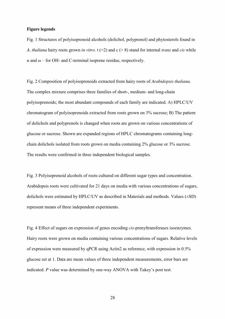
Fig. 1 Structures of polyisoprenoid alcohols (dolichol, polyprenol) and phytosterols found in
A. thaliana hairy roots grown in vitro. t (=2) and c (> 8) stand for internal trans and cis while
α and ω – for OH- and C-terminal isoprene residue, respectively.
Fig. 2 Composition of polyisoprenoids extracted from hairy roots of Arabidopsis thaliana.
The complex mixture comprises three families of short-, medium- and long-chain
polyisoprenoids; the most abundant compounds of each family are indicated. A) HPLC/UV
chromatogram of polyisoprenoids extracted from roots grown on 3% sucrose; B) The pattern
of dolichols and polyprenols is changed when roots are grown on various concentrations of
glucose or sucrose. Shown are expanded regions of HPLC chromatograms containing long-
chain dolichols isolated from roots grown on media containing 2% glucose or 3% sucrose.
The results were confirmed in three independent biological samples.
Fig. 3 Polyisoprenoid alcohols of roots cultured on different sugar types and concentration.
Arabidopsis roots were cultivated for 21 days on media with various concentrations of sugars,
dolichols were estimated by HPLC/UV as described in Materials and methods. Values (±SD)
represent means of three independent experiments.
Fig. 4 Effect of sugars on expression of genes encoding cis-prenyltransferases isoenzymes.
Hairy roots were grown on media containing various concentrations of sugars. Relative levels
of expression were measured by qPCR using Actin2 as reference, with expression in 0.5%
glucose set at 1. Data are mean values of three independent measurements, error bars are
indicated. P value was determined by one-way ANOVA with Tukey’s post test.

29
Fig. 5 Effect of hairy root culture growth on the content of polyisoprenoid alcohols. Roots
were grown on 2% glucose for indicated time, dolichols were estimated by HPLC/UV as
described in Materials and methods. Each bar represents the mean value of three independent
experiments. Error bars are indicated.
Fig. 6 Composition of free sterols and steryl esters in hairy roots. GC/FID chromatograms of
sterols isolated as free alcohols (top) and esters (bottom) from roots grown on 3% sucrose.
Representative chromatograms out of three for independent biological samples are shown.

Table 1. Primers used in qPCR to evaluate expression of Arabidopsis thaliana
cis-prenyltransferases
Name of Arabidopsis CPT Locus Tag Primer sequence
AtCPT1 At2g23410 F: 5’-GTGGCAACTTGCTTATTCCG-3’ R: 5’-CCTACGCTGATACGAAGC-3’
AtCPT2 At2g23400 F: 5’-TTGTCCGAGAGGAGGAGCTAC-3’ R: 5’-TGCCGTCGTCAATCCGTCTC-3’
AtCPT3 At2g17570 F: 5’-GCGCTTATGTCGATGCTG-3’ R: 5’-CAGACTCAACCTCCTCAGG-3’
AtCPT6 At5g58780 F: 5’-GACGATTATGACAACGAGCAAC-3’ R: 5’-ATGTCTTGGCATCAGCTCTC-3’
AtCPT7 At5g58770 F: 5’-TATCTCTACGAGTTCCTACTCC-3’ R: 5’-CTACTTAACCGCCATCGC-3’
AtCPT9 At5g58784 F: 5’-AGCATGTGGCGGTTATATTGG-3’ R: 5’-TTCTCCATGAGCCTTCTCG-3’
Actin2 At3g18780 F: 5’-GACCAGCTCTTCCATCGAGAA-3’ R: 5’-CAAACGAGGGCTGGAACAAG-3’
Table 1

Table 2. Dolichol spectrum in Arabidopsis hairy roots – effect of carbon source
Numbers in bold indicate dominating prenologues.
Carbon source Dolichols (number of isoprene units)
Glucose
0.5% 12 13 14 15 16 17 18 19 20 21 22 23 24 25 26 27 28 29 30 1.0% 12 13 14 15 16 17 18 19 20 21 22 23 24 25 26 27 28 29 30 31 32 33 34 35 1.5% 12 13 14 15 16 17 18 19 20 21 22 23 24 25 26 27 28 29 30 31 32 33 34 35 2.0% 12 13 14 15 16 17 18 19 20 21 22 23 24 25 26 27 28 29 30 31 32 33 34 35 2.5% 12 13 14 15 16 17 18 19 20 21 22 23 24 25 26 27 28 29 30 3.0% 12 13 14 15 16 17 18 19 20 21 22 23 24 25 26 27 28 29 30
Sucrose 3.0% 12 13 14 15 16 17 18 19 20 21 22 23 24 25 26 27 28 29 30
Table 2

Table 3. Composition of sterols accumulated as free alcohols and esters in hairy roots of A.
thaliana grown on 3% sucrose. Data are obtained from GC/MS estimation (see Materials and
methods).
Sterol
Sterol fraction
Free sterol Esterified sterol
µg/g d.w. % µg/g d.w. %
Total 2234 100 81 100
Brassicasterol 31.3 1.4 3.3 4.1
Campesterol 375.3 16.8 13.4 16.6
Campestanol 35.7 1.6 2.3 2.8
Stigmasterol 969.6 43.4 19.4 24.0
β-Sitosterol 661.3 29.6 36.7 45.3
Stigmastanol 71.5 3.2 5.9 7.2
Stigmast-4,22-dien-3-one 46.9 2.1 n.d.* 0
Stigmast-4-en-3-one 42.4 1.9 n.d.* 0
* n.d., not detected
Table 3
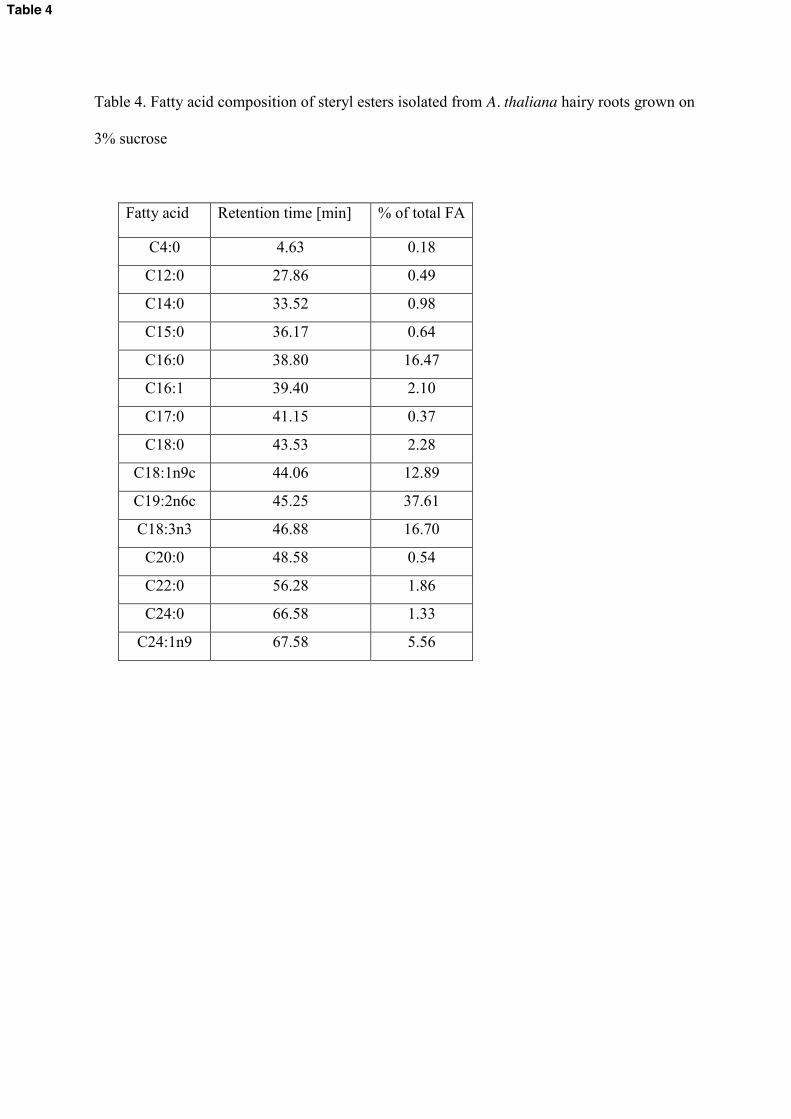
Table 4. Fatty acid composition of steryl esters isolated from A. thaliana hairy roots grown on
3% sucrose
Fatty acid Retention time [min] % of total FA
C4:0 4.63 0.18
C12:0 27.86 0.49
C14:0 33.52 0.98
C15:0 36.17 0.64
C16:0 38.80 16.47
C16:1 39.40 2.10
C17:0 41.15 0.37
C18:0 43.53 2.28
C18:1n9c 44.06 12.89
C19:2n6c 45.25 37.61
C18:3n3 46.88 16.70
C20:0 48.58 0.54
C22:0 56.28 1.86
C24:0 66.58 1.33
C24:1n9 67.58 5.56
Table 4

Table 5. Effect of sugar type and concentration on sterol content and profile (shown as ratio of
two dominating sterols) in A.thaliana hairy roots. Content of sterols was estimated using
HPLC/UV (see Materials and methods).
Sugar concentration Stigmasterol β-Sitosterol Total sterol content
[µg/g d.w.]
0.5% glucose 1.4 1 2129±234
2.0% glucose 1.3 1 2048±234
3.0% glucose 0.7 1 1476±126
3.0% sucrose 1.5 1 2234±268
Table 5
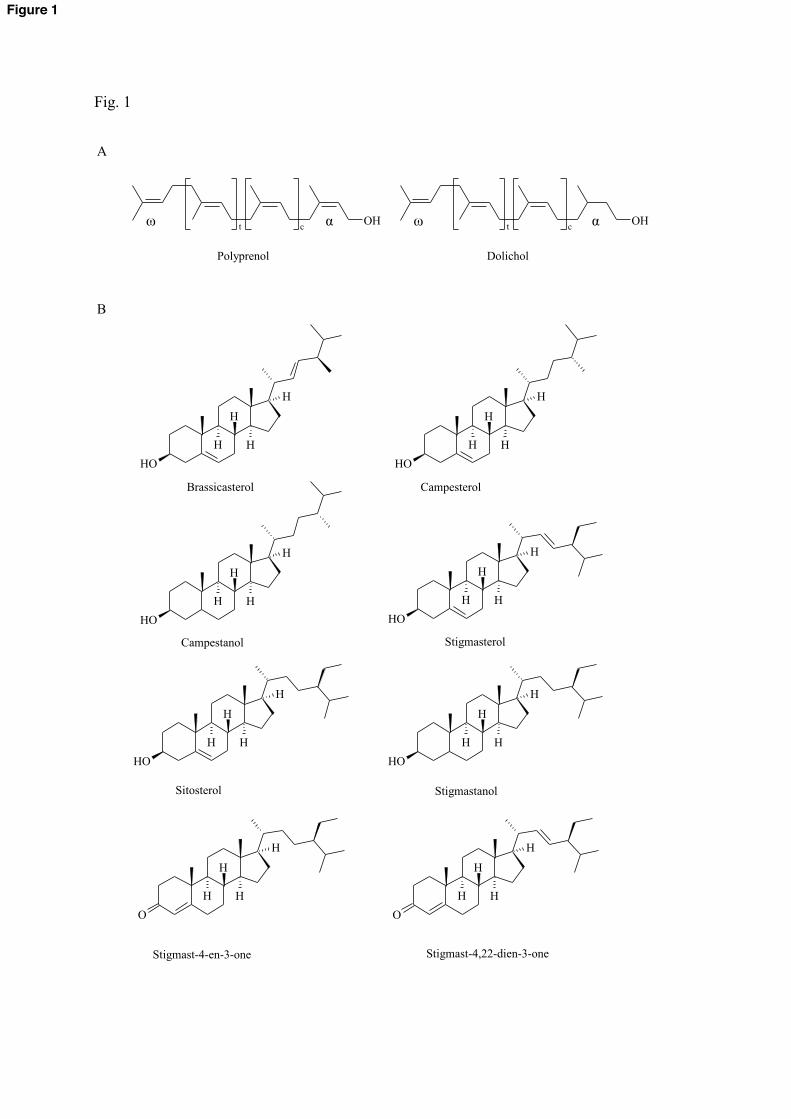
Fig. 1
OH
H
H H
H
OH
H
H H
H
OH
H
H H
H
OH
H
H H
H
OH
H
H H
H
OH
H
H H
H
O
H
H H
H
O
H
H H
H
OHt
c
OHt
c
Brassicasterol Campesterol
Campestanol Stigmasterol
Sitosterol Stigmastanol
Stigmast-4-en-3-one Stigmast-4,22-dien-3-one
Polyprenol Dolichol
A
B
ω ω α α
Figure 1

Fig. 2
A
B 2% Glucose 3% Sucrose
10 15 20 25 30 35 40 45 50
Det
ecto
r res
pons
e
Time [min.]
Dol-16
Dol-13 Pren-21 Dol-21
30 32 34 36 38
Det
ecto
rres
pons
e
Time [min.]28 30 32 34 36 38
Det
ecto
rres
pons
e
Time [min.]
Dol-23Pren-23 Dol-21Pren-21
Medium-chain polyisoprenoids
Long-chain polyisoprenoids
Short-chain polyisoprenoids
Figure 2

Fig. 3
0
10
20
30
40
50
60
70
3% sucrose 0.5% glucose 1% glucose 1.5% glucose 2% glucose 2.5% glucose 3% glucose Poly
isopr
enoi
ds [µ
g/g
dry
tissu
e]
Carbon source
Figure 3

Fig. 4
Figure 4
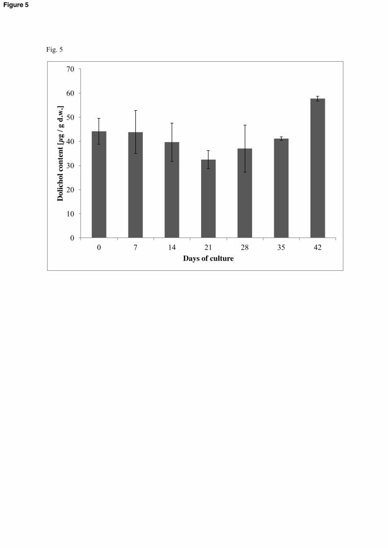
Fig. 5
0
10
20
30
40
50
60
70
0 7 14 21 28 35 42
Dol
icho
l con
tent
[µg
/ g d
.w.]
Days of culture
Figure 5

Fig. 6
20 22 24 26 28 30 32 34 36 38 40
Det
ecto
r res
pons
e
Time [min.]
free sterols
esterified sterols
β-sitosterol
stigmastanol
stigmasterol
campesterol
brassicasterol
campestanol
Figure 6