1 vascular and lymphatic system pathology. 2 blood flow systemic blood flow is a circuit : heart →...
Post on 18-Dec-2015
273 views
TRANSCRIPT

1
Vascular and Lymphatic System Pathology

2
Blood Flow
• Systemic blood flow is a circuit :
• Heart →Arteries→ Arterioles→ Capillaries→ Venules→ Veins→ Heart
• Artery – any vessels that carries blood away from the heart.
• Vein – any vessels that carries blood toward the heart

3

4
Structure of blood vessels• Tunica intima
– Endothelium and connective tissue
• Tunica media– Smooth muscle and elastic tissue
• Tunica externa or tunica adventitia– Connective and elastic tissue

5

6

7
Arteries
• Large arteries are elastic (conducting) arteries – pressure reservoirs
• Medium arteries are muscular (distributing) arteries – more smooth muscle
• Contraction or relaxation of muscle changes the size of the lumen, and so controls the blood pressure in the vessel.

8

9
Capillaries
• Only a single layer of endothelium and a basement membrane
• Connect arterioles and venules
• Functional part of system
• True capillaries begin at a precapillary sphincter which controls blood flow through the capillary

10

11
Veins
• Relatively thin; less elastic
• Larger in diameter than arteries
• Have valves to prevent backflow of blood
• Flow to heart is assisted by contraction of skeletal muscles

12

13

14

15
Control of systemic circulation
• Nervous control – innervated by sympathetic nervous system ONLY
• Cardiac control center (primarily in medulla oblongata)
• Heart has both Sympathetic and Parasympathetic innervations.

16
• Baroreceptors and chemoreceptors:– Monitor pressure
– Monitor blood levels of O2, CO2 and H+
– Send information to cardiovascular center, which responds

17
Compliance• The increase in volume a vessel can
accommodate for a given increase in pressure. – Depends on the ratio of elastic fibers to muscle
fibers in the vessel wall.• Elastic arteries more compliant than muscular arteries• Veins more compliant than either artery (blood reservoirs)
• Decreased compliance suggests an increased stiffness of vessel wall.
• Determines the vessel’s response to changes in pressure.

18
Blood pressure
• Mean arterial pressure is the average in pressure in the arteries throughout the cardiac cycle.
• Depends on the compliance of the arteries and the amount of blood in the arterial system.

19
Lymphatic System• A vascular system that runs “parallel” to the
blood vascular system
• Flow does not circulate – begins in tissue
• Returns to venous system at subclavian veins
• Fluid in vessels is lymph – mostly water and proteins
• Interstitial fluid→ lymphatic capillaries→ lymphatic vessels→ lymphatic trunks→ lymphatic ducts

20

21

22
Lymph nodes• Lie along lymphatic vessels
• Contain lymphocytes that filter lymph and eliminate microbes/damaged cells/ toxins
• Biological filtration

23

24
Diseases of Arteries and Veins• Thrombus- “clotting” in an unbroken vessel
– Maintains a point of attachment– Organized differently than a clot– usually due to damage to endothelium and
exposure of collagen in the basement membrane

25
Arterial thrombus
• Forms where blood is moving rapidly – see alternating lines of platelets and red cells trapped in fibrin
• Lines of Zahn

26
Venous thrombus
• Forms differently due to decreased blood flow
• Mixed region at site of attachment
• More blood clotting forms a downstream red cap

27
Factors that predispose to thrombosis
• Endothelial damage
– Bacterial damage
– Damage to the myocardium
– Wear and tear – hemodynamic stress
• Hypertension increases this
• Arteriosclerosis
–Inflammation– Tumors and irritation by their products

28
Factors that predispose to thrombosis
• Flow abnormalities– Increases platelet contact with endothelium– Reduction in flow:
• Arterial:–Cardiac damage and decreased
pumping action–Increased blood viscosity
• Venous:–Physical inactivity–Varicose veins

29
– Turbulence:
• Damaged heart valves
• Congenital heart defects
• Compression of the vessel
• Weakened arterial wall - aneurysm

30
Other Causes • Aging
• Immobilization
• Injury to vessel endothelium
• Increased clotting response
Effects:
• Decreased venous emptying
• Increased venous pressures
• Edema
• Pain

31
Sequelae of Thrombosis1 Resolution –
Anticoagulation system
Fibrolytic system
Moderate exercise increases thrombus resolution

32
2 Organization
The thrombus is digested by phagocytes and replaced by connective tissue – incorporating the thrombus into the vessel wall.
May recanalize – small channels open up and restore blood flow

33Recanalization

34
3 Propagation –
Thrombus extends further down the vessel, usually a vein.
Initial thrombus acts as a site for further platelet adherence.

35
Propagation

36
4 Infarction –
an infarct is an area of necrosis caused by ischemia and hypoxia.
More common in arteries than veins due to blood flow patterns
Collateral circulation and anastomosis prevent infarction

37

38
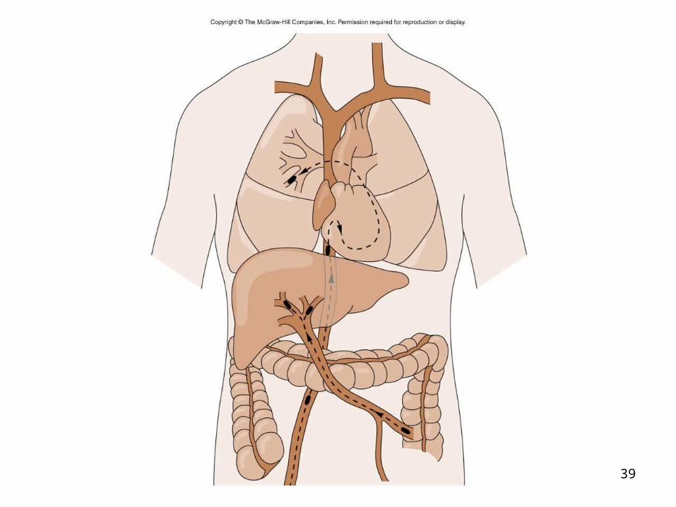
Embolism – obstruction of vessel by matter circulating in blood stream
– Matter could be fat, air, infant’s cells, in addition to pieces of clot – thromboemboli
– Thromboemoboli from the venous system tend to end up in the:
lungs and liver
39

40
Treatment
• Anticoagulants
• Fibrinolytics – t-Pas
• Prophylactic aspirin therapy

41

42
Arterial Occlusions
• Arteriosclerosis – abnormal thickening and hardening of the arterial walls– Smooth muscle cells and collagen fibers
migrate into the tunica intima, causing stiffening and thickening, narrowing the lumen
– Can exacerbate high blood pressure, and cause weakening and outpouching of vessel walls

43

44
Atherosclerosis
• A form of arteriosclerosis where soft deposits of intra-arterial fat and fibrin harden over time – atheroma
• May see build up in skin – Xanthoma or arcus in cornea.
• In general, patients suffer few symptoms unless > 60 % of blood supply is blocked

45
• Progressive over years– Starts with some injury to endothelium
• Smoking, hypertension, hyperlipidemia, diabetes, autoimmune disease, and infection
– Inflammation, release of enzymes by macrophages causes oxidation of LDL, which is then consumed by macrophages – foam cells – accumulate to form fatty streaks
– Fatty streaks of lipid material appear first as yellow streaks and spots
– Smooth muscle cells proliferate, and migrate over the streak forming a fibrous plaque

46
• Fibrous plaque results in necrosis of underlying tissue and narrowing of lumen
• Inflammation can result in ulceration and rupture of the plaque, resulting in platelet adherence to the lesion = complicated lesion
• Can result in rapid thrombus formation with complete vessel occlusion → tissue ischemia and infarction

47

48

49
Clinical manifestations
• Signs and symptoms of inadequate perfusion – TIAs, often associated with exercise or stress
• When lesion becomes complicated, can result in tissue infarction
– Coronary artery disease – myocardial ischemia
– In brain – major cause of stroke
50
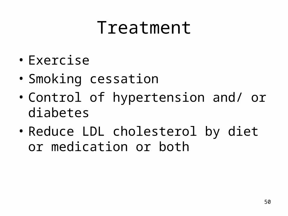
Treatment
• Exercise
• Smoking cessation
• Control of hypertension and/ or diabetes
• Reduce LDL cholesterol by diet or medication or both
51
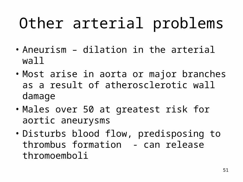
Other arterial problems
• Aneurism – dilation in the arterial wall
• Most arise in aorta or major branches as a result of atherosclerotic wall damage
• Males over 50 at greatest risk for aortic aneurysms
• Disturbs blood flow, predisposing to thrombus formation - can release thromoemboli

52
• Asymptomatic until rupture– Embolism– Death
• Treatment by surgical repair
• Aortic Dissection –bleeding into vessel wall, separating vessel layers– Men in 40-60 y.o. age group with
hypertension– Younger persons with connective tissue
disease or congenital defects– Presents with pain – life threatening

53
Systemic Hypertension
• A consistent increase in arterial blood pressure caused by increased Cardiac output or increased peripheral resistance or both
• Leads to damage of vessel walls
• If arteries constrict over a long time with increased pressure in vessel, the wall becomes thicker to withstand the stress.
• Results in narrowing of arterial lumen
• Leads to inflammatory response

54
• Causes one in eight deaths worldwide
• Third leading cause of death in the world
• Affects 50 million Americans

55
Primary hypertension• Also called essential or idiopathic
hypertension
• 92- 95 % of all cases
• No specific cause identified
• Can happen with retention of sodium and water → increased blood volume.
• Also low dietary potassium, calcium and magnesium intakes

56
Other risk factors• Smoking
– Nicotine is a vasoconstrictor
• Greater than 3 alcoholic drinks/ day
– 2-4 drinks / week lowers blood pressure

57
Suspected causes
• Interaction of genetics and environment
• Overactivity of sympathetic nervous system
• Overactivity of renin / angiotensin/ aldosterone system
• Salt and water retention by kidneys
• And others

58
Secondary hypertension• Caused by a systemic disease process
that raises peripheral resistance or cardiac output = 5 - 10 % of cases.
• Renal vascular disease
• Adrenocortical tumors
• Adrenomedullary tumors
• Drugs ( oral contraceptives, corticosteroids, antihistamines)

59
Complicated hypertension
• Sustained primary hypertension that damages the structure and function of the vessels themselves.
• Commonly affects heart, aorta, kidneys, eyes, brain, and lower extremities (target-organ damage).

60
Clinical manifestations
• None in early stages other than elevated BP
• Some individuals never have symptoms; others become very ill and die

61
Treatment
• Modification of life style
• Drugs
– Diuretics, beta-blockers, angiotensin converting enzyme inhibitor
• Compliance is often difficult – patients stop taking medication when they feel better – can get rebound effects

Venous Disorders
• Varicose veins – dilations, can lead to valvular insufficiency
• Can occur in superficial veins (saphenous) or deep veins
• Causes of secondary varicose veins:– Deep vein thrombosis– Congenital defects and pressure on
abdominal veins
62

Treatment• Prevention – little can be done after valves
become incompetent
• Avoid stressors, such as standing for long periods
• Elastic support stockings
• Sclerotherapy – injections of drugs to induce fibrosis of vessel
• Surgical removal - but only when deep vein are open.
63