1, yukako handa2 1 and yonehiro - peerj · 2d culture systems (achilli, meyer & morgan, 2012;...
TRANSCRIPT

Submitted 6 October 2017Accepted 3 December 2017Published 4 January 2018
Corresponding authorYonehiroKanemura, [email protected]
Academic editorElena González-Burgos
Additional Information andDeclarations can be found onpage 9
DOI 10.7717/peerj.4187
Copyright2018 Fukusumi et al.
Distributed underCreative Commons CC-BY 4.0
OPEN ACCESS
Small-scale screening of anticancerdrugs acting specifically on neuralstem/progenitor cells derived fromhuman-induced pluripotent stem cellsusing a time-course cytotoxicity testHayato Fukusumi1, Yukako Handa2, Tomoko Shofuda1 and YonehiroKanemura2,3,4
1Division of Stem Cell Research, Institute for Clinical Research, Osaka National Hospital, National HospitalOrganization, Osaka, Japan
2Division of Regenerative Medicine, Institute for Clinical Research, Osaka National Hospital,National Hospital Organization, Osaka, Japan
3Department of Neurosurgery, Osaka National Hospital, National Hospital Organization, Osaka, Japan4Department of Physiology, Keio University School of Medicine, Tokyo, Japan
ABSTRACTSince the development of human-induced pluripotent stem cells (hiPSCs), varioustypes of hiPSC-derived cells have been established for regenerative medicine and drugdevelopment. Neural stem/progenitor cells (NSPCs) derived from hiPSCs (hiPSC-NSPCs) have shown benefits for regenerative therapy of the central nervous system.However, owing to their intrinsic proliferative potential, therapies using transplantedhiPSC-NSPCs carry an inherent risk of undesired growth in vivo. Therefore, it isimportant to find cytotoxic drugs that can specifically target overproliferative trans-planted hiPSC-NSPCs without damaging the intrinsic in vivo stem-cell system. Here,we examined the chemosensitivity of hiPSC-NSPCs and human neural tissue—derivedNSPCs (hN-NSPCs) to the general anticancer drugs cisplatin, etoposide, mercaptop-urine, and methotrexate. A time-course analysis of neurospheres in a microspherearray identified cisplatin and etoposide as fast-acting drugs, and mercaptopurine andmethotrexate as slow-acting drugs. Notably, the slow-acting drugs were eventuallycytotoxic to hiPSC-NSPCs but not to hN-NSPCs, a phenomenon not evident in theconventional endpoint assay on day 2 of treatment. Our results indicate that slow-actingdrugs can distinguish hiPSC-NSPCs from hN-NSPCs and may provide an effectivebackup safety measure in stem-cell transplant therapies.
Subjects Neuroscience, ToxicologyKeywords Microsphere array, Time-course cytotoxicity test, Endpoint assay, ATP assay, Humanfetal neural tissue, Neural stem/progenitor cells, Anticancer drugs, Human-induced pluripotentstem cells, Neurosphere, Drug screening
INTRODUCTIONSince the development of human-induced pluripotent stem cells (hiPSCs) (Takahashi et al.,2007; Yu et al., 2007), various types of hiPSC-derived cells have been established that can be
How to cite this article Fukusumi et al. (2018), Small-scale screening of anticancer drugs acting specifically on neural stem/progenitorcells derived from human-induced pluripotent stem cells using a time-course cytotoxicity test. PeerJ 6:e4187; DOI 10.7717/peerj.4187

used in regenerative medicine and drug development, while avoiding many of the ethicalissues and technical difficulties involved with human tissue–derived cells. Human iPSC-derived neural stem/progenitor cells (hiPSC-NSPCs) (Fujimoto et al., 2012; Kobayashiet al., 2012; Oki et al., 2012; Tornero et al., 2013) and human fetal neural tissue–derivedNSPCs (hN-NSPCs) (Ishibashi et al., 2004; Iwanami et al., 2005; Ogawa et al., 2002) haveproven beneficial in treating various central nervous system diseases and injuries. However,the intrinsic proliferative potential of hiPSC-NSPCs, which makes them promising sourcesfor large numbers of cells in vitro, can be a double-edged sword in vivo: transplanted cellscan proliferate excessively before terminal differentiation in specific microenvironments.Although such undesired proliferation has not generally produced teratomas, malignantcarcinogenesis, or other serious adverse events (Nori et al., 2015; Sugai et al., 2016), thisinherent potential suggests the need for backup safety measures for stem cell–basedtherapies.
One strategy for reducing the risk of overgrowth is to transduce a gene that caninduce apoptosis, such as herpes simplex virus truncated thymidine kinase (HSV-tk)activated by ganciclovir (Cao et al., 2007) or a caspase-based artificial cell-death switch(iCaspase-9) activated by AP20187 (Krishnamurthy et al., 2010), into the stem-cell genome.However, inserting exogenous genes into the donor-cell genome contradicts the purpose ofintegration-free hiPSCs, which are generated to minimize the risk of genetic modificationor transgene re-activation, and transgenic strategies may create new risks despite the useof ‘genomic safe harbors’ for insertions in the human genome. Another strategy is to usedrugs to suppress the in vivo overgrowth of transplanted cells; for instance, pretreatinghiPSC-NSPCs with a γ-secretase inhibitor inhibits Notch signaling, which is required formaintaining NSPC stemness (Okubo et al., 2016). However, a single treatment prior totransplantation may not be sufficient to overcome the cells’ growth potential, and cannotregulate cell growth after transplantation. Therefore, a useful backup safety measure wouldbe a method to chemically ablate transplanted cells, preferably with a cytotoxic drug thatspecifically acts on transplanted hiPSC-NSPCs but not tissue-resident NSPCs.
In this study, we assessed four approved anticancer drugs, two cytotoxic (cisplatinand etoposide) and two cytostatic (mercaptopurine and methotrexate), as candidates forsuppressing the overgrowth of non-transgenic stem cells in vivo.
Although the efficacy of candidate drugs has conventionally been evaluated by cell-destructive methods, such as MTT or ATP assays, these methods cannot assess the effectsof a drug on the same cell population over time. To address this, previous studies haveassessed the time-course of pharmacological effects using cell-nondestructive methods,such as measurement of changes in impedance in two-dimensional (2D) adherent cellcultures (Caviglia et al., 2015) and image-based measurement of the spheroid size inthree-dimensional (3D) cultures of various cell types, including glioma cells (Vinci etal., 2012), hepatocytes (Bell et al., 2016), and cardiomyocytes (Beauchamp et al., 2015). Itis known that 3D culture systems mimic the in vivo environment more effectively than2D culture systems (Achilli, Meyer & Morgan, 2012; Pampaloni, Reynaud & Stelzer, 2007).Therefore, the present study used a conventional endpoint assay on day 2 of the treatmentand a 7-day time-course cytotoxicity test to determine the effects of cisplatin, etoposide,
Fukusumi et al. (2018), PeerJ, DOI 10.7717/peerj.4187 2/13

mercaptopurine, and methotrexate on 3D neurospheres derived from hiPSC-NSPCs andhN-NSPCs, which are considered to mimic the in vivo stem cell system.
MATERIALS AND METHODSEthics statementThis study was conducted in accordance with the principles of the Declaration of Helsinki.The use of hN-NSPCs and hiPSCs was approved by the OsakaNational Hospital hN-NSPCsand hiPSCs ethics committee (Nos. 110, 120, and 146).
Cell linesWe used two hN-NSPC lines (oh-NSC-3-fb and oh-NSC-7-fb) (Kanemura et al., 2002)and two hiPSC (201B7)-derived NSPC lines: the DSM line, which was established usingthe single SMAD-inhibition method with the Noggin alternative dorsomorphin (DSM)(Shofuda et al., 2013), and the dSMAD line, which was established by the dual SMAD-inhibition method with DSM and SB431542 (Fukusumi et al., 2016).
Cell cultureThe hN-NSPCs and hiPSC-NSPCs were propagated as neurospheres in Dulbecco’sModified Eagle’s Medium (DMEM)/F12 (D8062; Sigma-Aldrich, St. Louis, MO, USA) with15 mM HEPES (Sigma-Aldrich), epidermal growth factor (EGF, 20 ng/mL; PeproTech,Rocky Hill, NJ, USA), fibroblast growth factor 2 (FGF2, 20 ng/mL; PeproTech), leukemiainhibitory factor (LIF, 10 ng/mL; Millipore, Billerica, MA, USA), B27 supplement (B27,2%; Thermo Fisher Scientific,Waltham,MA, USA), and heparin (5µg/mL; Sigma-Aldrich)(Fukusumi et al., 2016; Kanemura et al., 2002; Shofuda et al., 2013). For hN-NSPCs, half ofthe medium was changed once a week. The neurospheres were dissociated into single cellsevery 14 days by incubating them with 0.05% trypsin/EDTA (Thermo Fisher Scientific) at37 ◦C for 20 min, after which soybean trypsin inhibitor (Roche, Basel, Switzerland) wasadded to stop the enzyme activity. The cells were then resuspended in 50% fresh mediumplus 50% conditioned medium at a density of 1 ×10⁵ cells/mL (Kanemura et al., 2002).For hiPSC-NSPCs, the medium was changed every 3–5 days. The cells were passaged every10–12 days using Accutase (Innovative Cell Technologies, San Diego, CA, USA) at 37 ◦Cfor 10 min for single-cell dissociation, after which the cells were resuspended in 100% freshmedium at a density of 1 ×10⁵ cells/mL (Fukusumi et al., 2016; Shofuda et al., 2013).
Drug preparationCisplatin (Sigma-Aldrich), etoposide (Sigma-Aldrich), mercaptopurine (Sigma-Aldrich),and methotrexate (LKT Laboratories, St. Paul, MN, USA) were dissolved in dimethylsulfoxide (DMSO) to generate 100 mM stock solutions.
Endpoint (ATP) assayThe neurospheres were dissociated into single cells and seeded into 96-well plates at adensity of 3×10⁴ cells/well (day −1). On day 0, cisplatin, etoposide, and methotrexatewere applied at 0, 0.1, 0.3, 1, 3, 10, 30, and 100 µM, andmercaptopurine was applied at 0, 1,3, 10, 30, 100, 300, and 1,000 µM (0 µM indicates DMSO only). ATP content was assayed
Fukusumi et al. (2018), PeerJ, DOI 10.7717/peerj.4187 3/13
321 70
Drug treatmenta
Endpointassay
Dose
A
B
C
D
E
F
G
H
1 2 3 4 5 6 7 8 9 10 11 12
b
Phase-contrast
Day 0 Day 7Day 3Day 2Day 1
Photo
●●●
●●●
●●●
●●●
Photo
●●●
●●●
●●●
●●●
Photo
●●●
●●●
●●●
●●●
Photo
●●●
●●●
●●●
●●●
Photo
●●●
●●●
●●●
●●●
<<Tracking same microwells>>Time-course measurement of neurosphere size Microsphere array (MSA)
» 652 microwells/array
Time-courseassay
Measurement of ATP content
A
B
C
D
E
F
G
H
1 2 3 4 5 6 7 8 9 10 11 12
Seeding cells-1Day
96-well plate
Neurospheres
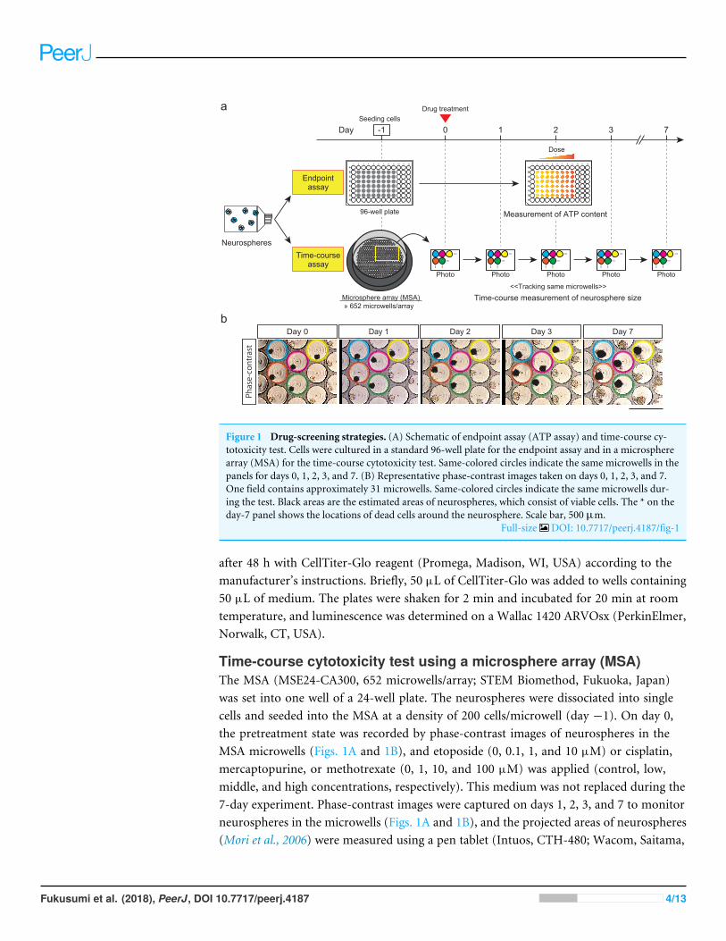
Figure 1 Drug-screening strategies. (A) Schematic of endpoint assay (ATP assay) and time-course cy-totoxicity test. Cells were cultured in a standard 96-well plate for the endpoint assay and in a microspherearray (MSA) for the time-course cytotoxicity test. Same-colored circles indicate the same microwells in thepanels for days 0, 1, 2, 3, and 7. (B) Representative phase-contrast images taken on days 0, 1, 2, 3, and 7.One field contains approximately 31 microwells. Same-colored circles indicate the same microwells dur-ing the test. Black areas are the estimated areas of neurospheres, which consist of viable cells. The * on theday-7 panel shows the locations of dead cells around the neurosphere. Scale bar, 500 µm.
Full-size DOI: 10.7717/peerj.4187/fig-1
after 48 h with CellTiter-Glo reagent (Promega, Madison, WI, USA) according to themanufacturer’s instructions. Briefly, 50 µL of CellTiter-Glo was added to wells containing50 µL of medium. The plates were shaken for 2 min and incubated for 20 min at roomtemperature, and luminescence was determined on a Wallac 1420 ARVOsx (PerkinElmer,Norwalk, CT, USA).
Time-course cytotoxicity test using a microsphere array (MSA)The MSA (MSE24-CA300, 652 microwells/array; STEM Biomethod, Fukuoka, Japan)was set into one well of a 24-well plate. The neurospheres were dissociated into singlecells and seeded into the MSA at a density of 200 cells/microwell (day −1). On day 0,the pretreatment state was recorded by phase-contrast images of neurospheres in theMSA microwells (Figs. 1A and 1B), and etoposide (0, 0.1, 1, and 10 µM) or cisplatin,mercaptopurine, or methotrexate (0, 1, 10, and 100 µM) was applied (control, low,middle, and high concentrations, respectively). This medium was not replaced during the7-day experiment. Phase-contrast images were captured on days 1, 2, 3, and 7 to monitorneurospheres in the microwells (Figs. 1A and 1B), and the projected areas of neurospheres(Mori et al., 2006) were measured using a pen tablet (Intuos, CTH-480; Wacom, Saitama,
Fukusumi et al. (2018), PeerJ, DOI 10.7717/peerj.4187 4/13

Concentration (µM)
Cisplatin Etoposide Mercaptopurine Methotrexate
DSMdSMAD
oh-NSC-7-fboh-NSC-3-fb
0
50
100
Cont 0.1 1 10 100 Cont 0.1 1 10 100 Cont 1 10 100 1000 Cont 0.1 1 10 100ATP
leve
ls re
lativ
e to
DM
SO c
ontro
l (%
)
Figure 2 Dose-response curves and IC50 values obtained from a conventional ATP assay on day 2. con-trol (%). The x-axis indicates the drug concentration (µM) in log scale, and the y-axis indicates ATP lev-els in the treated cells relative to the DMSO control (%). Colors indicate cell type. The log-logistic model(LL2.4) was used. Error bars represent the 95% CI.
Full-size DOI: 10.7717/peerj.4187/fig-2
Japan) with the TrakEM2 plugin (Cardona et al., 2012) in ImageJ (Fiji package) (Schindelinet al., 2012; Schindelin et al., 2015). Microwells containing multiple neurospheres on day 0were excluded from analysis. Outliers in a boxplot of neurosphere sizes on day 0 were alsoexcluded. At each time-point, the neurosphere size in the presence of each treatment wasexpressed as a percentage of the day 0 value. These data were then expressed as a percentageof the respective DMSO control.
Statistical analysisFor the endpoint assay (Fig. 2), predicted dose–response curves and 50% inhibitoryconcentration (IC50) values were obtained using the four-parameter log-logistic(LL2.4) and ED functions, respectively, of the drc package (Ritz et al., 2015) in R(R Core Team, 2015). For the time-course cytotoxicity test, changes in neurospheresize relative to those in the DMSO controls (Fig. 3) and day 0 neurospheres(Fig. S1) were analyzed with the four-parameter logisticmodel (L.4) and the Brain–Cousensfive-parameter model (BC.5) of the drc package in R, respectively. Data from the endpointassay (Fig. 2) and time-course cytotoxicity test (Figs. 3 and S1) were plotted with 95%confidence intervals (95% CI).
RESULTSThe IC50 of cisplatin, etoposide, mercaptopurine, and methotrexate in the two hiPSC-NSPC cell lines (DSM and dSMAD) and the two hN-NSPC cell lines (oh-NSC-3-fb andoh-NSC-7-fb; Fig. 2 and Table 1) was determined by endpoint (ATP) assay. Cisplatinand etoposide were preferentially toxic to hN-NSPCs and hiPSC-NSPCs, respectively.Interestingly, the two hiPSC-NSPC lines differed in sensitivity to these drugs (Table 1).Mercaptopurine was highly toxic to both types of NSPCs, while methotrexate had almostno effect on either type even at high concentrations. Of the two hiPSC-NSPC lines, DSMwas significantly more resistant to the drugs.
To follow the real-time effects of these drugs after treatment, we conducted a simpletime-course cytotoxicity test using an MSA (Figs. 1A and 1B). Similar to the ATP assay,
Fukusumi et al. (2018), PeerJ, DOI 10.7717/peerj.4187 5/13

0 1 2 3 4 5 6 7 0 1 2 3 4 5 6 7 0 1 2 3 4 5 6 7
Days after treatment
Neu
rosp
here
siz
e re
lativ
e to
DM
SO c
ontro
l (%
)
Cis
plat
inEt
opos
ide
Mer
capt
opur
ine
Met
hotre
xate
DSM dSMAD oh-NSC-3-fb oh-NSC-7-fb
Human iPSC-derived NSPCs Human fetal neural tissue-derived NSPCs
DoseControl
LowMiddleHigh
0
50
100
0
50
100
0
50
100
0
50
100
0 1 2 3 4 5 6 7
Figure 3 Results of time-course cytotoxicity test. The x-axis indicates days after treatment, and the y-axis indicates neurosphere size relative to the DMSO control (%). Colors indicate drug concentrations.The logistic model (L.4) was used. Error bars represent the 95% CI.
Full-size DOI: 10.7717/peerj.4187/fig-3
Table 1 IC50 values (µM) of drugs against hiPSC-NSPCs and hN-NSPCs.
Drug hiPSC-NSPCs hN-NSPCs
DSM dSMAD oh-NSC-3-fb oh-NSC-7-fb
Cisplatin 100< 72.3 14.6 15.2Etoposide 0.32 0.04 6.59 28.5Mercaptopurine 120 17.3 17.3 47.0Methotrexate 100< 100< 100< 100<
Fukusumi et al. (2018), PeerJ, DOI 10.7717/peerj.4187 6/13

we assessed the effect of cisplatin, etoposide, mercaptopurine, and methotrexate at fourdosage levels in the two hiPSC-NSPC and two hN-NSPCs lines, in this case by measuringneurosphere size on each day for seven days (Fig. 3 and Table S1). Because neurosphere sizeof the DMSO control increased during the 7-day assay (Fig. S1), smaller neurosphere sizesof the drug treatment group than those of the DMSO control indicate cytotoxicity of drugs.
Compared to the DMSO control, cisplatin showed toxic effects in hiPSC-NSPCs atlow to high concentrations and in hN-NSPCs at middle and high concentrations (Fig. 3).Notably, high concentrations of cisplatin killed hiPSC-NSPC neurospheres, as indicated bytheir disappearance (Fig. S1 shows changes in neurosphere size relative to day 0), whereashN-NSPC neurospheres were present throughout the assay period (Fig. S1). Etoposide alsoaffected the neurosphere size in both hiPSC-NSPCs and hN-NSPCs, but it showed earlierand stronger toxicity at lower concentrations compared to cisplatin (Fig. 3). However,the hN-NSPC neurosphere size remained constant even at the highest concentration ofetoposide or cisplatin (Fig. S1). Thus, there was a clear difference between hiPSC-NSPCsand hN-NSPCs at the highest concentrations of both cisplatin and etoposide.Moreover, lowconcentrations of etoposide affected the neurosphere size in dSMAD and oh-NSC-7-fb cellsmore strongly than that in DSM or oh-NSC-3-fb cells, respectively. Thus, the preferentialtoxicity of the drug differed not only between the two types of NSPCs, but also betweentwo cell lines of the same type.
Mercaptopurine and methotrexate, even at the highest concentrations, were only mildlytoxic to hN-NSPCs and did not stop their growth (Fig. S1). However, hiPSC-NSPCs wereaffected by both mercaptopurine and methotrexate. At low concentrations, methotrexatewas noticeably more toxic to hiPSC-NSPCs than was mercaptopurine (Fig. 3). Unlikecisplatin and etoposide, mercaptopurine and methotrexate had only a limited effect at eventhe highest concentrations until day 3, which allowed the growth of larger neurospheres(Fig. S1). As with cisplatin and etoposide, mercaptopurine and methotrexate proved tobe highly cytotoxic by the end of the assay. From these results, cisplatin and etoposidecan be classified as fast-acting drugs with early cellular toxicity, while mercaptopurine andmethotrexate can be classified as slow-acting drugs with later toxicity.
DISCUSSIONAlthough hiPSC-NSPC transplantation is effective for treating spinal cord injury andstroke, the use of stem cells poses certain risks due to their intrinsic proliferative potential.In particular, the artificial generation of hiPSCs may cause genetic and epigeneticabnormalities, which could potentially increase the risk of tumorigenesis (Nagoshi &Okano, 2017). These risks can be reduced prior to transplantation by inserting a ‘suicidegene’ into the donor cells, or by pretreating the donor cells with inhibitors that reduce theirstemness and direct differentiation. However, neither of these strategies is ideal. Transgenescan potentially introduce new risks via genome modification. Although pretreatment canreduce donor-cell stemness, we have not yet found a way to deal with the overgrowth ofgrafted cells after transplantation. Thus, as a backup safety measure for stem-cell therapies,it is essential to identify drugs that act specifically on the grafted cells, but not on resident
Fukusumi et al. (2018), PeerJ, DOI 10.7717/peerj.4187 7/13

stem cells. To this end, we conducted a small-scale screening of four anticancer drugs andexamined their effect on hiPSC-NSPCs and on hN-NSPCs, which are considered to mimicthe resident stem cells in the host body.
Based on ATP assay results, we classified the four drugs as follows: (1) more toxic tohN-NSPCs than hiPSC-NSPCs (cisplatin), (2)more toxic to hiPSC-NSPCs than hN-NSPCs(etoposide), (3) similar toxic effects on hiPSC-NSPCs and hN-NSPCs (mercaptopurine),and (4) almost no effect on hiPSC-NSPCs or hN-NSPCs (methotrexate). These resultsidentified etoposide as a candidate backup safety measure for stem cell-based therapies,since it was selectively toxic to hiPSC-NSPCs.However, the dosemay need to be adjusted forindividual cell lines, since different lines of the same type of NSPC differed in sensitivity toetoposide. Although an ATP assay is useful for characterizing drugs based on dose-responsecurves and IC50 values, the long-term monitoring of transplanted cells is necessary aftertreatment in vivo.
In this study, we monitored the effects of anticancer drugs on neurosphere size invitro for a period of seven days after treatment. Based on these results, we classifiedcisplatin and etoposide as fast-acting drugs with early cytotoxicity, and mercaptopurineand methotrexate as slow-acting drugs with late cytotoxicity; this classification is consistentwith the drug categories. Cisplatin and etoposide are cytotoxic drugs that act directlyby alkylating DNA and inhibiting topoisomerase, respectively, whereas mercaptopurineand methotrexate are cytostatic drugs that inhibit IMP dehydrogenase and dihydrofolatereductase, respectively. Compared to hiPSC-NSPCs, the hN-NSPCs were more resistantto high concentrations of cisplatin or etoposide; this difference might be due to thedifferent developmental stages of the cells. In fact, hiPSC-NSPCs recapitulate regularneural development along with cell proliferation after transplantation (Sugai et al., 2016),and this characteristic will likely be useful for developing drugs that specifically targettransplanted cells. The presence of mercaptopurine and methotrexate, which are cytostatic,eventually induced death in hiPSC-NSPCs, but only mildly limited hN-NSPC growthduring the 7-day assay. Mercaptopurine and the cytotoxic drugs cisplatin and etoposidedecreased the ATP level in both hN-NSPCs and hiPSC-NSPCs on day 2 of treatment(Fig. 2). Although ATP level is a useful index of cell viability, mercaptopurine-mediatedinhibition of de novo purine synthesis might also reduce the ATP level in the absence ofcell death, in contrast to other cytotoxic drugs. However, the effect of cytostatic drugsdistinguished hN-NSPCs and hiPSC-NSPCs in the time-course assay (Fig. 3). Therefore,we need to reassess cytostatic drugs from the viewpoint of their time-dependent action.Our findings indicate that methotrexate is preferable to mercaptopurine as a candidatesafety measure for hiPSC-NSPC transplantation because it was cytotoxic even at lowconcentrations.
This study has certain limitations. First, the mechanisms underlying the late toxicityof cytostatic drugs on hiPSC-NSPCs are unknown, although hN-NSPCs are reported toexpress high levels of ABCB1 transporter (Islam et al., 2005; Yamamoto et al., 2009), whichmay contribute to a development of tolerance to slow-acting drugs. Further studies areneeded to elucidate the mechanisms underlying the selective cytotoxic effects of cytostatic
Fukusumi et al. (2018), PeerJ, DOI 10.7717/peerj.4187 8/13

drugs on hiPSC-NSPCs. Second, these effects were obtained in vitro and therefore awaitconfirmation in vivo.
CONCLUSIONBased on a 7-day time-course cytotoxicity test, we classified four anticancer drugs asfast-acting or slow-acting. We found that the slow-acting drugs affected hiPSC-NSPCsand hN-NSPCs differently, which was not evident in a conventional ATP assay performedon day 2. As hN-NSPCs were more tolerant of slow-acting drugs than hiPSC-NSPCs, wepropose that slow-acting drugs such as methotrexate may provide drug candidates forbackup safety measures to prevent the undesirable proliferation of hiPSC-NSPCs aftertransplantation therapies.
ACKNOWLEDGEMENTSThe authors thank Ms. Ai Takada, Ms. Miho Sumida, Ms. Ema Yoshioka, Ms. Yui Inazawa,and Mr. Daisuke Kanematsu for technical support.
ADDITIONAL INFORMATION AND DECLARATIONS
FundingThis study was supported by the Research on Regulatory Harmonization and Evaluationof Pharmaceuticals, Medical Devices, Regenerative and Cellular Therapy Products,Gene Therapy Products, and Cosmetics from the Japan Agency for Medical Researchand Development (AMED), and the Advanced Research for Medical Products MiningProgramme of the National Institute of Biomedical Innovation (NIBIO). There was noadditional external funding received for this study. The funders had no role in study design,data collection and analysis, decision to publish, or preparation of the manuscript.
Grant DisclosuresThe following grant information was disclosed by the authors:Japan Agency for Medical Research and Development (AMED).National Institute of Biomedical Innovation (NIBIO).
Competing InterestsThe authors declare there are no competing interests.
Author Contributions• Hayato Fukusumi conceived and designed the experiments, performed the experiments,analyzed the data, contributed reagents/materials/analysis tools, wrote the paper,prepared figures and/or tables, reviewed drafts of the paper.
• Yukako Handa performed the experiments, analyzed the data, contributedreagents/materials/analysis tools, prepared figures and/or tables, reviewed drafts ofthe paper.
Fukusumi et al. (2018), PeerJ, DOI 10.7717/peerj.4187 9/13

• Tomoko Shofuda conceived and designed the experiments, performed the experiments,contributed reagents/materials/analysis tools, reviewed drafts of the paper.
• Yonehiro Kanemura conceived and designed the experiments, wrote the paper, revieweddrafts of the paper.
EthicsThe following information was supplied relating to ethical approvals (i.e., approving bodyand any reference numbers):
The use of hN-NSPCs and hiPSCs was approved by the Osaka National HospitalhN-NSPCs and hiPSCs ethics committee (Nos. 110, 120, and 146).
Data AvailabilityThe following information was supplied regarding data availability:
The raw data is included in Tables S2 and S3.
Supplemental InformationSupplemental information for this article can be found online at http://dx.doi.org/10.7717/peerj.4187#supplemental-information.
REFERENCESAchilli TM,Meyer J, Morgan JR. 2012. Advances in the formation, use and understand-
ing of multi-cellular spheroids. Expert Opinion on Biological Therapy 12:1347–1360DOI 10.1517/14712598.2012.707181.
Beauchamp P, MoritzW, Kelm JM, Ullrich ND, Agarkova I, Anson BD, Suter TM,Zuppinger C. 2015. Development and characterization of a scaffold-free 3Dspheroid model of induced pluripotent stem cell-derived human cardiomyocytes.Tissue Engineering Part C: Methods 21:852–861 DOI 10.1089/ten.TEC.2014.0376.
Bell CC, Hendriks DF, Moro SM, Ellis E, Walsh J, Renblom A, Fredriksson PuigvertL, Dankers AC, Jacobs F, Snoeys J, Sison-Young RL, Jenkins RE, Nordling A,Mkrtchian S, Park BK, KitteringhamNR, Goldring CE, Lauschke VM, Ingelman-SundbergM. 2016. Characterization of primary human hepatocyte spheroids asa model system for drug-induced liver injury, liver function and disease. ScientificReports 6:25187 DOI 10.1038/srep25187.
Cao F, Drukker M, Lin S, Sheikh AY, Xie X, Li Z, Connolly AJ, Weissman IL, Wu JC.2007.Molecular imaging of embryonic stem cell misbehavior and suicide geneablation. Cloning and Stem Cells 9:107–117 DOI 10.1089/clo.2006.0E16.
Cardona A, Saalfeld S, Schindelin J, Arganda-Carreras I, Preibisch S, Longair M,Tomancak P, Hartenstein V, Douglas RJ. 2012. TrakEM2 software for neural circuitreconstruction. PLOS ONE 7:e38011 DOI 10.1371/journal.pone.0038011.
Caviglia C, Zor K, Canepa S, Carminati M, Larsen LB, Raiteri R, Andresen TL, Heiska-nen A, Emneus J. 2015. Interdependence of initial cell density, drug concentrationand exposure time revealed by real-time impedance spectroscopic cytotoxicity assay.Analyst 140:3623–3629 DOI 10.1039/c5an00097a.
Fukusumi et al. (2018), PeerJ, DOI 10.7717/peerj.4187 10/13

Fujimoto Y, AbematsuM, Falk A, Tsujimura K, Sanosaka T, Juliandi B, Semi K,Namihira M, Komiya S, Smith A, Nakashima K. 2012. Treatment of a mousemodel of spinal cord injury by transplantation of human induced pluripotent stemcell-derived long-term self-renewing neuroepithelial-like stem cells. Stem Cells30:1163–1173 DOI 10.1002/stem.1083.
Fukusumi H, Shofuda T, Bamba Y, Yamamoto A, Kanematsu D, Handa Y, Okita K,NakamuraM, Yamanaka S, Okano H, Kanemura Y. 2016. Establishment of humanneural progenitor cells from human induced pluripotent stem cells with diversetissue origins. Stem Cells International 2016:7235757 DOI 10.1155/2016/7235757.
Ishibashi S, Sakaguchi M, Kuroiwa T, Yamasaki M, Kanemura Y, Shizuko I, ShimazakiT, Onodera M, Okano H, Mizusawa H. 2004.Human neural stem/progenitor cells,expanded in long-term neurosphere culture, promote functional recovery afterfocal ischemia in Mongolian gerbils. Journal of Neuroscience Research 78:215–223DOI 10.1002/jnr.20246.
IslamMO, Kanemura Y, Tajria J, Mori H, Kobayashi S, Shofuda T, Miyake J, HaraM, Yamasaki M, Okano H. 2005. Characterization of ABC transporter ABCB1expressed in human neural stem/progenitor cells. FEBS Letters 579:3473–3480DOI 10.1016/j.febslet.2005.05.019.
Iwanami A, Kaneko S, NakamuraM, Kanemura Y, Mori H, Kobayashi S, Yamasaki M,Momoshima S, Ishii H, Ando K, Tanioka Y, Tamaoki N, Nomura T, Toyama Y,Okano H. 2005. Transplantation of human neural stem cells for spinal cord injury inprimates. Journal of Neuroscience Research 80:182–190 DOI 10.1002/jnr.20436.
Kanemura Y, Mori H, Kobayashi S, IslamO, Kodama E, Yamamoto A, Nakanishi Y,Arita N, Yamasaki M, Okano H, HaraM,Miyake J. 2002. Evaluation of in vitroproliferative activity of human fetal neural stem/progenitor cells using indirect mea-surements of viable cells based on cellular metabolic activity. Journal of NeuroscienceResearch 69:869–879 DOI 10.1002/jnr.10377.
Kobayashi Y, Okada Y, Itakura G, Iwai H, Nishimura S, Yasuda A, Nori S, Hikishima K,Konomi T, Fujiyoshi K, Tsuji O, Toyama Y, Yamanaka S, NakamuraM, Okano H.2012. Pre-evaluated safe human iPSC-derived neural stem cells promote functionalrecovery after spinal cord injury in common marmoset without tumorigenicity.PLOS ONE 7:e52787 DOI 10.1371/journal.pone.0052787.
Krishnamurthy S, Dong Z, Vodopyanov D, Imai A, Helman JI, Prince ME,WichaMS,Nor JE. 2010. Endothelial cell-initiated signaling promotes the survival and self-renewal of cancer stem cells. Cancer Research 70:9969–9978DOI 10.1158/0008-5472.CAN-10-1712.
Mori H, Ninomiya K, Kino-okaM, Shofuda T, IslamMO, Yamasaki M, Okano H,TayaM, Kanemura Y. 2006. Effect of neurosphere size on the growth rate ofhuman neural stem/progenitor cells. Journal of Neuroscience Research 84:1682–1691DOI 10.1002/jnr.21082.
Nagoshi N, Okano H. 2017. iPSC-derived neural precursor cells: potential for celltransplantation therapy in spinal cord injury. Cellular and Molecular Life ScienceDOI 10.1007/s00018-017-2676-9.
Fukusumi et al. (2018), PeerJ, DOI 10.7717/peerj.4187 11/13

Nori S, Okada Y, Nishimura S, Sasaki T, Itakura G, Kobayashi Y, Renault-Mihara F,Shimizu A, Koya I, Yoshida R, Kudoh J, Koike M, Uchiyama Y, Ikeda E, Toyama Y,NakamuraM, Okano H. 2015. Long-term safety issues of iPSC-based cell therapy ina spinal cord injury model: oncogenic transformation with epithelial-mesenchymaltransition. Stem Cell Reports 4:360–373 DOI 10.1016/j.stemcr.2015.01.006.
Ogawa Y, Sawamoto K, Miyata T, Miyao S, WatanabeM, NakamuraM, BregmanBS, Koike M, Uchiyama Y, Toyama Y, Okano H. 2002. Transplantation of invitro-expanded fetal neural progenitor cells results in neurogenesis and functionalrecovery after spinal cord contusion injury in adult rats. Journal of NeuroscienceResearch 69:925–933 DOI 10.1002/jnr.10341.
Oki K, Tatarishvili J, Wood J, Koch P,Wattananit S, Mine Y, Monni E, Tornero D,Ahlenius H, Ladewig J, Brustle O, Lindvall O, Kokaia Z. 2012.Human-inducedpluripotent stem cells form functional neurons and improve recovery after graftingin stroke-damaged brain. Stem Cells 30:1120–1133 DOI 10.1002/stem.1104.
Okubo T, Iwanami A, Kohyama J, Itakura G, Kawabata S, Nishiyama Y, Sugai K,Ozaki M, Iida T, Matsubayashi K, MatsumotoM, NakamuraM, Okano H. 2016.Pretreatment with a gamma-secretase inhibitor prevents tumor-like overgrowth inhuman iPSC-derived transplants for spinal cord injury. Stem Cell Reports 7:649–663DOI 10.1016/j.stemcr.2016.08.015.
Pampaloni F, Reynaud EG, Stelzer EH. 2007. The third dimension bridges the gap be-tween cell culture and live tissue. Nature Reviews: Molecular Cell Biology 8:839–845DOI 10.1038/nrm2236.
R Core Team. 2015. R: a language and environment for statistical computing. Vienna: RFoundation for Statistical Computing. Available at https://www.R-project.org/ .
Ritz C, Baty F, Streibig JC, Gerhard D. 2015. Dose–response analysis using R. PLOSONE 10:e0146021 DOI 10.1371/journal.pone.0146021.
Schindelin J, Arganda-Carreras I, Frise E, Kaynig V, Longair M, Pietzsch T, PreibischS, Rueden C, Saalfeld S, Schmid B, Tinevez JY,White DJ, Hartenstein V, Eliceiri K,Tomancak P, Cardona A. 2012. Fiji: an open-source platform for biological-imageanalysis. Nature Methods 9:676–682 DOI 10.1038/nmeth.2019.
Schindelin J, Rueden CT, Hiner MC, Eliceiri KW. 2015. The ImageJ ecosystem: an openplatform for biomedical image analysis.Molecular Reproduction and Development82:518–529 DOI 10.1002/mrd.22489.
Shofuda T, Fukusumi H, Kanematsu D, Yamamoto A, Yamasaki M, Arita N, Kane-mura Y. 2013. A method for efficiently generating neurospheres from human-induced pluripotent stem cells using microsphere arrays. Neuroreport 24:84–90DOI 10.1097/WNR.0b013e32835cb677.
Sugai K, Fukuzawa R, Shofuda T, Fukusumi H, Kawabata S, Nishiyama Y, Higuchi Y,Kawai K, IsodaM, Kanematsu D, Hashimoto-Tamaoki T, Kohyama J, IwanamiA, Suemizu H, Ikeda E, MatsumotoM, Kanemura Y, NakamuraM, Okano H.2016. Pathological classification of human iPSC-derived neural stem/progenitor cellstowards safety assessment of transplantation therapy for CNS diseases.MolecularBrain 9:Article 85 DOI 10.1186/s13041-016-0265-8.
Fukusumi et al. (2018), PeerJ, DOI 10.7717/peerj.4187 12/13

Takahashi K, Tanabe K, Ohnuki M, Narita M, Ichisaka T, Tomoda K, Yamanaka S.2007. Induction of pluripotent stem cells from adult human fibroblasts by definedfactors. Cell 131:861–872 DOI 10.1016/j.cell.2007.11.019.
Tornero D,Wattananit S, GronningMadsenM, Koch P,Wood J, Tatarishvili J, MineY, Ge R, Monni E, Devaraju K, Hevner RF, Brustle O, Lindvall O, Kokaia Z.2013.Human induced pluripotent stem cell-derived cortical neurons integratein stroke-injured cortex and improve functional recovery. Brain 136:3561–3577DOI 10.1093/brain/awt278.
Vinci M, Gowan S, Boxall F, Patterson L, ZimmermannM, CourtW, Lomas C,Mendiola M, Hardisson D, Eccles SA. 2012. Advances in establishment and analysisof three-dimensional tumor spheroid-based functional assays for target validationand drug evaluation. BMC Biology 10:29 DOI 10.1186/1741-7007-10-29.
Yamamoto A, Shofuda T, IslamMO, Nakamura Y, Yamasaki M, Okano H, KanemuraY. 2009. ABCB1 is predominantly expressed in human fetal neural stem/progenitorcells at an early development stage. Journal of Neuroscience Research 87:2615–2623DOI 10.1002/jnr.22094.
Yu J, VodyanikMA, Smuga-Otto K, Antosiewicz-Bourget J, Frane JL, Tian S, NieJ, Jonsdottir GA, Ruotti V, Stewart R, Slukvin II, Thomson JA. 2007. Inducedpluripotent stem cell lines derived from human somatic cells. Science 318:1917–1920DOI 10.1126/science.1151526.
Fukusumi et al. (2018), PeerJ, DOI 10.7717/peerj.4187 13/13