10: ' # '8& *#1 & 9 - intechcdn.intechopen.com/pdfs-wm/35332.pdf · pelvic and...
TRANSCRIPT

3,350+OPEN ACCESS BOOKS
108,000+INTERNATIONAL
AUTHORS AND EDITORS114+ MILLION
DOWNLOADS
BOOKSDELIVERED TO
151 COUNTRIES
AUTHORS AMONG
TOP 1%MOST CITED SCIENTIST
12.2%AUTHORS AND EDITORS
FROM TOP 500 UNIVERSITIES
Selection of our books indexed in theBook Citation Index in Web of Science™
Core Collection (BKCI)
Chapter from the book The Role of Osteotomy in the Correction of Congenital andAcquired Disorders of the SkeletonDownloaded from: http://www.intechopen.com/books/the-role-of-osteotomy-in-the-correction-of-congenital-and-acquired-disorders-of-the-skeleton
PUBLISHED BY
World's largest Science,Technology & Medicine
Open Access book publisher
Interested in publishing with IntechOpen?Contact us at [email protected]
4
Pelvic and Hip Osteotomies in Children
I. Gavrankapetanovic Orthopedic and Traumatology Clinic,
Clinical Center University of Sarajevo, Bosnia and Herzegovina
1. Introduction
In this chapter four tipe of pelvic osteotomy in children will be addressed. We think that is
very important to share our 25 years long experience in this field with other colleagues and
help with variety of practical advices.
Congenital dysplasia of the hip involves the acetabular roof, which doesn't adequately
cover the femoral head. Many pelvic operations are done to achieve better coverage of the
femoral head. Among them are various types of pelvic osteotomies.
We have operated many children becouse of poor develope early ultrasound hip screening in past decades in our contry or failure in diagnose on early screening. In this chapter we would like to describe two types of pelvic osteotomies with case presentations for each.
We commenly use following two types of pelvic osteotomies:
1. Innominate Osteotomy of Salter 2. Chiari Medial Displacement Osteotomy of the Pelvis
2. Innominate osteotomy of salter
2.1 Definition
The Salter innominate osteotomy is sort of derotating osteotomy, usually performed in combination with an open reduction for dislocated hip in developmetnal dysplasia of the hip (DDH), and also in treatment of acetabular dysplasia in the child with a concentrically reduced hip.1
2.2 Anatomy
The line of osteotomy extends from the sciatic notch to the anterior inferior iliac spine, perpendicular to the sides of the ilium.
The acetabulum is reorientated without changing its size or shape. It is rotated to improve anterior and lateral femoral head coverage.
Bone graft increases anterior and lateral femoral head coverage.6
www.intechopen.com

The Role of Osteotomy in the Correction of Congenital and Acquired Disorders of the Skeleton
88
2.3 Indications
1. Age of patient: patient 18 months of age and older till 8 year old 2. Coplete and concentric reduction of the hip 3. Congruous hip joint 4. Absence of contracture
2.4 Pathogenesis
Acetabular dysplasia can have different couses:
• Lack of a reduced, spherical head within the growing acetabulum1
• Abnormal interstitial or appositional growth within the acetabular and triradiate cartilage1
• Abnormal development of the secondary centers of ossification of the ilium, pubis, and ischium1
2.5 Patient history and physical findings
- patient with DDH are usually female and first – born children - can be breech presentation at birth - physical examination findings:
• Hamstring tightness test – implies hip dislocation or flaccid paralysis of the hamstring muscles
• Gluteus medius lurch – positive test – trunk lean over the stance phase leg
• Galeazzi sing – positive test is when there are knees at different levels (nonspecifik sing)
• Limitation of hip abduction – asymmetric abduction signify unilateral hip dislocation, bilateral hip dislocation is shown by bilaterally decreased abduction
• Trendelenburg sing- the test is positive if the pelvis dips away from the affected leg during single limb stance (nonspecific sign)
• Inguinal skin fold – asymmetric skin fold (nonspecific sign)
2.6 Imiging and other diagnostic studies
- AP radiography, supine frog – leg lateral pelvis radiography
- to diagnose acetabular dysplasia it is necessary to measure acetabular index on the AP
film, also Shenton's line is inspected for discontinuity, which implies hip subluxation
- subtle cases of acetabular dysplasia can be identify by false – profile view of the hip
- in older children for assessing acetabular morphology can be useful 3D CT scanning of
the acetabulum, MRI of the hip
2.7 Diferential diagnosis
• Congenital short femur
• Proximal femoral focal deficiency
• Legg – Calve – Pertes disease
• Developmental coxa vara
www.intechopen.com

Pelvic and Hip Osteotomies in Children
89
2.8 Nonoperative management
• Observation of acetabular index after successful closed reduction during the period of 12 – 18 month
• Salter innominate osteotomy can be perfomed if residual acetabular dysplasia exist9 • Open reduction of dislocated hip can be performed if a child less than 18 months old
fails closed reduction • If acetabular development is deficient after observation for improvement in the
acetabular index Salter innominate osteotomy should be performed
2.9 Surgical management
2.9.1 Preoperative planing
• In cases of hip dysplasia before patient positioning confirmation of concentric reduction is done by hip arthrogram
• If concentric reduction is not achieve before performing a Salter innominate osteotomy than other procedures like open reduction, proximal femoral osteotomy sould be perfomed to achieve it
• Salter innominate osteotomy can be performed including intramuscular psoas lengthening without open hip reduction if patient is older than 18 monts and if a gentle concentric closed reduction is achieved
• To decide whether a concurrent femoral derotational osteotomy is necessary it is used fluoroscopy for estimation of femoral anteversion
• If femoral anteversion is greater than 45 degrees femoral derotational osteotomy is performed
2.10 Positioning
• We placed patient on the operating table with a gel roll under the thoracolumbar spine on the affected side.
• The area of sterile preparation is from the midline anterior and posterior, to the inferior rib cage proximaly, and entire leg distally
2.11 Approach
• It is used an anterior Smith – Peterson approach to the hip
Surgical incision is placed over the anterior aspect of the hip, centered about 2 cm below the anterior superior iliac spine. Proximaly it goes following the contour of the iliac crest but distal to it. Distally, incision curves between the sartorius and tensor fascia lata muscles.
2.12 Surgical techniques
- anterior surgical approach to the hip; - release of adductor and iliopsoas muscles; - Gigli saw is used for osteotomizing innominate bone from the sciatic notch to the
anterior inferior iliac spine; - acetabulum together with pubis and ischium is rotated anteriorly and laterally with
symphysis acting as a hinge;
www.intechopen.com

The Role of Osteotomy in the Correction of Congenital and Acquired Disorders of the Skeleton
90
- the osteotomy is opened anteriorly by external rotation of the femur; - osteotomy is held open anterolaterally by bone graft, and thus roof of acetabulum is
shifted more anteriorly and laterally; - bone graft is secured with Steinmann pins; - it is necessary to take intraoperative radiograph with the pins in place to ensure they do
not enter the triradiate cartilage
2.13 Pearls and pitfalls
- the osteotomy should be considered only for anterior and lateral acetabular deficiency in the concentrically reduced hip;
- the osteotomy should be avoided in conditions with known posterior hip displasia, such as myelomeningocele or cerebral palsy;
- it is essential strict subperiosteal exposure of the sciatic notch; - Gigli saw passage is facilitated by twisting the retractors in opposite directions; - to determine the proper osteotomy exit postition the anterior inferior iliac spine needs
to be fully exposed; - pulling the distal fragment anterior and keeping the posterior cortex of the osteotomy
opposed will hinge open osteotomy; - estimation of proper pin length; - obtain an intraoperative radiograph; - extra – articular pin placement shuld be ruled out by direct palpation or by placing the
hip through a full range of motion and feeling and listening for crepitus; - proper medial pin placement is achieved by pins aimed deep to the medial cortex in the
proximal and distal fragments and the graft; - proper posterior pin placement is achieved by pins aimed in the posterior half of the
graft
2.14 Postoperative care
- patient is immobilized in cast for about 6 weeks
2.15 Outcomes
- good to excellent functional outcomes scores at over 30 years are expected after Salter innominate osteotomy, in the abscence of avascular necrosis
2.16 Complications
- during surgical exposure it can be injured: neurovascular structures in the sciatic notch, lateral femoral cutaneous nerve;
- consequence of inadquate patient selection preoperatively or inadequate acetabular rotation intraoperatively is inadequate correction of acetabular dysplasia;7
- injury of the femoral nerve can appear due to prolonged retraction of the psoas muscle or incorrect identification of the psoas tendon during intramuscular tenotomy;
- pin penetaration into the hip joint or triradiate cartilage ; - nonunion at the osteotomy site, - migration of the graft,7
www.intechopen.com
Pelvic and Hip Osteotomies in Children
91
- avascular necrosis of the femoral epiphysis7 - growth arrest of the triradiate cartilage
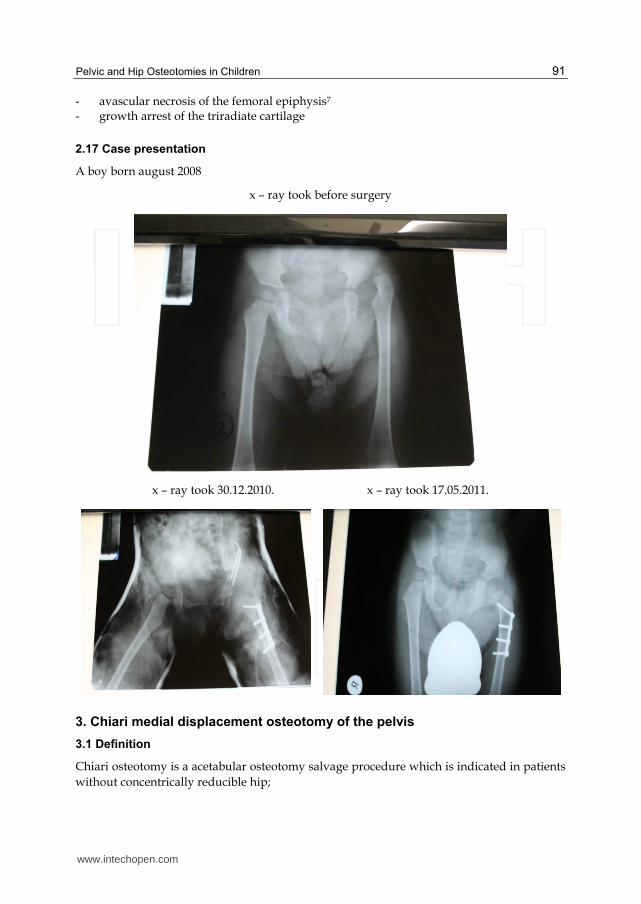
2.17 Case presentation
A boy born august 2008
x – ray took before surgery
x – ray took 30.12.2010. x – ray took 17.05.2011.
3. Chiari medial displacement osteotomy of the pelvis
3.1 Definition
Chiari osteotomy is a acetabular osteotomy salvage procedure which is indicated in patients
without concentrically reducible hip;
www.intechopen.com

The Role of Osteotomy in the Correction of Congenital and Acquired Disorders of the Skeleton
92
- goes through the iliac bone of the pelvis; ¹ - medial deplacement of acetabulum and hip joint; - improving posterior and lateral coverage; - shelf over the dysplastic, subluxated hip is formed by ilium; - goal is create a stable, pain – free hip
3.2 Indications
1. Age of patient: patient over 8 year old, adolescents, young adult 2. An irreducible lateral subluxation of the hip with moderate incongruity of the joint
3.3 Contraindications
- severe arthrosis; - age over 45; - greater proximal migration of the femoral head
3.4 Anatomy
• DDH – deficiency of the anterior and anterolateral acetabulum; • Spastic hip dysplasia – deficiency of lateral and posterolateral acetabulum; • When planning the shape and orientation of the osteotomy and positioning of the iliac
shelf over the hip joint it is necessary to consider location of acetabular deficiency; - Femoral head deformity usually involves coxa breva, coxa magna, or coxa plana; - Abductor mechanicd may be improved by simultaneous advancement of the greater
trochanter; - In cases of high dislocation of the hip and in the pelvis in patients with advanced
neurologic conditions Chiari osteotomy may not provide adequate coverage; - Additonal bone graft to supplement posterior, lateral and anterior coverage is usually
necessary; - Contcentric reduction of the femoral head into the acetabulum is not required; - Increasing the surfice area of coverage provides decreasing the force across the hip
joint; - Obligatory shortening of the gluteal muscle length and abductor moment arm is caused
by lateralizing the ilium to form a shelf that weakness the muscle and contribustes to postoperative Trendelenburg limp;
- Resting length of the gluteus medius can be restored by advancing the greater trochanter
3.5 Pathogenesis
Advanced hip disease requiring salvage surgery may be caused by following:
- late diagnosis of developmental dysplasia of the hip;
- spastic or neuromuscular hip dysplasia;
- failed prior hip procedures;
- acetabular trauma
Incomplete or incongruous femoral head coverage can be caused by following femoral head conditions:
www.intechopen.com

Pelvic and Hip Osteotomies in Children
93
- primary malformation,
- secondary avascular necrosis,
- slipped capital femoral epiphysis,
- epiphyseal – metaphyseal dysplasia,
- secondary malformation from longstanding subluxation or impingement
3.6 Patient history and physical findings
3.6.1 Main parts include
- family or personal history of DDH,
- other hip disorder,
- trauma,
- skeletal dysplasias
- cerebral palsy
- birth order and weight
3.7 The phyisical examination
- limb length,
- gait,
- strength,
- assistive devices
3.8 Specific hip tests
- Trendelenburg test: weakness in abductors
- Anterior apprehension test: patient's subjective feeling of fear or instability
- Strength of gluteus medius and maximus
- Anterior impingement test
- Bicycle test
- Range of motion: testing internal and external rotation at multiple degrees of flexion
because of variation of femoral head and acetabular deformities that can help in
location of pathologic articulation
- Galeazzi sign: shows hip subluxation or dislocation
- Preoperative requirements: flexion to 90 degrees, full extension, at least 10 to 20 degrees
of abduction
- Preoperative gait assessement: discern of limp antalgia what can be improve by Chiari
osteotomy
3.9 Imaging and other diagnostic studies
- Radiography includes: weight – bearing AP views of bilateral hips,false profile of hips,
AP of hips in maximal abduction and internal rotation what can provide assessment of
lateral and anterior coverage of the femoral head as well as congruency of the hip joint.
- 3D CT can be used in preoperatively assessing the amount and direction of acetabular
deficiency
www.intechopen.com

The Role of Osteotomy in the Correction of Congenital and Acquired Disorders of the Skeleton
94
- MRI can help in preoperative assessment of articular and labral cartilage
3.10 Diferential diagnosis
• DDH
• Spastic hip dysplasia
• Legg – Calve – Perthes diseas, avascular necrosis
• Posttraumatic hip or femoral dysplasia
• Multiple or spondyloepiphysealdysplasia
3.11 Nonoperative management
- The patient who is candidate for Chiari oteotomy usually have pain and arthrosis; - The onset of arthritic symptoms can be delayed by activity, job modification, weight loss - Physical therapy can be useful in increasing of range of motion and strength but can not
stop the onset of arthritis in the dysplastic hip
3.12 Surgical management
- Onset of spastic hip dysplasia can be delayed by hip adductor tenotomy, lengthening or Botox if performed before age 4 to 6 and if hip abduction is less than 45 degrees with hip flexed and extended;4
- Surgical correction is necessary in painful, unstable, moderate to severe dysplasia with incongruent articualtion with or without femoral head deformity;
- Arthrodesis, shelf procedures and arthroplasty are additional options to Chiari
3.13 Preoperative planning
- A complete physical examination and radiographs - If there is marked proximal migration, preoperative traction for 2 to 3 weeks can be
used to improve the position of the femoral head relative to the acetabulum
3.14 Positioning
- The position of the patient is supine ; - Antibiotic profilaxis and placement of catheter; - General anesthesia is one that we prefer
3.15 Approach
There are two approaches:
- ilioinguinal approach – begins from the iliac crest and continues medially for about 10 cm - iliofemoral approach provides better visualisation in larger patients and combined
pelvic and femoral procedures to be done through one incision
3.16 Surgical tehnique
- iliac osteotomy is angled from the sciatic notch to the ASIS – anterolateral distally to posteromedial proximally;
www.intechopen.com

Pelvic and Hip Osteotomies in Children
95
- placing iliac buttress into horizontal position should be avoided since this will cause
persistently unstable joint laterally;
- following osteotomy, a triangular osseous defect anteriorly which is stabilized with
curved plate of bone graft from iliac wing;
- consequence of inadequate stabilization of anterior defect is anterior instability;
- acetabulum is displaced medially;
- acetabulum is abducted into a more vertical and medial position and replaces it with
joint capsule supported by osseous buttress of the iliac wing;
- distal acetabular fragment is displaced medially and adducted;
- proximal iliac fragment sould not be move laterally;
- inferior surface of proximal fragment forms roof over femoral head;
- the osteotomy is fixed in place with cortical screws either along the iliac crest or along
the outer table of the ilium
3.17 Pearls and pitfalls
- adequate patient selection;
- Chiari osteotomy may incompletely manage severe arthrosis or proximal migration;
- we pay special attention when considering Chiari osteotomy in young patients or in
patients with neuromuscular disease as they may have insufficient thickness of ilium to
provide adequate coverage;
- the contents of the sciatic notch must be carefully protected by subperiosteal placement
of retractors;
- screws are placed at the iliac crest for fixation or along the outer table of the ilium in
increased displacement;
- for augmenting deficient coverage we use a corticocancellous segment of the inner table
of the ilium;
- after displacement our recommendation is palpation of the posterior edge of the
osteotomy to confirm there is no soft tissue entrapment
3.18 Postoperative care
- Patients are placed in long – leg casts held in abduction by a connector bar
3.19 Outcomes
- Good to exellent outcomes for pain relief;2,3,5,7
- We have better outcomes in younger patients with mobile hips and adequate corrected
coverage
3.20 Complications
• Infection;
• Heterotropic ossification;
• Incomplete correction and resubluxation;
• Neuropraxia of sciatic and of the lateral femoral cutaneous nerve obtained by sciatic
nerve entrapment or injury during osteotomy
www.intechopen.com

The Role of Osteotomy in the Correction of Congenital and Acquired Disorders of the Skeleton
96
3.21 Case presentation
A boy born in february 1998. Operatively treated out of our Institution, when he was one year old
x – ray took 19.10.2010. x – ray took 01.11.2010.
x – ray took 14.12.2010.
www.intechopen.com
Pelvic and Hip Osteotomies in Children
97
4. References
4.1 References part 1
[1] Flynn JM, Wiesel SW. Operative tchniques in pediatric orthopaedics (Lippincott
Williams &Wikins, 2011).
[2] Tukenmez M, Tezeren G. Salter innominate osteotomy for treatment of developmental
dysplasia of the hip. Journal of Orthopedic Surgery 2007;15(3):286-90.
[3] Barrett WP, Staheli LT, Chew DE. The effectiveness of the Salter innominate osteotomy
in the treatment of congenital dislocation of the hip. J Bone Joint Surg Am
1984;68A:79-87.
[4] Macnicol MF, Bertol P. The Salter innominate osteotomy: should it be combined with
concurrent open reduction? J Pediatr Orthop B 2005;14:415-421.
[5] Böhm P, Brzuske A. Salter Innominate Osteotomy for the Treatment of Development
Dysplasia of the Hip in Children: Results of Seventy – three Consecutive
Osteotomies After Twenty – sixt to Thirty five years of follow - up. JBJS
2002;84:178-186.
[6] Rab GT. Biomechanical aspects of Salter osteotomy. Clin Orthop Relat Res 1978;132:82-
87.
[7] Gur E, Sarlak O. The complications of Salter innominate osteotomy in the treatment of
congenital dislocation of the hip. Acta Orthop B 2005;14:415-421.
[8] Sarban S, Ozturk A, Tabur H, et al. Anteversion of the acetabulum and femoral neck in
early walking age patients with developmental dysplasia of the hip. J Pediatr
Orthop B 2005;14:410-414.
[9] Gillingham BL, Sanche AA, Wenger DR. Pelvic osteotomies for the treatment of hip
dysplasia in children and young adults. J Am Acad Orthop Surg 1999;7:325 –
337
4.2 References part 2
[1] Flynn JM, Wiesel SW. Operative tchniques in pediatric orthopaedics (Lippincott
Williams &Wikins, 2011).
[2] Yanagimoto S, Hotta H, Izumida R, Sakamaki T. Long – term results of the Chiari pelvic
osteotomy in patients with developmental dysplasia of the hip: indications for the
Chiari pelvic osteotomy according to disease stage and femoral head shape. J
Orthop Sci. 2005; 10(6):557-63.
[3] Ito H, Matsuno T, Minami A. Chiari pelvic osteotomy for advanced osteoarthritis in
patient with hip dysplasia. J Bone Joint Surg Am 2004; 86A: 1439 – 1445.
[4] Debnath UK, Guha AR, Karlakki S, et al. Combined femoral and Chiari osteotomies for
reconstruction of the painful subluxation or dislocation of the hip in cerebral palsy:
a long – term outcome study. J Bone Joint Surg Br 2006;88B:1373 – 1378.
[5] Bailey TE, Hall JE. Chiari medial displacement osteotomy. J Pediatr Orthop 1985;5:635 –
641.
[6] Windager R, Pongracz N, Schonecker W, et al. Chiari osteotomy for congenital
dislocation and subluxation of the hip: results after 20 to 34 years follow – up. J
Bone Joint Surg Br 1991;73B:890 – 895.
www.intechopen.com

The Role of Osteotomy in the Correction of Congenital and Acquired Disorders of the Skeleton
98
[7] Gagala J, Blacha J, Bednarek A. Chiari pelvic osteotomy in the treatment of hip dysplasia
in adults. Chir Narzadow Ruchu Ortop. Pol. 2006;71(3):183-5
www.intechopen.com
The Role of Osteotomy in the Correction of Congenital andAcquired Disorders of the SkeletonEdited by Prof. James Waddell
ISBN 978-953-51-0495-7Hard cover, 294 pagesPublisher InTechPublished online 11, April, 2012Published in print edition April, 2012
InTech EuropeUniversity Campus STeP Ri Slavka Krautzeka 83/A 51000 Rijeka, Croatia Phone: +385 (51) 770 447 Fax: +385 (51) 686 166www.intechopen.com
InTech ChinaUnit 405, Office Block, Hotel Equatorial Shanghai No.65, Yan An Road (West), Shanghai, 200040, China
Phone: +86-21-62489820 Fax: +86-21-62489821
This book demonstrates specific osteotomy techniques from the skull to the hallux. The role of osteotomy inthe correction of deformity is under appreciated in part because of the ubiquitous nature of joint replacementsurgery. It should be remembered, however, that osteotomy has a role to play in the correction of deformity inthe growing child, the active young adult, and patients of any age with post-traumatic deformity limiting functionand enjoyment of life. In this text we bring you a number of papers defining specific problems for whichosteotomy is found to be an effective and lasting solution. I hope you find it useful.
How to referenceIn order to correctly reference this scholarly work, feel free to copy and paste the following:
I. Gavrankapetanovic (2012). Pelvic and Hip Osteotomies in Children, The Role of Osteotomy in the Correctionof Congenital and Acquired Disorders of the Skeleton, Prof. James Waddell (Ed.), ISBN: 978-953-51-0495-7,InTech, Available from: http://www.intechopen.com/books/the-role-of-osteotomy-in-the-correction-of-congenital-and-acquired-disorders-of-the-skeleton/pelvic-and-hip-osteotomy-in-children