100 cases in paediatrics.pdf
TRANSCRIPT

8/20/2019 100 cases in paediatrics.pdf
http://slidepdf.com/reader/full/100-cases-in-paediatricspdf 1/320

8/20/2019 100 cases in paediatrics.pdf
http://slidepdf.com/reader/full/100-cases-in-paediatricspdf 2/320
100 CASESin Paediatrics

8/20/2019 100 cases in paediatrics.pdf
http://slidepdf.com/reader/full/100-cases-in-paediatricspdf 3/320
This page intentionally left blank

8/20/2019 100 cases in paediatrics.pdf
http://slidepdf.com/reader/full/100-cases-in-paediatricspdf 4/320
100 CASESin Paediatrics
J. E. Raine MD FRCPCH DCHConsultant Paediatrician, Whittington Hospital, London;Honorary Senior Lecturer, University College, London, UK
A. J. Cunnington BMBCh MA MRCPCH DTM&HSpecialist Registrar in Paediatrics, London Deanery;Clinical Research Fellow, Immunology Unit,London School of Hygiene and Tropical Medicine, London, UK
J. M. Walker BA FRCP FRCPCHConsultant Paediatrician, Portsmouth Hospitals NHS Trust,Portsmouth, UK
Volume Editor:
J. E. Raine
100 Cases Series Editor:
P. John Rees M FRCPDean of Medical Undergraduate Education, King’s College LondonSchool of Medicine at Guy’s, King’s and St Thomas’ Hospitals, London, UK
8/20/2019 100 cases in paediatrics.pdf
http://slidepdf.com/reader/full/100-cases-in-paediatricspdf 5/320
First published in Great Britain in 2009 byHodder Arnold, an imprint of Hodder Education, an Hachette UK Company,338 Euston Road, London NW1 3BH
http://www.hoddereducation.com
© 2009 Hodder Arnold
All rights reserved. Apart from any use permitted under UK copyright law, this publication may only bereproduced, stored or transmitted, in any form, or by any means with prior permission in writing of thepublishers or in the case of reprographic production in accordance with the terms of licences issued by theCopyright Licensing Agency. In the United Kingdom such licences are issued by the Copyright LicensingAgency: Saffron House, 6-10 Kirby Street, London EC1N 8TS.
Whilst the advice and information in this book are believed to be true and accurate at the date of going topress, neither the authors nor the publisher can accept any legal responsibility or liability for any errors oromissions that may be made. In particular (but without limiting the generality of the preceding disclaimer)every effort has been made to check drug dosages; however it is still possible that errors have been missed.Furthermore, dosage schedules are constantly being revised and new side-effects recognized. For these rea-sons the reader is strongly urged to consult the drug companies' printed instructions before administeringany of the drugs recommended in this book.
British Library Cataloguing in Publication DataA catalogue record for this book is available from the British Library
Library of Congress Cataloging-in-Publication DataA catalog record for this book is available from the Library of Congress
ISBN 978 0 340 96875 8
1 2 3 4 5 6 7 8 9 10
Commissioning Editor: Joanna KosterProject Editor: Francesca NaishProduction Controller: Karen TateCover Designer: Amina DudhiaIndexer: Laurence Errington
Typeset in 10/12 Optima by Macmillan Publishing Solutions(www.macmillansolutions.com)Printed & bound in India
What do you think about this book? Or any other Hodder Arnold title?Please visit our website: www.hoddereducation.com

8/20/2019 100 cases in paediatrics.pdf
http://slidepdf.com/reader/full/100-cases-in-paediatricspdf 6/320
v
To Laine, Kooks and Benjo

8/20/2019 100 cases in paediatrics.pdf
http://slidepdf.com/reader/full/100-cases-in-paediatricspdf 7/320

8/20/2019 100 cases in paediatrics.pdf
http://slidepdf.com/reader/full/100-cases-in-paediatricspdf 8/320
vii
CONTENTS
Preface xi Acknowledgements xiii List of abbreviations xv Normal values xvii
RespiratoryCase 1: An infant with noisy breathing 1Case 2: A child with noisy breathing 3Case 3: A chesty infant 7Case 4: A chronic cough 11Case 5: Recurrent chest infections 14Case 6: A wheezy teenager 19Case 7: Fever and breathlessness 22Case 8: A teenager with chest pain 27
CardiologyCase 9: A cyanosed newborn 29Case 10: A shocked neonate 31Case 11: A pale, breathless baby 33Case 12: An incidental murmur 37Case 13: A funny turn 41
Endocrinology and diabetesCase 14: A thirsty boy 45
Case 15: A tall boy 47Case 16: A short girl 50Case 17: An overweight boy 53Case 18: A girl with early puberty 56Case 19: A boy with delayed puberty 59Case 20: Is it a boy or a girl? 61Case 21: A boy with breasts 63Case 22: A boy with bow legs 66
GastroenterologyCase 23: A vomiting Infant 71Case 24: A child with bloody diarrhoea 73Case 25: A teenager with chronic diarrhoea 75Case 26: Acute diarrhoea and vomiting 79Case 27: Acute abdominal pain 83Case 28: Recurrent abdominal pain 85Case 29: A constipated toddler 89Case 30: An infant with poor weight gain 92Case 31: An infant with persistent jaundice 97

8/20/2019 100 cases in paediatrics.pdf
http://slidepdf.com/reader/full/100-cases-in-paediatricspdf 9/320
viii
Case 32: Abdominal pain and jaundice 99Case 33: A lump in the groin 101
NephrologyCase 34: Abdominal pain and dysuria 105Case 35: Red urine 107Case 36: A puffy face 111Case 37: A bed wetter 113Case 38: High blood pressure 115
InfectionsCase 39: Fever and a rash 117Case 40: Fever in a returning traveller 119Case 41: Sticky eyes 121
Case 42: A persistent fever 123Case 43: Recurrent infections 125Case 44: Unexplained weight loss 128
DermatologyCase 45: An itchy rash 133Case 46: Deteriorating eczema 136Case 47: An infant with blisters 140
HaematologyCase 48: A pale child 143Case 49: Easy bruising 147Case 50: A child with chest pain 150
OncologyCase 51: An abdominal mass 155Case 52: An unsteady child 157Case 53: Lymphadenopathy 161Case 54: A swollen shoulder 163
Case 55: Fever in a patient on chemotherapy 165Case 56: Aches and pains 169
Bones and jointsCase 57: A girl with a limp 172Case 58: Pain and fever 177Case 59: Swollen joints 179Case 60: A spinal deformity 183
NeurologyCase 61: A fitting child 185Case 62: A febrile, drowsy child 187Case 63: A big head 190Case 64: A child in a coma 194Case 65: A late walker 197Case 66: A child with learning difficulties 199Case 67: Sudden weakness 201Case 68: Chronic headaches 205Case 69: A funny smile 207
Contents
8/20/2019 100 cases in paediatrics.pdf
http://slidepdf.com/reader/full/100-cases-in-paediatricspdf 10/320
ix
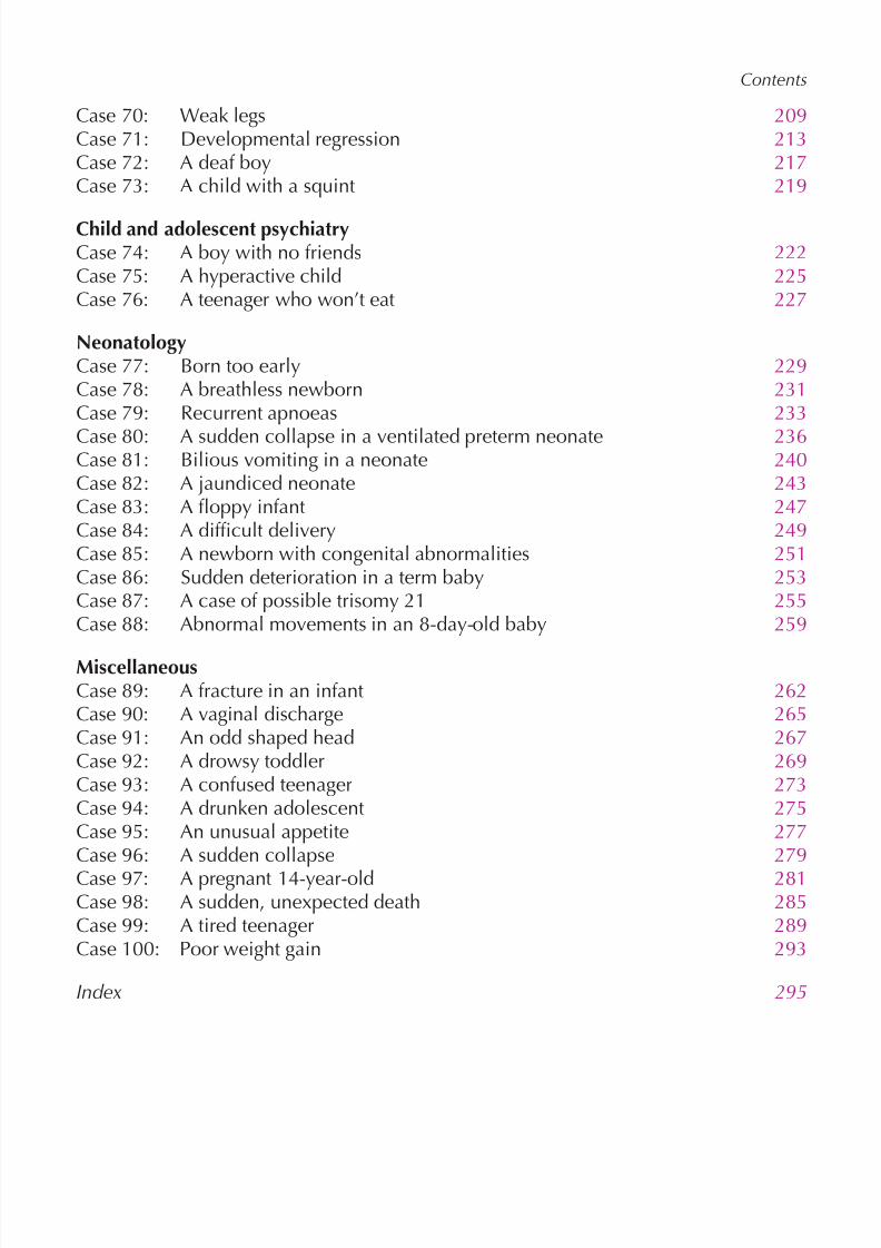
Case 70: Weak legs 209Case 71: Developmental regression 213Case 72: A deaf boy 217Case 73: A child with a squint 219
Child and adolescent psychiatryCase 74: A boy with no friends 222Case 75: A hyperactive child 225Case 76: A teenager who won’t eat 227
NeonatologyCase 77: Born too early 229Case 78: A breathless newborn 231Case 79: Recurrent apnoeas 233
Case 80: A sudden collapse in a ventilated preterm neonate 236Case 81: Bilious vomiting in a neonate 240Case 82: A jaundiced neonate 243Case 83: A floppy infant 247Case 84: A difficult delivery 249Case 85: A newborn with congenital abnormalities 251Case 86: Sudden deterioration in a term baby 253Case 87: A case of possible trisomy 21 255Case 88: Abnormal movements in an 8-day-old baby 259
MiscellaneousCase 89: A fracture in an infant 262Case 90: A vaginal discharge 265Case 91: An odd shaped head 267Case 92: A drowsy toddler 269Case 93: A confused teenager 273Case 94: A drunken adolescent 275Case 95: An unusual appetite 277Case 96: A sudden collapse 279Case 97: A pregnant 14-year-old 281Case 98: A sudden, unexpected death 285Case 99: A tired teenager 289Case 100: Poor weight gain 293
Index 295
Contents

8/20/2019 100 cases in paediatrics.pdf
http://slidepdf.com/reader/full/100-cases-in-paediatricspdf 11/320
This page intentionally left blank

8/20/2019 100 cases in paediatrics.pdf
http://slidepdf.com/reader/full/100-cases-in-paediatricspdf 12/320
xi
PREFACE
Paediatrics is a fascinating and multifaceted speciality. As well as dealing with thestandard medical conditions that arise in children, it covers neonatology and the pre-term infant (often newcomers to a neonatal intensive care unit see it as ‘another world’),genetics, ethics, child development, child protection, and child and adolescent psych-
iatry. During the short medical student paediatric attachments, and in the era of decreas-ing hours of work for junior doctors, the exposure to the diverse range of paediatricconditions is limited.
In the 100 cases that follow, we have tried to cover the majority of the key areas in paedi-atrics. We have tackled problems that arise in different settings, such as primary care, andemergency departments, paediatric outpatients, the paediatric ward and the maternity andneonatal intensive care units. We hope to have done so in a way that is interesting andthat brings the cases alive. After reading through the case and questions, the reader shouldcarefully consider their answer and ideally commit their thoughts to paper, prior to look-
ing at the answers over the page. We have also tried to demonstrate how a senior paedi-atrician would approach and work their way through the clinical problem and to explainsome underlying principles in a way that will help cement understanding and knowledge.
The book is aimed at medical students, foundation year doctors doing paediatrics and junior doctors studying for their MRCPCH. We hope that these cases will be enjoyableand that they will keep the ‘grey cells’ stimulated!
J E Raine A J Cunnington
J M Walker October 2008

8/20/2019 100 cases in paediatrics.pdf
http://slidepdf.com/reader/full/100-cases-in-paediatricspdf 13/320
This page intentionally left blank
8/20/2019 100 cases in paediatrics.pdf
http://slidepdf.com/reader/full/100-cases-in-paediatricspdf 14/320

8/20/2019 100 cases in paediatrics.pdf
http://slidepdf.com/reader/full/100-cases-in-paediatricspdf 15/320
This page intentionally left blank
8/20/2019 100 cases in paediatrics.pdf
http://slidepdf.com/reader/full/100-cases-in-paediatricspdf 16/320
xv
ABBREVIATIONS
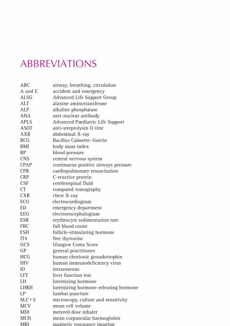
ABC airway, breathing, circulation A and E accident and emergency ALSG Advanced Life Support Group ALT alanine aminotransferase ALP alkaline phosphatase ANA anti-nuclear antibody APLS Advanced Paediatric Life Support ASOT anti-streptolysin O titre AXR abdominal X-ray BCG Bacillus Calmette–GuerinBMI body mass indexBP blood pressureCNS central nervous systemCPAP continuous positive airways pressure
CPR cardiopulmonary resuscitationCRP C-reactive proteinCSF cerebrospinal fluidCT computed tomography CXR chest X-ray ECG electrocardiogramED emergency departmentEEG electroencephalogramESR erythrocyte sedimentation rate
FBC full blood countFSH follicle-stimulating hormonefT4 free thyroxineGCS Glasgow Coma ScoreGP general practitioner HCG human chorionic gonadotrophinHIV human immunodeficiency virusIO intraosseousLFT liver function testLH luteinizing hormoneLHRH luteinizing hormone-releasing hormoneLP lumbar punctureM,C S microscopy, culture and sensitivity MCV mean cell volumeMDI metered dose inhaler MCH mean corpuscular haemoglobinMRI magnetic resonance imagingMSU midstream urineNAI non-accidental injury

8/20/2019 100 cases in paediatrics.pdf
http://slidepdf.com/reader/full/100-cases-in-paediatricspdf 17/320
xvi
NG nasogastricNAD no abnormality detectedPCR polymerase chain reactionPEFR peak expiratory flow rate
PICU paediatric intensive care unitp.r.n. as requiredRAST radioallergosorbent testSLE systemic lupus erythematosusTB tuberculosisTFT thyroid function testTSH thyroid-stimulating hormoneU Es urea and electrolytesURTI upper respiratory tract infectionUS ultrasoundUTI urinary tract infection
WBC white blood cells
Abbreviations

8/20/2019 100 cases in paediatrics.pdf
http://slidepdf.com/reader/full/100-cases-in-paediatricspdf 18/320
xvii
NORMAL VALUES
Respiratory rate at rest Age (years) Respiratory rate (breaths/min)
1 30–401–2 25–352–5 25–305–12 20–25
12 15–20
Heart rateAge (years) Heart rate (beats/min)
1 110–1601–2 100–1502–5 95–1405–12 80–120
12 60–100
Systolic blood pressureAge (years) Systolic blood pressure (mmHg)
1 70–901–2 80–952–5 80–1005–12 90–110
12 100–120
(Reproduced with kind permission of the Advanced Life Support Group from Advanced Paediatric Life Support , Blackwell Publishing, 2005.)
NORMAL VALUES

8/20/2019 100 cases in paediatrics.pdf
http://slidepdf.com/reader/full/100-cases-in-paediatricspdf 19/320
This page intentionally left blank

8/20/2019 100 cases in paediatrics.pdf
http://slidepdf.com/reader/full/100-cases-in-paediatricspdf 20/320
1
RESPIRATORY
CASE 1:AN INFANT WITH NOISY BREATHING
HistoryMohammed is a 3-month-old boy, brought to a paediatric rapid referral clinic because ofpersistent noisy breathing. He was born in the UK at term after an uneventful pregnancy andis the fifth child of non-consanguineous Somalian parents. His birth weight was 3.7 kg (75thcentile). Since he was a few weeks old, he has had noisy breathing, which hasn’t affected hisfeeding, and his parents were repeatedly reassured that it would get better. He has continuedto have intermittent noisy breathing, especially when agitated, and sometimes during sleep.Over the last few days, his breathing has been noisier than usual. Otherwise he has been wellwithout any fevers. All of his siblings have recently had coughs and colds.
ExaminationHe is active and smiles responsively. Oxygen saturations are 95 per cent in air and histemperature is 36.9ºC. He is coryzal and has intermittent stridor. There is a small ‘straw-berry’ haemangioma on his forehead. Respiratory rate is 45/min, there is subcostal reces-sion and mild tracheal tug. Air entry is symmetrical in the chest, with no crackles orwheeze. Cardiovascular examination is unremarkable. His weight is 6.7 kg (75th centile).
Questions• What is the most likely cause of his stridor?• What other important diagnoses need to be considered?• How can the diagnosis be confirmed?
8/20/2019 100 cases in paediatrics.pdf
http://slidepdf.com/reader/full/100-cases-in-paediatricspdf 21/320
2
100 Cases in Paediatrics
ANSWER 1Stridor is an inspiratory sound due to narrowing of the upper airway. Mohammed is mostlikely to have stridor due to laryngomalacia. This means that the laryngeal cartilage is softand floppy, with an abnormal epiglottis and/or arytenoid cartilages. The larynx collapsesand narrows during inspiration (when there is a negative intrathoracic pressure), resulting ininspiratory stridor. It is usually a benign condition with noisy breathing but no major prob-lems with feeding or significant respiratory distress. Most cases resolve spontaneously withina year as the larynx grows and the cartilaginous rings stiffen. The reason Mohammed nowhas respiratory distress is that he has an intercurrent viral upper respiratory tract infection.
A very important diagnosis to consider in this boy is a haemangioma in the upper airway.The majority of haemangiomas are single cutaneous lesions, but they can also occur at
other sites and the upper airway is one position where they can enlarge with potentiallylife-threatening consequences. The presence of one haemangioma increases the likelihoodof a second one. This boy should be referred for assessment by an ENT surgeon.
There are many other possible congenital causes of stridor which affect the structure orfunction of the upper airway. Infectious causes of stridor, such as croup and epiglottitis,are very rare in this age group.
!
Differential diagnosis of stridor in an infant• Laryngomalacia• Laryngeal cyst, haemangioma or web• Laryngeal stenosis• Vocal cord paralysis• Vascular ring• Gastro-oesophageal reflux• Hypocalcaemia (laryngeal tetany)• Respiratory papillomatosis• Subglottic stenosis
The diagnosis of laryngomalacia can be confirmed by visualization of the larynx usingflexible laryngoscopy. This can be done by an ENT surgeon as an outpatient procedure.This demonstrates prolapse over the airway of an omega-shaped epiglottis or arytenoidcartilages. Congenital structural abnormalities may also be seen. Lesions below the vocalcords may require bronchoscopy, CT or an MRI scan for diagnosis.
KEY POINTS
• The commonest cause of congenital stridor is laryngomalacia.• Laryngomalacia can be exacerbated by intercurrent respiratory infections.

8/20/2019 100 cases in paediatrics.pdf
http://slidepdf.com/reader/full/100-cases-in-paediatricspdf 22/320
3
CASE 2:A CHILD WITH NOISY BREATHING
HistoryEwa is a 4-year-old child who presents to the ED with a sudden onset of noisy breathing.She has had a runny nose for 2 days, a cough for 1 day and developed noisy breathing3 hours earlier. Her mother feels that she is getting progressively more breathless. Her fatherhad a cold the previous week. She is otherwise well but has troublesome eczema which istreated with emulsifiers and steroid creams. Her mother states that she is allergic to peanuts,as they lead to a deterioration of the eczema within 1–2 hours. She avoids peanuts and alltypes of nuts. She is fully immunized. Her 8-year-old sister has asthma.
ExaminationOxygen saturation is 89 per cent in air. Her temperature is 38.0 C. There is loud noisy breath-ing, mainly on inspiration. Her respiratory rate is 52/min with supracostal and intercostalrecession. On auscultation, there are no crackles or wheezes. There are no other signs.
Questions• What is the most likely diagnosis?• What is the differential diagnosis?
• What is the treatment?
8/20/2019 100 cases in paediatrics.pdf
http://slidepdf.com/reader/full/100-cases-in-paediatricspdf 23/320
4
100 Cases in Paediatrics
ANSWER 2The most likely diagnosis is laryngotracheobronchitis (croup). This child has stridor, whichis an inspiratory sound secondary to narrowing of the upper airway. In contrast, wheezeis an expiratory sound caused by narrowing of the lower airways. The effort required toshift air through the narrowed airway has resulted in tachypnoea and recession.
The upper airway of a child with stridor should not be examined and the child shouldnot be upset by performing painful procedures such as blood tests. This is becausethere is a small risk that this may lead to a deterioration, causing partial obstruction toprogress to complete obstruction and a respiratory arrest.
! Differential diagnosis of acute stridor
• Laryngotracheobronchitis• Inhaled foreign body• Anaphylaxis• Epiglottitis• Rare causes include:
– Bacterial tracheitis– Severe tonsillitis with very large tonsils– Inhalation of hot gases (e.g. house fire)
– Retropharyngeal abscess
Croup typically occurs in children aged 6 months to 5 years. It is characterized by anupper respiratory tract infection that is followed by a barking-type cough, a hoarse
voice, stridor and a low-grade fever. Croup is most commonly caused by the parainflu-enza virus.
When a foreign body is inhaled, there is usually a history of sudden coughing and/or chok-ing in a child that was previously well. There may be accompanying cyanosis. The foreignbody is usually a food (e.g. peanut) but may be a small toy. On examination there may be aunilateral wheeze with decreased air entry on one side.
This case is not typical of anaphylaxis, in that there is no history of the child havinghad peanuts. Nor are there features that often accompany anaphylaxis, such as an itchyurticarial rash, facial swelling, vomiting, wheeze or hypotension.
Epiglottitis would be very unlikely in a fully immunized child who would have receivedthe Haemophilus influenzae vaccine.
Initial management deals with the ABC. As the oxygen saturation is low, high-flow 100per cent oxygen will be needed to elevate the saturation to 95 per cent.
The first step in the treatment of croup is oral dexamethasone. A less frequently usedalternative is nebulized budesonide. If 2–3 hours later the child has improved and theoxygen saturation is 95 per cent in air, the child can be discharged. In some casesa further dose of steroids can be administered 12–24 hours later. If the child deterio-rates then nebulized adrenaline can be administered. If adrenaline is required then seniorhelp and an anaesthetist should be summoned urgently. If the child deteriorates further(increasing tachypnoea, recession and exhaustion) then intubation and ventilation are

8/20/2019 100 cases in paediatrics.pdf
http://slidepdf.com/reader/full/100-cases-in-paediatricspdf 24/320
5
Respiratory
required to secure the airway and to prevent hypoxia and its sequelae. If intubation isunsuccessful, an ENT surgeon will be required to perform an emergency tracheostomy.
KEY POINTS• Stridor is due to upper airway obstruction.• The upper airway of a child with stridor should not be examined as this may
precipitate total obstruction.• Laryngotracheobronchitis is the commonest cause of acute stridor.

8/20/2019 100 cases in paediatrics.pdf
http://slidepdf.com/reader/full/100-cases-in-paediatricspdf 25/320
This page intentionally left blank

8/20/2019 100 cases in paediatrics.pdf
http://slidepdf.com/reader/full/100-cases-in-paediatricspdf 26/320
7
CASE 3:A CHESTY INFANT
HistoryMax is a 3-month-old boy seen in the community by his GP. He developed a runny noseand bit of a cough 2 days ago but has become progressively more chesty and has nowgone off his feeds and is having far fewer wet nappies. He has two older siblings whoalso have colds. He was born at 34 weeks’ gestation but had no significant neonatalproblems and went home at 2 weeks of age. Both parents smoke but not in the house.His mother had asthma as a child.
ExaminationMax is miserable but alert. His airway is clear. He is febrile (37.8 C) and has copious clearnasal secretions and a dry wheezy cough. His respiratory rate is 56 breaths/min with tra-cheal tug and intercostal and subcostal recession. On auscultation, there are widespreadfine crackles and expiratory wheeze. The remainder of the examination is unremarkable.
Questions• What is the most likely diagnosis?• What is the commonest causative organism?
• What are the indications for referral to hospital?• What is the management in hospital?
8/20/2019 100 cases in paediatrics.pdf
http://slidepdf.com/reader/full/100-cases-in-paediatricspdf 27/320
8
100 Cases in Paediatrics
ANSWER 3This baby has the characteristic clinical features of acute bronchiolitis – a seasonal viralillness occurring from early autumn to spring, principally affecting infants.
The commonest causative organism is respiratory syncytial virus (RSV), which is respon-sible for about 80 per cent of infections. In hospital, a nasopharyngeal aspirate (NPA) maybe sent for viral immunofluorescence, polymerase chain reaction (PCR) or culture. This islargely for infection control and epidemiology and does not affect acute management.
Around 2–3 per cent of all infants are admitted each year with RSV-positive bronchiolitisbut many more are managed at home. Prevention is possible with monoclonal RSV immuno-globulin (Palivizumab) but this is reserved for high-risk infants, e.g. oxygen-dependentsurvivors of prematurity, as it is extremely expensive. There is no immunization.
! Indications for hospital referral
• Apnoeic episodes (commonest in babies 2 months and may be the presentingfeature)
• Intake 50 per cent of normal in preceding 24 hours• Cyanosis• Severe respiratory distress – grunting, nasal flaring, severe recession, respiratory
rate 70/min• Congenital heart disease, pre-existing lung disease or immunodeficiency• Significant hypotonia, e.g. trisomy 21 – less likely to cope with respiratory
compromise• Survivor of extreme prematurity• Social factors
Babies usually deteriorate over the first 48–72 hours. Hence there is a low threshold foradmitting any baby 2 months of age on day 1–2 of their illness as they may deteri-orate and become exhausted and apnoeic.
Management is supportive. Investigations are rarely indicated apart from an NPA. A chest X-ray is only needed if the clinical course is unusual and often leads to unnec-essary antibiotic prescriptions. Blood tests are only required if there is diagnostic uncer-tainty, e.g. if the infant has a temperature 39 C and a superadded bacterial respiratoryinfection is suspected. Oxygen saturations should be kept at 92 per cent and theinfant should be nasogastrically fed if they cannot maintain 50 per cent of normalintake. Intravenous fluids are used in severe cases. All fluids are restricted to two-thirds
of maintenance. Nasal and oral suction is helpful. There is no evidence that broncho-dilators, oral or inhaled steroids modify the clinical course or any important outcomessuch as the need for ventilation or the length of stay. A capillary blood gas should bechecked if the infant is deteriorating. Every season a small proportion of infants needhigh-dependency or intensive care – most respond well to continuous positive airwayspressure (CPAP), avoiding the need for intubation.
Babies are discharged when they are well enough to continue recovering at home butmany continue to cough and wheeze for weeks and get similar symptoms with subse-quent upper respiratory tract infections. Response to conventional asthma treatment is

8/20/2019 100 cases in paediatrics.pdf
http://slidepdf.com/reader/full/100-cases-in-paediatricspdf 28/320
9
Respiratory
variable. Leukotriene antagonists may have a role. Exposure to tobacco smoke must beavoided.
KEY POINTS
• Bronchiolitis is a clinical diagnosis.• Numerous well-conducted studies have shown no benefit from any drug intervention
in the acute phase or in the prevention of long-term sequelae.• Monoclonal RSV immunoglobulin (Palivizumab) may be given for prevention to high-
risk infants, but the costs of widespread use outweigh the benefits.

8/20/2019 100 cases in paediatrics.pdf
http://slidepdf.com/reader/full/100-cases-in-paediatricspdf 29/320
This page intentionally left blank

8/20/2019 100 cases in paediatrics.pdf
http://slidepdf.com/reader/full/100-cases-in-paediatricspdf 30/320
11
CASE 4:A CHRONIC COUGH
HistoryDonna is a 12-year-old girl seen in the GP surgery with her mother. This is her fourth visitin 3 months. Her initial presentation was with a headache, fever, malaise, a sore throat anda symmetrical non-pruritic rash on her arms and hands. The lesions varied in size and char-acter, some being simple red macules and others being up to 2 cm in diameter with a central,slightly dusky centre and a surrounding ‘halo’ of varying erythema. A diagnosis of a viralinfection was made. However, these symptoms progressed to include a cough productive ofwhite sputum. The computer records show that the emergency GP they consulted at the timeheard some crackles throughout the chest and prescribed a course of clarithromycin. All ofher symptoms have resolved, except for her cough. This is mostly during the day and isnot waking her or her family. However, it is disrupting her life because she is being senthome from school and her parents have excluded her from sport. It is a spasmodic unpro-ductive cough that comes in bouts, which are occasionally severe enough to cause vomiting.
Another GP gave her a trial of inhaled salbutamol but with no apparent improvement. Shehas never had any obvious nasal symptoms. Donna is otherwise well and recently started herperiods. She is fully immunized. Her father has a history of asthma. Her mother smokes but‘not around the children’. There is no history of recent foreign travel and no family history or
contact with tuberculosis.
ExaminationDonna looks well. Her height is on the 91st centile and her weight is on the 75th cen-tile. There has been appropriate weight gain since her illness began. She is not clubbedor anaemic. She is afebrile. There is no significant lymphadenopathy. Examination of theears, nose and throat is normal. Her pulse is 72 beats/min, her heart sounds are normaland there are no murmurs. Inspection of the chest is normal and her respiratory rate is18 breaths/min. Expansion, percussion and auscultation are normal. Examination of the
abdomen is unremarkable.
Questions• What is the differential diagnosis?• What is the most likely diagnosis?• What was the rash?• What is the management?
INVESTIGATIONS
Full blood count NormalC-reactive protein NormalErythrocyte sedimentation rate NormalChest X-ray 6 weeks previously Normal

8/20/2019 100 cases in paediatrics.pdf
http://slidepdf.com/reader/full/100-cases-in-paediatricspdf 31/320
12
100 Cases in Paediatrics
ANSWER 4Cough is one of the commonest symptoms in childhood and indicates irritation of nervereceptors within the airway.
! Differential diagnosis of a recurrent or persistent cough in childhood
• Recurrent viral URTIs – very common in all age groups but more so in infantsand toddlers
• Asthma – unlikely without wheeze or dyspnoea• Allergic rhinitis – often nocturnal due to ‘post-nasal drip’
• Chronic non-specific cough – probably post-viral with increased cough receptorsensitivity• Post-infectious – a ‘pertussis (whooping cough)-like’ illness can continue for
months following pertussis, adenovirus, mycoplasma and chlamydia• Recurrent aspiration – gastro-oesophageal reflux• Environmental – especially smoking, active or passive• Suppurative lung disease – cystic fibrosis or primary ciliary dyskinesia• Tuberculosis• Habit
Donna is otherwise healthy with no evidence of any chronic disease and she has a normalchest X-ray. Although her father has asthma, she has no convincing features of atopy andshe did not respond to inhaled salbutamol. The history is not that of recurrent aspiration.The abrupt onset of symptoms with systemic features suggests infection, and the descrip-tion of her cough as spasmodic bouts with occasional vomiting is that of a ‘pertussis-like’illness. This can continue for months following an infection, as can a chronic, non-specificcough following a viral infection.
The acute history is very typical, although not specific, for Mycoplasma pneumoniae infec-tion. This aetiology is supported by the rash, which has the characteristic clinical featuresof erythema multiforme (EM). As expected from the name, EM has numerous morpho-logical features but the diagnosis is made on finding the classic target-like papules with anerythematous outer border, an inner pale ring and a dusky purple to necrotic centre. It isoccasionally mistaken for urticaria, but EM is largely asymptomatic and the lesions do notfade within 24 hours. Infection, frequently mycoplasma, is one of numerous causes. Notreatment is indicated.
These symptoms cause understandable distress and anxiety and a belief that there mustbe something wrong and that treatment is necessary. As in this case, children must have athorough clinical evaluation to exclude serious and treatable pathology. This, plus an expla-nation that they can expect the cough to take months to resolve, is usually adequate toreassure families. Explore the reasons behind their anxiety and encourage a return to nor-mality knowing that no harm is being done. A watch-and-wait policy is best, resisting anypressure to investigate further or to try other treatments such as inhaled steroids. The onething the family can do is to ban smoking in the house and this could be the spur for hermother to give up altogether.

8/20/2019 100 cases in paediatrics.pdf
http://slidepdf.com/reader/full/100-cases-in-paediatricspdf 32/320
13
Respiratory
KEY POINTS
• Cough is one of the commonest symptoms in childhood and is usually due to viralrespiratory tract infections.
• A chronic cough may indicate a serious disorder and all such children should have athorough clinical review to exclude significant pathology.

8/20/2019 100 cases in paediatrics.pdf
http://slidepdf.com/reader/full/100-cases-in-paediatricspdf 33/320
14
CASE 5:RECURRENT CHEST INFECTIONS
HistoryConor is a 4-year-old boy who is admitted to the paediatric ward from the A&E depart-ment with pneumonia. This is his fourth hospital admission. In the first year of life, hewas admitted twice with bronchiolitis, requiring several days on oxygen, and about 6months ago he was admitted with pneumonia, again requiring oxygen and intravenousantibiotics. He has had many courses of oral antibiotics over the last few years fromhis GP for chest infections. He also has recurrent abdominal pain and his parents reportlarge offensive stools. His parents both smoke 20–30 cigarettes/day. He is unimmunizedas his parents are worried about potential side-effects.
ExaminationHe is small (height ninth centile, weight second centile), pale-looking, miserable and
very clingy to his mother. He has finger clubbing. His temperature is 38.7 C, respiratoryrate 40 breaths/min, heart rate 140 beats/min and oxygen saturation 89 per cent in air(95 per cent in facemask oxygen). There is reduced air entry at the left base with bron-chial breath sounds in the left midzone, and coarse crackles are heard on both sides ofthe chest. Cardiovascular examination is unremarkable. His abdomen is mildly distended
but non-tender.
INVESTIGATIONS
Normal
Haemoglobin 10.1 g/dL 11.0–13.8 g/dLWhite cell count 19.7 109 /L 6–17 109 /LPlatelets 401 109 /L 210–490 109 /LImmunoglobulin G 10.2 g/L 5.0–15.0 g/L
Immunoglobulin A 2.5 g/L 0.3–3.0 g/LImmunoglobulin M 1.8 g/L 0.4–2.0 g/LChest radiograph – see Figure 5.1
Figure 5.1 Conor’s chest radiograph.

8/20/2019 100 cases in paediatrics.pdf
http://slidepdf.com/reader/full/100-cases-in-paediatricspdf 34/320
15
Respiratory
Questions• What does the chest radiograph show?• What is the likely underlying diagnosis?• What investigation would you do to confirm the diagnosis?
• What are the other manifestations of this disease?

8/20/2019 100 cases in paediatrics.pdf
http://slidepdf.com/reader/full/100-cases-in-paediatricspdf 35/320
16
100 Cases in Paediatrics
! Presentations of cystic fibrosis
Neonatal • Meconium ileus• Intestinal atresia• Hepatitis/prolonged jaundice
Infant • Rectal prolapse (may be recurrent)• Failure to thrive• Malabsorption and vitamin deficiency (A, D, E, K)
Older children
• Recurrent chest infections
• ‘Difficult’ asthma• Haemoptysis• Nasal polyps• Distal intestinal obstruction syndrome• Liver disease• Diabetes mellitus
ANSWER 5The chest radiograph shows consolidation with some collapse of the left lower lobe andfurther consolidation in the right middle lobe. There are small bilateral pleural effusions.These features are consistent with the clinical diagnosis of pneumonia.
The combination of clubbing and recurrent chest infections is strongly suggestive ofcystic fibrosis (CF). This is the commonest cause of clubbing in children in the UK. Normalimmunoglobulins exclude antibody deficiencies such as X-linked hypogammaglobulinae-mia. Malabsorption (with bulky, offensive stools) is another common feature of CF.
Cystic fibrosis is an autosomal recessive condition affecting 1 in 2500 children born inthe UK. It is the commonest autosomal recessive disorder in the Caucasian population.Cystic fibrosis is caused by defects in the gene for the CF transmembrane conductanceregulator (CFTR). This gene encodes for a protein that functions as a chloride channel andis regulated by cyclic AMP. Cystic fibrosis causes dysfunction of multiple organs – mostprominently lung, intestine, pancreas and liver. Clinical phenotypes of CF can be very
variable, affecting the age at presentation, symptoms and the severity of different organinvolvement. Thus CF should be considered in any of the situations listed below.
Diagnosis can be made by the sweat test, which will demonstrate elevated sweat sodiumand chloride concentrations, and by genetic testing. National newborn screening usingblood spots collected on day 5 of life are now tested for immunoreactive trypsinogen (atthe same time as testing for phenylketonuria, congenital hypothyroidism and sickle celldisease). This is now leading to the identification of cases before the onset of clinicaldisease.
Once a child is diagnosed with CF, he or she will need multidisciplinary team manage-ment under the supervision of a paediatric respiratory consultant. Optimal care will aimto maintain lung function by treating respiratory infections and removing mucus from

8/20/2019 100 cases in paediatrics.pdf
http://slidepdf.com/reader/full/100-cases-in-paediatricspdf 36/320
17
Respiratory
the airways with physiotherapy, and to maintain adequate growth and nutrition withpancreatic enzyme and nutritional supplements.
KEY POINTS
• Cystic fibrosis should be considered in children with recurrent chest infections ormalabsorption.
• Cystic fibrosis is the commonest cause of finger clubbing in children.

8/20/2019 100 cases in paediatrics.pdf
http://slidepdf.com/reader/full/100-cases-in-paediatricspdf 37/320
This page intentionally left blank
8/20/2019 100 cases in paediatrics.pdf
http://slidepdf.com/reader/full/100-cases-in-paediatricspdf 38/320
19
CASE 6:A WHEEZY TEENAGER
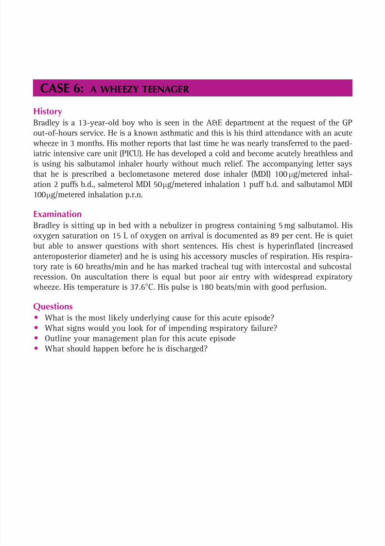
HistoryBradley is a 13-year-old boy who is seen in the A&E department at the request of the GPout-of-hours service. He is a known asthmatic and this is his third attendance with an acutewheeze in 3 months. His mother reports that last time he was nearly transferred to the paed-iatric intensive care unit (PICU). He has developed a cold and become acutely breathless andis using his salbutamol inhaler hourly without much relief. The accompanying letter saysthat he is prescribed a beclometasone metered dose inhaler (MDI) 100 µg/metered inhal-ation 2 puffs b.d., salmeterol MDI 50 µg/metered inhalation 1 puff b.d. and salbutamol MDI100 µg/metered inhalation p.r.n.
ExaminationBradley is sitting up in bed with a nebulizer in progress containing 5 mg salbutamol. Hisoxygen saturation on 15 L of oxygen on arrival is documented as 89 per cent. He is quietbut able to answer questions with short sentences. His chest is hyperinflated (increasedanteroposterior diameter) and he is using his accessory muscles of respiration. His respira-tory rate is 60 breaths/min and he has marked tracheal tug with intercostal and subcostalrecession. On auscultation there is equal but poor air entry with widespread expiratory
wheeze. His temperature is 37.6 C. His pulse is 180 beats/min with good perfusion.
Questions• What is the most likely underlying cause for this acute episode?• What signs would you look for of impending respiratory failure?• Outline your management plan for this acute episode• What should happen before he is discharged?

8/20/2019 100 cases in paediatrics.pdf
http://slidepdf.com/reader/full/100-cases-in-paediatricspdf 39/320
20
100 Cases in Paediatrics
ANSWER 6This boy has another acute exacerbation of asthma. Much the most likely underlyingcause is poor adherence to home treatment. This is common in all age groups but par-ticularly in teenagers with their growing independence and risk-taking behaviour.
! Signs of impending respiratory failure
• Exhaustion (this is a clinical impression)• Unable to speak or complete sentences• Colour – cyanosis pallor• Hypoxia despite high-flow humidified oxygen
• Restlessness and agitation are signs of hypoxia, especially in small children• Silent chest – so little air entry that no wheeze is audible• Tachycardia• Drowsiness• Peak expiratory flow rate (PEFR) persistently 30 per cent of predicted for height
(tables are available) or personal best. Children 7 years cannot perform PEFRreliably and technique in sick children is often poor
Acute management goals are to correct hypoxia, reverse airway obstruction and pre- vent progression. Reassurance and calm are crucial because he will be frightened. Givehigh-flow oxygen via mask and monitor saturations. Start a regular inhaled β-agonist(e.g. salbutamol) via a nebulizer. Beta-agonists can be given continuously. If so, cardiacmonitoring is needed as side-effects include irritability, tremor, tachycardia and hypoka-laemia. Inhaled ipratropium bromide can be added. Give oral prednisolone or intraven-ous (IV) hydrocortisone. Frequent clinical review is paramount. Blood gases (capillaryor venous) and a chest X-ray may be required. If there is no improvement or the childdeteriorates, additional treatment is needed. These include IV salbutamol, IV magnesiumsulphate (a smooth muscle relaxant) and IV aminophylline, although the effectiveness ofthe latter two is still controversial. His precipitating ‘cold’ is almost certainly viral andantibiotics are unlikely to be beneficial.
Before discharge a thorough review of his asthma is needed:
• How often does he miss his regular drugs?• Is there parental supervision?• What device does he use? Children rarely use MDIs effectively and need a spacer.
However, he is unlikely to use one because they are cumbersome and not ‘cool’. Agree an alternative ‘breath-activated’ device with the proviso that, if acutelywheezy, he must use a spacer.
• Consider changing to a combined steroid/long-acting β-agonist inhaler. This shouldimprove adherence.
• Ask about smoking – him and his family. Adults should be encouraged to stopsmoking or to smoke outside.
• Educate about allergen avoidance, e.g. daily vacuuming to reduce house dust mites.Consider measuring total IgE and specific allergen IgE (RAST) if the history suggestsallergies.
• All asthmatics should have a written home management plan.

8/20/2019 100 cases in paediatrics.pdf
http://slidepdf.com/reader/full/100-cases-in-paediatricspdf 40/320
21
Respiratory
• Provide an asthma symptom diary and arrange hospital follow-up until controlimproves. Most children can and should be managed in primary care. Primary careand hospital-based asthma specialist nurses are very helpful.
KEY POINTS
• The commonest cause of an acute deterioration in chronic asthma is poor adherenceto treatment.
• Home management should be reviewed during any acute admission.

8/20/2019 100 cases in paediatrics.pdf
http://slidepdf.com/reader/full/100-cases-in-paediatricspdf 41/320
22
CASE 7:FEVER AND BREATHLESSNESS
HistoryNiall is a 3-year-old boy from a travelling family referred to the paediatric day unit bythe out-of-hours GP service. He was seen 4 days ago with a cough and fever and diag-nosed with a viral upper respiratory tract infection (URTI). The following day he returnedand was commenced on oral antibiotics. He is now complaining of tummy ache and has
vomited once. The GP is worried that he is becoming dehydrated and may have a urin-ary tract infection or intra-abdominal pathology. He has not been immunized but thereis no other medical history of note.
ExaminationNiall is miserable, flushed, toxic and febrile (38.8 C) with a capillary refill time of 2 s. Hispulse is 140 beats/min, his oxygen saturation is 91 per cent in air and his blood pressure is85/60 mmHg. He seems to be in pain, especially when he coughs, and his respiratory rateis 48 breaths/min with nasal flaring. There is dullness to percussion in the right lower zoneposteriorly with decreased breath sounds and bronchial breathing. He seems reluctant tohave his abdomen examined but bowel sounds are normal.
INVESTIGATIONSNormal
Haemoglobin 11.8 g/dL 11.5–15.5 g/dLWhite cell count 25.4 109 /L 6–17.5 109 /LNeutrophils 20.8 109 /L 3–5.8 109 /LPlatelets 467 109 /L 150–400 109 /LSodium 126 mmol/L 138–146 mmol/LPotassium 3.5 mmol/L 3.5–5.0 mmol/LUrea 2.5 mmol/L 1.8–6.4 mmol/LCreatinine 48 µmol/L 27–62 µmol/L
Glucose 4.5 mmol/L 3.3–5.5 mmol/LC-reactive protein 387 mg/L 6 mg/LChest X-ray – see Figure 7.1
Figure 7.1 Niall’s chest X-ray.
8/20/2019 100 cases in paediatrics.pdf
http://slidepdf.com/reader/full/100-cases-in-paediatricspdf 42/320
23
Respiratory
Questions• What are the chest X-ray findings?• What is the most likely causative organism?• What complication may have arisen and how would you confirm it?
• List the steps in management.

8/20/2019 100 cases in paediatrics.pdf
http://slidepdf.com/reader/full/100-cases-in-paediatricspdf 43/320
24
100 Cases in Paediatrics
ANSWER 7The chest X-ray shows loss of the right hemidiaphragm, right lower zone consolidationand a normal right heart border – the characteristic features of right lower lobe pneu-monia. Lower lobe pneumonia should always be on the list of differential diagnoses forabdominal pain in children. Young children rarely localize pain but observation shouldestablish whether they have pleuritic pain – they may have shallow breathing or maysimply sit very still, hardly moving the affected side.
The most likely causative organism is Streptococcus pneumoniae . However, a diagnosisis rarely made from sputum analysis because children tend to swallow their sputum.Blood cultures may be positive. Children in the UK are immunized against Streptococcus
pneumoniae but not all strains are covered, there is emerging antibiotic resistance and
also ‘failed’ immunizations. Any immunized child with positive cultures should be inves-tigated for an underlying immune problem, including an absent spleen.
Niall has hyponatraemia. There are no pointers to excess sodium loss (e.g. diarrhoea orsignificant vomiting) and hence the most likely cause is the syndrome of inappropri-ate antidiur etic hormone secretion (SIADH), a known association with pneumonia. Thehyponatraemia is dilutional. First, the result should be confirmed – taking blood fromchildren can be difficult and unexpected results should be repeated. At the same time,urine should be sent for osmolality and sodium. His serum osmolality is calculated asfollows:
2 ([Na] [K]) [urea] [glucose] 266 mosmol/kg (normal 278–305)
Normally a fall in serum osmolality would suppress antidiuretic hormone secretion to allowexcretion of excess water as dilute urine. In SIADH, urine osmolality is inappropriately high( 320 mosmol/kg) and urine sodium is usually 40 mmol/L (unlike hypovolaemic stateswhere it is 20 mmol/L).
! Steps in management• Oxygen to maintain saturation at 92 per cent• Adequate pain relief for pleuritic pain• Intravenous antibiotics according to local guidelines, e.g. co-amoxiclav• Initial fluid restriction to two-thirds maintenance to help correct the hyponatraemia.
Fluid restrict even if no hyponatraemia, as SIADH may still develop• Fluid balance, regular urea and electrolytes – adjust fluids accordingly. Weigh
twice daily• Physiotherapy, e.g. bubble blowing. Encourage mobility• Monitor for development of a pleural effusion. If the chest X-ray is suspicious,
an ultrasound will be diagnostic. If present, a longer course of antibiotics isrecommended to prevent empyema (a purulent pleural effusion). A chest drainmay be necessary if there is worsening respiratory distress, mediastinal shift onthe chest X-ray, a large effusion or failure to respond to adequate antibiotics
• Ensure adequate nutrition – children have often been anorectic for several days.Low threshold for supplementary feeds probably via nasogastric tube
• Organise immunization programme before discharge• Arrange a follow-up chest X-ray in 6–8 weeks for those with lobar collapse and/
or an effusion. If still abnormal, consider an inhaled foreign body

8/20/2019 100 cases in paediatrics.pdf
http://slidepdf.com/reader/full/100-cases-in-paediatricspdf 44/320
25
Respiratory
KEY POINTS
• A lower lobe pneumonia should always be considered in the differential diagnosis ofan acute abdomen in children.
• ‘Vaccine failures’ should be investigated for an underlying immune problem.

8/20/2019 100 cases in paediatrics.pdf
http://slidepdf.com/reader/full/100-cases-in-paediatricspdf 45/320
This page intentionally left blank

8/20/2019 100 cases in paediatrics.pdf
http://slidepdf.com/reader/full/100-cases-in-paediatricspdf 46/320
27
CASE 8:A TEENAGER WITH CHEST PAIN
HistoryFabio is a 13-year-old boy who presents to outpatients with a 6-month history of chestpain. The pain can occur at rest or on exercise and is central with no radiation. It lasts forup to an hour. He occasionally gets palpitations after exercise, which he describes as regu-lar. The pain is not accompanied by light-headedness and he has never fainted. He has norespiratory or gastrointestinal symptoms. He had asthma as a child and has a salbutamolinhaler at home but has had no symptoms in the past few years. There is a family historyof hypertension and his grandfather died of a myocardial infarction a year ago.
ExaminationHis pulse is 86/min, regular, his blood pressure is 124/82 mmHg and his heart soundsare normal. There is no hepatomegaly. Femoral pulses are palpable and his chest is clear.His peak expiratory flow rate (PEFR) is 460 L/min (child’s height 1.62 m, PEFR range320–570 L/min). On palpation there is no chest tenderness.
INVESTIGATIONS
His ECG and chest X-ray are both normal.
Questions• What is the most likely diagnosis and the differential diagnosis?• What treatment would you suggest?

8/20/2019 100 cases in paediatrics.pdf
http://slidepdf.com/reader/full/100-cases-in-paediatricspdf 47/320
28
100 Cases in Paediatrics
ANSWER 8The most likely diagnosis in this case is idiopathic chest pain. This is one of the common-est causes of chest pain in children. Psychological chest pain, which may be a replicationof the pain his grandfather used to have, and may be secondary to various stresses suchas bullying, is a further possibility. Costochondritis is due to inflammation of the cartilagethat connects the inner end of each rib with the sternum. There may be tenderness onpalpation of the cartilage in the anterior chest wall and the pain may be worse on move-ment or coughing. The cause is unknown and the condition is self-limiting.
The lack of respiratory symptoms and signs with the normal PEFR would go against arespiratory cause. PEFR is related to height, and charts with normal values exist. Childrenhave to be 5 or more years of age in order to perform this test in an effective and con-
sistent manner, and normally the best reading out of three is obtained. Pneumonia withpleurisy is a common cause of chest pain that is typically worse on inspiration. In thecase of a pneumothorax, the pain is sudden and associated with shortness of breath. Asevere cough from whatever cause can lead to musculoskeletal chest pain.
The lack of gastrointestinal symptoms such as vomiting would go against gastro-oesophageal reflux. Cardiac disease is a rare cause of chest pain in children. The palpita-tions described are probably secondary to tachycardia during exercise.
! Differential diagnosis of chest pain
• Trauma, e.g. fractured rib• Exercise, e.g. overuse injury• Idiopathic• Psychological, e.g. anxiety• Costochondritis• Pneumonia with pleural involvement• Asthma• Severe cough• Pneumothorax
• Reflux oesophagitis• Sickle cell disease with chest crisis and/or pneumonia• Rare: pericarditis, angina, e.g. from severe aortic stenosis, osteomyelitis, tumour
The patient and his family should be reassured. Ibuprofen could be used on an as nece-ssary basis for its analgesic and anti-inflammatory properties for the more prolongedbouts of pain. The child should be reviewed in about 2 months to monitor progress.
KEY POINTS
• Chest pain in children is often idiopathic, psychological or musculoskeletal in origin.• Pulmonary causes are a further common cause of chest pain.• Cardiac disease is a rare cause of chest pain in children.• A chest X-ray and ECG should be done to rule out significant pathology.

8/20/2019 100 cases in paediatrics.pdf
http://slidepdf.com/reader/full/100-cases-in-paediatricspdf 48/320
29
CARDIOLOGY
CASE 9: A CYANOSED NEWBORN
A 5-hour-old male newborn on the postnatal ward is noticed by the midwife becausehe looks blue around the lips and tongue. He is the first child of a 27-year-old motherwith asthma who was taking inhaled steroids throughout pregnancy. Antenatal scanswere unremarkable. She went into spontaneous labour at 41 weeks and there was thinmeconium staining of the liquor when the membranes ruptured 1 hour before delivery.Cardiotocograph monitoring during labour revealed normal variability of fetal heart rate.The baby was born by normal vaginal delivery and weighed 3.3 kg. The Apgar scoreswere 7 at 1 min and 8 at 5 min.
ExaminationThe baby is not dysmorphic. His temperature is 36.6 C and his central capillary refilltime is 2 s. His lips, tongue and extremities are cyanosed. He is crying normally and hasno signs of increased respiratory effort. Heart rate is 160 beats/min, femoral pulses arepalpable, heart sounds are normal and no murmur is audible. Oxygen saturation is 70per cent in air and does not rise with facial oxygen, which has been administered by themidwife. There is no hepatosplenomegaly.
Questions• What is the likely diagnosis and differential diagnosis?• How do you interpret the blood gas results?• What is the emergency management?
INVESTIGATIONS
Arterial blood gas Normal In air pH 7.25 7.35–7.42
P aO2 4.7 kPa 9.3–13.3 kPaP aCO2 5.0 kPa 4.7–6.0 kPa
After 10 min in high-flow facemask oxygen pH 7.23 7.35–7.42
P aO2 5.3 kPa 9.3–13.3 kPaP aCO2 5.2 kPa 4.7–6.0 kPa
8/20/2019 100 cases in paediatrics.pdf
http://slidepdf.com/reader/full/100-cases-in-paediatricspdf 49/320
30
ANSWER 9This baby is most likely to have transposition of the great arteries. There are few con-genital cyanotic heart conditions that present on the first day of life, because in mostcases the ductus arteriosus remains open at this stage, maintaining pulmonary bloodflow in conditions such as pulmonary and tricuspid atresia where pulmonary blood flowwould be severely restricted or absent. Severe Ebstein’s anomaly and obstructed totalanomalous pulmonary venous drainage can produce early cyanosis, but these conditionsare associated with significant respiratory distress. Persistent fetal circulation also resultsin cyanosis, usually with respiratory distress in the context of a newborn who has suf-fered a significant hypoxic, hypothermic or hypoglycaemic insult, who has pulmonaryhypoplasia or sepsis, or sometimes for unknown reasons.
Transposition of the great arteries accounts for up to 5 per cent of congenital heart dis-ease and is the commonest cardiological cause of cyanosis in the newborn. The aortaand pulmonary arteries are transposed such that the right ventricle delivers deoxy-genated blood, returning from the systemic circulation, into the aorta and back to thesystemic circulation. The left ventricle delivers oxygenated blood, arriving from thelungs, into the pulmonary arteries and back to the lungs. These two parallel circula-tions would result in the vital organs never receiving oxygenated blood, and hence rapiddeath, if it was not for mixing in the atria via the foramen ovale and between the aortaand pulmonary arteries via the ductus arteriosus. Chest X-ray may show a narrowedupper mediastinum due to the abnormal position of the aorta and pulmonary arteries.Echocardiography by an experienced operator is necessary to confirm the diagnosis.
The hyperoxia test provides a means of diagnosing whether cyanosis is due to cardiacor respiratory disease. Normally arterial P aO2 is greater than 9 kPa and rises to more than20 kPa after exposure to 90–100 per cent oxygen. If the P aO2 fails to rise, this is stronglysuggestive of cyanotic heart disease. Persistent fetal circulation can also result in a lackof response. There is already evidence of tissue hypoxia in this case, as there is a meta-bolic acidosis.
Emergency management involves commencing a prostaglandin infusion to maintain
patency of the ductus arteriosus, and to correct the metabolic acidosis. An emergencyballoon atrial septostomy will probably be needed to improve mixing of oxygenated anddeoxygenated blood at the atrial level. Ultimately, an arterial switch operation will needto be performed to provide an anatomical correction.
100 Cases in Paediatrics
KEY POINTS
• Measure oxygen saturations if you have any suspicion that a baby may be cyanosed.
• The absence of a murmur does not exclude congenital heart disease.

8/20/2019 100 cases in paediatrics.pdf
http://slidepdf.com/reader/full/100-cases-in-paediatricspdf 50/320
31
CASE 10:A SHOCKED NEONATE
HistoryFreddie is 3 days old. He is brought by ambulance to the resuscitation room in A&E.He was found in his cot this morning looking mottled and breathing very fast. He hadbeen well until yesterday when he did not feed as well as usual. He was born at 39weeks’ gestation by normal vaginal delivery in a midwife-led birthing unit and was dis-charged home the same day. There was no prolonged rupture of membranes and he didnot require any resuscitation at birth. He has been exclusively breast-fed. He did notreceive vitamin K due to parental objection.
ExaminationFreddie is grunting and has a respiratory rate of 70/min with subcostal, intercostaland sternal recession. His lung fields sound clear. Oxygen saturation monitoring doesnot pick up a trace. He looks mottled, cyanosed peripherally and his limbs feel cold.Capillary refill time is 5 s, heart rate is 180/min, blood pressure is unrecordable, and thefemoral pulses cannot be felt. The heart sounds are unremarkable. The liver edge is pal-pable 3 cm below the costal margin and his temperature is 35.0 C.
INVESTIGATIONS
Normal Arterial blood gas pH 7.01 7.35–7.42
P aCO2 5.3. kPa 4.7–6.0 kPaP aO2 8.1 kPa 9.3–13.3 kPa
HCO 3 10 mmol/L 18–20 mmol/L Base excess 18 2.5 to 2.5 mmol/L Glucose 3.8 mmol/L 3.3–5.5 mmol/L
Questions• What is the interpretation of the blood gas result?• What is the most likely diagnosis and what is the differential?• What is the initial management of a collapsed neonate?
8/20/2019 100 cases in paediatrics.pdf
http://slidepdf.com/reader/full/100-cases-in-paediatricspdf 51/320
32
ANSWER 10The pH is 7.01, which is a severe acidosis. The P aCO2 is normal, so the acidosis is notrespiratory in origin. The low bicarbonate and large negative base excess indicate thatthis is a metabolic acidosis. There is also a degree of hypoxaemia. The baby’s circulationis so poor that pulses are not palpable, the saturation meter cannot pick up a pulse traceand a severe metabolic acidosis has developed due to hypoperfusion.
This is most likely to be a case of cardiogenic shock due to a congenital left heartobstructive lesion. The clues are in the age of the baby and the absence of the femoralpulses. Left-sided obstructive cardiac lesions rely on the ductus arteriosus to perfuse thesystemic circulation by passage of blood from the pulmonary arteries into the distal endof the aortic arch. As the ductus arteriosus closes, the systemic perfusion becomes dra-
matically reduced, resulting in collapse and shock. Freddie actually had hypoplastic leftheart syndrome (HLHS). The absence of a murmur does not rule out a cardiac cause, andmurmurs are not always present in HLHS.
100 Cases in Paediatrics
! Congenital cardiac lesions presenting with neonatal collapse
• Severe aortic coarctation• Aortic arch interruption• Hypoplastic left heart syndrome• Critical aortic stenosis
! Differential diagnosis of a collapsed neonate
• Infection – e.g. group B Streptococcus , herpes simplex• Cardiogenic – e.g. hypoplastic left heart syndrome, supraventricular tachycardia• Hypovolaemic – e.g. dehydration, bleeding• Neurogenic – e.g. meningitis, subdural haematoma (‘shaken baby’)• Lung disorder – e.g. congenital diaphragmatic hernia (late presentation)
• Metabolic – e.g. propionic acidaemia, methylmalonic acidaemia• Endocrine – e.g. panhypopituitarism
In any collapsed neonate, it is essential to adopt a standard approach to resuscitation.The airway should be maintained, high-flow oxygen administered, intravenous accessobtained and fluid resuscitation should be given for the shock. Blood glucose measure-ment must be checked early and corrected if low. A blood gas sample should be analysed.Intravenous antibiotics should be given promptly as sepsis is a possible treatable cause. Ifthere is any suspicion of a duct-dependent cardiac lesion, a prostaglandin infusion shouldbe commenced, as this is life-saving. Early involvement of senior paediatricians, ananaesthetic team and paediatric intensive care services will help appropriate management.
KEY POINTS
• Congenital cardiac disease can present as shock in a neonate after several days.• The absence of a murmur does not rule out congenital heart disease.• A prostaglandin infusion can maintain patency of the ductus arteriosus and can be
life-saving.

8/20/2019 100 cases in paediatrics.pdf
http://slidepdf.com/reader/full/100-cases-in-paediatricspdf 52/320
33
CASE 11:A PALE
,BREATHLESS BABY
History Alfie is a 7-month-old baby who is seen in the A&E department with a day’s history ofpallor. Over the past few hours, he has also become restless and breathless. He is feedingpoorly. He has no cough or wheeze. There is no past medical history of note and he ison no medication. His 3-year-old sister has a cold but a 5-year-old brother and the restof the family are well.
ExaminationHe is apyrexial, pale and his oxygen saturation is 91 per cent in air. The heart rate is 270beats/min, blood pressure is 84/44 mmHg, heart sounds are normal and femoral pulses arepalpable. The peripheral capillary refill is 4 s. Respiratory rate is 62 breaths/min and thechest is clear. The liver is palpable 4 cm below costal margin. There are no other signs.
INVESTIGATIONS
Normal
Haemoglobin 12.8 g/dL 10.5–13.5 g/dLWhite cell count 7.0 109 /L 4.0–11.0 109 /LPlatelets 323 109 /L 150–400 109 /LSodium 138 mmol/L 135–145 mmol/LPotassium 3.9 mmol/L 3.5–5.0 mmol/LUrea 6.4 mmol/L 1.8–6.4 mmol/LCreatinine 60 µmol/L 20–80 µmol/LC-reactive protein 5 mg/L 6 mg/LECG – see Figure 11.1
Figure 11.1 Alfie’s electrocardiogram (lead 2).
Questions• What does the ECG show and what is the diagnosis?• What treatments can be used to manage this condition?

8/20/2019 100 cases in paediatrics.pdf
http://slidepdf.com/reader/full/100-cases-in-paediatricspdf 53/320
34
100 Cases in Paediatrics
ANSWER 11The ECG shows a narrow QRS complex tachycardia with a rate of approximately 220beats/min. There are no P waves.
The diagnosis is supraventricular tachycardia (SVT). This is the commonest pathologicalarrhythmia in childhood (sinus arrhythmia is a normal variant). Diagnosis is based onthe presence of a narrow QRS complex tachycardia. P waves are only visible in abouthalf of cases. It is usually secondary to a re-entrant tachycardia using an accessory path-way. It is also associated with the Wolff–Parkinson–White syndrome (see Fig. 11.2). Thiscondition can be diagnosed by a short PR interval and a slow upstroke of the QRS (deltawave). These are normally only seen when the patient does not have a tachycardia.
Figure 11.2 Intermittent pre-excitation in Wolff–Parkinson–White syndrome. The first two beatsshow the short PR interval and delta wave. The middle two beats are normal and the abnormalityreturns in the final two beats. (Reproduced with permission from Worrell DA, ed, Oxford Textbook of Medicine , 2003, Oxford University Press.)
The heart rate in SVT is typically 200–300 beats/min. Arrhythmias can last from a few secondsto days. In older children, they can be associated with palpitations, light-headedness andchest discomfort. However, in young infants, the inability to report symptoms may leadto presentation with heart failure and shock, as in this case. In a third of cases there isassociated congenital heart disease so an echocardiogram should be performed. Supra-
ventricular tachycardia needs to be differentiated from sinus tachycardia. In the latter,the heart rate is usually 220 beats/min, there is greater variability in the heart rate andthere is often a history consistent with shock.
! Features of heart failure
• Tachycardia• Tachypnoea• Hepatomegaly• Poor feeding• Sweating• Excessive weight gain (acutely)• Poor weight gain (chronically)• Gallop rhythm• Cyanosis• Heart murmur

8/20/2019 100 cases in paediatrics.pdf
http://slidepdf.com/reader/full/100-cases-in-paediatricspdf 54/320
35
Cardiology
Initial treatment in this emergency follows the standard resuscitation guidelines and thebaby should be administered oxygen. Capillary refill is slightly prolonged but shouldimprove swiftly following the correction of the tachycardia.
Specific treatment comprises vagal stimulation, for instance by eliciting the ‘diving reflex’which will increase vagal tone, slow atrioventricular conduction and abort the tachycar-dia. This can be done by placing a rubber glove filled with iced water over the baby’sface. If this fails, the baby’s face can be immersed in iced water for 5 s. Second-line treat-ment comprises intravenous adenosine that can be administered in escalating doses. Ifthis fails, synchronized DC cardioversion will almost always stop the tachycardia. Oncesinus rhythm is achieved, maintenance treatment using drugs such as amiodarone shouldbe started following consultation with a cardiologist. Infants ( 1 year old) with SVT areless likely to relapse than older children. They are often treated for 1 year, followingwhich the medication is slowly tapered. In the majority of cases, the SVT does not recur.
Definitive treatment comprises catheterization with radiofrequency ablation.
KEY POINTS
• SVT is the commonest pathological arrhythmia in childhood.• It can lead to heart failure and shock.• Treatments comprise vagal manoeuvres, adenosine and DC cardioversion.

8/20/2019 100 cases in paediatrics.pdf
http://slidepdf.com/reader/full/100-cases-in-paediatricspdf 55/320
This page intentionally left blank
8/20/2019 100 cases in paediatrics.pdf
http://slidepdf.com/reader/full/100-cases-in-paediatricspdf 56/320
37
CASE 12:AN INCIDENTAL MURMUR
HistoryLola is a 3-year-old girl referred to outpatients by her GP, who heard a murmur when shepresented to him with a fever and a cough. Looking through her notes, the murmur was notheard at her 6-week check and she has only been seen twice since, with minor infections. Hebrought her back a week later to listen again, the murmur was still present and he has referredher on. She is otherwise entirely healthy with no significant past medical or family history.
ExaminationLola looks generally healthy and her height and weight are on the 75th centiles. She isnot clinically anaemic, jaundiced or cyanosed. Her pulse is 88/min and her blood pressure90/50 mmHg. She has normal femoral pulses. Examination of the praecordium shows nothrills but there is a heave at the lower left sternal border. The apex beat is in the fifth inter-costal space in the mid-clavicular line. Both heart sounds are present but the pulmonary com-ponent of the second sound is quiet. There is a click immediately after the first heart soundand an ejection systolic murmur which is heard loudest in the pulmonary area. This radiatesinto both lung fields and is heard in the back between the scapulae. Examination of the respi-ratory and abdominal systems is normal with no hepatomegaly.
I
II
III
AVR
AVL
AVF
V1
V2
V3
V5
V6
V4
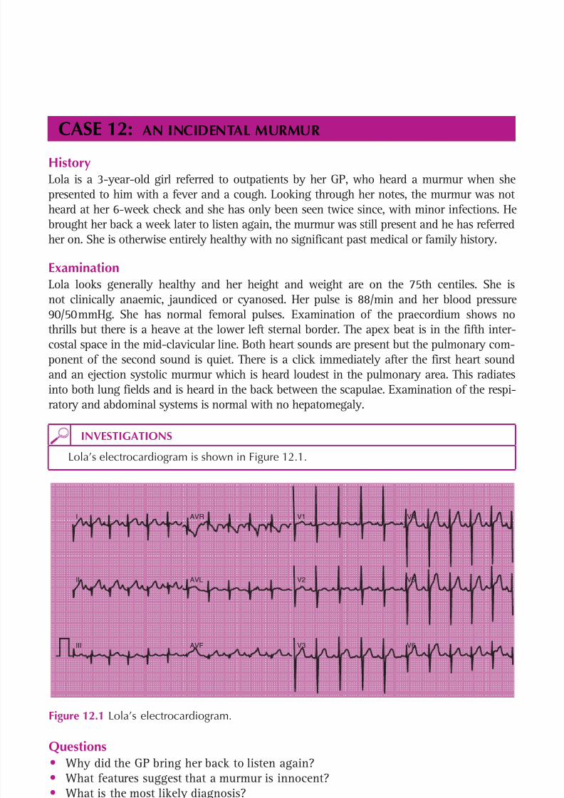
Figure 12.1 Lola’s electrocardiogram.
INVESTIGATIONS
Lola’s electrocardiogram is shown in Figure 12.1.
Questions• Why did the GP bring her back to listen again?• What features suggest that a murmur is innocent?• What is the most likely diagnosis?• What does the ECG show?• What should happen next?

8/20/2019 100 cases in paediatrics.pdf
http://slidepdf.com/reader/full/100-cases-in-paediatricspdf 57/320
38
100 Cases in Paediatrics
ANSWER 12Up to 40 per cent of children will have a murmur heard at some time during childhood,particularly if examined at a time of high cardiac output – e.g. fever, anaemia or anxiety.These ‘innocent’ murmurs have certain reassuring features and the heart is structurallynormal. It is good practice to re-examine asymptomatic healthy children 1–2 weeks laterwhen they are well. If the murmur persists, they should be referred. That it was not heardat the 6-week check is irrelevant – murmurs in children are often difficult to hear. A mur-mur can be diagnosed as innocent on the basis of the certain clinical findings (see box).
! Clinical findings in innocent murmurs
• Asymptomatic• No thrills or heaves• Normal heart sounds, normally split with no added clicks• Quiet and soft• Systolic (isolated diastolic murmurs are never innocent)• Short, ejection (pansystolic murmurs are pathological)• Single site with no radiation to neck, lung fields or back• Varies with posture (decreases or disappears when patient sits up, loudest when
they’re lying)
Further investigations are rarely indicated. Nevertheless parents are understandably anx-ious and must be reassured that an innocent murmur is simply a ‘noise’. It may neverdisappear but this does not matter because the heart is normal. All other murmurs shouldbe investigated. However, a normal ECG and chest X-ray do not exclude pathology. Anechocardiogram is the definitive test.
The findings are characteristic of moderate pulmonary valve stenosis (PS). This is com-mon, accounting for 7–10 per cent of all congenital heart defects. Unless the stenosis
is severe, children are otherwise healthy and asymptomatic. An ‘opening’ click may beheard at the beginning of systole. With increasing severity, the clinical features changeto reflect the progressive narrowing and the resulting right ventricular hypertrophy(RVH). The pulmonary component of the second heart sound diminishes and a lower leftsternal heave develops. In severe PS there may be evidence of right-sided heart failurewith hepatomegaly.
The ECG will show the same progression. This child's ECG shows right axis deviation andevidence of RVH (an ‘R' in V1 > 20 millimetres, an ‘S' in V6 > 5 millimetres and uprightT waves across all the right precordial leads).
She needs an echocardiogram to:
• confirm the clinical diagnosis• assess severity to guide further investigation and treatment• exclude any associated cardiac lesions, e.g. ventricular septal defect.
Doppler echocardiography measures velocity across the valve – the higher this is, the greaterthe need for intervention. Catheter balloon valvuloplasty is the treatment of choice in the

8/20/2019 100 cases in paediatrics.pdf
http://slidepdf.com/reader/full/100-cases-in-paediatricspdf 58/320
39
Cardiology
majority and the outcomes are good. Milder degrees of PS need regular re-evaluation tomonitor any progression. Antibiotic prophylaxis to prevent infective endocarditis is no longerrecommended in this condition.
KEY POINTS
• Cardiac murmurs are heard at some time in up to 40 per cent of children.• The majority of murmurs in children are innocent.• A normal ECG and chest X-ray do not exclude a pathological murmur.• An echocardiogram is the definitive investigation.

8/20/2019 100 cases in paediatrics.pdf
http://slidepdf.com/reader/full/100-cases-in-paediatricspdf 59/320
This page intentionally left blank
8/20/2019 100 cases in paediatrics.pdf
http://slidepdf.com/reader/full/100-cases-in-paediatricspdf 60/320
41
CASE 13:A FUNNY TURN
Sian is a 15-year-old girl, referred to paediatric outpatients by her GP. The letter says,‘Thank you for seeing this girl who has had her first fit.’ Three weeks ago she was atschool when the fire alarm went off unexpectedly. She felt faint and clammy. Friendstried to help her to walk out of the classroom, but after about a minute she collapsed tothe floor and had a brief episode of jerking movements affecting all four limbs. Therewas no incontinence or tongue biting. She regained consciousness within a minute butcontinued to feel weak for a few hours afterwards.
Sian had a febrile convulsion at 18 months of age and she has fainted on about five occa-sions, mostly in emotional situations or when it has been hot. She describes herself as ananxious girl, and has a sensation of her heart racing every few weeks. She has never beenin hospital, but has seen her GP for heavy periods. There is no family history of epilepsy,but her aunt collapsed and died at the age of 28 in Canada. She is top of her year at schooland hopes to be a doctor or lawyer.
ExaminationHer height is 170 cm (91st centile) and her weight is 53kg (50 th). Cardiovascular, respi-ratory, abdominal and neurological examinations are normal.
Questions• What are the possible causes of her collapse?• What is the most important investigation to perform at this stage?

8/20/2019 100 cases in paediatrics.pdf
http://slidepdf.com/reader/full/100-cases-in-paediatricspdf 61/320
42
100 Cases in Paediatrics
ANSWER 13
! Causes of a funny turn
Distinguishing features in the history Epileptic seizure Aura, incontinence, tongue biting, family historyCardiac arrhythmia Palpitations, sudden collapse, exercise-relatedNeurally mediated syncope Preceding stimulus, dizziness, nauseaPanic attack Hyperventilation, paraesthesia, carpopedal spasmBreath-holding attack Usually a toddler, upset/cryingReflex anoxic seizure Usually infant/toddler, painful stimulusPseudoseizures Psychological problems
Other causes
HypoglycaemiaOther metabolic derangementsDrugs, alcohol
In this case, the history is most suggestive of a neurally mediated (vasovagal) syncope,a cardiac arrhythmia or perhaps a panic attack. The history is less typical of an epilepticseizure, and the most likely reason for the brief convulsion is hypoperfusion of the braindue to low blood pressure while her friends held her upright – an anoxic seizure. Thisterminated quickly when she was supine after collapsing.
The single most important investigation is an ECG, as this may show a potentially life-threatening cardiac cause. An EEG is generally not performed after a first seizure becauseit may be normal in those who go on to have epilepsy and abnormal in those who neverhave another seizure, with the EEG adding little to prognosis at this stage. Blood testsare often performed for glucose, electrolytes, calcium and magnesium, but are frequentlyunhelpful. Sian’s ECG is shown in Figure 13.1.
II
Figure 13.1 Sian’s electrocardiogram.
Her ECG shows a prolonged corrected QT interval, making the diagnosis of a long QTsyndrome. This disorder has a number of genetic causes, and in some cases an arrhythmiacan be precipitated by a sudden loud noise. This girl went on to require anti-arrhythmicdrug treatment and an implantable cardiodefibrillator.
The QT interval is measured from the start of the QRS complex to the end of the T-wave;the QT interval is corrected (QTc) for heart rate by: QTc QT/( R – R interval). QTc isnormally around 0.41 s and should be less than 0.45 s. In this case it is 0.52 s.
8/20/2019 100 cases in paediatrics.pdf
http://slidepdf.com/reader/full/100-cases-in-paediatricspdf 62/320
43
Cardiology
KEY POINTS
• The commonest cause of fainting in a teenager is neurally mediated (vasovagal)syncope.
• Cardiac causes should always be considered in the differential of epilepsy andfunny turns.
• An ECG is almost always indicated.

8/20/2019 100 cases in paediatrics.pdf
http://slidepdf.com/reader/full/100-cases-in-paediatricspdf 63/320
This page intentionally left blank
8/20/2019 100 cases in paediatrics.pdf
http://slidepdf.com/reader/full/100-cases-in-paediatricspdf 64/320
8/20/2019 100 cases in paediatrics.pdf
http://slidepdf.com/reader/full/100-cases-in-paediatricspdf 65/320
46
100 Cases in Paediatrics
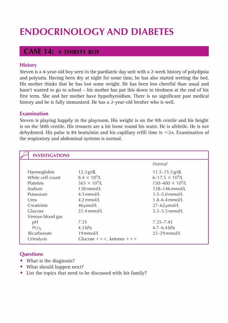
ANSWER 14The diagnosis is type 1 diabetes mellitus (T1DM), by far the commonest form of diabetes inchildhood characterized by pancreatic β-cell dysfunction with insulin deficiency. The precisemechanism is not understood, but environmental factors probably ‘trigger’ a T-cell-mediatedautoimmune process in those genetically susceptible (although only about 10 per cent havea family history at presentation). There may be a history of other autoimmune diseases. Theincidence is rising. The incidence of type 2 is also rising alongside obesity.
Although this boy has ketonuria, he is not acidotic and does not have diabetic ketoacidosis(DKA) – a widely accepted biochemical definition being a pH 7.30 and/or a bicarbonate
15 mmol/L. Nor is he vomiting or showing signs of sepsis. Therefore he does not needintravenous fluids or intravenous insulin. Only about 10–20 per cent of children present
with DKA, usually pre-school children in whom the diagnosis has not been considered.Childhood DKA has a mortality rate of approximately 0.2 per cent, usually from cerebraloedema due to the use of excessive fluids and/or rapid changes in blood glucose.
He now needs to start regular subcutaneous insulin. Current best practice suggests thatinsulin is best delivered by a ‘basal bolus’ regime – background ‘basal’ insulin givenonce daily with rapid-acting ‘bolus’ insulin at mealtimes. Alternatives are twice-dailyinjections of pre-mixed long and rapidly acting insulins or insulin via a pump.
The impact of the diagnosis of T1DM should never be underestimated (see Case 87,p. 255 in relation to the breaking of bad news to families). At diagnosis, he and his fam-ily will start a detailed education programme. Education is provided by a multidiscipli-nary team, including specialist nurses and dieticians. The aspects of management thatneed to be discussed include:
• how to give insulin – to a probably reluctant 4-year-old• regular finger-prick blood testing – up to four times daily • interpreting blood glucose results and altering insulin• recognition and treatment of hypoglycaemia – ‘hypos’ really worry parents and older
patients, especially at night
• management during intercurrent illness, e.g. flu
• blood or urine ketone estimations• who to contact in an emergency • diet – matching insulin to carbohydrate in a ‘basal-bolus’ regime• school – trusting staff in everyday care• peer group relationships – diabetes risks making children feel ‘different’• exercise• the ‘honeymoon’ period• long-term complications – discuss even at outset because many families know about
and fear complications and need objective information• using glycosylated haemoglobin (HBA1c) to measure overall control• membership of local and/or national support groups.
KEY POINTS
• The cardinal features of type 1 diabetes are polydypsia and polyuria.• Most patients do not present in DKA.• Patients with DKA are acidotic (pH 7.30) as well as having ketonuria.• Education and support from a multidisciplinary specialist team are crucial.

8/20/2019 100 cases in paediatrics.pdf
http://slidepdf.com/reader/full/100-cases-in-paediatricspdf 66/320
47
CASE 15:A TALL BOY
HistoryZak is a 10-year-old boy referred to outpatients by his GP with tall stature. He is seen withhis 28-year-old mother who is not particularly concerned because his 29-year-old fatheris 1.93 m, although he does not come from a tall family himself. However, friends andfamily have begun to comment and the parents are seeking reassurance. When asked, theboy admits that he is self-conscious about his stature and is fed up with being treated asolder than his true age. At school, everyone expects him to take more responsibility thanhe thinks is fair. He is otherwise healthy although he was seen in infancy with a cardiacmurmur that was thought to be innocent. He did not have an echocardiogram. He is doingwell academically at school. His 7-year-old sister is apparently on the 75th percentile forheight. His mother is measured in clinic and is 1.66 m tall. This puts her between the 50thand 75th centiles for a female.
ExaminationZak is tall and slender. His height is 1.58 m ( 99.6th centile) and weight 35 kg (75th cen-tile). His arm span is 168 cm. He has long, tapering fingers and his thumb, when completelyopposed within the clenched hand, projects beyond the ulnar border. He has pectus excava-
tum (funnel chest) but an otherwise normal respiratory examination. He has a midsystolicclick with a late systolic murmur heard at the apex. Examination of the abdomen is normaland he is prepubertal.
Questions• What is the most likely diagnosis?• What further investigations does he need?• Who else should be examined and by whom?

8/20/2019 100 cases in paediatrics.pdf
http://slidepdf.com/reader/full/100-cases-in-paediatricspdf 67/320
48
100 Cases in Paediatrics
ANSWER 15The most likely diagnosis is the connective tissue disease Marfan syndrome. This is aninherited synthetic disorder of fibrillin-1, a glycoprotein constituent of the microfibrilsof elastic fibres that anchor non-elastic tissue such as the aortic adventitia and the sus-pensory ligament of the eye.
By definition, tall stature includes all children with a length or height above the 98thcentile. However, uncovering pathology in such children is rare and the vast majorityhave simple familial tall stature. This is characterized by a normal physical examinationand serial height measurements that show no growth acceleration. Skeletal age (assessedby a bone age X-ray of the wrist) is usually slightly advanced in relation to chronologi-cal age, the child enters puberty relatively (not abnormally) early and completes their
growth phase relatively early, with a final height within the expected range for theirfamily. The differential diagnosis of familial tall stature is a syndrome associated withtall stature, such as Marfan or Klinefelter (47, XXY) syndrome, or a child with growthacceleration for which an endocrine cause is likely, e.g. sexual precocity, thyrotoxicosisor growth hormone excess. Children with familial tall stature rarely benefit from furtherinvestigations or monitoring.
The diagnosis of Marfan syndrome is made on accepted clinical criteria – only about60–70 per cent of patients who fulfil these have an identifiable mutation in the fibrillingene. This boy displays many of the musculoskeletal features – arm span to height ratio
1.05, arachnodactyly with a positive thumb sign and pectus excavatum – but manyother systems may be involved. Most important are the eyes and heart. Lens disloca-tion is one of the major criteria for diagnosis and he needs a slit-lamp examination. Hismurmur is typical of mitral valve prolapse (MVP) and echocardiography is essential fordiagnosis and to monitor the high risk of progressive aortic root dilatation.
Marfan syndrome is an autosomal dominant disorder, although about 30 per cent arisesporadically. There is the distinct possibility that this boy has inherited it from his father,who is unusually tall for his family and should be investigated. There is significant mor-bidity and reduced longevity, mainly from the cardiovascular complications, but young
adults like him may still be asymptomatic. However, making such a diagnosis will havefar-reaching social, financial and psychological consequences for an individual and theirfamily. Referral to the genetics team is mandatory: first, to obtain informed consent forinvestigations; second, to confirm the diagnosis; and third, to counsel about recurrenceand inheritance risk – in Marfan this is 50 per cent. Refuting a diagnosis is obviously
just as important in uncertain cases and the geneticists are best placed to do so.
KEY POINTS
• Pathological causes of tall stature are rare.• Families who may have serious inheritable disorders must be referred to genetics
teams for diagnosis and counselling.

8/20/2019 100 cases in paediatrics.pdf
http://slidepdf.com/reader/full/100-cases-in-paediatricspdf 68/320
This page intentionally left blank
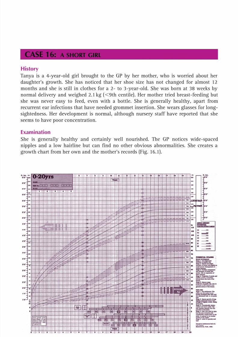
8/20/2019 100 cases in paediatrics.pdf
http://slidepdf.com/reader/full/100-cases-in-paediatricspdf 69/320
50
CASE 16:A SHORT GIRL
HistoryTanya is a 4-year-old girl brought to the GP by her mother, who is worried about herdaughter’s growth. She has noticed that her shoe size has not changed for almost 12months and she is still in clothes for a 2- to 3-year-old. She was born at 38 weeks bynormal delivery and weighed 2.1 kg ( 9th centile). Her mother tried breast-feeding butshe was never easy to feed, even with a bottle. She is generally healthy, apart fromrecurrent ear infections that have needed grommet insertion. She wears glasses for long-sightedness. Her development is normal, although nursery staff have reported that sheseems to have poor concentration.
ExaminationShe is generally healthy and certainly well nourished. The GP notices wide-spacednipples and a low hairline but can find no other obvious abnormalities. She creates agrowth chart from her own and the mother’s records (Fig. 16.1).
Figure 16.1 Tanya’s growth chart showing available height and weight data. (Chart reproducedwith kind permission of the Child Growth Foundation.)

8/20/2019 100 cases in paediatrics.pdf
http://slidepdf.com/reader/full/100-cases-in-paediatricspdf 70/320
51
Endocrinology and diabetes
Questions• How did the GP plot the parents’ heights?• What are the clinical signs that suggest a pathological cause for short stature?• What is the most likely diagnosis?

8/20/2019 100 cases in paediatrics.pdf
http://slidepdf.com/reader/full/100-cases-in-paediatricspdf 71/320
52
100 Cases in Paediatrics
! Clinical signs suggesting a pathological cause for short stature
• Extreme short stature – on or below 0.4th centile.• Short for family size – outside target range for parents• Short and relatively overweight – suggests an endocrinopathy• Short and very underweight – suggests poor nutrition malabsorption• Growth failure – crossing the centiles downwards• Dysmorphic features• Skeletal disproportion – charts available for ratio between sitting height
and leg length. Significant disproportion suggests a skeletal dysplasia, e.g.achondroplasia
• Signs of systemic disease, e.g. clubbing
ANSWER 16Plotting the parents’ centiles establishes whether a child falls into the expected heightrange for their family. Both parents should be measured, but realistically this is oftendifficult. To plot the father’s centile on a girls’ chart, 14 cm is subtracted from hisheight, and on a boys’ chart the same measurement is added to the mother’s height.
Alternatively, their centile is taken from the relevant chart. The ‘target centile range’ canalso be calculated. This is the midpoint of the two plotted parental centiles 8.5 cm for agirl and 10 cm for a boy. With normal parents and healthy children, final heights willbe normally distributed within these 95 per cent confidence limits.
Growth is unique to childhood and an excellent tool for confirming health as well asspotting disease. Children who are healthy grow and gain weight normally and followcentiles. The challenge in assessing a short child is to pick out those with a pathologicalcause from the majority without. Usually there is no one defining feature and pieces ofthe clinical jigsaw are put together from the history, examination, growth pattern andany investigations. A short child following the 0.4th to 2nd centiles from a short fam-ily is less worrying than one who is crossing the centiles downwards due to abnormallyslow growth. The causes are numerous – chronic disease (coeliac disease, uncontrolledasthma), drug effects (steroids), endocrine disorders (hypothyroidism, growth hormonedeficiency) and emotional abuse. Thorough clinical assessment is crucial.
This girl is extremely short, crossing the centiles, short for family and has some subtledysmorphic features. Pathology is probable. She is also relatively overweight for height,suggesting an endocrinopathy. The GP should refer her to a growth clinic.
The most likely diagnosis is Turner syndrome (TS) caused by the complete or partialabsence of one of the X chromosomes. The incidence is 1 in 3000 live female births.Short stature is universal and there are specific Turner growth charts. The dysmorphicfeatures vary in frequency (e.g. only 50 per cent have neck webbing) and are often sub-
tle. This girl has wide-spaced nipples and a low hairline. She also has some of the otherassociations – low birth weight, difficult to feed in infancy, middle ear disease, visualproblems and poor concentration. None of these are specific, but put together they makeTS likely. Investigations must include chromosome analysis.
KEY POINTS
• Most children seen with short stature do not have a pathological cause.• Dysmorphic features in Turner syndrome may be subtle and there should be a low
threshold for chromosome analysis in all short girls.

8/20/2019 100 cases in paediatrics.pdf
http://slidepdf.com/reader/full/100-cases-in-paediatricspdf 72/320
53
CASE 17:AN OVERWEIGHT BOY
HistoryMarlon is a 7-year-old boy who presents to paediatric outpatients because he’s over-weight. His father became concerned 1 year ago. His mother is overweight, hypertensiveand has type 2 diabetes, but his father’s weight is average. His father says the boy’s dietis generally good but that his grandmother spoils him. He does sports twice a week atschool. He is bullied at school about his weight. His birth weight was 3.8 kg and therewere no problems in the neonatal period. He snores every night but his parents have notnoticed any sleep apnoea. His development is normal. He is on no medication. No otherdiseases run in the family.
ExaminationThere are no dysmorphic features. There are some pink abdominal stretch marks. Thereis no acanthosis nigricans and no goitre. His blood pressure is 116/75 mmHg. His tonsilsare large but with a good gap between them. There are no other signs. His height is125 cm (75th centile) and his weight is 38.7 kg ( 99.6th centile).
INVESTIGATIONS
Full blood count, urea and electrolytes, liver and thyroid function tests are normal. Hisfasting glucose, insulin, cholesterol and triglycerides are normal.
Questions• What is this child’s body mass index (BMI)?• What is the most likely cause of this child’s obesity?• How should this child be treated?
8/20/2019 100 cases in paediatrics.pdf
http://slidepdf.com/reader/full/100-cases-in-paediatricspdf 73/320
54
ANSWER 17Marlon’s BMI weight (kg)/height (m) 2 38.7/(1.25) 2 24.8 kg/m 2. There are stand-ard BMI charts for males and females from 0 to 20 years. BMI varies according to age.Children with a BMI above the 91st centile are overweight and those with a BMI abovethe 98th centile are obese.
The most likely cause of this child’s obesity is simple obesity – obesity due to caloricintake exceeding energy expenditure. It is the commonest cause of obesity and is notdue to any underlying pathology. Children with simple obesity tend to be tall and over-weight. Those who are short and overweight are more likely to have underlying path-ology, such as an endocrine disorder.
Stretch marks are secondary to the obesity and are usually pink. Violaceous stretchmarks are associated with Cushing’s syndrome. Large tonsils and obstructive sleepapnoea are associated with obesity. The child does not have acanthosis nigricans, whichis a thickening and darkening of the skin in the axilla, neck or groin. It is usually a signof insulin resistance. Children with obesity should be assessed for co-morbidities suchas hypertension, breathlessness on exertion, obstructive sleep apnoea, hyperinsulinaemiaand type 2 diabetes (in children 10 years), dyslipidaemia, knock-knees or bow legs,polycystic ovary syndrome and psychosocial dysfunction.
100 Cases in Paediatrics
! Causes of obesity
• Simple obesity• Genetic, i.e. one or both parents is obese• Endocrine disease, e.g. hypothyroidism, Cushing’s syndrome, growth hormone
deficiency, pseudohypoparathyroidism• Drugs, e.g. steroids, sodium valproate• Syndromes, e.g. Down’s, Präder–Willi and Laurence–Moon–Biedl• Disorders associated with immobility, e.g. cerebral palsy• Hypothalamic damage, e.g. secondary to trauma or brain tumours• Rarely, single gene mutations, such as those of the melanocortin-4 receptor and
leptin
Treatment should be multidisciplinary. Dietetic input is very important. Children shouldbe encouraged to exercise for 60 min/day. Sedentary activities such as playing com-puter games should be discouraged. Obesity can lead to low self-esteem and, in our case,Marlon is being bullied. He would therefore benefit from seeing a psychologist, whocould also initiate behavioural therapies to help treat the obesity.
As the enlarged tonsils are not leading to sleep apnoea, tonsillectomy is not currentlyindicated. The systolic blood pressure is below the 95th centile for a 7-year-old malewith a height on the 75th centile and therefore does not need treatment.
Drug treatment with drugs such as Orlistat or sibutramine is reserved for children 12 years of age and is only recommended in the presence of severe physical or psychologicalco-morbidities. Post-puberty, bariatric surgery, such as gastric stapling, can be consideredin severe cases.

8/20/2019 100 cases in paediatrics.pdf
http://slidepdf.com/reader/full/100-cases-in-paediatricspdf 74/320
55
Endocrinology and diabetes
KEY POINTS
• Body mass index is calculated by weight (kg)/height (m) 2.• A child with a BMI above the 91st centile is overweight and one with a BMI above
the 98th centile is obese.• The commonest cause of obesity is simple obesity.
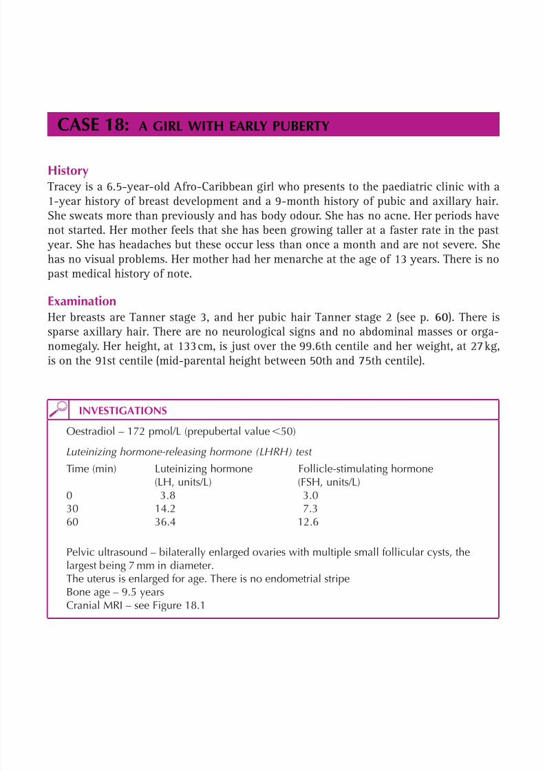
8/20/2019 100 cases in paediatrics.pdf
http://slidepdf.com/reader/full/100-cases-in-paediatricspdf 75/320
56
CASE 18:A GIRL WITH EARLY PUBERTY
HistoryTracey is a 6.5-year-old Afro-Caribbean girl who presents to the paediatric clinic with a1-year history of breast development and a 9-month history of pubic and axillary hair.She sweats more than previously and has body odour. She has no acne. Her periods havenot started. Her mother feels that she has been growing taller at a faster rate in the past
year. She has headaches but these occur less than once a month and are not severe. Shehas no visual problems. Her mother had her menarche at the age of 13 years. There is nopast medical history of note.
ExaminationHer breasts are Tanner stage 3, and her pubic hair Tanner stage 2 (see p. 60 ). There issparse axillary hair. There are no neurological signs and no abdominal masses or orga-nomegaly. Her height, at 133 cm, is just over the 99.6th centile and her weight, at 27 kg,is on the 91st centile (mid-parental height between 50th and 75th centile).
INVESTIGATIONS
Oestradiol – 172 pmol/L (prepubertal value 50)
Luteinizing hormone-releasing hormone (LHRH) test Time (min) Luteinizing hormone Follicle-stimulating hormone (LH, units/L) (FSH, units/L)0 3.8 3.030 14.2 7.360 36.4 12.6
Pelvic ultrasound – bilaterally enlarged ovaries with multiple small follicular cysts, thelargest being 7 mm in diameter.The uterus is enlarged for age. There is no endometrial stripeBone age – 9.5 yearsCranial MRI – see Figure 18.1
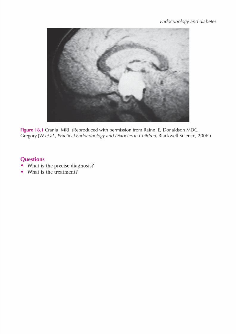
8/20/2019 100 cases in paediatrics.pdf
http://slidepdf.com/reader/full/100-cases-in-paediatricspdf 76/320
57
Endocrinology and diabetes
Questions• What is the precise diagnosis?• What is the treatment?
Figure 18.1 Cranial MRI. (Reproduced with permission from Raine JE, Donaldson MDC,Gregory JW et al. , Practical Endocrinology and Diabetes in Children , Blackwell Science, 2006.)

8/20/2019 100 cases in paediatrics.pdf
http://slidepdf.com/reader/full/100-cases-in-paediatricspdf 77/320
58
100 Cases in Paediatrics
ANSWER 18Tracey has gonadotrophin-dependent precocious puberty secondary to a hypothalamichamartoma. This condition differs from gonadotrophin-independent precocious puberty,which is caused by the abnormal secretion of sex steroids independent of the hypothalamo-pituitary axis. Precocious puberty is defined in the UK as puberty commencing beforethe age of 8 years in females and 9 years in males. In the United States these criteriahave been recently revised to below 7 years in a Caucasian girl and below 6 years in an
Afro-Caribbean girl. There may be a family history.
The first sign of puberty in girls is breast development, and in boys is testicular enlarge-ment with a testicular volume of 4 mL denoting the start of puberty.
Tracey’s puberty started at 5 years and 6 months. Sweating, body odour, acne and aheight spurt (height velocity increases from 5 cm/year prepubertally to as much as 12 cm/
year) are all part of puberty. The high height centile relative to the mid-parental centileand the advanced bone age all support early puberty. The LHRH test can help diagnosegonadotrophin-dependent precocious puberty. An LH value 8 units/L with a predom-inant LH response is diagnostic. The ultrasound is also in keeping with puberty. In themajority of girls ( 90 per cent) the cause is idiopathic. The earlier the onset of puberty,the greater the likelihood of there being a cause. Investigation should be considered inall girls below 8 years of age. The presence of neurological features should also promptinvestigations, including a cranial MRI. In a minority of girls, a hypothalamic hamar-
toma can lead to early puberty. Rarely, malignant brain tumours can cause precociouspuberty. Acquired neurological injuries, such as encephalitis, hydrocephalus and radi-ation, can also lead to early puberty.
In contrast, in boys early puberty is usually secondary to a cranial lesion and MRI scan-ning is mandatory.
Treatment is with monthly injections of an LHRH analogue. This should halt pubertyand may lead to some regression. Although currently tall, this girl is at risk of prematurefusion of her epiphysis with a short adult height. Psychosocial considerations are also an
important indication for treatment. Coping with the emotional changes of puberty andpossible early menarche is difficult in young girls. In girls with mild, slowly progressiveearly puberty, no treatment may be necessary. In boys, treatment is also with an LHRHanalogue. Any underlying lesion should be treated. Treatment is often continued until11 years of age.
Hypothalamic hamartomas are benign and often remain static in size or grow slowly,producing no other signs. Neurosurgical intervention is not indicated except in rarepatients with intractable seizures.
KEY POINTS
• Precocious puberty is defined as puberty starting below the age of 8 years in girls and9 years in boys.
• In females it is usually idiopathic, but in males it is usually secondary to a craniallesion.
• Treatment is with LHRH analogues (any underlying lesion may also need treatment).

8/20/2019 100 cases in paediatrics.pdf
http://slidepdf.com/reader/full/100-cases-in-paediatricspdf 78/320
59
CASE 19:A BOY WITH DELAYED PUBERTY
HistoryPaul, who is 14 years and 6 months old, comes to see his GP for the first time in years.He is worried about his short stature and lack of pubertal development. He feels he islosing out in team sports and wants to become a physical education teacher. The GPrecords show that his father is on the 9th centile and his mother is between the 9th and25th centiles. There are no other symptoms or past history of note.
ExaminationPaul is a fit, healthy, slim boy. General and systems examination are normal. His penisis about 5 cm long and the skin of the scrotum is lax and slightly pigmented. Both testesare descended but are quite small. There is no pubic or axillary hair. Both height andweight are on the 2nd centile. The GP puts together a growth chart from the availabledata (see Fig. 19.1).
Figure 19.1 Paul’s growth chart. (Chart reproduced with kind permission of the Child GrowthFoundation.)
Questions• What is the most likely diagnosis?• What should the GP do next?• Is there any treatment available?
8/20/2019 100 cases in paediatrics.pdf
http://slidepdf.com/reader/full/100-cases-in-paediatricspdf 79/320
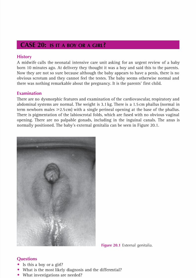
8/20/2019 100 cases in paediatrics.pdf
http://slidepdf.com/reader/full/100-cases-in-paediatricspdf 80/320
61
CASE 20:IS IT A BOY OR A GIRL
?History
A midwife calls the neonatal intensive care unit asking for an urgent review of a babyborn 10 minutes ago. At delivery they thought it was a boy and said this to the parents.Now they are not so sure because although the baby appears to have a penis, there is noobvious scrotum and they cannot feel the testes. The baby seems otherwise normal andthere was nothing remarkable about the pregnancy. It is the parents’ first child.
ExaminationThere are no dysmorphic features and examination of the cardiovascular, respiratory andabdominal systems are normal. The weight is 3.1 kg. There is a 1.5 cm phallus (normal interm newborn males 2.5 cm) with a single perineal opening at the base of the phallus.There is pigmentation of the labioscrotal folds, which are fused with no obvious vaginalopening. There are no palpable gonads, including in the inguinal canals. The anus isnormally positioned. The baby’s external genitalia can be seen in Figure 20.1.
Figure 20.1 External genitalia.
Questions• Is this a boy or a girl?• What is the most likely diagnosis and the differential?• What investigations are needed?
8/20/2019 100 cases in paediatrics.pdf
http://slidepdf.com/reader/full/100-cases-in-paediatricspdf 81/320
62
100 Cases in Paediatrics
ANSWER 20 At this stage, no one can or should say whether this baby is a boy or a girl. It is impera-tive not to guess as it could be either a virilized female (a normal girl exposed in uteroto excess androgens) or an undervirilized male. Disorders of sexual differentiation (DSD)are a medical and social emergency and families find it exceptionally difficult not know-ing the sex of their baby. Pending urgent tests they should not name the baby or registerthe birth.
In the absence of palpable gonads, but with evidence of virilization with a small phallusand pigmentation, it is most likely that this is a virilized female. Much the commonestaetiology is congenital adrenal hyperplasia (CAH). If the baby is an undervirilized male,there is a much larger differential, including disorders of gonadotrophin production (e.g.
Kallman syndrome), defects of testosterone synthesis and end-organ insensitivity due toandrogen receptor abnormalities. With very severe undervirilization, it is possible thatthe sex of rearing should be female. Such babies need urgent referral to a specialist centrewith a team of endocrinologists, urologists, geneticists and psychologists.
Congenital adrenal hyperplasia is a group of autosomal recessive inborn errors of metab-olism (IEM) (therefore commoner if parents are consanguineous), within the adrenalsteroidogenic pathways that produce mineralocorticoids (aldosterone), glucocorticoids(cortisol) and androgens (testosterone). Ninety-five per cent of cases are due to 21-hydroxylase deficiency (21-OHD) that catalyses late steps in the first two pathways. The
consequences are the same as for any IEM – deficiency of the end product, continueddrive through the pathway, build-up of precursors and diversion down alternative path-ways. Lack of cortisol causes hypoglycaemia and a poor stress response. Elevated pre-cursors divert to the androgen pathway and testosterone virilizes both male and femalefetuses. Most babies are also aldosterone-deficient. Those missed at birth (usually viri-lized boys) classically present in the second week of life with a salt-wasting (Addisonian)crisis – vomiting and shocked with severe hyponatraemia, hyperkalaemia and acidosis.
Patients with 21-OHD need lifelong steroid replacement, initially with hydrocortisoneand fludrocortisone (a mineralocorticoid analogue), and endocrine monitoring.
The timing of any surgery to reduce clitoromegaly and create a vaginal orifice iscontroversial.
Investigations in the first few days consider the differential diagnosis and monitor forcomplications:
• karyotype – result in 48 hours if laboratory warned• pelvic and abdominal ultrasound – extremely helpful and immediately available• 17-hydroxyprogesterone – sent after 48 hours as raised in all newborns• full male hormone profile• urine steroid profile – confirms site of block in steroidogenic pathway • electrolytes and glucose – from day 2; monitor bedside glucose• plasma renin activity – best estimation of salt status.
KEY POINTS
• Never guess the sex of a newborn baby with abnormal genitalia.• Virilization of a female infant is almost always congenital adrenal hyperplasia due to
21-hydroxylase deficiency.

8/20/2019 100 cases in paediatrics.pdf
http://slidepdf.com/reader/full/100-cases-in-paediatricspdf 82/320
63
CASE 21:A BOY WITH BREASTS
History Anthony is a 15-year-old boy who presents to an outpatient clinic with an 18-monthhistory of gynaecomastia. There is occasional breast tenderness. There is no history ofgalactorrhoea. He has stopped doing sports at school as he is too embarrassed to undressin front of his classmates in the changing room. He is a little behind at school andrequires extra help. His aunt has breast cancer and his grandmother died of breast can-cer. He is on no medication and has no allergies.
ExaminationThere is moderate symmetrical gynaecomastia. Pubertal staging is as follows (see p. 60 ):pubic hair, Tanner stage 4; genitalia, Tanner stage 3. Testes are both 15 mL in volume(using the Prader orchidometer). There are no abdominal masses and no other signs.
His weight, at 78 kg, is between the 91st and 98th centiles and his height, at 169 cm, ison the 50th centile.
INVESTIGATIONS
Normal
Follicle-stimulating hormone 2.9 units/L 1.8–10.6 units/LLuteinizing hormone 3.3 units/L 0.4–7.0 units/LTestosterone 9.2 nmol/L 7.6–21.5 nmol/LOestradiol 98 pmol/L 30–130 pmol/L
Questions
• What is the diagnosis?• What is the treatment?
8/20/2019 100 cases in paediatrics.pdf
http://slidepdf.com/reader/full/100-cases-in-paediatricspdf 83/320
64
ANSWER 21 Anthony has pubertal gynaecomastia. All males have small amounts of oestrogen, justas all females have small amounts of adrenal androgens (which, in females, leads to thedevelopment of pubic and axillary hair). Gynaecomastia is common in pubertal malesand is due to a decreased ratio of testosterone to oestrogen in puberty.
Klinefelter’s syndrome (XXY) can be associated with gynaecomastia and learning dif-ficulties. These patients also have small testes and this makes that diagnosis unlikelyin our patient. Gynaecomastia is occasionally familial. Very rarely, oestrogen-secretingtumours can lead to gynaecomastia, e.g. a feminizing adrenal tumour or a Leydig celltumour of the testis. The absence of abdominal signs, of a unilaterally enlarged testisand the normality of the oestradiol level make these diagnoses very unlikely. Drugs such
as oestrogen and spironolactone and drugs of abuse, such as marijuana, can also causegynaecomastia.
Prolactinomas are not usually accompanied by gynaecomastia and the absence of galac-torrhoea makes this diagnosis unlikely. Breast cancer would be exceptionally rare inpuberty.
Mild-to-moderate cases of pubertal gynaecomastia need no investigation. The conditionis transient and usually lasts for several months to 2 years. Anthony is somewhat over-weight and a proportion of the breast enlargement could be accounted for by adiposerather than breast tissue. Advice on weight loss could therefore be helpful. In mild-to-moderate cases, reassurance usually suffices. If the gynaecomastia is severe, or is lead-ing to psychosocial problems, e.g. severe teasing, plastic surgery is indicated. Mammaryreduction by either liposuction or a subareolar incision with removal of the excess tissuecan be performed.
KEY POINTS
• Gynaecomastia is common in pubertal boys.
• Reassurance usually suffices, but occasionally plastic surgery is indicated.
100 Cases in Paediatrics

8/20/2019 100 cases in paediatrics.pdf
http://slidepdf.com/reader/full/100-cases-in-paediatricspdf 84/320
This page intentionally left blank
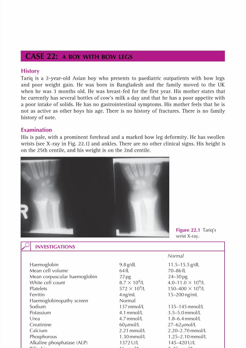
8/20/2019 100 cases in paediatrics.pdf
http://slidepdf.com/reader/full/100-cases-in-paediatricspdf 85/320
66
CASE 22:A BOY WITH BOW LEGS
HistoryTariq is a 2-year-old Asian boy who presents to paediatric outpatients with bow legsand poor weight gain. He was born in Bangladesh and the family moved to the UKwhen he was 3 months old. He was breast-fed for the first year. His mother states thathe currently has several bottles of cow’s milk a day and that he has a poor appetite witha poor intake of solids. He has no gastrointestinal symptoms. His mother feels that he isnot as active as other boys his age. There is no history of fractures. There is no familyhistory of note.
ExaminationHis is pale, with a prominent forehead and a marked bow leg deformity. He has swollenwrists (see X-ray in Fig. 22.1) and ankles. There are no other clinical signs. His height ison the 25th centile, and his weight is on the 2nd centile.
INVESTIGATIONS
Normal
Haemoglobin 9.8 g/dL 11.5–15.5 g/dLMean cell volume 64 fL 70–86 fLMean corpuscular haemoglobin 22 pg 24–30 pgWhite cell count 8.7 109 /L 4.0–11.0 109 /LPlatelets 572 10 9 /L 150–400 109 /LFerritin 4 ng/mL 15–200 ng/mLHaemoglobinopathy screen NormalSodium 137 mmol/L 135–145 mmol/LPotassium 4.1 mmol/L 3.5–5.0 mmol/LUrea 4.7 mmol/L 1.8–6.4 mmol/LCreatinine 60 µmol/L 27–62 µmol/LCalcium 2.21 mmol/L 2.20–2.70 mmol/LPhosphorous 1.30 mmol/L 1.25–2.10 mmol/LAlkaline phosphatase (ALP) 1372 U/L 145–420 U/LBilirubin 16 µmol/L 2–26 µmol/LAlanine aminotransferase (ALT) 32 U/L 10–40 U/L
Figure 22.1 Tariq’swrist X-ray.

8/20/2019 100 cases in paediatrics.pdf
http://slidepdf.com/reader/full/100-cases-in-paediatricspdf 86/320
67
Endocrinology and diabetes
Questions• Name the two disorders that affect this child.• What is the treatment?
8/20/2019 100 cases in paediatrics.pdf
http://slidepdf.com/reader/full/100-cases-in-paediatricspdf 87/320
68
100 Cases in Paediatrics
ANSWER 22This type of nutritional history in an Asian child, the accompanying clinical signs, thehigh ALP, the borderline calcium and the wrist X-ray all point to rickets. Furthermore,the poor nutritional intake, the pallor and the microcytic hypochromic anaemia aretypical of iron deficiency anaemia.
Rickets is a disorder characterized by a failure in the mineralization of growing bones.The majority of cases are due to inadequate sunlight exposure coupled with nutritionaldeficiency. The condition is more common in dark-skinned infants who need greatersun exposure to synthesize adequate amounts of vitamin D. Prolonged breast-feedingincreases the risk of rickets as breast milk is a poor source of vitamin D. Infant for-mulas are often supplemented with vitamin D, but cow’s milk is also a poor source of
vitamin D. Diseases that interfere with the metabolic conversion and activation of vita-min D, such as severe renal and liver disease, can lead to rickets. Illnesses associatedwith malabsorption, such as coeliac disease, can also lead to rickets. Other diseases thatinterfere with calcium and phosphorus homeostasis can cause rickets, e.g. renal tubu-lar defects, such as hypophosphataemic rickets, in which there is failure of phosphateabsorption, or Fanconi’s syndrome in which there is a defect in the reabsorption ofphosphate, glucose and amino acids.
Clinical features include swollen wrists and ankles, frontal bossing, prominent costo-chondral junctions (a rickety rosary), bow legs or knock-knees, craniotabes, muscle
weakness, tetany and hypocalcaemic fits.The radiograph demonstrates the typical appearance in rickets with widening of thegrowth plate and cupping, splaying and fraying of the metaphysis.
A vitamin D level to confirm the nutritional aetiology of the rickets and anti-transglutaminase antibody levels to rule out coeliac disease would be helpful.
Iron deficiency anaemia is said to occur in a quarter of children under 5 years of age.Cow’s milk is a poor source of iron. Although usually nutritional, blood loss shouldbe considered as a possible cause, especially in older children. This may be due tooccult gastrointestinal blood loss secondary to a peptic ulcer, a Meckel’s diverticulumor inflammatory bowel disease. A ferritin level should be done to confirm the diagno-sis and is likely to be very low. A haemoglobinopathy screen should be performed inhigh-risk groups to exclude disorders such as thalassaemia that can also cause a micro-cytic hypochromic anaemia and that may coexist with iron deficiency. Infants maybe pale, tired, irritable and anorexic. In severe cases they can be tachypnoeic. There isevidence that iron deficiency anaemia can also have adverse effects on attention spanand learning.
Treatment consists of a 3-month course of high-dose oral vitamin D. Bone chemistryshould be measured 2 weeks after starting treatment and regularly thereafter to avoidhypercalcaemia and to ensure that the biochemical and haematological parameters nor-malize. This should be followed by maintenance doses of vitamin D for the long term.The prognosis for complete resolution of the deformities is good. In cases where theserum calcium is low, calcium is also given initially until levels normalize.
There is often an accompanying iron deficiency anaemia, as in this case. Red meat andgreen vegetables are good sources of iron and some foods, such as cereals, are oftenfortified with iron. The anaemia should be treated with a 3-month course of oral iron.

8/20/2019 100 cases in paediatrics.pdf
http://slidepdf.com/reader/full/100-cases-in-paediatricspdf 88/320
69
Endocrinology and diabetes
A dietetic referral and screening of the siblings for rickets and iron deficiency wouldalso be advisable.
KEY POINTS
• Rickets is usually secondary to inadequate exposure to sunlight and poor nutrition.• Swollen wrists, a rickety rosary and bow legs are typical clinical features.• Treatment is with vitamin D.• Iron deficiency is very common in young children and can be successfully treated
with dietary advice and oral iron.

8/20/2019 100 cases in paediatrics.pdf
http://slidepdf.com/reader/full/100-cases-in-paediatricspdf 89/320
This page intentionally left blank
8/20/2019 100 cases in paediatrics.pdf
http://slidepdf.com/reader/full/100-cases-in-paediatricspdf 90/320
71
GASTROENTEROLOGY
CASE 23: A VOMITING INFANT
HistoryTom is a 7-week-old infant who presents to the A&E department with a 1-week history ofnon-bilious vomiting. His mother describes the vomit as ‘shooting out’. He has a good appe-tite but has lost 300 g since he was last weighed a week earlier. He has mild constipation.The family have recently returned from Spain. There is no vomiting in any other membersof the family. His sister suffers from vesicoureteric reflux and urinary tract infections.
ExaminationTom is apyrexial and mildly dehydrated. His pulse is 170 beats/min, blood pressure 82/43 mmHg,and peripheral capillary refill 2 s. There is no organomegaly, masses or tenderness on abdom-inal examination. There are no signs in the other systems.
INVESTIGATIONS
Normal
Haemoglobin 11.7 g/dL 10.5–13.5 g/dLWhite cell count 10.0 109 /L 4.0–11.0 10 9 /L
Platelets 332 109
/L 150–400 109
/LSodium 134 mmol/L 135–145 mmol/LPotassium 3.1 mmol/L 3.5–5.0 mmol/LChloride 81 mmol/L 98–106 mmol/LUrea 9.0 mmol/L 1.8–6.4 mmol/LCreatinine 60 µmol/L 18–35 µmol/LCapillary gas pH 7.56 7.36–7.44
P CO2 6.0 kPa 4.0–6.5 kPaP O2 3.2 kPa 12–15 kPa
HCO3 38 mmol/L 22–29 mmol/LBase excess 10 ( 2.5)–( 2.5) mmol/LUrine dipstick No abnormality detected
Questions• What is the likely diagnosis?• What is the differential diagnosis?• How would you confirm the diagnosis?• What is the treatment?

8/20/2019 100 cases in paediatrics.pdf
http://slidepdf.com/reader/full/100-cases-in-paediatricspdf 91/320
8/20/2019 100 cases in paediatrics.pdf
http://slidepdf.com/reader/full/100-cases-in-paediatricspdf 92/320
73
CASE 24:A CHILD WITH BLOODY DIARRHOEA
HistoryBobby is a 7-month-old child who is referred to the paediatric rapid referral clinic with a2-day history of diarrhoea with blood and mucus in the stool. His mother states that he hasperiods of inconsolable crying which are getting worse and more frequent. In the last fewhours, he had started to vomit and the last vomit was bile-stained. There is no history ofcontact with gastroenteritis, of travel or of bleeding disorders. He had neonatal meningitisand subsequently developed epilepsy, which is treated with sodium valproate. He has had nofits in the past month. His mother is in good health but his father has type 1 diabetes.
ExaminationHe has a temperature of 37.9 C. His pulse rate is 186 beats/min, blood pressure is80/44 mmHg and capillary refill is 4 s. He is difficult to examine due to frequent crying,but when examined during a quiet period, a mass is felt on the right side of the abdo-men. The anus appears normal and there are no other signs.
INVESTIGATIONS
Normal Haemoglobin 12.8 g/dL 10.5–13.5 g/dLWhite cell count 7.0 109 /L 4.0–11.0 109 /LPlatelets 457 109 /L 150–400 109 /LSodium 138 mmol/L 135–145 mmol/LPotassium 3.9 mmol/L 3.5–5.0 mmol/LUrea 9.5 mmol/L 1.8–6.4 mmol/LCreatinine 60 µmol/L 20–80 µmol/LC-reactive protein 12 mg/L 6 mg/L
Questions• What is the diagnosis?• What is the key investigation?• What is the treatment?

8/20/2019 100 cases in paediatrics.pdf
http://slidepdf.com/reader/full/100-cases-in-paediatricspdf 93/320
74
100 Cases in Paediatrics
ANSWER 24The commonest causes of passing blood per rectum are gastroenteritis and an anal fissure.Gastroenteritis is a possibility in this case, but the lack of contact with gastroenteritis andof recent travel makes it somewhat less likely. The normality of the anus makes an analfissure unlikely. The age of the child, the rectal bleeding and the mass (a late feature ofintussusception) make an intussusception the most likely diagnosis.
The typical age in intussusception is 3 months to 3 years. It is due to a part of thebowel telescoping into the adjacent distal bowel. Most intussusceptions are ileocolic.They result in stretching of the mesenteric vasculature that can potentially lead to bowelischaemia or infarction. For this reason, investigation and treatment need to be insti-tuted urgently. Typically the child has paroxysmal pain during which he or she becomes
very pale; stool containing a mixture of blood and mucus (hence the name redcurrant jelly stool); and a sausage-shaped mass (the intussusception itself ) in the upper rightquadrant of the abdomen. As the disease progresses, bowel obstruction, peritonitis andsepticaemia may develop. The diagnostic investigation is an ultrasound.
! Causes of rectal bleeding
• Gastroenteritis• Anal fissure• Intussusception
• Cow’s milk protein allergy• Meckel’s diverticulum• Inflammatory bowel disease• Polyp• Clotting abnormality• Sexual abuse
This infant has clinical signs (tachycardia and prolonged capillary refill) and biochemicalevidence (raised urea) of dehydration, and initial treatment consists of intravenous fluidresuscitation and intravenous antibiotics, such as penicillin, gentamicin and metronida-zole. A nasogastric tube should be inserted and the stomach emptied. In the majority ofcases, reduction can be achieved by a radiologist with an air enema. The antibiotics aregiven because of the possibility of sepsis and the small risk of a perforation during thereduction. If this procedure fails, or if the child is unstable or has signs of peritonitis or aperforation, then surgery is indicated. There is a recurrence rate of 10 per cent followingan air enema reduction, and of 2–5 per cent following a surgical reduction.
KEY POINTS
• Intussusception should be considered in any child aged 3 months to 3 years withbloody stool.
• There should be a low threshold for carrying out an ultrasound.• An air enema reduction is successful in the majority of cases.

8/20/2019 100 cases in paediatrics.pdf
http://slidepdf.com/reader/full/100-cases-in-paediatricspdf 94/320
75
CASE 25:A TEENAGER WITH CHRONIC DIARRHOEA
HistoryLevi is a 14-year-old boy who presents to the paediatric rapid referral clinic with a 3-weekhistory of diarrhoea and cramp-like abdominal pain. He has no blood or mucus in his stooland has had no vomiting. His appetite is poor and he has lost 3.5 kg in 3 weeks. He hasintermittent fevers. The family recently returned from France where his father contracteddiarrhoea, from which he is now recovering. There is no other medical history of note. Hismother suffers from irritable bowel syndrome.
ExaminationHe looks generally unwell. There is no anaemia, jaundice, clubbing or lymphadenopathy.His temperature is 37.9 C. There is generalized abdominal tenderness but no guarding orrebound. There is no organomegaly. Inspection of the mouth and anus is normal. His puber-tal assessment shows him to have testes that are 5 mL in volume and pubic hair and geni-talia that are Tanner stage 2.
His weight, at 35 kg, is on the second centile and his height, at 151 cm, is on the ninthcentile (mid-parental height is on the 50th centile).
INVESTIGATIONS
Normal
Haemoglobin 10.9 g/dL 14–18 g/dLWhite cell count 15.2 109 /L 4.0–11.0 109 /LPlatelets 623 109 /L 150–400 109 /LMean cell volume 70 fL 76–96 fLErythrocyte sedimentation rate (ESR) 87 mm/h 15 mm/hC-reactive protein (CRP) 36 mg/L 6 mg/LFerritin 14 ng/ml 20–300 ng/mlAlbumin 31 g/L 35–50 g/LUrea and electrolytes NormalStool – no bacterial growth, no ova, cysts or parasites
Questions• What is the most likely diagnosis?• What investigations would you do?• What is the initial treatment?
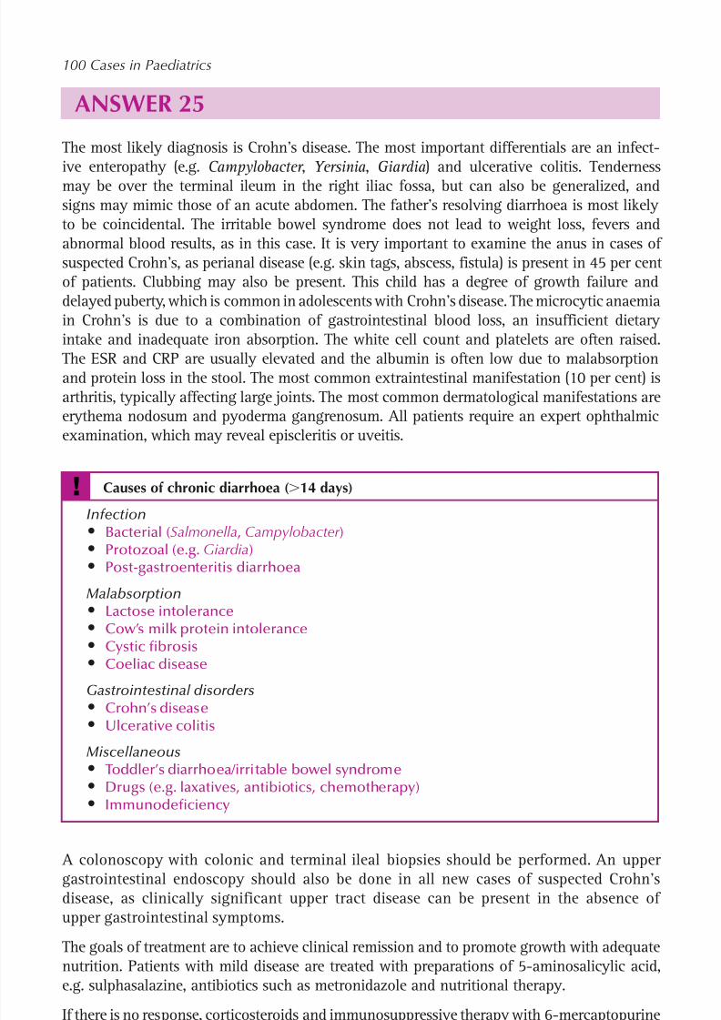
8/20/2019 100 cases in paediatrics.pdf
http://slidepdf.com/reader/full/100-cases-in-paediatricspdf 95/320
76
! Causes of chronic diarrhoea ( 14 days)
Infection
• Bacterial ( Salmonella , Campylobacter )• Protozoal (e.g. Giardia )• Post-gastroenteritis diarrhoea
Malabsorption• Lactose intolerance• Cow’s milk protein intolerance• Cystic fibrosis• Coeliac disease
Gastrointestinal disorders
• Crohn’s disease• Ulcerative colitis
Miscellaneous• Toddler’s diarrhoea/irritable bowel syndrome• Drugs (e.g. laxatives, antibiotics, chemotherapy)• Immunodeficiency
ANSWER 25The most likely diagnosis is Crohn’s disease. The most important differentials are an infect-ive enteropathy (e.g. Campylobacter , Yersinia , Giardia ) and ulcerative colitis. Tendernessmay be over the terminal ileum in the right iliac fossa, but can also be generalized, andsigns may mimic those of an acute abdomen. The father’s resolving diarrhoea is most likelyto be coincidental. The irritable bowel syndrome does not lead to weight loss, fevers andabnormal blood results, as in this case. It is very important to examine the anus in cases ofsuspected Crohn’s, as perianal disease (e.g. skin tags, abscess, fistula) is present in 45 per centof patients. Clubbing may also be present. This child has a degree of growth failure anddelayed puberty, which is common in adolescents with Crohn’s disease. The microcytic anaemiain Crohn’s is due to a combination of gastrointestinal blood loss, an insufficient dietaryintake and inadequate iron absorption. The white cell count and platelets are often raised.
The ESR and CRP are usually elevated and the albumin is often low due to malabsorptionand protein loss in the stool. The most common extraintestinal manifestation (10 per cent) isarthritis, typically affecting large joints. The most common dermatological manifestations areerythema nodosum and pyoderma gangrenosum. All patients require an expert ophthalmicexamination, which may reveal episcleritis or uveitis.
A colonoscopy with colonic and terminal ileal biopsies should be performed. An uppergastrointestinal endoscopy should also be done in all new cases of suspected Crohn’sdisease, as clinically significant upper tract disease can be present in the absence ofupper gastrointestinal symptoms.
The goals of treatment are to achieve clinical remission and to promote growth with adequatenutrition. Patients with mild disease are treated with preparations of 5-aminosalicylic acid,e.g. sulphasalazine, antibiotics such as metronidazole and nutritional therapy.
If there is no response, corticosteroids and immunosuppressive therapy with 6-mercapto purineor methotrexate can be tried. The latter two agents can also have corticosteroid-sparingeffects. Surgery is considered when medical therapy fails. Indications include intractable
100 Cases in Paediatrics

8/20/2019 100 cases in paediatrics.pdf
http://slidepdf.com/reader/full/100-cases-in-paediatricspdf 96/320
77
KEY POINTS
• The perianal area should always be examined in cases of suspected Crohn’s disease.• Growth failure and delayed puberty are common in Crohn’s disease.• Colonoscopy and upper gastrointestinal endoscopy are key investigations.• Initial treatment consists of preparations of 5-aminosalicylic acid, antibiotics and
nutritional therapy.
disease with growth failure, obstruction (due to strictures or adhesions), abscess drainage,fistula, intractable haemorrhage and perforation.
Gastroenterology

8/20/2019 100 cases in paediatrics.pdf
http://slidepdf.com/reader/full/100-cases-in-paediatricspdf 97/320
This page intentionally left blank

8/20/2019 100 cases in paediatrics.pdf
http://slidepdf.com/reader/full/100-cases-in-paediatricspdf 98/320
79
CASE 26:ACUTE DIARRHOEA AND VOMITING
HistoryLuca is a 24-month-old boy with Down’s syndrome, brought to the A&E department byhis mother. He has had diarrhoea and vomiting for 1 day. In the last 8 hours, he has drunk200 ml milk, vomited five times and passed six liquid stools. The vomit is not bilious andthere is no blood or mucus in the stools. It has been hard to tell if he is passing urinebecause every nappy is soiled. He has no cardiac problems and no other medical problemsexcept glue ear. There is no history of foreign travel. His two older siblings have recentlyhad diarrhoea and vomiting.
ExaminationHe is miserable and lethargic. His heart rate is 120 beats/min, respiratory rate is 25breaths/min, and his temperature is 37.7 C. He has dry mucous membranes, his eyes areslightly sunken, his skin turgor appears normal and his capillary refill time is less than2 s. His abdomen is soft with no masses palpable. His weight is 11 kg (50th centile on theDown’s syndrome growth chart).
An oral fluid challenge is commenced in the emergency department. He drinks 60 ml ofelectrolyte solution over 2 hours and vomits once on the floor. He does not pass urineinto a urine bag during this period.
Questions• What is the most likely diagnosis?• How dehydrated is this child?• How would you manage this child now?• How would you calculate the fluid requirements for this child over the next 24 hours?
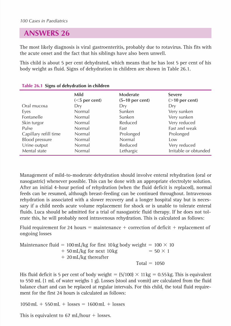
8/20/2019 100 cases in paediatrics.pdf
http://slidepdf.com/reader/full/100-cases-in-paediatricspdf 99/320
80
100 Cases in Paediatrics
ANSWERS 26The most likely diagnosis is viral gastroenteritis, probably due to rotavirus. This fits withthe acute onset and the fact that his siblings have also been unwell.
This child is about 5 per cent dehydrated, which means that he has lost 5 per cent of hisbody weight as fluid. Signs of dehydration in children are shown in Table 26.1.
Management of mild-to-moderate dehydration should involve enteral rehydration (oral ornasogastric) whenever possible. This can be done with an appropriate electrolyte solution.
After an initial 4-hour period of rehydration (when the fluid deficit is replaced), normalfeeds can be resumed, although breast-feeding can be continued throughout. Intravenousrehydration is associated with a slower recovery and a longer hospital stay but is neces-sary if a child needs acute volume replacement for shock or is unable to tolerate enteralfluids. Luca should be admitted for a trial of nasogastric fluid therapy. If he does not tol-erate this, he will probably need intravenous rehydration. This is calculated as follows:
Fluid requirement for 24 hours maintenance correction of deficit replacement ofongoing losses
Maintenance fluid 100 mL/kg for first 10 kg body weight 100 10 50 mL/kg for next 10 kg 50 1 20 mL/kg thereafter
Total 1050
His fluid deficit is 5 per cent of body weight (5/100) 11 kg 0.55 kg. This is equivalentto 550 mL (1 mL of water weighs 1 g). Losses (stool and vomit) are calculated from the fluidbalance chart and can be replaced at regular intervals. For this child, the total fluid require-ment for the first 24 hours is calculated as follows:
1050 mL 550 mL losses 1600 mL losses
This is equivalent to 67 mL/hour losses.
Table 26.1 Signs of dehydration in children
Mild Moderate Severe( 5 per cent) (5–10 per cent) ( 10 per cent)
Oral mucosa Dry Dry DryEyes Normal Sunken Very sunkenFontanelle Normal Sunken Very sunkenSkin turgor Normal Reduced Very reducedPulse Normal Fast Fast and weakCapillary refill time Normal Prolonged ProlongedBlood pressure Normal Normal LowUrine output Normal Reduced Very reducedMental state Normal Lethargic Irritable or obtunded

8/20/2019 100 cases in paediatrics.pdf
http://slidepdf.com/reader/full/100-cases-in-paediatricspdf 100/320
81
Gastroenterology
KEY POINTS
• Rotavirus is the most common cause of gastroenteritis in infants and young children.• Clinical assessment of dehydration is based on multiple physical signs.• Where possible, enteral rehydration with an electrolyte solution should be used for
children with gastroenteritis.• Fluid replacement must account for the deficit plus the maintenance requirement
plus ongoing losses.

8/20/2019 100 cases in paediatrics.pdf
http://slidepdf.com/reader/full/100-cases-in-paediatricspdf 101/320
This page intentionally left blank

8/20/2019 100 cases in paediatrics.pdf
http://slidepdf.com/reader/full/100-cases-in-paediatricspdf 102/320
83
CASE 27:ACUTE ABDOMINAL PAIN
HistorySarah is a 13-year-old girl who is brought to the A&E department by her mother, witha 2-day history of abdominal pain and vomiting. She got back from a family holidayin Devon yesterday evening and has been feeling unwell since. Initially she had centralabdominal pain which was coming and going, but now she has more constant pain onthe right side of her abdomen. She has vomited three times, but her stool has been nor-mal. She has no significant medical history, was well during the 2 weeks in Devon andher last period was about 2 weeks ago. Her family are all well and report that they allate the same food over the last few weeks.
ExaminationShe is flushed and has a temperature of 37.9 C. Her heart rate is 95 beats/min, respi-ratory rate is 18 breaths/min, capillary refill time is less than 2 s, and blood pressure is105/67 mmHg. She appears obese (weight 68 kg, 98th centile; height 151 cm, 25th centile).Her abdomen is tender in the right lower quadrant but there is no guarding or reboundtenderness. Pressing in the left lower quadrant elicits pain on the right.
INVESTIGATIONS
Normal Haemoglobin 12.3 g/dL 12.1–15.1 g/dLWhite cell count 16.3 109 /L 4.5–13 109 /LNeutrophil count 10 109 /L 1.5–6 109 /LPlatelets 210 109 /L 180–430 109 /LSodium 133 mmol/L 135–145 mmol/LPotassium 3.4 mmol/L 3.5–5.6 mmol/LUrea 4.3 mmol/L 2.5–6.6 mmol/LCreatinine 76 µmol/L 20–80 µmol/LBilirubin 5 mmol/L 1.7–26 mmol/LAlkaline phosphatase 264 IU/dL 25–800 IU/dLAspartate aminotransferase 20 IU/dL 10–45 IU/dLAlbumin 35 g/L 37–50 g/LC-reactive protein 16 mg/L 5 mg/LUrineLeucocytes NegativeNitrites Negative NegativeBlood Negative NegativeUltrasound abdomen – liver, spleen and kidneys appear normal. There are a few slightlyenlarged mesenteric lymph nodes. The appendix is not seen.
Questions• What is the most likely diagnosis?• What other diagnoses should be considered?• How should she be managed?
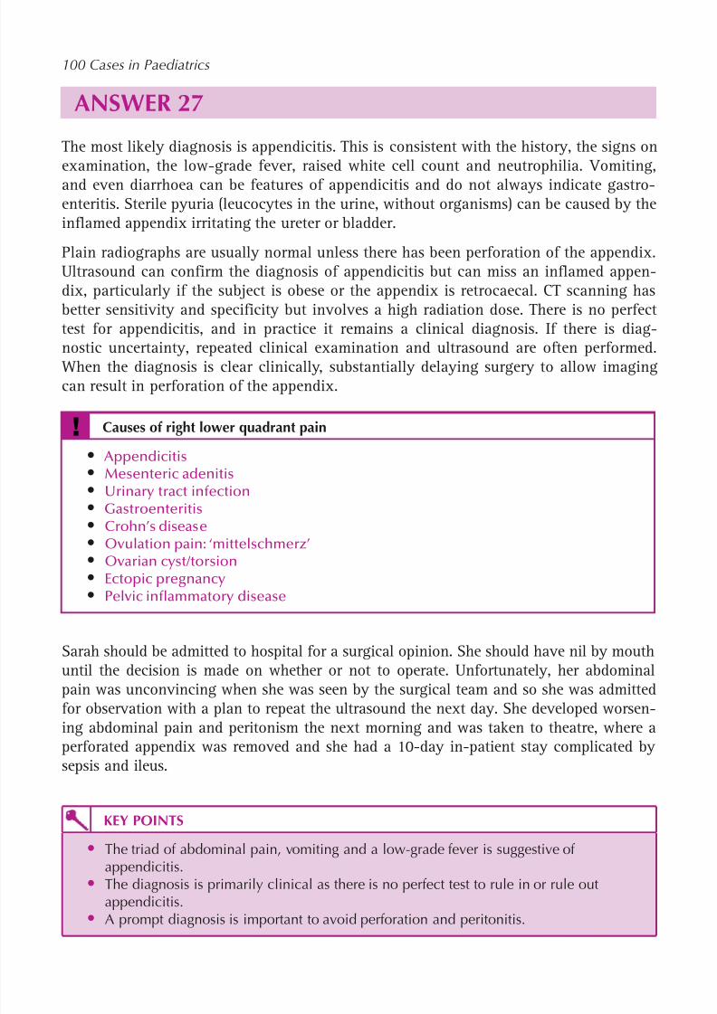
8/20/2019 100 cases in paediatrics.pdf
http://slidepdf.com/reader/full/100-cases-in-paediatricspdf 103/320
84
100 Cases in Paediatrics
ANSWER 27The most likely diagnosis is appendicitis. This is consistent with the history, the signs onexamination, the low-grade fever, raised white cell count and neutrophilia. Vomiting,and even diarrhoea can be features of appendicitis and do not always indicate gastro-enteritis. Sterile pyuria (leucocytes in the urine, without organisms) can be caused by theinflamed appendix irritating the ureter or bladder.
Plain radiographs are usually normal unless there has been perforation of the appendix.Ultrasound can confirm the diagnosis of appendicitis but can miss an inflamed appen-dix, particularly if the subject is obese or the appendix is retrocaecal. CT scanning hasbetter sensitivity and specificity but involves a high radiation dose. There is no perfecttest for appendicitis, and in practice it remains a clinical diagnosis. If there is diag-
nostic uncertainty, repeated clinical examination and ultrasound are often performed. When the diagnosis is clear clinically, substantially delaying surgery to allow imagingcan result in perforation of the appendix.
! Causes of right lower quadrant pain
• Appendicitis• Mesenteric adenitis• Urinary tract infection• Gastroenteritis
• Crohn’s disease• Ovulation pain: ‘mittelschmerz’• Ovarian cyst/torsion• Ectopic pregnancy• Pelvic inflammatory disease
Sarah should be admitted to hospital for a surgical opinion. She should have nil by mouthuntil the decision is made on whether or not to operate. Unfortunately, her abdominalpain was unconvincing when she was seen by the surgical team and so she was admitted
for observation with a plan to repeat the ultrasound the next day. She developed worsen-ing abdominal pain and peritonism the next morning and was taken to theatre, where aperforated appendix was removed and she had a 10-day in-patient stay complicated bysepsis and ileus.
KEY POINTS
• The triad of abdominal pain, vomiting and a low-grade fever is suggestive ofappendicitis.
• The diagnosis is primarily clinical as there is no perfect test to rule in or rule outappendicitis.• A prompt diagnosis is important to avoid perforation and peritonitis.
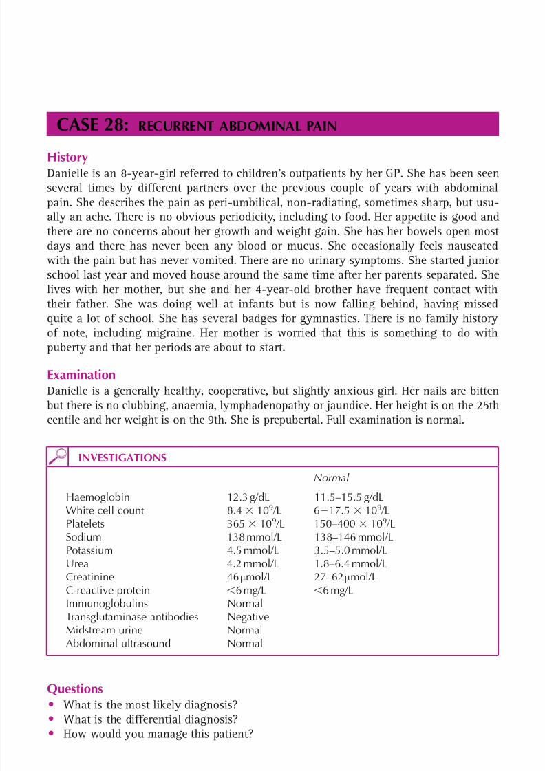
8/20/2019 100 cases in paediatrics.pdf
http://slidepdf.com/reader/full/100-cases-in-paediatricspdf 104/320
85
CASE 28:RECURRENT ABDOMINAL PAIN
HistoryDanielle is an 8-year-girl referred to children’s outpatients by her GP. She has been seenseveral times by different partners over the previous couple of years with abdominalpain. She describes the pain as peri-umbilical, non-radiating, sometimes sharp, but usu-ally an ache. There is no obvious periodicity, including to food. Her appetite is good andthere are no concerns about her growth and weight gain. She has her bowels open mostdays and there has never been any blood or mucus. She occasionally feels nauseatedwith the pain but has never vomited. There are no urinary symptoms. She started juniorschool last year and moved house around the same time after her parents separated. Shelives with her mother, but she and her 4-year-old brother have frequent contact withtheir father. She was doing well at infants but is now falling behind, having missedquite a lot of school. She has several badges for gymnastics. There is no family historyof note, including migraine. Her mother is worried that this is something to do withpuberty and that her periods are about to start.
ExaminationDanielle is a generally healthy, cooperative, but slightly anxious girl. Her nails are bitten
but there is no clubbing, anaemia, lymphadenopathy or jaundice. Her height is on the 25thcentile and her weight is on the 9th. She is prepubertal. Full examination is normal.
INVESTIGATIONS
Normal
Haemoglobin 12.3 g/dL 11.5–15.5 g/dLWhite cell count 8.4 109 /L 6 17.5 109 /LPlatelets 365 10 9 /L 150–400 109 /L
Sodium 138 mmol/L 138–146 mmol/LPotassium 4.5 mmol/L 3.5–5.0 mmol/LUrea 4.2 mmol/L 1.8–6.4 mmol/LCreatinine 46 µmol/L 27–62 µmol/LC-reactive protein 6 mg/L 6 mg/LImmunoglobulins NormalTransglutaminase antibodies NegativeMidstream urine NormalAbdominal ultrasound Normal
Questions• What is the most likely diagnosis?• What is the differential diagnosis?• How would you manage this patient?
8/20/2019 100 cases in paediatrics.pdf
http://slidepdf.com/reader/full/100-cases-in-paediatricspdf 105/320
86
100 Cases in Paediatrics
ANSWER 28The most likely diagnosis is chronic abdominal pain of childhood or recurrent abdomi-nal pain (RAP). This is a benign, very common but potentially debilitating condition.
The pointers to this not being organic are the chronicity of the characteristic symptomsin an otherwise healthy, physically active child. The commonest identifiable cause forRAP is psychosomatic, as seems likely from the description of this girl who has hadmany recent changes in her life. However, it is a diagnosis of exclusion and investiga-tions exclude conditions that can have an insidious onset, such as coeliac disease, andreassure the child and family that there is no sinister cause. The latter is crucial becausethe symptoms are very real and there is understandable anxiety.
Among the differentials, abdominal migraine differs in that it is usually associated withpallor and vomiting. There is almost always a family history of migraine. An organiccause is also more likely the further the pain is from the umbilicus.
Mothers often worry that RAP heralds menarche but this comes towards the end ofpuberty. Sexual abuse should be considered in the differential.
! Differential diagnosis of chronic abdominal pain
• Psychosomatic• Urinary tract infections• Constipation• Gastro-oesophageal reflux• Coeliac disease• Inflammatory bowel disease• Cow’s milk intolerance• Abnormal renal anatomy, e.g. pelviureteric junction obstruction• Abdominal migraine• Peptic ulcer• Sexual or other abuse
Management focuses on explanations and reassurance. It is helpful to liken the conditionto tension headaches in adults – common and unpleasant but not serious. Acknowledgethat there is no question of fabrication. Children rarely fabricate symptoms and, ifthey do, abuse should be considered. The classic time for RAP is in the morning beforeschool. Most parents recognize this and have often already asked questions about bul-lying or other worries, but sometimes a cycle sets in where missing school and fallingbehind worsen the symptoms and cause more anxiety. The child may be recognized as a‘worrier’ and most can understand the concept of psychosomatic symptoms.
Some families still find it difficult to accept that there is nothing medically wrong andpursue a diagnosis such as food allergy. They may seek advice from practitioners ofalternative medicine and it is important to discuss openly the dangers of dietary exclu-sions that they may suggest. In the absence of other symptoms, such as diarrhoea or anassociation with eating, there is no evidence that such measures are effective.
Once a diagnosis has been reached, the child should be discharged from hospital follow-up to prevent the risk of reinforcing a medical diagnosis. Referral to a psychologist orpsychiatrist is sometimes necessary.

8/20/2019 100 cases in paediatrics.pdf
http://slidepdf.com/reader/full/100-cases-in-paediatricspdf 106/320
87
Gastroenterology
KEY POINTS
• Children can have psychosomatic symptoms just as adults – however, these are adiagnosis of exclusion.

8/20/2019 100 cases in paediatrics.pdf
http://slidepdf.com/reader/full/100-cases-in-paediatricspdf 107/320
This page intentionally left blank

8/20/2019 100 cases in paediatrics.pdf
http://slidepdf.com/reader/full/100-cases-in-paediatricspdf 108/320
89
CASE 29:A CONSTIPATED TODDLER
HistoryTanya is a 4-year-old girl who presents to outpatients with a 2-year history of constipa-tion. She opens her bowels about once every 5 days and strains. She soils her knickers onmost days. She has intermittent abdominal pain, which is relieved by opening her bowels.Recently, there has been fresh blood on the toilet tissue. Lactulose has been used, with lit-tle success. Her mother states that she did not have a dirty nappy until 40 hours of age.
She has recently had a urine infection diagnosed by her GP. The illness was mild and
responded well to antibiotics. She was delivered by emergency Caesarean sectionbecause of fetal distress and meconium staining.
Examination A faecal mass is palpable in the left iliac fossa. The anus appears normal. Rectal examination –hard stool palpated. The back is normal. Blood pressure is 101/62 mmHg. There are noother signs. Weight is on the 50th centile and height is on the 25th centile.
Questions
• What is the most likely diagnosis?
• Would you carry out any investigations?• What is the treatment?

8/20/2019 100 cases in paediatrics.pdf
http://slidepdf.com/reader/full/100-cases-in-paediatricspdf 109/320
90
100 Cases in Peadiatrics
ANSWER 29The most likely diagnosis is functional constipation. This condition is associated withfaecal masses in the lower abdomen. Severe constipation can lead to an anal fissure(and vice versa). This is the likely cause of the bleeding. It may be high up in the anusand therefore not visible on inspection. The soiling is involuntary and due to liquid stoolleaking from above the hard stool mass in the rectum. Hirschsprung’s disease should besuspected if meconium has not been passed in the first 24 hours of life. However, in thiscase, the passage of meconium in utero , the lack of symptoms in the first 2 years of lifeand the normal weight make that diagnosis unlikely. Furthermore, in Hirschsprung’s dis-ease the rectum is usually empty.
! Causes of constipation
• Dietary• Dehydration• Anal fissure/stenosis• Hirschsprung’s disease• Intestinal obstruction e.g. stricture post-necrotizing enterocolitis• Spinal cord lesion• Cystic fibrosis (meconium ileus equivalent)
• Cow’s milk intolerance• Drugs, e.g. opiates, vincristine, lead poisoning• Hypothyroidism• Sexual abuse
No investigations are necessary. Usually, clinical assessment suffices to make the diagnosis.In children who refuse a rectal examination, or if there is doubt about the diagnosis, thenan abdominal X-ray is useful to assess the degree of faecal loading. Children with consti-pation are more likely to get urinary infections. In a 4-year-old with a history of one, non-severe urinary tract infection, no investigations are required.
Dietary advice needs to be given, encouraging a good fluid intake, a daily high-fibrecereal and fruit and vegetables. Star charts may also help.
Initial drug treatment consists of an osmotic laxative such as lactulose. If that is ineffect-ive, as in this case, a stimulant laxative such as senna should be added. If the patientremains constipated, a more powerful osmotic laxative such as Movicol can be used asa single agent. The doses of these medications can be titrated to the frequency of bowelactions, with the aim being for the child to open their bowels daily in a pain-free man-ner without soiling.
If the child has pain secondary to an anal fissure, lidocaine ointment should help. Whenever possible, treatment is administered orally. However, in some cases glycerine sup-positories or phosphate enemas are required to help disimpact hard stool in the rectum.
In very severe cases, a bowel-cleansing solution such as Klean-Prep may be needed, andin extreme cases a manual evacuation may need to be performed in theatre.
8/20/2019 100 cases in paediatrics.pdf
http://slidepdf.com/reader/full/100-cases-in-paediatricspdf 110/320
91
Gastroenterology
Constipation often starts at the age of 2 years when the child is being toilet-trained.Toilet training may lead to ‘power struggles’ between the child and the family and thusto constipation. In some cases, psychological intervention is helpful.
KEY POINTS
• Functional constipation is very common.• Investigations are usually unnecessary.• Initial treatment consists of dietary advice and lactulose.

8/20/2019 100 cases in paediatrics.pdf
http://slidepdf.com/reader/full/100-cases-in-paediatricspdf 111/320
92
CASE 30:AN INFANT WITH POOR WEIGHT GAIN
HistoryTommy is a 9-month-old boy who is referred to the outpatient clinic with failure tothrive. His birth weight was 3.17 kg, he gained weight well initially and was weanedat 6 months. His appetite has been poor over the last couple of months and his motherdescribes him as miserable. He has no vomiting and opens his bowels four times a day.The stool is loose and smelly but contains no blood or mucus. His mother breast-feduntil 4 months of age. The family have recently returned from Turkey but his motherstates that the poor feeding and weight loss preceded their holiday. His father hasCrohn’s disease, for which he has required several operations.
ExaminationTommy is a thin, pale-looking infant. He is miserable and cries easily, and his abdo-men is distended. There are no signs of tenderness, masses or organomegaly. The oralcavity and perianal area appear normal. There are no other signs. His centile chart is inFigure 30.1.
INVESTIGATIONS
Normal Haemoglobin 8.2 g/dL 9.0–14.0 g/dLMean cell v olume 68 fL 70–86 fLWhite cell count 9.8 109 /L 4.0–11.0 109 /LPlatelets 432 109 /L 150–400 109 /LC-reactive protein 9 mg/L 6 mg/LFerritin 6 ng/ml 15–200 ng/mlUrea and electrolytes Normal
Liver function tests NormalBone chemistry NormalImmunoglobulins NormalCoeliac screenTransglutaminase IgA Antibody 100 U/mL 0–8 U/mLUrine dip NegativeStool – no bacterial growth, no ova, cysts or parasites

8/20/2019 100 cases in paediatrics.pdf
http://slidepdf.com/reader/full/100-cases-in-paediatricspdf 112/320
93
Gastroenterology
Questions• What is the definition of failure to thrive?• What are the causes of failure to thrive?• What is the likely diagnosis and how could it be confirmed?
Figure 30.1 Centile chart. (Chart reproduced with kind permission of the Child GrowthFoundation.)

8/20/2019 100 cases in paediatrics.pdf
http://slidepdf.com/reader/full/100-cases-in-paediatricspdf 113/320
94
100 Cases in Paediatrics
ANSWER 30Failure to thrive is a descriptive term and not a diagnosis. It is often defined as a weight,or a rate of weight gain, that is significantly below what is normal for a child of thatage and sex. Some authors define failure to thrive as a weight that has fallen two centilelines on the standard growth charts. Although this term can refer to height as well asweight, it is usually used in relation to weight in children under 2 years of age.
! Causes of failure to thrive
Inadequate intake of food • Poor feeding
• Mechanical problem such as a cleft palate or bulbar palsyMalabsorption• Coeliac disease• Cystic fibrosis
Excessive loss of nutrients• Vomiting due to gastro-oesophageal reflux• Protein-losing enteropathy, e.g. cow’s milk protein intolerance
Increased nutrient requirement • Congestive cardiac failure
• Chronic infection, e.g. HIVMiscellaneous causes• Dysmorphic syndromes, e.g. Russell–Silver syndrome• Inborn errors of metabolism
Psychosocial • Child abuse and neglect• Emotional and social deprivation
It is important to take a full dietary history. The commonest cause of failure to thriveis a poor nutrient intake which may be due to feeding mismanagement or psychosocialproblems. Failure to thrive may also be the result of a combination of the factors in theabove box, and in some cases no cause is found. Crohn’s disease is very rare in this agegroup and the father’s illness is coincidental.
The likely diagnosis is coeliac disease. Steatorrhoea (with pale, greasy, smelly stool) anddiarrhoea are typical. The abdominal distension is due to distension of the intestinalloops with fluids and gas. The centile chart shows that the weight has crossed two cen-tile lines since 6 months and that there has also been weight loss. This would correspondwith weaning at 6 months and the introduction of gluten in wheat and other foods.The anaemia is secondary to the poor nutrient intake and malabsorption. Folate as wellas iron deficiency may occur. Anti-tissue transglutaminase is 95 per cent accurate indiagnosing coeliac disease (it is important to simultaneously measure IgA, as anti-tissuetransglutaminase is an IgA antibody and IgA deficiency would invalidate the test).However, the definitive diagnosis is based on an endoscopy with a jejunal biopsy. Incases of coeliac disease, there is subtotal villous atrophy, crypt hypertrophy and a lam-ina propria plasma cell infiltrate. Treatment is with a gluten-free diet.

8/20/2019 100 cases in paediatrics.pdf
http://slidepdf.com/reader/full/100-cases-in-paediatricspdf 114/320
95
Gastroenterology
KEY POINTS
• The commonest cause of failure to thrive is a poor nutrient intake.• Serological testing is very useful in the diagnosis of coeliac disease, but definitive
diagnosis is with a jejunal biopsy.• Treatment of coeliac disease is with a gluten-free diet.

8/20/2019 100 cases in paediatrics.pdf
http://slidepdf.com/reader/full/100-cases-in-paediatricspdf 115/320
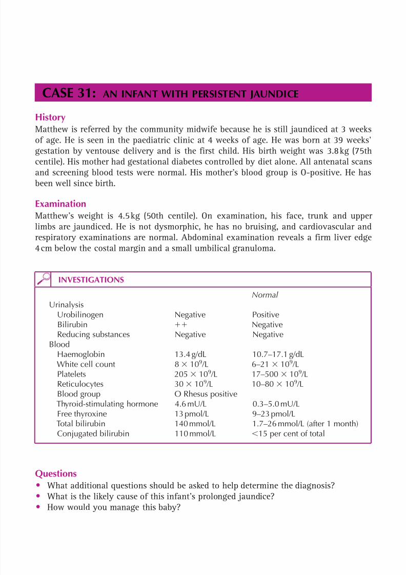
8/20/2019 100 cases in paediatrics.pdf
http://slidepdf.com/reader/full/100-cases-in-paediatricspdf 116/320
97
CASE 31:AN INFANT WITH PERSISTENT JAUNDICE
HistoryMatthew is referred by the community midwife because he is still jaundiced at 3 weeksof age. He is seen in the paediatric clinic at 4 weeks of age. He was born at 39 weeks’gestation by ventouse delivery and is the first child. His birth weight was 3.8 kg (75thcentile). His mother had gestational diabetes controlled by diet alone. All antenatal scansand screening blood tests were normal. His mother’s blood group is O-positive. He hasbeen well since birth.
ExaminationMatthew’s weight is 4.5 kg (50th centile). On examination, his face, trunk and upperlimbs are jaundiced. He is not dysmorphic, he has no bruising, and cardiovascular andrespiratory examinations are normal. Abdominal examination reveals a firm liver edge4 cm below the costal margin and a small umbilical granuloma.
INVESTIGATIONS
Normal Urinalysis Urobilinogen Negative Positive Bilirubin Negative Reducing substances Negative NegativeBlood Haemoglobin 13.4 g/dL 10.7–17.1 g/dL White cell count 8 109 /L 6–21 109 /L Platelets 205 109 /L 17–500 109 /L Reticulocytes 30 10 9 /L 10–80 109 /L Blood group O Rhesus positive Thyroid-stimulating hormone 4.6 mU/L 0.3–5.0 mU/L Free thyroxine 13 pmol/L 9–23 pmol/L Total bilirubin 140 mmol/L 1.7–26 mmol/L (after 1 month) Conjugated bilirubin 110 mmol/L 15 per cent of total
Questions• What additional questions should be asked to help determine the diagnosis?• What is the likely cause of this infant’s prolonged jaundice?• How would you manage this baby?

8/20/2019 100 cases in paediatrics.pdf
http://slidepdf.com/reader/full/100-cases-in-paediatricspdf 117/320
98
100 Cases in Peadiatrics
ANSWER 31The history should establish when jaundice first occurred, as the causes of early-onset
jaundice are different from the causes of prolonged jaundice. Parents should be askedspecifically about the presence of pale stools and dark urine. Pale stool indicates thatbile pigments are not being excreted into the gut, and dark urine occurs with the accu-mulation of water-soluble conjugated bilirubin in the urine. These indicate that thereis obstruction to bile flow out of the liver. The urine dipstick shows the presence ofbilirubin but the absence of urobilinogen, so this child would be expected to have palestools and dark urine.
! Neonatal jaundice requiring investigation• Early onset, 24–36 hours after birth• Severe jaundice, at any time (definition of severe jaundice varies with age)• Prolonged, 14 days
The causes of neonatal jaundice, including prolonged jaundice, are outlined in Case 82(p. 243 ).
Prolonged conjugated hyperbilirubinaemia in a baby who appears well is likely to bedue to extrahepatic biliary atresia. Progressive destruction of the extrahepatic bile ductsoccurs, with obstruction of bile flow and, subsequently, rapid progression of damagewithin the liver and cirrhosis.
It is important to pick up cases of extrahepatic biliary atresia early, so that the diagno-sis can be confirmed by ultrasound and liver biopsy and then surgery can be performedbefore irreversible damage to the liver has occurred. The outcome of the Kasai procedure(portoenterostomy) is much better for surgery performed before 60 days of age. For thisreason, the case needs to be discussed promptly with the local specialist centre.
KEY POINTS
• In prolonged neonatal jaundice with a raised conjugated bilirubin fraction, thediagnosis must be extrahepatic biliary atresia until proven otherwise.
• The outcome of the Kasai procedure is much better if surgery is performed early.

8/20/2019 100 cases in paediatrics.pdf
http://slidepdf.com/reader/full/100-cases-in-paediatricspdf 118/320
99
CASE 32:ABDOMINAL PAIN AND JAUNDICE
HistoryFerdinand is a 12-year-old boy who is brought to the A&E department by his par-ents. This is his third attendance in the last 2 weeks. Today he has been complaining ofabdominal pain and his parents noticed that the whites of his eyes look yellow. He hasbeen scratching himself a lot, although he has not had a rash. The notes from his previ-ous attendances show that he had a flu-like illness with a fever up to 38.5 C, nauseaand a poor appetite over the last 10 days. However, there were no abnormal findings onexamination. He has previously been healthy, but he did travel to the Philippines in theschool holidays about 5 weeks ago. He was born in the UK and received all his routinechildhood immunizations.
ExaminationHe is jaundiced and appears in discomfort. His temperature is 36.8 C, heart rate is 90beats/min, respiratory rate 20/min, oxygen saturation 98 per cent in air, and his bloodpressure is 118/70 mmHg. Cardiovascular and respiratory examinations are normal, buthis liver is palpable 4 cm below the costal margin and is tender. Ears, nose and throatare normal and he is alert and fully orientated.
INVESTIGATIONS
Normal
Haemoglobin 12.3 g/dL 12.1–16.6 g/dLWhite cell count 11.9 109 /L 4.5–13 109 /LPlatelets 250 109 /L 180–430 109 /LBilirubin 89 mmol/L 1.7–26 mmol/LAlkaline phosphatase 1050 25–800 IU/dLAspartate aminotransferase 3798 10–45 IU/dLAlbumin 35 37–50 g/L
Questions• What other questions are important to ask the patient and family?• What additional tests would be useful?• What is the most likely diagnosis?• How could this illness have been prevented?

8/20/2019 100 cases in paediatrics.pdf
http://slidepdf.com/reader/full/100-cases-in-paediatricspdf 119/320
100
ANSWER 32The history can be very helpful in establishing whether jaundice is caused by a pre-hepatic,hepatic or posthepatic problem. Bilirubin is produced by degradation of haem and is ini-tially unconjugated, and hence water-insoluble. Unconjugated bilirubin is transported boundto albumin and is conjugated in the liver parenchymal cells to make it water-soluble andexcretable in bile. Conjugated bilirubin is excreted and converted to urobilinogen, urobilinand stercobilinogen in the gut, which can be reabsorbed, leading to the enterohepatic circu-lation of bile pigments. These pigments give stool its normal coloration. If there is increasedunconjugated bilirubin production from increased red cell destruction (e.g. haemolysis) thenthere will be jaundice with normally pigmented stool and urine. If there is obstruction tobile excretion (e.g. common bile duct stone), conjugated bilirubin accumulates (which causesitching), overflows into urine (causing it to appear dark), and does not reach the gut (result-
ing in pale stools). Hepatic causes may produce a combination of these patterns. Thus it isimportant to ask about stool and urine colour, as well as factors that may predispose to eachtype of cause. In this case, a full travel history is necessary and additional questions shouldbe asked about pre-travel immunization and drug history.
! Differential diagnosis of abdominal pain and jaundice
Pre-hepatic• Abdominal pain is an uncommon feature• Possible causes: malaria, sickle cell crisis
Hepatic• Often pale stools and dark urine will be present• Acute hepatitis due to infection, drugs, toxins
Posthepatic• Pale stools and dark urine• Bile duct stones, cholecystitis, choledochal cyst• Cholangitis must be considered in a febrile child
Additional tests should include urine dipstick to confirm bilirubinuria, amylase, deter-
mination of the conjugated and unconjugated fractions of serum bilirubin, serologicalinvestigation for viral hepatitis (most importantly, hepatitis A IgM), a clotting profileand abdominal ultrasound.
The most likely diagnosis is hepatitis A, which is uncommon in the UK but endemicthroughout much of the world and is most likely to cause symptomatic acute hepatitisin older children and adults. There is usually a flu-like prodromal phase, with nauseaand anorexia starting 2–6 weeks after exposure, followed by an icteric phase when theremay be tender hepatomegaly. Usually children make a complete recovery. Other infec-tions (e.g. hepatitis B, or C, and Epstein–Barr virus) can also cause acute hepatitis but
are less common.Hepatitis A can be prevented by immunization and good hygiene practices, as it is trans-mitted by the faeco-oral route.
KEY POINTS
• Always ask about urine and stool colour in a jaundiced child.• Hepatitis A is endemic throughout much of the world.• Hepatitis A is most likely to cause symptomatic disease in older children and adults
100 Cases in Paediatrics
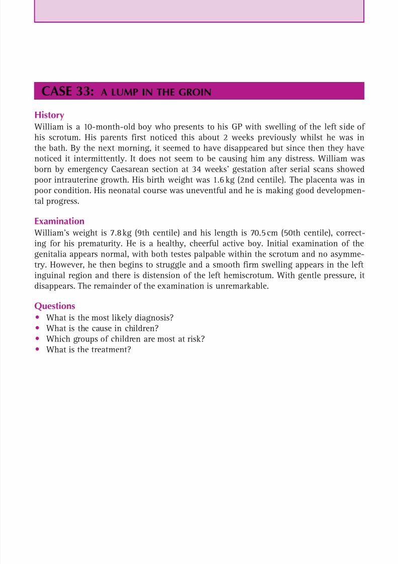
8/20/2019 100 cases in paediatrics.pdf
http://slidepdf.com/reader/full/100-cases-in-paediatricspdf 120/320
101
CASE 33:A LUMP IN THE GROIN
History William is a 10-month-old boy who presents to his GP with swelling of the left side ofhis scrotum. His parents first noticed this about 2 weeks previously whilst he was inthe bath. By the next morning, it seemed to have disappeared but since then they havenoticed it intermittently. It does not seem to be causing him any distress. William wasborn by emergency Caesarean section at 34 weeks’ gestation after serial scans showedpoor intrauterine growth. His birth weight was 1.6 kg (2nd centile). The placenta was inpoor condition. His neonatal course was uneventful and he is making good developmen-tal progress.
Examination William’s weight is 7.8 kg (9th centile) and his length is 70.5 cm (50th centile), correct-ing for his prematurity. He is a healthy, cheerful active boy. Initial examination of thegenitalia appears normal, with both testes palpable within the scrotum and no asymme-try. However, he then begins to struggle and a smooth firm swelling appears in the leftinguinal region and there is distension of the left hemiscrotum. With gentle pressure, itdisappears. The remainder of the examination is unremarkable.
Questions• What is the most likely diagnosis?• What is the cause in children?• Which groups of children are most at risk?• What is the treatment?
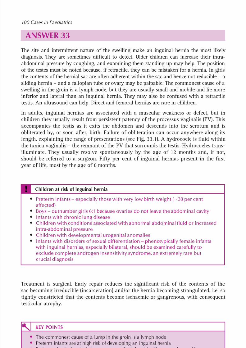
8/20/2019 100 cases in paediatrics.pdf
http://slidepdf.com/reader/full/100-cases-in-paediatricspdf 121/320
102
ANSWER 33The site and intermittent nature of the swelling make an inguinal hernia the most likelydiagnosis. They are sometimes difficult to detect. Older children can increase their intra-abdominal pressure by coughing, and examining them standing up may help. The positionof the testes must be noted because, if retractile, they can be mistaken for a hernia. In girlsthe contents of the hernial sac are often adherent within the sac and hence not reducible – asliding hernia – and a fallopian tube or ovary may be palpable. The commonest cause of aswelling in the groin is a lymph node, but they are usually small and mobile and lie moreinferior and lateral than an inguinal hernia. They may also be confused with a retractiletestis. An ultrasound can help. Direct and femoral hernias are rare in children.
In adults, inguinal hernias are associated with a muscular weakness or defect, but in
children they usually result from persistent patency of the processus vaginalis (PV). Thisaccompanies the testis as it exits the abdomen and descends into the scrotum and isobliterated by, or soon after, birth. Failure of obliteration can occur anywhere along itslength, explaining the range of presentations (see Fig. 33.1). A hydrocoele is fluid withinthe tunica vaginalis – the remnant of the PV that surrounds the testis. Hydrocoeles trans-illuminate. They usually resolve spontaneously by the age of 12 months and, if not,should be referred to a surgeon. Fifty per cent of inguinal hernias present in the first
year of life, most by the age of 6 months.
Treatment is surgical. Early repair reduces the significant risk of the contents of thesac becoming irreducible (incarceration) and/or the hernia becoming strangulated, i.e. sotightly constricted that the contents become ischaemic or gangrenous, with consequent
testicular atrophy.
! Children at risk of inguinal hernia
• Preterm infants – especially those with very low birth weight ( 30 per centaffected)
• Boys – outnumber girls 6:1 because ovaries do not leave the abdominal cavity• Infants with chronic lung disease• Children with conditions associated with abnormal abdominal fluid or increased
intra-abdominal pressure• Children with developmental urogenital anomalies
• Infants with disorders of sexual differentiation – phenotypically female infants with inguinal hernias, especially bilateral, should be examined carefully toexclude complete androgen insensitivity syndrome, an extremely rare butcrucial diagnosis
100 Cases in Paediatrics
KEY POINTS
• The commonest cause of a lump in the groin is a lymph node• Preterm infants are at high risk of developing an inguinal hernia• Early repair of a hernia is important to reduce the risk of incarceration and/or
strangulation
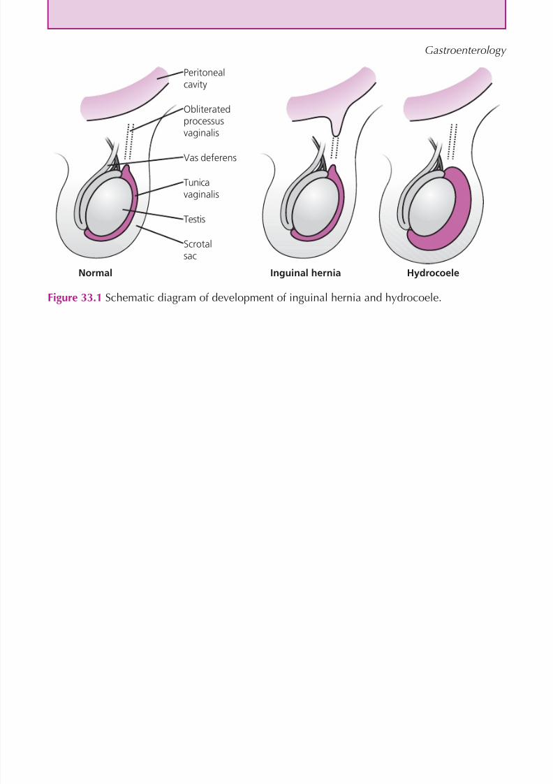
8/20/2019 100 cases in paediatrics.pdf
http://slidepdf.com/reader/full/100-cases-in-paediatricspdf 122/320
103
Gastroenterology
Peritonealcavity
Obliteratedprocessus
vaginalis
Vas deferens
Tunicavaginalis
Testis
Scrotalsac
Normal Inguinal hernia Hydrocoele
Figure 33.1 Schematic diagram of development of inguinal hernia and hydrocoele.

8/20/2019 100 cases in paediatrics.pdf
http://slidepdf.com/reader/full/100-cases-in-paediatricspdf 123/320
This page intentionally left blank
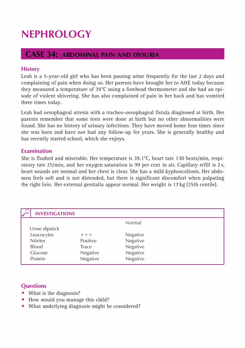
8/20/2019 100 cases in paediatrics.pdf
http://slidepdf.com/reader/full/100-cases-in-paediatricspdf 124/320
105
NEPHROLOGY
CASE 34: ABDOMINAL PAIN AND DYSURIA
HistoryLeah is a 5-year-old girl who has been passing urine frequently for the last 2 days andcomplaining of pain when doing so. Her parents have brought her to A&E today becausethey measured a temperature of 39 C using a forehead thermometer and she had an epi-sode of violent shivering. She has also complained of pain in her back and has vomitedthree times today.
Leah had oesophageal atresia with a tracheo-oesophageal fistula diagnosed at birth. Herparents remember that some tests were done at birth but no other abnormalities werefound. She has no history of urinary infections. They have moved home four times sinceshe was born and have not had any follow-up for years. She is generally healthy andhas recently started school, which she enjoys.
ExaminationShe is flushed and miserable. Her temperature is 39.1 C, heart rate 130 beats/min, respi-ratory rate 25/min, and her oxygen saturation is 99 per cent in air. Capillary refill is 2 s,heart sounds are normal and her chest is clear. She has a mild kyphoscoliosis. Her abdo-men feels soft and is not distended, but there is significant discomfort when palpatingthe right loin. Her external genitalia appear normal. Her weight is 17 kg (25th centile).
INVESTIGATIONS
Normal Urine dipstick Leucocytes Negative Nitrites Positive Negative Blood Trace Negative Glucose Negative Negative Protein Negative Negative
Questions• What is the diagnosis?• How would you manage this child?• What underlying diagnosis might be considered?
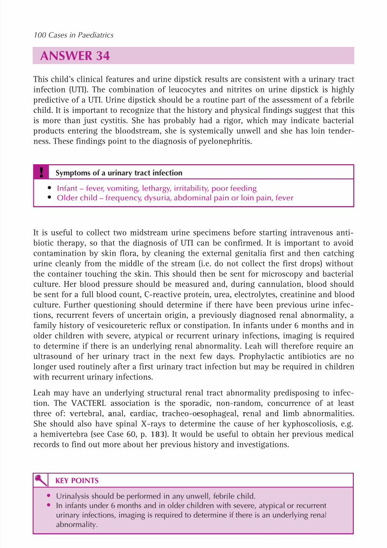
8/20/2019 100 cases in paediatrics.pdf
http://slidepdf.com/reader/full/100-cases-in-paediatricspdf 125/320
106
ANSWER 34This child’s clinical features and urine dipstick results are consistent with a urinary tractinfection (UTI). The combination of leucocytes and nitrites on urine dipstick is highlypredictive of a UTI. Urine dipstick should be a routine part of the assessment of a febrilechild. It is important to recognize that the history and physical findings suggest that thisis more than just cystitis. She has probably had a rigor, which may indicate bacterialproducts entering the bloodstream, she is systemically unwell and she has loin tender-ness. These findings point to the diagnosis of pyelonephritis.
! Symptoms of a urinary tract infection
• Infant – fever, vomiting, lethargy, irritability, poor feeding• Older child – frequency, dysuria, abdominal pain or loin pain, fever
It is useful to collect two midstream urine specimens before starting intravenous anti-biotic therapy, so that the diagnosis of UTI can be confirmed. It is important to avoidcontamination by skin flora, by cleaning the external genitalia first and then catchingurine cleanly from the middle of the stream (i.e. do not collect the first drops) withoutthe container touching the skin. This should then be sent for microscopy and bacterial
culture. Her blood pressure should be measured and, during cannulation, blood shouldbe sent for a full blood count, C-reactive protein, urea, electrolytes, creatinine and bloodculture. Further questioning should determine if there have been previous urine infec-tions, recurrent fevers of uncertain origin, a previously diagnosed renal abnormality, afamily history of vesicoureteric reflux or constipation. In infants under 6 months and inolder children with severe, atypical or recurrent urinary infections, imaging is requiredto determine if there is an underlying renal abnormality. Leah will therefore require anultrasound of her urinary tract in the next few days. Prophylactic antibiotics are nolonger used routinely after a first urinary tract infection but may be required in childrenwith recurrent urinary infections.
Leah may have an underlying structural renal tract abnormality predisposing to infec-tion. The VACTERL association is the sporadic, non-random, concurrence of at leastthree of: v ertebral, a nal, c ardiac, t racheo-o e sophageal, r enal and limb abnormalities.She should also have spinal X-rays to determine the cause of her kyphoscoliosis, e.g.a hemivertebra (see Case 60, p. 183 ). It would be useful to obtain her previous medicalrecords to find out more about her previous history and investigations.
KEY POINTS• Urinalysis should be performed in any unwell, febrile child.• In infants under 6 months and in older children with severe, atypical or recurrent
urinary infections, imaging is required to determine if there is an underlying renalabnormality.
100 Cases in Paediatrics

8/20/2019 100 cases in paediatrics.pdf
http://slidepdf.com/reader/full/100-cases-in-paediatricspdf 126/320
107
CASE 35:RED URINE
HistoryFiona is a 6-year-old girl who presents to the A&E department where her mother statesthat Fiona’s urine has turned red. She has brought a jar of urine which contains whatappears to be reddish-brown urine. She has no dysuria, frequency or abdominal painand there is no history of trauma. She is otherwise well but she and the rest of the fam-ily have recently had colds. There have been no nosebleeds or abnormal bruising. Shehas had no problems with her joints. She had a urinary tract infection at the age of 4
years, but following a normal renal ultrasound she was discharged from clinic. She ison no medication. There is no family history of renal problems but her grandmother hashypertension.
ExaminationThere is no anaemia. She is apyrexial. There is no skin rash or bruising. There is nooedema of the legs. There are no abdominal signs or joint abnormalities. Blood pressureis 124/80 mmHg. There are no other signs.
INVESTIGATIONSNormal
Haemoglobin 11.7 g/dL 11.5–15.5 g/dLWhite cell count 8.7 109 /L 4.0–11.0 109 /LPlatelets 372 109 /L 150–400 109 /LSodium 137 mmol/L 135–145 mmol/LPotassium 4.1 mmol/L 3.5–5.0 mmol/LUrea 11.3 mmol/L 1.8–6.4 mmol/LCreatinine 145 mol/L 27–62 mol/L
Alkaline phosphatase 372 U/L 145–420 U/LBilirubin 16 mol/L 2–26 mol/LAlanine aminotransferase (ALT) 32 U/L 10–40 U/LAlbumin 41 g/L 37–50 g/LClotting NormalUrine dipstick Blood 4 Protein 2 Leucocytes 1 Nitrites Nil
Microscopy – red blood cells and red blood cell casts seen
Questions• What further investigations should be performed?• What is the most likely diagnosis?• What is the treatment?

8/20/2019 100 cases in paediatrics.pdf
http://slidepdf.com/reader/full/100-cases-in-paediatricspdf 127/320
108
ANSWER 35The following investigations should be performed:
• throat swab• anti-streptolysin O titre (ASOT), C3 and C4 – ASOT is raised and C3 is reduced in
post-streptococcal glomerulonephritis• ESR and ANA – will be abnormal in vasculitides, e.g. SLE• abdominal X-ray and renal US – will demonstrate normality of kidneys and help
exclude calculi.
The most likely diagnosis is a post-streptococcal glomerulonephritis. This condition typi-cally occurs approximately 2 weeks after a streptococcal upper respiratory tract infec-
tion (it may also follow a streptococcal skin infection). It can be associated with varyingdegrees of oliguria and renal failure (in this case there is mild renal failure). The castsindicate renal involvement.
In cases of red urine or blood in the nappy, one should first ensure the blood is from theurinary tract and not from the rectum (e.g. due to constipation) or from the vagina (e.g.periods or abuse). Foods such as beetroot can lead to red urine. Haemoglobinuria due tohaemolysis will also lead to red urine which will be dipstick-positive for blood but therewill be no red blood cells on microscopy.
The causes of haematuria are:
• urinary tract infection• nephritis – post-streptococcal glomerulonephritis
– Henoch–Schönlein purpura– IgA nephropathy – nephrotic syndrome (20 per cent have haematuria at presentation)
• calculi• trauma
• haematological – clotting disorders, haemolytic uraemic syndrome
• anatomical causes – polycystic kidneys, hydronephrosis• tumours, e.g. Wilms’ tumour • drugs, e.g. cyclophosphamide, aspirin• factitious illness• recurrent benign haematuria (a diagnosis by elimination).
Treatment is primarily symptomatic. A 10-day course of oral penicillin should be givenbut this will not alter the natural history of the glomerulonephritis. Fluids should berestricted to 1 L/day and the diet should have no added salt. Frusemide is helpful incases of hypertension (this girl’s blood pressure is greater than the 95th centile for a6-year-old girl) or oedema and will increase urine output. If hypertension persists, a cal-cium channel blocker such as amlodipine may be helpful. If heavy proteinuria developsor if renal function deteriorates, a renal opinion should be sought. Indications for dialy-sis include life-threatening hyperkalaemia and the clinical manifestations of uraemia.Outpatient follow-up will necessitate the monitoring of blood pressure, urea and electro-lytes/creatinine and urine dipstick. More than 95 per cent of patients make a completerecovery. Complications include nephrotic range proteinuria and renal failure.
100 Cases in Paediatrics
8/20/2019 100 cases in paediatrics.pdf
http://slidepdf.com/reader/full/100-cases-in-paediatricspdf 128/320
109
KEY POINTS
• Urinary tract infections, post-streptococcal glomerulonephritis and Henoch–Schönlein purpura account for the majority of cases of haematuria.
• Post-streptococcal glomerulonephritis has a very good prognosis.
Nephrology

8/20/2019 100 cases in paediatrics.pdf
http://slidepdf.com/reader/full/100-cases-in-paediatricspdf 129/320
This page intentionally left blank
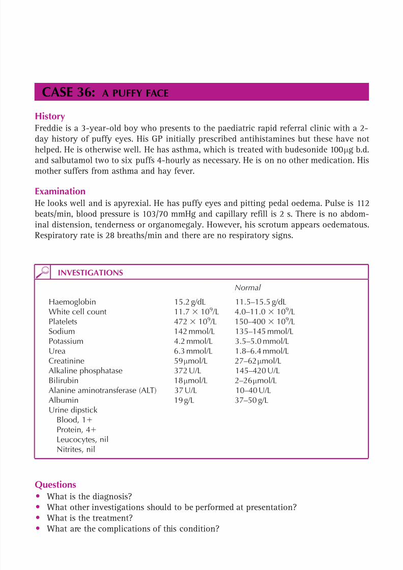
8/20/2019 100 cases in paediatrics.pdf
http://slidepdf.com/reader/full/100-cases-in-paediatricspdf 130/320
111
CASE 36:A PUFFY FACE
HistoryFreddie is a 3-year-old boy who presents to the paediatric rapid referral clinic with a 2-day history of puffy eyes. His GP initially prescribed antihistamines but these have nothelped. He is otherwise well. He has asthma, which is treated with budesonide 100 µg b.d.and salbutamol two to six puffs 4-hourly as necessary. He is on no other medication. Hismother suffers from asthma and hay fever.
ExaminationHe looks well and is apyrexial. He has puffy eyes and pitting pedal oedema. Pulse is 112beats/min, blood pressure is 103/70 mmHg and capillary refill is 2 s. There is no abdom-inal distension, tenderness or organomegaly. However, his scrotum appears oedematous.Respiratory rate is 28 breaths/min and there are no respiratory signs.
INVESTIGATIONS
Normal
Haemoglobin 15.2 g/dL 11.5–15.5 g/dLWhite cell count 11.7 109 /L 4.0–11.0 109 /LPlatelets 472 109 /L 150–400 109 /LSodium 142 mmol/L 135–145 mmol/LPotassium 4.2 mmol/L 3.5–5.0 mmol/LUrea 6.3 mmol/L 1.8–6.4 mmol/LCreatinine 59 µmol/L 27–62 µmol/LAlkaline phosphatase 372 U/L 145–420 U/LBilirubin 18 µmol/L 2–26 µmol/LAlanine aminotransferase (ALT) 37 U/L 10–40 U/LAlbumin 19 g/L 37–50 g/LUrine dipstick Blood, 1 Protein, 4 Leucocytes, nil Nitrites, nil
Questions
• What is the diagnosis?• What other investigations should to be performed at presentation?• What is the treatment?• What are the complications of this condition?
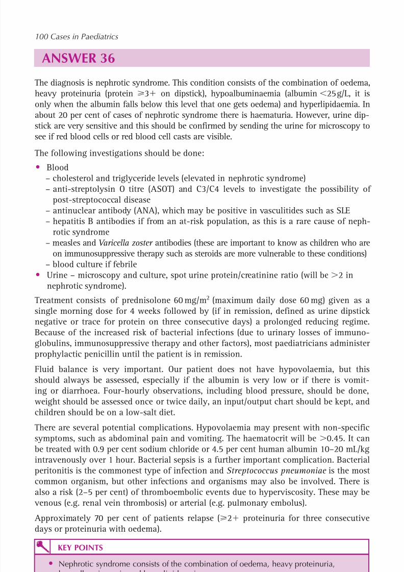
8/20/2019 100 cases in paediatrics.pdf
http://slidepdf.com/reader/full/100-cases-in-paediatricspdf 131/320

8/20/2019 100 cases in paediatrics.pdf
http://slidepdf.com/reader/full/100-cases-in-paediatricspdf 132/320
113
CASE 37:A BED WETTER
History Alvin is a 9-year-old black British boy who has been referred to paediatric outpatientsbecause of bedwetting. He is accompanied by his father. He wets the bed most nights,does not wake up when it happens and there is a large pool of urine. He has no previ-ous medical problems and no recent illnesses. His father says he is unsure if there is anyfamily history of bedwetting. They tried using an enuresis alarm 2 years ago, and thealarm woke him up but he was already wet so they gave up after 1 week. Alvin is quiteupset about his bedwetting, particularly because he recently wet the bed when he stayedat a friend’s house and has been teased about this at school. Alvin’s father is despairingand has started waking him at night to go to toilet, but sometimes he is already wet. Hesays this is affecting his ability to work and he feels that Alvin is being lazy and shouldbe able to control his bladder at this age.
ExaminationHis height is 140 cm (75th centile) and his weight is 35 kg (75th centile). His bloodpressure is 112/70 mmHg. Cardiovascular, respiratory and abdominal examinations areunremarkable.
INVESTIGATIONS
Normal
Urine analysisLeucocytes Negati ve NegativeNitrites Negative NegativeBlood Negative NegativeGlucose Negative NegativeSpecific gravity 1.002 1.002–1.035
Questions• What further history is required?• What further examination is necessary?• What are the options for management?

8/20/2019 100 cases in paediatrics.pdf
http://slidepdf.com/reader/full/100-cases-in-paediatricspdf 133/320
114
100 Cases in Paediatrics
ANSWER 37Nocturnal enuresis is defined as involuntary voiding of urine during sleep at least threetimes per week in a child aged 5 years or older. Approximately 10 per cent of 5-year-olds and 5 per cent of 10-year-olds are affected. It is essential to establish if this isprimary or secondary. Primary nocturnal enuresis indicates that he has never achieveddryness at night and three mechanisms may contribute to this: lack of arousal fromsleep, bladder instability or low functional bladder capacity, and nocturnal polyuria dueto low vasopressin levels. Secondary enuresis indicates that he had previously achieveddryness at night for at least 6 months and something has happened to cause bedwettingagain. Secondary causes include constipation, urinary tract infection, diabetes mellitusor psychosocial stresses, such as bullying or a recent parental separation. It is impor-tant to establish if there are daytime symptoms of urgency, frequency, dysuria or wet-
ting, which suggest bladder instability or urinary tract infections. An accurate diary offluid intake and voiding throughout the day is helpful. A family history of bedwetting,renal problems and sickle cell trait/disease (associated with reduced urinary concentrat-ing ability) should be sought.
Examination should include plotting the child’s height and weight on a growth chartand a urine analysis. In addition, the genitalia and spine should be inspected for abnor-malities, and lower limb neurology should be assessed.
! Management options for primary nocturnal enuresis• Self help measures – regular daytime fluid intake and voiding, avoid caffeinated
drinks• Enuresis clinic support• Nocturnal polyuria – desmopressin (synthetic analogue of antidiuretic hormone)• Lack of arousal – enuresis alarm with star chart• Bladder instability – bladder retraining, anticholinergic medication
In this case, there are features of both nocturnal polyuria and lack of arousal from sleep.Unfortunately the father is not sympathetic and unlikely to have the commitment tomake an enuresis alarm succeed. In this setting, desmopressin may provide some respiteto the family. This will reduce nocturnal urine production. It is important to emphasizeearly that this is not something Alvin has any control over, that desmopressin is not asolution in the very long term, and that ultimately he may need other methods of treat-ment. Desmopressin can be withdrawn in a structured fashion, assisted by the use of anenuresis alarm, when the family are ready to support Alvin and invest some effort in histreatment. It may take a long time to achieve consistent dryness through the night. Thefamily may need psychological support.
Desmopressin can cause headache, nausea and abdominal pain, and may be dangerous ifthere is excessive fluid intake.
KEY POINTS
• Nocturnal enuresis is very common and should only be investigated in children olderthan 5 years.
• It is essential to establish if nocturnal enuresis is primary or secondary.

8/20/2019 100 cases in paediatrics.pdf
http://slidepdf.com/reader/full/100-cases-in-paediatricspdf 134/320
115
CASE 38:HIGH BLOOD PRESSURE
HistoryNikita is a 12-year-old Asian girl who is referred to paediatric outpatients by her GPbecause she has high blood pressure and is overweight. She came to see her GP becauseshe had an ear infection and the GP measured her BP because she is overweight. It wasraised and he repeated it a week later when Nikita had recovered and the reading was thesame. Nikita is otherwise well but gets headaches about once every 2 weeks. There isno nausea or vomiting. The headaches have been occurring for about 2 years. She hadgrommets inserted when she was 4 years old. She started her periods 1 year ago and herperiods are regular. She is on no medication. Her mother has hypertension.
ExaminationHer height is 167 cm (91st centile), weight 85.3 kg ( 99.6th centile) and body mass index(BMI) is 30.6 kg/m 2. Her blood pressure is 146/82 mmHg. There are no dysmorphic fea-tures. She has no cardiovascular signs and femoral pulses are palpable. There are pinkabdominal striae. There is no organomegaly or abdominal masses. There are no neuro-logical or respiratory signs.
INVESTIGATIONS(Done by the GP)
Normal
Full blood count NormalSodium 137 mmol/L 135–145 mmol/LPotassium 4.1 mmol/L 3.5–5.0 mmol/LUrea 6.2 mmol/L 1.8–6.4 mmol/LCreatinine 67 µmol/L 44–88 µmol/LBone chemistry Normal
Liver function tests NormalThyroid function tests NormalUrine dipstick Blood – nil Protein – trace Leucocytes – nil Nitrites – nil
Questions• What is the most likely cause of the hypertension in this child?• What further investigations would you perform?• What would be your management plan?

8/20/2019 100 cases in paediatrics.pdf
http://slidepdf.com/reader/full/100-cases-in-paediatricspdf 135/320
116
100 Cases in Paediatrics
ANSWER 38This child is most likely to have essential (idiopathic) hypertension. This is associatedwith obesity. Hypertension in pubertal children is most often essential but in prepubertalchildren there is often a cause.
However, first the diagnosis of hypertension needs to be confirmed. The blood pressureshould only be measured after the child has been seated and calm for 5 min. The BP cuffshould cover more than three-quarters of the upper right arm and the bladder 50 percent of the arm circumference (too small a cuff will lead to an artificially high bloodpressure). The reading should be repeated at least once. In cases of mild hypertensionwith no target organ damage (the majority), the blood pressure should be repeated atleast three times at weekly intervals to determine if the elevation is sustained. Ideally,
24-hour ambulatory blood pressure monitoring should also be carried out to confirm thediagnosis and to rule out white-coat hypertension. There are charts available with cen-tiles for childrens’ blood pressure which are dependent on sex, age and height centile. Asystolic or diastolic blood pressure 95th centile denotes hypertension. Headaches canoccur in hypertension. In Nikita’s case, the cause may be hypertension or the commonertension type headache. Any obese child can get striae; in Cushing’s disease they tend tobe purple rather than pink.
! Causes of hypertension (use the mnemonic – CREED)
• Cardiological, e.g. coarctation of the aorta• Renal, e.g. glomerulonephritis, renal artery stenosis• Essential• Endocrine, e.g. thyrotoxicosis, Cushing’s disease, phaeochromocytoma• Drugs, e.g. steroids, contraceptive pill, amphetamines
To confirm the diagnosis, 24-hour ambulatory blood pressure monitoring should be per-formed. Renal disease is the commonest cause of hypertension and a renal ultrasound(ideally with Doppler studies of the renal vessels) should be done. Cardiac pathology is
the second commonest cause. The presence of palpable femoral pulses makes a coarc-tation unlikely. However, an ECG should still be performed to see if the hypertension hasled to left ventricular hypertrophy. To investigate the consequences of obesity, a fastingglucose (to rule out type 2 diabetes) and fasting cholesterol and triglyceride levels shouldbe done. Further investigations will depend on confirmation of the hypertension by theambulatory monitoring and clinical symptomatology.
The management plan should include lifestyle changes. A referral to a dietician shouldbe done and advice given about a low-salt and a low-fat diet. A total of 1 hour of exer-cise every day should be recommended. If the raised blood pressure is confirmed, a beta-
blocker or calcium channel blocker could be used.
KEY POINTS
• The blood pressure should be measured with the child calm, with the right size ofcuff, and be repeated.
• Published centile charts should be consulted.• Hypertension in pubertal children is most often essential but in prepubertal children
there is usually a cause (most commonly renal).
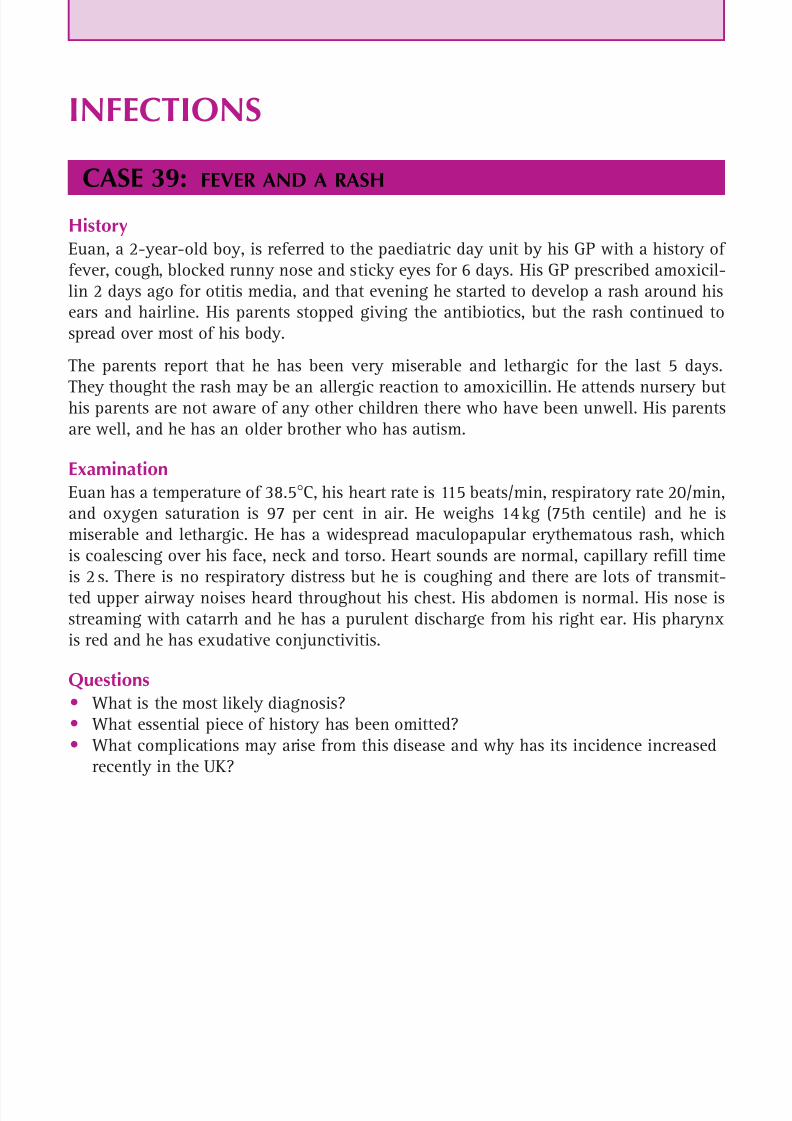
8/20/2019 100 cases in paediatrics.pdf
http://slidepdf.com/reader/full/100-cases-in-paediatricspdf 136/320
117
INFECTIONS
CASE 39: FEVER AND A RASH
HistoryEuan, a 2-year-old boy, is referred to the paediatric day unit by his GP with a history offever, cough, blocked runny nose and sticky eyes for 6 days. His GP prescribed amoxicil-lin 2 days ago for otitis media, and that evening he started to develop a rash around hisears and hairline. His parents stopped giving the antibiotics, but the rash continued tospread over most of his body.
The parents report that he has been very miserable and lethargic for the last 5 days.
They thought the rash may be an allergic reaction to amoxicillin. He attends nursery buthis parents are not aware of any other children there who have been unwell. His parentsare well, and he has an older brother who has autism.
ExaminationEuan has a temperature of 38.5 C, his heart rate is 115 beats/min, respiratory rate 20/min,and oxygen saturation is 97 per cent in air. He weighs 14 kg (75th centile) and he ismiserable and lethargic. He has a widespread maculopapular erythematous rash, whichis coalescing over his face, neck and torso. Heart sounds are normal, capillary refill timeis 2 s. There is no respiratory distress but he is coughing and there are lots of transmit-ted upper airway noises heard throughout his chest. His abdomen is normal. His nose isstreaming with catarrh and he has a purulent discharge from his right ear. His pharynxis red and he has exudative conjunctivitis.
Questions• What is the most likely diagnosis?• What essential piece of history has been omitted?• What complications may arise from this disease and why has its incidence increased
recently in the UK?

8/20/2019 100 cases in paediatrics.pdf
http://slidepdf.com/reader/full/100-cases-in-paediatricspdf 137/320
118
100 Cases in Paediatrics
ANSWER 39This is a case of measles. The history is typical, commencing with a catarrhal prodromephase of fever, conjunctivitis, cough and coryza, preceding development of the rash 3–5days later. During the catarrhal phase, Koplik’s spots may be seen as small white spotson the buccal mucosa. Although pathognomonic of measles, they can be very hard tofind and have usually disappeared within 1 day of the rash starting. The rash is maculo-papular and starts around the hairline and behind the ears, spreading downwards acrossthe body. It often becomes confluent on the upper body, resulting in a blotchy appear-ance. Usually children with measles are very miserable.
! Differential diagnosis of measles
Clinical distinguishing featuresKawasaki disease Not catarrhalRubella Much milder prodrome, occipital
lymphadenopathyEpstein–Barr virus Tonsillitis, lymphadenopathy, not catarrhalRoseola infantum (human herpes Fever ends as rash appearsvirus 6, HHV6)Scarlet fever Pharyngitis or tonsillitis, not catarrhal
An immunization history is an essential part of any paediatric history and no detailshave been provided here. In fact, Euan had not received the combined measles, mumpsand rubella (MMR) vaccine, which is usually given at 13 months of age.
! Complications of measles
• Pneumonia• Corneal ulceration• Suppurative otitis media• Gastroenteritis
• Febrile convulsions• Encephalomyelitis (rare) and subacute sclerosing panencephalitis (very rare)
Public confidence and uptake of the MMR vaccine fell in the UK following media pub-licity about a study in 1998 suggesting a link between the vaccine and autism. Despitemany publications demonstrating the safety of MMR, and specifically that there is nolink to autism, the rate of MMR vaccination in the population fell below that needed toafford herd immunity and cases of measles increased. Measles is a notifiable disease, andprompt notification of suspected cases to the Health Protection Agency allows steps tobe taken to minimize further spread and protect those exposed.
KEY POINTS
• Measles has become more common in the UK following a reduction in MMR vaccineuptake.
• Measles has a typical catarrhal phase before onset of the rash.• The rash evolves in a characteristic fashion, starting around the hairline and behind
the ears and spreading downwards.• Measles is a notifiable disease.
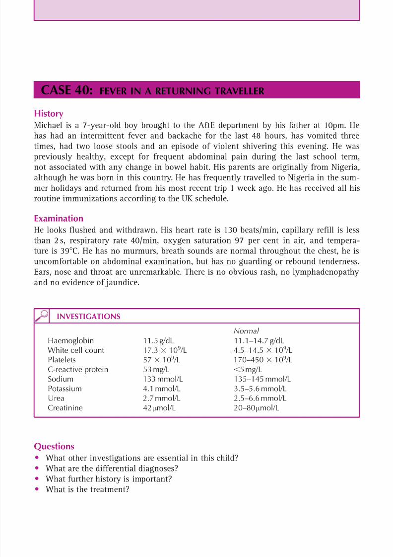
8/20/2019 100 cases in paediatrics.pdf
http://slidepdf.com/reader/full/100-cases-in-paediatricspdf 138/320
119
CASE 40:FEVER IN A RETURNING TRAVELLER
HistoryMichael is a 7-year-old boy brought to the A&E department by his father at 10pm. Hehas had an intermittent fever and backache for the last 48 hours, has vomited threetimes, had two loose stools and an episode of violent shivering this evening. He waspreviously healthy, except for frequent abdominal pain during the last school term,not associated with any change in bowel habit. His parents are originally from Nigeria,although he was born in this country. He has frequently travelled to Nigeria in the sum-mer holidays and returned from his most recent trip 1 week ago. He has received all hisroutine immunizations according to the UK schedule.
ExaminationHe looks flushed and withdrawn. His heart rate is 130 beats/min, capillary refill is lessthan 2 s, respiratory rate 40/min, oxygen saturation 97 per cent in air, and tempera-ture is 39 C. He has no murmurs, breath sounds are normal throughout the chest, he isuncomfortable on abdominal examination, but has no guarding or rebound tenderness.Ears, nose and throat are unremarkable. There is no obvious rash, no lymphadenopathyand no evidence of jaundice.
Questions• What other investigations are essential in this child?• What are the differential diagnoses?• What further history is important?
• What is the treatment?
INVESTIGATIONS
Normal Haemoglobin 11.5 g/dL 11.1–14.7 g/dLWhite cell count 17.3 109 /L 4.5–14.5 10 9 /LPlatelets 57 109 /L 170–450 109 /LC-reactive protein 53 mg/L 5 mg/LSodium 133 mmol/L 135–145 mmol/L
Potassium 4.1 mmol/L 3.5–5.6 mmol/LUrea 2.7 mmol/L 2.5–6.6 mmol/LCreatinine 42 µmol/L 20–80 µmol/L

8/20/2019 100 cases in paediatrics.pdf
http://slidepdf.com/reader/full/100-cases-in-paediatricspdf 139/320
120
100 Cases in Paediatrics
ANSWER 40In any traveller returning from a malaria endemic area, it is absolutely essential to doan urgent thick blood film to look for malaria parasites, and many hospitals now alsouse a rapid diagnostic test kit to identify the presence of malaria antigens in the blood.It is also important to obtain blood cultures, liver function tests, urine dipstick and, in
view of this patient’s tachypnoea, a blood gas sample (to look for acidosis) and possiblya chest X-ray. Michael’s blood film showed 1 per cent parasitaemia with Plasmodium
falciparum and the rapid diagnostic test was positive. Malaria cannot be diagnosed orexcluded on clinical grounds, and may coexist with other infections. Blood films must bedone and three negative films are required to exclude the diagnosis. Thrombocytopeniais commonly seen in malaria, but cannot be relied upon to make the diagnosis.
! Non-specific symptoms occuring in children with malaria
• Fever• Diarrhoea• Vomiting• Cough• Tachypnoea• Headache• Lethargy• Coma
• Jaundice• Haematuria• Myalgia• Pallor
The differential diagnosis of fever in the returning child traveller can be very wide,including haematuria, malaria, typhoid and all of the causes of childhood fever intheir home country (e.g. tonsillitis, myalgia, pneumonia, urinary tract infection, etc).Frequently the cause of the fever is not an unusual or exotic infection at all, even if it
was acquired abroad. An adequate travel history includes details of exactly where the child visited (especiallywhether urban or rural), what sort of accommodation they stayed in, what activitiesthey did during their visit, and what immunizations and malaria prophylaxis they had.Michael did not take any prophylaxis because his parents felt malaria was not a seriousillness.
Treatment should be started promptly, guided by advice from an expert in infectious dis-eases. A variety of oral treatment options are available for uncomplicated malaria, butintravenous quinine remains the standard treatment for severe disease. Broad-spectrumantibiotic cover should also be given in the case of a seriously unwell child.
KEY POINTS
• Fever in a traveller returning from a malaria-endemic area is malaria until provenotherwise.
• Non-specific symptoms should not be interpreted as ruling out the diagnosis.• Prompt initiation of treatment is essential.

8/20/2019 100 cases in paediatrics.pdf
http://slidepdf.com/reader/full/100-cases-in-paediatricspdf 140/320
121
CASE 41:STICKY EYES
HistoryJenna is a 5-day-old girl who is brought to her GP because she has had sticky eyes for2 days. The GP notes that there is a purulent discharge filling both eyes, and it is veryhard to see the conjunctiva clearly. He refers her to the on-call paediatric team. She wasborn at term by normal vaginal delivery to a 19-year-old first-time mother after anuneventful pregnancy. Her birth weight was 2.75 kg (ninth centile). Her mother smokedthroughout pregnancy and drank some alcohol. She had erratic attendance for antenatalcare and had refused antenatal screening blood tests. She was discharged from hospitalafter 12 hours and the baby has been bottle-fed since.
ExaminationJenna looks healthy apart from swollen eyelids with profuse purulent discharge and ery-thematous conjunctiva. She weighs 2.8 kg. She is afebrile, with a heart rate of 140/min,a respiratory rate of 35/min, normal heart sounds and normal breath sounds. Her abdo-men is soft and the liver is palpable 1 cm below the costal margin. Her anterior fonta-nelle is normotensive.
INVESTIGATIONS
Normal
Haemoglobin 13.3 g/dL 13.4–19.8 g/dLWhite cell count 9.1 109 /L 6–21 109 /LPlatelets 353 109 /L 170–500 109 /LC-reactive protein 11 mg/L 5 mg/LMicroscopy of pus from the eye – pus cells ; Gram-negative diplococci
Questions• What is the diagnosis?• How should the baby be managed?• What advice should be given to the mother?

8/20/2019 100 cases in paediatrics.pdf
http://slidepdf.com/reader/full/100-cases-in-paediatricspdf 141/320
122
100 Cases in Paediatrics
ANSWER 41Jenna has ophthalmia neonatorum (neonatal conjunctival infection) caused by Neisseriagonorrhoeae . The differential diagnosis of sticky eyes in a neonate includes nasolacrimalduct obstruction, Chlamydia trachomatis , herpes simplex and other bacterial conjunctiv-itis (e.g. Staphylococcus aureus , Streptococcus pneumoniae ). Inflammation of the con-
junctiva is not seen in nasolacrimal duct obstruction, although the eyes may be sticky.Ophthalmia neonatorum is an emergency and requires prompt management to preventpermanent visual impairment and to treat possible systemic infection.
In cases of ophthalmia neonatorum, it is essential to take adequate microbiologi-cal specimens and then commence prompt antibiotic treatment. Swabs for chlamydiaare usually different from those used for other bacteria and it is necessary to look for
both as chlamydia and gonococcal infections can coexist. Gonococcal ophthalmia neo-natorum should be treated with intravenous ceftriaxone or cefotaxime and frequent eyeirrigation with saline solution. Many experts would also add antibiotic eye drops. Theneonate should be evaluated for disseminated infection, although this is unlikely in thisinfant, who is afebrile and appears systemically well. Ophthalmia neonatorum is a noti-fiable disease.
Jenna’s mother should be told that this is a sexually transmitted infection (STI), whichJenna has almost certainly acquired from the birth canal. It can cause asymptomatic infec-tion in women, but also causes symptomatic infection of the genital tract, pelvic inflam-
matory disease and perihepatitis. It is very important that she attends a genitourinarymedicine clinic for treatment and screening for other STIs and considers discussing thiswith her partner(s). It is also important to have a more thorough discussion about hersocial circumstances and why she declined antenatal screening blood tests.
KEY POINTS
• Ophthalmia neonatorum requires prompt recognition and systemic treatment.• Appropriate specimens must be sent for both gonococcus and chlamydia.• The mother must be advised that she and her partner(s) will require screening.

8/20/2019 100 cases in paediatrics.pdf
http://slidepdf.com/reader/full/100-cases-in-paediatricspdf 142/320
8/20/2019 100 cases in paediatrics.pdf
http://slidepdf.com/reader/full/100-cases-in-paediatricspdf 143/320
124
ANSWER 42Fever and rash are very common in paediatrics. Most rashes are non-specific viral rashesbut some illnesses are accompanied by typical rashes. For instance, chickenpox is char-acterized by a maculopapular rash that evolves into vesicles and meningococcal septi-caemia by a petechial non-blanching rash.
The most likely diagnosis in this case is Kawasaki’s disease which is a vasculitis. This dis-order occurs mainly in young children (80 per cent 5 years). It is diagnosed clinically.
Criteria for diagnosis are:
• The presence of a fever for 5 or more days and four of the following five features:– non-purulent conjunctivitis
– cervical lymphadenopathy – skin rash– erythema of the oral and pharyngeal mucosa– erythema and swelling of the hands and feet (followed a week later by skin
desquamation).
Accompanying features are a raised WCC, CRP and ESR. In the second week of the ill-ness a thrombocytosis usually develops.
When assessing a child with a prolonged fever ( 7 days), the following conditions shouldbe considered.
! Causes of a prolonged fever
• Infections, e.g. tuberculosis, HIV• Malignant diseases, e.g. lymphoma• Autoimmune diseases, e.g. juvenile idiopathic arthritis• Miscellaneous, e.g. drugs, inflammatory bowel disease
Treatment consists of an infusion of immunoglobulins on the day of diagnosis, initiallyhigh-dose aspirin at anti-inflammatory doses followed by low-dose aspirin at anti-thrombotic doses.
The main complication of this disorder is coronary artery aneurysms that can, in somecases, lead to myocardial infarction and sudden death. A prolonged fever ( 16 days),male sex, age 1 year, cardiomegaly, raised inflammatory markers and raised plateletsare all risk factors. An echocardiogram at diagnosis and follow-up echocardiograms arerequired to rule out this complication. The prognosis is related to the cardiac complica-tions. The risk of cardiac complications if treatment with immunoglobulins was com-menced within 10 days of diagnosis is 10 per cent.
KEY POINTS
• Kawasaki’s disease should be considered in all children with a prolonged fever.• Treatment consists of immunoglobulins and aspirin.• The main long-term complication is coronary artery aneurysms.
100 Cases in Paediatrics
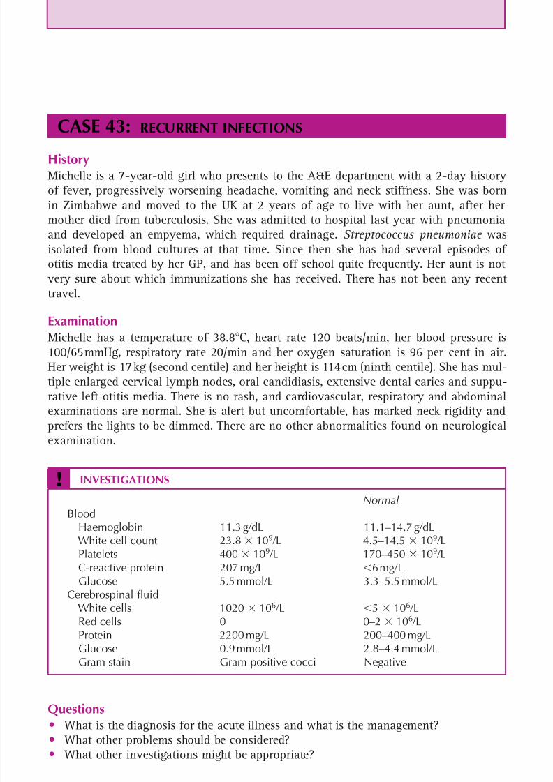
8/20/2019 100 cases in paediatrics.pdf
http://slidepdf.com/reader/full/100-cases-in-paediatricspdf 144/320
125
CASE 43:RECURRENT INFECTIONS
HistoryMichelle is a 7-year-old girl who presents to the A&E department with a 2-day historyof fever, progressively worsening headache, vomiting and neck stiffness. She was bornin Zimbabwe and moved to the UK at 2 years of age to live with her aunt, after hermother died from tuberculosis. She was admitted to hospital last year with pneumoniaand developed an empyema, which required drainage. Streptococcus pneumoniae wasisolated from blood cultures at that time. Since then she has had several episodes ofotitis media treated by her GP, and has been off school quite frequently. Her aunt is not
very sure about which immunizations she has received. There has not been any recenttravel.
ExaminationMichelle has a temperature of 38.8 C, heart rate 120 beats/min, her blood pressure is100/65 mmHg, respiratory rate 20/min and her oxygen saturation is 96 per cent in air.Her weight is 17 kg (second centile) and her height is 114 cm (ninth centile). She has mul-tiple enlarged cervical lymph nodes, oral candidiasis, extensive dental caries and suppu-rative left otitis media. There is no rash, and cardiovascular, respiratory and abdominal
examinations are normal. She is alert but uncomfortable, has marked neck rigidity andprefers the lights to be dimmed. There are no other abnormalities found on neurologicalexamination.
Questions• What is the diagnosis for the acute illness and what is the management?• What other problems should be considered?• What other investigations might be appropriate?
! INVESTIGATIONS
Normal Blood Haemoglobin 11.3 g/dL 11.1–14.7 g/dL White cell count 23.8 109 /L 4.5–14.5 109 /L Platelets 400 109 /L 170–450 109 /L C-reactive protein 207 mg/L 6 mg/L Glucose 5.5 mmol/L 3.3–5.5 mmol/LCerebrospinal fluid White cells 1020 106 /L 5 106 /L Red cells 0 0–2 106 /L Protein 2200 mg/L 200–400 mg/L Glucose 0.9 mmol/L 2.8–4.4 mmol/L Gram stain Gram-positive cocci Negative

8/20/2019 100 cases in paediatrics.pdf
http://slidepdf.com/reader/full/100-cases-in-paediatricspdf 145/320
126
ANSWER 43This child has acute bacterial meningitis, which is most likely to be due to S. pneumo-niae . This diagnosis is strongly suspected from the acute history and the blood results,and confirmed by the CSF findings (see Case 62, p. 187 ). It may have developed second-ary to the otitis media. She should be commenced on an appropriate antibiotic (mostcommonly intravenous ceftriaxone) and on intravenous dexamethasone.
Michelle has a history of recurrent infections with two of these being severe. Any childwith unusual, severe, recurrent or persistent infections must be evaluated for the pos-sibility of an underlying immunodeficiency. In this case, recurrent S. pneumoniae infec-tions raise concerns about hyposplenism, antibody deficiency and HIV infection. Thefindings of severe dental caries, oral candidiasis and cervical lymphadenopathy, together
with the history of immigration from Zimbabwe and her mother dying from TB, stronglysuggest HIV. Testing for HIV is done with informed consent of the person with parentalresponsibility for the child. If the test is positive, the child will gradually be given infor-mation and prepared for disclosure of the diagnosis when they are able to comprehendthe implications of having HIV.
! Examples of factors predisposing to recurrent infections
Primary Secondary Antibody deficiency HIVComplement deficiency Immunosuppressive drugsNeutropenia MalnutritionChronic granulomatous disease HyposplenismCellular immunodeficiency Cystic fibrosisAtaxia telangiectasia Anatomical anomalies, e.g. skull base defect
KEY POINTS
• Consider immunodeficiency in all children with unusual, severe, persistent and
recurrent infections.• HIV testing should be part of the assessment of a child with recurrent infections.
100 Cases in Paediatrics

8/20/2019 100 cases in paediatrics.pdf
http://slidepdf.com/reader/full/100-cases-in-paediatricspdf 146/320
This page intentionally left blank
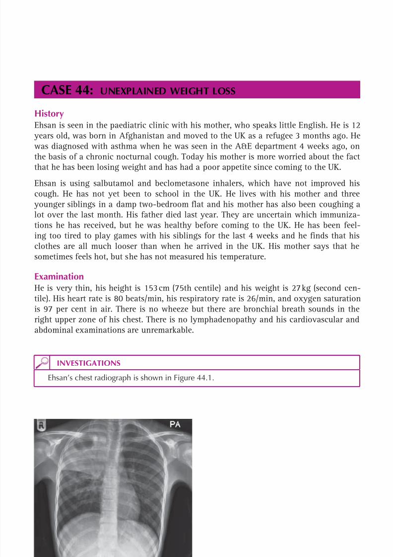
8/20/2019 100 cases in paediatrics.pdf
http://slidepdf.com/reader/full/100-cases-in-paediatricspdf 147/320
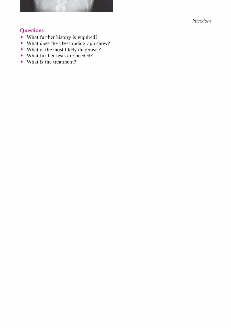
8/20/2019 100 cases in paediatrics.pdf
http://slidepdf.com/reader/full/100-cases-in-paediatricspdf 148/320
129
Questions• What further history is required?• What does the chest radiograph show?• What is the most likely diagnosis?
• What further tests are needed?• What is the treatment?
Infections

8/20/2019 100 cases in paediatrics.pdf
http://slidepdf.com/reader/full/100-cases-in-paediatricspdf 149/320
130
100 Cases in Paediatrics
ANSWER 44 A history of weight loss with a chronic cough needs to be fully investigated. It is import-ant to ask about the past medical history, family history and contact history with directquestions about tuberculosis. What happened to this boy’s father? (In fact, he died in
Afghanistan after suddenly coughing up a large amount of blood.) Even if the immun-ization history is unknown, they may know if Ehsan has received the BCG (as this leavesa distinctive scar) and whether he has ever been treated for TB. Ask about the onset ofthe cough, whether it is productive, whether there is bloodstained sputum and if there isany chest pain or dyspnoea. Ask about exacerbating and relieving factors. Ask when theweight loss started and whether it is associated with abdominal pain, diarrhoea, malab-sorptive (bulky, offensive) stools, nausea or vomiting. Also ask about night sweats.
! Causes of weight loss s
• Inadequate nutrition/neglect• Gastro-oesophageal reflux• Coeliac disease• Inflammatory bowel disease• Cystic fibrosis• Anorexia nervosa• Cardiac failure• Chronic renal failure
• Diabetes mellitus• Malignancy• Infections, e.g. tuberculosis, HIV
The chest radiograph shows dense consolidation and cavitation in the right upper lobe.The most likely diagnosis is pulmonary tuberculosis. Ehsan’s father probably died frompulmonary tuberculosis and Ehsan’s mother probably also has pulmonary tuberculosis.
Ehsan will require admission for investigation and treatment, with isolation whilst he
may have mycobacteria in his sputum. The possibility of multidrug-resistant TB shouldbe considered in view of his recent immigration from Afghanistan.
The gold standard for diagnosis of tuberculosis is culture of the mycobacteria from clin-ical specimens. Unfortunately this is much more difficult to achieve in children than inadults, and only possible in less than 50 per cent of cases. More often the diagnosis isbased on suggestive clinical and radiological features, history of exposure to TB, andresults of tuberculin skin testing (TST). Interpretation of the TST is affected by priorBCG vaccination, and new tests based on the release of interferon-gamma from bloodmononuclear cells in response to antigens present in TB but not in BCG may aid diag-
nosis further. Ehsan will require a tuberculin skin test, sputum to be collected for micro-scopy and culture, erythrocyte sedimentation rate, C-reactive protein, full blood countand liver function tests. It is more likely that his sputum will show acid-fast bacilli onmicroscopy, because he has cavitating pulmonary disease. Cavities are often teemingwith mycobacteria. This presentation would be much rarer in younger children, whorarely have cavitating disease. If he is unable to expectorate sputum by himself, tech-niques to induce sputum production may be attempted, and gastric aspirates may be sentfor mycobacterial culture (but their positive yield is much lower). Drug sensitivity testing

8/20/2019 100 cases in paediatrics.pdf
http://slidepdf.com/reader/full/100-cases-in-paediatricspdf 150/320
131
Infections
will be needed on cultured specimens. Ehsan and his mother should be counselled for anHIV test, because HIV is an important risk factor for development of TB.
Standard treatment for pulmonary tuberculosis should commence with four drugs: isoni-azid, rifampicin, ethambutol and pyrazinamide. Pyridoxine is often given to adolescentsto reduce the risk of isoniazid causing peripheral neuropathy. The rest of the familywill need to be screened for evidence of active or latent tuberculosis. Latent tubercu-losis occurs when a person has been infected with TB, but, rather than causing disease,the mycobacteria become dormant. Individuals with latent tuberculosis are generallynot infectious to others but are at risk of developing active tuberculosis in the future.Chemoprophylaxis (a course of one or two anti-tuberculous drugs) is advised for latentinfection.
Tuberculosis is a notifiable disease and the incidence has been increasing in the UK. Themajority of cases occur in non-UK-born young adults.
KEY POINTS
• Suspicion of tuberculosis should be high in children from high-incidence countrieswith a compatible clinical history.
• The likelihood of obtaining a positive sputum smear or culture in children withpulmonary tuberculosis is much lower than in adults.
• Diagnosis of tuberculosis is often made on the basis of likelihood of exposure,clinical and radiological findings and tuberculin skin testing.

8/20/2019 100 cases in paediatrics.pdf
http://slidepdf.com/reader/full/100-cases-in-paediatricspdf 151/320
This page intentionally left blank

8/20/2019 100 cases in paediatrics.pdf
http://slidepdf.com/reader/full/100-cases-in-paediatricspdf 152/320

8/20/2019 100 cases in paediatrics.pdf
http://slidepdf.com/reader/full/100-cases-in-paediatricspdf 153/320
134
ANSWER 45The most likely diagnosis is scabies. This is caused by the mite Sarcoptes scabiei and ishighly contagious. Intense itching is typical. However, in children under 1 year this maypresent as irritability. A history of contact may be present. The classic features are bur-rows, papules, vesicles and pustules. Thread-like, linear burrows, typically in the fingerwebs and wrists, are pathognomonic but are often difficult to see. Definitive diagnosisinvolves removal of the mite from the burrow and examination under the microscope,but this, too, is difficult. Secondary bacterial infection due to scratching is common.In infants, the rash usually affects the palms, soles, axilla and scalp. In older children,lesions typically involve the web space between the fingers, the flexor aspects of thewrist and arm, the axilla and the waistline. The face is rarely involved in those over 5
years of age. Other areas of the body can also be affected. Although there is a strong
family history of atopy, the patient has never had eczema and it would be unusual foreczema to present for the first time at this age in this way.
Common causes of an itchy rash in a child are:
• eczema – very common, often involves face, elbow and knee flexures• seborrhoeic dermatitis – affects infants, often in association with cradle cap• scabies – itchy where mite has burrowed• insect bites – affects uncovered areas such as arms and legs• drug allergy
• urticaria – idiopathic or secondary to allergens, consists of wheal (raised and white)and flare (red)• fungal infections – e.g. tinea capitis or tinea pedis (athlete’s foot)• chickenpox.
Treatment consists of the application of scabicidal medication, e.g. permethrin. Familymembers and ‘kissing contacts’ should also be treated. Topical or oral antibiotics mayalso be required to treat secondary bacterial infection. Oral antihistamines and topicalsteroids, e.g. 1 per cent hydrocortisone, may be needed to help treat the itching. All cloth-ing and bed linen should be laundered to remove eggs and mites. It can take 4–6 weeksfor the itching to resolve. If lesions are still present at this time, persistent infection orreinfection should be suspected and treatment may need repeating.
KEY POINTS
• Scabies infestation causes severe itching.• The classic features are linear burrows, papules, vesicles and pustules.• The treatment of choice is permethrin.
100 Cases in Paediatrics

8/20/2019 100 cases in paediatrics.pdf
http://slidepdf.com/reader/full/100-cases-in-paediatricspdf 154/320
This page intentionally left blank

8/20/2019 100 cases in paediatrics.pdf
http://slidepdf.com/reader/full/100-cases-in-paediatricspdf 155/320
136
CASE 46:DETERIORATING ECZEMA
HistoryGeorge is a 2-year-old boy who presents to the paediatric rapid referral clinic with aworsening of his eczema. The latter was diagnosed by the GP when he was 6 monthsold. He has been treated with a variety of emollients and topical steroids which areapplied on an intermittent, as-required basis. The parents report that over the last 3days the eczema has gradually deteriorated and also become more itchy. George also hasasthma which is treated with inhaled budesonide twice a day and inhaled salbutamol onan as-required basis. His mother had eczema as a child but grew out of it and his fatherhas hay fever.
ExaminationGeorge is well hydrated. His temperature is 38.3 C, he is itchy and miserable. He haswidespread eczema all over his body, which is worst on his face, hands and arms wherethe skin feels moist (see Fig. 46.1). A number of vesicles and some punched-out lesionscan be seen on the face, hands and arms. There are also pustules and some areas ofhoney-coloured crusting in those areas. In some areas the lesions have coalesced. Themarked scratching that has taken place makes the exact nature of the lesions more dif-
ficult to determine. His eyes are puffy and there are some surrounding lesions. The eyesthemselves appear normal but are difficult to assess fully. There is cervical, axillary andinguinal lymphadenopathy. His chest is clear.
Figure 46.1 Eczemaon George’s face.(Reproduced withkind permission fromKane KSMet al. , Color
Atlas and Synopsis of
Pediatric Dermatology ,McGraw Hill, 2002.)
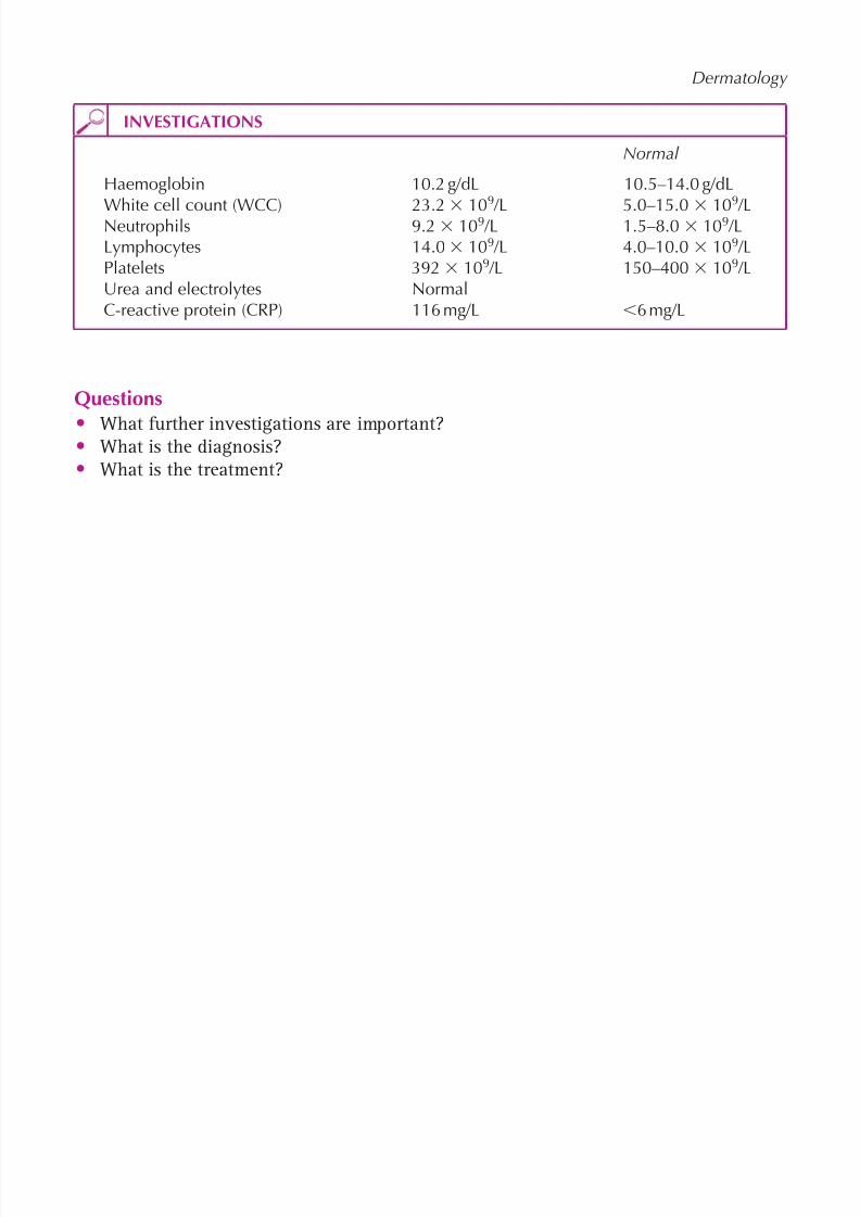
8/20/2019 100 cases in paediatrics.pdf
http://slidepdf.com/reader/full/100-cases-in-paediatricspdf 156/320
8/20/2019 100 cases in paediatrics.pdf
http://slidepdf.com/reader/full/100-cases-in-paediatricspdf 157/320
138
100 Cases in Paediatrics
ANSWER 46 A blood culture should have been done at the same time as the other blood tests, asthere is a small possibility that he may have a septicaemia. Skin swabs should be takenfor bacteriology and virology (virology swabs require special viral medium).
The diagnosis is eczema herpeticum with superadded bacterial infection. It would beworth asking if the child has had similar previous episodes or if anyone in the familyhas herpetic cold sores. Eczema herpeticum is caused by herpes simplex virus infectionof eczematous skin. The infection spreads along the skin and haematogenously. Vesiclesand pustules occur and these may coalesce, erode the skin and become haemorrhagicand crusted. The lesions can disseminate rapidly and may cause life-threatening infec-tion. Eczema herpeticum may also affect the conjunctiva and cornea and can cause a
keratitis that, if left untreated, may lead to blindness. The diagnosis is sometimes madewhen a patient fails to respond to antibacterial therapy. The raised temperature, WCCand CRP indicate that the infection is severe, and the lymphocytosis is suggestive of a
viral infection. The diagnosis can be made by microscopy, culture or viral PCR.
Bacterial infection can also lead to an acute deterioration in eczema (and is more commonthan herpetic infection). The pathogen is usually Staphylococcus aureus and occasionallyStreptococcus . Staphylococcal infection can lead to honey-coloured crusting, as in thiscase. In our case, the secondary bacterial infection is most likely to be due to scratching,leading to the Staphylococcus that often colonizes the skin, causing infection.
In some cases it can be difficult to determine if the eczema has been infected by bacteriaor viruses and both need to be treated.
The lymphadenopathy is secondary to the infected eczema.
In some cases of widespread infected eczema, there can be marked fluid loss from theskin, which may be exacerbated in the presence of a temperature. In this case the patientis well hydrated, but hydration should be carefully monitored (the situation is some-what analogous to fluid loss in a burn). Intravenous acyclovir should be administered.If there is any concern about possible eye involvement, as in this case, an urgent oph-thalmic opinion should be sought. In addition, intravenous antibiotics (e.g. co-amoxiclav)should be given because of the superadded bacterial skin infection and possible septicae-mia. Analgesics, antipyretics and anti-pruritic agents, i.e. antihistamines, should be pre-scribed. Topical treatment with steroids and other immunosuppressants (e.g. tacrolimus)should be discontinued (this is in contrast to bacterial infections alone when steroidcreams are usually continued alongside the antibiotic). Topical treatments can usually berestarted after 1 week when the patient has improved.
KEY POINTS
• Herpetic infection can lead to severe worsening of eczema (eczema herpeticum).• Bacterial infection is the commonest cause for an acute deterioration in eczema, with
the commonest pathogen being Staphylococcus aureus .

8/20/2019 100 cases in paediatrics.pdf
http://slidepdf.com/reader/full/100-cases-in-paediatricspdf 158/320
This page intentionally left blank
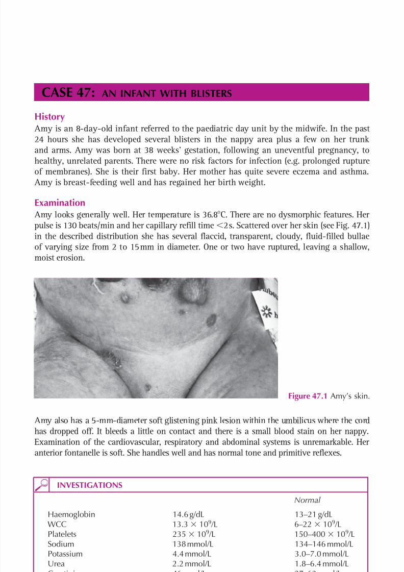
8/20/2019 100 cases in paediatrics.pdf
http://slidepdf.com/reader/full/100-cases-in-paediatricspdf 159/320
140
CASE 47:AN INFANT WITH BLISTERS
History Amy is an 8-day-old infant referred to the paediatric day unit by the midwife. In the past24 hours she has developed several blisters in the nappy area plus a few on her trunkand arms. Amy was born at 38 weeks’ gestation, following an uneventful pregnancy, tohealthy, unrelated parents. There were no risk factors for infection (e.g. prolonged ruptureof membranes). She is their first baby. Her mother has quite severe eczema and asthma.
Amy is breast-feeding well and has regained her birth weight.
Examination Amy looks generally well. Her temperature is 36.8 C. There are no dysmorphic features. Herpulse is 130 beats/min and her capillary refill time 2 s. Scattered over her skin (see Fig. 47.1)in the described distribution she has several flaccid, transparent, cloudy, fluid-filled bullaeof varying size from 2 to 15 mm in diameter. One or two have ruptured, leaving a shallow,moist erosion.
INVESTIGATIONS
Normal Haemoglobin 14.6 g/dL 13–21 g/dLWCC 13.3 109 /L 6–22 109 /LPlatelets 235 109 /L 150–400 10 9 /LSodium 138 mmol/L 134–146 mmol/LPotassium 4.4 mmol/L 3.0–7.0 mmol/LUrea 2.2 mmol/L 1.8–6.4 mmol/LCreatinine 46 µmol/L 27–62 µmol/LCRP 6 mg/L 6 mg/L
Amy also has a 5-mm-diameter soft glistening pink lesion within the umbilicus where the cordhas dropped off. It bleeds a little on contact and there is a small blood stain on her nappy.Examination of the cardiovascular, respiratory and abdominal systems is unremarkable. Heranterior fontanelle is soft. She handles well and has normal tone and primitive reflexes.
Figure 47.1 Amy’s skin.
8/20/2019 100 cases in paediatrics.pdf
http://slidepdf.com/reader/full/100-cases-in-paediatricspdf 160/320
141
Dermatology
Questions• What is the most likely cause of the bullae?• What is the management?• What is the umbilical lesion likely to be, and how would you treat it?

8/20/2019 100 cases in paediatrics.pdf
http://slidepdf.com/reader/full/100-cases-in-paediatricspdf 161/320
142
100 Cases in Paediatrics
ANSWERThese flaccid, fragile blisters are characteristic of bullous impetigo. This is the localizedpresentation of the staphylococcal scalded-skin syndrome and patients are otherwisewell. At the other end of the spectrum are those who present with generalized cutaneousinvolvement and systemic illness. Both are caused by Staphylococcus aureus, most ofwhich are from phage group 2. In bullous impetigo the organism can be cultured fromthe lesions but the systemic form is mediated by exfoliative toxins and swabs are sterile.
The common weepy, golden, crusty forms of impetigo start on skin that has been dam-aged, e.g. by insect bites, chickenpox or abrasions. It spreads easily to other sites and toother children. The causative organism is also usually Staphylococcus aureus but gener-ally not from phage group 2. Group A β-haemolytic Streptococcus can also cause impetigo.
Patients with eczema are at increased risk of infective exacerbations due to these organisms.It is likely that this baby’s infection was transmitted from her mother, either because of hereczema or because she has mastitis.
A much rarer cause of blistering in the neonatal period is epidermolysis bullosa. Thereare several different types that differ in severity, clinical features, distribution and inher-itance patterns, but all are characterized by blisters that develop with trauma and areexacerbated in warm weather. Blisters present at birth would be strongly supportive ofthis diagnosis.
Treatment is with a β-lactamase-resistant antibiotic such as co-amoxiclav. This infant iswell and so the oral route can be safely used whilst monitoring for a satisfactory response.In systemic scalded skin syndrome, antibiotics are administered parenterally. Such patientsneed careful monitoring because they are at risk of significant heat and fluid losses. Theyalso require adequate pain relief.
The umbilical lesion is likely to be a granuloma. The umbilical cord usually dries and separ-ates within 6–8 days after birth and the surface epithelializes. Where this is incompleteor there is mild infection, granulation tissue can develop and persist. A granuloma is notpainful. A number of treatments are available, including cauterization using a silver nitrate
stick. This can be repeated once a week until the lesion has resolved. It is crucial to dif-ferentiate the common umbilical granuloma from the rare umbilical polyp which resultsfrom persistence of the omphalomesenteric duct or the urachus. If it communicates with theileum or bladder, there may a faeculant or urinary discharge. Treatment is surgical.
KEY POINTS
• Skin infections due to Staphyococcus aureus have a range of presentations from mild,localized lesions to severe, systemic infections.
• Blistering present at birth is likely to be due to epidermolysis bullosa.

8/20/2019 100 cases in paediatrics.pdf
http://slidepdf.com/reader/full/100-cases-in-paediatricspdf 162/320
143
HAEMATOLOGY
CASE 48: A PALE CHILD
HistorySarah is a 4-year-old girl who is referred to the paediatric day unit by her GP with a 2-dayhistory of widespread bruising. She has also had two nosebleeds in the preceding 24 hours.There are no known injuries, although her mother says she is quite ‘hectic’, especially whenplaying with her younger brother. They both had a cold and a sore throat 2 weeks previ-ously. She is otherwise very well. There is no significant past medical or family history andshe is on no medication.
ExaminationSarah is drawing pictures and is cheerful and cooperative. She has no dysmorphic fea-tures. Her height and weight are on the 25th centiles. There is no jaundice and she isafebrile. She is pale and clinically anaemic with a few 1.0 cm lymph nodes in the cervicaland inguinal regions. There is widespread bruising mainly on her limbs but no evidenceof active bleeding. There is no hepatosplenomegaly. Her pulse is 96 beats/min and bothheart sounds are normal. She has a grade 2/6 ejection systolic murmur. Examination of therespiratory system is normal.
INVESTIGATIONS
Normal
Haemoglobin 6.2 g/dL 11.5–15.5 g/dLWhite cell count 1.2 109 /L 6.0–17.5 109 /LNeutrophils 0.2 109 /L 3.0–5.8 109 /LLymphocytes 0.8 109 /L 1.5–3.0 109 /LPlatelets 6 109 /L 150–400 109 /LBlood film No blastsProthrombin time 12 s 11–15 sPartial thromboplastin time 32 s 25–35 sUrea and electrolytes NormalLiver function tests NormalC-reactive protein 6 mg/L 6 mg/L
Questions• What is the differential diagnosis?• What investigations are indicated?• What are her major current risks?

8/20/2019 100 cases in paediatrics.pdf
http://slidepdf.com/reader/full/100-cases-in-paediatricspdf 163/320
144
100 Cases in Paediatrics
ANSWER 48Sarah has pancytopenia, loss of all bone marrow (BM) elements. This is not a diagnosisand needs further investigation.
! Differential diagnosis of pancytopenia
Bone marrow failure• Inherited – all rare. Commonest is Fanconi’s anaemia. Excess chromosome
breaks. Defective DNA repair, decreased cell survival and susceptible to oxidantstress. Associated physical abnormalities, e.g. skeletal (absent thumbs), shortstature. Only cure is a BM transplant.
• Acquired – viral, e.g. hepatitis, herpes, Epstein–Barr – drugs – idiosyncratic, e.g. chloramphenicol, anticonvulsants, or predictable,
e.g. chemotherapy; 80 per cent are ‘idiopathic aplastic anaemia’.
Bone marrow infiltration• Malignancy, e.g. leukaemia or neuroblastoma. Rarely myelofibrosis and
myelodysplasia.
Bone marrow infiltration causes marrow expansion and pain unless it is slowly progressiveas in myelofibrosis. There are also likely to be other symptoms and signs, e.g. lymphaden-
opathy or hepatosplenomegaly in leukaemia. Significantly enlarged lymph nodes are usu-ally 1 cm in diameter. Despite her pallor, this child is playing happily, making BM failuremore likely. About 20 per cent of patients with inherited pancytopenias have none of theassociated features so an inherited cause cannot be excluded clinically, but these condi-tions are rare. This girl probably has an acquired pancytopenia – probably idiopathic aplas-tic anaemia (AA). Most cases appear to be caused by activated T-lymphocytes, producingcytokines that suppress haematopoiesis. Her mild tachycardia and murmur are most likelydue to a hyperdynamic circulation secondary to anaemia. An echocardiogram may be indi-cated if it does not disappear or diminish following transfusion.
! Investigation of pancytopenia
• Blood film – detailed morphology of all cell lines• Red cell indices – the anaemia in AA is normocytic or mildly macrocytic• Reticulocytes – 20 109 /L suggests severe aplastic anaemia• Viral titres – hepatitis, Epstein–Barr, parvovirus (usually causes red cell aplasia)• Chromosomes for breakage analysis• BM aspirate and trephine – to assess morphology and cellularity of the cells and
to exclude infiltration. In AA it is hypocellular but the remaining cells are normal
Management pending a diagnosis and treatment is supportive. Haemorrhage and infec-tion are her two major risks. However, there is a danger with multiple platelet transfu-sions of developing antibodies (alloimmunization). These can cause a typical transfusionreaction with fevers and rigors, but more importantly can diminish the increment inplatelet count. This risk is minimized by restricting transfusions to episodes of activebleeding and/or a platelet count of 5 10 9/L. Neutropenia, especially if prolonged,puts her at risk of serious life-threatening bacterial infections and the family should begiven instructions about regular monitoring of her temperature and when to seek advice.
8/20/2019 100 cases in paediatrics.pdf
http://slidepdf.com/reader/full/100-cases-in-paediatricspdf 164/320
145
Haematology
Spontaneous recovery of idiopathic aplastic anaemia is rare. The decision to embark ondefinitive treatment depends upon the severity. BM transplant (BMT) offers the best chancefor those with a matched sibling donor. Otherwise, immunosuppressive treatment withantithymocyte globulin and cyclosporin is the main alternative. If this fails, a matched,
unrelated-donor BMT is the only option.
KEY POINTS
• Pancytopenia can be due to bone marrow failure or bone marrow infiltration.• The most important risks from pancytopenia are bleeding and infection.

8/20/2019 100 cases in paediatrics.pdf
http://slidepdf.com/reader/full/100-cases-in-paediatricspdf 165/320
This page intentionally left blank

8/20/2019 100 cases in paediatrics.pdf
http://slidepdf.com/reader/full/100-cases-in-paediatricspdf 166/320
147
CASE 49:EASY BRUISING
History Ahmed is a 2-year-old boy who presents to the paediatric rapid referral clinic with easybruising. His mother states that over the last 2 days bruises have been appearing on hisbody spontaneously or with minimal trauma and that he also had two short nose bleedsthe previous day. He is otherwise well but his mother says that he had a cold about2 weeks ago. He has had no previous illnesses but had a circumcision at 2 months ofage for religious reasons with no excessive bleeding. He is on no medication. There is nofamily history of bleeding disorders.
ExaminationHe is well, playing and apyrexial. There is no pallor. He has widespread purpura andbruising over the flexor and extensor surfaces of all four limbs, trunk and face. There issome blood crusted around his nose. There is no lymphadenopathy or hepatosplenomegaly.There are no respiratory or cardiological signs and there are no joint abnormalities.
INVESTIGATIONS
Normal Haemoglobin 10.2 g/dL 10.5–14.0 g/dLWhite cell count 9.6 109 /L 5.0–15.0 109 /LNeutrophils 4.2 109 /L 1.7–7.5 109 /LPlatelets 6 109 /L 150–400 109 /LClotting screen NormalUrea and electrolytes NormalC-reactive protein 5 mg/L 6 mg/LBlood film – large (young) platelets; no blasts seenUrine dipstick Blood – negative Protein – negative Leucocytes – negative Nitrites – negative
Questions• What is the diagnosis?• What are the causes of purpura in a child?• Are any further investigations necessary?
• What is the treatment?
8/20/2019 100 cases in paediatrics.pdf
http://slidepdf.com/reader/full/100-cases-in-paediatricspdf 167/320
8/20/2019 100 cases in paediatrics.pdf
http://slidepdf.com/reader/full/100-cases-in-paediatricspdf 168/320
149
A repeat full blood count should be performed at 7–10 days to ensure that there is noevolving bone marrow disorder, e.g. aplasia. ITP is usually a self-limiting disorder. Fiftyper cent resolve in 2 months, 75 per cent in 4 months and 90 per cent in 6 months.
KEY POINTS
• Treatment in ITP should be guided by symptoms rather than the platelet count.• The most serious complication is an intracranial haemorrhage.• Meningococcal septicaemia is an important cause of purpura that requires urgent
treatment.
Haematology
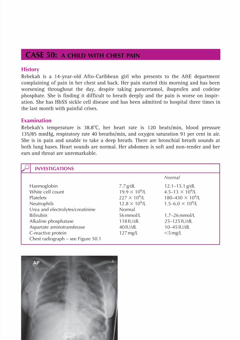
8/20/2019 100 cases in paediatrics.pdf
http://slidepdf.com/reader/full/100-cases-in-paediatricspdf 169/320
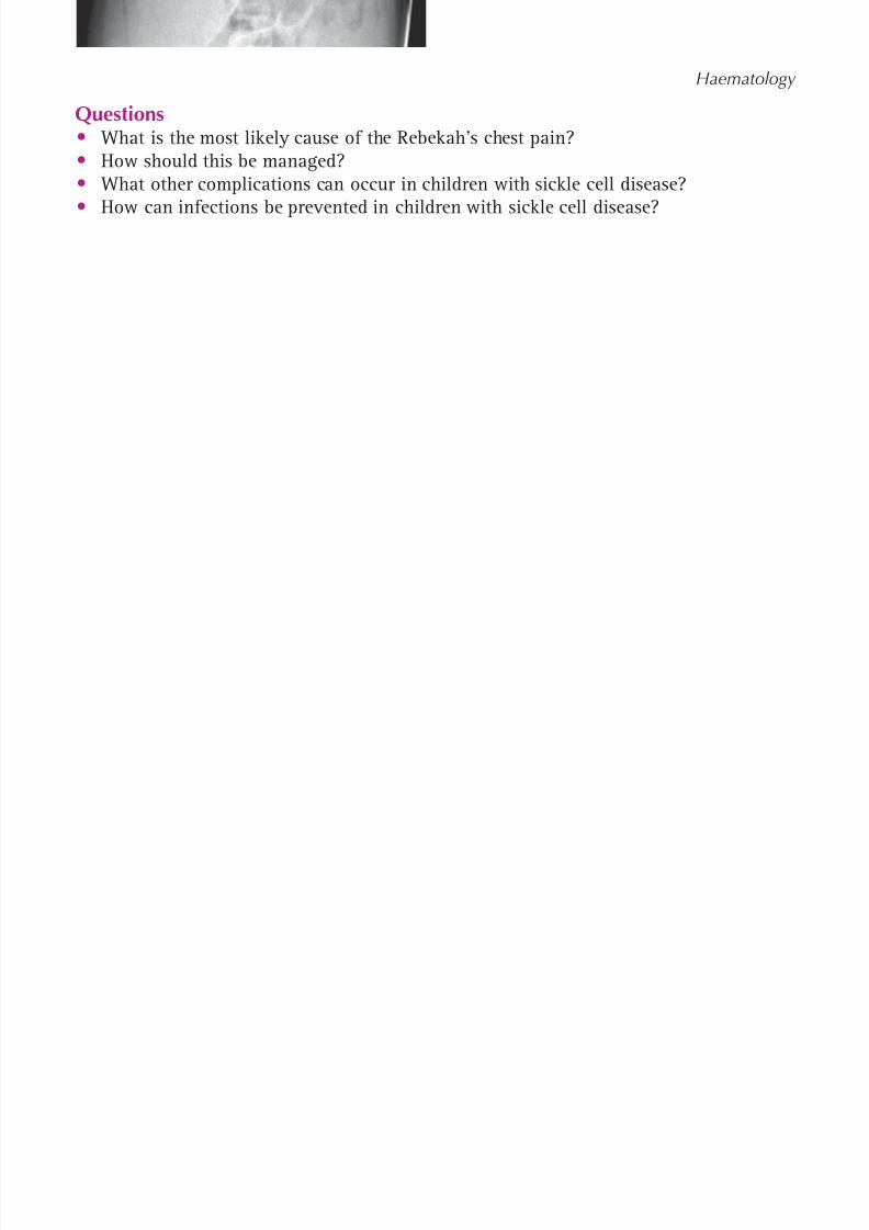
8/20/2019 100 cases in paediatrics.pdf
http://slidepdf.com/reader/full/100-cases-in-paediatricspdf 170/320
151
Haematology
Questions• What is the most likely cause of the Rebekah’s chest pain?• How should this be managed?• What other complications can occur in children with sickle cell disease?
• How can infections be prevented in children with sickle cell disease?

8/20/2019 100 cases in paediatrics.pdf
http://slidepdf.com/reader/full/100-cases-in-paediatricspdf 171/320
152
100 Cases in Paediatrics
ANSWER 50Rebekah has acute sickle chest syndrome. Thrombosis, infection and fat embolism tothe lung produce a syndrome of pleuritic chest pain, shortness of breath and fever. Thepathology often evolves from the lung bases and produces consolidation, which may beclinically apparent before radiographic changes appear. Hypoxia is a frequent feature,and failure to recognize and manage this syndrome aggressively may lead to a rapiddeterioration. Compare the initial chest radiograph (which shows just a small patch ofconsolidation in the left lower lobe behind the heart) with the one taken 2 days later(which shows more extensive bi-basal consolidation – see Fig. 50.2).
Sickling of red blood cells and their sequestration in vessels is exacerbated by dehydra-tion, hypoxia and acidosis, so these factors need to be corrected with hyperhydrationand supplemental oxygen. Adequate analgesia (often intravenous morphine) and physio-therapy help the patient to breathe deeply enough to ventilate the affected areas of thelungs. Empirical antibiotic treatment should be started, e.g. co-amoxiclav and clari-thromycin. Children with acute sickle chest syndrome should be discussed with a senior paediatrician and a haematologist. If they deteriorate, they may require ventilatorysupport (continuous positive airways pressure (CPAP) or intubation and ventilation).
A top-up or exchange blood transfusion may also be needed to reduce the percentage ofsickling red blood cells.
Figure 50.2 Rebekah’s chest radiograph2 days later.
! Complications of sickle cell disease in children• Painful crises, avascular necrosis of the hips and shoulders• Chest syndrome, abdominal syndrome, girdle syndrome• Splenic sequestration, aplastic anaemic crisis• Stroke, retinal vein occlusion• Priapism, haematuria, enuresis, chronic renal failure• Pigment gallstones, cholecystitis, biliary colic• Hyposplenism, sepsis, osteomyelitis• Delayed puberty

8/20/2019 100 cases in paediatrics.pdf
http://slidepdf.com/reader/full/100-cases-in-paediatricspdf 172/320
153
Haematology
Children with sickle cell disease develop hyposplenism, which makes them particularly vulnerable to infection with encapsulated bacteria such as Haemophilus influenzae andStreptococcus pneumoniae . They should receive all routine immunizations, pneumococcalpolysaccharide vaccine, and regular penicillin V prophylaxis. Hepatitis B vaccine is rec-
ommended, and if they travel to a malaria-endemic area, it is essential they take adequateprophylaxis.
KEY POINTS
• Sickle chest syndrome is an emergency. Early recognition and treatment can preventsevere consequences.
• Children with sickle cell disease are vulnerable to infections with encapsulatedbacteria.
• Complications of sickle cell disease can affect any organ.

8/20/2019 100 cases in paediatrics.pdf
http://slidepdf.com/reader/full/100-cases-in-paediatricspdf 173/320
This page intentionally left blank

8/20/2019 100 cases in paediatrics.pdf
http://slidepdf.com/reader/full/100-cases-in-paediatricspdf 174/320
155
ONCOLOGY
CASE 51: AN ABDOMINAL MASS
HistoryChristopher is a 3-year-old boy who presents to the children’s day unit via his GP. Theprevious evening his mother noticed a lump in his abdomen while drying him after abath. He has no urinary symptoms and his bowels open normally twice daily. He is other-wise entirely fit and well, and there is no other history of note.
ExaminationChristopher is generally healthy with no anaemia, jaundice or lymphadenopathy. Hisheight and weight are on the 75th centile. His blood pressure is 115/75 mmHg. Bothheart sounds are normal with no murmurs and his chest is clear. In his abdomen there isa 12-cm-diameter smooth, firm mass in the right flank that extends across the midline.There is no hepatosplenomegaly. The external genitalia are normal.
INVESTIGATIONS
Normal
Haemoglobin 11.7 g/dL 11.5–15.5 g/dLWhite cell count 8.4 109 /L 5.5–15.5 109 /LPlatelets 365 109 /L 150–400 109 /LSodium 139 mmol/L 138–146 mmol/LPotassium 4.3 mmol/L 3.5–5.0 mmol/LUrea 2.0 mmol/L 1.8–6.4 mmol/LCreatinine 45 mol/L 27–62 mol/LC-reactive protein 6 mg/L 6 mg/LMidstream urine 10 red cells 1–10 white cells No growthAbdominal ultrasound – there is an 11 cm, apparently well-capsulated, solidheterogeneous mass arising from the right kidney. There is no evidence of calcification
Questions• What is the commonest cause of an abdominal mass in childhood?• What is the most likely diagnosis in this case?• Where should he be managed?• What complication has arisen?
8/20/2019 100 cases in paediatrics.pdf
http://slidepdf.com/reader/full/100-cases-in-paediatricspdf 175/320
156
100 Cases in Paediatrics
ANSWER 51The commonest cause of an abdominal mass – occasionally huge – in childhood is faeces.It is characteristically craggy, mobile and in the lower abdomen. However, abdominalmasses in children should be assumed to be malignant until proven otherwise, and ifthere is any doubt, imaging with ultrasound is required.
In an otherwise healthy asymptomatic child, the most likely diagnosis is a Wilms’tumour or nephroblastoma, a neoplasm of the kidney comprising approximately 6 percent of childhood cancers; 7 per cent are bilateral. It is not unusual for these to presentafter parents have found the mass incidentally. Wilms’ tumours may be associated withhemihypertrophy, aniridia (hypoplastic iris commonly associated with macular and opticnerve hypoplasia) and other congenital abnormalities, usually of the genitourinary tract.
Children with Beckwith–Wiedemann syndrome (mutations at 11p15.5, hemihypertrophy,macroglossia and visceromegaly) have such an increased risk of Wilms’ (approximately4 per cent) that they are screened regularly during early childhood.
The main differential diagnosis is neuroblastoma (NB) – an embryonal cancer of the periph-eral sympathetic nervous system. Most arise in the abdomen either in an adrenal gland orin the retroperitoneal sympathetic ganglia. The mass usually causes discomfort. Sadly, chil-dren with NB often present with symptoms of metastatic spread, such as fever, bone pain,irritability and weight loss – unusual in Wilms’. Tumour markers and imaging help distin-guish the two. Ninety-five per cent of NB patients have raised urine vanillylmandelic acid
(VMA) and homovanillic acid (HVA). Unlike NB, Wilms’ tumours rarely calcify.Surgery is the mainstay of treatment for Wilms’ tumour with pre- and post-surgerychemotherapy and radiotherapy depending on the stage. The prognosis for Wilms’ isexcellent, with stage 1–3 patients having an overall cure rate of 88–98 per cent. Thosewith stage 4 disease, with distant metastases, have a poorer but still relatively favourableprognosis of about 75 per cent.
There is good evidence that prognosis in all childhood cancers improves in childrenmanaged by specialist units, although some care can be local on a ‘shared care’ basis.
Centralization of care, multidisciplinary teams, entry into randomized controlled trialsand the use of national or international protocols have steadily improved the prognosisin all childhood cancers.
His BP is high for a child of his age. Children dislike having their BP measured so it canbe difficult to measure accurately. There are electronic devices commonly in use buttheir results should be interpreted with caution and every effort made to measure BPwith standard equipment. The cuff width should be at least two-thirds of the distancefrom the elbow to the shoulder. Normative ranges according to sex, age and height cen-tile exist for children and adolescents. They are presented as percentiles, with the 95th
percentile defining hypertension. In Wilms’ tumour, BP is often raised at presentation,probably due to ischaemia. It may persist for several months even after the primary hasbeen removed and may need drug treatment.
KEY POINTS
• Abdominal masses in children are malignant until proven otherwise.• Wilms’ tumour can be associated with congenital anomalies.• The care of all children with cancer should be coordinated by specialist centres.
8/20/2019 100 cases in paediatrics.pdf
http://slidepdf.com/reader/full/100-cases-in-paediatricspdf 176/320
157
CASE 52:AN UNSTEADY CHILD
HistoryToby is a 4.5-year-old boy referred to children’s outpatients by his GP with a history ofapparent stiffness in his neck. About 6 weeks previously he complained of a ‘dizzy head’and became reluctant to walk home after school. Subsequently he seemed to find it dif-ficult to look upwards but seemed fine when looking from side to side. He has also beenrubbing his head and crying. In the past 3 weeks Toby has had several episodes of vom-iting and at the time holds his neck in pain. There have been no other gastrointestinalsymptoms nor any fevers and there has been some improvement with paracetamol.
Toby has become increasingly clumsy and has had several unexpected falls even whensitting. Having walked up stairs independently, he now wants to hold the rail or some-one’s hand and no longer runs with confidence. He has fallen off his tricycle twice andno longer wants to ride it.
Toby is the youngest of three children and has no significant past medical or family his-tory. He had chickenpox 3 years ago. Up to now his development has been normal andhe is fully immunized.
ExaminationToby appears a generally healthy boy with his height and weight on the 25th centilesand his head circumference on the 50th centile. There is no cervical lymphadenopathyand passive movements of his neck are normal, although he is reluctant to look up tothe ceiling. His pulse is 88 beats/min and his blood pressure is 85/60 mmHg. There areno murmurs, and examination of the respiratory and abdominal systems is unremark-able. There are no neurocutaneous markers. There is no nystagmus and external ocularmovements are normal. Fundoscopy is very difficult because he screws up his eyes, buta brief glimpse of the right fundus suggests blurred disc margins. The remainder of the
cranial nerves appear normal. He has a broad-based, unsteady gait and cannot walk heelto toe. However, muscle bulk, tone, power and reflexes are normal in both arms andlegs. He is very unsteady when standing with his eyes shut. He tries very hard with thefinger–nose test but misses both every time, nor can he touch his nose accurately withhis eyes shut. He can pick up small objects without a problem.
Questions• What movement disorder do these symptoms and signs describe?• Where is the pathology likely to be located and what complication has arisen?• What investigation does he need?
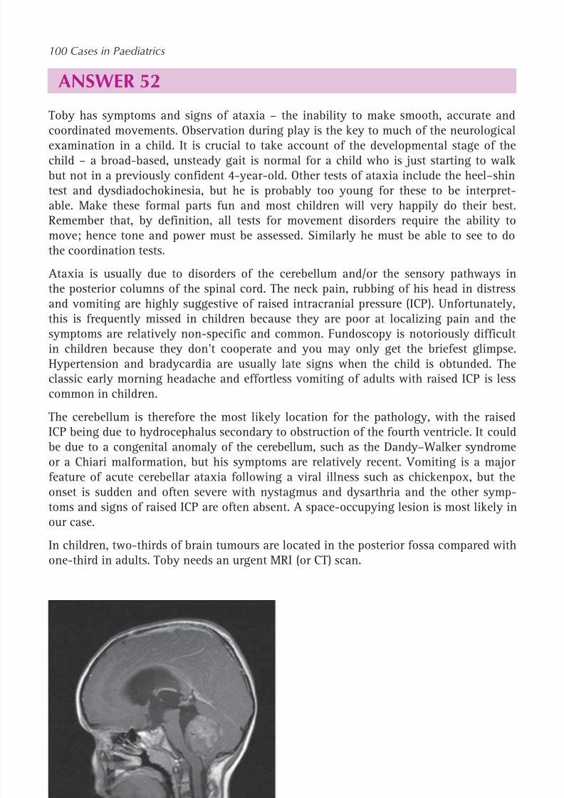
8/20/2019 100 cases in paediatrics.pdf
http://slidepdf.com/reader/full/100-cases-in-paediatricspdf 177/320
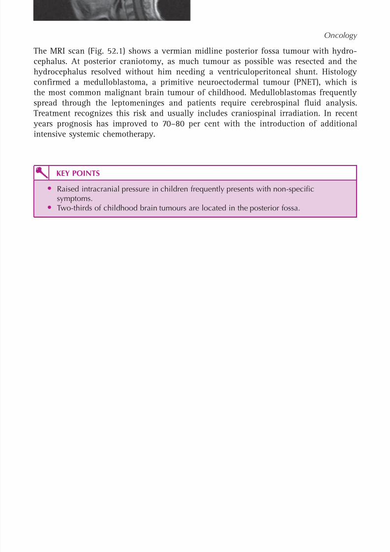
8/20/2019 100 cases in paediatrics.pdf
http://slidepdf.com/reader/full/100-cases-in-paediatricspdf 178/320
159
Oncology
The MRI scan (Fig. 52.1) shows a vermian midline posterior fossa tumour with hydro-cephalus. At posterior craniotomy, as much tumour as possible was resected and thehydrocephalus resolved without him needing a ventriculoperitoneal shunt. Histologyconfirmed a medulloblastoma, a primitive neuroectodermal tumour (PNET), which is
the most common malignant brain tumour of childhood. Medulloblastomas frequentlyspread through the leptomeninges and patients require cerebrospinal fluid analysis.Treatment recognizes this risk and usually includes craniospinal irradiation. In recent
years prognosis has improved to 70–80 per cent with the introduction of additionalintensive systemic chemotherapy.
KEY POINTS
• Raised intracranial pressure in children frequently presents with non-specificsymptoms.• Two-thirds of childhood brain tumours are located in the posterior fossa.

8/20/2019 100 cases in paediatrics.pdf
http://slidepdf.com/reader/full/100-cases-in-paediatricspdf 179/320
This page intentionally left blank
8/20/2019 100 cases in paediatrics.pdf
http://slidepdf.com/reader/full/100-cases-in-paediatricspdf 180/320

8/20/2019 100 cases in paediatrics.pdf
http://slidepdf.com/reader/full/100-cases-in-paediatricspdf 181/320
162
ANSWER 53The lump is consistent with enlarged lymph nodes. The differential diagnosis is:
• infection – bacterial, viral or mycobacteria (tuberculosis and non-tuberculousmycobacteria)• malignancy – lymphoma or leukaemia.
Lymph nodes are often palpable in children. Cervical and axillary lymph nodes 1 cm indiameter and inguinal lymph nodes 1.5 cm in diameter are considered to be enlarged.
Any enlargement in any other lymph nodes, e.g. supraclavicular, requires further inves-tigation. All children get several upper respiratory tract infections a year, and the minorcold several weeks earlier is probably irrelevant. Statistically, a bacterial cervical lym-phadenitis is the most likely diagnosis. However, usually there is a clear history of a cur-rent or recent upper respiratory tract infection. In bacterial infections, the overlying skinis usually erythematous and the lump is warm and tender. Occasionally the swelling maybecome fluctuant and this is indicative of pus formation and an abscess. In such a case,a referral should be made to a surgeon for incision and drainage. In viral infections,the nodes are tender but are not usually warm or erythematous. The Paul–Bunnell testis similar to the Monospot test and is a test for glandular fever. Viral serology for HIV,cytomegalovirus, toxoplasmosis (which often produce more generalized lymphadeno-pathy) and cat scratch disease (due to Bartonella henselae ) would be helpful if there wasstill no diagnosis after the initial investigations.
In tuberculosis, the lump is cold and non-tender. Munira has had her BCG vaccination butit is important to remember that, although this vaccination provides reasonable protectionagainst miliary TB and TB meningitis (50–80 per cent), it only provides 50 per cent protec-tion for pulmonary TB and only minimal (if any) protection against non-tuberculous myco-bacteria. A lymphoma can present with non-tender cervical lymphadenopathy. The lymphnodes tend to be firmer than inflammatory nodes and may be adherent to the overlyingskin or underlying structures. The absence of systemic symptoms, such as fevers, nightsweats and weight loss, makes the diagnosis of tuberculosis and lymphoma less likely.
At the first visit, it would be worth giving a 10-day course of an antibiotic, e.g. co-amoxiclav, that would treat streptococcal and staphylococcal infection. This would treatthe most likely diagnosis of a bacterial lymphadenitis.
The white blood cell count is normal and the CRP and ESR are only mildly elevated.The ASOT, Paul–Bunnell test and Mantoux are negative. These results make an infectionunlikely and a malignancy the most likely diagnosis.
If there was no diminution in the size of the lump following the 10-day course of treat-ment (which would suggest that the diagnosis is not a bacterial lymphadenitis), thepatient should be referred to a surgeon for an urgent biopsy. This was the case withMunira in whom the lymph node biopsy was diagnostic of Hodgkin’s lymphoma.
KEY POINTS
• Most children have some small palpable lymph nodes.• The commonest cause of cervical lymphadenitis is streptococcal or staphylococcal
infection.• Patients with lymphoma commonly present with non-tender firm cervical
lymphadenopathy.
100 Cases in Paediatrics

8/20/2019 100 cases in paediatrics.pdf
http://slidepdf.com/reader/full/100-cases-in-paediatricspdf 182/320
163
CASE 54:A SWOLLEN SHOULDER
HistoryNaomi is a 13-year-old girl who presents to the A&E department of her local hospitalfor the third time in 8 weeks. On the first occasion she had slipped from her horse whilstmounting and sustained some minor bruising mainly to the right arm, hip and leg. Therewas no evidence of any serious bone injury. Over the course of the next 2 weeks shedeveloped worsening pain and immobility in her right shoulder. Examination from that
visit was recorded as showing a decreased range of movement but little else. Her symp-toms were thought to be due to either a ‘frozen shoulder’ or to be functional, and shewas prescribed ibuprofen for pain and referred for routine physiotherapy. The physio-therapists have sent her back with increasing pain and now swelling.
ExaminationNaomi is clearly in pain and is reluctant to move her right shoulder. There is a general-ized, smooth, warm, firm swelling of the right shoulder with a decreased range of move-ment in all directions. There are some distended visible veins and the overlying skin isshiny but not red. The remainder of the examination is normal.
INVESTIGATIONS
Naomi’s shoulder X-ray is shown in Figure 54.1.
Figure 54.1 Naomi’sshoulder X-ray.
Questions• What is the diagnosis?• What is the management?

8/20/2019 100 cases in paediatrics.pdf
http://slidepdf.com/reader/full/100-cases-in-paediatricspdf 183/320
164
100 Cases in Paediatrics
ANSWER 54The diagnosis is an osteosarcoma of the right humerus. There is no differential. This is amalignant tumour that classically presents in adolescence and often comes to light aftera relatively minor injury. It is still not uncommon for there to be a delay in diagnosisbecause it is rare and not considered in the differential diagnosis of limb and joint pain.The characteristic features on the X-ray are:
• bone destruction• gross swelling• elevation of the periosteum with creation of Codman’s triangle (marked with an
arrow on the X-ray)• ‘sunray’ spicules of new bone visible in the muscle and subcutaneous tissues.
The first step is regular adequate pain relief. Non-steroidal anti-inflammatory agentsmay be adequate but it is highly likely that opioids will be needed. This is an aggressivetumour with rapid expansion of the periosteum which is exquisitely painful.
Secondly, a senior paediatrician – preferably from the local ‘shared care’ oncology team –should break the news to the girl and her family. They will be devastated and quite pos-sibly angry at the perceived delay in diagnosis. The local team will have the experienceand knowledge to answer any immediate questions. They will also know that the bestroute for onward referral is to a specialist paediatric oncology centre and not, at thisstage, to orthopaedics. Although a tissue diagnosis is required, she needs to be screenedfor metastases as this may alter the management significantly. Osteosarcomas usuallyspread to lung and bone and she will need a chest CT scan and a bone scan plus anMRI scan of the shoulder to guide future surgery. The biopsy should only be taken in aspecialist centre (or with their explicit agreement) and reviewed by a team of specialisthistopathologists.
The specific management of osteosarcoma is initial chemotherapy to reduce tumourbulk and to clear any metastases, followed by surgery followed by further chemother-apy. Definitive surgery is probably contraindicated if metastases have not cleared orsignificantly reduced with initial chemotherapy, although some patients with isolatedlung secondaries do extremely well following their resection. Modern surgery is largelysuccessful at avoiding mutilating amputation using prostheses that can be externally‘lengthened’ as the patient grows.
Prognosis depends upon the presence of metastatic spread at presentation – bone sec-ondaries carry a particularly grave outlook – and on response to chemotherapy asevidenced by tumour necrosis at surgical resection. More than 90 per cent necrosis isassociated with an improved outcome. Overall prognosis is a 5-year survival rate ofapproximately 60 per cent.
KEY POINTS
• Children do not get ‘frozen shoulder’ and functional pain is a diagnosis of exclusion.• Bone tumours are rare but should be considered in any patient with unexpectedly
severe symptoms following a minor injury.• The care of children with cancer should be coordinated by a specialist centre.• Centralization of care, entry into randomized controlled trials and the use of national
or international protocols have steadily improved the prognosis in childhood cancer.
8/20/2019 100 cases in paediatrics.pdf
http://slidepdf.com/reader/full/100-cases-in-paediatricspdf 184/320
165
CASE 55:FEVER IN A PATIENT ON CHEMOTHERAPY
HistoryThe parents of 6-year-old Lucy, who has B-cell lymphoma, have brought her to thechildren’s ward of their local hospital as they have recorded a temperature at home of38.7 C. They have since given her a dose of paracetamol. Lucy completed her secondcourse of chemotherapy 6 days previously. They know that her last blood count 2 daysago showed that she was neutropenic (neutrophils 1.0 10 9/L). The whole family,including the patient, have had symptoms of a viral illness with a sore throat, a runnynose and a bit of a cough, but she has also had three bouts of diarrhoea in the preced-ing 24 hours and some lower abdominal pain. There are four other children at home,ranging in age from 6 months to 9 years. The mother works part-time in the eveningsserving behind a bar, while the father is a self-employed painter and decorator. Theirdaughter was very sick at presentation and spent 5 weeks in the regional paediatriconcology centre 40 miles away. She has only been home since the end of the recentcourse of chemotherapy.
ExaminationLucy is playing in a side-room with her dolls. She is reluctant to be examined. Her
temperature is 37.4 C. Her pulse is 130 beats/min and her blood pressure 95/60 mmHg.Oxygen saturation is 97 per cent in air. The indwelling central-line exit site is slightlyerythematous and tender. There are no murmurs and examination of the respiratory andabdominal systems is unremarkable. Her ears, nose and throat are normal.
INVESTIGATIONS
Normal
Haemoglobin 7.4 g/dL 11.5–15.5 g/dLWhite cell count 0.6 109 /L 6.0–17.5 109 /LNeutrophils 0.1 109 /L 3.0–5.8 109 /LPlatelets 24 109 /L 150–400 109 /LSodium 134 mmol/L 138–146 mmol/LPotassium 3.8 mmol/L 3.5–5.0 mmol/LUrea 6.2 mmol/L 1.8–6.4 mmol/LCreatinine 64 mol/L 27–62 mol/LC-reactive protein 67 mg/L 6 mg/L
Questions• What factors increase the risk of infection in children with cancer?• What is the management?
8/20/2019 100 cases in paediatrics.pdf
http://slidepdf.com/reader/full/100-cases-in-paediatricspdf 185/320
166
ANSWER 55Once a child with cancer undergoing treatment is in remission, the commonest cause ofdeath is infection. Families have written instructions about temperature monitoring andwhen to seek advice. All admitting hospitals have protocols for treatment based on thedegree of fever (e.g. 38.5 C) and the neutrophil count (e.g. 0.75 10 9/L).
! Contributing factors to infection in children with cancer
• Neutropenia – the more severe ( 0.5 109/L) and prolonged, the greater the risk.There may be loss of the inflammatory response, e.g. insufficient white blood cellsto form an abscess
• Mucositis – following radiotherapy or chemotherapy. Can affect the wholegastrointestinal tract. There is a risk that intestinal bacteria will breach the gut
wall and enter the bloodstream• Indwelling central lines – crucial for giving chemotherapy and blood sampling,
but significant source of infection, both blood-borne and at exit site• Frequent hospital admissions• Poor nutrition• Generalized immunosuppression, inhibiting the usual host defences – viruses
that rarely cause severe illness in immunocompetent children can be life-threatening in oncology patients, e.g. varicella, measles and cytomegalovirus.
Chemotherapy also predisposes children to ‘opportunistic infections’ – organismsthat only infect an immunocompromised host. Prophylaxis against Pneumocystiscarinii is included in protocols for cancers where the chemotherapy regimen isparticularly immunosuppressive
The reduction in temperature is irrelevant – the protocols are initiated on the recordedtemperature at home. That she is playing is also irrelevant – she has worrying symp-toms and signs. Abdominal pain and diarrhoea suggest mucositis and she is tachycardic.
Although a visible source of sepsis is her central line site, this rarely causes significantsystemic features. Gram-negative sepsis is the real threat and she requires admission andthe immediate administration of intravenous broad-spectrum antibiotics that includecover for Gram-negative bacteria (e.g. Pseudomonas aeruginosa , Klebsiella ), as well asGram-positive bacteria that may be causing a line infection.
Blood cultures and swabs (e.g. throat and line site) must be taken first and prefera-bly urine and stool samples. However, collecting these should never delay antibiotics.
A chest X-ray is needed if there are respiratory symptoms or signs. Careful monitoring
and frequent re-evaluation are required, changing the regimen according to responseand culture results, e.g. coagulase-negative staphylococci frequently cause infections incentral lines. If Lucy is still febrile and significantly neutropenic after about 5 days,antifungal agents may be added. Haemopoietic growth factors, e.g. granulocyte colony-stimulating factor, are sometimes given. She will almost certainly need blood and/orplatelet transfusions. Again, protocols vary but an example would be a haemoglobin
7 g/dL and platelets 10 10 9/L or active bleeding.
Once afebrile and recovering, she can be discharged home where, if necessary, the par-ents or community nurses can continue antibiotics. Life for such families is inevitably
100 Cases in Paediatrics

8/20/2019 100 cases in paediatrics.pdf
http://slidepdf.com/reader/full/100-cases-in-paediatricspdf 186/320
167
KEY POINTS• Once in remission, infection is the commonest cause of death in children receiving
treatment for cancer.• Febrile neutropenia in an immunocompromised child is a medical emergency.• Common viral illnesses can kill immunocompromised children.
Oncology
disrupted socially and financially – the main breadwinner here is self-employed. Theywill be supported by specific staff and charities.

8/20/2019 100 cases in paediatrics.pdf
http://slidepdf.com/reader/full/100-cases-in-paediatricspdf 187/320
This page intentionally left blank
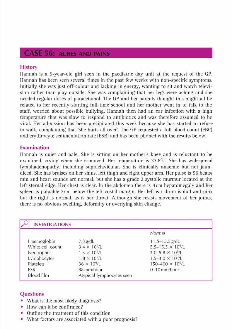
8/20/2019 100 cases in paediatrics.pdf
http://slidepdf.com/reader/full/100-cases-in-paediatricspdf 188/320
169
CASE 56:ACHES AND PAINS
HistoryHannah is a 5-year-old girl seen in the paediatric day unit at the request of the GP.Hannah has been seen several times in the past few weeks with non-specific symptoms.Initially she was just off-colour and lacking in energy, wanting to sit and watch televi-sion rather than play outside. She was complaining that her legs were aching and sheneeded regular doses of paracetamol. The GP and her parents thought this might all berelated to her recently starting full-time school and her mother went in to talk to thestaff, worried about possible bullying. Hannah then had an ear infection with a hightemperature that was slow to respond to antibiotics and was therefore assumed to be
viral. Her admission has been precipitated this week because she has started to refuseto walk, complaining that ‘she hurts all over’. The GP requested a full blood count (FBC)and erythrocyte sedimentation rate (ESR) and has been phoned with the results below.
ExaminationHannah is quiet and pale. She is sitting on her mother’s knee and is reluctant to beexamined, crying when she is moved. Her temperature is 37.8 C. She has widespreadlymphadenopathy, including supraclavicular. She is clinically anaemic but not jaun-
diced. She has bruises on her shins, left thigh and right upper arm. Her pulse is 96 beats/min and heart sounds are normal, but she has a grade 2 systolic murmur located at theleft sternal edge. Her chest is clear. In the abdomen there is 4 cm hepatomegaly and herspleen is palpable 2 cm below the left costal margin. Her left ear drum is dull and pinkbut the right is normal, as is her throat. Although she resists movement of her joints,there is no obvious swelling, deformity or overlying skin change.
INVESTIGATIONS
Normal
Haemoglobin 7.3 g/dL 11.5–15.5 g/dLWhite cell count 3.4 109 /L 5.5–15.5 109 /LNeutrophils 1.3 109 /L 3.0–5.8 109 /LLymphocytes 1.8 109 /L 1.5–3.0 109 /LPlatelets 36 109 /L 150–400 109 /LESR 88 mm/hour 0–10 mm/hourBlood film Atypical lymphocytes seen
Questions• What is the most likely diagnosis?• How can it be confirmed?• Outline the treatment of this condition• What factors are associated with a poor prognosis?
8/20/2019 100 cases in paediatrics.pdf
http://slidepdf.com/reader/full/100-cases-in-paediatricspdf 189/320
170
ANSWER 56Hannah’s blood count shows pancytopenia, which is due to either bone marrow (BM)infiltration or failure (see Case 48 , A pale child, p. 143 ). She has the clinical symptomsand signs of infiltration – the bone pain is due to marrow expansion. Children may notlocalize the pain, and generalized non-specific symptoms culminating in a reluctanceto move and weight-bear are common. There may be bone tenderness. Hannah has evi-dence of extramedullary involvement with hepatosplenomegaly and lymphadenopathy.Supraclavicular nodes are always suspicious. Bruises below the knees are very commonin children, but those at sites other than the shins may be pathological. The murmur isalmost certainly a flow murmur due to anaemia. The most likely diagnosis is acute leu-kaemia, probably lymphoblastic (ALL) because this accounts for 80 per cent of leukae-mias in childhood.
Leukaemic ‘blast’ cells may not be observed on a routine peripheral blood film examin-ation or may be misinterpreted. The peripheral FBC may even be normal, causingdiagnostic difficulty in differentiating it from conditions that present with similar fea-tures, e.g. infectious mononucleosis or rheumatoid arthritis. Analysis of a BM aspirate isessential. This differentiates ALL from acute myeloid leukaemia (AML) and other malig-nant causes of BM infiltration, such as neuroblastoma. It also enables classification of
ALL using cell membrane markers and cytogenetic and molecular genetic features whichinfluence treatment decisions and prognosis.
Current treatment for ALL is in four phases:1. Remission induction . Four weeks of combination chemotherapy. More than 95 per cent
show BM remission by day 28.2. Consolidation and CNS protection . Oral chemotherapy is commenced. Cytotoxic drugs
penetrate poorly into the CNS. A course of weekly intrathecal (IT) drugs is given toprevent later CNS relapse. IT drugs are also given during induction and throughoutmaintenance.
3. Delayed intensification(s) . Remission at day 28 is often only apparent. One or twocourses (depending on risk) of intense chemotherapy are dovetailed with blocks of
maintenance chemotherapy.4. Continuing maintenance. Chemotherapy continues for a total of 2 years in girls and
3 years in boys. Patients are immunosuppressed and at risk of opportunistic infec-tions. Co-trimoxazole is given to prevent Pneumocystis carinii pneumonia.
! Poor prognostic factors in acute lymphoblastic leukaemia
• Total white cell count 100 109 /L at presentation
• Outside age range 2–10 years• Male sex• T-cell (15 per cent) or mature B-cell (1 per cent) origin (Common-ALL
(85 per cent) from B-cell progenitors has best prognosis)• Certain chromosome abnormalities – hypodiploidy, presence of Philadelphia
chromosome (Others, e.g. hyperdiploidy, confer a favourable prognosis)• CNS disease at presentation• Slow response to treatment, 5% blasts persisting on day 28 BM
Overall prognosis for ALL is excellent with a 5-year survival 80 per cent.
100 Cases in Paediatrics

8/20/2019 100 cases in paediatrics.pdf
http://slidepdf.com/reader/full/100-cases-in-paediatricspdf 190/320
171
KEY POINTS
• Acute leukaemia can present with very non-specific and indolent clinical features.• A bone marrow aspirate is essential to confirm or refute a diagnosis of acute
leukaemia.
Oncology
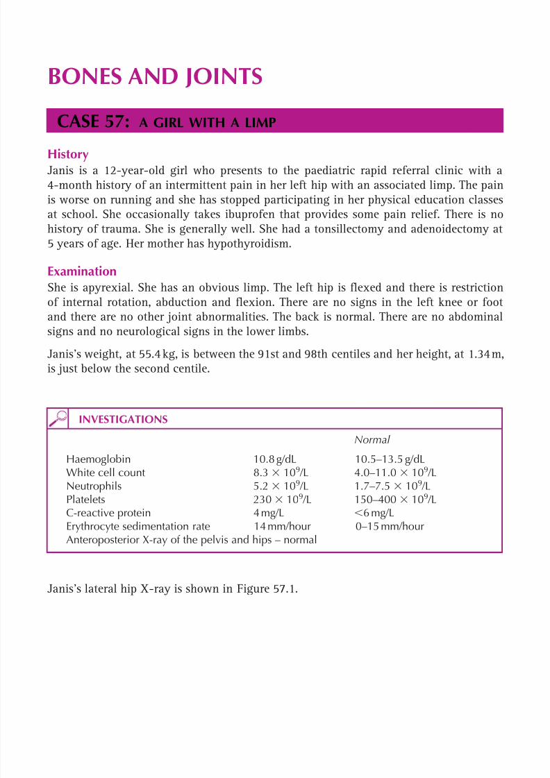
8/20/2019 100 cases in paediatrics.pdf
http://slidepdf.com/reader/full/100-cases-in-paediatricspdf 191/320
172
BONES AND JOINTS
CASE 57: A GIRL WITH A LIMP
HistoryJanis is a 12-year-old girl who presents to the paediatric rapid referral clinic with a4-month history of an intermittent pain in her left hip with an associated limp. The painis worse on running and she has stopped participating in her physical education classesat school. She occasionally takes ibuprofen that provides some pain relief. There is nohistory of trauma. She is generally well. She had a tonsillectomy and adenoidectomy at5 years of age. Her mother has hypothyroidism.
ExaminationShe is apyrexial. She has an obvious limp. The left hip is flexed and there is restrictionof internal rotation, abduction and flexion. There are no signs in the left knee or footand there are no other joint abnormalities. The back is normal. There are no abdominalsigns and no neurological signs in the lower limbs.
Janis’s weight, at 55.4 kg, is between the 91st and 98th centiles and her height, at 1.34 m,is just below the second centile.
INVESTIGATIONS
Normal
Haemoglobin 10.8 g/dL 10.5–13.5 g/dLWhite cell count 8.3 109 /L 4.0–11.0 109 /LNeutrophils 5.2 109 /L 1.7–7.5 109 /LPlatelets 230 109 /L 150–400 109 /LC-reactive protein 4 mg/L 6 mg/LErythrocyte sedimentation rate 14 mm/hour 0–15 mm/hour
Anteroposterior X-ray of the pelvis and hips – normal
Janis’s lateral hip X-ray is shown in Figure 57.1.
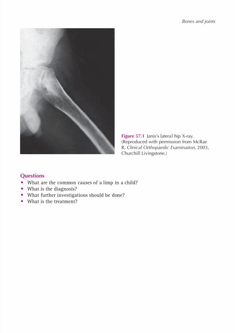
8/20/2019 100 cases in paediatrics.pdf
http://slidepdf.com/reader/full/100-cases-in-paediatricspdf 192/320
173
Bones and joints
Figure 57.1 Janis’s lateral hip X-ray.(Reproduced with permission from McRaeR, Clinical Orthopaedic Examination , 2003,Churchill Livingstone.)
Questions• What are the common causes of a limp in a child?• What is the diagnosis?• What further investigations should be done?• What is the treatment?
8/20/2019 100 cases in paediatrics.pdf
http://slidepdf.com/reader/full/100-cases-in-paediatricspdf 193/320
174
100 Cases in Paediatrics
ANSWER 57 A limp is a common problem in childhood. It is important to be aware that it can be dueto an abnormality anywhere from the back to the foot.
Causes of a limp in a child are best considered according to age:
• 0–3 years– Trauma, e.g. fracture (may be accidental or non-accidental)– Infection: septic arthritis, osteomyelitis or discitis– Malignancy – Developmental dysplasia of the hip– Neuromuscular disease, e.g. cerebral palsy
•4–10 years– Trauma– Transient synovitis (irritable hip)– Infection – septic arthritis, osteomyelitis or discitis– Perthe’s disease– Juvenile idiopathic arthritis– Malignancy, e.g. leukaemia– Neuromuscular disease, e.g. cerebral palsy
• 10–18 years– Trauma– Infection: septic arthritis, osteomyelitis or discitis– Slipped upper femoral epiphysis– Juvenile idiopathic arthritis– Malignancy, e.g. osteogenic sarcoma
The diagnosis is a slipped upper femoral epiphysis (SUFE). SUFE is commoner in boysand in obese individuals. Pain can be felt in the groin, hip or thigh or there may bereferred pain in the knee. Classically the affected hip is flexed and the leg is externallyrotated. A small slip may be missed on the anteroposterior view and a lateral or frog leg
view of the hip is therefore also necessary. The lateral X-ray shows displacement of theproximal femoral epiphysis relative to the femoral neck. Approximately 25 per cent ofpatients will also have a contralateral slip and the other hip must therefore be carefullyassessed.
A minority of patients with SUFE have an underlying endocrinopathy or metabolicabnormality (hypothyroidism, hypogonadism, growth hormone abnormalities, panhypo-pituitarism or renal osteodystrophy) and if this is suspected then the appropriate inves-tigations should be performed. Janis is short and overweight and has a family history ofthyroid disease. A more detailed examination demonstrated that she had a small goitre.Thyroid function tests revealed a free T4 of 9.2 pmol/L (12–22), a thyroid-stimulatinghormone (TSH) of 64 mU/L (0.5–6.0) and thyroid peroxidase (TPO) antibodies of 1342 IU/mL (0–34). These results are compatible with a diagnosis of Hashimoto’s (autoimmune)thyroiditis. Obese individuals should have their fasting glucose measured and this testshould also be done on Janis.
Janis should be admitted and told not to weight-bear. She should be referred urgentlyto the orthopaedic surgeons. Although it would seem that the SUFE has been present for4 months, one can get an acute or chronic slip of the epiphysis that can lead to avascular
8/20/2019 100 cases in paediatrics.pdf
http://slidepdf.com/reader/full/100-cases-in-paediatricspdf 194/320
175
Bones and joints
necrosis of the femoral head. Surgery is therefore urgent and consists of pinning thefemoral head to the femoral neck. Thyroxine should also be started.
KEY POINTS• In SUFE, a lateral or frog leg view, as well as an anteroposterior view, of the hip is
necessary to avoid missing minor slips.• Urgent treatment is necessary in SUFE to try to avoid the complication of avascular
necrosis of the femoral head

8/20/2019 100 cases in paediatrics.pdf
http://slidepdf.com/reader/full/100-cases-in-paediatricspdf 195/320
This page intentionally left blank

8/20/2019 100 cases in paediatrics.pdf
http://slidepdf.com/reader/full/100-cases-in-paediatricspdf 196/320
8/20/2019 100 cases in paediatrics.pdf
http://slidepdf.com/reader/full/100-cases-in-paediatricspdf 197/320
178
100 Cases in Paediatrics
ANSWER 58This boy has signs of sepsis with fever, prolonged capillary refill time and tachycardia.His investigations support this. The history of pain with movement is strongly sugges-tive of musculoskeletal pathology. In acute sepsis, the emphasis is usually on diagnos-ing meningitis or an acute abdomen and it is easy to overlook this system. Examine forany swelling, redness of the skin, warmth and tenderness. There may be nothing to seesuperficially, so observe the posture and whether the child is keeping any limb flexed orstill. Gently examine the range of movement of all joints.
The most likely diagnosis is osteomyelitis or septic arthritis. The legs are much thecommonest site and most infections are blood-borne. There is often a recent history ofminor trauma or a breach in the skin, which probably explains why boys are more often
affected. The commonest bacteria involved is Staphylococcus aureus .Children with sickle cell disease have a higher incidence of skeletal infections. The dif-ferential depends on the site but includes soft-tissue infections and psoas abscess. In theless obviously septic child, remember trauma, both accidental and non-accidental, andbone infiltration with cancers such as leukaemia.
! Possible imaging modalities
• Plain X-rays – excludes trauma. In osteomyelitis, bones are unlikely to beabnormal until 10–14 days after onset. In arthritis they are often normal but mayshow widening of joint space local soft tissue or fat changes
• Ultrasound (US) – highly sensitive in detecting joint effusion and guidingaspiration. It may demonstrate swelling and distortion of soft tissues andsubperiosteal region
• MRI – the best technique to differentiate between soft tissue and bone infection;also to detect a joint effusion where US is normal. However, the boy will needsedation or general anaesthetic at this age
• Technetium ( 99mTc) bone scan – accumulates as ‘hot spots’ in areas of increasedbone turnover so useful in osteomyelitis, especially multifocal. Infection closeto growth plates can limit specificity
First, he needs fluid resuscitation and adequate pain relief. If the clinical diagnosis is osteo-myelitis, he should start intravenous antibiotics, e.g. co-amoxiclav. However, if there isevidence of septic arthritis, he should ideally have an immediate US and, if there is an effu-sion, an urgent arthrotomy before starting antibiotics. If there is no effusion, the diagnosisis more likely to be osteomyelitis and antibiotics can be started. They should not be delayedin order to establish a definitive diagnosis with imaging or cultures, because this increasesthe risk of long-term effects on the growth and function of the affected limb, especially ifthe growth plate is involved. The length of treatment and route of administration of anti-
biotics for both depend upon the clinical and biochemical (WCC, ESR, CRP) responses. Atotal of 6 weeks is common practice. Long-term follow-up is necessary because problemsmay not be apparent for several years. Most children make an uneventful recovery.
KEY POINTS
• Bone and/or joint sepsis should be considered in the differential diagnosis of allchildren presenting with sepsis.
• Delay in treatment of osteomyelitis or septic arthritis can cause significant long-termeffects on the growth and function of the affected limb.
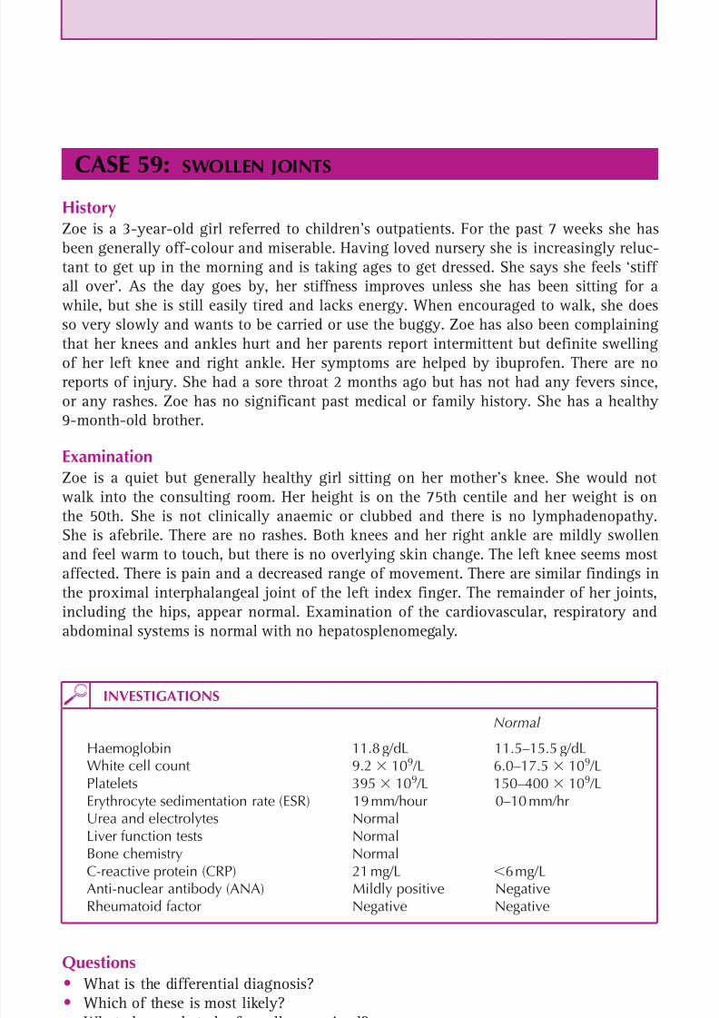
8/20/2019 100 cases in paediatrics.pdf
http://slidepdf.com/reader/full/100-cases-in-paediatricspdf 198/320
179
CASE 59:SWOLLEN JOINTS
HistoryZoe is a 3-year-old girl referred to children’s outpatients. For the past 7 weeks she hasbeen generally off-colour and miserable. Having loved nursery she is increasingly reluc-tant to get up in the morning and is taking ages to get dressed. She says she feels ‘stiffall over’. As the day goes by, her stiffness improves unless she has been sitting for awhile, but she is still easily tired and lacks energy. When encouraged to walk, she doesso very slowly and wants to be carried or use the buggy. Zoe has also been complainingthat her knees and ankles hurt and her parents report intermittent but definite swellingof her left knee and right ankle. Her symptoms are helped by ibuprofen. There are noreports of injury. She had a sore throat 2 months ago but has not had any fevers since,or any rashes. Zoe has no significant past medical or family history. She has a healthy9-month-old brother.
ExaminationZoe is a quiet but generally healthy girl sitting on her mother’s knee. She would notwalk into the consulting room. Her height is on the 75th centile and her weight is onthe 50th. She is not clinically anaemic or clubbed and there is no lymphadenopathy.
She is afebrile. There are no rashes. Both knees and her right ankle are mildly swollenand feel warm to touch, but there is no overlying skin change. The left knee seems mostaffected. There is pain and a decreased range of movement. There are similar findings inthe proximal interphalangeal joint of the left index finger. The remainder of her joints,including the hips, appear normal. Examination of the cardiovascular, respiratory andabdominal systems is normal with no hepatosplenomegaly.
INVESTIGATIONS
Normal
Haemoglobin 11.8 g/dL 11.5–15.5 g/dLWhite cell count 9.2 109 /L 6.0–17.5 109 /LPlatelets 395 109 /L 150–400 109 /LErythrocyte sedimentation rate (ESR) 19 mm/hour 0–10 mm/hrUrea and electrolytes NormalLiver function tests NormalBone chemistry NormalC-reactive protein (CRP) 21 mg/L 6 mg/L
Anti-nuclear antibody (ANA) Mildly positive NegativeRheumatoid factor Negative Negative
Questions• What is the differential diagnosis?• Which of these is most likely?• What else needs to be formally examined?• What are the principles of management?
8/20/2019 100 cases in paediatrics.pdf
http://slidepdf.com/reader/full/100-cases-in-paediatricspdf 199/320
180
100 Cases in Paediatrics
ANSWER 59Zoe has a polyarthritis – pain, swelling, warmth and restricted movement of several
joints. The much commoner arthralgia is joint pain without the other features.
! Differential diagnosis of chronic polyarthritis
• Connective tissue disorder – juvenile idiopathic arthritis (JIA), systemic lupuserythematosus, dermatomyositis, psoriatic arthritis
• Infection – bacterial causes are unlikely with this chronic history. Viruses such ashepatitis and also Lyme disease should be excluded
• Malignant disorders – children with leukaemia can have joint pain due tometaphyseal marrow expansion months before peripheral blood count changes.A bone marrow examination may be indicated
• Inflammatory bowel disease – can present with arthritis• Vasculitis – Henoch–Schönlein purpura
Zoe has other features in the history that make JIA the most likely diagnosis. Therehas been an insidious onset of early morning stiffness, ‘gelling’ (stiffness) after inactiv-ity, slow walking and progressive joint involvement. Juvenile idiopathic arthritis is agroup of conditions in which there is a chronic arthritis lasting 6 consecutive weeksbefore the age of 16 years. Infection and other causes must obviously be excluded. Her
sore throat 2 months earlier is probably incidental although a reactive arthritis follow-ing Streptococcus tonsillitis is well recognized and the anti-streptolysin O titre (ASOT)should be assayed. Juvenile idiopathic arthritis is classified according to its onset as sys-temic, polyarticular ( four joints) or oligo(pauci)articular ( four joints).
Zoe has oligoarticular JIA, the commonest subtype, which typically affects the knees andankles and less often the elbows or a single finger joint in young children 6 years,particularly girls. In systemic or polyarticular JIA, there is often laboratory evidence ofchronic inflammation with anaemia, thrombocytosis and an elevated ESR and C-reactiveprotein, but in oligoarticular JIA these may be normal or only mildly abnormal. Patients
with oligoarticular JIA have an increased risk of one of the complications of JIA, chronicanterior uveitis, and this is increased still further in girls who are ANA-positive. Shetherefore needs formal ophthalmology screening and frequent regular review. The courseof the uveitis is independent of the joints.
! Principles of management
• Pain control with non-steroidal anti-inflammatory drugs (NSAIDs)• Physiotherapy, hydrotherapy and occupational therapy. Splints may help prevent
contractures and deformities• Intra-articular depot corticosteroid injections to reduce swelling and deformity• Disease-modifying drugs are needed if not controlled with NSAIDs, e.g.
methotrexate, sulphasalazine. Cytokine inhibitors (e.g. anti-tumour necrosisfactor) may be needed for refractory polyarticular or systemic JIA. All of theseneed careful monitoring in a specialist setting
• Regular monitoring of growth and nutrition as for any chronic disease• Multidisciplinary team approach, including psychosocial support. The impact
of all forms of JIA is significant, although the prognosis for oligoarthritis isgenerally better than for other groups
8/20/2019 100 cases in paediatrics.pdf
http://slidepdf.com/reader/full/100-cases-in-paediatricspdf 200/320
181
Bones and joints
KEY POINTS
• Juvenile idiopathic arthritis can only be diagnosed after 6 continuous weeks ofarthritis and after the exclusion of other causes.
• The management of JIA requires a multidisciplinary team with as much emphasis onthe therapies as on drug interventions.

8/20/2019 100 cases in paediatrics.pdf
http://slidepdf.com/reader/full/100-cases-in-paediatricspdf 201/320
This page intentionally left blank

8/20/2019 100 cases in paediatrics.pdf
http://slidepdf.com/reader/full/100-cases-in-paediatricspdf 202/320
183
CASE 60:A SPINAL DEFORMITY
HistoryMelissa is a 12-year-old who presents to her GP with a persistent cough that has beengoing on for 3 weeks. The whole family have had a flu-like illness but her symptomsseem to have gone on for longer. Her cough is non-productive, she has not been short ofbreath and she hasn’t complained of chest pain. She had a temperature early on but thishas resolved. There is no significant past medical history and she rarely attends the sur-gery. She has not yet started her periods. She takes no regular medication.
ExaminationMelissa is a generally healthy girl who is not distressed at rest. She is pink in room air andnot clubbed. There is no significant lymphadenopathy. The GP examines her respiratory sys-tem and can find no abnormalities. However, he notices that she appears to have a lateralcurvature to the left of her thoracic spine. There are no neurocutaneous features.
Questions• What is this deformity?• What else should now be documented in the history and examination?
• What are the management options?
8/20/2019 100 cases in paediatrics.pdf
http://slidepdf.com/reader/full/100-cases-in-paediatricspdf 203/320
184
100 Cases in Paediatrics
ANSWER 60This patient has a abnormality in spinal alignment in the coronal plane – a scoliosis. Thespine has normal curves in the sagittal plane that provide stability and balance. The cer-
vical and lumbar spines are convex anteriorly, termed lordosis. The thoracic and sacralspines are convex posteriorly, termed kyphosis. These normal curves can be exaggerated,e.g. in congenital deformities such as achondroplasia where there is almost always a sig-nificant lumbar lordosis.
Having found a scoliosis, the next question is whether it is postural (common) or struc-tural (rare). A postural scoliosis may be caused by pain or be secondary to a leg-lengthdiscrepancy and these conditions should be ruled out first. Leg-length discrepancy isusually easily managed with a shoe raise. Provided both are absent, a postural scoliosis
will disappear when the child bends forwards to try to touch her toes and no furtherintervention is required. In a structural scoliosis, the curvature persists in the forward-bending test and there is rotation of the vertebral bodies towards the convexity of thecurve, causing a ‘rib hump’. Most cases of structural scoliosis are idiopathic but some arerelated to an underlying condition and these need exclusion.
! Differential diagnosis of a structural scoliosis
• Idiopathic
• Neuromuscular, e.g. cerebral palsy or Duchenne muscular dystrophy• Congenital structural abnormalities of the vertebrae, e.g. hemivertebrae – there
is a high risk of associated spinal cord defects, e.g. spinal dysraphism and othercongenital abnormalities, especially genitourinary
• Syndromic, e.g. neurofibromatosis and Marfan syndrome
Therefore, children with a structural scoliosis need a thorough neurological evaluationbefore concluding that the scoliosis is idiopathic.
Treatment assumes that a progressive scoliosis will cause unacceptable deformities, anincreased risk of early joint disease and cardiorespiratory compromise. In idiopathicstructural scoliosis, the risk of progression depends upon sex, age, pubertal status and thedegree of curvature. Premenarchal girls are at highest risk. All patients with, or at risk of,structural scoliosis should be monitored regularly. Bracing and surgical correction are thetwo treatment options. Surgery is recommended earlier in those with neuromuscular orcongenital scoliosis.
KEY POINTS
• Scoliosis is often picked up incidentally and most are postural.• Patients found to have a structural scoliosis must have a thorough neurological
evaluation although most cases are idiopathic.• A structural scoliosis in a premenarchal girl is the most likely to progress.

8/20/2019 100 cases in paediatrics.pdf
http://slidepdf.com/reader/full/100-cases-in-paediatricspdf 204/320
185
NEUROLOGY
CASE 61: A FITTING CHILD
HistoryTom is a 4-year-old child known to have epilepsy. He is admitted to the resuscitationroom in A&E with a fit. He was born at 26 weeks’ gestation and had a periventricularhaemorrhage which has led to moderate learning difficulties. He has had a runny nose,a cough and a fever in the last few days. The coughing sometimes leads to vomiting andhis mother states that he has vomited up several of his drug doses in the last few days.He is on sodium valproate and lamotrigine. He has had previous admissions with fits buthis mother says this is his longest fit. She has already administered buccal midazolam athome with no effect. No one in the family has had cold sores in the past few weeks.
ExaminationTom is in the midst of having a generalized fit which started 35 min before he reachedhospital. His temperature is 40.2 C, oxygen saturation is 90 per cent in air, respiratory rate30/min, heart rate 180/min, blood pressure 105/77 mmHg and peripheral capillary refill 5 s.
He has a runny nose and an erythematous pharynx with enlarged but non-pustular ton-sils. His chest is clear. He has no neck stiffness or rash. There are no other signs.
High-flow (15 L/min) 100 per cent facemask oxygen elevates the saturation to 97 per centand an intravenous injection of lorazepam terminates the fit after a further 5 min, fol-lowing which Tom is postictal.
INVESTIGATIONS
Normal
Haemoglobin 11.7 g/dL 11.5–15.5 g/dLWhite cell count 24.4 109 /L 5.5–15.5 109 /LNeutrophils 19.2 109 /L 1.5–8.0 109 /LPlatelets 435 109 /L 150–400 109 /LUrea and electrolytes NormalBone chemistry NormalLiver function tests NormalGlucose 7.1 mmol/L 3.5–6.5 mmol/LC-reactive protein (CRP) 106 mg/L 6 mg/LVenous gas: pH 7.26 7.35–7.45
P aCO2 6.8 kPa 4.7–6.4 kPaP aO2 3.1 kPa 11–14.4 kPa
HCO 3 19 mmol/L 22–29 mmol/L Base excess 7 mmol/L ( 3)–( 3) mmol/L
Questions• What is the most likely cause of this child’s prolonged fit?• Would you do a lumbar puncture (LP)?• What further treatment is required?

8/20/2019 100 cases in paediatrics.pdf
http://slidepdf.com/reader/full/100-cases-in-paediatricspdf 205/320
186
100 Cases in Paediatrics
ANSWER 61The most likely cause is poorly controlled epilepsy. A fever (secondary to an upperrespiratory tract infection in this case) can decrease the threshold for a fit in a childwith epilepsy. The vomiting of medication would further increase the risk of a fit. Therewould be no point doing drug levels in this case. The absence of neck stiffness and arash would go against meningitis. Encephalitis is rare. There is no history of recent her-petic cold sores in the family. Encephalitis is often accompanied by a focal fit or focalneurological signs as well as a diminished conscious level and a fever. An LP should notbe performed because of the length of the fit.
Although this child’s fit is probably due to his epilepsy, the possibility of meningitis orencephalitis cannot be totally ruled out. He has a very high temperature, poor peripheral
perfusion, has had his longest ever fit, has a raised white cell count with a neutrophiliaand a raised CRP. An LP would help diagnose meningitis and encephalitis and would iden-tify the organism and its sensitivities. However, it should not be performed if there is a riskof raised intracranial pressure, as it may lead to coning and death. A magnetic resonanceimaging (MRI) or computed tomography (CT) scan may need to be performed to help ruleout raised intracranial pressure prior to an LP. Furthermore, an LP often does not alter theinitial management and can be done the next day if the patient is better, in order to eluci-date the diagnosis and help determine the type and length of antimicrobial treatment.
Intravenous fluids, initially 0.9 per cent saline 20 ml/kg, should be administered to improveperfusion and to help normalize the capillary refill time. Intravenous ceftriaxone, clarithro-mycin and aciclovir should be administered. Antipyretics should also be given. Clinicalprogress and an LP when the patient is better will help determine the diagnosis and thenecessary treatment.
Measuring the blood glucose at the bedside is essential to rule out hypoglycaemia in fit-ting children. One should never give more than a total of two doses of benzodiazepines,because of the risk of respiratory depression. Status epilepticus is one of the commonestpaediatric emergencies and it is important to be familiar with its treatment.
!Relative contraindications to a lumbar puncture (reproduced with modificationsfrom APLS manual, agreed by ALSG and Blackwell Publishing 2005):
• Prolonged or focal seizure• Focal neurological signs• A widespread purpuric rash• A Glasgow Coma Scale score 13• Abnormal posture or movement e.g. decerebrate posture• An inappropriately low pulse, raised blood pressure and irregular breathing
(suggesting impending brain herniation)• Thrombocytopenia or clotting disorder
• Pupillary dilatation• Papilloedema• Hypertension
KEY POINTS
• A glucose level should always be measured in a fitting child.• A lumbar puncture should not be performed if there is concern about raised
intracranial pressure, e.g. papilloedema or coma.
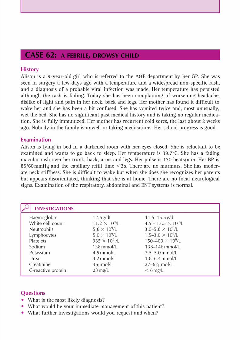
8/20/2019 100 cases in paediatrics.pdf
http://slidepdf.com/reader/full/100-cases-in-paediatricspdf 206/320
187
CASE 62:A FEBRILE
,DROWSY CHILD
History Alison is a 9-year-old girl who is referred to the A&E department by her GP. She wasseen in surgery a few days ago with a temperature and a widespread non-specific rash,and a diagnosis of a probable viral infection was made. Her temperature has persistedalthough the rash is fading. Today she has been complaining of worsening headache,dislike of light and pain in her neck, back and legs. Her mother has found it difficult towake her and she has been a bit confused. She has vomited twice and, most unusually,wet the bed. She has no significant past medical history and is taking no regular medica-tion. She is fully immunized. Her mother has recurrent cold sores, the last about 2 weeksago. Nobody in the family is unwell or taking medications. Her school progress is good.
Examination Alison is lying in bed in a darkened room with her eyes closed. She is reluctant to beexamined and wants to go back to sleep. Her temperature is 39.7 C. She has a fadingmacular rash over her trunk, back, arms and legs. Her pulse is 130 beats/min. Her BP is85/60 mmHg and the capillary refill time 2 s. There are no murmurs. She has moder-ate neck stiffness. She is difficult to wake but when she does she recognizes her parents
but appears disorientated, thinking that she is at home. There are no focal neurologicalsigns. Examination of the respiratory, abdominal and ENT systems is normal.
INVESTIGATIONS
Haemoglobin 12.6 g/dL 11.5–15.5 g/dLWhite cell count 11.2 109 /L 4.5 – 13.5 109 /LNeutrophils 5.6 109 /L 3.0–5.8 109 /LLymphocytes 5.0 109 /L 1.5–3.0 109 /LPlatelets 365 109 /L 150–400 109 /LSodium 138 mmol/L 138–146 mmol/LPotassium 4.5 mmol/L 3.5–5.0 mmol/LUrea 4.2 mmol/L 1.8–6.4 mmol/LCreatinine 46 µmol/L 27–62 µmol/LC-reactive protein 23 mg/L 6 mg/L
Questions• What is the most likely diagnosis?• What would be your immediate management of this patient?• What further investigations would you request and when?

8/20/2019 100 cases in paediatrics.pdf
http://slidepdf.com/reader/full/100-cases-in-paediatricspdf 207/320
188
100 Cases in Paediatrics
ANSWER 62The most likely diagnosis is a viral meningoencephalitis. Typically there is a prodrome ofa non-specific febrile illness, possibly accompanied by a rash, followed by the onset ofa progressive headache and photophobia. Any virus can be responsible, but at least 80per cent of cases are caused by enteroviruses. Of the other infectious agents mimicking
viral meningoencephalitis, by far the most important are bacteria, because they requireprompt treatment with antibiotics. The other major differential is a brain abscess.
Remember the maxim ‘treat the treatable’. Pending culture results, the assumption hasto be that a bacterial cause is possible even when, as in this case, the blood tests are notsupportive. A broad-spectrum antibiotic such as cefotaxime should be started immedi-ately following blood cultures. Most causes of viral meningoencephalitis have no spe-
cific treatment and the management is supportive. However, herpes simplex virus type 1(HSV-1) is an important cause (note her mother’s cold sores) and without treatment 70per cent progress to coma and death. Brain involvement is focal and patients frequentlypresent with focal seizures. All patients with meningoencephalitis should therefore bestarted on aciclovir. Mycoplasma pneumoniae can also cause meningoencephalitis and
Alison should also be treated with a macrolide antibiotic such as clarithromycin.
An EEG is valuable in supporting the diagnosis. Typically it shows slow-wave activ-ity without focal features, except in HSV-1 infection where there may be evidence oftemporal lobe involvement. A magnetic resonance imaging (MRI) or computed tomog-
raphy (CT) scan may show brain swelling and is mandatory if there are focal signs. Itwill also exclude a brain abscess. At some stage a lumbar puncture (LP) will be neces-sary but it is currently contraindicated because this girl is drowsy and disorientated, islikely to have raised intracranial pressure and could cone if an LP is performed. Thereis no urgency because she is already receiving the appropriate treatment. Cerebrospinalfluid (CSF) analysis should help differentiate between the various potential diagnoses(see Table 62.1).
Table 62.1 Cerebrospinal fluid findings in the commonest central nervous system infections
Condition Pressure White cells Protein Glucose Comments(mmH 2O) (mm 3) (g/L) (mmol/L)
Normal 50–80 5 0.2–0.4 70 per cent Clear andblood glucose colourless
Viral Normal or Rarely 1000. 0.5–2.0 Normal Clear. Viralslightly ↑ Neutrophils early Grossly culture and
but thereafter mostly ↑ in HSV PCRlymphocytes
Bacterial Usually ↑ 100– 10 000 1.0–5.0 ↓ 50 per cent Turbid.
Mostly neutrophils blood glucose Organismsseen on Gramstain. Cultureand PCR
HSV, herpes simplex virus; PCR, polymerase chain reaction.
Most patients with viral meningoencephalitis make a complete recovery, except for HSV-1 where sequelae, sometimes severe, are common.
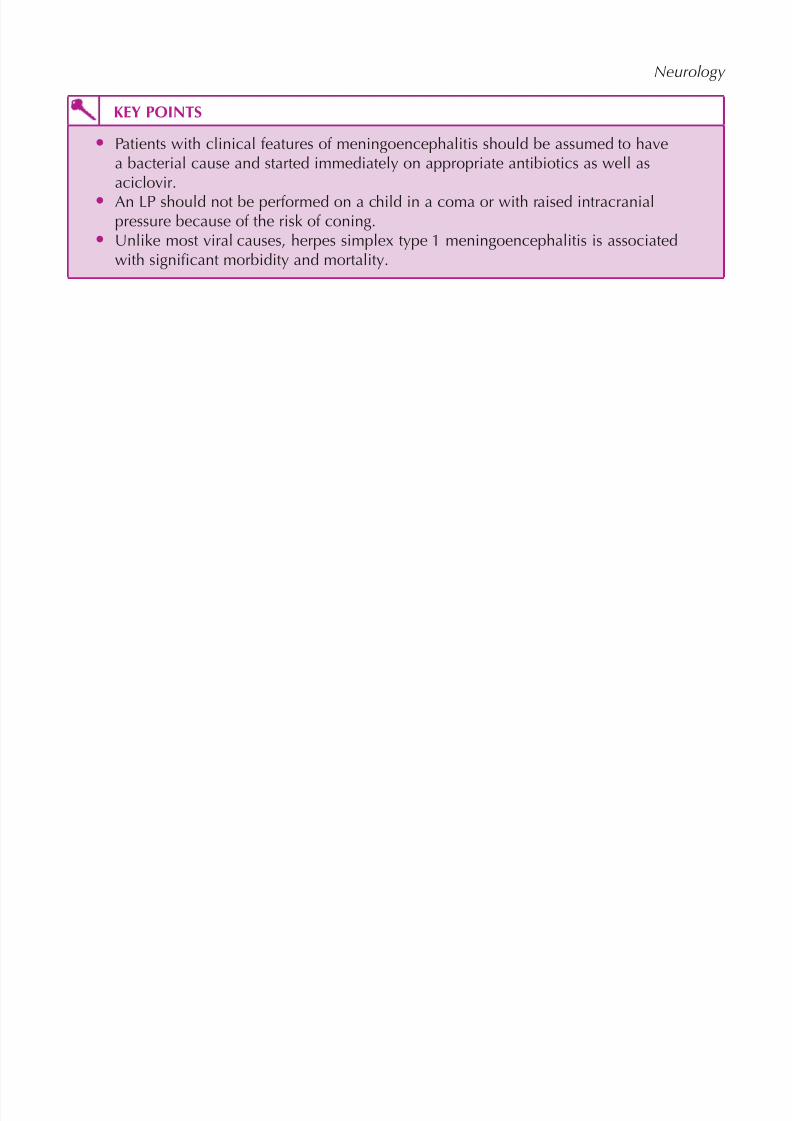
8/20/2019 100 cases in paediatrics.pdf
http://slidepdf.com/reader/full/100-cases-in-paediatricspdf 208/320
189
Neurology
KEY POINTS
• Patients with clinical features of meningoencephalitis should be assumed to havea bacterial cause and started immediately on appropriate antibiotics as well asaciclovir.
• An LP should not be performed on a child in a coma or with raised intracranialpressure because of the risk of coning.
• Unlike most viral causes, herpes simplex type 1 meningoencephalitis is associatedwith significant morbidity and mortality.
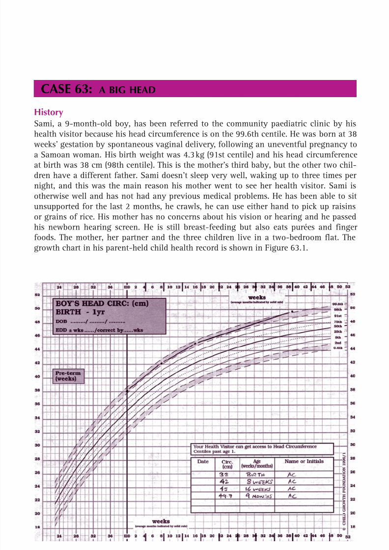
8/20/2019 100 cases in paediatrics.pdf
http://slidepdf.com/reader/full/100-cases-in-paediatricspdf 209/320
190
CASE 63:A BIG HEAD
HistorySami, a 9-month-old boy, has been referred to the community paediatric clinic by hishealth visitor because his head circumference is on the 99.6th centile. He was born at 38weeks’ gestation by spontaneous vaginal delivery, following an uneventful pregnancy toa Samoan woman. His birth weight was 4.3 kg (91st centile) and his head circumferenceat birth was 38 cm (98th centile). This is the mother’s third baby, but the other two chil-dren have a different father. Sami doesn’t sleep very well, waking up to three times pernight, and this was the main reason his mother went to see her health visitor. Sami isotherwise well and has not had any previous medical problems. He has been able to situnsupported for the last 2 months, he crawls, he can use either hand to pick up raisinsor grains of rice. His mother has no concerns about his vision or hearing and he passedhis newborn hearing screen. He is still breast-feeding but also eats purées and fingerfoods. The mother, her partner and the three children live in a two-bedroom flat. Thegrowth chart in his parent-held child health record is shown in Figure 63.1.
Figure 63.1 Sami’s growth chart (reproduced with permission of the Child Growth Foundation).

8/20/2019 100 cases in paediatrics.pdf
http://slidepdf.com/reader/full/100-cases-in-paediatricspdf 210/320
191
ExaminationSami is a well-looking child, who smiles and babbles continuously. He is not dysmor-phic. His weight is 10.5 kg (91st centile), his length is 76.2 cm (98th centile) and his headcircumference is 49.7 cm (99.6th centile). Cardiovascular and respiratory examinations
are normal. His anterior fontanelle is almost closed. Neurological examination revealsnormal tone, power and reflexes in the upper and lower limbs.
Questions• What features in the history help to distinguish between the causes of a big head?• What other features should be sought on examination?• What is the most likely explanation for the large head circumference?
Neurology

8/20/2019 100 cases in paediatrics.pdf
http://slidepdf.com/reader/full/100-cases-in-paediatricspdf 211/320
192
ANSWER 63 A large head may be a normal variant, often with a familial tendency, or may be causedby pathological processes. In an infant, rising intracranial pressure will cause the skullwith unfused sutures to expand rapidly. If possible, it should be determined from pre-
vious measurements whether the head circumference is enlarging and crossing centiles(a worrying feature) or whether it is just growing steadily along the same centile. Theremay be other symptoms of raised intracranial pressure: vomiting, lethargy, irritability,poor feeding. Developmental delay or regression (loss of previously attained develop-mental milestones) suggests metabolic, genetic and syndromic causes. Social concernsabout a family raise the possibility of a subdural haematoma from non-accidental injury.Parents should be asked whether they or the siblings have large heads – do they havedifficulty buying hats to fit them?
The causes of a large head (macrocephaly) are listed below. When performing the exami-nation, these should be borne in mind. The shape of the head, and the size, shape andpatency of the fontanelles and cranial sutures should be assessed. The presence of dys-morphic features, birthmarks or other congenital anomalies should be sought. Centraland peripheral nervous systems should be examined and development assessed. Weightand height should be measured and plotted on a centile chart, as should parental headcircumferences.
! Causes of macrocephalyPossible examination finding
Hydrocephalus Tense fontanelle, distended scalp veinsVascular malformations Cranial bruitTumour Abnormal neurologySubdural haematoma Bruising, other injuriesFragile X syndrome Developmental delay, large earsNeurofibromatosis Café-au-lait patches, axillary frecklesOvergrowth syndrome, Developmental delay, large hands/feet e.g. Soto’s syndromeMetabolic, e.g. mucopolysaccharidoses Developmental delay, coarse featuresFamilial macrocephaly Parent has large head circumference
Sami has normal development and his growth chart shows that he has always been alarge baby with a large head. The change from the 98th centile to 99.6th centile is notconcerning, considering the slow rate of change and the likelihood of small errors inmeasurement. The cause of his large head size is likely to be familial and this was sup-ported by his mother’s head circumference lying on the 98th centile.
KEY POINTS
• The commonest cause of a large head is familial macrocephaly.• Check the growth chart and developmental history for a child with a big head.
100 Cases in Paediatrics

8/20/2019 100 cases in paediatrics.pdf
http://slidepdf.com/reader/full/100-cases-in-paediatricspdf 212/320
This page intentionally left blank

8/20/2019 100 cases in paediatrics.pdf
http://slidepdf.com/reader/full/100-cases-in-paediatricspdf 213/320
194
CASE 64:A CHILD IN A COMA
HistoryKyle is a 7-month-old boy who presents to the A&E department of a district generalhospital in a coma. He has been somewhat lethargic in the last few days, has vomitedseveral times and has gradually got worse. His parents could not rouse him and broughthim straight to A&E. There is no past medical history of note and no diseases run in thefamily.
ExaminationHis airway and breathing are stable. Oxygen saturation is 96 per cent in air. His pulse is176 beats/min, his blood pressure is 82/56 mmHg and the capillary refill is 5 s. The AVPUscore is P. He is apyrexial and has no rash. The anterior fontanelle is bulging with dis-tended veins over the scalp. There are no focal neurological signs and no other signs.
INVESTIGATIONS
Normal
Haemoglobin 9.6 g/dL 10.5–14.0 g/dLWhite cell count 9.2 109 /L 4.0–11.0 109 /LPlatelets 332 109 /L 150–400 109 /LClotting NormalUrea and electrolytes NormalLiver function tests NormalBone NormalGlucose 5.6 mmol/L 3.5–7 mmol/LC-reactive protein 4 mg/L 6 mg/LLactate 3.6 mmol/L 1.1–2.3 mmol/LAmmonia 70 µmol/L 18–74 µmol/LCapillary gas pH 7.35 7.36–7.44
P O2 3.1 kPa (note capillary sample)P CO2 6.2 kPa 4.0–6.5 kPa
Base excess 7.2 ( 2.5)–( 2.5) mmol/LCT scan – see Figure 64.1
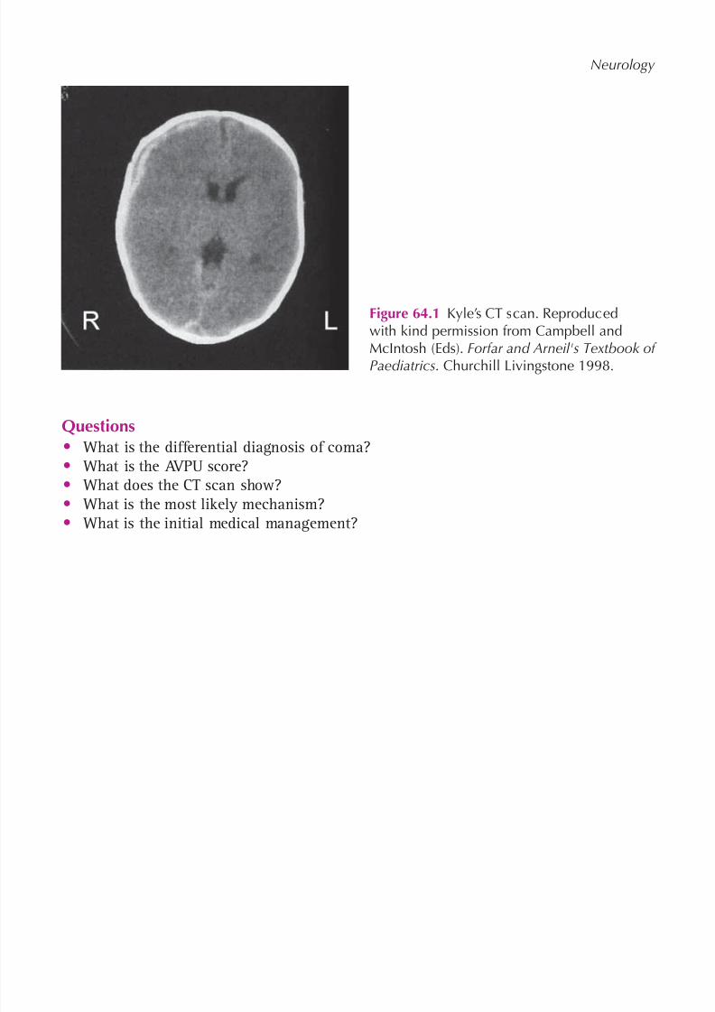
8/20/2019 100 cases in paediatrics.pdf
http://slidepdf.com/reader/full/100-cases-in-paediatricspdf 214/320
195
Neurology
Questions• What is the differential diagnosis of coma?• What is the AVPU score?• What does the CT scan show?• What is the most likely mechanism?
• What is the initial medical management?
Figure 64.1 Kyle’s CT scan. Reproducedwith kind permission from Campbell and
McIntosh (Eds). Forfar and Arneil's Textbook ofPaediatrics . Churchill Livingstone 1998.
8/20/2019 100 cases in paediatrics.pdf
http://slidepdf.com/reader/full/100-cases-in-paediatricspdf 215/320
196
ANSWER 64
! Differential diagnosis of coma in children
• Hypoxic-ischaemic brain injury, e.g. following a respiratory arrest• Epileptic seizure/postictal state• Trauma, e.g. intracranial haemorrhage, cerebral oedema• Infections, e.g. meningitis, encephalitis, abscess• Metabolic – renal and hepatic failure, hypoglycaemia, diabetic ketoacidosis,
inborn errors• Poisoning• Vascular lesions, e.g. stroke
The AVPU score is a score used for the rapid assessment of the conscious level of a child(especially 5 years of age). A A lert, V responds to V oice, P responds only toP ain, U U nresponsive to all stimuli. P or less corresponds to a GCS of 8 or less.
The CT shows an acute subdural haemorrhage over the right hemisphere.
The most likely mechanism is a shaking injury (the ‘shaken baby syndrome’) or possiblya direct impact.
The pulse is raised and the capillary refill time is prolonged. This would fit in with an
intracranial bleed and vomiting (note the haemoglobin is low and the lactate and baseexcess are raised due to poor perfusion). A bolus of 20 mL/kg of 0.9 per cent salineshould therefore be administered. In any child with an AVPU score of P or U, the airwayis at risk and an anaesthetist should therefore be called.
A broad-spectrum antibiotic, e.g. cefotaxime, should be given as soon as possible tocover the possibility of sepsis.
The bulging anterior fontanelle indicates raised intracranial pressure. In infants, unfusedsutures and a patent anterior fontanelle allow the cranial volume to increase. Raised intra-
cranial pressure is therefore better tolerated in its initial stages and large bleeds may occurbefore neurological features develop. Management of raised intracranial pressure includessitting the infant at a 30 º angle, restricting fluids to two-thirds maintenance, and electiveintubation and ventilation to maintain the P CO2 at 4–4.5 kPa (intubation will also secure theairway and help ensure a safe transfer). The child should also be well oxygenated and nor-moglycaemic. An urgent discussion needs to be held with the regional neurosurgical centreto discuss whether the infant should be administered mannitol or hypertonic saline to helpdecrease the intracranial pressure. Arrangements will need to be made for an urgent neuro-surgical assessment to determine whether surgery is required to evacuate the haematoma.
This injury is most likely to have been intentional. Social services should therefore becontacted and child protection procedures followed.
KEY POINTS
• Urgent transfer of acutely ill children with an intracranial bleed to a neurosurgicalunit is mandatory, as a haematoma may need to be surgically evacuated.
• The majority of serious intracranial injuries in the first year of life are the result ofnon-accidental injury.
100 Cases in Paediatrics
8/20/2019 100 cases in paediatrics.pdf
http://slidepdf.com/reader/full/100-cases-in-paediatricspdf 216/320
197
CASE 65:A LATE WALKER
HistoryJustin is an 18-month-old boy referred to the community paediatric clinic because he isnot yet walking. He is the first child of white British parents, born at 41 weeks’ gestationfollowing an uneventful pregnancy. There was meconium staining of the amniotic fluidbut he cried immediately at birth and didn’t need any resuscitation. He has been gener-ally healthy apart from normal coughs and colds, and he had chickenpox 6 months ago.His parents recall that he could roll over at about 4 months, sat up alone at 10 months,crawled from 13 months and was able to ‘bear walk’ on hands and feet and pull himselfto standing from around 17 months. He can take a few steps holding on to furniture butcannot walk independently yet. He is able to scribble with a crayon with either handand can slot pieces into a shape-sorter toy. He uses lots of single words appropriately,understands simple instructions, feeds himself with a spoon and will also feed his teddybear. The parents have no concerns about his vision or hearing. He attends nursery 3 daysper week and is described by the nursery staff as a likeable boy, although they have beenconcerned that he still doesn’t walk. There is no family history of delayed walking. Justin’smother is an accountant and his father is an architect and both are in their early 40 s.They are hoping to have another child soon.
ExaminationJustin looks well and enjoys interacting with you. He is not dysmorphic. His weight is10.8 kg (25th centile), height is 80 cm (25th centile) and head circumference is 49 cm (50thcentile). Cardiovascular, respiratory and abdominal examinations are normal. His spineand joints appear normal, and there are no unusual birthmarks. Neurological examin ationshows normal cranial nerves and normal posture, tone, power and reflexes in the upperand lower limbs. He does not have any tremor. When he is helped to stand, holding on tothe side of a low table, Justin takes a few steps whilst holding on with both hands.
Questions• What comments can you make about Justin’s developmental history?• What further assessment is necessary?• What is the most important investigation to perform?• What are the most common causes of delayed walking?
8/20/2019 100 cases in paediatrics.pdf
http://slidepdf.com/reader/full/100-cases-in-paediatricspdf 217/320
198
ANSWER 65Justin’s gross motor development is delayed. Although he rolled at the normal age,he was slightly late to sit independently and crawl. The average age for walking is 13months (range 9–18 months) and failure to walk by 18 months is a developmental warn-ing sign. The rest of his development seems appropriate for his current age, althoughthere is little detail given. Further assessment of his development in fine motor, speech,language, communication and social domains will reveal whether he has isolated delayin walking or he has other areas of delay too.
The most important investigation is a blood test for creatine kinase (CK) measurement.This is because boys with Duchenne muscular dystrophy (DMD) will have a very elevatedCK and the diagnosis would be strongly suspected if this were found. As Justin’s par-
ents are planning to have another child, they could have appropriate genetic counsellingabout the risk of another child being affected. DMD is an X-linked recessive conditionaffecting about 1:4000 births and it usually results in progressive muscle weakness, lossof ambulation and death in the teens or early 20s. Many children with DMD will alsohave a degree of learning difficulty and may exhibit delay in other areas of development.
Delayed walking is a common problem. Ninety-seven per cent of normal children will bewalking by 18 months of age, which means 3 per cent will not. Most children not walkingby 18 months will be normal, but up to 10 per cent will have an underlying neurological
problem and 10 per cent will have developmental delay in other areas. Less than 1 per centwill turn out to have muscular dystrophy.
! Causes of delayed walking
ExampleGeneralized developmental delay Fragile-X syndrome, Down’s syndromeNeuromuscular cause Cerebral palsy, Duchenne muscular dystrophyMusculoskeletal Congenital dislocation of the hipNormal ‘late walker’ The majority of cases
KEY POINTS
• Failure to walk by 18 months is a developmental warning sign.• Duchenne muscular dystrophy is the most important diagnosis to exclude in boys.
100 Cases in Paediatrics

8/20/2019 100 cases in paediatrics.pdf
http://slidepdf.com/reader/full/100-cases-in-paediatricspdf 218/320
199
CASE 66:A CHILD WITH LEARNING DIFFICULTIES
HistorySimon is 7 years old. He has been brought to the community paediatric clinic at the localchildren’s centre by his mother and grandmother. He was referred for a medical assessmentto help prepare a statement of special educational needs. His notes indicate that he has beenseen previously at the children’s centre by speech and language therapists due to speechdelay, and by the occupational therapist because he was very clumsy. The correspondenceindicates that a lot of appointments were missed and eventually he was lost to follow-up. He is the second child of non-consanguineous parents. He attends the same main-stream school as his 9-year-old brother, who has no problems at school. His father worksas a delivery driver but his mother does not work. Simon was born at term and has long-standing problems with eczema and constipation. He was admitted to hospital once afterscalding himself with boiling water from a kettle. He is reported to be fully immunized,although his Red Book has been lost. His mother has difficulty recalling his developmentalmilestones, but thinks that he walked at about 20 months and spoke his first word at about30 months. He managed to potty-train at about 4.5 years, although he still soils himselfoccasionally. He used to be a contented child who would sit quietly for long periods, buthis behaviour became increasingly aggressive when he started school. His mother thinks his
eyesight is fine and his hearing is fine when he wants to listen. His mother and grandmotherthink that, overall, he is a slow learner, but they aren’t particularly worried about him.
ExaminationSimon appears quite slight for his age, his height is 115 cm (ninth centile) and his weightis 18 kg (second centile). He has quite a long, thin face and large ears but otherwise lookssimilar to his mother. He sits quietly beside his mother through most of the consultation.He speaks very little voluntarily and generally answers questions with a ‘yes’ or a ‘no’.Cardiovascular, respiratory and abdominal examinations are normal. Neurological exam-ination reveals normal tone, power and reflexes in the upper and lower limbs. He hasrather flat feet, and his knees hyperextend when standing still. His gait is normal, but hecannot stand on one leg for more than a few seconds and he seems quite clumsy whenrunning. His eye movements and facial movements appear normal. He will not toleratefundoscopy and is generally uncooperative with further cranial nerve assessment.
Questions• What is a statement of educational needs?• What further assessment is important?
• What causes should be considered for Simon’s problems?

8/20/2019 100 cases in paediatrics.pdf
http://slidepdf.com/reader/full/100-cases-in-paediatricspdf 219/320
200
ANSWER 66If a child’s school cannot provide all of their needs, the local authority may carry outan assessment to find out what the child’s special educational needs (SEN) are and howthey can be supported. The findings are documented in a statement of SEN (usually justcalled a ‘statement’). This describes the child’s SEN and the assistance he or she requires.The process requires assessments by the child’s school, an educational psychologist, adoctor and other relevant professionals.
The medical assessment should identify any features that may indicate specific under-lying diagnoses, look for any treatable contributing factors and describe the child’s cur-rent level of function and medical needs. It is essential to check hearing and vision.
The history suggests Simon may have developmental delay in multiple areas and heprobably has global learning disability. He may have ligamentous laxity, with associateddyspraxia. He has chronic eczema and constipation, which may affect his ability to partici-pate fully at school. His behavioural problems may be secondary to his learning difficul-ties and frustration at school, or may be an independent problem. Fragile X syndrome andDuchenne muscular dystrophy need to be considered as underlying diagnoses, but muchmore assessment is also needed.
! Examples of causes of learning difficulties
Congenital causes• Chromosome disorder• Fragile X syndrome• Duchenne muscular dystrophy• Congenital infection• Fetal alcohol syndrome
Acquired causes• Traumatic brain injury• Meningitis• Psychosocial deprivation
! Examples of contributing factors in learning difficulties
Problem SolutionHearing impairment Hearing aids, specialist teachingVisual impairment Glasses, modification of classroomChronic medical condition Optimize medical careEpilepsy Make diagnosis, anti-epileptic drugsAutistic spectrum disorders Specific teaching strategies
Dyspraxia Occupational therapyNeglect Intervention by social services
100 Cases in Paediatrics
KEY POINTS
• Children with learning difficulties may need a statement of special educational needs.• There may be remediable problems contributing to a child’s overall difficulties,
e.g. deafness.
8/20/2019 100 cases in paediatrics.pdf
http://slidepdf.com/reader/full/100-cases-in-paediatricspdf 220/320
201
CASE 67:SUDDEN WEAKNESS
HistoryLeroy is a 2.5-year-old West Indian boy brought to the A&E department by ambulance.He was in the children’s playground at the park apparently fit and well. He suddenlyfell over whilst running to his mother and when he tried to get up, his right arm andleg were not moving and he was unable to speak. His speech has returned a little butis still far from normal and he is still not using his right side. He recognizes his parentsbut is a little drowsy. He is visibly upset. Nothing like this has ever happened beforeand his development to date has been normal. He is right-handed. Leroy was born atterm following an uneventful pregnancy to healthy, unrelated parents. Apart from anorchidopexy at 13 months, he has no significant past medical history. He is on no regu-lar medication and has no known allergies. He has two older sisters.
ExaminationLeroy is a generally healthy Afro-Caribbean boy. His airway is patent and he is breath-ing without support. His oxygen saturation is 100 per cent in 15 L of oxygen via facemask. His pulse is 92 beats/min and regular with a capillary refill time of 2 s. His bloodpressure is 80/54 mmHg. His temperature is 37 C. Examination of the cardiovascular, res-
piratory and abdominal systems is normal. He is quite sleepy but knows his parents andresponds appropriately to attempts to examine him. He has drooping of the right sideof his mouth but his right eye closes when he cries. He will reach out with his left handto his favourite toy when it is held to his left but ignores it if it is introduced from hisright-hand side until it is almost in the midline. Tone, power, sensation and reflexesare normal on his left side. He has increased tone in his right arm and leg but power isweak. Reflexes are brisk throughout his right side. His plantar is flexor on the left butextensor on the right.
Questions• What is the clinical diagnosis?• List the possible causes• What important and relevant screening test would he have had as a neonate?• What investigation does he need?• What are the key points in the acute and long-term management?
8/20/2019 100 cases in paediatrics.pdf
http://slidepdf.com/reader/full/100-cases-in-paediatricspdf 221/320
202
100 Cases in Paediatrics
ANSWER 67Leroy has an acute right hemiplegia and hemianopia. He has an upper motor neurone
VIIth nerve palsy as evidenced by eye closure despite weakness of the facial muscles.The clinical diagnosis is an acute stroke. Most childhood strokes are due to arterialocclusion (thrombotic or embolic), but venous thrombosis and haemorrhage do occur.The commonest cause of childhood stroke is sickle cell disease (SCD), affecting at least10 per cent of such children. Many children with stroke have another medical condition.
! Causes of acute stroke in childhood
• Haematological abnormalities – SCD, polycythaemia, leukaemia, disorders ofcoagulation
• Cardiac disease – congenital, especially cyanotic and acquired, e.g. endocarditisor Kawasaki disease. Usually embolic
• Infection – meningitis, local head and neck infections or bacteraemia• Intracerebral vascular pathology – ruptured aneurysm, arteriovenous
malformation• Autoimmune disease – systemic lupus erythematosus, juvenile idiopathic
arthritis, sarcoidosis• Metabolic disease – homocystinuria, mitochondrial disorders
• Trauma
Sickle cell disease is now part of the neonatal screening programme. Previously all chil-dren from populations with a high incidence would be checked prior to any procedurethat could precipitate a sickling crisis, such as surgery.
In acute stroke it is crucial to distinguish between ischaemic and other categoriesbecause this alters the investigations and management. A brain MRI should be under-taken as soon as possible, but if it is not available within 48 hours, a CT scan is anacceptable initial alternative. Scanning should be undertaken urgently if there is adepressed level of consciousness at presentation or a deterioration.
Further investigations will depend on the scan findings. For example, all children witharterial ischaemic stroke should have MRI angiography of the cervical and proximalintracranial arterial vasculature and cardiac echocardiography. They should also beinvestigated for an underlying prothrombotic tendency, such as protein C deficiency, as
should those with venous thrombosis. A platelet count and clotting studies are needed inintracerebral haemorrhage.
The acute medical management aims to minimize future disability. He needs regularneurological observations and monitoring of his vital signs with urgent scanning andadmission to the paediatric intensive care unit if there is deterioration. If an arterialischaemic stroke is confirmed and haemorrhage and SCD are excluded, aspirin is started.
Anticoagulation should be considered in specific circumstances, such as cerebral venoussinus thrombosis.

8/20/2019 100 cases in paediatrics.pdf
http://slidepdf.com/reader/full/100-cases-in-paediatricspdf 222/320
203
Neurology
The long-term management of stroke is a prime example of the benefits of a system-atic, multidisciplinary approach to assess and monitor the medical, social, emotional andeducational needs of the child. All children should have an assessment within 72 hoursof admission and the professionals involved should have early liaison with their coun-
terparts in the community to ensure smooth transition of care. A key worker from oneof the teams should coordinate the package of care and act as a central point of contactfor the family.
KEY POINTS
• MRI (or CT) scanning should be performed within 48 hours where there is a clinicaldiagnosis of childhood stroke.
• At least 10 per cent of patients with sickle cell disease will have a stroke during
childhood.• The management of childhood stroke should be multidisciplinary.

8/20/2019 100 cases in paediatrics.pdf
http://slidepdf.com/reader/full/100-cases-in-paediatricspdf 223/320
This page intentionally left blank
8/20/2019 100 cases in paediatrics.pdf
http://slidepdf.com/reader/full/100-cases-in-paediatricspdf 224/320
205
CASE 68:CHRONIC HEADACHES
HistoryTom is a 10-year-old boy referred to outpatients by his GP with a 2-year history ofrecurrent headaches. He describes the pain as being of gradual onset, frontal, right-sidedand throbbing. He has not noticed any visual symptoms. He takes paracetamol but it isof limited value. The headache is often preceded or accompanied by nausea and vomit-ing. His mother has noticed that he becomes very pale and quiet. He takes himself offto bed and sleeps for several hours after which he is fine. The episodes can last up to12 hours. In between episodes Tom is fit and well. He is happy at school and doing wellbut missed 10 days last term due to headaches. He takes part in numerous after-schoolactivities and loves performing arts, especially dance. He was admitted with a minorhead injury at the age of 7 having fallen off his bike. There is no other significant pastmedical history and he takes no regular medication. His mother suffered with migraineas a teenager.
ExaminationTom is a healthy, well-grown boy. His height and weight are both on the 75th centiles.His pulse is 68 beats/min and regular and his blood pressure 96/58 mmHg. Examination
of the cardiovascular, respiratory and abdominal systems is normal. His cranial nervesare intact and fundoscopy is unremarkable. His peripheral nervous system is normal.
Questions• What is the most likely diagnosis?• What is the differential diagnosis?• What investigations would you request?• Is there any treatment that would reduce the frequency of these episodes?
8/20/2019 100 cases in paediatrics.pdf
http://slidepdf.com/reader/full/100-cases-in-paediatricspdf 225/320
206
ANSWER 68These are the characteristic features of common migraine – recurrent headache withsymptom-free intervals, often unilateral, typically throbbing and accompanied by nau-sea and vomiting. Abdominal pain is often a feature. This is the commonest form ofmigraine in childhood and over 90 per cent have a family history. The diagnosis shouldbe reconsidered if this is absent. Classic migraine has aura as an additional feature.
Auras are usually visual and include small areas of visual loss in a visual field (scotoma)and brilliant white zigzag lines (fortification spectra).
Headaches are common in children and their frequency increases with age. In youngchildren, certainly pre-school, an organic cause is highly likely.
! Differential diagnosis of chronic headaches• Tension headaches – commonest of all, described as a ‘band’ or 'pressure’ and
bilateral. Usually otherwise asymptomatic. May last weeks and tend to worsenas day progresses. Child is otherwise healthy
• Sinusitis – percussing over affected sinuses causes pain and discomfort• Refractive errors – ensure vision has been checked recently• Raised intracranial pressure – a brain tumour is most parents’ underlying worry.
Unlike migraine and tension headaches, pain is worse when lying down. Itis almost always accompanied by other abnormal symptoms, e.g. change inbehaviour, or neurological signs, e.g. papilloedema
• Solvent or drug abuse• Hypertension – blood pressure must be checked
A thorough history and examination are the mainstays in making a diagnosis in a child ofthis age and he probably needs no investigations. Obviously if there are focal neurologicalsymptoms or signs (apart from visual aura), an MRI should be performed. Most childrencan lie still enough for this from about 7–8 years. Otherwise, sedation or a general anaes-thetic is needed. The younger the child, the lower the threshold for scanning because anorganic cause is more likely and they are poor at communicating the characteristics oftheir headache. His previous head injury is irrelevant and not an indication to scan.
Prevention of migraine should focus on lifestyle changes. Reassurance that there isno serious intracranial pathology is often enough to improve symptoms. Keeping adiary is a simple way to try to identify any trigger factors, including, rarely, a spe-cific food. Blanket exclusion diets should be avoided. Stress, tiredness and anxiety arethe commonest precipitants. Problems at school, including bullying, should be explored.Relaxation and biofeedback techniques may help. Finally, if all else fails, school attend-ance is affected and the migraines are unacceptably intrusive, prevention with drugs
may be considered. There are very few well-designed clinical trials, but drugs such aspropranolol (contraindicated in asthma), pizotifen and some of the newer anticonvul-sants (gabapentin or topiramate) may be effective. All have side-effects.
KEY POINTS
• Headaches in young children are more likely to be organic in origin and there is alower threshold for scanning.
• Lifestyle changes, not drugs, should be the focus for the prevention of migraine.
100 Cases in Paediatrics
8/20/2019 100 cases in paediatrics.pdf
http://slidepdf.com/reader/full/100-cases-in-paediatricspdf 226/320
207
CASE 69:A FUNNY SMILE
HistoryOliver is a 7-year-old boy who presents to a paediatric rapid referral clinic with ‘a lop-sided face’. His mother noticed that he was drooling from the left side of his mouth for2 days and his left eyelid has been droopy. When he smiles, his face becomes very asym-metric. He is otherwise well but the drooling has made eating and drinking difficult anda little embarrassing. His left eye also feels dry. The rest of the family are well but younotice that his mother is recovering from a cold sore. There is no history of travel or ofany bites. He had an appendicectomy last year and had pyloric stenosis operated on ininfancy.
ExaminationThe child’s mouth droops on the left side. His smile, in particular, is asymmetric. It iswell delineated with the normal facial creases on the right side of the face, but on theleft side it is poorly formed and weak. He is also unable to shut his left eye. There areno other cranial nerve abnormalities and the neurological system is otherwise normal.Otoscopy is normal and his blood pressure is 107/67 mmHg. There are no other signs.
Questions• What is the likely diagnosis?• What is the treatment?• What is the prognosis?

8/20/2019 100 cases in paediatrics.pdf
http://slidepdf.com/reader/full/100-cases-in-paediatricspdf 227/320
208
ANSWER 69The most likely diagnosis is a left-sided Bell’s palsy. This is a lower motor neurone palsyof the facial nerve. In lower motor neurone palsies, the whole side of the face is weak.Taste on the anterior two-thirds of the tongue on the involved side is lost in about halfof cases and there may be facial numbness. In upper motor neurone facial nerve palsies,there is preserved forehead power and eye closure. The facial palsy is said to occur dueto oedema of the facial nerve as it crosses the facial canal in the temporal bone. It mayoccur 2 weeks after a viral infection. Herpes simplex and varicella zoster are said to becausative. Epstein–Barr virus, Lyme disease and mumps have also been implicated.
In chronic cases, other causes, such as otitis media, hypertension, tumours (facial nerveor brain stem tumours), leukaemia and trauma, should also be considered.
If the facial palsy is thought to be due to an upper motor neurone facial palsy then cra-nial imaging with a magnetic resonance imaging (MRI) scan should be undertaken.
If the child presents within 1 week then steroid treatment with prednisolone 1 mg/kgonce daily (up to 60 mg/day) for 7 days is effective. Treatment with other agents such asaciclovir is controversial. Some paediatricians would only administer it if there is a clearhistory of contact with herpes, as in our case, or varicella. Eye care is important. As itmay not be possible for the child to close his eye, treatment with artificial tears such ashypromellose eye drops and taping of the eye at night may be necessary to prevent anexposure keratitis.
Most patients recover within a few weeks. Eighty-five per cent make a complete recov-ery, 10 per cent are left with mild facial weakness and 5 per cent are left with permanentsignificant facial weakness. Recovery may take up to 6 months.
100 Cases in Paediatrics
KEY POINTS
• A lower motor neurone facial palsy affects the forehead and eye as well as the lower
part of the face.• The commonest cause is a Bell’s palsy.• Treatment with steroids within 1 week of diagnosis is beneficial.
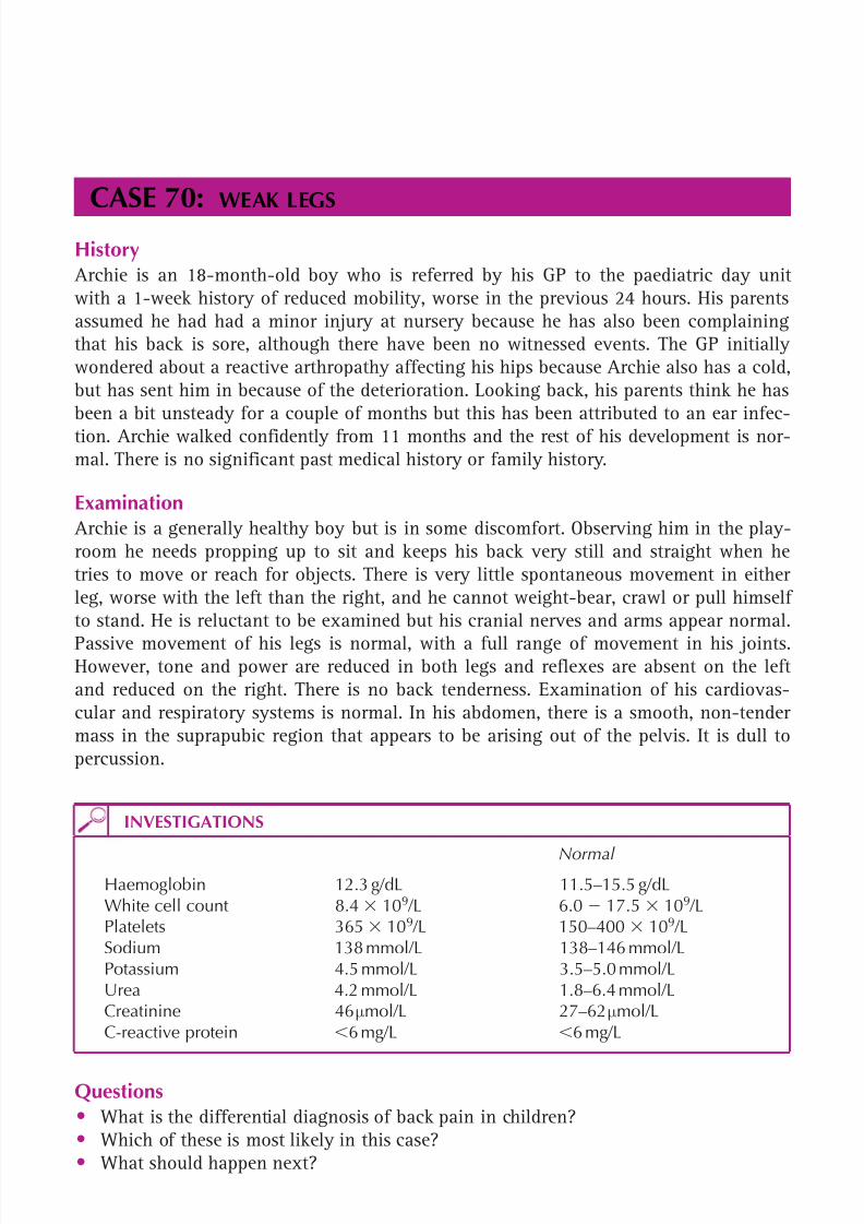
8/20/2019 100 cases in paediatrics.pdf
http://slidepdf.com/reader/full/100-cases-in-paediatricspdf 228/320
209
CASE 70:WEAK LEGS
History Archie is an 18-month-old boy who is referred by his GP to the paediatric day unitwith a 1-week history of reduced mobility, worse in the previous 24 hours. His parentsassumed he had had a minor injury at nursery because he has also been complainingthat his back is sore, although there have been no witnessed events. The GP initiallywondered about a reactive arthropathy affecting his hips because Archie also has a cold,but has sent him in because of the deterioration. Looking back, his parents think he hasbeen a bit unsteady for a couple of months but this has been attributed to an ear infec-tion. Archie walked confidently from 11 months and the rest of his development is nor-mal. There is no significant past medical history or family history.
Examination Archie is a generally healthy boy but is in some discomfort. Observing him in the play-room he needs propping up to sit and keeps his back very still and straight when hetries to move or reach for objects. There is very little spontaneous movement in eitherleg, worse with the left than the right, and he cannot weight-bear, crawl or pull himselfto stand. He is reluctant to be examined but his cranial nerves and arms appear normal.
Passive movement of his legs is normal, with a full range of movement in his joints.However, tone and power are reduced in both legs and reflexes are absent on the leftand reduced on the right. There is no back tenderness. Examination of his cardiovas-cular and respiratory systems is normal. In his abdomen, there is a smooth, non-tendermass in the suprapubic region that appears to be arising out of the pelvis. It is dull topercussion.
INVESTIGATIONS
Normal Haemoglobin 12.3 g/dL 11.5–15.5 g/dLWhite cell count 8.4 109 /L 6.0 17.5 109 /LPlatelets 365 10 9 /L 150–400 109 /LSodium 138 mmol/L 138–146 mmol/LPotassium 4.5 mmol/L 3.5–5.0 mmol/LUrea 4.2 mmol/L 1.8–6.4 mmol/LCreatinine 46 µmol/L 27–62 µmol/LC-reactive protein 6 mg/L 6 mg/L
Questions• What is the differential diagnosis of back pain in children?• Which of these is most likely in this case?• What should happen next?
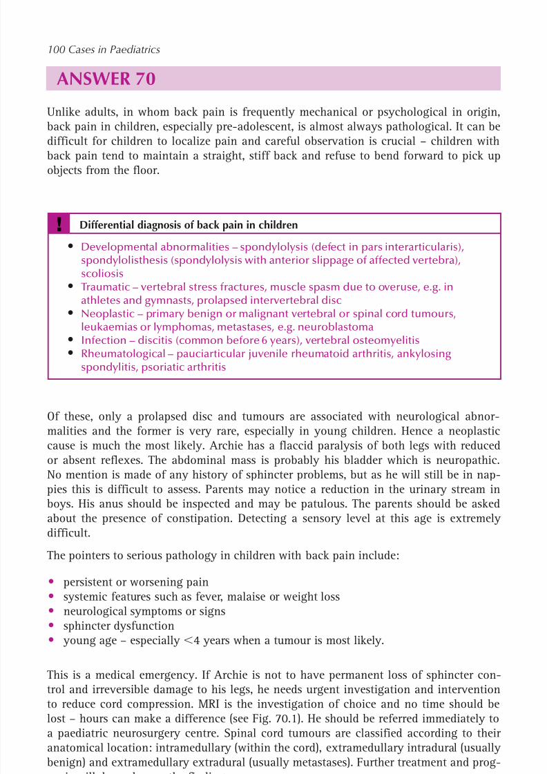
8/20/2019 100 cases in paediatrics.pdf
http://slidepdf.com/reader/full/100-cases-in-paediatricspdf 229/320
210
100 Cases in Paediatrics
ANSWER 70Unlike adults, in whom back pain is frequently mechanical or psychological in origin,back pain in children, especially pre-adolescent, is almost always pathological. It can bedifficult for children to localize pain and careful observation is crucial – children withback pain tend to maintain a straight, stiff back and refuse to bend forward to pick upobjects from the floor.
! Differential diagnosis of back pain in children
• Developmental abnormalities – spondylolysis (defect in pars interarticularis),spondylolisthesis (spondylolysis with anterior slippage of affected vertebra),scoliosis
• Traumatic – vertebral stress fractures, muscle spasm due to overuse, e.g. inathletes and gymnasts, prolapsed intervertebral disc
• Neoplastic – primary benign or malignant vertebral or spinal cord tumours,leukaemias or lymphomas, metastases, e.g. neuroblastoma
• Infection – discitis (common before 6 years), vertebral osteomyelitis• Rheumatological – pauciarticular juvenile rheumatoid arthritis, ankylosing
spondylitis, psoriatic arthritis
Of these, only a prolapsed disc and tumours are associated with neurological abnor-malities and the former is very rare, especially in young children. Hence a neoplasticcause is much the most likely. Archie has a flaccid paralysis of both legs with reducedor absent reflexes. The abdominal mass is probably his bladder which is neuropathic.No mention is made of any history of sphincter problems, but as he will still be in nap-pies this is difficult to assess. Parents may notice a reduction in the urinary stream inboys. His anus should be inspected and may be patulous. The parents should be askedabout the presence of constipation. Detecting a sensory level at this age is extremely difficult.
The pointers to serious pathology in children with back pain include:
• persistent or worsening pain• systemic features such as fever, malaise or weight loss• neurological symptoms or signs• sphincter dysfunction• young age – especially 4 years when a tumour is most likely.
This is a medical emergency. If Archie is not to have permanent loss of sphincter con-trol and irreversible damage to his legs, he needs urgent investigation and interventionto reduce cord compression. MRI is the investigation of choice and no time should belost – hours can make a difference (see Fig. 70.1). He should be referred immediately toa paediatric neurosurgery centre. Spinal cord tumours are classified according to theiranatomical location: intramedullary (within the cord), extramedullary intradural (usuallybenign) and extramedullary extradural (usually metastases). Further treatment and prog-nosis will depend upon the findings.
This boy was found to have a diffuse intramedullary spinal tumour from T7 to T12.
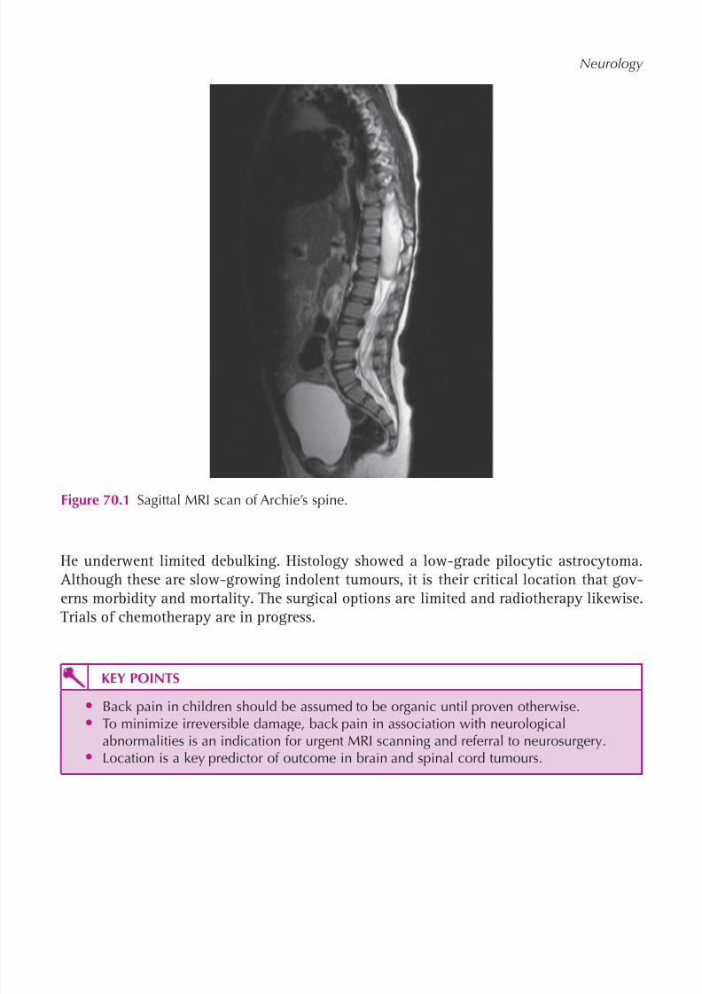
8/20/2019 100 cases in paediatrics.pdf
http://slidepdf.com/reader/full/100-cases-in-paediatricspdf 230/320
211
Neurology
He underwent limited debulking. Histology showed a low-grade pilocytic astrocytoma. Although these are slow-growing indolent tumours, it is their critical location that gov-erns morbidity and mortality. The surgical options are limited and radiotherapy likewise.Trials of chemotherapy are in progress.
Figure 70.1 Sagittal MRI scan of Archie’s spine.
KEY POINTS
• Back pain in children should be assumed to be organic until proven otherwise.• To minimize irreversible damage, back pain in association with neurological
abnormalities is an indication for urgent MRI scanning and referral to neurosurgery.• Location is a key predictor of outcome in brain and spinal cord tumours.

8/20/2019 100 cases in paediatrics.pdf
http://slidepdf.com/reader/full/100-cases-in-paediatricspdf 231/320
This page intentionally left blank
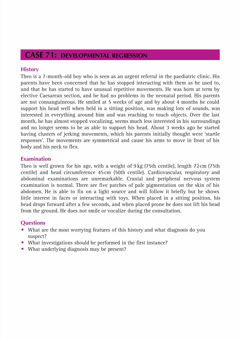
8/20/2019 100 cases in paediatrics.pdf
http://slidepdf.com/reader/full/100-cases-in-paediatricspdf 232/320
213
CASE 71:DEVELOPMENTAL REGRESSION
HistoryTheo is a 7-month-old boy who is seen as an urgent referral in the paediatric clinic. Hisparents have been concerned that he has stopped interacting with them as he used to,and that he has started to have unusual repetitive movements. He was born at term byelective Caesarean section, and he had no problems in the neonatal period. His parentsare not consanguineous. He smiled at 5 weeks of age and by about 4 months he couldsupport his head well when held in a sitting position, was making lots of sounds, wasinterested in everything around him and was reaching to touch objects. Over the lastmonth, he has almost stopped vocalizing, seems much less interested in his surroundingsand no longer seems to be as able to support his head. About 3 weeks ago he startedhaving clusters of jerking movements, which his parents initially thought were ‘startleresponses’. The movements are symmetrical and cause his arms to move in front of hisbody and his neck to flex.
ExaminationTheo is well grown for his age, with a weight of 9 kg (75th centile), length 72 cm (75thcentile) and head circumference 45 cm (50th centile). Cardiovascular, respiratory and
abdominal examinations are unremarkable. Cranial and peripheral nervous systemexamination is normal. There are five patches of pale pigmentation on the skin of hisabdomen. He is able to fix on a light source and will follow it briefly but he showslittle interest in faces or interacting with toys. When placed in a sitting position, hishead drops forward after a few seconds, and when placed prone he does not lift his headfrom the ground. He does not smile or vocalize during the consultation.
Questions• What are the most worrying features of this history and what diagnosis do you
suspect?• What investigations should be performed in the first instance?• What underlying diagnosis may be present?
8/20/2019 100 cases in paediatrics.pdf
http://slidepdf.com/reader/full/100-cases-in-paediatricspdf 233/320
214
ANSWER 71It is clear from the history that there has been developmental regression – loss of pre-
viously attained developmental milestones. The abnormal movements are suggestiveof infantile spasms, a seizure type causing contraction of axial muscles, resulting in aspectrum of abnormal movements from a small nod of the head to a full ‘jack-knifespasm’. This combination makes it likely that this child has West syndrome, an epilepsysyndrome characterized by infantile spasms, developmental regression and hypsarrhyth-mia (a chaotic pattern) on EEG. West syndrome starts in the first year of life and canhave a variety of underlying causes, including structural brain abnormalities, neuromet-abolic disease, neurocutaneous syndromes and acquired brain injury. The seizures aredifficult to control with medication, and the developmental outlook is poor. This is notnecessarily the case with all causes of developmental regression, and it is very important
to try to identify any treatable underlying disease, as this may dramatically change theneurodevelopmental outlook.
! Causes of developmental regression
ExamplesAcquired brain injury Hypoxia, meningitisSeizure disorders West syndrome, Landau–Kleffner
syndromeNeurometabolic/neurodegenerative Mitochondrial disorders, Tay–Sachs diseaseToxic substances Heavy metal poisoning, e.g. leadInfectious Subacute sclerosing panencephalitis, prion diseasePervasive developmental disorder Autism, childhood disintegrative
disorder, Rett’s syndrome
In the first instance, this child will require an EEG to look for hypsarrhythmia and anMRI scan to look for structural brain abnormalities. Blood tests should include a fullblood count, renal and liver function tests, calcium, phosphate, glucose, lactate, ammoniaand amino acids. Urine should be sent for urine amino and organic acids. Examinationwith a Wood’s (ultraviolet) light will help to determine if there are hypopigmented areasof skin, which may be difficult to see in normal light and could indicate an underlyingneurocutaneous syndrome. Further investigations should be directed by a paediatricianwith experience in neurology and epilepsy.
The presence of depigmented patches on the skin (possibly ash leaf macules) raises thepossibility of tuberous sclerosis. Seizures are a common presenting feature and indi-cate the presence of hamartomas in the central nervous system. This neurocutaneoussyndrome is autosomal dominantly inherited, although many cases are new mutations.Clinical features are due to hamartomas which occur in multiple organs, including brain,skin, eyes, kidneys and heart. Tuberous sclerosis arises from defects in either of the twogenes encoding the interacting proteins (tuberin on chromosome 16, and hamartin onchromosome 9) involved in the regulation of cellular growth and differentiation. The
100 Cases in Paediatrics
8/20/2019 100 cases in paediatrics.pdf
http://slidepdf.com/reader/full/100-cases-in-paediatricspdf 234/320
215
KEY POINTS
• Developmental regression requires prompt and thorough evaluation by a specialist.• Infantile spasms are usually associated with diseases with a poor prognosis.• Tuberous sclerosis is one of the commonest causes of developmental regression and
infantile spasms.
other two most common neurocutaneous syndromes are neurofibromatosis and Sturge– Weber syndrome.
Neurology

8/20/2019 100 cases in paediatrics.pdf
http://slidepdf.com/reader/full/100-cases-in-paediatricspdf 235/320
This page intentionally left blank

8/20/2019 100 cases in paediatrics.pdf
http://slidepdf.com/reader/full/100-cases-in-paediatricspdf 236/320
217
CASE 72:A DEAF BOY
HistoryRonan is a 3-year-old boy with Down’s syndrome. He is brought to his GP because hisparents are worried about his hearing. He does not seem to notice when his parents callhis name if he is not looking directly at them, his speech has become less clear and hisnursery teachers are concerned that his behaviour has become more difficult to manage.He has also started to snore when asleep.
Ronan was born at 36 weeks’ gestation. He required admission to the neonatal unit for
poor feeding and also needed a partial volume dilutional exchange transfusion for poly-cythaemia with hypoglycaemia and seizures. He had a normal newborn hearing screen.He has a small ventricular septal defect and he had bacterial endocarditis 6 monthsago, necessitating a long course of intravenous antibiotics. His mother is a 30-year-oldteacher and his father is a 43-year-old manager of a computer company. They are bothwell. Ronan has been fully immunized, including an influenza vaccine last winter.
ExaminationRonan’s weight and length are on the 25th centile on the Down’s syndrome growthchart. He smiles at you when you approach and seems to follow visual cues and beaffectionate with his parents. His speech is difficult to understand. Cardiovascular exam-ination reveals a heart rate of 110 beats/min, a grade 3 pansystolic murmur, loudest atthe lower left sternal edge, and no signs of cardiac failure. His respiratory and abdom-inal examinations are unremarkable. Both tympanic membranes have a dull retractedappearance with loss of the light reflex and some bubbles visible behind the membrane.He has large tonsils that almost touch in the middle.
Questions• What is the most likely cause of his hearing difficulties?• What other possibilities are there?• How could his hearing be assessed?

8/20/2019 100 cases in paediatrics.pdf
http://slidepdf.com/reader/full/100-cases-in-paediatricspdf 237/320
218
ANSWER 72Ronan is most likely to have conductive hearing loss secondary to chronic otitis mediawith an effusion (OME, or ‘glue ear’). This diagnosis is consistent with the fact that hishearing has recently deteriorated, that he snores (indicating partial upper airway obstruc-tion in sleep) and the findings on examination of his ears. Children with Down’s syndromeare particularly vulnerable to OME because their large adenoids, small nasopharynx andnarrow Eustachian tubes make aeration of the middle ear less efficient. For these reasons,children with Down’s syndrome should have regular screening for hearing impairment.
In addition to conductive deafness, sensorineural deafness is more common in childrenwith Down’s syndrome. The normal newborn hearing screen (otoacoustic emissions) makescongenital sensorineural deafness unlikely. Sensorineural hearing loss can be acquired, and
a potential risk factor is the use of aminoglycoside antibiotics, e.g. gentamicin, which mayhave been used in the treatment of his bacterial endocarditis. Aminoglycosides cause dose-related ototoxicity and some individuals also have an underlying genetic susceptibility.
Another possibility is that Ronan has autism. This is more common in children with Down’ssyndrome and can present with abnormal speech, language and behaviour, which may ini-tially be attributed to hearing difficulties.
! Features of conductive and sensorineural hearing loss
Conductive Sensorineural
Causes Glue ear Genetic Congenital ear Infections: congenital / postnatal abnormalities Birth asphyxia Head injurySeverity Milder; intermittent More severe; static or progressive 20–60 dB loss 20–120 dB lossManagement Conservative Hearing aid Grommets Cochlear implant
Ronan should be referred urgently to a paediatric audiology clinic, so that there is minimaldelay in detection of any significant hearing impairment, and to minimize further delay ofhis speech and language development. Ideally, he would already have regular follow-up insuch a clinic. His hearing will be assessed using techniques appropriate to his developmentalage. These may include visual reinforcement audiometry (where a visual signal reinforces acorrect head-turning response to localize a sound), free field conditioning (where an audi-tory stimulus is the cue for an action in a simple game), and speech discrimination tests(where toys are named at a set volume and identified by the child). He would also havetympanometry performed to confirm negative middle ear pressures typical of OME. It wouldalso be important to ask if Ronan has sleep apnoea, which is associated with severe upperairway obstruction and which may necessitate an adenotonsillectomy.
100 Cases in Paediatrics
KEY POINTS
• It is important to recognize and refer hearing problems at an early stage.• Conductive hearing loss due to glue ear is the most common cause of hearing
impairment.• Children with Down’s syndrome have an increased risk of hearing problems and
require surveillance.

8/20/2019 100 cases in paediatrics.pdf
http://slidepdf.com/reader/full/100-cases-in-paediatricspdf 238/320
219
CASE 73:A CHILD WITH A SQUINT
HistoryRachel is a 9-month-old girl who is brought to her GP practice by her mother. Over thelast month her parents have noticed that her eyes don’t always seem to be looking inthe same direction. She was born at 35 weeks’ gestation after an emergency Caesareansection for suspected placental abruption, but was in good condition at birth. There wereno medical problems after delivery. She had chickenpox 1 month ago, but has neverreally been to the surgery otherwise, except for her immunizations. Her mother reportsthat she his happy with her development, as Rachel is doing everything at about thesame age that her 3-year-old brother did.
ExaminationRachel is a healthy-looking child. She has an obvious right convergent squint. Figure 73.1illustrates the appearance of her eyes as she looks around.
Questions• What is a convergent squint?• What are the possible causes of a squint in a child this age?• What simple assessment can be performed?• What should be done next?
Looking left
Looking right
Looking up
Looking down
Looking straightahead
Figure 73.1 Appearance of Rachel’s eyes as shelooks around.
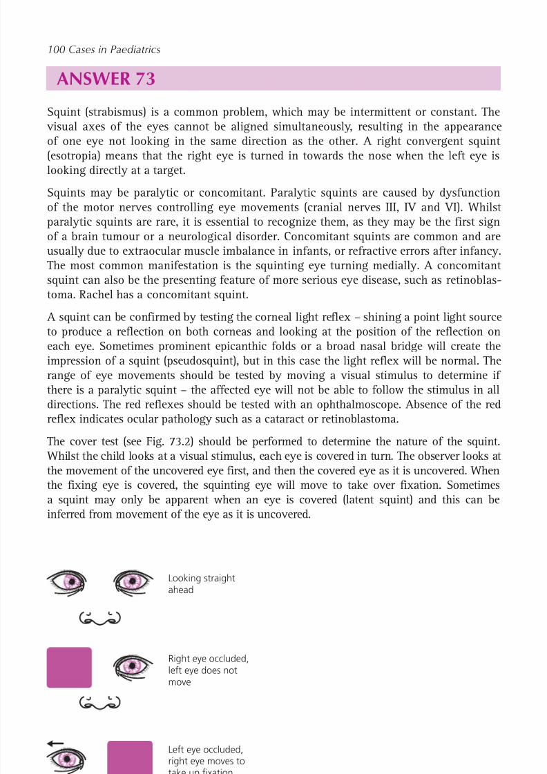
8/20/2019 100 cases in paediatrics.pdf
http://slidepdf.com/reader/full/100-cases-in-paediatricspdf 239/320
220
ANSWER 73Squint (strabismus) is a common problem, which may be intermittent or constant. The
visual axes of the eyes cannot be aligned simultaneously, resulting in the appearanceof one eye not looking in the same direction as the other. A right convergent squint(esotropia) means that the right eye is turned in towards the nose when the left eye islooking directly at a target.
Squints may be paralytic or concomitant. Paralytic squints are caused by dysfunctionof the motor nerves controlling eye movements (cranial nerves III, IV and VI). Whilstparalytic squints are rare, it is essential to recognize them, as they may be the first signof a brain tumour or a neurological disorder. Concomitant squints are common and areusually due to extraocular muscle imbalance in infants, or refractive errors after infancy.
The most common manifestation is the squinting eye turning medially. A concomitantsquint can also be the presenting feature of more serious eye disease, such as retinoblas-toma. Rachel has a concomitant squint.
A squint can be confirmed by testing the corneal light reflex – shining a point light sourceto produce a reflection on both corneas and looking at the position of the reflection oneach eye. Sometimes prominent epicanthic folds or a broad nasal bridge will create theimpression of a squint (pseudosquint), but in this case the light reflex will be normal. Therange of eye movements should be tested by moving a visual stimulus to determine ifthere is a paralytic squint – the affected eye will not be able to follow the stimulus in all
directions. The red reflexes should be tested with an ophthalmoscope. Absence of the redreflex indicates ocular pathology such as a cataract or retinoblastoma.
The cover test (see Fig. 73.2) should be performed to determine the nature of the squint. Whilst the child looks at a visual stimulus, each eye is covered in turn. The observer looks atthe movement of the uncovered eye first, and then the covered eye as it is uncovered. Whenthe fixing eye is covered, the squinting eye will move to take over fixation. Sometimesa squint may only be apparent when an eye is covered (latent squint) and this can beinferred from movement of the eye as it is uncovered.
Left eye occluded,right eye moves totake up fixation
Right eye occluded,left eye does notmove
Looking straightahead
Figure 73.2 Cover test.
100 Cases in Paediatrics
8/20/2019 100 cases in paediatrics.pdf
http://slidepdf.com/reader/full/100-cases-in-paediatricspdf 240/320
221
Mild and intermittent squint may be present in the neonatal period and disappear with-out treatment. Any child over 3 months of age with a squint, or parental concern abouta squint, should be referred to an orthoptist or ophthalmologist for further assessment.The aim is to detect any serious underlying pathology and to prevent the development
of amblyopia. In the developing brain, the image from the squinting eye is suppressedto avoid diplopia. In an untreated squint, this leads to irreversible suppression of the visual pathways, visual impairment in that eye and possible blindness. This phenomenonis known as amblyopia. Treatments include correction of refractive errors with glasses,patching of the good eye to force use of the other eye, and surgery on the extraocularmuscles.
KEY POINTS
• Squint is a common childhood problem.• It is essential to determine if it is a paralytic or non-paralytic squint.• Referral to a specialist is important to prevent development of amblyopia.
Neurology

8/20/2019 100 cases in paediatrics.pdf
http://slidepdf.com/reader/full/100-cases-in-paediatricspdf 241/320
222
CHILD AND ADOLESCENT PSYCHIATRY
CASE 74: A BOY WITH NO FRIENDS
History Adrian is a 10-year-old boy with difficult behaviour at home, who comes for areview to the community paediatric clinic. He was thought to have attention deficithyperactivity disorder (ADHD) after an initial consultation in the clinic. However,information obtained from his school indicated that his teacher did not find his behav-iour to be hyperactive or inattentive. His mother had mentioned that he didn’t seem tohave any friends to play with and that this seemed to be the reason he was always caus-ing trouble at home. It is decided that it might be helpful to observe his behaviour in
school.
You observe Adrian at school over a 2-hour period. He does not know that youare specifically watching him. Initially he has a games lesson: the children arepractising football skills. Adrian seems interested, but whenever the task involves acomplex instruction, he doesn’t seems to be able to follow it. When this happens, hestands still or does his own thing. He doesn’t ask anyone else to explain what he issupposed to do. He doesn’t join in with the playground banter between the otherchildren and the teacher. At the end of the lesson, there is a short game of footballand he is the last to be picked for a team. He seems to find it hard to anticipate theflow of the game and runs around almost aimlessly. When he does get the ball he runsstraight down the pitch with it and ends up colliding with another child who is trying totackle him.
Next he is observed in the classroom, where he is doing mathematics. Again he par-ticipates very little in the classroom discussion and he doesn’t really speak to the otherchildren. He sits quietly, fidgets and slouches frequently, but doesn’t leave his seat. Heplaces his pens and ruler in a neat row and gets quite upset when another child borrowshis ruler and disrupts the pattern. When he is asked a question, he mumbles only half of
the answer. His book shows that he has worked out most of the simple sums correctlybut has struggled with the questions that required more reasoning.
His teacher reports that the staff find Adrian quite bewildering. Frequently he justdoesn’t understand the task he is asked to do and he often does his own thing, butsometimes he is able produce complex pieces of work. Even with one-to-one teachingit has been impossible to determine his true academic potential, and sometimes he gets
very upset when teachers try to give him extra guidance. His social interaction is alwaysodd, he doesn’t have any real friends, and although the other children will tolerate him
joining in their games, this frequently results in trouble. Sometimes he will just play onhis own, e.g. opening and closing the lock on the playground gate. He won’t participatein discussions or drama and when he does speak in front of the class he may speakwith a strange intonation or say things that are not appropriate to the context. He gets
very anxious about apparently minor things such as changes in the day’s timetable. Last

8/20/2019 100 cases in paediatrics.pdf
http://slidepdf.com/reader/full/100-cases-in-paediatricspdf 242/320
223
Child and adolescent psychiatry
week he got upset several times each day because the classroom clock had not been setto the right time.
Questions
• What underlying causes may explain Adrian’s problems?• What additional assessment would be helpful?• How might making a diagnosis help Adrian to achieve his full potential at school?

8/20/2019 100 cases in paediatrics.pdf
http://slidepdf.com/reader/full/100-cases-in-paediatricspdf 243/320
224
100 Cases in Paediatrics
ANSWER 74 Adrian displays evidence of difficulties with social communication, social interaction andobsessional behaviour. He may have an autistic spectrum disorder, possibly Asperger’ssyndrome. This diagnosis cannot be made with certainty from the brief history, andfurther assessment would be needed. Other underlying or contributing problems mightinclude hearing difficulties, specific learning difficulties, social and emotional problems.
Autism is characterized by the triad of impairment of social communication, social inter-action and rigidity of thought and behaviour. Unusual routines and interests, abnormalsensory sensitivity and learning or other developmental difficulties may coexist. Theterm autistic spectrum disorder is now widely accepted and recognizes that there can beconsiderable variation in the severity of symptoms.
Autistic disorder is the most severe end of the spectrum and is usually diagnosed in earlychildhood. Affected children are often non-verbal, with cognitive impairment and highlystereotyped behaviours. At the milder end of the spectrum there may be relatively littleimpairment of communication, leading to a diagnosis being made later in life, if at all.
Asperger’s syndrome is at the mild end of the autistic spectrum and individuals are usu-ally of average or above-average intelligence, have difficulties understanding non-verbalcommunication cues, find it difficult to form friendships and difficult to empathize withothers. Autistic spectrum disorders may affect up to 1 per cent of children in the UK.
Additional assessment should include a hearing test and assessment by a paediatricianwith an interest in autistic spectrum disorders (usually a community paediatrician) or achild and adolescent mental health team. Often a clinical psychologist or a multidisci-plinary team of other professionals will be involved in the assessment. Detailed devel-opmental, medical, family and social histories should be taken. Adrian’s social skills,communication and behaviour will be observed. Sometimes this is done in a standard-ized way using an observational assessment. The process needs the support of the fam-ily, who should understand that the eventual outcome might be a diagnosis of autisticspectrum disorder.
Making a diagnosis of autistic spectrum disorder can be very emotive. It can bring greatrelief to parents and the child, but can also be met with denial or dismay. Understandingthe diagnosis will enable the teachers at Adrian’s school to understand why he behavesin the way he does. It will allow the school to implement strategies to help Adrian to beincluded as fully as possible in the curriculum. This will include individualized measuresto support social communication and behavioural interventions to assist the develop-ment of adaptive skills. The special educational needs coordinator at the school will helpto develop an individual education plan to help staff to know what Adrian’s needs areand how they can best be met.
KEY POINTS
• Autism is characterized by impairments of social communication, social interactionand rigidity of thought and behaviour.
• Autistic spectrum disorders may remain undiagnosed in late childhood andadolescence.
• Making the diagnosis may help to find strategies to improve learning and behaviour.
8/20/2019 100 cases in paediatrics.pdf
http://slidepdf.com/reader/full/100-cases-in-paediatricspdf 244/320
225
CASE 75:A HYPERACTIVE CHILD
HistoryRichard is a 6-year-old boy who is brought to the community paediatric clinic by hismother. She asked her GP to refer Richard because she thinks he is hyperactive. Shedescribes him as ‘always on the go’, unable to sit still or concentrate on anything, andnever seeming to think before he acts. He is particularly difficult at home in the even-ings and rarely goes to bed before midnight. His behaviour has been getting worse overthe last year. His mother says that she just can’t cope with his behaviour any more andwould like him to be put on some medicine to calm him down.
Richard was born at 35 weeks’ gestation, and his only medical problem has beeneczema. This has been difficult to control and he is under follow-up by a paediatricdermatologist. He currently uses a combination of emollients and a topical steroid. Hehas two teenage half-brothers and a 1-year-old half-sister. His mother is separated fromher partner and now looks after the children alone. She works part-time as a beautician.The family lives in a small flat and Richard attends his local primary school. There havebeen problems with anti-social neighbours, and Richard’s oldest brother has recentlybeen in trouble with the police.
Richard sits quietly whilst his mother gives this history. When asked directly, he saysthat he enjoys school and gets on well with his brothers and sister. He misses his father,whom he doesn’t see anymore. His mother says that he’s better behaved when he is out-side the home, and other people don’t see what he’s really like.
Questions• What is attention deficit hyperactivity disorder (ADHD)?• What additional questions will help to determine if this is the diagnosis?• What treatment can be used for ADHD?

8/20/2019 100 cases in paediatrics.pdf
http://slidepdf.com/reader/full/100-cases-in-paediatricspdf 245/320
226
ANSWER 75 Attention deficit hyperactivity disorder is characterized by inattention, hyperactivityand/or impulsiveness, which are excessive for the child’s developmental age and causesignificant social or academic problems. The problems must have begun before 7 yearsof age, have been present for at least 6 months, and must be present in more than onesetting, which usually means at home and at school. Children often appear to be con-stantly ‘on the go’, fidgety, unable to sustain concentration or organize themselves andunable to wait their turn. ADHD affects 2–5 per cent of school-age children with boysfour times more commonly affected. In later life it is associated with an increased risk ofunemployment, criminality and substance misuse.
Many children have behaviour which overlaps with some of the features of ADHD. It
is important to distinguish between overactivity, one end of the normal spectrum, andhyperactivity. Rarely, a physical disorder such as thyrotoxicosis will be misdiagnosedas ADHD. In Richard’s case the history indicates that there are lots of potential psycho-social and emotional stresses that could cause difficult behaviour: a new sister, separatedparents, a mother trying to divide her attention between four children of very differentages, disturbances by neighbours, poor sleep and a chronic medical problem.
It is important to establish when the problems first started, and how the behaviour prob-lems have evolved in each of the domains of inattention, hyperactivity and impulsive-ness. It is essential to discover whether the problems also occur at school, and this is
best done by contacting Richard’s teacher (with parental consent). There are standard-ized questionnaires that can be completed by parents and teachers to assess his behav-iour more objectively. More family and social history will be useful to determine theimpact of all of the different stresses in his life. His bedtime routine and sleep patternsneed to be addressed because children who do not get enough sleep can often behavein a hyperactive way. Richard’s progress at school, his development and his hearingneed to be carefully assessed. Finally, his mother’s agenda must be explored. Often thechild’s behaviour is more of a problem for the parents than the child. In fact, Richard’sbehaviour at school was fairly normal and he did not have ADHD. His problems wereattributed to his family circumstances and dynamics, and he was referred to a childpsychologist.
Treatments for ADHD are pharmacological and behavioural. Paradoxically, stimulantmedication helps to keep the child focused on a task and relieves many of the symp-toms of ADHD. Methylphenidate is the usual first choice, but should only be commencedunder specialist supervision. Side-effects include loss of appetite, poor sleep and abdom-inal pain. Behavioural treatment aims to help the child and family modify their behav-iour to minimize the impact of ADHD. Changes to how the child is dealt with in thehome and school environments may be needed.
KEY POINTS
• Attention deficit hyperactivity disorder (ADHD) is a common condition.• Similar symptoms can have many other causes.• Stimulant medication should only be commenced under specialist supervision.
100 Cases in Paediatrics

8/20/2019 100 cases in paediatrics.pdf
http://slidepdf.com/reader/full/100-cases-in-paediatricspdf 246/320
227
CASE 76:A TEENAGER WHO WON
’T EAT
HistoryClaire is a 15-year old girl who is urgently referred to the paediatric clinic because ofweight loss, nausea and a poor appetite. She is accompanied by her mother and father,who do most of the talking. They were shocked when they took her on holiday 3 weeksago and they saw how little Claire ate. When they forced her to join them for meals inrestaurants, she complained of feeling sick and having no appetite. She has become anincreasingly picky eater over the last year or so, first becoming vegetarian and thenrarely eating with her parents because mealtimes clashed with her running training.During that time she became the county cross-country running champion, and she trainsat least once every day. Her mother thinks she goes to the toilet more often than normaland is worried that she has not menstruated for the last 4 months. Claire has lost a lot ofweight and all her clothes from 1 year ago are very loose on her.
Claire is quiet while her parents talk, but when asked directly she says that she doesn’tthink there is a problem. She says that she has lost some weight over the last year, butthat was necessary for her running. She eats when she is hungry and is very carefulabout her diet because she needs the right foods to optimize her performance. She saysthat she feels healthier than she ever has, although she admits that her running perform-ance has deteriorated over the last two months. She enjoys her school, works hard andachieves top grades. She says that things are fine at home and that her parents shouldbe proud of her rather than worried. She is quite pleased that she hasn’t menstruated for4 months as periods are quite a nuisance. She says she is not sexually active and deniesany drug use. She is otherwise healthy and just takes laxatives occasionally for consti-pation. She has a 12-year-old brother whom she likes. Her father manages his own com-pany and her mother is a dance teacher.
ExaminationClaire’s weight is 39 kg ( second centile) and her height is 166 cm (75th centile). Herskin is dry and her bones appear very prominent. She has fine downy hair over herface. Her heart rate is 55 beats/min and regular, but otherwise cardiovascular, respira-tory, abdominal and ear, nose and throat examinations are normal.
Questions• What is Claire’s body mass index?• What is the most likely diagnosis and what questions would help to confirm this?• What other diagnoses must be considered?
• What physical complications can arise from this condition?
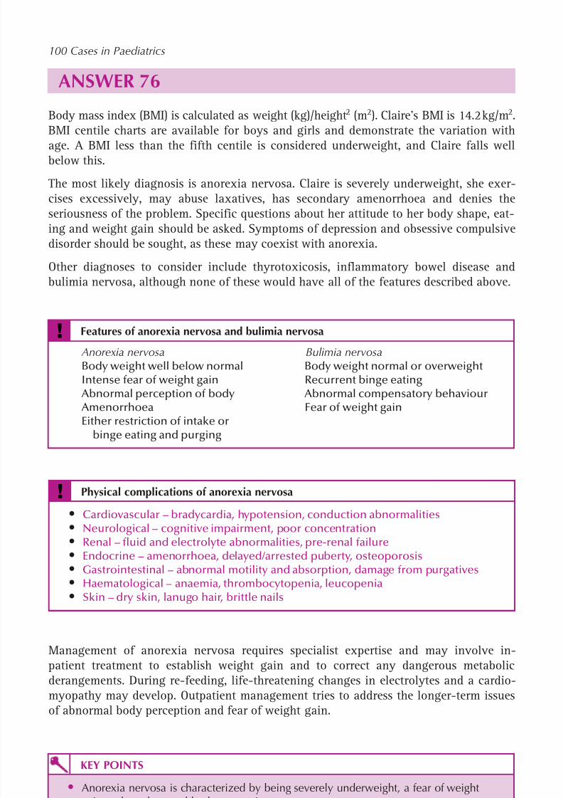
8/20/2019 100 cases in paediatrics.pdf
http://slidepdf.com/reader/full/100-cases-in-paediatricspdf 247/320
228
ANSWER 76Body mass index (BMI) is calculated as weight (kg)/height 2 (m2). Claire’s BMI is 14.2 kg/m 2.BMI centile charts are available for boys and girls and demonstrate the variation withage. A BMI less than the fifth centile is considered underweight, and Claire falls wellbelow this.
The most likely diagnosis is anorexia nervosa. Claire is severely underweight, she exer-cises excessively, may abuse laxatives, has secondary amenorrhoea and denies theseriousness of the problem. Specific questions about her attitude to her body shape, eat-ing and weight gain should be asked. Symptoms of depression and obsessive compulsivedisorder should be sought, as these may coexist with anorexia.
Other diagnoses to consider include thyrotoxicosis, inflammatory bowel disease andbulimia nervosa, although none of these would have all of the features described above.
! Features of anorexia nervosa and bulimia nervosa
Anorexia nervosa Bulimia nervosaBody weight well below normal Body weight normal or overweightIntense fear of weight gain Recurrent binge eatingAbnormal perception of body Abnormal compensatory behaviourAmenorrhoea Fear of weight gainEither restriction of intake or
binge eating and purging
! Physical complications of anorexia nervosa
• Cardiovascular – bradycardia, hypotension, conduction abnormalities• Neurological – cognitive impairment, poor concentration• Renal – fluid and electrolyte abnormalities, pre-renal failure
• Endocrine – amenorrhoea, delayed/arrested puberty, osteoporosis• Gastrointestinal – abnormal motility and absorption, damage from purgatives• Haematological – anaemia, thrombocytopenia, leucopenia• Skin – dry skin, lanugo hair, brittle nails
Management of anorexia nervosa requires specialist expertise and may involve in-patient treatment to establish weight gain and to correct any dangerous metabolicderangements. During re-feeding, life-threatening changes in electrolytes and a cardio-
myopathy may develop. Outpatient management tries to address the longer-term issuesof abnormal body perception and fear of weight gain.
KEY POINTS
• Anorexia nervosa is characterized by being severely underweight, a fear of weightgain and an abnormal body perception.
• Anorexia nervosa can lead to dangerous physical complications.
100 Cases in Paediatrics

8/20/2019 100 cases in paediatrics.pdf
http://slidepdf.com/reader/full/100-cases-in-paediatricspdf 248/320
229
NEONATOLOGY
CASE 77: BORN TOO EARLY
HistoryMrs Richardson is a 23-year old lady in her first pregnancy, who is admitted to hospitalat 26 weeks’ gestation because she has had spontaneous rupture of membranes. Forty-eight hours later she goes into spontaneous labour and a baby boy is delivered, weighing800 g (25th centile). At delivery he is making spontaneous respiratory effort with markedsubcostal and sternal recession. He is stabilized and transported to the neonatal unit.
Questions• What can be done before delivery to optimize the outcome for a premature baby?
• What are the management priorities for the baby at delivery?• What is the lung pathology seen in the chest radiograph?• What complications of prematurity does this baby face?
Figure 77.1 Chest radiograph at 4 hours of age.
INVESTIGATIONS
Baby Richardson’s chest radiograph is shown in Figure 77.1.
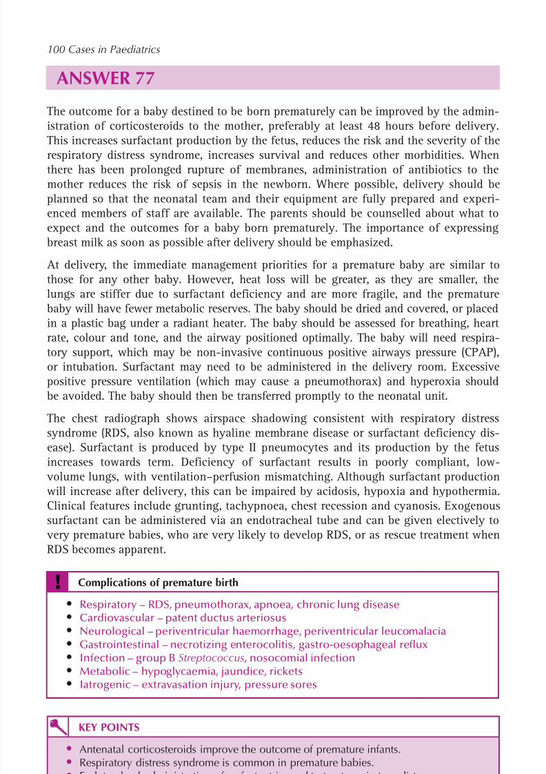
8/20/2019 100 cases in paediatrics.pdf
http://slidepdf.com/reader/full/100-cases-in-paediatricspdf 249/320
230
ANSWER 77The outcome for a baby destined to be born prematurely can be improved by the admin-istration of corticosteroids to the mother, preferably at least 48 hours before delivery.This increases surfactant production by the fetus, reduces the risk and the severity of therespiratory distress syndrome, increases survival and reduces other morbidities. Whenthere has been prolonged rupture of membranes, administration of antibiotics to themother reduces the risk of sepsis in the newborn. Where possible, delivery should beplanned so that the neonatal team and their equipment are fully prepared and experi-enced members of staff are available. The parents should be counselled about what toexpect and the outcomes for a baby born prematurely. The importance of expressingbreast milk as soon as possible after delivery should be emphasized.
At delivery, the immediate management priorities for a premature baby are similar tothose for any other baby. However, heat loss will be greater, as they are smaller, thelungs are stiffer due to surfactant deficiency and are more fragile, and the prematurebaby will have fewer metabolic reserves. The baby should be dried and covered, or placedin a plastic bag under a radiant heater. The baby should be assessed for breathing, heartrate, colour and tone, and the airway positioned optimally. The baby will need respira-tory support, which may be non-invasive continuous positive airways pressure (CPAP),or intubation. Surfactant may need to be administered in the delivery room. Excessivepositive pressure ventilation (which may cause a pneumothorax) and hyperoxia shouldbe avoided. The baby should then be transferred promptly to the neonatal unit.
The chest radiograph shows airspace shadowing consistent with respiratory distresssyndrome (RDS, also known as hyaline membrane disease or surfactant deficiency dis-ease). Surfactant is produced by type II pneumocytes and its production by the fetusincreases towards term. Deficiency of surfactant results in poorly compliant, low-
volume lungs, with ventilation–perfusion mismatching. Although surfactant productionwill increase after delivery, this can be impaired by acidosis, hypoxia and hypothermia.Clinical features include grunting, tachypnoea, chest recession and cyanosis. Exogenoussurfactant can be administered via an endotracheal tube and can be given electively to
very premature babies, who are very likely to develop RDS, or as rescue treatment whenRDS becomes apparent.
! Complications of premature birth
• Respiratory – RDS, pneumothorax, apnoea, chronic lung disease• Cardiovascular – patent ductus arteriosus• Neurological – periventricular haemorrhage, periventricular leucomalacia• Gastrointestinal – necrotizing enterocolitis, gastro-oesophageal reflux• Infection – group B Streptococcus , nosocomial infection
• Metabolic – hypoglycaemia, jaundice, rickets
• Iatrogenic – extravasation injury, pressure sores
KEY POINTS
• Antenatal corticosteroids improve the outcome of premature infants.• Respiratory distress syndrome is common in premature babies.• Endotracheal administration of surfactant is used to treat respiratory distress
syndrome.
100 Cases in Paediatrics
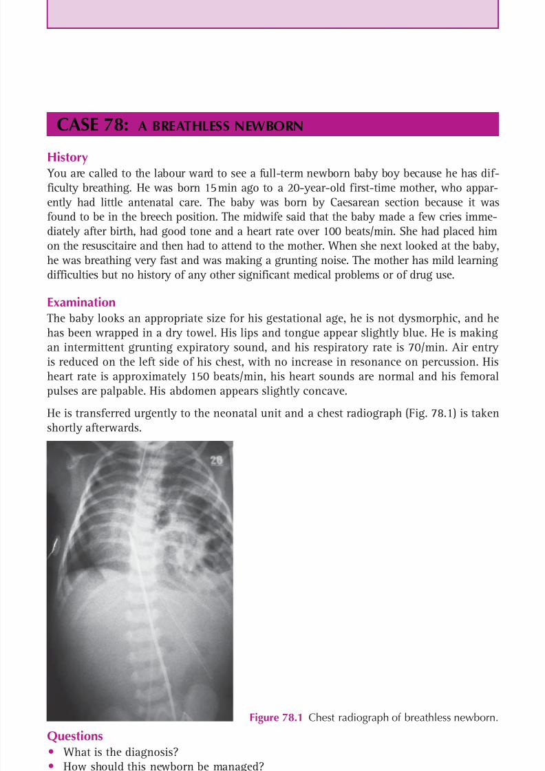
8/20/2019 100 cases in paediatrics.pdf
http://slidepdf.com/reader/full/100-cases-in-paediatricspdf 250/320
231
CASE 78:A BREATHLESS NEWBORN
History You are called to the labour ward to see a full-term newborn baby boy because he has dif-ficulty breathing. He was born 15 min ago to a 20-year-old first-time mother, who appar-ently had little antenatal care. The baby was born by Caesarean section because it wasfound to be in the breech position. The midwife said that the baby made a few cries imme-diately after birth, had good tone and a heart rate over 100 beats/min. She had placed himon the resuscitaire and then had to attend to the mother. When she next looked at the baby,he was breathing very fast and was making a grunting noise. The mother has mild learningdifficulties but no history of any other significant medical problems or of drug use.
ExaminationThe baby looks an appropriate size for his gestational age, he is not dysmorphic, and hehas been wrapped in a dry towel. His lips and tongue appear slightly blue. He is makingan intermittent grunting expiratory sound, and his respiratory rate is 70/min. Air entryis reduced on the left side of his chest, with no increase in resonance on percussion. Hisheart rate is approximately 150 beats/min, his heart sounds are normal and his femoralpulses are palpable. His abdomen appears slightly concave.
He is transferred urgently to the neonatal unit and a chest radiograph (Fig. 78.1) is takenshortly afterwards.
Figure 78.1 Chest radiograph of breathless newborn.
Questions• What is the diagnosis?• How should this newborn be managed?• What other conditions can cause respiratory distress in a term newborn after an
unremarkable delivery?

8/20/2019 100 cases in paediatrics.pdf
http://slidepdf.com/reader/full/100-cases-in-paediatricspdf 251/320
232
ANSWER 78The most common cause of respiratory distress in a term baby following a Caesareansection is transient tachypnoea of the newborn due to delayed clearance of lung fluid.However, this does not produce the clinical or radiographic features seen in this case.Here the diagnosis is congenital diaphragmatic hernia. Although many cases are pickedup through antenatal screening, herniation of bowel through defects in the diaphragmcan occur late in gestation or even after delivery. It usually presents with respiratorydistress at, or shortly after, birth. The abdomen appears concave because some of itscontents are within the thorax, the mediastinum and apex beat may be displaced andbowel sounds may be heard in the chest. The chest radiograph shows mediastinal shiftwith air-filled bowel loops occupying the left hemithorax. The baby has been intubated,a nasogastric tube is in place, and umbilical venous and arterial catheters have been
inserted.It is important to intubate the baby early to prevent the swallowing of air, which cancause expansion of the bowel within the chest and further respiratory compromise. Ifthe diagnosis is known before delivery, the use of bag-and-mask ventilation should beavoided for the same reason. A nasogastric tube should be placed to aspirate gastric fluidand air. Meticulous intensive care is needed to optimize the baby before surgery is per-formed because the lungs are usually hypoplastic (partly due to compression by bowelwithin the thorax) and pulmonary hypertension is a frequent complication. Mortalityrates of 20 per cent are expected even with optimal care.
! Causes of unexpected respiratory distress in the term newborn
• Transient tachypnoea of the newborn• Pneumothorax• Congenital pneumonia, sepsis• Lung malformations, congenital diaphragmatic hernia• Oesophageal atresia, tracheo-oesophageal fistula
• Choanal atresia, other upper airway malformations• Congenital heart disease• Anaemia, polycythaemia• Cerebral haemorrhage
KEY POINTS
• Transient tachypnoea of the newborn is the most common cause of respiratorydistress in a term baby, most often following Caesarean section.• A newborn with congenital diaphragmatic hernia should be intubated promptly to
prevent swallowed air distending the bowel within the thorax.• Prognosis in congenital diaphragmatic hernia is mainly related to the degree of lung
hypoplasia, and the mortality rate is approximately 20 per cent.
100 Cases in Paediatrics
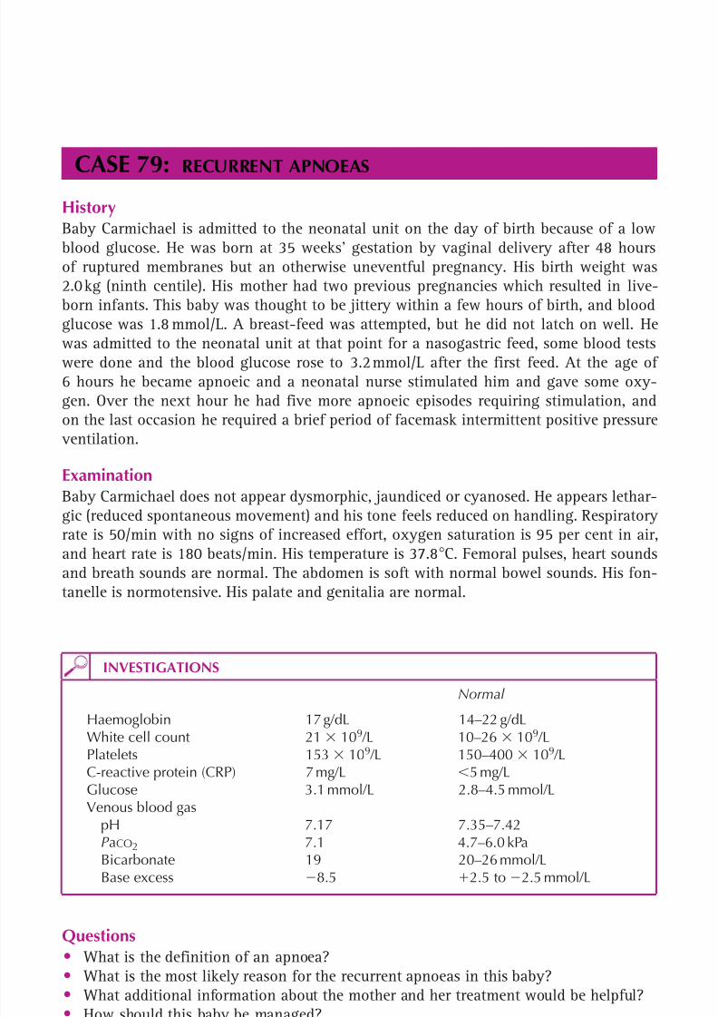
8/20/2019 100 cases in paediatrics.pdf
http://slidepdf.com/reader/full/100-cases-in-paediatricspdf 252/320
233
CASE 79:RECURRENT APNOEAS
HistoryBaby Carmichael is admitted to the neonatal unit on the day of birth because of a lowblood glucose. He was born at 35 weeks’ gestation by vaginal delivery after 48 hoursof ruptured membranes but an otherwise uneventful pregnancy. His birth weight was2.0 kg (ninth centile). His mother had two previous pregnancies which resulted in live-born infants. This baby was thought to be jittery within a few hours of birth, and bloodglucose was 1.8 mmol/L. A breast-feed was attempted, but he did not latch on well. Hewas admitted to the neonatal unit at that point for a nasogastric feed, some blood testswere done and the blood glucose rose to 3.2 mmol/L after the first feed. At the age of6 hours he became apnoeic and a neonatal nurse stimulated him and gave some oxy-gen. Over the next hour he had five more apnoeic episodes requiring stimulation, andon the last occasion he required a brief period of facemask intermittent positive pressure
ventilation.
ExaminationBaby Carmichael does not appear dysmorphic, jaundiced or cyanosed. He appears lethar-gic (reduced spontaneous movement) and his tone feels reduced on handling. Respiratory
rate is 50/min with no signs of increased effort, oxygen saturation is 95 per cent in air,and heart rate is 180 beats/min. His temperature is 37.8 C. Femoral pulses, heart soundsand breath sounds are normal. The abdomen is soft with normal bowel sounds. His fon-tanelle is normotensive. His palate and genitalia are normal.
INVESTIGATIONS
Normal
Haemoglobin 17 g/dL 14–22 g/dLWhite cell count 21 109 /L 10–26 109 /LPlatelets 153 109 /L 150–400 109 /LC-reactive protein (CRP) 7 mg/L 5 mg/LGlucose 3.1 mmol/L 2.8–4.5 mmol/LVenous blood gas pH 7.17 7.35–7.42 P aCO2 7.1 4.7–6.0 kPa Bicarbonate 19 20–26 mmol/L Base excess 8.5 2.5 to 2.5 mmol/L
Questions• What is the definition of an apnoea?• What is the most likely reason for the recurrent apnoeas in this baby?• What additional information about the mother and her treatment would be helpful?• How should this baby be managed?

8/20/2019 100 cases in paediatrics.pdf
http://slidepdf.com/reader/full/100-cases-in-paediatricspdf 253/320
234
100 Cases in Paediatrics
ANSWER 79 An apnoea is an episode of cessation of breathing lasting more than 20 s, or a shortertime if there is bradycardia or a colour change. This is distinct from the normal newbornpattern of periodic breathing, where fast respiration alternates with pauses of up to 10 s.
In this case, the most likely diagnosis is sepsis. The baby was born prematurely afterprolonged rupture of membranes – both risk factors for sepsis. He was hypoglycaemic, acommon finding in septic newborns, and he has had recurrent and worsening apnoeas.He is lethargic and has a slightly raised temperature. White cell count and CRP are unre-markable, but this is often the case early in the course of neonatal sepsis. The blood gasshows a mixed metabolic and respiratory acidosis, indicating hypoperfusion and hypo-
ventilation. This would be typical of early-onset group B streptococcal (GBS) sepsis.
There is no information provided about whether the mother had any microbiologicalsamples taken, received intrapartum antibiotics, had a fever or had evidence of chorio-amnionitis. These are all factors which would affect the risk of sepsis in the newborn. Itis also important to know whether the mother received any drugs during labour whichmay suppress neonatal respiration, e.g. pethidine, or whether she uses any illicit drugs.
Given the presence of two risk factors for early-onset GBS sepsis, if the mother hadnot received adequate intrapartum antibiotic prophylaxis, the baby should have beenassessed, cultures taken and antibiotics commenced at birth. If this was not done as soonas the baby showed the first signs of jitteriness and hypoglycaemia, sepsis should havebeen suspected. This baby is now in a dangerous situation, where apnoeas may progressto a respiratory arrest. Management priorities are to secure the airway with optimal posi-tioning of the head, ensure adequate breathing with either CPAP or intub ation, estab-lish vascular access, give fluid boluses to achieve cardiovascular stability and administer
antibiotics as early as possible. After this, further assessment for the source of the sepsis,and to exclude other causes of apnoea, can be undertaken.
! Causes of apnoea in a newborn
• Apnoea of prematurity• Lung disease, e.g. respiratory distress syndrome• Congenital heart disease• Sepsis• Hypoglycaemia
• Hypothermia• Sedative drugs (administered to mother in labour, or to baby)• Neurological insults – cerebral haemorrhage, oedema or seizures• Anaemia• Gastro-oesophageal reflux
KEY POINTS
• Apnoeas are a final common manifestation of many disease processes in the neonate.• Any neonate with apnoeas requires prompt evaluation.• Antibiotics must be administered urgently to a neonate with suspected early-onset
sepsis.

8/20/2019 100 cases in paediatrics.pdf
http://slidepdf.com/reader/full/100-cases-in-paediatricspdf 254/320
This page intentionally left blank
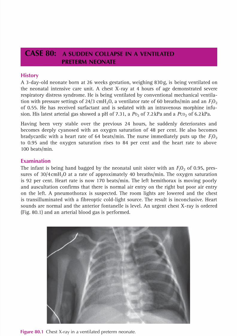
8/20/2019 100 cases in paediatrics.pdf
http://slidepdf.com/reader/full/100-cases-in-paediatricspdf 255/320
236
CASE 80: A SUDDEN COLLAPSE IN A VENTILATEDPRETERM NEONATE
History A 3-day-old neonate born at 26 weeks gestation, weighing 830 g, is being ventilated onthe neonatal intensive care unit. A chest X-ray at 4 hours of age demonstrated severerespiratory distress syndrome. He is being ventilated by conventional mechanical ventila-tion with pressure settings of 24/3 cmH 2O, a ventilator rate of 60 breaths/min and an F iO2of 0.55. He has received surfactant and is sedated with an intravenous morphine infu-sion. His latest arterial gas showed a pH of 7.31, a P O2 of 7.2 kPa and a P CO2 of 6.2 kPa.
Having been very stable over the previous 24 hours, he suddenly deteriorates andbecomes deeply cyanosed with an oxygen saturation of 48 per cent. He also becomesbradycardic with a heart rate of 64 beats/min. The nurse immediately puts up the F iO2to 0.95 and the oxygen saturation rises to 84 per cent and the heart rate to above100 beats/min.
ExaminationThe infant is being hand bagged by the neonatal unit sister with an F iO2 of 0.95, pres-
sures of 30/4 cmH 2O at a rate of approximately 40 breaths/min. The oxygen saturationis 92 per cent. Heart rate is now 170 beats/min. The left hemithorax is moving poorlyand auscultation confirms that there is normal air entry on the right but poor air entryon the left. A pneumothorax is suspected. The room lights are lowered and the chestis transilluminated with a fibreoptic cold-light source. The result is inconclusive. Heartsounds are normal and the anterior fontanelle is level. An urgent chest X-ray is ordered(Fig. 80.1) and an arterial blood gas is performed.
Figure 80.1 Chest X-ray in a ventilated preterm neonate.
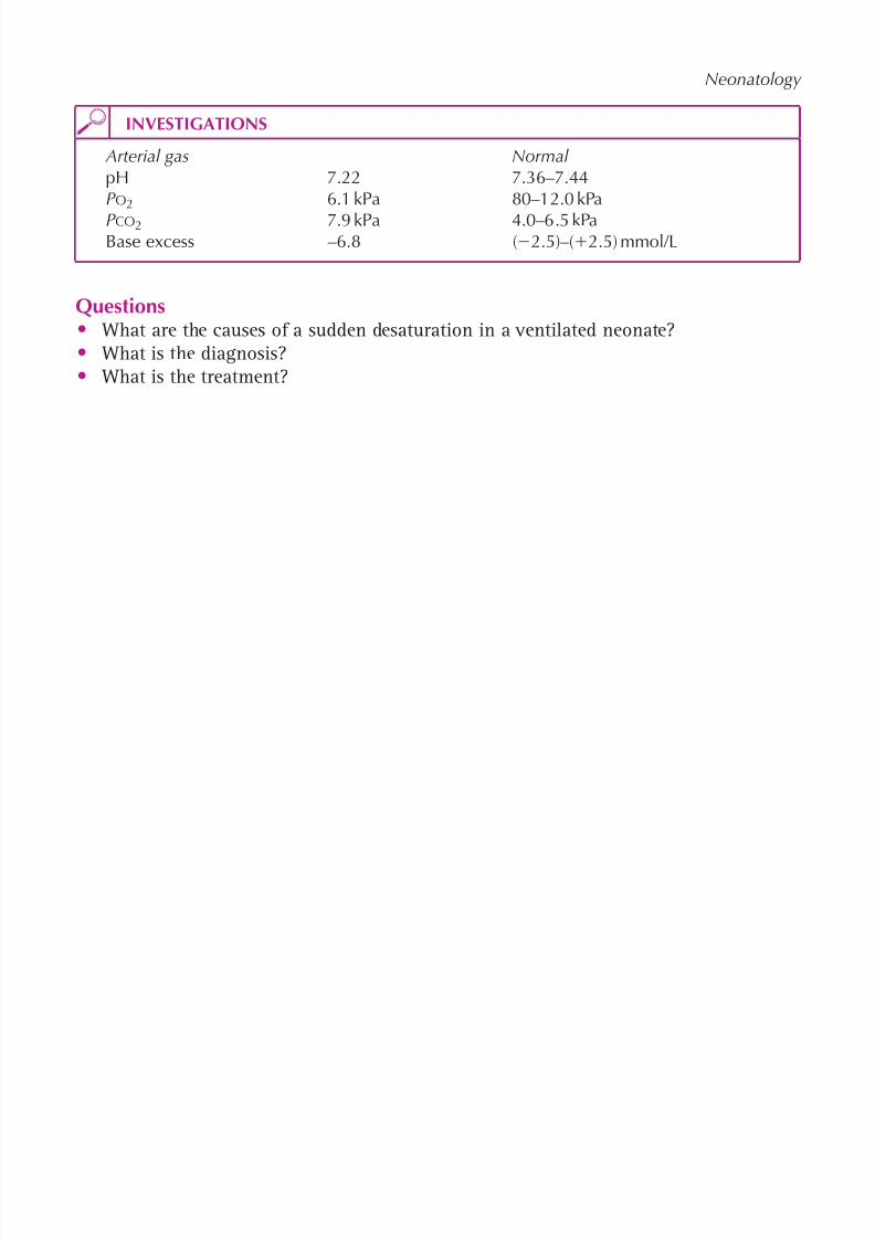
8/20/2019 100 cases in paediatrics.pdf
http://slidepdf.com/reader/full/100-cases-in-paediatricspdf 256/320
237
Neonatology
Questions• What are the causes of a sudden desaturation in a ventilated neonate?• What is the diagnosis?• What is the treatment?
INVESTIGATIONS
Arterial gas Normal pH 7.22 7.36–7.44P O2 6.1 kPa 80–12.0 kPaP CO2 7.9 kPa 4.0–6.5 kPaBase excess –6.8 ( 2.5)–( 2.5) mmol/L

8/20/2019 100 cases in paediatrics.pdf
http://slidepdf.com/reader/full/100-cases-in-paediatricspdf 257/320
238
100 Cases in Paediatrics
ANSWER 80This question is best answered by considering whether the chest is not moving, whetherchest movement is decreased or asymmetrical, or whether chest movement is normal:
• no chest movement– ventilator not working or tubing disconnected or kinked– endotracheal tube blocked or dislocated
• chest movement decreased or asymmetrical– pneumothorax– worsening respiratory disease
• chest movement normal– right-to-left shunt, e.g. across a patent ductus arteriosus– large periventricular haemorrhage– severe sepsis.
The diagnosis in this case is a left-sided tension pneumothorax with mediastinal shiftto the right (away from the side of the pneumothorax). A chest drain should be insertedimmediately.
If a pneumothorax is suspected clinically and cold-light transillumination demonstratesthat the chest ‘lights up’ on that side (the two sides should be compared), this is indicativeof a pneumothorax and a chest drain should be inserted and a chest X-ray is unneces-sary. If the child deteriorates, a 23g butterfly needle can be inserted into the second inter-
costal space in the mid-clavicular line. Air can then be aspirated from the pleural spaceand this can ‘buy time’ pending the insertion of a chest drain. The largest possible chestdrain should be used (usually 8, 10 or 12 FG depending on the size of the infant).
The drain should be inserted above the rib as the intercostal vessels lie immediatelybelow the rib. It should be inserted into the third, fourth or fifth intercostal space inthe mid-axillary line. Following insertion and the appropriate connections to an under-water seal, air bubbles should be seen, swinging should be observed on breathing andthe infant should rapidly improve. A chest X-ray should be done to ensure that the lunghas re-inflated and to check the position of the drain.
KEY POINTS
• A pneumothorax is a common cause of a sudden deterioration in a ventilated pretermneonate.
• Diagnosis is by cold light transillumination and/or a chest X-ray.• Urgent treatment is necessary. A needle aspiration can be done in the first instance if
the child is very unwell or deteriorating, but definitive treatment is with a chest drain.

8/20/2019 100 cases in paediatrics.pdf
http://slidepdf.com/reader/full/100-cases-in-paediatricspdf 258/320
This page intentionally left blank
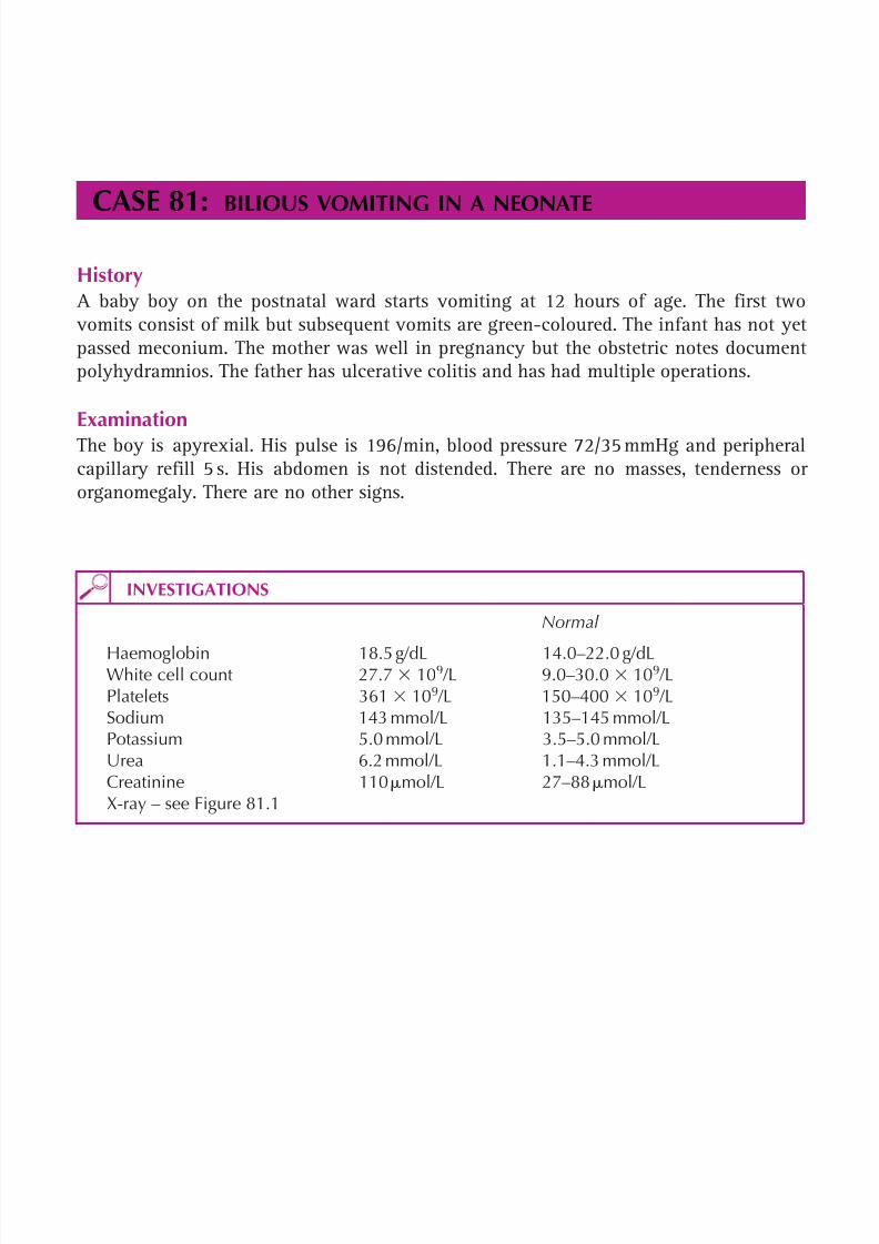
8/20/2019 100 cases in paediatrics.pdf
http://slidepdf.com/reader/full/100-cases-in-paediatricspdf 259/320
240
CASE 81:BILIOUS VOMITING IN A NEONATE
History A baby boy on the postnatal ward starts vomiting at 12 hours of age. The first two vomits consist of milk but subsequent vomits are green-coloured. The infant has not yetpassed meconium. The mother was well in pregnancy but the obstetric notes documentpolyhydramnios. The father has ulcerative colitis and has had multiple operations.
ExaminationThe boy is apyrexial. His pulse is 196/min, blood pressure 72/35 mmHg and peripheralcapillary refill 5 s. His abdomen is not distended. There are no masses, tenderness ororganomegaly. There are no other signs.
INVESTIGATIONS
Normal
Haemoglobin 18.5 g/dL 14.0–22.0 g/dLWhite cell count 27.7 109 /L 9.0–30.0 109 /LPlatelets 361 109 /L 150–400 109 /LSodium 143 mmol/L 135–145 mmol/LPotassium 5.0 mmol/L 3.5–5.0 mmol/LUrea 6.2 mmol/L 1.1–4.3 mmol/LCreatinine 110 mol/L 27–88 mol/LX-ray – see Figure 81.1
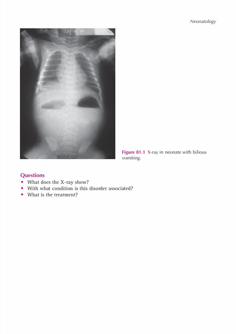
8/20/2019 100 cases in paediatrics.pdf
http://slidepdf.com/reader/full/100-cases-in-paediatricspdf 260/320
241
Neonatology
Questions• What does the X-ray show?• With what condition is this disorder associated?• What is the treatment?
Figure 81.1 X-ray in neonate with biliousvomiting.

8/20/2019 100 cases in paediatrics.pdf
http://slidepdf.com/reader/full/100-cases-in-paediatricspdf 261/320
242
100 Cases in Paediatrics
ANSWER 81The X-ray demonstrates the classic double bubble appearance due to dilatation of thestomach and the proximal duodenum found in duodenal atresia. It is an erect film which
also shows a fluid level in the stomach and duodenum. There is a paucity of gas inthe remainder of the abdomen. The polyhydramnios is secondary to the inability of theinfant to swallow amniotic fluid (conversely, inability to excrete amniotic fluid in theurine due to renal agenesis or dysgenesis leads to oligohydramnios). As the obstructionis in the upper gastrointestinal tract, there is no abdominal distension.
Down’s syndrome is associated with duodenal atresia in 20 per cent of cases. Duodenalatresia is also associated with malrotation, oesophageal atresia and congenital heart disease.Contrast studies are occasionally needed, primarily to rule out malrotation and volvulus.
The pulse rate is raised (normal in a neonate is 100–180 beats/min), the blood pres-sure is normal and the peripheral capillary refill is prolonged. The urea and creatinineare slightly raised. Initial treatment is with intravenous rehydration to correct the dehy-dration. A nasogastric tube is inserted and the stomach emptied. Where there is onecongenital abnormality, one should look for other abnormalities. Duodenal atresia isassociated with heart disease and this needs excluding prior to surgery.
KEY POINTS• Bilious vomiting in a newborn should be regarded as a surgical emergency until
proven otherwise.• Duodenal atresia leads to a double bubble appearance on the X-ray.• There is a strong association with Down’s syndrome.

8/20/2019 100 cases in paediatrics.pdf
http://slidepdf.com/reader/full/100-cases-in-paediatricspdf 262/320
243
CASE 82:A JAUNDICED NEONATE
History Armstrong is a 4-day-old Afro-Caribbean infant who is referred by his midwife to thepaediatric day unit because of jaundice. His mother thinks that the jaundice may havecommenced within the first 24 hours of life, but she was told by the first midwife thatshe saw that the baby was fine. The child has also been a little sleepy and has notbreast-fed as well as previously. His birth weight was 3.70 kg at term. His mother had asplenectomy after falling off a horse as a teenager. This is her first pregnancy. There isno family history of jaundice.
ExaminationThe sclera are markedly yellow and the infant is somewhat lethargic. He is well perfusedand apyrexial. There is no hepatosplenomegaly and there are no other signs. He weighs3.40 kg.
INVESTIGATIONS
Normal
Haemoglobin 12.1 g/dL 14.0–22.0 g/dLWhite cell count 27.7 109 /L 9.0–30.0 109 /LPlatelets 361 10 9 /L 150–400 109 /LReticulocytes 12 per cent 2 per centBlood film Occasional spherocytesSodium 143 mmol/L 135–145 mmol/LPotassium 5.0 mmol/L 3.5–5.0 mmol/LUrea 6.2 mmol/L 1.1–4.3 mmol/L
Creatinine 110 mol/L 27–88 mol/LBilirubin 560 mol/L 205 mol/LAlbumin 30 g/L 25–34 g/LAlanine aminotransferase 48 U/L 6–50 U/LAlkaline phosphatase 367 U/L 145–420 U/LC-reactive protein 5 mg/L 6 mg/LBaby’s blood group AMaternal blood group ODirect antiglobulin test (DAT) PositiveGlucose-6-phosphate
dehydrogenase (G6PD) 7.4 IU/g Hb 4.6–13.5 IU/g HbUrine dipstick No leucocytes or nitrites
Questions• What are the causes of jaundice in a neonate?• Why is neonatal jaundice potentially dangerous?• What is the cause in this infant?• What is the treatment?

8/20/2019 100 cases in paediatrics.pdf
http://slidepdf.com/reader/full/100-cases-in-paediatricspdf 263/320
244
100 Cases in Paediatrics
ANSWER 82The causes of jaundice are best considered by dividing jaundice into early-onset, normal-onset and late-onset jaundice.
! Causes of jaundice
Early onset (first 24 hours, haemolytic jaundice)• Rhesus haemolytic disease• ABO incompatibility• G6PD deficiency (commonest in those of African, Asian, or Mediterranean
descent)
• Hereditary spherocytosisNormal onset • Physiological (all newborns get a degree of jaundice peaking at 4–5 days)• Bruising• Polycythaemia• Causes of early jaundice
Late onset ( 14 days, prolonged jaundice)• Persistence of a pathological earlier jaundice• Breast milk jaundice• Neonatal hepatitis• Biliary atresia• Hypothyroidism• Galactosaemia
Jaundice can also be a non-specific marker of neonatal infection at any stage.
Neonatal jaundice can be dangerous, as unconjugated bilirubin can cross the blood–brainbarrier. Very high levels can lead to kernicterus which can cause deafness and choreo-athetoid cerebral palsy.
The cause of the jaundice in this infant is ABO incompatibility. His mother is O andwill therefore have anti-A and anti-B antibodies in her blood. Theses antibodies cancross the placenta and lead to haemolysis in infants with either blood group A or B. Thisis further confirmed by the positive DAT (though, on occasion, this test can be nega-tive). The decreased haemoglobin and raised reticulocyte count provide further evidenceof haemolysis. The presence of a few spherocytes is common in ABO incompatibility.The normal temperature, normal inflammatory markers and normal urine dipstick make
infection unlikely. Unlike Rhesus haemolytic disease, it can occur with the first preg-nancy and does not get worse with successive pregnancies.
There are graphs that provide guidelines stating at what bilirubin level treatment withphototherapy and an exchange transfusion are indicated. The guidelines depend onthe age of the infant, their weight, gestation and on whether they are well or ill. Thisinfant’s bilirubin level is well above the exchange transfusion line. Phototherapy shouldbe commenced immediately and blood taken for cross-match (for O blood to minim-ize further haemolysis). The fall in birth weight of 8 per cent and the raised urea andcreatinine suggest a degree of dehydration which could be exacerbating the jaundice.

8/20/2019 100 cases in paediatrics.pdf
http://slidepdf.com/reader/full/100-cases-in-paediatricspdf 264/320
245
Neonatology
Maintenance intravenous fluids should therefore be commenced and breast-feeding canbe continued. Appropriate lines should be inserted to enable the exchange transfusionto take place. The blood for the transfusion usually has to be ordered from the regionalblood transfusion centre. It is possible that the bilirubin will have fallen markedly by
the time the blood has arrived, in which case the exchange transfusion may be avoided.Following discharge, the haemoglobin should be monitored, a hearing test arranged anddevelopment followed up.
KEY POINTS
• Neonatal jaundice is a potential cause of kernicterus that can lead to deafness andcerebral palsy.
• Jaundice may be more difficult to diagnose in Asian and Afro-Caribbean babies.
• Treatment in severe cases consists of phototherapy an exchange transfusion.

8/20/2019 100 cases in paediatrics.pdf
http://slidepdf.com/reader/full/100-cases-in-paediatricspdf 265/320
This page intentionally left blank
8/20/2019 100 cases in paediatrics.pdf
http://slidepdf.com/reader/full/100-cases-in-paediatricspdf 266/320

8/20/2019 100 cases in paediatrics.pdf
http://slidepdf.com/reader/full/100-cases-in-paediatricspdf 267/320
248
ANSWER 83This baby has a very abnormal neurological examination. The key findings are severehypotonia and weakness with preservation of facial and eye movements, absent deeptendon reflexes, poor sucking reflex and fasciculation of the tongue. These features indi-cate a process affecting the lower motor neurones of the entire spinal cord and selectivecranial nerve motor nuclei (IX and XII).
In this case it is likely that there is an underlying genetic cause for the baby’s condition,so further questions should be asked to try to substantiate this. Parental consanguinity,history of previous pregnancies ending in miscarriage or stillbirth, other family memberswith medical problems and pregnancies in other family members should be asked about. Inthe current pregnancy, there may have been reduced fetal movements. The parents should
be asked in detail about whether they have any medical problems, and direct questionsabout symptoms of neuromuscular disease, including weakness, fatiguability and musclecramps. In assessing any floppy infant, it is important to establish whether an acquiredcause may be present, so an assessment should be made of the maternal drug history andof any drugs given during labour, whether any brain or spinal cord injury may have beensustained during delivery, and any risk factors for sepsis or features of metabolic disease.
An infant can be floppy due to ‘central’ or ‘peripheral’ (neuromuscular) causes. In theformer, deep tendon reflexes are present, and in the latter they are absent and the childhas marked weakness. Central causes are much more common than peripheral causes. Inthis case, the clinical findings are of a peripheral cause and the most likely diagnosis isinfantile spinal muscular atrophy (SMA type 1). SMA is an autosomal recessive condition,with type 1 affecting about 1 per 10 000 live births. Sixty per cent of affected infants willbe floppy at birth. Motor neurone degeneration leads to progressive weakness and deathusually occurs from respiratory complications, e.g. pneumonia, within the first year of life.
! Causes of a floppy infant
‘Central’ – hypotonic but not ‘Peripheral’– hypotonic, weak, areflexicsignificantly weak
Chromosomal disorder, Infantile spinal muscular atrophye.g. Down’s syndrome,Prader-Willi syndrome
Brain injury, e.g. hypoxic-ischaemic Congenital myastheniaencephalopathy
Brain infection or sepsis Congenital myotonic dystrophyMetabolic disturbance, Congenital myopathy
e.g. hypoglycaemiaDrug exposure, e.g. pethidine Congenital muscular dystrophy
KEY POINTS
• An infant can be floppy due to central or peripheral (neuromuscular) causes.• Assessment of muscle power and deep tendon reflexes helps to classify the likely
causes.• Central causes of hypotonia are the most common.
100 Cases in Paediatrics

8/20/2019 100 cases in paediatrics.pdf
http://slidepdf.com/reader/full/100-cases-in-paediatricspdf 268/320
249
CASE 84:A DIFFICULT DELIVERY
History A 34-year-old woman with gestational diabetes in her first pregnancy is admitted to thelabour ward at 41 weeks’ gestation in active labour. She is obese (weight 105 kg). Her notesindicate that she was recommended dietary control of her gestational diabetes. Her attend-ance at antenatal clinic was erratic, and she did not regularly measure her blood glucoseconcentration. Induction of labour had been discussed previously, as it was felt that thefetus was large. The mother had declined this, stating that she wanted to have a naturalbirth. The fetal anomaly scan at 20 weeks had been normal, but she had not attended fortests of fetal well-being after 38 weeks’ gestation.
The delivery is complicated by severe shoulder dystocia and extra help is summoned,including a neonatal ‘crash call’. On arrival of the neonatal team, the baby is still notdelivered and the obstetric consultant has just arrived. After 15 min have elapsed fromdelivery of the head to delivery of the body, a large baby boy is delivered.
Questions• What immediate problems should be anticipated and why do they occur?
• What will this baby look like at delivery?• How should the baby be immediately assessed and managed?• What are the possible complications for the baby?

8/20/2019 100 cases in paediatrics.pdf
http://slidepdf.com/reader/full/100-cases-in-paediatricspdf 269/320
250
ANSWER 84This baby will almost certainly have suffered an acute asphyxial insult as a result of cordcompression while delivery of the body was prevented by the shoulder dystocia. The neo-natal team should anticipate that the baby will require significant resuscitation and shouldensure that a senior neonatologist is present. Obstruction of the fetoplacental circula-tion for more than a few minutes causes hypoxia, the fetus ceases breathing movements(primary apnoea), becomes bradycardic and circulation is directed to essential organs.
Acidosis develops and, after a longer period of asphyxia, episodic reflex gasping occurs. Ifaeration of the lungs does not occur, these gasps diminish and a phase of terminal apnoeaoccurs, from which the fetus can only be saved by delivery and artificial ventilation. Atdelivery this baby will probably be apnoeic, bradycardic or asystolic, pale and floppy.
Resuscitation of the newborn baby is unique. If there has never been aeration of thelungs, this must be achieved before any other interventions will work. The baby shouldbe dried, covered and assessed for colour, tone, breathing and heart rate. If the baby isnot breathing, the airway should be opened by placing the head in the neutral pos itionand five inflation breaths should be given. The first few inflation breaths may not pro-duce chest movement because they act to displace lung fluid. The effectiveness of infla-tion breaths should be assessed by checking for an increase in heart rate. If this has notoccurred it should be confirmed that the chest is being inflated before commencing car-diac compressions. This baby may also require adrenaline and sodium bicarbonate to beadministered via an emergency umbilical venous catheter in order to achieve a return ofcardiac output. The blood glucose should also be checked, especially as his mother haddiabetes in pregnancy. After immediate resuscitation, the baby will need to be trans-ferred to the neonatal unit for supportive and neuroprotective care.
The outcome of acute asphyxia can be good if the episode is brief and effective resusci-tation is provided. More severe asphyxia can lead to hypoxic-ischaemic encephalopathy(HIE) and death. In addition, shoulder dystocia is associated with brachial plexus injuryand fracture of the clavicle and humerus.
! Complications of birth asphyxia
First days after resuscitation Long term (mostly after severe HIE)Encephalopathy, seizures, cerebral oedema Cerebral palsyHypoventilation SeizuresMyocardial dysfunction Sensorineural hearing lossPersistent fetal circulation Visual impairmentRenal dysfunction Learning difficultiesHypoglycaemia, hyponatraemia
100 Cases in Paediatrics
KEY POINTS
• If a baby is likely to have suffered severe asphyxia, senior assistance is required.• Newborn resuscitation has a unique algorithm because the lungs have never been
inflated with air.• Birth asphyxia affects multiple organs, although most long-term complications are
neurological.

8/20/2019 100 cases in paediatrics.pdf
http://slidepdf.com/reader/full/100-cases-in-paediatricspdf 270/320
251
CASE 85:A NEWBORN WITH CONGENITAL ABNORMALITIES
History A midwife on the postnatal ward asks for a male infant to be reviewed on the day ofbirth because she is concerned that his ears look abnormal. The baby was born at termin good condition after a normal vaginal delivery. His mother is a 28-year-old salesmanager, and this was her first pregnancy. She received routine antenatal care and herbooking blood tests and anomaly scan were unremarkable. She and her partner have nomedical problems. She is worried that the baby’s ears look abnormal and wants to knowwhether anything can be done to improve their appearance.
ExaminationThe birth weight was 2.6 kg (second centile) and head circumference 33.5 cm (ninth cen-tile). The baby has small, abnormal-looking ears and an iris coloboma (a defect in theiris). Respiratory examination is unremarkable. Cardiac examination reveals a heart rateof 140/min, normal femoral pulses, normal apex beat, but a grade 3/6 pansystolic murmurthat is loudest at the lower left sternal edge. Abdominal examination reveals a very smallpenis with undescended testes. Neurological examination is normal, the palate is normaland the hips are normal.
Questions• What explanation should be given to the parents at this stage?• What different mechanisms can lead to congenital abnormalities?• How should a child with multiple congenital abnormalities be assessed?

8/20/2019 100 cases in paediatrics.pdf
http://slidepdf.com/reader/full/100-cases-in-paediatricspdf 271/320
252
ANSWER 85This infant has multiple congenital abnormalities. The explanation given to the parentsby the junior doctor must be honest but gentle, and should avoid speculation aboutprognosis. It would be appropriate to explain that congenital abnormalities are quitecommon, but in addition to the ear abnormalities the parents have noticed, there areother abnormalities, which need to be assessed by a more senior colleague. The iris colo-boma and the genital abnormalities can be pointed out and the heart murmur should bementioned. The combination of multiple abnormalities suggests an underlying cause orsyndrome. The parents will be upset by the news, but will appreciate honesty and hardwork in order to give them further information as soon as possible.
Congenital anomalies affect about 3 per cent of live births, although not all of these areobvious at the time of birth. They can be divided into malformations due to errors duringmorphogenesis, and deformations which affect a previously normally formed part of thefetus. Most malformations do not have a defined aetiology and are considered sporadic.
! Causes of congenital malformations
• Genetic – chromosomal abnormalities, e.g. 22q11 deletion (DiGeorge syndrome) – gene defects, e.g. CHD7 (chromodomain helicase DNA-binding protein 7
mutations causing CHARGE syndrome)• Environmental – maternal factors, e.g. diabetes (cardiac malformations) – prescribed drugs, e.g. phenytoin (fetal hydantoin syndrome) – recreational drugs, e.g. alcohol (fetal alcohol syndrome) – environmental toxins, e.g. dioxins (central nervous system anomalies) – infections (e.g. rubella, cytomegalovirus)• Sporadic
Assessment of a child with multiple congenital abnormalities should determine whetherthere are other abnormalities, the severity of those already detected, and the under-lying cause if possible. Sometimes a characteristic pattern of abnormalities will revealthe diagnosis. Karyotyping should be performed and other specific genetic tests may berequired. A clinical geneticist should be asked to assess the child if a unifying diagnosisis not apparent. In this case the findings suggest CHARGE syndrome (coloboma, heartdefects, atresia choanae, retardation of growth and development, genital and ear abnor-malities). Although there is no respiratory distress, choanal atresia should be excluded(by attempting to pass a nasogastric tube through each nostril). The cardiovascularexamination findings would be consistent with a ventricular septal defect, the common-est congenital heart defect. Further assessments by a paediatric cardiologist, ophthalmolo-gist, endocrinologist, urologist, audiological physician and developmental paediatricianwill be needed. Making a diagnosis may allow future problems to be anticipated andallows genetic counselling about the recurrence risk in future pregnancies.
100 Cases in Paediatrics
KEY POINTS
• Congenital abnormalities are common.• If one congenital abnormality is present, you should look for other ones.• Recognizing a pattern of abnormalities comprising a syndrome is important for
prognosis and genetic counselling.

8/20/2019 100 cases in paediatrics.pdf
http://slidepdf.com/reader/full/100-cases-in-paediatricspdf 272/320
253
CASE 86:SUDDEN DETERIORATION IN A TERM BABY
HistoryThe midwife finds a 3-day-old male baby on the postnatal ward to be very lethargic andbreathing fast. The baby is taken to the neonatal unit for further evaluation. He was bornat term by emergency Caesarean section for undiagnosed breech presentation. There wasno prolonged rupture of membranes. The pregnancy had been unremarkable to that pointand all antenatal scans had been normal. Mother is blood group O-positive, had negativeHIV, hepatitis B and syphilis serology and was rubella immune. The delivery was attendedby a paediatrician but the baby cried immediately and needed no resuscitation. His birthweight was 3.5 kg (50th centile) and the first-day check was unremarkable. He has beenbreast-fed and although there were problems latching on initially, he fed well on day 2.He seems rather sleepy and has had frequent non-bilious vomits today. His mother is a23-year-old woman from Pakistan who speaks good English and this is her first baby.
ExaminationThe baby’s temperature is 36.3 C, his heart rate is 160 beats/min and his respiratoryrate is 70 breaths/min. Oxygen saturation is 99 per cent in air, capillary refill is 2–3 s,and blood pressure is 60/40 mmHg. There is subcostal and mild sternal recession. He
responds to vigorous stimulation and pain, but otherwise is very lethargic. The chest isclear, heart sounds and femoral pulses are normal, and the abdomen is normal. The fon-tanelle is normotensive. He has a strong odour which is slightly unpleasant.
Questions
• What are the possible causes of this type of presentation?
• How do you interpret the blood gas analysis?• What is the most likely cause in this case?• What is the emergency management?
INVESTIGATIONS
Normal Venous blood gas analysis pH 6.98 7.35–7.42P aCO2 2.62 4.7–6.0 kPa
Base excess 25.5 2.5 to 2.5 mmol/L Lactate 2.1 1.0–1.8 mmol/L Glucose 5.2 2.2–4.4 mmol/LUrinalysis Glucose Negative NegativeProtein Negative Negative
Ketones Negative
8/20/2019 100 cases in paediatrics.pdf
http://slidepdf.com/reader/full/100-cases-in-paediatricspdf 273/320
254
ANSWER 86The history describes a term neonate who became very ill over a short period of time.The important diagnoses to consider are discussed in Case 10 (p. 31–32 ).
! Approach to blood gas interpretation
In this case
Look at the pH pH 7.35, acidosisLook at the P CO 2 P CO 2 4.7 kPa, respiratory compensationLook at the base excess Lower than 2.5, metabolic acidosis
The blood gas analysis shows a severe metabolic acidosis, with unsuccessful respiratorycompensation. This fits with the clinical picture of a tachypnoeic baby.
Important points to note are the absence of risk factors for sepsis, the absence of signsof circulatory compromise, a lethargic baby with a strong odour, ketonuria and a severemetabolic acidosis without significant hyperlactataemia or hypoglycaemia. These find-ings point to an inborn error of metabolism, where excess acid is accumulating in theblood due to a metabolic defect. Possible causes include propionic, methylmalonic andisovaleric acidaemias. Remembering the names is less important than recognizing thistype of presentation.
Emergency management consists of stopping feeds and starting an infusion of 10 per centdextrose. Whilst sepsis is unlikely to be the sole cause of the illness, it could be a contribut-ing factor and antibiotics should be administered. The baby needs to be assessed promptlyby an experienced paediatrician as he is likely to require intubation and ventilationif he starts to tire or cannot protect his airway. It is likely that sodium bicarbonate wouldbe beneficial to partially correct the acidosis, and the case should be discussed urgentlywith the regional paediatric metabolic centre, as specific therapies may be recommendedeven before a certain diagnosis is available. To assist in making a diagnosis, bloodshould be sent for ammonia, plasma amino acid profile, and urine for amino and organic
acid profiles.This baby had consanguineous parents (first cousins), increasing the risk of an autosomalrecessive inborn error of metabolism. He required intubation and ventilation until theacidosis was corrected and he was then transferred to the regional metabolic unit. Thediagnosis turned out to be isovaleric acidaemia.
KEY POINTS
• Metabolic disease can present as a sudden deterioration in the neonatal period.
• Metabolic disease is rare and management must be discussed with an expert.
100 Cases in Paediatrics
8/20/2019 100 cases in paediatrics.pdf
http://slidepdf.com/reader/full/100-cases-in-paediatricspdf 274/320

8/20/2019 100 cases in paediatrics.pdf
http://slidepdf.com/reader/full/100-cases-in-paediatricspdf 275/320
256
ANSWER 87Dysmorphology is the study of human congenital malformations and dysmorphic featuresare abnormal body characteristics. Some are common in the general population and in isola-tion are not significant, e.g. single palmar crease. A syndrome is a well-characterized groupof such abnormalities occurring together that often point to a single condition as the cause.
! Characteristic dysmorphic features of trisomy 21
• Hypotonia• Flat face• Upward slanting palpebral fissures• Epicanthic folds• White speckles in the iris (Brushfield spots)• Short broad hands• Single palmar and plantar fissures (Simian crease)
There are several associated features, the commonest of which are low IQ (average IQabout 50), congenital heart disease, duodenal atresia and short stature.
Chromosomes must be sent for urgent analysis. This is to confirm the clinical suspicions,look for mosaicism ( 1 per cent) that might alter the prognosis and to exclude a trans-location. If a translocation is identified, parental chromosomes must be analysed in caseone is a carrier with a high risk of recurrence. The incidence of cardiac anomalies is sohigh (35 per cent) that all children with trisomy 21 should have a cardiac echocardio-gram even if no murmur is detected. An atrioventriculoseptal defect (AVSD) is the char-acteristic association with trisomy 21, but a ventriculoseptal defect is the commonestabnormality.
Although the risk of Down’s syndrome rises with maternal age, most mothers of childrenwith Down’s syndrome are 30 years as this is the group that has the greatest numberof children.
Breaking bad news is never easy but parents will always remember the first discussion.They should be seen by a senior paediatrician with the midwife who knows them welland other family members if they wish. If English is not their first language, an inter-preter should be supplied – this should not be a family member as this person needsto be impartial. The discussion should take place in a private room with no phones orbleeps and plenty of time should be allowed. Avoid jargon and use diagrams to helpexplanations.
It is worth exploring if they have noticed anything. Expect distress, anger, guilt, shame –in fact, any emotion. They are likely to ask how this could happen when they weregiven a negative screen result. There is a very common misconception amongst the pub-lic and professionals that a negative screen completely excludes the diagnosis. Antenatalscreening programmes identify those at high risk to whom a definitive diagnostic test(e.g. amniocentesis) should be offered.
100 Cases in Paediatrics
8/20/2019 100 cases in paediatrics.pdf
http://slidepdf.com/reader/full/100-cases-in-paediatricspdf 276/320
257
KEY POINTS
• Negative antenatal screening does not mean zero risk.• Chromosomes must always be analysed even if the clinical findings are absolutely
characteristic of a syndrome.
Acknowledge that it is normal to forget much of this first meeting. Arrange another meet-ing soon afterwards and suggest they write down any questions that arise in the meantime.
Written material is available from Down’s syndrome charities and their websites.
Neonatology

8/20/2019 100 cases in paediatrics.pdf
http://slidepdf.com/reader/full/100-cases-in-paediatricspdf 277/320
This page intentionally left blank
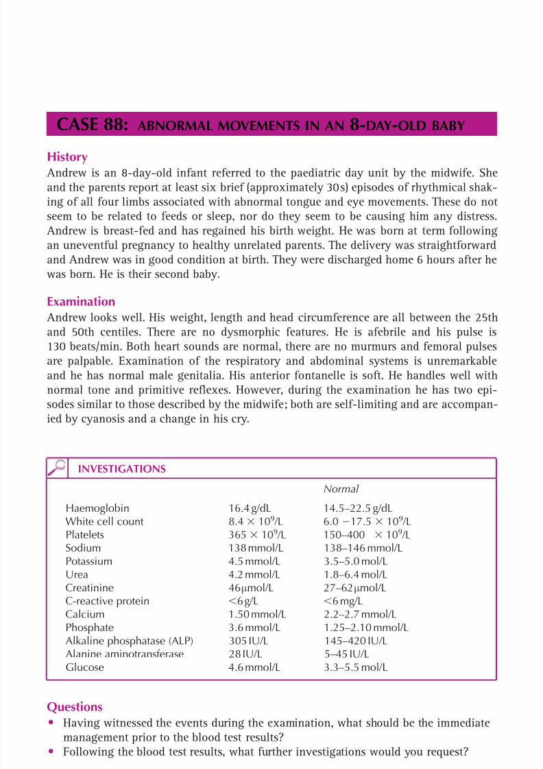
8/20/2019 100 cases in paediatrics.pdf
http://slidepdf.com/reader/full/100-cases-in-paediatricspdf 278/320
259
CASE 88:ABNORMAL MOVEMENTS IN AN
8-DAY
-OLD BABY
History Andrew is an 8-day-old infant referred to the paediatric day unit by the midwife. Sheand the parents report at least six brief (approximately 30 s) episodes of rhythmical shak-ing of all four limbs associated with abnormal tongue and eye movements. These do notseem to be related to feeds or sleep, nor do they seem to be causing him any distress.
Andrew is breast-fed and has regained his birth weight. He was born at term followingan uneventful pregnancy to healthy unrelated parents. The delivery was straightforwardand Andrew was in good condition at birth. They were discharged home 6 hours after hewas born. He is their second baby.
Examination Andrew looks well. His weight, length and head circumference are all between the 25thand 50th centiles. There are no dysmorphic features. He is afebrile and his pulse is130 beats/min. Both heart sounds are normal, there are no murmurs and femoral pulsesare palpable. Examination of the respiratory and abdominal systems is unremarkableand he has normal male genitalia. His anterior fontanelle is soft. He handles well withnormal tone and primitive reflexes. However, during the examination he has two epi-
sodes similar to those described by the midwife; both are self-limiting and are accompan-ied by cyanosis and a change in his cry.
INVESTIGATIONS
Normal
Haemoglobin 16.4 g/dL 14.5–22.5 g/dLWhite cell count 8.4 109 /L 6.0 17.5 109 /L
Platelets 365 109
/L 150–400 109
/LSodium 138 mmol/L 138–146 mmol/LPotassium 4.5 mmol/L 3.5–5.0 mol/LUrea 4.2 mmol/L 1.8–6.4 mol/LCreatinine 46 µmol/L 27–62 µmol/LC-reactive protein 6 g/L 6 mg/LCalcium 1.50 mmol/L 2.2–2.7 mmol/LPhosphate 3.6 mmol/L 1.25–2.10 mmol/LAlkaline phosphatase (ALP) 305 IU/L 145–420 IU/LAlanine aminotransferase 28 IU/L 5–45 IU/LGlucose 4.6 mmol/L 3.3–5.5 mol/L
Questions• Having witnessed the events during the examination, what should be the immediate
management prior to the blood test results?• Following the blood test results, what further investigations would you request?

8/20/2019 100 cases in paediatrics.pdf
http://slidepdf.com/reader/full/100-cases-in-paediatricspdf 279/320
260
ANSWER 88 Andrew is having seizures. Remember these are a symptom, not a diagnosis, and thecauses are numerous. The approach to a fitting child is:
1. Give high-flow oxygen, place on a cardiac monitor.2. Obtain intravenous access. Send relevant tests, starting with the basic and common.3. Treat immediately what can be treated. Check blood glucose and treat hypoglycaemia
after taking specimens to identify a cause. Start intravenous broad-spectrum antibiot-ics, e.g. benzylpenicillin and gentamicin – although sepsis is unlikely, it is a possibil-ity that needs to be treated.
4. Control the seizures with appropriate anticonvulsants. In neonates this is usually aloading dose of phenobarbital, followed, if necessary, by maintenance.
The results show that Andrew has significant hypocalcaemia, low enough to cause seiz-ures. It is likely to be real because the phosphate is high but, as with all unusual results,it should be repeated. If confirmed, the hypocalcaemia should be treated with intraven-ous calcium but only after taking samples to investigate the cause. It is crucial not tolose this opportunity to make a diagnosis because endocrine and metabolic pathwayscorrect very rapidly.
! Causes of hypocalcaemia in infancy
• Prematurity• Hypoxic ischaemic encephalopathy (HIE)• Hypoparathyroidism – transient (associated with prematurity, HIE or maternal
diabetes) or permanent• Hypomagnesaemia – magnesium facilitates release of parathormone (PTH)• Exchange transfusion – citrate in transfused blood chelates calcium to prevent
clotting• Familial activating mutations of the calcium-sensing receptor – the calcium level
at which PTH is released is lower than normal; some cases labelled as ‘familialhypoparathyroidism’ are probably this disorder• Maternal hypercalcaemia – suppresses fetal PTH• Maternal vitamin D deficiency• In older children, vitamin D deficiency is the commonest cause, but the ALP
would be high due to the increased bone turnover in rickets (see Case 22, p. 66).
Most of these can be excluded from the history. The next tests to send are magnesium,parathyroid hormone (PTH), vitamin D and maternal bone biochemistry. However, thehigh phosphate (PTH is phosphaturic) and normal ALP suggest hypoparathyroidism. Thecauses of permanent hypoparathyroidism that present in the neonatal period includeDiGeorge syndrome – a disorder associated with microdeletions of chromosome 22q11.2,congenital heart disease and thymic aplasia. PTH is not available therapeutically sotreatment of hypoparathyoidism is with vitamin D, but this does not stop the renal lossof calcium that PTH normally prevents. To minimize the risk of nephrocalcinosis, cal-cium is maintained at low normal or mildly subnormal levels and the urinary calcium/creatinine ratio is monitored.
100 Cases in Paediatrics
8/20/2019 100 cases in paediatrics.pdf
http://slidepdf.com/reader/full/100-cases-in-paediatricspdf 280/320
261
All children with neonatal seizures of any cause are at risk of developmental problemsand need surveillance.
KEY POINTS• In any case of hypocalcaemia or hypoglycaemia, relevant samples should be taken
before treatment to maximize the opportunity of making a definitive diagnosis.• Children with neonatal seizures of any cause are at risk of developmental problems
and need surveillance.
Neonatology

8/20/2019 100 cases in paediatrics.pdf
http://slidepdf.com/reader/full/100-cases-in-paediatricspdf 281/320
262
MISCELLANEOUS
CASE 89: A FRACTURE IN AN INFANT
HistoryBobby is a 13-week-old baby brought to the A&E department one evening by his parentsbecause he is not moving his left leg. They both report that he seemed fine during hisbath the previous evening and went to bed as usual. He was awake on and off during theevening, but with a recent cold has been more difficult to settle. They share the baby’scare and last night it was his father’s turn, but he is adamant that he noticed nothinguntoward when he fed him and changed his nappy. Bobby’s mother recalls being wokenby him crying but is not sure of the time. They both noticed the leg when they woke
up this morning. Bobby’s mother wanted to bring him in straight away but his fatherpersuaded her that it was probably the way he had slept and that they should wait awhile.
Bobby was born at 37 weeks. He was in good condition at birth and they have no con-cerns about his development. He has been bottle-fed from birth and is often difficult tofeed, with quite severe symptoms of gastro-oesophageal reflux. The GP has prescribedGaviscon but without much improvement. He is on no other regular medication. No dis-eases run in the family. His father is aged 19 and his mother is 18. They recently movedfrom a more rural area in the hope of finding employment but with no success yet.Neither of them has any family locally nor has anyone else taken care of Bobby in thepast couple of days. They are living in a two-room flat and the baby sleeps in a cot intheir bedroom.
ExaminationBobby is a healthy, well-grown baby. The parent-held record shows that he has beengaining weight along the 9th centile. His length is on the 25th centile, as is his head cir-cumference. The only abnormality is that he is not moving his left leg and cries in painwhen it is moved. There is soft tissue swelling overlying the left femur. His temperature
is 37.7 C and he is a bit coryzal. His sclerae are normal.
INVESTIGATIONS
An X-ray of Bobby’s left leg is shown in Figure 89.1.
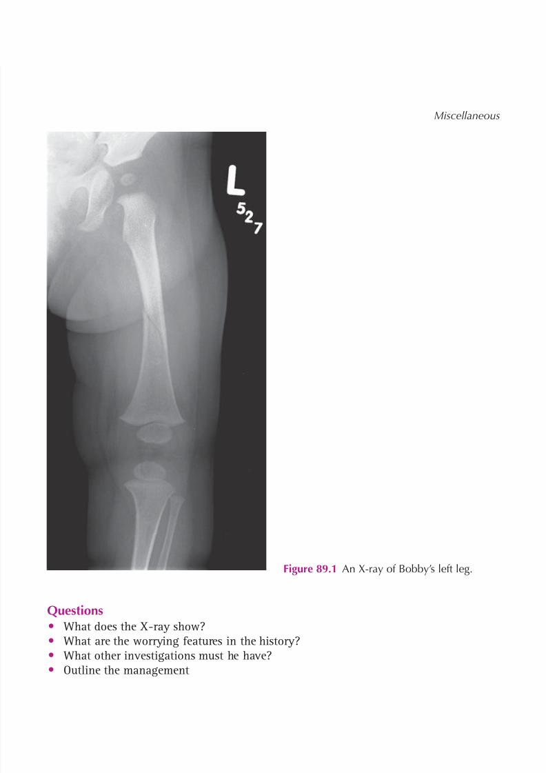
8/20/2019 100 cases in paediatrics.pdf
http://slidepdf.com/reader/full/100-cases-in-paediatricspdf 282/320
263
Miscellaneous
Questions• What does the X-ray show?• What are the worrying features in the history?• What other investigations must he have?• Outline the management
Figure 89.1 An X-ray of Bobby’s left leg.
8/20/2019 100 cases in paediatrics.pdf
http://slidepdf.com/reader/full/100-cases-in-paediatricspdf 283/320
264
100 Cases in Paediatrics
ANSWER 89The X-ray shows a mid-shaft spiral fracture of the left femur. The majority of childhoodfractures are accidental with an explanation that fits the injury. However, non-acciden-tal injury (NAI) must always be considered and there are certain patterns of presentationand findings that ring alarm bells. The child’s age and mobility are very important fac-tors. The fracture site and type are also important. Metaphyseal and posterior rib frac-tures are very suspicious and a spiral fracture suggests a twisting injury – unlikely in anon-weight-bearing infant. So the most worrying features in this history are an unex-plained spiral long bone fracture in an infant who is immobile and under constant adultsupervision. There has also been a delay in reporting the injury. All of these are featuresthat make any injury more likely to be non-accidental. Others include:
• a history that is not consistent with the injury • a history that changes with time• inappropriate reaction(s) of carer(s) – vague, elusive or aggressive• history of a suspicious injury or NAI in child and/or siblings.
Any infant with suspected NAI must have a full skeletal survey with an expert radi ol-ogy assessment because not all fractures are detectable clinically. Rib fractures may beinconspicuous initially and a bone scan and/or a repeat chest X-ray may also be needed.These investigations should also help exclude the rare differential of osteogenesis imper-fecta. He should also have a brain CT scan to exclude injuries such as a subdural haem-
orrhage. If there is bruising, a full blood count and a clotting screen are necessary.
! Steps in management
• Admit to manage the fracture and as a place of safety• Pain relief and immobilization in traction• Check the child protection register• Document, date and sign the history, examination and investigations• Inform a senior paediatrician, who will confirm the findings and explain openly
and non-judgmentally why there are concerns, what is going to happen nextand who will be involved
• Refer immediately to Social Care. Where appropriate, they will involve thepolice
• Attend the multidisciplinary child protection conference to which the parentsare invited. Information is shared between all professional groups. Outcomesare the decision of the conference, not of individuals. The aim is always to act inthe child’s best interest
KEY POINTS
• The possibility of non-accidental injury should be considered in any childhood fracture.• Thorough documentation is vital in all possible cases of non-accidental injury.• Communication between all professional groups is essential in deciding the outcome
for the child and family.
This family have other risk factors for NAI. They are young, have no local support, andthere have been difficulties with feeding. The father admitted to losing his temper in
the middle of the night after neighbours complained about the crying. He wrenched thebaby’s leg trying to get it out of his sleep suit.

8/20/2019 100 cases in paediatrics.pdf
http://slidepdf.com/reader/full/100-cases-in-paediatricspdf 284/320
8/20/2019 100 cases in paediatrics.pdf
http://slidepdf.com/reader/full/100-cases-in-paediatricspdf 285/320
266
ANSWER 90The likely diagnosis is vulvovaginitis. Prepubertal girls may have a clear sticky dischargethat resolves with menarche. However, a malodorous discharge indicates pathology. It ismost likely that a Streptococcus was the cause of the upper respiratory tract infectionand that this infection was transmitted to the genital area by the child’s fingers. It is notuncommon to get a degree of dysuria in association with vulvovaginitis. Occasionally
vaginal bleeding may occur. To definitively diagnose a urinary tract infection, there hasto be a pure growth of 10 5 colony-forming units/mL WBCs. Jasmine’s results do notfulfil this criteria. Poor perineal hygiene may cause or exacerbate vulvovaginitis, whichoften presents shortly after girls have started self-toileting. Masturbation may also leadto vulvovaginitis. Pinworms may be associated with perianal itching, especially at night,and in about 20 per cent of cases there is an associated vulvovaginitis. If no worms
are visible, Sellotape can be applied to the perianal skin, stuck onto a glass slide andthen sent for microscopy to look for enterobius ova. Herpes infection can also leadto vulvovaginitis. It leads to vesicles and to discrete ulcers. These are usually due todigital contact with cold sores, but the possibility of sexual abuse should also beborne in mind. Viral swabs should be taken. Recurrent vulvovaginitis usually ceases atpuberty.
The differential consists of candidal infection, a foreign body, sexually transmitted dis-ease and the very rare botryoid sarcoma. Candida leads to a creamy white discharge,
vulval itching and dysuria. It is rare prepubertally but may occur in diabetics or fol-lowing antibiotic use. A foreign body may consist of toilet paper or a toy. In youngchildren it may be visible on inspection, but on occasion may require an examinationunder anaesthetic. Evidence of infection with an organism such as chlamydia or gonor-rhoea suggests sexual abuse and should prompt a child protection investigation. A sar-coma usually presents with a bloodstained vaginal discharge. On examination, a fleshyhaemorrhagic lesion is seen, which is often described as ‘grape-like’.
Treatment consists of a 1-week course of penicillin. Basic hygiene measures should alsobe advocated, such as cleaning oneself from front to back after a bowel motion, usingloose-fitting cotton knickers, having daily baths or showers using simple soaps (avoidingbubble baths or other irritants) and allowing air to dry. A short course of topical oestro-gen cream may also be helpful.
KEY POINTS
• Vulvovaginitis is common in prepubertal girls.• Advice on hygiene antibiotics is effective in treating this condition.• In children with a vaginal discharge, the possibility of sexual abuse should also be
considered.
100 Cases in Paediatrics

8/20/2019 100 cases in paediatrics.pdf
http://slidepdf.com/reader/full/100-cases-in-paediatricspdf 286/320
267
CASE 91:AN ODD SHAPED HEAD
HistoryColin is a 5-month-old boy who is referred to the paediatric clinic by his GP for assess-ment of an abnormal head shape. He was born at 38 weeks’ gestation by forceps deliv-ery. Apart from a small bruise across his forehead, no abnormality was noted at thattime. He has not had any medical problems since birth, but his parents have noted thatover the last few months his head shape has become increasingly asymmetrical. They are
very worried about this cosmetically, and the GP has worried them further by suggest-ing that he may have craniosynostosis. Colin is the first child of American parents whoboth work in banking. He was bottle-fed from birth because his mother found breast-feeding very difficult. He is described as a contented child who sleeps well and cries little.Developmental history reveals that he can sit with support from cushions, although heprefers lying on his back, he can pick up objects with the palm of either hand and putsthem in his mouth, he holds his milk bottle, he enjoys vocal turn-taking and makes arange of sounds. When he was younger, he mainly lay on the right side of his head, butthis is less obvious now as he is able to move about more.
ExaminationColin is a healthy-looking boy. His weight is 7.5 kg (50th centile), his length is 67 cm (75thcentile) and his head circumference is 43.5 cm (50th centile). His head is parallelogram-shaped when viewed from above, with the right occipital region appearing flattened (seeFig. 91.1). There is no ridging over any of the cranial sutures. His face appears symmet-rical and there are no dysmorphic features. He does not have a squint and cranial nervefunction appears normal. His anterior fontanelle is normal.
Questions• What is the most likely cause of the abnormal head shape?• What is craniosynostosis and what complications can it cause?
• What treatment might be available for Colin?
Figure 91.1 Colin’s head shape. (Reproducedwith permission of The London OrthoticConsultancy Ltd.)
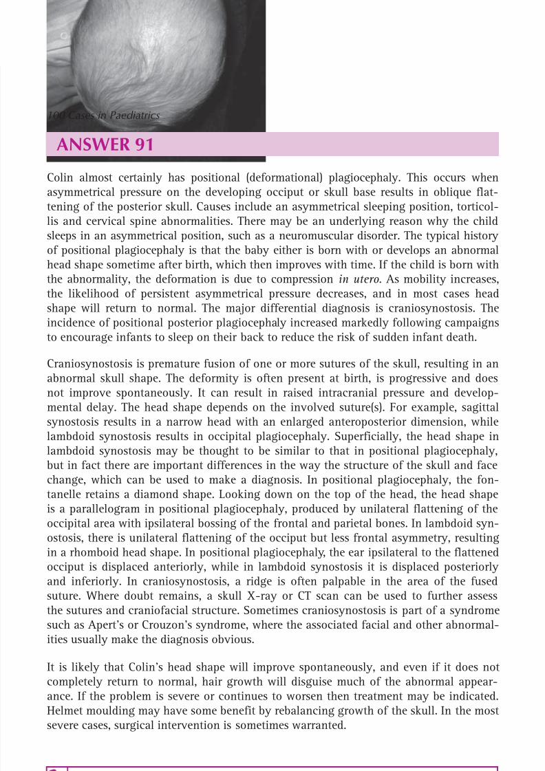
8/20/2019 100 cases in paediatrics.pdf
http://slidepdf.com/reader/full/100-cases-in-paediatricspdf 287/320
268
ANSWER 91Colin almost certainly has positional (deformational) plagiocephaly. This occurs whenasymmetrical pressure on the developing occiput or skull base results in oblique flat-tening of the posterior skull. Causes include an asymmetrical sleeping position, torticol-lis and cervical spine abnormalities. There may be an underlying reason why the childsleeps in an asymmetrical position, such as a neuromuscular disorder. The typical historyof positional plagiocephaly is that the baby either is born with or develops an abnormalhead shape sometime after birth, which then improves with time. If the child is born withthe abnormality, the deformation is due to compression in utero . As mobility increases,the likelihood of persistent asymmetrical pressure decreases, and in most cases headshape will return to normal. The major differential diagnosis is craniosynostosis. Theincidence of positional posterior plagiocephaly increased markedly following campaigns
to encourage infants to sleep on their back to reduce the risk of sudden infant death.
Craniosynostosis is premature fusion of one or more sutures of the skull, resulting in anabnormal skull shape. The deformity is often present at birth, is progressive and doesnot improve spontaneously. It can result in raised intracranial pressure and develop-mental delay. The head shape depends on the involved suture(s). For example, sagittalsynostosis results in a narrow head with an enlarged anteroposterior dimension, whilelambdoid synostosis results in occipital plagiocephaly. Superficially, the head shape inlambdoid synostosis may be thought to be similar to that in positional plagiocephaly,
but in fact there are important differences in the way the structure of the skull and facechange, which can be used to make a diagnosis. In positional plagiocephaly, the fon-tanelle retains a diamond shape. Looking down on the top of the head, the head shapeis a parallelogram in positional plagiocephaly, produced by unilateral flattening of theoccipital area with ipsilateral bossing of the frontal and parietal bones. In lambdoid syn-ostosis, there is unilateral flattening of the occiput but less frontal asymmetry, resultingin a rhomboid head shape. In positional plagiocephaly, the ear ipsilateral to the flattenedocciput is displaced anteriorly, while in lambdoid synostosis it is displaced posteriorlyand inferiorly. In craniosynostosis, a ridge is often palpable in the area of the fusedsuture. Where doubt remains, a skull X-ray or CT scan can be used to further assessthe sutures and craniofacial structure. Sometimes craniosynostosis is part of a syndromesuch as Apert’s or Crouzon’s syndrome, where the associated facial and other abnormal-ities usually make the diagnosis obvious.
It is likely that Colin’s head shape will improve spontaneously, and even if it does notcompletely return to normal, hair growth will disguise much of the abnormal appear-ance. If the problem is severe or continues to worsen then treatment may be indicated.Helmet moulding may have some benefit by rebalancing growth of the skull. In the mostsevere cases, surgical intervention is sometimes warranted.
100 Cases in Paediatrics
KEY POINTS
• Positional posterior plagiocephaly is the most common cause of abnormal headshape.
• Craniosynostosis usually causes progressive deformation with a ridge palpable overthe fused suture(s).
• Positional plagiocephaly usually improves spontaneously.

8/20/2019 100 cases in paediatrics.pdf
http://slidepdf.com/reader/full/100-cases-in-paediatricspdf 288/320
269
CASE 92:A DROWSY TODDLER
HistoryJonathan is a 21-month-old boy referred to the paediatric day unit by his GP. He was seenin the surgery the previous day with diarrhoea and vomiting and seemed to have tummyache. The vomit contained a small amount of fresh blood, but as he was otherwise welland cardiovascularly stable, admission had been deferred because his mother is 36 weeksinto her fourth pregnancy. Instead, later that day, the GP rang to check how he was andwas reassured to hear that he seemed to have recovered and was tolerating drinks andsome food. However, this morning he seems lethargic and a finger-prick blood glucosetest performed by his GP was only 3.1 mmol/L. Jonathan is the youngest of three children,the older two being 6 and 3 years old. His father is in the navy and is currently at sea.
ExaminationHe looks unwell. His airway is patent, his respiratory rate is 26 breaths/min and hispulse rate is 180 beats/min with a capillary refill time of 5 s. His blood pressure is60/35 mmHg. He is jaundiced. Both heart sounds are present and normal. Examinationof the respiratory and abdominal systems is normal. He is drowsy but knows his motherand responds to her voice. He resists examination and withdraws to pain. There is no
meningism and there are no focal neurological signs.
INVESTIGATIONS
Normal
Haemoglobin 12.3g/dL 11.5–15.5 g/dLWhite cell count 8.4 109 /L 6.0 –17.5 109 /LPlatelets 140 109 /L 150–400 109 /LProthrombin time 19 s 11–15 sPartial thromboplastin time 32 s 25–35 s
Sodium 138 mmol/L 138–146 mmol/LPotassium 3.6 mmol/L 3.5–5.0 mmol/LUrea 8.2 mmol/L 1.8–6.4 mmol/LCreatinine 33 µmol/L 27–62 µmol/LGlucose 2.9 mmol/L 3.3–5.5 mmol/LBilirubin 85 mmol/L 2–26 mmol/LAlanine aminotransferase (ALT) 1875 IU/L 5–45 IU/LAlkaline phosphatase 2624 IU/L 145–420 IU/LAlbumin 32 g/L 39–50 g/LC-reactive protein 6 mg/L 6 mg/L
Lactate 3.2 0.8–1.5 mmol/LVenous blood gas on 15 L/min of oxygen pH 7.28 7.35–7.45
P CO2 3.8 kPa 4.5–6.0 kPa Bicarbonate 17 mmol/L 22–29 mmol/L
Questions• What pathological processes are evident from his clinical signs and investigations?• What is the most likely unifying cause?• How is this condition managed?

8/20/2019 100 cases in paediatrics.pdf
http://slidepdf.com/reader/full/100-cases-in-paediatricspdf 289/320
270
ANSWER 92Firstly, this boy has acute liver dysfunction. He has a prolonged prothrombin time,hypoglycaemia and low albumin – these are markers of the manufacturing processesof the liver. There is liver inflammation with an elevated ALT and bilirubin. Secondly,he has clinical and biochemical evidence of tissue underperfusion and increased anaer-obic metabolism. He has a tachycardia, borderline hypotension and a prolonged capil-lary refill time with a metabolic acidosis and a high lactate. There is a compensatoryreduction in the P CO2 (see Case 86, p. 253 ). Thirdly, there are clinical symptoms andsigns suggestive of mild encephalopathy, although his drowsiness could be due to thehypoglycaemia. Finally, note the high urea. This could be due to dehydration but hisdiarrhoea and vomiting were resolving and the creatinine is normal. It is more likelyto be due to the digestion of blood following upper gastrointestinal tract bleeding. Ask
about melaena stools.The most likely unifying cause is some form of poisoning. This history is classical foriron toxicity. Iron is corrosive to the gastrointestinal mucosa, causing abdominal pain,nausea, vomiting and diarrhoea within a few hours of ingestion with haematemesis andbloody diarrhoea in more severe toxicity. Thereafter, there is an interlude with an appar-ent recovery from about 8–16 hours. This boy has entered the third stage (16–24 hours)with progressive systemic involvement due to the vasodilator effects of iron and mito-chondrial poisoning. Toxicity is related to the amount of ferrous iron ingested, which
varies according to the particular iron salt. All preparations carry this information, e.g.a ferrous fumarate tablet 210 mg contains 65 mg of ferrous iron. Toxicity is unlikely if
20 mg/kg is ingested but the risk increases steadily thereafter. Not many tablets areneeded to cause significant poisoning.
This child’s mother was receiving iron treatment during her pregnancy and found thepack in his bedroom. Iron is one of the most common causes of childhood accidentalpoisoning because iron-containing preparations are widely available and often resemblesweets. Some units ask a health visitor to undertake a home visit after any case of acci-dental poisoning to assess overall home safety and to remind parents about keeping alldrugs out of reach of children.
! Management of iron poisoning
• Give oxygen and intravenous fluids to manage poor perfusion• Treat hypoglycaemia• Send the family home to check for missing drugs and to bring in any remaining
drugs for identification• Whole iron tablets are radio-opaque – an abdominal X-ray to confirm ingestion
may help• Try to remove the poison. If taken within 1 hour of presentation or tablets
visible in stomach on X-ray, consider gastric lavage with a wide-bore tube• Measure serum iron urgently, but remember that it may be spuriously low if
taken 8 hours post-ingestion• Try to estimate the quantity of iron ingested and the risk of toxicity• Discuss with National Poisons Information Service (24 hour service) who will
advise about treatment with desferrioxamine, a specific chelator of iron
• Discuss with a specialist liver unit
100 Cases in Paediatrics

8/20/2019 100 cases in paediatrics.pdf
http://slidepdf.com/reader/full/100-cases-in-paediatricspdf 290/320
271
Miscellaneous
KEY POINTS
• Advice should be sought from the National Poisons Information Service if there is anydoubt about the management of a case of poisoning.
• Iron is one of the most common causes of childhood poisoning.

8/20/2019 100 cases in paediatrics.pdf
http://slidepdf.com/reader/full/100-cases-in-paediatricspdf 291/320
This page intentionally left blank
8/20/2019 100 cases in paediatrics.pdf
http://slidepdf.com/reader/full/100-cases-in-paediatricspdf 292/320

8/20/2019 100 cases in paediatrics.pdf
http://slidepdf.com/reader/full/100-cases-in-paediatricspdf 293/320
274
100 Cases in Paediatrics
ANSWER 93This previously healthy teenage boy has developed an acute confusional state. One ofthe most obvious causes is drug ingestion, either recreational or a deliberate overdose,possibly with his brother’s lamotrigine. It is important to ask the family specific ques-tions. Has he been out of their sight? Are there any other drugs in the house? If neces-sary, send them home to check and make sure none are missing. Phone the school andhis friends. All these steps could be life-saving. A urine screen for ‘drugs of abuse’ andtoxicology, including lamotrigine, should be requested but the result will take some time.
However, never jump to conclusions – there are numerous other possibilities. Withan acute history, metabolic problems are unlikely although they must be excluded.Hyponatraemia could be due to inappropriate antidiuretic hormone secretion following
an unreported head injury. Hypercalcaemia and renal failure are likely to have a moreinsidious onset and other symptoms, such as tiredness. Thyroid disease is a possibil-ity. Thyrotoxicosis can present with an isolated behaviour disturbance such as schoolphobia. At this age, a previously undiagnosed inborn error of metabolism is unlikely butis certainly possible, especially in a child with learning difficulties. Measure blood sugar(bedside and laboratory), a venous pH, lactate and ammonia. Keep urine for amino andorganic acid analysis. Ask the laboratory to keep any spare blood in case it is needed forfuture analysis. Most will do so for up to 6 months.
Even in the absence of clinical signs, bacterial and viral infections are possible diag-
noses. They are also treatable. Send a full blood count, C-reactive protein, blood cul-tures, a midstream urine and acute viral titres. If indicated, convalescent titres can besent 10–14 days later. It is safest to start treatment with broad-spectrum antibiotics, e.g.cefotaxime, and aciclovir. A lumbar puncture is currently contraindicated because he is
very confused and raised intracranial pressure cannot be excluded. It may be indicatedonce he has improved.
An acute neurological event must also be excluded, including seizures and migraine. Hemerits an urgent CT and/or a MRI scan and an EEG.
This boy’s EEG the following day showed generalized bursts of spike and slow-waveactivity with a frontotemporal emphasis. He recovered within 24 hours but went onto have further similar episodes of confusion, culminating in a generalized seizure.The final diagnosis was complex partial epilepsy with secondary generalization. Heresponded very well to anticonvulsants.
KEY POINTS
• Do everything possible to identify any drug that a child might have takeninadvertently or deliberately. It might save their life.
• Never assume that an acute confusional state is due to drug ingestion at any age.

8/20/2019 100 cases in paediatrics.pdf
http://slidepdf.com/reader/full/100-cases-in-paediatricspdf 294/320
275
CASE 94:A DRUNKEN ADOLESCENT
HistoryLaura is a 14-year-old girl who is admitted to the A&E department. Her friend whoaccompanies her states that she collapsed on the pavement having had a bottle of vodkaand then disappears. She has vomited once in the ambulance and once in casualty. Thereis no other history available. Her parents are not present.
Examination Airway, breathing and circulation are stable. Glasgow Coma Score (GCS) is 6.Temperature is 36.4 C. There are no signs of trauma, no focal neurological signs and noother signs.
Questions• Should this child be ventilated?• What other investigation should be performed?• What treatment should be administered?• What wider aspects of this adolescent’s case should be looked into?
INVESTIGATIONS
Urea and electrolytes NormalBone chemistry NormalLiver function tests NormalBlood glucose 5.6 mmol/LBlood alcohol level 210 mg/100 mL.
8/20/2019 100 cases in paediatrics.pdf
http://slidepdf.com/reader/full/100-cases-in-paediatricspdf 295/320
276
100 Cases Paediatrics
ANSWER 94The airway of any child with a GCS of 8 or less is at risk. An anaesthetist should becalled urgently to monitor the airway and breathing. A blood gas may help the assess-ment. Most children in this setting will manage without being intubated and slowly,spontaneously recover.
A cranial computed tomography (CT) scan should be done. There is very little history. Although there are no external marks of a head injury or focal neurological signs, itis possible that Laura sustained a head injury when she fell on the pavement and thatthis partly accounts for her low GCS. The vomiting is probably secondary to the alcoholingestion but may be secondary to a head injury.
Maintenance intravenous fluids (5 per cent dextrose/0.45 per cent saline) should beadministered to help avoid the hangover headache that is primarily due to dehydration.Hourly GCS observations should be performed.
Binge-drinking is very common in teenagers. It is important to try to find out the child’sname and contact their family as soon as possible. Social services should be contacted toinform them of what has happened and to find out if she is known to them. When she isawake it would be worth asking whether she takes recreational drugs and consider doingurine toxicology, if appropriate. One should also ask about the possibility of sexual abusewhilst she was drunk and unconscious. However, if there are no allegations of sexualabuse and no evidence to suggest it (e.g. missing or torn knickers) then this need not bepursued. If there are concerns about the possibility of sexual abuse, a forensic medicalexaminer should be contacted to do the necessary examination and to take the neces-sary forensic swabs. There is an association between alcohol abuse, behavioural disordersand depression. In some cases, psychiatric and/or social work input and follow-up isrequired.
KEY POINTS
• Binge-drinking is very common in adolescents.• In drunken teenagers, always measure the blood glucose level.• In children with a diminished GCS, a cranial CT should be done to rule out the
possibility of a coexisting intracranial injury.• Remember to consider the psychosocial aspects of the case.
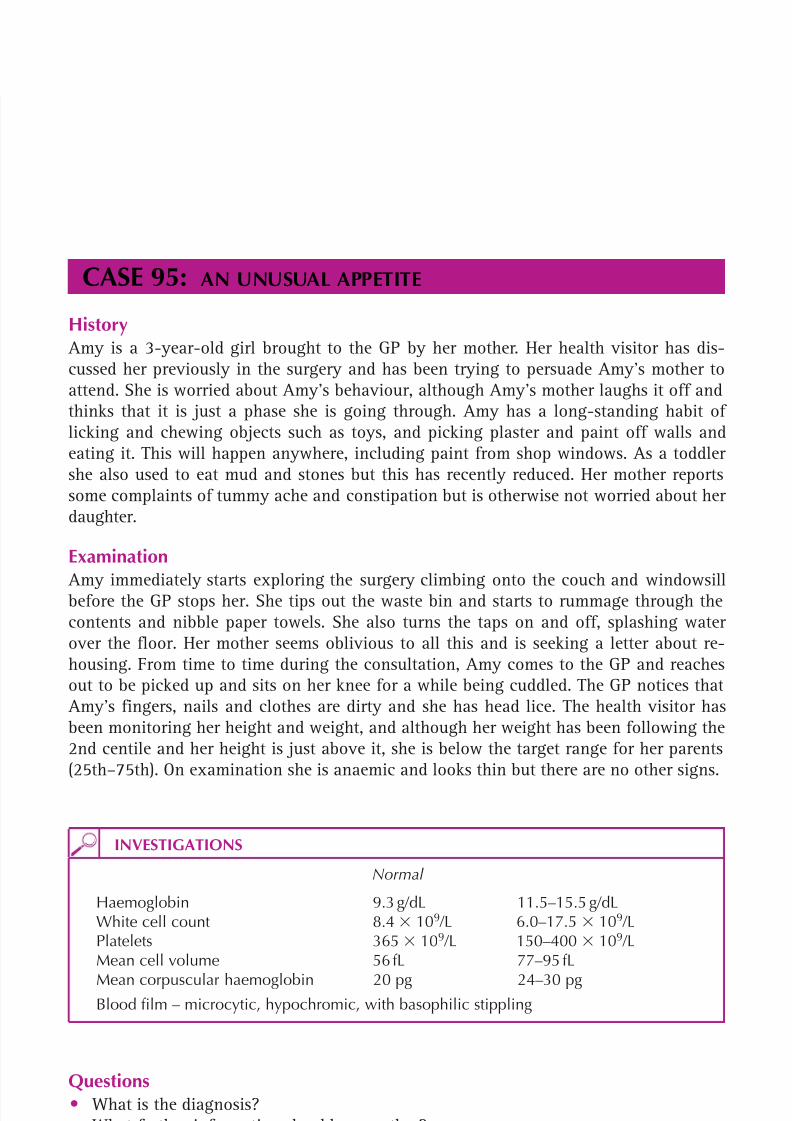
8/20/2019 100 cases in paediatrics.pdf
http://slidepdf.com/reader/full/100-cases-in-paediatricspdf 296/320
277
CASE 95:AN UNUSUAL APPETITE
History Amy is a 3-year-old girl brought to the GP by her mother. Her health visitor has dis-cussed her previously in the surgery and has been trying to persuade Amy’s mother toattend. She is worried about Amy’s behaviour, although Amy’s mother laughs it off andthinks that it is just a phase she is going through. Amy has a long-standing habit oflicking and chewing objects such as toys, and picking plaster and paint off walls andeating it. This will happen anywhere, including paint from shop windows. As a toddlershe also used to eat mud and stones but this has recently reduced. Her mother reportssome complaints of tummy ache and constipation but is otherwise not worried about herdaughter.
Examination Amy immediately starts exploring the surgery climbing onto the couch and windowsillbefore the GP stops her. She tips out the waste bin and starts to rummage through thecontents and nibble paper towels. She also turns the taps on and off, splashing waterover the floor. Her mother seems oblivious to all this and is seeking a letter about re-housing. From time to time during the consultation, Amy comes to the GP and reaches
out to be picked up and sits on her knee for a while being cuddled. The GP notices that Amy’s fingers, nails and clothes are dirty and she has head lice. The health visitor hasbeen monitoring her height and weight, and although her weight has been following the2nd centile and her height is just above it, she is below the target range for her parents(25th–75th). On examination she is anaemic and looks thin but there are no other signs.
INVESTIGATIONS
Normal Haemoglobin 9.3 g/dL 11.5–15.5 g/dLWhite cell count 8.4 109 /L 6.0–17.5 109 /LPlatelets 365 109 /L 150–400 109 /LMean cell volume 56 fL 77–95 fLMean corpuscular haemoglobin 20 pg 24–30 pgBlood film – microcytic, hypochromic, with basophilic stippling
Questions• What is the diagnosis?• What further information should you gather?• What complications have arisen?• What is the management?
8/20/2019 100 cases in paediatrics.pdf
http://slidepdf.com/reader/full/100-cases-in-paediatricspdf 297/320
278
100 Cases in Paediatrics
ANSWER 95 Amy has pica, the repeated or chronic ingestion of non-nutritive substances. This isalmost universal in infants and toddlers but abnormal after the age of 2 years and needsinvestigation.
It is very unusual for a child to spontaneously reach out to a relatively unknown adultfor comfort. This behaviour should always raise the possibility of emotional and socialneglect and these are known predisposing factors for pica. Others include autism andlearning difficulties and the GP needs to ensure that there is no significant developmentaldelay. Her social behaviour is not typical of autism. The difficulty persuading the motherto attend, her indifference to the problem and her behaviour during the consultation alladd to the concerns. Amy is also small for her family and thin. Emotional and physical
neglect can lead to psychosocial dwarfism. A full social history should be gathered aboutthe family and their circumstances. Who else is at home? Does the mother have a partnerand is he the father of Amy? Is there a history of drug or alcohol abuse or domestic vio-lence? Are there other children? Is this family known to Social Care?
Anaemia and lead poisoning have developed. The microcytic, hypochromic anaemia isstrongly indicative of iron deficiency, which is common in childhood and almost alwaysdietary in origin. It is even more common in pica. Basophilic stippling is characteristic oflead poisoning, another known complication of pica. Sources include lead-containing paintfrom both buildings and imported painted toys. Symptoms range from colicky abdominal
pain and constipation to headache, drowsiness, fits and coma in lead encephalopathy. Oncein the intestine, lead competes with iron and calcium for binding sites, so if either or bothof these are deficient, lead absorption is enhanced. Hyperactivity, as in this case, is fre-quently a manifestation of lead poisoning in pre-school and young school-aged children.
The management of this child has two components, medical and psychosocial:
• Medical management – urgent referral to hospital– exclude other causes for poor growth and weight gain, e.g. coeliac disease– measure blood lead and seek advice from the National Poisons Information Service
about the need for chelating agents– check blood ferritin and a haemoglobinopathy screen and give a 3-month course
of iron– measure bone biochemistry and vitamin D and consider treatment with vitamin D
or a multivitamin preparation to minimize lead absorption• Psychosocial management
– referral to Social Care for a multidisciplinary assessment of the whole family – try to establish the source of lead and remove it or restrict access to it – involve the
Health Protection Agency – likely to need referral to Child and Family Therapy service for behaviour management.
KEY POINTS
• Exclude lead poisoning in any child with a history of pica.• In lead poisoning, investigate and treat any coexisting calcium and iron deficiency to
minimize lead absorption.• Emotional and social neglect are known predisposing factors for pica.
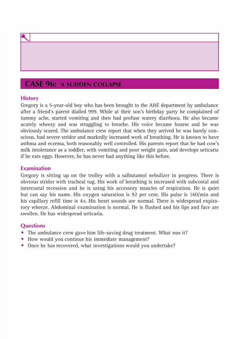
8/20/2019 100 cases in paediatrics.pdf
http://slidepdf.com/reader/full/100-cases-in-paediatricspdf 298/320
279
CASE 96:A SUDDEN COLLAPSE
HistoryGregory is a 5-year-old boy who has been brought to the A&E department by ambulanceafter a friend’s parent dialled 999. While at their son’s birthday party he complained oftummy ache, started vomiting and then had profuse watery diarrhoea. He also becameacutely wheezy and was struggling to breathe. His voice became hoarse and he wasobviously scared. The ambulance crew report that when they arrived he was barely con-scious, had severe stridor and markedly increased work of breathing. He is known to haveasthma and eczema, both reasonably well controlled. His parents report that he had cow’smilk intolerance as a toddler, with vomiting and poor weight gain, and develops urticariaif he eats eggs. However, he has never had anything like this before.
ExaminationGregory is sitting up on the trolley with a salbutamol nebulizer in progress. There isobvious stridor with tracheal tug. His work of breathing is increased with subcostal andintercostal recession and he is using his accessory muscles of respiration. He is quietbut can say his name. His oxygen saturation is 92 per cent. His pulse is 160/min andhis capillary refill time is 4 s. His heart sounds are normal. There is widespread expira-
tory wheeze. Abdominal examination is normal. He is flushed and his lips and face areswollen. He has widespread urticaria.
Questions• The ambulance crew gave him life-saving drug treatment. What was it?• How would you continue his immediate management?• Once he has recovered, what investigations would you undertake?
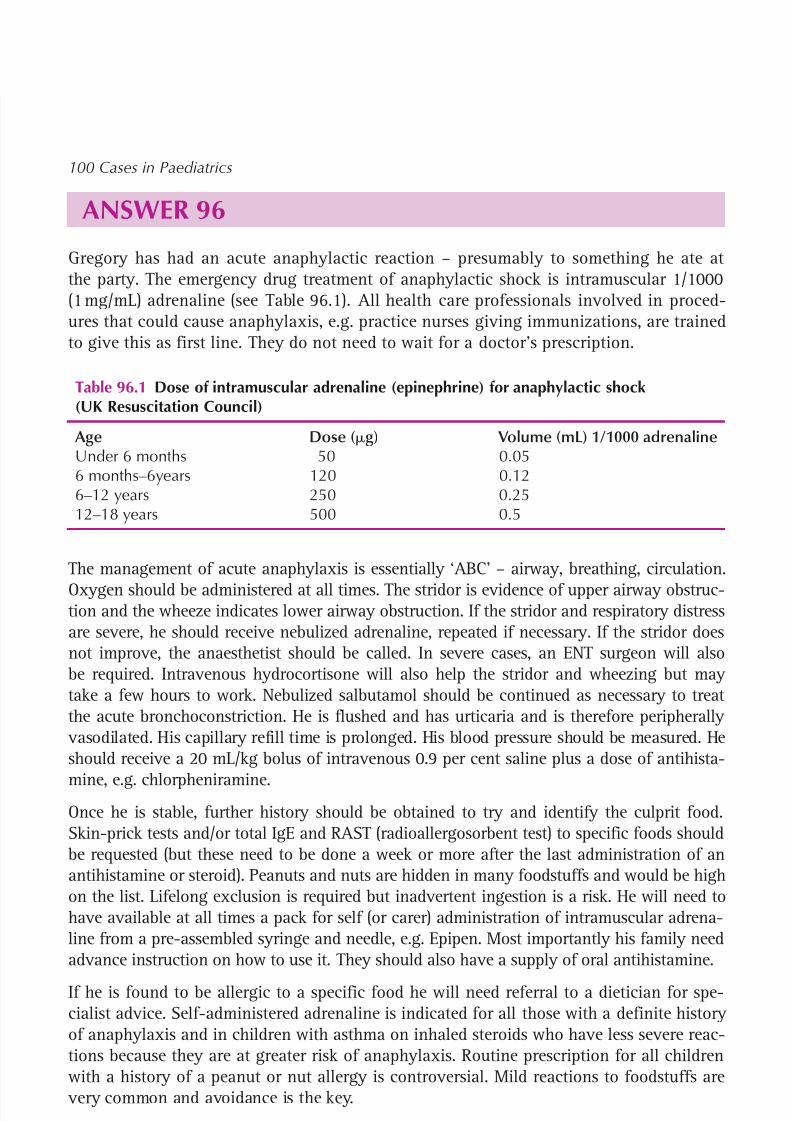
8/20/2019 100 cases in paediatrics.pdf
http://slidepdf.com/reader/full/100-cases-in-paediatricspdf 299/320
280
ANSWER 96Gregory has had an acute anaphylactic reaction – presumably to something he ate atthe party. The emergency drug treatment of anaphylactic shock is intramuscular 1/1000(1 mg/mL) adrenaline (see Table 96.1). All health care professionals involved in proced-ures that could cause anaphylaxis, e.g. practice nurses giving immunizations, are trainedto give this as first line. They do not need to wait for a doctor’s prescription.
Table 96.1 Dose of intramuscular adrenaline (epinephrine) for anaphylactic shock(UK Resuscitation Council)
Age Dose ( g) Volume (mL) 1/1000 adrenalineUnder 6 months 50 0.056 months–6years 120 0.126–12 years 250 0.2512–18 years 500 0.5
The management of acute anaphylaxis is essentially ‘ABC’ – airway, breathing, circulation.Oxygen should be administered at all times. The stridor is evidence of upper airway obstruc-tion and the wheeze indicates lower airway obstruction. If the stridor and respiratory distressare severe, he should receive nebulized adrenaline, repeated if necessary. If the stridor doesnot improve, the anaesthetist should be called. In severe cases, an ENT surgeon will alsobe required. Intravenous hydrocortisone will also help the stridor and wheezing but maytake a few hours to work. Nebulized salbutamol should be continued as necessary to treatthe acute bronchoconstriction. He is flushed and has urticaria and is therefore peripherally
vasodilated. His capillary refill time is prolonged. His blood pressure should be measured. Heshould receive a 20 mL/kg bolus of intravenous 0.9 per cent saline plus a dose of antihista-mine, e.g. chlorpheniramine.
Once he is stable, further history should be obtained to try and identify the culprit food.Skin-prick tests and/or total IgE and RAST (radioallergosorbent test) to specific foods shouldbe requested (but these need to be done a week or more after the last administration of an
antihistamine or steroid). Peanuts and nuts are hidden in many foodstuffs and would be highon the list. Lifelong exclusion is required but inadvertent ingestion is a risk. He will need tohave available at all times a pack for self (or carer) administration of intramuscular adrena-line from a pre-assembled syringe and needle, e.g. Epipen. Most importantly his family needadvance instruction on how to use it. They should also have a supply of oral antihistamine.
If he is found to be allergic to a specific food he will need referral to a dietician for spe-cialist advice. Self-administered adrenaline is indicated for all those with a definite historyof anaphylaxis and in children with asthma on inhaled steroids who have less severe reac-tions because they are at greater risk of anaphylaxis. Routine prescription for all children
with a history of a peanut or nut allergy is controversial. Mild reactions to foodstuffs are very common and avoidance is the key.
KEY POINT
• Intramuscular adrenaline is the first-line treatment for acute anaphylaxis.
100 Cases in Paediatrics

8/20/2019 100 cases in paediatrics.pdf
http://slidepdf.com/reader/full/100-cases-in-paediatricspdf 300/320
281
CASE 97:A PREGNANT
14-YEAR
-OLD
History Anna is a 14-year-old girl who presents to her GP worried that she may be pregnant.She started her periods at the age of 11 and they have been pretty regular, but she hasnow missed two and has been suffering from nausea most mornings. She met a 19-year-old man called Ryan at a party 6 months ago and they have been seeing each othersince, mainly at his flat which he shares with three other people, but occasionally ather house when her parents are at work. She remembers a night a couple of months agowhen she and Ryan had had a few drinks and the condom slipped off. They thought itwould be fine. He works as a chef and she has been playing truant and missing schoolif he is working in the evening. Up until now she has been doing well at school, but sheanticipates that her grades will slip this year.
Ryan is not her first sexual partner and she has always relied on condoms as she is wor-ried that the contraceptive pill will make her put on weight. Anna has not told her familyabout Ryan knowing that they will disapprove. She has no significant past medical history.Unfortunately, Ryan is also a smoker and she has started smoking cigarettes. She deniestaking any other recreational drugs. Anna is adamant that she does not want the baby andis equally adamant that she does not want to involve her parents or Ryan in any decisionabout a termination. None of them knows she is seeing the GP.
Examination Anna is generally healthy. There is nothing abnormal to find on examination and theuterus is not palpable.
INVESTIGATIONS
Urine -human chorionic gonadotrophin is positive.
Questions• Where does the GP stand legally regarding Anna and Ryan’s relationship?• May Anna make up her own mind about a termination?
8/20/2019 100 cases in paediatrics.pdf
http://slidepdf.com/reader/full/100-cases-in-paediatricspdf 301/320
282
ANSWER 97Ryan has broken the Sexual Offences Act (2003) by having sexual intercourse with a
young person under 16 years. However, the Act aims to reduce sexual exploitation andabuse of children and young people, not to criminalize normal adolescent behaviour. Ifall young people known to be sexually active were reported to the police, they wouldprobably be less likely to access contraceptive and sexual health services, leaving themmore vulnerable to unintended pregnancy and other health risks. Under the Act, allthose under 16 have the right to confidential advice, and the person offering it, med icalor non-medical, is not guilty of any offence, provided they are protecting the child’sphysical and/or emotional well-being.
The key words are ‘exploitation’ and ‘abuse’, and in the rare event of these being present,
the patient must understand that absolute confidentiality cannot be guaranteed. Todetermine whether a relationship presents a risk that needs referral to Social Care and/orpolice, the GP needs to consider:
• whether the young person is competent to understand and consent to the sexualactivity – according to the Act, children under 13 are not and anyone involved inpenetrative sex can be convicted of rape
• power imbalances through differences in size, age and development – even 16 and17-year-olds cannot give informed consent if the perpetrator is in a position of trust,such as a teacher or youth worker
• whether there was aggression, manipulation or bribery including the use of drugsand/or alcohol
• attempts to secure unreasonable secrecy • whether the partner is known by agencies to have worrying relationships with other
young people• evidence of parental neglect or lack of supervision in a child under 13• whether the relationship involves behaviours considered to be ‘grooming’ in the
context of sexual exploitation.
From Anna’s history there are no obvious legal or safeguarding children’s anxietiesabout her relationship with Ryan, although it is far from ideal. Therefore the GP doesnot have to report it to anyone.
In UK law the legal age of consent to medical treatment is 16 years. There is a legal prece-dent for younger children to give valid consent provided they fulfil approved criteria. Tobe ‘Gillick’ competent they must demonstrate sufficient maturity and intelligence (capacity)to understand the nature and implications of the proposed treatment, including the risksand alternatives, and the consequences of not having it.
This means that Anna could have a termination without her parents’ (or her boyfriend’s)knowledge, although she should be actively encouraged to tell them.
Note that a child is not considered to have the capacity to refuse investigation or treat-ment against the judgment of their parents or doctors. A 15-year-old cannot refuseblood tests or treatment, although every effort should be made to understand their fearsand wishes.
100 Cases in Paediatrics
8/20/2019 100 cases in paediatrics.pdf
http://slidepdf.com/reader/full/100-cases-in-paediatricspdf 302/320
283
KEY POINTS
• All children under 16 years have the right to confidential sexual health advice.• Only where there is evidence of exploitation or abuse should cases of underage sex
be referred to Social Care and/or police.• Young people can, under specific circumstances, consent to treatment independently
of their parents but they should always be encouraged to involve them.
Miscellaneous

8/20/2019 100 cases in paediatrics.pdf
http://slidepdf.com/reader/full/100-cases-in-paediatricspdf 303/320
This page intentionally left blank

8/20/2019 100 cases in paediatrics.pdf
http://slidepdf.com/reader/full/100-cases-in-paediatricspdf 304/320
285
CASE 98:A SUDDEN
,UNEXPECTED DEATH
HistoryZara is a 4-month-old infant who is brought to the A&E department and taken straightto the resuscitation bay. Her parents say that they had gone to bed with Zara lying inthe bed between them and when they woke up she was blue, not breathing and lifeless.She had been generally well the previous day but had not fed as well as usual. Zara wasborn at 32 weeks gestation, weighing 1.90 kg, and had been in the special care baby unitfor 5 weeks. She had required oxygen for 2 days and had been nasogastrically fed for 3weeks. The parents are not consanguineous and Zara is their first child.
The ambulance crew state that the baby was not breathing and asystolic when theyarrived. Zara has had basic life support during the 10-min journey to the hospital.
ExaminationThere is no respiratory rate and no air entry. Oxygen saturation is unrecordable. There isno heart rate and a flat ECG trace. The baby is limp and unresponsive, with a tempera-ture of 35.8 C.
Questions• What is your immediate management?• Which investigations should be performed?• What is the likely outcome?• How would you proceed if the child did not survive?
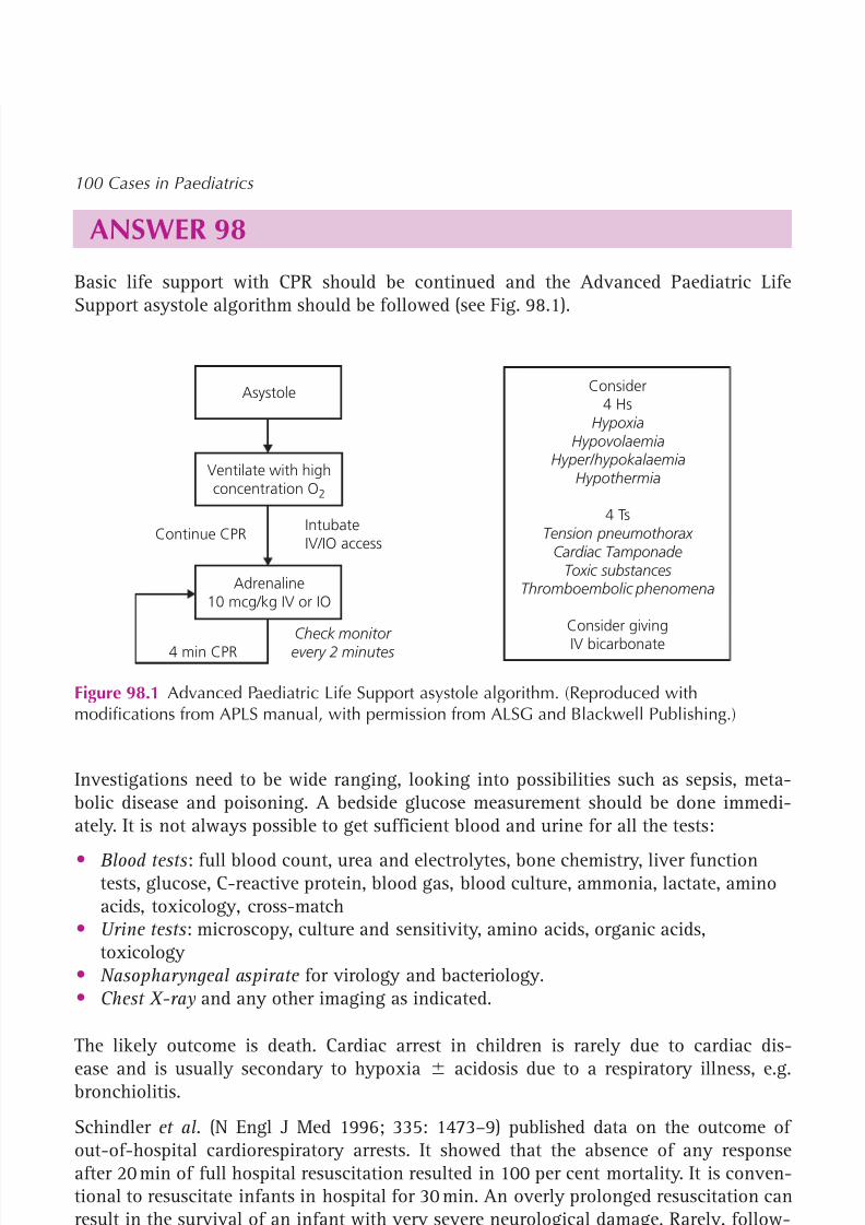
8/20/2019 100 cases in paediatrics.pdf
http://slidepdf.com/reader/full/100-cases-in-paediatricspdf 305/320
286
ANSWER 98Basic life support with CPR should be continued and the Advanced Paediatric LifeSupport asystole algorithm should be followed (see Fig. 98.1).
Continue CPRIntubateIV/IO access
4 min CPRCheck monitor every 2 minutes
Ventilate with highconcentration O 2
Adrenaline10 mcg/kg IV or IO
Asystole Consider4 Hs
HypoxiaHypovolaemia
Hyper/hypokalaemiaHypothermia
4 Ts
Tension pneumothorax Cardiac Tamponade
Toxic substancesThromboembolic phenomena
Consider givingIV bicarbonate
Figure 98.1 Advanced Paediatric Life Support asystole algorithm. (Reproduced withmodifications from APLS manual, with permission from ALSG and Blackwell Publishing.)
Investigations need to be wide ranging, looking into possibilities such as sepsis, meta-bolic disease and poisoning. A bedside glucose measurement should be done immedi-ately. It is not always possible to get sufficient blood and urine for all the tests:
• Blood tests : full blood count, urea and electrolytes, bone chemistry, liver functiontests, glucose, C-reactive protein, blood gas, blood culture, ammonia, lactate, aminoacids, toxicology, cross-match
• Urine tests : microscopy, culture and sensitivity, amino acids, organic acids,toxicology
• Nasopharyngeal aspirate for virology and bacteriology.• Chest X-ray and any other imaging as indicated.
The likely outcome is death. Cardiac arrest in children is rarely due to cardiac dis-ease and is usually secondary to hypoxia acidosis due to a respiratory illness, e.g.bronchiolitis.
Schindler et al. (N Engl J Med 1996; 335: 1473–9) published data on the outcome ofout-of-hospital cardiorespiratory arrests. It showed that the absence of any response
after 20 min of full hospital resuscitation resulted in 100 per cent mortality. It is conven-tional to resuscitate infants in hospital for 30 min. An overly prolonged resuscitation canresult in the survival of an infant with very severe neurological damage. Rarely, follow-ing drowning or poisoning, resuscitation for longer than 30 min is necessary.
If the child does not survive, the parents need to be talked to sensitively and told that allsudden unexpected deaths in infancy (SUDI, the new name for the sudden infant deathsyndrome which was previously also known as cot death) are referred to the coroner andthat the baby by law requires a postmortem. The parents also need to be told that it ispossible that no cause will be found for the death. They should be told that the police
100 Cases in Paediatrics

8/20/2019 100 cases in paediatrics.pdf
http://slidepdf.com/reader/full/100-cases-in-paediatricspdf 306/320
287
will be automatically informed. The child protection register should be checked. Mosthospitals have a list of people who need to be informed after an infant has died, e.g.general practitioner.
The family need to be followed up to discuss the results of the postmortem. Bereavementcounselling should also be offered.
KEY POINTS
• Cardiac arrest in children is usually secondary to hypoxia due to respiratory disease.• Following an out-of-hospital cardiac arrest, lack of a response after 20 min of hospital
resuscitation almost invariably results in death.• All cases of SUDI require a referral to the coroner and a postmortem.
Miscellaneous

8/20/2019 100 cases in paediatrics.pdf
http://slidepdf.com/reader/full/100-cases-in-paediatricspdf 307/320
This page intentionally left blank
8/20/2019 100 cases in paediatrics.pdf
http://slidepdf.com/reader/full/100-cases-in-paediatricspdf 308/320
289
CASE 99:A TIRED TEENAGER
HistoryCharlotte is 14 years old. She is referred to the paediatric outpatient department by herGP. Over the last 6 months she has been complaining of feeling tired all the time, shehas a sore throat, headaches, pains in her arms, legs and abdomen, and feels weak. Shehas not been to school for the last 6 weeks because she can’t manage the walk to the busstop and now she needs her mother’s assistance to walk around her house. The referralletter says that they have attended the GP’s surgery five times over this period, and themother is very anxious about what is causing this. The GP did some blood tests and saysthat a full blood count, renal and liver function tests were normal.
Further history reveals that this all started with a sore throat and fever for a few days.From then on, she started to get headaches, worse in the evening, and became weakand lethargic. She seemed to get worse each week, until she was hardly doing anythingherself, staying in bed or watching television. Despite sleeping a lot, she doesn’t feelrefreshed after sleeping. Her mother has to help her to shower and dress, as she feels soweak. She aches all over and finds it very hard to concentrate on anything. Her periodshave become irregular.
Charlotte was getting top grades at school before all this and wants to be a doctor. Sheenjoys school and denies any bullying. She lives with her mother and stepfather. Shehas always been very reliant on her mother. Her sister was successfully treated for acutelymphoblastic leukaemia 3 years ago. Charlotte has never been in hospital before andusually does swimming team training five times per week.
Her mother is very worried that this could be something sinister and she has looked onthe internet and thinks it could be a brain tumour or thyroid problem. She is upset thatthe GP hasn’t taken it seriously and only did blood tests because she made a fuss.
ExaminationCharlotte is rather quiet, but will participate in conversation when prompted. Her weightis 45 kg (25th centile) and her height is 164 cm (75th centile). Otherwise, physical exam-ination is unremarkable.
Questions• What is the most likely diagnosis?• Would you request any more investigations?• What is the prognosis?

8/20/2019 100 cases in paediatrics.pdf
http://slidepdf.com/reader/full/100-cases-in-paediatricspdf 309/320

8/20/2019 100 cases in paediatrics.pdf
http://slidepdf.com/reader/full/100-cases-in-paediatricspdf 310/320

8/20/2019 100 cases in paediatrics.pdf
http://slidepdf.com/reader/full/100-cases-in-paediatricspdf 311/320
This page intentionally left blank
8/20/2019 100 cases in paediatrics.pdf
http://slidepdf.com/reader/full/100-cases-in-paediatricspdf 312/320
293
CASE 100:POOR WEIGHT GAIN
HistoryCharlie is a 14-month-old boy who is referred by his GP to the paediatric day unitbecause of diarrhoea and weight loss. He has had numerous previous admissions. Charliewas born at 32 weeks after a pregnancy complicated by recurrent bleeding. He neededheadbox oxygen for 5 days. By discharge home at 6 weeks, he was bottle-fed and hisweight was on the 25th centile. Over the next 2 months he had several admissions withpossible apnoeas, but investigations including an EEG and brain MRI were normal andnone were witnessed during observation on the ward. At 4 months he had two episodesof haematemesis confirmed on inspection of the towels his mother brought with herand had a normal full blood count, clotting screen and barium meal. His weight by thenhad dropped to the 2nd centile and it has not been above it since, and has occasionallyfallen to below the 0.4th centile.
He has persistent, frequent, loose stools often containing visible blood. A diagnosis ofcow’s milk protein intolerance was made, but a dairy-free diet made no difference. He isnow on a hydrolysated (pre-digested) formula. Solids were introduced at 5 months.
During his admissions the nurses have fed him and report that he is always hungryand takes good amounts, but there has been no significant weight gain. Charlie has hadextensive normal investigations, including an abdominal ultrasound, small bowel biopsyand a colonoscopy, when no blood was seen. There are no concerns about his devel-opment. His 4-year-old brother was also investigated for poor weight gain. With thepermission of the parents, reports have been obtained from the tertiary centre to whichthe 4-year-old was referred – he, too, had exhaustive tests but no diagnosis was evermade. His growth chart shows a rapid improvement in weight gain from about 2 years.Charlie’s mother is a pharmacy technician and his father is a lorry driver. The staff knowthem well and his mother is always happy to stay.
ExaminationCharlie looks skinny. He has redundant skin folds over his thighs and buttocks. Hisweight is just below the 0.4th centile, and his length and head circumference are on the50th centiles. He is not clubbed and not clinically anaemic. His pulse is 180 beats/min.Examination is otherwise unremarkable.
Questions• What is the most likely diagnosis?• What could be the mechanisms contributing to Charlie’s history and current clinical
state?• What should happen next?
8/20/2019 100 cases in paediatrics.pdf
http://slidepdf.com/reader/full/100-cases-in-paediatricspdf 313/320
294
ANSWER 100Charlie has had several unexplained episodes of potentially serious illness and now hasevidence of ongoing faltering weight gain with blood in his stools but not at colono-scopy. Exhaustive investigations are normal. This is on the background of being bornpreterm after a complicated pregnancy and having an older brother who had a similarpattern of illness in the first 2 years of life. This is all highly consistent with a diagnosisof fabricated or induced illness (FII), an uncommon and difficult to diagnose form ofchild abuse. The perpetrator may:
• fabricate a medical history • cause symptoms by repeatedly exposing the child to a toxin, medication, infectious
agent or physical trauma, including smothering
• alter laboratory samples or temperature measurements.
The mother is almost always responsible and a significant percentage have connectionswith the health services. Sadly the disturbed parent seems to obtain a perverse satisfac-tion from the close association with hospital care and staff. The possible mechanisms inthis case include the blood in the vomit and stools being his mother’s, possibly even hermenstrual blood. Specimens can be analysed for ‘foreign’ blood. The vaginal bleedingduring pregnancy may have been self-induced. The apnoeas could have many explan-ations and covert surveillance by the police may be needed if episodes are ongoing and
smothering is a possibility. Faltering weight gain means that energy expenditure exceedsenergy intake. Possibilities include diluting his feeds, throwing feeds away or poisoningwith laxatives or an agent that increases metabolic rate such as thyroxine – to whichthis mother, as a pharmacy technician, would have access. This possibility is supportedby his tachycardia. Thyrotoxicosis is extraordinarily rare in this age group and thyroidfunction tests may be normal if doses are not given every day. Some mothers becomehighly sophisticated at FII – and this mother may have had prior experience with herolder boy. A phone call to the GP may reveal a history of abuse during the mother’schildhood or prior mental health problems, but these do not mean that this is FII. It is allsupportive, not diagnostic, evidence.
However, it is crucial to make the diagnosis because this disorder can be very dam agingto the child, not just from the impact of the unnecessary and invasive investigations butbecause of long-term behavioural and other problems. Other children may also be atrisk. The case must be referred to Social Care, who have a statutory duty to undertake athorough investigation according to strict guidelines. The child protection register shouldbe checked and the health visitor should be contacted for further information. At thesame time, it is worth obtaining a second opinion from another consultant paediatrician.
KEY POINTS
• Fabricated and induced illness should be considered in any child with recurrentunexplained symptoms, but it may be difficult to diagnose.
• All potential cases of fabricated and induced illness must be referred to Social Care.
100 Cases in Paediatrics

8/20/2019 100 cases in paediatrics.pdf
http://slidepdf.com/reader/full/100-cases-in-paediatricspdf 314/320
295
References are by case number with relevant page number(s) following in brackets. Referenceswith a page range e.g. 25(68–70) indicate that although the subject may be mentioned only onone page, it concerns the whole case.
INDEX
abdominal mass 51( 155 –6)abdominal migraine 28( 86), 68(206 )abdominal pain
acute 27( 83–4)chronic/recurrent 28( 85–7)dysuria and 34( 105–6)
ABO incompatibility 82( 244 )abortion 97( 281–3)aches and pain 56( 169 –71)
see also headacheadolescents (teenagers)
abdominal pain (acute) 27( 83–4)chest pain 8( 27–8), 50(150 –3)confusion 93( 273 –4)diarrhoea (chronic) 25( 75–7)drunkenness 94( 275 –6)eating disorder 76funny turn 13( 41–3)
limp, causes 57( 174 )pregnancy 97( 281–3)shoulder swelling 54( 163 –4)tiredness 99( 289 –91)wheezing 6( 19–21)see also puberty
adrenal hyperplasia, congenital 20( 61–3)adrenaline, anaphylaxis 96( 280 )
Advanced Paediatric Life Support asystolealgorithm 98( 286 )
alcohol abuse 94( 275 –6)allergy, food 96( 279 –80)anaemia
idiopathic aplastic 48( 143 –5)iron deficiency 22( 67–9)
anal fissure 24( 74), 29(100)anaphylaxis 2( 4), 96(279 –80)anorexia nervosa 76( 228 )antidiuretic hormone (ADH), syndrome of
inappropriate secretion 7( 24)
aplastic anaemia, idiopathic 48( 143 –5)apnoeas, recurrent neonatal 79( 233 –4)appendicitis 27( 83–4)appetite disorders 76, 95( 277 –8)arrhythmia 13 (41–3)
infant 11( 33–5)arthritis
polyarticular 59( 179 –81)septic 58( 178 )
Asperger’s syndrome 74( 224 )asphyxia, birth 84( 249 –50)asthma exacerbation 6( 19– 21)asystole, infant 98( 285 –7)ataxia 52( 157 –9)attention deficit hyperactivity
disorder 75( 225 –6)autistic spectrum disorder 74( 222 –4)
AVPU score 64( 194 , 196 )
back pain 70( 209 –11 )bacterial infection
CNS (incl. meninges) 43( 125 –6),62(188 )
cutaneous, secondary 46( 138 )Beckwith–Wiedemann syndrome
51(156 )bedwetting 37( 113 –14)Bell’s palsy 69( 207–8)
biliary atresia 31( 98)bilious vomiting, neonatal 81( 240 –2)bilirubin, excess 31( 98), 82(243 –5)binge-drinking 94( 276 )birth asphyxia 84( 249 –50)blisters 47( 140 –2)blood
in faeces (bloody diarrhoea) 24( 73–4)in urine 35( 107 –9)
blood pressure, high 38( 115 –16)
8/20/2019 100 cases in paediatrics.pdf
http://slidepdf.com/reader/full/100-cases-in-paediatricspdf 315/320
296
Index
bone(s) 57–60( 172 –84)cancer 54( 163 –4)fracture 64( 194 –6), 89(262 –5)
bone marrowaspiration in acute leukaemia 56(170)failure or infiltration, causes 48( 144 )
bow legs 22( 66–9)brain tumours 52( 157 –9)
headache 68( 206 )precocious puberty 18( 56–8)pubertal gynaecomastia 21( 64)
breast developmentboy 21( 63–4)early 18( 56–8)
breathing, noisy see stridor
breathlessness (dyspnoea)fever and 7( 22–5)neonatal 78( 231–2)pallor and 11( 33–5)
bronchiolitis, acute 3( 7–9)bruising, easy 49( 147 –9)bulimia nervosa 76( 228 )bullous impetigo 47( 140 –2)
cancer 51–6( 155 –71)
cardiac arrest, infant 98( 285 –7)cardiac disease 9–13( 29– 43)
Down’s syndrome 78( 256 )Marfan’s syndrome 15( 47, 48)
centile chart see growth (centile) chartcerebellar lesion 52( 158 )cerebrospinal fluid examination (lumbar
puncture) for CNS infections 61( 186 ),62(188 )
cervical lymphadenopathy 53( 161–2)
CHARGE syndrome 85( 252 )chemotherapy in acute lymphoblastic leukaemia 56( 170)fever in patient on 55( 165 –7)
chest pain, teenager 8( 27–8)child abuse 100( 293 –4)
physical 64( 194 –6), 89(262 –5)chronic fatigue syndrome 99( 289 –91)coeliac disease 30( 94)collapse, sudden 96( 279 –80)
ventilated preterm neonate 80( 238 –40)conductive hearing loss 72( 218)confused teenager 93( 273 –4)congenital adrenal hyperplasia 20( 61–3)congenital causes of learning
difficulties 66( 200 )congenital malformations
diaphragmatic hernia 78( 231–2)duodenal atresia 81( 240 –2)heart 9–10( 29– 32), 12(37–9)
multiple 85( 251–2)upper airway 2( 2)
conjugated bilirubin 32( 100)excess 31( 98), 32(100)
conjunctivitis, neonatal 41( 121–2)consent, legal age of 97( 282 )constipated toddler 29( 89–91)constitutional delay of growth and
puberty 19( 59– 60)costochondritis 8( 28)cough, chronic 4( 11– 13)cover test 73( 220 )craniosynostosis 91( 268 )Crohn’s disease 25( 74–6)croup 2( 3–5)
Cushing’s syndrome 17( 54)cutaneous disorders 45–7( 133 –42)cyanosis, neonatal 9( 29– 30)cystic fibrosis 5( 14– 17)
deafness 72( 217–18)death, sudden unexpected infant 98( 285 –7)dehydration, signs and management 26( 80)delivery, difficult 84( 249 –50)dermatology 45–7( 133 –42)
desmopressin 37( 114 )developmental regression 71( 213–15)diabetes mellitus type 1 14( 45–6)diaphragmatic hernia, congenital 78( 231–2)diarrhoea
acute, infants/toddlers 26( 79– 81)and diarrhoea 26( 79– 81)
bloody, infant 24( 73–4)chronic, teenager 25( 75–7)
diet see food; nutrient
DiGeorge syndrome 88( 260 )Down’s syndrome (trisomy 21)deafness 72( 217–18)otitis media with effusion 51( 155 –6)possible neonatal case 87( 255 –6)
viral gastroenteritis 26( 79– 81)drowsiness 92( 269 –71)
fever and 62( 187 –9)drug abuse 93( 274 )drunken adolescent 94( 275 –6)
Duchenne muscular dystrophy 65( 198 )ductus arteriosus, patent 9( 30)duodenal atresia 81( 240 –2)dysmorphology, Down’s syndrome
case 87( 255 , 256 )dyspnoea (breathlessness) and fever 7( 22–5)dysuria and abdominal pain 34( 105–6)
ear, glue 72( 217–18)eating disorders 76, 95( 277 –8)

8/20/2019 100 cases in paediatrics.pdf
http://slidepdf.com/reader/full/100-cases-in-paediatricspdf 316/320
297
Index
Ebstein’s anomaly 9( 30)ECG see electrocardiogramechocardiogram
Marfan’s syndrome 15( 48)pulmonary valve stenosis 12( 38)
eczema herpeticum 46( 136 –8)electrocardiogram (ECG)
funny turn 13( 42)murmur (incidental) 12( 37, 38)pale breathless baby 11( 33, 34)
encephalitis 61( 181)see also meningoencephalitis
endocrinology 14–22( 45– 69)epidermolysis bullosa 47( 142 )epiglottitis 2( 4)
epileptic fits and seizures see seizuresepinephrine, anaphylaxis 96( 280 )erythema multiforme 4( 12)eyes
puffy 36( 111 –12)squint 73( 219–21)sticky 41( 121–2)
fabricated or induced illness 100( 293 –4)face, puffy 36( 111 –12)
facial nerve palsy 69( 207–8)factitious illness 100( 293 –4)failure to thrive 30( 92–5), 100(293 –4)fatigued teenager 99( 289 –91)femur
fracture 89( 262 –5)slipped upper epiphysis 57( 172 –5)
fetal circulation, persistent 9( 30)fever (pyrexia)
breathlessness and, toddler 7( 22–5)
chemotherapy patient 55( 165 –7)drowsiness and 62( 187 –9)pain and, infant 58( 177 –8)rash and, infants 39( 117 –18)
persistent 42( 123 –4)returning traveller 40( 119 –20)
fits see seizuresfloppy infant 83( 247 –8)fluid replacement 26( 80)food
allergy 96( 279 –80)failure to thrive due to poor intake 30( 94)foreign body, inhaled 2( 4)foreign travel, fever on return 40( 119 –20)fossa, posterior, tumour 52( 157 –9)fracture, non-accidental 89( 262– 4)funny turn 13( 41–3)
gastroenteritis 24( 74) viral 26( 80)
gastroenterology 23–33( 71– 103)gastro-oesophageal reflux 23( 71)genetic disorders
of metabolism 86( 253 –4)multiple congenital malformations due
to 85( 252 )genitalia, ambiguous 20( 61–2)genu varum (bow legs) 22( 66–9)Gillick competence 97( 282 )glomerulonephritis,
post-streptococcal 35( 108–9)glue ear 72( 217–18)gluten enteropathy (coeliac disease) 30( 94)gonadotrophin-dependent precocious
puberty 18( 56–8)
gonococcal ophthalmia neonatorum41(122 )
granuloma, umbilical 47( 142 )great arteries, transposition 9( 30)groin lump 33( 101 –3)growth (centile) chart
infant with big head 63( 190, 192 )infant with poor weight gain 30( 93)pubertal delay 19( 59)
growth disorders 15–16( 47– 52)
see also constitutional delay of growth andpuberty; failure to thrivegynaecomastia 21( 63–4)
haemangioma, upper airway 2( 2)haematology 48–50( 143 –53)haematuria 35( 107 –9)haemolytic jaundice 82( 244 )hamartomas, hypothalamic 18( 56–8)head
big 63( 190 –2)odd shape 91( 267 –8)headaches, chronic 68( 205 –6)hearing loss 72( 217–18)heart see entries under cardiacheight see staturehepatitis A 32( 100)hereditary disorders see genetic disordershernia
congenital diaphragmatic 78( 231–2)inguinal 33( 101 –3)
herpes simplex virus infection, eczematousskin 46( 136 –8)
hip pathology, limp 57( 172 –5)Hirschsprung’s disease 29( 100)HIV testing
recurrent infection 43( 126 )tuberculosis 44( 131)
Hodgkin’s lymphoma 53( 161–2)human immunodeficiency virus see HIV

8/20/2019 100 cases in paediatrics.pdf
http://slidepdf.com/reader/full/100-cases-in-paediatricspdf 317/320
298
Index
hyaline membrane disease see respiratorydistress syndrome
hydrocele 33( 102, 103)hyperactive child 75( 225 –6)hyperbilirubinaemia 82( 243 –5)
conjugated 31( 98)unconjugated 32( 100), 82(244 )
hypertension 38( 115 –16)hypocalcaemia 88( 260 )hyponatraemia in syndrome of inappropriate
ADH secretion 7( 24)hypoparathyroidism 88( 259 –61)hypoplastic left heart syndrome 9( 32)hypothalamic hamartomas 18( 56–8)
impetigo, bullous 47( 140 –2)inborn errors of metabolism 86( 253 –4)infants and toddlers
abdominal mass 51( 155 –6)appetite disorder 95( 277 –8)bruising 49( 147 –9)chesty 3( 7–9)coma 64( 194 –6)constipation 29( 89–91)cystic fibrosis presentation 5( 16)
deafness 72( 217–18)developmental regression 71( 213–15)diarrhoea see diarrhoeadrowsiness 92( 269 –71)eczema herpeticum 46( 136 –8)failure to thrive 30( 92–5), 100(293 –4)fever see fever groin lump 33( 101 –3)head abnormalities see headitchy rash 45( 133 –4)
joint swelling 59( 179 –81)limp, causes 57( 172 –5)murmur 12( 37–9)non-accidental injury 64( 194 –6), 89(262 –5)pale breathless 11( 33–5)puffy eyes/face 36( 111 –12)squint 73( 219–21)stridor 1( 1–2)sudden unexpected death 98( 285 –7)
vomiting see vomiting
walking delay 65( 197 –8)weakness (sudden) 67 (201–3)see also neonates
infections 39–44( 117 –31)chemotherapy patient 55( 166 )CNS, lumbar puncture for 61( 186 ),
62(188 )confusion due to 93( 274 )cutaneous 45–7( 133 –42)gastrointestinal 24( 74), 25(76)
glomerulonephritis following 35( 108)hepatic 32( 100)osteoarticular 58( 178 )recurrent 43( 125 –6)respiratory 2–5( 3–17), 7(22–5)urinary 34( 105–6)
vaginal 90( 265 –6)see also specific pathogens and conditions
inguinal hernia 33 (101–3)inherited disorders see genetic disordersinjury, non-accidental 64( 194 –6),
89(262 –5)insulin-dependent (type 1) diabetes
mellitus 14( 217–18)intracranial pressure, raised
ataxic child 52( 158 )comatose infant 64( 196 )headache 68( 206 )
intussusception 24( 74)iron
deficiency causing anaemia 22( 67–9)poisoning 92( 269 –71)
ischaemic stroke 67( 202 )itchy rash 45( 133 –4)
jaundice 32( 99– 100)neonatal 82( 243 –57)
persistent 31( 97–8) joints, swollen 59( 179 –81)
see also septic arthritis juvenile idiopathic arthritis 59( 179 –81)
Kawasaki disease 42( 123 –4)kidney 34–8( 105–16)
malignancy 51( 155 –6)
Klinefelter’s syndrome 21( 64)
laryngomalacia 1( 1–2)laryngotracheobronchitis 2( 3–5)laxatives 29( 100)lead poisoning 95( 278 )learning difficulties 66( 199– 200 )legs
bow 22( 66–9)fracture 64( 194 –6), 89(262 –5)
weak 70( 209 –11 )leukaemia, acute 56( 169 –71)limp 57( 172 –5)liver
poisoning causing acutedysfunction 92( 269 –71)
viral infections 32( 100)long QT syndrome 13( 41–3)(QT)lumbar puncture (CNS infections) 61( 186 ),
62(188 )

8/20/2019 100 cases in paediatrics.pdf
http://slidepdf.com/reader/full/100-cases-in-paediatricspdf 318/320
299
Index
luteinising hormone-releasing hormoneanalogue 18( 58)
lymphadenopathy 53( 161–2)lymphoblastic leukaemia, acute 56(169 –71)lymphoma
fever in patient on chemotherapy55(166 –7)
Hodgkin’s 53( 161–2)
macrocephaly 63( 190 –2)malabsorption 25( 76)malaria 40( 119 –20)malignant tumours 51–6( 155 –71)measles 39( 117 –18)medulloblastoma 52( 157 –9)
meningitis 61( 181)S. pneumoniae 43(125 –6)
meningococcal septicaemia 49( 147 –9)meningoencephalitis, viral 62( 187 –9)metabolic disorders, inherited 86 (253–4)migraine 68( 205 –6)
abdominal 28( 86), 68 (206)mitral valve prolapse 15 (48)MMR vaccination 39( 117 –18)movement abnormalities
brain tumour causing 52( 157 –9)neonatal 88( 259 –61)
Münchausen syndrome by proxy 100( 293 –4)murmur
incidental 12 (37–9)Marfan’s syndrome 15 (47, 48)
muscular atrophy, infantile spinal 83 (248)muscular dystrophy, Duchenne 65( 198)musculoskeletal disorders 57–60( 172 –84)myalgic encephalopathy 99( 289 –91)
Mycobacterium tuberculosis 44(128 –31),53(162)Mycoplasma pneumoniae 4(11– 13), 62(188 )
neck lump (lymphadenopathy) 53( 161–2)neonates (newborns) 77–88( 229 –61)
ambiguous sex 20 (61–2)blisters 47( 140 –2)cardiogenic shock 10( 31–2)conjunctivitis 41( 121–2)
cyanosis 9( 29– 30)cystic fibrosis presentation 5( 16) jaundice see jaundicesee also infants
neoplasms 51–6( 155 –71)nephritis, haematuria 35( 108)nephroblastoma 51( 155 –6)nephrology 34–8( 105–16)nephrotic syndrome 36( 111– 12)neuroblastoma 51( 156)
neurocutaneous syndromes 71( 214–15)neurology 61–73( 185– 221)
brain tumour see brain tumoursfloppy infant 83( 247 –8)
neutropenia, chemotherapy-induced 55( 166 )newborns see neonatesnocturnal enuresis 37( 113 –14)noisy breathing see stridor non-accidental injury 64( 194 –6), 89(262 –5)nutrient, failure to thrive due to inadequate
intake/excessive loss/increasedrequirement 30( 94)
obesity, see also overweightoligoarticular juvenile idiopathic
arthritis 59( 180)oncology 51–6( 155 –71)ophthalmia neonatorum 41( 121–2)organic acid disorders 86( 253 –4)orthopaedics 57–60( 172 –84)osteomyelitis 58( 178 )osteosarcoma 54( 163 –4)otitis media with effusion 51( 155 –6)overweight (incl obesity) 17( 53–5)
hypertension and 38( 115 –16)
painabdominal see abdominal painaches and 56( 169 –71)back 70( 209 –11)chest, teenagers 8( 27–8), 50(150 –3)see also headaches
pallor 48( 143 –5)breathlessness and 11( 33–5)
pancytopenia 48( 143 –5)
in acute lymphoblastic leukaemia 56( 170)parathyroid disorders 88( 259 –61)physical abuse 64( 194 –6), 89(262 –5)pica 95 (277–8)plagiocephaly,
positional/deformational 91( 267 –8)pneumonia
in cystic fibrosis 5( 14)mycoplasmal 4( 11– 13)streptococcal 7( 22–5)
pneumothorax, tension 80 (238–40)poisoningiron 92 (269–71)lead 95 (278)
polyarthritis 59( 179 –81)posterior fossa tumour 52( 157 –9)post-streptococcal glomerulonephritis 35( 108)PR interval, short 11( 34)precocious puberty 18( 56–8)pregnancy, teenage 97( 281–3)

8/20/2019 100 cases in paediatrics.pdf
http://slidepdf.com/reader/full/100-cases-in-paediatricspdf 319/320
300
Index
premature birth, complications 77( 230)respiratory distress syndrome see respiratory
distress syndromeprimitive neuroectodermal tumour 52( 159)prolactinoma 21( 64)pruritic (itchy) rash 45( 133 –4)psychiatry 74–6
appetite/eating disorders 76, 95( 277 –8)psychosocial causes of failure to thrive 30( 94)puberty
delayed 19( 59– 60)early 18( 56–8)gynaecomastia 21( 63–4)
puffy eyes/face 36( 111– 12)pulmonary valve stenosis 12( 37– 9)
pulmonary venous drainage, totalanomalous 9( 30)
purpura, idiopathic thrombocytopenic 49( 148)pyloric stenosis 23( 71)pyrexia see fever
QRS complex tachycardia, narrow 11( 34)QT interval, prolonged 13( 41–3)(QT)
rash
fever and see fever itchy 45( 133 –4)
red urine 35( 107 –9)rehydration 26( 80)renal problems see kidney respiratory disorders 1–8( 1–28)
cyanosis due to (vs cardiac disease) 9( 30)neonatal 77–80( 229– 38)
respiratory distress, unexpectedneonatal 78( 231–2)
respiratory distress syndrome (hyalinemembrane disease; surfactantdeficiency) 777( 229– 30)
ventilation, sudden collapse 80( 238– 40)respiratory failure in asthma, impending,
signs 6( 20)respiratory syncytial virus 3( 8)resuscitation
anaphylaxis 96( 280)birth asphyxia 84( 250)
rickets 22( 66– 9)rotavirus 26( 80)RSV 3(8)
sarcoma, bone 54( 163 –4)scabies 45( 133 –4)scoliosis 60( 183 –4)seizures and fits 61( 185 –6), 71(213– 15),
93(274 –5)neonatal 88( 259– 61)
sensorineural hearing loss 72( 218)sepsis, neonatal 79( 233 –4)septic arthritis 58( 178)septicaemia, meningococcal 49( 147 –9)sex, ambiguous 20( 61–2)sexual intercourse, under-16 years 97( 281–3)sexually-transmitted infection 41( 122)shaken baby syndrome 64( 194 –6)shock, neonatal cardiogenic 10( 31–2)short girl 16( 50–2)shoulder dystocia 84( 249, 250)shoulder swelling 54( 163 –4)sickle cell disease 50( 150 –3), 58(178) , 67(202)skin disorders 45–7( 133– 42)smile, funny 69( 207–8)
social communication difficulties 74( 222 –4)special educational needs 66( 200)spinal cord tumour 70( 209 –11)spinal deformity 60( 183 –4)spinal muscular atrophy, infantile 83( 248)squint 73( 219–21)staphylococcal infection of skin 47( 142)
secondary 46( 138)stature
short 16( 50– 2)
tall 15( 47– 8)status epilepticus 61( 186) , 71(213–15)sticky eyes 41( 121–2)strabismus 73( 219– 21)Streptococcus
glomerulonephritis following infectionwith 35( 108)
S. pneumoniae 7(22– 5), 43(125 –6)stridor (noisy breathing) 2( 3–5)
infants 1( 1–2)
stroke 67( 201–3)substance abuse 93( 274)supraventricular tachycardia 11( 33–5)surfactant deficiency see respiratory distress
syndromesyndrome of inappropriate ADH
secretion 7( 24)
tachycardia, supraventricular 11( 33– 5)tall stature 15( 47– 8)
teenagers see adolescentstension headache 68( 206)tension pneumothorax 80( 238– 40)thirsty boy 14( 45– 6)thoracic spinal deformity 60( 183 –4)thrombocytopenic purpura, idiopathic 49( 148)tired teenager 99( 289 –91)toxicology see poisoningtransposition of great arteries 9( 30)trauma, non-accidental 64( 194 –6), 89(262 –5)

8/20/2019 100 cases in paediatrics.pdf
http://slidepdf.com/reader/full/100-cases-in-paediatricspdf 320/320