10152 2007 73 article-web 1. - link.springer.com2fs10152-007-0073-8.pdf · on z. brightae nov. sp....
TRANSCRIPT

Helgol Mar Res (2007) 61:247–265
DOI 10.1007/s10152-007-0073-8ORIGINAL ARTICLE
Tubulideres seminoli gen. et sp. nov. and Zelinkaderes brightae sp. nov. (Kinorhyncha, Cyclorhagida) from Florida
Martin V. Sørensen · Iben Heiner · Ole Ziemer · Birger Neuhaus
Received: 27 November 2006 / Revised: 15 March 2007 / Accepted: 10 May 2007 / Published online: 20 June 2007© Springer-Verlag and AWI 2007
Abstract One new kinorhynch genus and species and onenew species from the genus Zelinkaderes are describedfrom sandy sediment oV Fort Pierce, Florida. The newgenus and species, Tubulideres seminoli gen. et sp. nov. ischaracterized by the presence of the Wrst trunk segmentconsisting of a closed ring, the second segment of a benttergal plate with a midventral articulation and the followingnine segments consisting of a tergal and two sternal plates.Cuspidate spines are not present, but Xexible tubules arelocated on several segments, and in particular concentratedon the ventral side of the second segment. Middorsal spinesare present on all trunk segments and are alternatinglyoVset to a position slightly lateral to the middorsal line.Zelinkaderes brightae nov. sp. is characterized by its spineformula in having middorsal spines on trunk segments 4, 6and 8–11, lateroventral acicular spines on segment 2, lateralaccessory cuspidate spines on segments 2 and 8, ventrolat-eral cuspidate spines on segments 4–6 and 9, lateroventralacicular spines present on segments 8 and 9, and midterminal,
lateral terminal and lateral terminal accessory spines onsegment 11. The spine formula of Z. brightae nov. sp.places it in a position in between Z. submersus and a cladeconsisting of Z. klepali and Z. Xoridensis. The new Wndingson Z. brightae nov. sp. have led us to propose an emendeddiagnosis for the genus.
Keywords Kinorhyncha · Tubulideres seminoli · Zelinkaderes brightae · New genus · Marine meiofauna
Introduction
By the end of 2006, the phylum Kinorhyncha comprisesabout 150 species, based on descriptions of adult stages(Neuhaus and Higgins 2002; Sørensen et al. 2005; Søren-sen 2006). The species are distributed on 10 families and 16genera. The phylum is divided into two orders, Homalo-rhagida and Cyclorhagida, of which the latter is the largestboth concerning number of genera and species. It also dis-plays the broadest morphological disparity. Cyclorhagidacontains a total of 10 genera, and one of them, Echinoderes,nests with its more than 50 species more than one-third ofthe total number of valid kinorhynch species. None of theremaining cyclorhagid genera contains more than 10 spe-cies. The most recent summary of the kinorhynch biodiver-sity was given by Adrianov and Malakhov (1999), butnewer contributions should be consulted as well to obtain acomplete overview (Pardos et al. 1998; Sørensen et al.2000, 2005; Adrianov et al. 2002a, b; Lemburg 2002;Neuhaus and Higgins 2002; Martorelli and Higgins 2004;Neuhaus 2004; Sørensen 2006; Neuhaus and Blasche2006). Of the 16 known kinorhynch genera, 5 have beendescribed within the last 20 years, and all of them havebeen assigned to the Cyclorhagida (Higgins 1990; Higgins
Communicated by Peter Funch.
M. V. Sørensen (&)Ancient DNA and Evolution Group, Niels Bohr Institute and Biological Institute, University of Copenhagen, Juliane Maries Vej 30, 2100 Copenhagen, Denmarke-mail: [email protected]
I. Heiner · O. ZiemerInvertebrate Department, Zoological Museum, University of Copenhagen, Copenhagen, Denmark
B. NeuhausMuseum für Naturkunde der Humboldt-Universität zu Berlin, Berlin, Germany
123

248 Helgol Mar Res (2007) 61:247–265
and Shirayama 1990; Nebelsick 1990; Adrianov and Mal-akhov 1999; Neuhaus and Blasche 2006). Nebelsick (1990)described the genus Antygomonas from subtidal medium-grained sand in the Adriatic Sea, and within the same yeartwo other new kinorhynch genera were described. Higgins(1990) established the new genus Zelinkaderes, partlybased on a newly discovered species, Zelinkaderes Xorid-ensis Higgins (1990), and partly based on a species, Z. sub-mersus (Gerlach 1969) that previously was assigned toCateria (see Gerlach 1969). Higgins and Shirayama (1990)described the new species and genus Dracoderes abei fromsubtidal sandy mud oV the coast of Japan. Nine years later,Adrianov re-evaluated the generic position of Echinoderesasiaticus Adrianov (1989) and revealed several uniquecharacters, which prompted the establishment of the genusCephalorhyncha, to which the species was reassigned(Adrianov and Malakhov 1999). Most recently, Neuhausand Blasche (2006) described Fissuroderes, including Wvespecies from New Zealand and Costa Rica, and assignedthe new genus to the family Echinoderidae together withEchinoderes and Cephalorhyncha.
The ongoing discovery of new taxa and informationobtained by exploration of new habitats such as the deepsea (Neuhaus 2004; Neuhaus and Blasche 2006), indicatethat we still have not revealed the full morphologicaland taxonomical variety of the kinorhynchs. At presentour knowledge on the kinorhynch phylogeny is stillextremely limited (Neuhaus and Higgins 2002), hencediscoveries of new taxa and further information aboutthe morphological disparity are extremely valuable factorsto obtain an improved understanding of the kinorhynchrelationships.
In this paper, we report the Wnding of a new cyclorhagidgenus and species, Tubulideres seminoli gen. et sp. nov.,and one new species, Zelinkaderes brightae sp. nov., fromsandy sediments at 15 m depth oV Fort Pierce, Florida. Thenew entities are described and the kinorhynch relationshipsare discussed in the light of this new Wnding.
Materials and methods
The material was taken with a meiofauna dredge from theresearch vessel R/V Sunburst, belonging to the SmithsonianMarine Station at Fort Pierce. It was collected from sandysediments (Dentalium sand) on 22 July 2003 at 15.1 m inthe Atlantic Ocean, on the locality Inlet Sand Spoil oV FortPierce, Florida (27°29.56�N, 080°12.23�W). The watertemperature was 15.3°C and the salinity was 35‰. Addi-tional material was collected in April 2004 and May 2006on the same, and nearby localities (5 mile station27°29.96�N, 080°12.67� W; 6 mile station 27°29.11� N,080°11.02� W). The specimens in the samples were either
freshwater-shocked or anesthetized with a 35‰ solution ofmagnesium sulphate and decanted through a net with63 �m mesh width.
Animals were sorted under an Olympus SZX12 dissect-ing microscope. Specimens for light microscopy weredehydrated through a graded series of glycerine andmounted in Fluoromount G®. Fluoromount G® is a non-Xuorescent mounting medium that usually is used to mountspecimens for Xuorescence or confocal microscopy. How-ever, it has proven to be useful for kinorhynchs as well,since it is viscous like other mounting media, such asFaure’s and Hoyer’s medium, but lacks the chloral hydrate,that often bleaches the mounted specimens so much thatthey may disappear after a certain time. The mounted spec-imens were examined and photographed using NomarskidiVerential interference contrast with an Olympus BX51microscope equipped with an Olympus C-5060SIS digitalcamera. Photos for the light microscope plates were madefrom Z-projections of 2–3 photos taken in slightly diVerentfocus planes. The projections were made with SIS cell^Dimaging software. Habitus drawings were made with acamera lucida.
Specimens for SEM were dehydrated through a gradedseries of ethanol, transferred to acetone and critical pointdried. The dried specimens were mounted on aluminiumstubs, sputter coated and examined with a JEOL JSM-6335F Weld emission scanning electron microscope.
From the Natural History Museum in Vienna and Dr.Monika Bright’s personal collection, specimens of Antygo-monas oreas were borrowed for comparison with Tubuli-deres seminoli gen. et sp. nov. Specimens of ZelinkaderesXoridensis and Z. submersus were borrowed from theNational Museum of Natural History, Smithsonian Institu-tion, Washington DC.
In the following descriptions we have adopted the termi-nology used by Brown (1989) for the mouth cone append-ages, whereas we follow the system proposed by Bauer-Nebelsick (1995, 1996) in the naming of rings in the mouthcone and introvert. For the naming of main body regionsand segments we follow Neuhaus and Higgins (2002) andNeuhaus and Blasche (2006), hence dividing the adult kin-orhynch specimen into a head, a neck, and a trunk consist-ing of segments 1–11. We prefer to use this numbering inthe present and in future contributions, and encourage otherauthors to adopt this system as well.
Descriptions
Order Cyclorhagida Zelinka, 1896
Suborder Cyclorhagae Zelinka, 1896
Genus Tubulideres gen. nov.
123

Helgol Mar Res (2007) 61:247–265 249
Diagnosis Neck consisting of 16 placids; placids fusedbasally with Wrst trunk segment. First trunk segment circu-lar in cross-section, consisting of one complete ring; ante-rior margin straight, posterior margin with wedge-shapedmidventral extension. Second segment consisting of a ter-gal plate with midventral articulation. Trunk segments 3–10more triangular in cross-section, consisting of one tergalplate and two sternal plates; posterior margins of segmentsdeeply fringed with distinct ventromedial and lateroventralnotches. Middorsal spines present on trunk segments 1through 11, located near posterior margins of segments, alt-ernatingly oVset from midline; posterior margins withsmall, paired notches at base of middorsal spine. Segment 2with two pairs of thick, ventromedial tubules and one pairof thick, lateroventral tubules located close to pair of thin,Xexible spines; group with ventromedial tubules and latero-ventral tubule/spine separated by wedge-shaped, fringedextensions from posterior margin of segment. Segments 5
and 7 with lateroventral tubules, and segments 4 through 9with lateroventral acicular spines. Segment 10 in maleswith middorsal and subdorsal spines; spines are Xexible,appearing segmented or moniliform in distal ends; femaleswith middorsal and subdorsal acicular spines. Terminaltrunk segment with middorsal spine, midterminal spine, lat-eral terminal spines and lateral terminal associated spineslateral terminal spines and lateral terminal accessory spinesof same lengths.
Type species Tubulideres seminoli gen. et sp. nov.Etymology The preWx “Tubuli-” is derived from the
Latin “tubulus” = tube, and refers to the numerous ventraltubules on trunk segment 2. The suYx “-deres” is fromGreek, meaning “neck”, and is used in the naming of mostcyclorhagid genera.
Tubulideres seminoli gen. et sp. nov. (Figs. 1, 2, 3, 4,5, 6; Tables 1, 2, 3)
Fig. 1 Tubulideres seminoli gen. et sp. nov. a Male dorsal view. b Male ventral view. c Segment 11 and terminal spines, drawn in same scale as a and b. d Female, left part of trunk segments 10–11, dorsal view. e Female, left part of trunk segments 10–11 ventral view
123

250 Helgol Mar Res (2007) 61:247–265
Diagnosis Same as genus diagnosis.Etymology The species name “seminoli” refers to the
Floridian Seminole Indians.Material examined All type material was obtained from
a sample taken on 22 July 2003 in the Atlantic Ocean, at InletSand Spoil oV Fort Pierce, Florida. Position 27°29.559�N,080°12.233�W. Holotype adult male, mounted in Fluoro-
mount G® (ZMUC KIN-172). Allotype adult female,mounted in Fluoromount G® (ZMUC KIN-173). ParatypesWve adult females and Wve adult males, mounted in Fluoro-mount G® (ZMUC KIN-174 to KIN-183). Additional mate-rial was collected at the same and nearby localities (5 and 6mile station) in April 2004 and May 2006. All types arestored at the Zoological Museum, University of Copenhagen.
Fig. 2 Light microscope photos of Tubulideres seminoli gen. et sp. nov. Holotypic male: a ven-tral view. b Trunk segments 1–8 dorsal view. c Trunk segments 1–8 ventral view. d Trunk seg-ments 7–11 dorsal view. e Trunk segments 7–11 ventral view. f Introvert and trunk segments 1–2 dorsal view. g Trunk segments 10–11 dorsal view. Allotypic female: h Trunk segments 10–11 ventral view. go Gonopore; la lateral articulation; lv lateroventral tubules; mvp midventral placid; sds subdorsal spine (male type); vm ventromedial tubules
123

Helgol Mar Res (2007) 61:247–265 251
Additional material is stored in the Museum für Naturk-unde der Humboldt-Universität zu Berlin and in MVS’personal collection.
Description The adult specimens possess a head, a neckand eleven trunk segments (Figs. 1a, b, 2a, 4a, b ). All mea-surements and dimensions are given in Tables 1, 2. A sum-mary of spine, tubule and sensory spot locations is given inTable 3.
The head consists of a mouth cone with one ring ofhelioscalids and three rings of oral styles and an introvertwith seven rings of scalids (Figs. 2f, 3, 4c–e). In the follow-ing description the ring with the outer oral styles will deWnering 0, hence rings in the mouth cone will be numbered¡01, ¡02, and ¡03 for the innermost ring, whereas ringsin the introvert will be referred to as ring 01 (spinoscalids)to ring 07 (trichoscalids). The innermost ring (ring ¡03)carries Wve helioscalids, followed by two rings with inneroral styles (Wve and ten, respectively). The oral styles havea torsion to the right, but with diVering windings. All inneroral styles have a terminal pore. The fourth ring (ring 0) hasnine, much longer outer oral styles with fringed bases(Fig. 4d–e). All outer styles consist of two segments, and aclaw-like structure on the tip (Fig. 4d). A small pore is
located right below the claw. The Wrst scalid ring (ring 01)consists of ten spinoscalids. The spinoscalids appear stoutand divide the rest of the introvert into ten sections (Fig. 3).The scalids of the rings 02–06 are all pointed and decreasein size gradually. The number of scalids is indicated inFig. 3. The seventh and most posterior ring consists of 14trichoscalids in similar sizes and appearance (Figs. 3, 4e).The position of scalids alternates in each ring, so that twoscalids in two following rows never sit in equivocal posi-tions. Only the midventral radius arm has four ordinary sca-lids in one row (rings 02, 04, 06 and 07). In summary, thedistribution of scalids is: 5, 5, 10, 9/10, 10, 10, 15, 15, 15,14.
The arrangement of scalids in the introvert can bedescribed section-wise instead of ring-wise. The midventralsection will be section 1, followed in clockwise directionby section 2, and then section 3, etc. (Fig. 3). All sections(except the middorsal section 6) would contain one oralstyle, and section 1 would furthermore have seven scalidsand one trichoscalid; sections 2, 4, 8, 10: six scalids andone trichoscalid; sections 3, 5, 7, 9: seven scalids and twotrichoscalids, and the middorsal section 6: six scalids andone trichoscalid.
The neck consists of 16 placids, measuring 13–14 �m inlength. The midventral placid is wider than the remainingplacids, measuring 11 �m at the base (Fig. 2f); the remain-ing placids measure 6 �m at their bases. The placids arefused with the Wrst trunk segment and the margin betweenthe neck and Wrst trunk segment is visible as an indistinctline only. The basal parts of the lateral placid margins arevisible as indistinct lines on the anterior part of segment 1.
Segment 1 consists of one complete ring. The posteriormargin is fringed with paired sinuate incisions on the ven-tral side. Midventrally the fringes form a wedge-shapedextension (Figs. 1b, 4b, 5c). A middorsal spine with a pairof paradorsal sensory spots is present on the posterior mar-gin of the segment. The base of the middorsal spine isXanked by a pair of notches (Figs. 5a, 6d). The middorsalspines on this and the following segments are not locatedexactly on the middorsal line, but are alternatingly slightlyoVset to the midline (Figs. 1a, 2b, d, 5a, b). This trait ismost distinct in live and moving specimens. A pair of ven-tromedial sensory spots is present (Figs. 1b, 5c). The sen-sory spots are droplet-shaped and consist of numerous shortpapillae and 1 (2?) central pore. Cuticular hairs are shortand thin, and densely scattered over the surface of the entiresegment.
Segment 2 consists of one tergal plate that bends andfuses midventrally (Figs. 1b, 2c). The trunk cuticle appearsto be of medium thickness, but still relatively Xexible. Asecondary fringe is located anteriorly on the segment. Theposterior margin of the dorsal side has a pectinate fringe,similar to the one on segment 1, and a middorsal spine,
Fig. 3 Diagram of mouth cone and introvert showing distribution oforal styles and scalids in Tubulideres seminoli gen. et sp. nov
123

252 Helgol Mar Res (2007) 61:247–265
Xanked by paired paradorsal sensory spots and basalnotches. A pair of large, rounded sensory spots, consistingof numerous short papillae and 1 central pore, is present ina laterodorsal position (Figs. 1a, 6b). The posterior marginof the lateral and ventral sides is more conspicuouslyfringed and characterized by the presence of deep latero-ventral and ventromedial incisions. The two incisions oneach plate are separated by a narrow fringed, truncated Xap,whereas a broader Xap is located close to the midventralline. The Xaps around the midventral line carry prominentcombs or fringes that are distinctively longest at the mid-ventral line. Two pairs of ventromedial tubules and one pairof lateroventral tubules next to a pair of acicular spines arelocated in their respective incisions near the posterior mar-gin of the segment (Figs. 1b, 2c, 5c, 6a). The shape and sizeof the tubules may vary, but in most species they are thickand prominent. The most lateral tubule appears to beslightly larger than the two midventral ones (Fig. 6a). The
thick tubules consist of a broad prominent basis withouthairs and a slightly thinner distal part which is densely cov-ered with cuticular hairs. The lateroventral acicular spinesresemble the tubules, but are much thinner and shorter, withmore inconspicuous hair patterns and diVerentiation into abasis and a distal part, and without the distal pore that oth-erwise characterize the tubules. Short cuticular hairs aredensely scattered over the segment. The hairs on the tergalplate are slightly longer than those on the sternal plates.
Segment 3 and the following segments consist of onetergal plate and probably two sternal plates. The lateral ter-gal–sternal junctions are very indistinct, whereas the mid-ventral junction is conspicuous. The posterior margin of thedorsal side has a fringe and a middorsal spine, Xanked by apair of paradorsal sensory spots and basal notches. Sensoryspots are not present on the tergal plate. The posteriormargins of the lateral and ventral side have two pairs ofdeep incisions in the same position as those on the previous
Fig. 4 Scanning electron micrographs of Tubulideres seminoli gen. et sp. nov. a Male dorsal view. b Male ventral view. c Mouth cone and oral styles frontal view. d. Mouth cone and oral styles laterofron-tal view. e Introvert lateral view of sectors 5 and 6. hs helioscalid; j joint; mc mouth cone; os outer oral style; sp spinoscalid; ts trichoscalid
123

Helgol Mar Res (2007) 61:247–265 253
segment, but tubules are not present (Fig. 5c). An area withdensely set, short papillae in the ventromedial incision maybe a modiWed sensory spot (Fig. 6e). Cuticular hairs coverthe segment completely, except in an elongate midlateralarea that is totally devoid of hairs. On this and the follow-ing segments the hairs gradually become shorter and morescale-like.
Segment 4 possesses a prominent secondary fringe at theanterior margin of the segment. The posterior margin of thedorsal side shows a fringe and a middorsal spine, Xankedby a pair of paradorsal sensory spots and basal notches.Otherwise, sensory spots are not present on the tergal plate.The posterior margins of the lateral and ventral side havetwo pairs of deep incisions in the same position as those onthe previous segment. A short, Xexible lateroventral acicu-lar spine is present in the lateroventral incision (Figs. 1b,2c, 5c). The spine has a smooth base and a Xexible distalpart with short hairs, hence resembling the short lateroven-
tral spine on segment 2. A sensory spot is located in theventromedial incision. Cuticular hairs are short, scale-likeand evenly distributed over the segment, except in the lesshirsute midlateral areas.
Segment 5 shows a prominent secondary fringe at theanterior margin of the segment. A fringe and a middorsalspine, Xanked by a pair of paradorsal sensory spots andbasal notches, are located at the posterior margin of the dor-sal side. Otherwise, sensory spots are not present on the ter-gal plate. The posterior margins of the lateral and ventralside have two pairs of deep incisions in the same position asthose on the previous segment. A short, Xexible acicularspine and a slightly larger tubule are present in each of thelateroventral incisions (Figs. 1b, 2c, 5c). The spine islocated closest to the midventral line. A sensory spot islocated in the ventromedial incision. Cuticular hairs areshort and scale-like, evenly distributed over the segment,except in the less hirsute midlateral areas.
Fig. 5 Scanning electron micrographs of Tubulideres seminoli gen. et sp. nov. a Male, trunk segments 1–5 dorsal view. b Male, trunk segments 7–11 dorsal view. c Male, trunk seg-ments 1–6 ventral view. d Male, trunk segments 7–11 ventral view. e. Female, trunk segments 10–11 lateral view. f Male, trunk segments 10–11 lateral view. g Female, trunk segments 9–11 ventral view. h Female, terminal trunk segment ventral view. go gonopore; ld laterodorsal spine; ltas lateral terminal accessory spine; lts lateral terminal spine; lv lateroventral tubule; md middorsal spine; mss modiWed sensory spot; mve midventral extension; sd subdorsal spines in male (m) and female (f); ss sensory spot; vm ventromedial tubules. Digits after spines/tu-bules refer to segment number
123

254 Helgol Mar Res (2007) 61:247–265
Segment 6 has a middorsal spine, Xanked by a pair ofparadorsal sensory spots and basal notches. A pair of elon-gate sensory spots composed of numerous minute papillaeand one pore is located in a sublateral position (Fig. 1b).One acicular spine is present in the lateroventral notch(Figs. 1b, 2c, 5c). The secondary fringe, middorsal spine(Fig. 6d), cuticular hairs and notches on the posterior mar-gin of the ventral side are otherwise as on the previous seg-ment.
Segment 7 possesses a middorsal spine, Xanked by a pairof paradorsal sensory spots and basal notches. Pairs of lat-eroventral tubules and acicular spines are located in the lat-eroventral notch (Figs. 1b, 2e, 5d). The acicular spine isclosest to the midventral line. The secondary fringe, mid-dorsal spine, cuticular hairs and notches on the posteriormargin of the ventral side are otherwise as on the previoussegment.
Segment 8 shows a middorsal spine, Xanked by a pair ofparadorsal sensory spots and basal notches. One pair oflong lateroventral acicular spines is located in the latero-ventral notch (Figs. 1b, 2e, 5d). Tubules are absent. Thesecondary fringe, middorsal spine, cuticular hairs and
notches on the posterior margin of the ventral side are oth-erwise as on the previous segment.
Segment 9 has a middorsal spine, Xanked by a pair ofparadorsal sensory spots and basal notches. One pair oflong lateroventral acicular spines is located in the latero-ventral notch (Figs. 1b, 2e, 5d). The ventromedial and ven-trolateral notches are located closer to each other and areonly separated by a small, pointed Xap. A very inconspicu-ous laterodorsal pore may indicate the location of the proto-nephridial opening (Fig. 6c). The secondary fringe,middorsal spine, cuticular hairs and notches on the poster-ior margin of the ventral side are otherwise as on the previ-ous segment.
Segment 10 possesses a middorsal spine, Xanked by apair of paradorsal sensory spots and basal notches. Latero-ventral acicular spines are not present. Females have anunpaired acicular middorsal spine, which is considerablyshorter than the middorsal spines on the previous segments,and one pair of subdorsal acicular spines (Figs. 1d, 5e). Themiddorsal and paired subdorsal spines are present in malesalso but these are more Xexible, and appear segmented ormoniliform in their distal ends (Figs. 1a, 2g, 5f). The
Fig. 6 Scanning electron micrographs of details in Tubuli-deres seminoli gen. et sp. nov. a Tubules on posterior margin of sternal plate of trunk segment 2. b Laterodorsal sensory spot on trunk segment 2. c Cuticular pore on trunk segment 9. d Para-dorsal sensory spots close to middorsal spine on trunk seg-ment 6. e Ventromedial sensory spot on trunk segment 3. f Para-dorsal sensory spot on terminal trunk segment. lv Lateroventral spine/tubule; po pore; ss sensory spot; vm ventromedial tubules. Digits after spines/tubules refer to segment number
123

Helgol Mar Res (2007) 61:247–265 255
middorsal and subdorsal spines in both genders are locatedin deep notches near the posterior margin of the segment. Asmall, rounded sensory spot is located in a laterodorsalposition. A slightly larger ventromedial sensory spot ispresent as well (Fig. 1b, e), but it is not associated with aventromedial notch as on the previous segments. Ventro-medial and lateroventral notches on the posterior margin ofthe segment are not present. Female gonopores are presenton the border between segment 10 and 11 (Figs. 2h, 5g).The secondary fringe and cuticular hairs appear as those onthe previous segment.
Segment 11 shows a long middorsal spine, paired lateralterminal spines, lateral terminal accessory spines and anunpaired midterminal spine (Figs. 1, 2a, g–h, 5d–g). Themidterminal spine is very long, Xexible and Xagellar in itsdistal end (Figs. 1c, 2a). On the dorsal side the segment ter-minates into two, paired cone-shaped extensions with distalsensory spots (type 3 sensory spots), located dorsal to themidterminal spine (Fig. 5e). A pair of small sensory spots ispresent in a somewhat paradorsal position near the bases ofthe cone-shaped extensions (Figs. 1a, 6f). Paired concentra-tions of papillae lateral to the middorsal spine indicate thepresence of a second pair of paradorsal modiWed sensoryspots (type 3). ModiWed sensory spots (type 3) are furthermore
located laterally at the bases of the lateral terminal spines(Fig. 5h). A secondary fringe with conspicuously longfringes exists in the anterior part of the tergal plate.
Phylogenetic remarks Tubulideres gen. nov. is charac-terized by having trunk segments consisting of a closed ringin the Wrst segment, an arched tergal plate with a midventralarticulation and wedge-shaped posterior extension in thesecond segment, and following segments consisting of atergal and two sternal plates. Cuspidate spines are absent,but several segments have Xexible tubules, and especiallythe second segment is equipped with a concentration ofthree pairs of minute tubules and one pair of even shorter,
Table 1 Measurements of adult Tubulideres seminoli nov. gen et sp.,including number of measured specimens (n) and SD
LTAS Lateral terminal accessory spine; LTS lateral terminal spine;MSW maximum sternal width; MTS midterminal spine; SW standardwidth; S1–11 segment lengths of trunk segments 1–11; TL trunk length
Character n Range Mean SD
TL 10 331–424 �m 366 �m 24.70 �m
SW 6 53–80 �m 64 �m 10.60 �m
SW/TL 6 14.7–22.4% 17.5% 3.22%
MSW-6 6 49–74 �m 62.5 �m 9.67 �m
MSW/TL 6 13.6–19.3% 17.0% 2.20%
LTS/TL 10 22.4–33.4% 28.1% 2.94%
LTAS/TL 10 23.8–34.6% 30.9% 3.10%
LTAS/LTS 10 84.5–147.0% 110.% 16.93%
MTS/TL 7 81.4–117.8% 100.% 13.06%
S1 9 31–42 �m 36 �m 3.60 �m
S2 10 30–41 �m 36 �m 3.43 �m
S3 10 32–44 �m 36 �m 4.05 �m
S4 10 34–44 �m 37 �m 3.40 �m
S5 10 31–42 �m 38 �m 3.62 �m
S6 10 36–45 �m 40 �m 2.30 �m
S7 10 37–53 �m 44 �m 4.22 �m
S8 10 40–49 �m 45 �m 2.88 �m
S9 10 43–52 �m 47 �m 2.70 �m
S10 10 37–50 �m 41 �m 4.08 �m
S11 10 26–38 �m 32 �m 4.32 �m
Table 2 Spine/tubule measurements of adult Tubulideres seminolinov. gen et sp. including number of measured specimens (n) and SD
When diVerences in size occur, closely set tubules/spines are markedwith i = inner and o = outer, where “inner” means closest to themidventral line and “outer” means closest to the lateral side. The nota-tions (f) = female and (m) = male refer to the speciWc gender when sex-ual dimorphism occurs
LTAS Lateral terminal accessory spine; LTS lateral terminal spine; LVlateroventral; MD middorsal; MTS midterminal spine; SD subdorsal;VM ventromedial
Character n Range (�m) Mean (�m) SD (�m)
MD 1 10 32–40 36 2.45
MD 2 10 36–61 47 7.52
MD 3 10 47–70 56 6.67
MD 4 10 54–74 65 6.48
MD 5 10 58–74 66 5.27
MD 6 9 59–75 67 5.96
MD 7 9 55–69 61 5.14
MD 8 9 50–75 65 8.59
MD 9 9 53–73 62 7.28
MD 10 (f) 4 51–62 58 5.00
MD 10 (m) 3 35–38 36 1.53
SD 10 (f) 5 43–46 44 1.30
SD 10 (m) 5 37–41 39 1.64
MD 13 6 161–186 177 9.40
LV 2 (o) 8 12–16 15 1.39
LV 2 (i) 10 19–23 21 1.45
VM 2 10 14–20 17 2.04
LV 4 10 19–24 21 1.85
LV 5 (o) 10 16–23 21 1.85
LV 5 (i) 10 22–28 24 1.76
LV 6 10 35–44 39 3.30
LV 7 (o) 10 22–28 25 1.93
LV 7 (i) 10 41–48 44 2.18
LV 8 9 55–65 61 3.03
LV 9 9 55–69 63 4.25
LTS 10 83–119 103 11.00
LTAS 10 101–124 112 8.60
MTS 7 309–393 364 30.87
123
256 Helgol Mar Res (2007) 61:247–265
Xexible spines. This trait is unique among kinorhynchs. Themiddorsal spines are alternatingly oVset to a position lateralto the middorsal line, and the number of sensory spots isrelative low. The combination of these characters and inparticular the tubule concentration on the second segmentjustify the erection of a new genus.
Tubulideres gen. nov. clearly belongs to the Cyclorha-gae, based on the presence of a broad midventral placid andthe Wrst trunk segment consisting of a closed ring. How-ever, it is diYcult to assign the genus to any existing fam-ily. Alternatingly relocated middorsal spines are found inDracoderidae only (Higgins and Shirayama 1990; Adrianovand Malakhov 1999), but since Tubulideres seminoli gen. etsp. nov. apparently does not share any other synapomor-phies with species of this rather diVerent genus, we con-sider this similarity as being a convergence.
The presence of a midterminal spine and middorsalspines suggests a position close to the Centroderidae. TheCentroderidae is currently also the only group characterizedby having the Wrst trunk segment consisting of a closedring, midventral articulation on the second segment and (atleast) the following segments consisting of a tergal and twosternal plates. However, centroderids have a second seg-ment consisting of a tergal and two sternal plates (Higgins1969; Martorelli and Higgins 2004; Neuhaus 2004),whereas the second segment in Tubulideres seminoli gen. etsp. nov. consists of a tergal plate with a midventral articula-tion only. The cuticle in centroderids furthermore appearsmuch thicker than in Tubulideres seminoli gen. et sp. nov.(see Figs. 5.11–5.13 in Adrianov and Malakhov 1999 anddiscussion below), and all centroderids lack the conspicu-ous lateroventral notches in the posterior margin of the
tergal plate that otherwise characterize Tubulideres semin-oli gen. et sp. nov. (Fig. 5c, d). Hence, a close relationshipwith species of the Centroderidae is not likely.
The trunk segments in Antygomonas incomitata and A.oreas are reported to consist of a closed ring in the Wrst seg-ment and a single plate with a midventral articulation in thefollowing ten segments (Nebelsick 1990; Bauer-Nebelsick1996). Reinvestigation of the type material revealed, how-ever, that extremely indistinct articulations can be observedventrolaterally on segments 3–11 (see also Nebelsick 1990,Fig. 13) which suggests that those segments consist of a ter-gal and two sternal plates as in Tubulideres seminoli gen. etsp. nov. As in Tubulideres seminoli gen. et sp. nov. the lat-eral articulation lines in species of Antygomonas are veryeasily overlooked, hence it is not surprising that they weremissed in the original descriptions. However, unpublishedobservations on additional species of Antygomonas fromthe Central American East PaciWc doubt the existence ofthese lateral articulations (B. Neuhaus, personal observa-tion) and studies of sectioned specimens are probablyrequired to conWrm whether or not sternal plates are pres-ent.
Furthermore, Tubulideres seminoli gen. et sp. nov. andspecies of Antygomonas have a very similar architecturerelated to the middorsal spines, i.e., middorsal spines on theposterior margin of the segment, Xanked by paired notchesand paradorsal sensory spots (Fig. 6d), and lateroventralnotches in the tergal plate (Fig. 5c, d; Nebelsick 1990;Bauer-Nebelsick 1996). These similarities may be inter-preted as potential synapomorphic characters of species ofTubulideres gen. nov. and Antygomonas. However, “mid-dorsal spines on all segments” also occur in species ofSphenoderes, Semnoderes, Centroderes and Campyloderes(Zelinka 1928; Higgins 1967, 1969; Nebelsick 1990; Neu-haus 2004; Bauer-Nebelsick 1996). Lateroventral notchesalso exist in species of Cateria (Gerlach 1956; Higgins1968). Therefore, these characters may belong to theground pattern of Kinorhyncha and could be evaluated assymplesiomorphic characters.
Tubulideres seminoli gen. et sp. nov. also diVers fromspecies of Antygomonas at several points. For example, aremain placids in Tubulideres seminoli gen. et sp. nov. fusedwith segment 1, and it lacks interstitial placids, cuspidatespines and the prominent dorsal and ventral indentations inthe anterior margin of Wrst trunk segment, whereas speciesof Antygomonas (see Nebelsick 1990; Bauer-Nebelsick1996) do not possess the peculiar tubules on the posteriormargin of segment 2 (Figs. 1b, 2c, 5c). These diVerencesstress that Tubulideres seminoli gen. et sp. nov. and speciesof Antygomonas cannot be considered congeneric. On theother hand, one might be tempted to assign Tubulideresseminoli gen. et sp. nov. to the family Antygomonidae,based on the presence of notches in the posterior segment
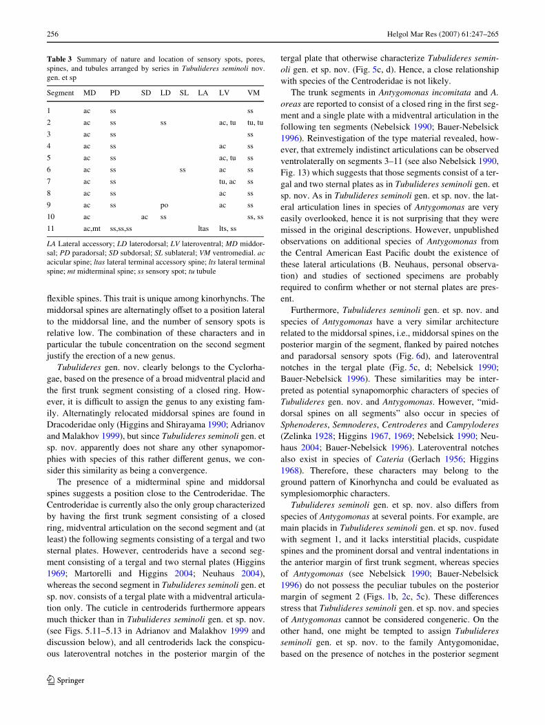
Table 3 Summary of nature and location of sensory spots, pores,spines, and tubules arranged by series in Tubulideres seminoli nov.gen. et sp
LA Lateral accessory; LD laterodorsal; LV lateroventral; MD middor-sal; PD paradorsal; SD subdorsal; SL sublateral; VM ventromedial. acacicular spine; ltas lateral terminal accessory spine; lts lateral terminalspine; mt midterminal spine; ss sensory spot; tu tubule
Segment MD PD SD LD SL LA LV VM
1 ac ss ss
2 ac ss ss ac, tu tu, tu
3 ac ss ss
4 ac ss ac ss
5 ac ss ac, tu ss
6 ac ss ss ac ss
7 ac ss tu, ac ss
8 ac ss ac ss
9 ac ss po ac ss
10 ac ac ss ss, ss
11 ac,mt ss,ss,ss ltas lts, ss
123

Helgol Mar Res (2007) 61:247–265 257
margins and the general similarities of the body cuticle inspecies of Antygomonas and Tubulideres seminoli gen. etsp. nov. However, as discussed above, we hesitate in doingso as these and other similarities all could be considered assymplesiomorphies. Hence, at present we prefer not toassign Tubulideres seminoli gen. et sp. nov. to any speciWcfamily, but to consider it as a species incertae sedis for theclass Cyclorhagida.
Morphological remarks The distribution of scalids inthe introvert of Tubulideres seminoli gen. et sp. nov. fol-lows a 5, 5, 10, 9 pattern for the mouth cone and a 10, 10,10, 15, 15, 15, 14 pattern of the introvert. A similar patternis present in various other species such as Antygomonasoreas (see Bauer-Nebelsick 1996) and the homalorhagidspecies Kinorhynchus phyllotropis, Paracentrophyes prae-dictus, and probably Pycnophyes communis (see Brown1989; Neuhaus 1995). Also Echinoderes capitatus has avery similar pattern, with the only exception that the mid-ventral trichoscalid is replaced by the normal scalid of ring06, meaning that ring 06 possesses 14 scalids only (Nebel-sick 1993). It should, however, be stressed that the scalidpatterns apparently vary greatly, even on generic levels [seeNeuhaus (1995) for a summary], hence nothing can be con-cluded at the present state.
The articulation of the outer oral styles with only twosegments and a claw-like structure diVers from the oralstyles of A. oreas that are reported to have three segmentsand no pore (Bauer-Nebelsick 1996). Since the pore mightbe confused with a second articulation, especially whenobserved in light microscopy, it is not unlikely that the oralstyles in A. oreas and A. incomitata consist of only two seg-ments and a pore as well. However, it would require rein-vestigations with SEM to conWrm this hypothesis.
As discussed in previous papers (see, e.g., Sørensen2006), consideration of new characters and character com-plexes is important in order to obtain substantial informa-tion for analyses of the phylogenetic relationships withinKinorhyncha. An interesting character concerns the thick-ness of the cuticle. Cuticle thickness has previously beendiscussed in relation to kinorhynch phylogeny and evolu-tion (Adrianov and Malakhov 1995; Neuhaus 1995). Athick and well-developed cuticle is typically present in spe-cies of the cyclorhagid families Echinoderidae and Centro-deridae and in the homaloragid Pycnophyidae, whereas athinner and more Xexible cuticle is found in species ofCateria, Zelinkaderes, Antygomonas, Semnoderidae andthe homalorhagid Neocentrophyidae (Higgins 1968, 1969;Kristensen and Higgins 1991; Nebelsick 1990; Adrianovand Malakhov 1995, 1999; Bauer-Nebelsick 1995, 1996;Neuhaus 1995; GaOrdónez et al. 2000). The cuticle in Tub-ulideres seminoli gen. et sp. nov. certainly belongs to thethin and Xexible type, with rather indistinct articulationsbetween the segmental plates (Figs. 2a–e, 5a–d). It has been
suggested that the thin cuticle displays the ancestral kin-orhynch state, whereas the thicker type evolved indepen-dently within the cyclorhagid and homalorhagid lineages(Adrianov and Malakhov 1995; Neuhaus 1995). This wouldplace Tubulideres seminoli gen. et sp. nov., and probably itsclose allied species of Antygomonas, among the most basalkinorhynchs. However, the exact position cannot be deter-mined yet as the notches in the posterior margin of mosttrunk segments resemble the pattern in species of Cateria(see Higgins 1968), whereas the general appearance of thecuticle points towards a position closer to Antygomonas.
Order Cyclorhagida Zelinka, 1896
Suborder Cyclorhagae Zelinka, 1896
Family Zelinkaderidae Higgins, 1990
Zelinkaderes brightae sp. nov.
(Figs. 7, 8, 9, 10, 11, 12; Tables 4, 5, 6),Diagnosis Trunk segments 1 through 11 with serially
arranged patches with cuticular bristles displaying a longi-tudinally striated pattern. Middorsal spines present on seg-ments 4, 6, 8–11; lateroventral short acicular spines presenton segment 2; lateral accessory cuspidate spines present onsegments 2 and 8; ventrolateral cuspidate spines present onsegments 4–6 and 9; lateroventral acicular spines presenton segments 8, 9 and 11 (=lateral terminal spine); midter-minal and lateral terminal accessory spines present on seg-ment 11. Females with acicular middorsal and subdorsalspines on segment 10; males with spines in same positions,but spines are more Xexible, appearing segmented or monil-iform in their distal ends. Lateral terminal accessory spinesshorter than lateral terminal spines, ratio = 72.1–87.5%.
Etymology The species is named after Dr. MonikaBright in recognition of her excellent work on Zelinkaderesand Antygomonas—the latter being another intriguing kin-orhynch genus.
Material examined All type material was obtained from asample taken on 22 July 2003 in the Atlantic Ocean, ca. 6miles oV Fort Pierce, Florida. Position: 27°29.559�N,080°12.233�W. Holotype: adult male, mounted in Fluoro-mount G® (ZMUC KIN-184). Allotype: adult female,mounted in Fluoromount G® (ZMUC KIN-185). Paratypes: 2adult females, 2 adult males and one juvenile, mounted in Flu-oromount G® (ZMUC KIN-186 to KIN-190). All types arestored at the Zoological Museum, University of Copenhagen.
Description The adult specimens possess a head, a neckand eleven trunk segments (Fig. 7a, b, 8a, 11a, b). All mea-surements and dimensions are given in Tables 4, 5. A sum-mary of spine and sensory spot locations is given inTable 6.
The head consists of a narrow mouth cone and an intro-vert with seven rings of scalids (Figs. 8f, 9, 10). Helioscalids
123

258 Helgol Mar Res (2007) 61:247–265
and the Wrst ring of inner oral styles (rings ¡03 and ¡02)are feebly visible in the examined specimens, but the sec-ond ring (ring ¡01) of inner oral styles appears to consist often styles that resemble the larger outer oral styles. The fol-lowing ring (ring 00) consists of nine outer oral styles withtwo segments and conspicuously pointed tips (Figs. 8f,10a). The spinoscalids (ring 01) are equipped with densefringes on their bases (Figs. 9, 10c). Distally to the fringes,a pair of spines extends from the basal plate of each spino-scalid (Fig. 10c). Ring 02 and 03 have no recognizable sca-lids. However, ring 02 carries ten pairs of spines, similar tothose of the spinoscalids. It furthermore has rows offringes, aligned to the radius arms and hence locateddirectly under the spines. Ring 03 has twenty single spinesfollowed by rows of fringes like in ring 02. Rings 04, 05and 06 consist of Wfteen regular sized scalids, with rows of
fringes between the rings (Figs. 9, 10c, d). The scalids ofring 05 are furthermore accompanied by eyelid-like struc-tures (Fig. 10d) that might correspond to basal plates. Ring07 has 14 rather small trichoscalids with one in a midven-tral position. In summary, the distribution of scalids perring can currently be reported as: ?, ?, 10, 9/10, *, *, 15, 15,15, 14. Described section-wise (Fig. 9) the scalid arrange-ment in the introvert is as follows: All sections (except themiddorsal section 6) with 1 oral style; section 1 furthermorewith four scalids and one trichoscalid; sections 2, 4, 6, 8,10: Wve scalids and one trichoscalid; and sections 3, 5, 7, 9:four scalids and two trichoscalids.
The neck consists of 16 soft and indistinct placids of uni-form size (Fig. 10b). The placids have two longitudinalfolds, mostly visible in SEM, but are otherwise smooth.The basal parts of the lateral placid margins are visible as
Fig. 7 Zelinkaderes brightae sp. nov. a Female dorsal view. b Female ventral view. c Male, left part of trunk segments 10–11 dorsal view. d. Male, left part of trunk segments 10–11 ventral view
123

Helgol Mar Res (2007) 61:247–265 259
indistinct lines on the anterior part of trunk segment 1. Allplacids are fused with segment 1 and the segment border isindistinct in LM. However, the border is easily seen inSEM because of the conspicuous striated surface sculptur-ing of segment 1 diVers from the smooth surface on theplacids (Figs. 10b, 11d).
Segment 1 consists of one complete cuticular ring(Figs. 7a, b, 11a–b). This and the following segments arecovered with small scale-like, longitudinally arranged
bristles that give the animal a conspicuous striated appear-ance (Figs. 7a, b, 8b–e, 11c–h). The lateral edges of thebristles are slightly swollen, which may give the erroneousimpression that each longitudinal line is formed by a doublerow of Wne hairs. Other kinds of cuticular hairs are absent.Posteriorly, the segment terminates into a pectinate fringe.
Segment 2 has one pair of very short and Xexible latero-ventral acicular spines and a pair of lateral accessory cuspidatespines (Figs. 7b, 11d, 12b). The acicular spines are
Fig. 8 Light microscope photos of Zelinkaderes brightae gen. et sp. nov. Holotypic male: a dor-sal view. b Trunk segments 1–6 dorsal view. c Trunk segments 1–6 ventral view. d Terminal trunk segment 8–11 dorsal view. e Trunk segments 8–11 ventral view. f Introvert and trunk seg-ments 1–2 dorsal view. g Termi-nal trunk segment in male dorsal view. Allotypic female: h termi-nal trunk segment in female dor-sal view. hp hairless patch; ld laterodorsal spines in male (m) and female (f); os outer oral style
123

260 Helgol Mar Res (2007) 61:247–265
extremely small and inconspicuous (Fig. 12b), whereas thecuspidate spines are considerably larger, and the broadbase constitutes more than 50% of the spine. An oval mid-
dorsal sensory spot is located slightly posterior to the midof the segment. The surface sculpturing is similar to thesculpturing on segment 1. However, it has several patcheswithout cuticular bristles or with bristles displaying adiVerent pattern and appearance (Fig. 7). All these patchesare elongate and extend posteriorly from the anteriormargin of the segment. In the ventromedial position thesegment has one pair of large, less hirsute patches (corre-sponding patches on segments 9 and 10 are documented onFig. 11f). Laterally it has two smaller patches totallydevoid of cuticular hairs or bristles. These patches are alsoelongate and extend posteriorly from the anterior marginof the segment, but the patches are otherwise much smallerthan the laterodorsal ones. In the laterodorsal position ithas a pair of patches that are lanceolate in shape, starting atthe anterior margin of the segment and extending posteri-orly through three-fourth of the segment length. The integ-ument surface in the anterior part of the patches is smoothand without any kind of hair or bristle structures. On theposterior part, the periphery is covered with small cuticularbristles, whereas the center is hairless (correspondingpatches on segments 9 and 10 are documented on Fig. 11e).Three further pairs of hairless patches are located moredorsally (corresponding paradorsal patch on segment 10 isdocumented on Fig. 12f).
Segment 3 and the following 7 segments consist of atergal plate with midventrally fused sides (Fig. 7b). Thesegment has no spines. An oval middorsal sensory spot islocated slightly posterior to the mid of the segment. Thesurface sculpturing displays the same striation as present onthe previous segments, and several patches with diVeringsurface sculpturing are present. A pair of large patches
Fig. 10 Scanning electron micrographs of details in the introvert of Zelinkaderes brigh-tae sp. nov. a Head with intro-vert and oral styles extended dorsal view. b Placids and closed head aperture. c Detail of spinoscalid attachment sites in section 4 of the introvert. d Detail in the middorsal section 6 of scalid rings in introvert. el eyelid-like structure; fr fring-es in introvert; mvp midventral placid; os outer oral style; s spines in introvert; sc scalids; sp spinoscalid in introvert ring 01. Digits refer to the speciWc introvert ring of each structure
Fig. 9 Diagram of mouth cone and introvert showing distribution oforal styles and scalids in Zelinkaderes brightae sp. nov
123

Helgol Mar Res (2007) 61:247–265 261
covered with cuticular bristles is located very close to themidventral line. These patches are equivalent with the ven-tromedial patches on the previous segment. The laterodor-sal patches with bristles are repeated from segment 2, andthe smaller hairless patches are present as two pairs in aventrolateral position, two pairs in a lateral position andthree pairs in the subdorsal position.
Segment 4 has one middorsal spine located near the pos-terior segment margin, and one pair of ventrolateral cuspi-date spines (Figs. 7a, b, 8b–c, 11c). The middorsal spine iscomposed of a less hirsute shaft and a distal part withdensely set cuticular hairs (corresponding middorsal spineon segment 10 is documented on Fig. 12a). One pair ofsmall paradorsal sensory spots is located at the base of thespine. The ventrolateral cuspidate spines are similar tothose on segment 2. One pair of elongate midlateral sensoryspots is located mesially on the segment (Fig. 12e). Thepatches without hairs or bristles are repeated from segment3.
Segment 5 has no middorsal spine but one pair of ven-trolateral cuspidate spines (Figs. 7b, 11c). The cuspidatespines (Fig. 12c) are similar to those on segment 4. Midlat-eral sensory spots are absent. The patches without hairs orbristles are repeated from segment 4.
Segment 6 has one middorsal spine located near the pos-terior margin, and one pair of ventrolateral cuspidate spines(Figs. 7a, b, 8b, c, 11c). The cuspidate spines are similar tothose on segment 5. One pair of small paradorsal sensoryspots is located at the base of the middorsal spine, whereasanother larger, elongate pair is located in a midlateral posi-tion mesially on the segment. The patches without hairs orbristles are repeated from segment 5.
Segment 7 has no spines (Figs. 7a, b, 8b, c, 11c). Onepair of elongate midlateral sensory spots is located mesiallyon the segment. The patches without hairs or bristles arerepeated from segment 6.
Segment 8 has one middorsal spine located near the pos-terior margin, one pair of closely set lateral accessory cus-pidate spines and one pair of lateroventral acicular spines(Figs. 7a, b, 8d–e, 11e). A pair of small paradorsal sensoryspots is located at the base of the middorsal spine. Other-wise, the segment has no sensory spots. The patches with-out hairs or bristles are repeated from segment 7.
Segment 9 has one middorsal spine located near the pos-terior margin, one pair of ventrolateral cuspidate spines andone pair of lateroventral acicular spines (Figs. 7a, b, 8d, e,11e, f). One pair of small paradorsal sensory spots islocated at the base of the middorsal spine. Furthermore, onepair of elongate midlateral sensory spots (Fig. 11e) and onepair of much smaller, rounded ventromedial sensory spotsare located slightly behind the mesial part of the segment.The patches without hairs or bristles are repeated fromsegment 8.
Segment 10 has one middorsal spine located near theposterior margin and one pair of laterodorsal acicularspines (Figs. 7a, b, 8d, e, 11e, g–h). Ventral and more lat-eral spines are absent. In females the middorsal and latero-dorsal spines are regular acicular (Figs. 8h, 11g), whereasthose in the males appear more Xexible and segmented ormoniliform in their distal ends (Figs. 7c, d, 8g, 11h). Onepair of small paradorsal sensory spots is located at the baseof the middorsal spine, two pairs of much larger oval sen-sory spots are located in a paradorsal (Fig. 12f) and midlat-eral position, respectively. The posterior margins of thesternal plates are broadly concave, not straight like on theprevious segments. The patches without hairs or bristles arerepeated from segment 9.
Segment 11 tapers strongly to the base of the midtermi-nal spine. The segment consists of one tergal and two ster-nal plates. One middorsal spine, without associatedparadorsal sensory spots, is located on the mid of the tergalplate. Furthermore, a midterminal spine, paired lateral ter-minal spines and lateral terminal accessory spines are pres-ent (Figs. 7, 8a, g, h, 11a, g, h). A pair of paradorsal sensoryspots with two pores is located mesially on the segment,anterior to a pair of modiWed sensory spots (type 3 sensoryspots), located at the base of the midterminal spine(Fig. 12d). The surface sculpturing diVers from the previ-ous segments. The most anterior part of the tergal plate hasthree paired patches with very densely set short cuticularhairs (Fig. 12d). The posterior margins of the patches arevery distinct, whereas the anterior margins are covered bythe pectinate fringe of segment 10. The most dorsal pair ofpatches each contains one conspicuous circular area with-out hairs. Another two pairs of patches are located betweenthe middorsal spine and the lateral accessory terminalspines. The rest of the plate is covered with short,extremely delicate hairs. The sternal plates are broad attheir anterior ends, and taper gradually towards a medial tipat the fringed posterior margins of each plate. The surfacesculpturing consists of two medial patches on the anteriorpart of each plate. The patches have distinct posterior mar-gins and are covered with densely set short cuticular hairsposteriorly. The anterior parts are less hirsute. Close to themidventral line of the segment two large patches extendfrom the anterior segment margin till the mid of the seg-ment. Each patch is rounded posteriorly and covered withdensely set short cuticular hairs, except in the center thatlacks hairs completely. Lateral to the patches the cuticle iscovered with less densely set hairs. In females the gonop-ores are visible as two strongly cuticularized areas at theintersegmental junction between segments 10 and 11. Theposterior halves of the sternal plates lack hairs completely.
Phylogenetic remarks Based on the presence of trunksegments 1 and 2 consisting of closed cuticular rings, 16placids that are fused with segment 1, and scale-like cuticular
123
262 Helgol Mar Res (2007) 61:247–265
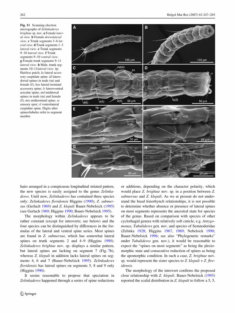
hairs arranged in a conspicuous longitudinal striated pattern,the new species is easily assigned to the genus Zelinka-deres. Until now, Zelinkaderes has contained three speciesonly: Zelinkaderes Xoridensis Higgins (1990), Z. submer-sus (Gerlach 1969) and Z. klepali Bauer-Nebelsick (1995)(see Gerlach 1969; Higgins 1990; Bauer-Nebelsick 1995).
The morphology within Zelinkaderes appears to berather constant (except for introverts; see below) and thefour species can be distinguished by diVerences in the for-mulas of the lateral and ventral spine series. Most spinesare found in Z. submersus, which has somewhat lateralspines on trunk segments 2 and 4–9 (Higgins 1990).Zelinkaderes brightae nov. sp. displays a similar pattern,but lateral spines are lacking on segment 7 (Fig. 7b),whereas Z. klepali in addition lacks lateral spines on seg-ments 4, 6 and 7 (Bauer-Nebelsick 1995). ZelinkaderesXoridensis has lateral spines on segments 5, 8 and 9 only(Higgins 1990).
It seems reasonable to propose that speciation inZelinkaderes happened through a series of spine reductions
or additions, depending on the character polarity, whichwould place Z. brightae nov. sp. in a position between Z.submersus and Z. klepali. As we at present do not under-stand the basal kinorhynch relationships, it is not possibleto determine whether absence or presence of lateral spineson most segments represents the ancestral state for speciesof the genus. Based on comparison with species of othercyclorhagid genera with relatively soft cuticle, e.g. Antygo-monas, Tubulideres gen. nov. and species of Semnoderidae(Zelinka 1928; Higgins 1967, 1969; Nebelsick 1990;Bauer-Nebelsick 1996; see also “Phylogenetic remarks”under Tubulideres gen. nov.), it would be reasonable toexpect the “spines on most segments” as being the plesio-morphic state and consecutive reduction of spines as beingthe apomorphic condition. In such a case, Z. brightae nov.sp. would represent the sister species to Z. klepali + Z. Xor-idensis.
The morphology of the introvert conWrms the proposedclose relationship with Z. klepali. Bauer-Nebelsick (1995)reported the scalid distribution in Z. klepali to follow a 5, 5,
Fig. 11 Scanning electron micrographs of Zelinkaderes brightae sp. nov. a Female later-al view. b Female dorsolateral view. c Trunk segments 3–6 lat-eral view. d Trunk segments 1–3 lateral view. e Trunk segments 9–10 lateral view. f Trunk segments 9–10 ventral view. g Female trunk segments 9–11 lateral view. h Male, trunk seg-ments 10–11lateral view. hp Hairless patch; la lateral acces-sory cuspidate spine; ld latero-dorsal spines in male (m) and female (f); ltas lateral terminal accessory spine; lv lateroventral acicular spine; md middorsal spines in male (m) and female (f); mts midterminal spine; ss sensory spot; vl ventrolateral cuspidate spine. Digits after spines/tubules refer to segment number
123

Helgol Mar Res (2007) 61:247–265 263
10, 9 / 10, *, *, 15, 15, 15, 14 pattern, which is identicalwith the pattern in Z. brightae (Fig. 9), but stands in a sharpcontrast to Z. Xoridensis which displays a 5, 5, 10, 9/10, 5,10, 15, *, *, 14 pattern (Higgins 1990).
Comparison of introvert morphology within Zelinka-deres and with species of other genera is hampered by theapparent reduction of certain scalid rings (ring 02 and 03 inZ. brightae nov. sp. and Z. klepali, and ring 05 and 06 inZ. Xoridensis). To establish the ground pattern for scaliddistribution in Zelinkaderes it would be useful to know thenumber of scalids that originally was present in the reducedrings. Here, we might be able to get a clue from the pres-ence of spines and fringes in Z. brightae nov. sp. (Figs. 9,10c, d) that could be interpreted as markers after reductionof scalids. This would suggest that the original oral style/scalid pattern prior to the reduction was ?, ?, 10, 9 / 10,(10), (20), 15, 15, 15, 14. Interestingly, this pattern is verysimilar to the pattern in species of Antygomonas and Tubu-lideres seminoli gen. et sp. nov. (except for ring 03 thatconsists of 10 scalids only; Nebelsick 1990; Bauer-Nebel-sick 1996; this paper), which could indicate a relationship
between these taxa. However, data is scarce among speciesof other kinorhynchs (see, e.g., Neuhaus 1995).
Morphological and taxonomic remarks
The complex scalid pattern in the introvert of Z. brightaenov. sp., prompted the introduction of two new terms forcomponents in the introvert. The term “spines” was usedto characterise stiV extensions of the basal plates(Fig. 10c). They can be distinguished from the scalids bythe lack of any articulation and their attachment to thebasal plates. They are glabrous and have a pointed tip.The component referred to as “eyelid-like structure” isused for the structures in ring 05 that look like eyelids orwings (Fig. 10d). The structures resemble a halved scalidbasal plate.
The general morphology of Z. brightae nov. sp. corre-sponds very well with the original diagnosis of Zelinka-deres that was given by Higgins (1990). However, evenmore traits appear to be characteristic for species of this
Fig. 12 Scanning electron micrographs of details in Zelinkaderes brightae sp. nov. a Trunk segment 8, middorsal spine. b Trunk segment 2, lateral accessory cuspidate spine and lateroventral acicular spine. c Trunk segment 5, lateroventral cuspidate spine. d Trunk seg-ment 11 lateral view. e. Trunk segment 4, midlateral sensory spot. f Trunk segment 10, large paradorsal sensory spot and hair-less patch. hp hairless patch; la lateral accessory cuspidate spine; lv lateroventral acicular spine; mss modiWed sensory spot; ss sensory spot
123

264 Helgol Mar Res (2007) 61:247–265
genus. These include the presence of ventrolateral cuspidatespines on trunk segment 5, lateral accessory cuspidatespines and lateroventral acicular spines on segment 8, andlateroventral acicular spines and ventrolateral cuspidatespines on segment 9.
The lateroventral spines on segment 10 have beenmoved to a more laterodorsal position, compared to thespines on the previous segments. This displacement alsoappears to be consistent for all four Zelinkaderesspecies. In Z. Xoridensis and Z. submersus, Higgins(1990) refers to their position as subdorsal, but accord-ing to the new and more precise spine terminology forspines locations, proposed by Pardos et al. (1998), theirposition is rather laterodorsal as in Z. brightae nov. sp.and Z. klepali.
Also the cuticular sculpturing is highly characteristic forspecies of the genus. The presence of cuticular bristlesarranged in longitudinal rows gives the cuticle aconspicuous striated appearance. This striation wasdescribed from both Z. Xoridensis and Z. submersus, butthe descriptions were solely based on light microscopy,hence Higgins (1990) interpreted the striation as an internalpattern. Bauer-Nebelsick (1995) conWrmed this assumptionby TEM studies of Z. klepali (Bauer-Nebelsick 1995:Figs. 40–42) and also conWrmed the presence of longitudi-nally arranged rows of biWd cuticular bristles. Similar bris-tles are also found in Z. brightae nov. sp.
Table 4 Measurements of adult Zelinkaderes brightae nov. sp.,including number of measured specimens (n) and SD
LTAS Lateral terminal accessory spine; LTS lateral terminal spine;MTS midterminal spine; S1–11 segment lengths of trunk segments 1–11; TL trunk length
Character n Range Mean SD
TL 5 505–650 �m 564 �m 55.69 �m
LTS/TL 5 18.2–21.2% 20.1% 1.3%
LTAS/TL 5 15.1–18.6% 16.3% 1.42%
LTAS/LTS 5 72.1–87.5% 81.1% 5.86%
MTS 2 425–497 �m 461 �m 50.91 �m
MTS/TL 2 65.4–86.0% 75.7% 14.57%
S1 5 52–66 �m 59 �m 6.16 �m
S2 5 43–53 �m 47 �m 4.38 �m
S3 5 46–51 �m 48 �m 2.07 �m
S4 5 48–60 �m 52 �m 4.97 �m
S5 5 49–60 �m 53 �m 4.30 �m
S6 5 53–63 �m 58 �m 4.00 �m
S7 5 55–72 �m 62 �m 6.47 �m
S8 5 57–75 �m 66 �m 7.33 �m
S9 5 63–83 �m 69 �m 8.41 �m
S10 5 57–72 �m 63 �m 5.59 �m
S11 5 42–55 �m 48 �m 5.10 �m
Table 5 Spine/tubule measurements of adult Zelinkaderes brightaenov. sp. including number of measured specimens (n) and SD
The notations (f) = female and (m) = male refer to the speciWc sexwhen sexual dimorphism occurs
LA Lateral accessory; LTAS lateral terminal accessory spine; LTS lat-eral terminal spine; LV lateroventral; MD middorsal; MTS midterminalspine; VL ventrolateral
Character n Range (�m) Mean (�m) SD (�m)
MD 4 5 64–88 73 9.04
MD 6 5 76–101 87 10.67
MD 8 5 62–84 74 8.62
MD 9 5 65–87 76 9.59
MD 10 (f) 2 55–60 58 3.54
MD 10 (m) 3 47–60 55 7.23
LD 10 (f) 2 46–50 48 2.83
LD 10 (m) 3 53–63 55 6.81
MD 13 5 109–175 139 24.26
LA 2 5 20–26 23 2.59
LV 2 4 10–16 13 2.50
VL 4 5 24–27 26 1.14
VL 5 5 25–28 27 1.30
VL 6 5 26–32 29 2.50
LA 8 5 28–33 31 2.28
LV 8 5 41–68 50 10.67
LV 9 5 47–92 50 17.10
VL 9 5 29–33 31 1.48
LTS 5 105–136 113 13.17
LTAS 5 84–96 91 6.46
MTS 5 425–497 461 50.91
Table 6 Summary of nature and location of sensory spots, spines, andtubules arranged by series in Zelinkaderes brightae n. sp
LA Lateral accessory; LD laterodorsal; LV lateroventral; MD middor-sal; ML midlateral; PD paradorsal; SD subdorsal; VL ventrolateral; VMventromedial. ac acicular spine; cu cuspidate spine; ltas lateral termi-nal accessory spine; lts lateral terminal spine; mt midterminal spine; sssensory spot
Segment MD PD LD ML LA LV VL VM
1
2 ss cu ac
3 ss
4 ac ss ss cu
5 cu
6 ac ss ss cu
7 ss
8 ac ss cu ac
9 ac ss ss ac cu ss
10 ac ss, ss ac ss
11 ac, mt ss, ss ltas lts
123

Helgol Mar Res (2007) 61:247–265 265
Genus Zelinkaderes
Emended diagnosis Oral styles consisting of two seg-ments. Neck consisting of 16 indistinct placids, all fusedwith Wrst trunk segment; midventral placid broader thanremaining ones. Segments 1 and 2 consisting of one closedcuticular ring. Segments 3 to 11 with lateral margins of sin-gle tergal plate articulating midventrally. Middorsal spinespresent on segments 4, 6, 8–11. Lateroventral cuspidatespines present on segment 5. Midlateral cuspidate spinesand sublateral acicular spines present on segment 8. Lateralaccessory acicular spines and lateroventral cuspidate spinespresent on segment 9. Laterodorsal acicular spines (monili-form in males) present on segment 10. Midterminal spinespresent in juveniles and adults. Lateral terminal and lateralterminal accessory spines present in both sexes in adults.Segments 1 through 11 with conspicuous longitudinal stria-tion formed by rows of cuticular bristles.
Acknowledgments We are indebted to Valerie Paul and the staV atthe Smithsonian Marine Station at Fort Pierce for providing us withexcellent research facilities during our stays at the station, and to thecrew on R/V Sunburst, Hugh Reichardt and Woody Lee. We also thankDr. Reinhardt M. Kristensen, Jesper G. Hansen and Thomas M. Jesper-sen for their help collecting the samples. We thank Dr. Helmut Satt-mann at the Museum of Natural History in Vienna and Dr. MonikaBright for providing type and supplementary material of Antygomonasoreas and Cheryl Bright at the National Museum of Natural History,Smithsonian Institution, for providing type specimens of ZelinkaderesXoridensis and Z. submersus. This is contribution no. 693 from theSmithsonian Marine Station at Fort Pierce. Funding was provided bythe Danish Natural Science Research Council (Grants No. 21–04–0331and 21–04–0047). The compound microscope, camera lucida andimaging software were funded by the Carlsberg Foundation (Grant No.2005–1–545) and the Novo Nordisk Foundation.
References
Adrianov AV (1989) First record of kinorhynchs from the Sea ofJapan. Zool Zhurn 68:17–27. [In Russian]
Adrianov AV, Malakhov VV (1995) The phylogeny and classiWcationof the class Kinorhyncha. Zoosyst Ross 4:23–44
Adrianov AV, Malakhov VV (1999) Cephalorhyncha of the worldocean. KMK LTD, Moscow
Adrianov AV, Murakami C, Shirayama Y (2002a) Echinoderes aureusn. sp. (Kinorhyncha: Cyclorhagida) from Tanabe Bay (HonshuIsland), Japan, with a key to the genus Echinoderes. Spec Div7:47–66
Adrianov AV, Murakami C, Shirayama Y (2002b) Taxonomic study ofthe Kinorhyncha in Japan. III. Echinoderes sensibilis n. sp (Kin-orhyncha: Cyclorhagida) from Tanabe Bay. Zool Sci 19:463–473
Bauer-Nebelsick M (1995) Zelinkaderes klepali sp.n., from shallowwater sands of the Red Sea. Ann Nat Hist Mus Wien 97B:57–74
Bauer-Nebelsick M (1996) Antygomonas oreas sp.n., a new deep-seakinorhynch from the PaciWc Ocean. Ann Nat Hist Mus Wien98B:5–22
Brown R (1989) Morphology and ultrastructure of the sensory append-ages of a kinorhynch introvert. Zool Scr 18:471–482
GaOrdónez D, Pardos B, Benito J (2000) Cuticular structures and epi-dermal glands of Echinoderes cantabricus and E. hispanicus(Kinorhyncha, Cyclorhagida) with special reference to their taxo-nomic value. J Morphol 246:161–178
Gerlach SA (1956) Über einen aberranten Vertreter der Kinorhynchenaus dem Küstengrundwasser. Kieler Meeresforsch 12:120–124
Gerlach SA (1969) Cateria submersa sp.n., ein cryptorhager Kin-orhynch aus dem sublitoralen Mesopsammal der Nordsee. VerInst Meeresforsch, Bremerhaven 12:161–168
Higgins RP (1967) The Kinorhyncha of New-Caledonia. ExpéditionFrancaise sur les Recifs coralliens de la Nouvelle-Calédonie. Par-is Fondation Singer-Polignac 2:75–90
Higgins RP (1968) Taxonomy and postembryonic development of theCryptorhagae, a new suborder for the mesopsammic kinorhynchgenus Cateria. Trans Am Microsc Soc 87:21–39
Higgins RP (1969) Indian Ocean Kinorhyncha:1, Condyloderes andSphenoderes, new cyclorhagid genera. Smithsonian Contr Zool14:1–13
Higgins RP (1990) Zelinkaderidae, a new family of cyclorhagid Kin-orhyncha. Smithsonian Contr Zool 500:1–26
Higgins RP, Shirayama Y (1990) Dracoderidae, a new family of thecyclorhagid Kinorhyncha from the Island Sea of Japan. Zool SciJpn 7:1–13
Kristensen RM, Higgins RP (1991) Kinorhyncha. In: Harrison FW,Ruppert EE (eds) Microscopic anatomy of invertebrates, vol 4.Wiley-Liss, New York, pp 377–404
Lemburg C (2002) A new kinorhynch Pycnophyes australensis sp. n.(Kinorhyncha: Homalorhagida: Pycnophyidae) from MagneticIsland, Australia. Zool Anz 241:173–189
Martorelli S, Higgins RP (2004) Kinorhyncha from the stomach of theshrimp Pleoticus muelleri (Bate, 1888) from Comodoro Rivad-avia, Argentina. Zool Anz 243:85–98
Nebelsick M (1990) Antygomonas incomitata gen. et sp. n. (Cyclo-rhagida, Kinorhyncha) and its phylogenetic relationships. ZoolScr 19:143–152
Nebelsick M (1993) Introvert, mouth cone, and nervous system of Ech-inoderes capitatus (Kinorhyncha, Cyclorhagida) and implicationsfor the phylogenetic relationships of Kinorhyncha. Zoomorphol-ogy 113:211–232
Neuhaus B (1995) Postembryonic development of Paracentrophyespraedictus (Homalorhagida): neoteny questionable among theKinorhyncha. Zool Scr 24:179–192
Neuhaus B (2004) Description of Campyloderes cf. vanhoeVeni (Kin-orhyncha, Cyclorhagida) from the Central American East PaciWcdeep sea with a review of the genus. Meiofauna Marina 13:3–20
Neuhaus B, Blasche T (2006) Fissuroderes, a new genus of Kinorhyn-cha (Cyclorhagida) from the deep sea and continental shelf ofNew Zealand and from the continental shelf of Costa Rica. ZoolAnz 245:19–52
Neuhaus B, Higgins RP (2002) Ultrastructure, biology, and phyloge-netic relationships of Kinorhyncha. Integr Comp Biol 42:619–632
Pardos F, Higgins RP, Benito J (1998) Two new Echinoderes (Kin-orhyncha, Cyclorhagida) from Spain, including a reevaluation ofkinorhynch taxonomic characters. Zool Anz 237:195–208
Sørensen MV (2006) New kinorhynchs from Panama, with a discus-sion of some phylogenetically signiWcant cuticular structures.Meiofauna Marina 15:51–77
Sørensen MV, Jørgensen A, Boesgaard TM (2000) A new Echinoderes(Kinorhyncha: Cyclorhagida) from a submarine cave in NewSouth Wales, Australia. Cah Biol Mar 41:167–179
Sørensen MV, Heiner I, Ziemer O (2005) A new species of Echino-deres from Florida (Kinorhyncha: Cyclorhagida). Proc Biol SocWash 118:499–508
Zelinka K (1928) Monographie der Echinodera. Verlag von WilhelmEngelmann in Leipzig, Leipzig
123