102e-cpg-may2001
TRANSCRIPT
8/3/2019 102E-CPG-May2001
http://slidepdf.com/reader/full/102e-cpg-may2001 1/4
8/3/2019 102E-CPG-May2001
http://slidepdf.com/reader/full/102e-cpg-may2001 2/4
likelihood of preterm birth include multiple gestation, poly-
hydramnios, vaginal bleeding, certain uterine anomalies, and
excessive uterine contractions. Despite these important asso-
ciations, maternal risk scoring fails to detect up to 70 percent
of infants who are delivered spontaneously before term.4
Other screening strategies that have been suggested include
measuring of biochemical markers of inflammation and cul-
turing for infections.5
ULTRASOUND VERSUS
DIGITAL CERVICAL MEASUREMENT
Digital assessment of the cervix has been commonly used to
diagnose premature labour or to evaluate women perceived to
be at increased risk of preterm labour. Digital assessment of cer-
vical length is subjective, varies between examiners, and under-
estimates true anatomic length. Digital exams before
hysterectomy underestimated cervical length by approximate-
ly14 mm, whereas transvaginal ultrasound measured lengthaccurately.6 Investigations using transvaginal ultrasound mea-
surement as the standard confirmed that digital examination
underestimates cervical length.7,8 This underestimation may
result from an inability to digitally assess the cervix length
beyond the vaginal fornices unless there is two cm or more of
dilation and the entire intracervical canal is examined.
COMPARISON BETW EEN TRANSVAGINAL,
TRANSABDOMINAL,AND TRANSPERINEAL
ULTRASOUND CERVICAL MEASUREMENT
Ultrasound assessment of the cervix was initially transabdomi-nal, but specific disadvantages led to a preference for the trans-
vaginal examination.9,10
• Transabdominal ultrasound requires filling the bladder to
assess the cervix adequately, but this may spuriously length-
en the cervix by opposing the anterior and posterior lower
uterine segments,9 concealing cervical shortening or fun-
nelling. In contrast, transvaginal ultrasound is performed
with the bladder empty.10
• Transabdominal resolution is hampered significantly by
maternal obesity, shadowing from fetal parts, and the need
for lower frequency transducers.10
Transperineal ultrasound has been performed as less invasive
than the transvaginal examination but most studies have found
that both approaches are acceptable to women.11,12 Since image
resolution is better transvaginally, transperineal ultrasound
should be reserved for and offered to women at increased risk
of preterm birth for whom vaginal assessment is unacceptably
invasive or uncomfortable.13,14,15
NORMAL CERVICAL LENGTH
Cervical length is normally distributed and remains relatively
constant until the third trimester.16,17,18 Heath found at 23 weeks
a mean length of 38 mm.19 Iams found a mean length at
24 weeks of 35 mm and at 28 weeks of 34 mm.20 If funnelling
is present, measurement should exclude the funnel and be takenfrom the funnel tip to the external os.21
TRANSVAGINAL CERVICAL MEASUREMENT
IN ASYMPTOMATIC PREGNANT WOMEN
Cervical length is inversely related to preterm birth risk in asymp-
tomatic women.8,19,20,22 The largest study of this relationship20
noted relative risks, compared to women above the 75th per-
centile, of approximately four if length was less than 30 mm
(25th percentile), six if less than 26 mm (10th percentile), nine
if less than 22 mm (5th percentile), and 14 if less than 13 mm
(1st percentile). The positive predictive value was poor (35%).Heath and colleagues19 studied women who were not at
increased risk of preterm birth and, using transvaginal ultrasound
at 23 weeks, found that 1.7 percent had a cervix length less than
or equal to 15 mm. These women accounted for 90 percent of
deliveries at less than 28 weeks and 60 percent of deliveries at
less than or equal to 32 weeks. This suggests that the positive
predictive value of a short cervix (≤ 15 mm) is much greater for
extreme prematurity (≤ 28 weeks). The authors have created a
formula to predict the risk of spontaneous delivery at less than
or equal to 32 weeks based on cervical length at 23 weeks.
Although transvaginal ultrasound screening of cervical length
can predict increased risk of preterm birth,23 there is no evidencethat this information can be used to improve outcomes. Consul-
tation and the proposed location of birth should be considered.
Other management options, such as cerclage, activity restriction,
tocolytics, and prophylactic steroids await appropriate evaluation
by randomized trials. The significant association between cervical
length and preterm birth risk may not apply to women who have
undergone cervical surgery resulting in permanent shortening.
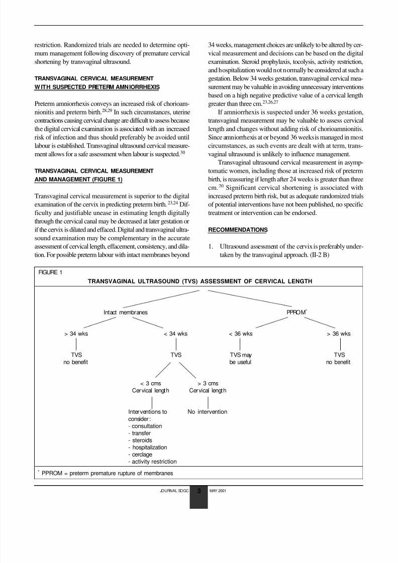
TRANSVAGINAL CERVICAL MEASUREMENT
W ITH SUSPECTED PRETERM LABOUR
Transvaginal ultrasound is superior to digital assessment of cer-
vical length but has limited ability to assess texture and dilation.
Of all variables assessed by digital or ultrasound examination,
transvaginal cervical length measurement is the best preterm
birth predictor.23 Prediction of preterm birth by internal os fun-
nelling has been found by some investigators21,23 but not
others.24,25 Cervical length greater than three cm has a high neg-
ative predictive value for delivery less than 34 weeks.23,26,27 This
information may help patients avoid unnecessary interventions
of unproven value such as tocolysis, hospitalization, and activity
JOURNAL SOGC MAY 2001
2
8/3/2019 102E-CPG-May2001
http://slidepdf.com/reader/full/102e-cpg-may2001 3/4

8/3/2019 102E-CPG-May2001
http://slidepdf.com/reader/full/102e-cpg-may2001 4/4
2. Routine prenatal cervical length screening by transvaginal
ultrasound is not supported by available evidence; how-
ever, it is indicated for women identified to be at increased
risk of preterm birth. Cervical shortening is associated with
increased preterm birth risk. (II-2 B)
3. Transvaginal ultrasound measurement of the cervix has a
high negative predictive value if length is greater than threecm after 24 weeks. This information can be used to avoid
unnecessary interventions. (II-2 B)
CONCLUSION
Transvaginal ultrasound cervical measurement is a safe and
effective technique to predict increased risk of preterm delivery
in selected patients. However, routine prenatal transvaginal
ultrasound screening of cervical length is not supported by avail-
able evidence. Normal results can help avoid unnecessary inter-
ventions, but randomized trials are needed to determine
whether interventions can improve maternal and neonatal out-comes, given cervical shortening.
J Soc Obstet Gynaecol Can 2001;23(5):418-21
REFERENCES
1. Berkowitz GS,Papienik E.Epidemiology of preterm birth.Epidemiol Rev
1993;15:414-3.
2. Joseph KS,Kramer MS,Wu Wen S,Alexander D.Determinants of
preterm bir th in Canada from 1981 through 1992,and 1992 through
1994.N Engl JMed 1998;339:1434-9.
3. Consensus Statement on Preterm Birth Prevention.Collaborative Con-
ference on Preterm Birth.Ot tawa,April 1998.
4. Armson BA,Dodds L.Prediction of preterm birth in a population of
Canadian women.Int JGynecol Obstet 1994:46(Suppl 2):93.
5. Goldenberg RL,Iams JD,Mercer BM,Meis PJ,Moawad AH,Cooper RL,
et al.The Preterm Prediction Study:The value of new vs standard risk
factors in predicting early and all spontaneous preterm births.NICHD
MFMU Network.Am JPublic Health 1998;88(2):233-8.
6. Jackson GM,Ludnir J,Bader TF.The accuracy of digital examination and
ultrasound in the evaluation of cervical length.Obstet Gynecol 1992;
72:214-8.
7. Sonek JD,Iams JD,Blumenfeld M,Johnson F,Landon M,Gabbe S.Measure-
ment of cervical length in pregnancy.Comparison between vaginal ult ra-
sonography and digital examination.Obstet Gynecol 1990;76:172-5.
8. Anderson HF,Nugent CE,Wanty SB,Hayashi RH.Prediction of r isk for
preterm delivery by ultrasonographic measurement of cervical length.
Am JObstet Gynecol 1990;163:859-67.
9. Anderson HF.Transvaginal and transabdominal ultrasonography of theuterine cervix dur ing pregnancy.JClin Ultrasound 1991;19:77-83.
10. To MS,Skentou C,Cicero S,Nicolaides KH.Cervical assessment at the
routine 23 weeks’ scan:problems with transabdominal sonography.
Ultrasound Obstet Gynecol 2000;15(4):292-6.
11. Rosati P,Guariglia L.Acceptability of early tr ansvaginal or abdominal
sonography in the first half of pregnancy.Arch Gynecol Obstet 2000;
264(2):80-3.
12. Braithwaite JM,Economides DL.Acceptability of patients of transvaginal
sonography in the elective assessment of the first t rimester fetus.Ult ra-
sound Obstet Gynecol 1997;9(2):76-9.
13. Kurtzman JT,Goldsmith LJ,Gall SA,Spinnato JA. Transvaginal versus
transperineal ultrasonography:a blinded comparison in the assessment
of cervical length at mid gestation.Am JObstet Gynecol 1998;
179(4):852-7.
14. Owen J,Neely C,Northen A.Transperineal versus endovaginal
ultrasonographic examination of the cervix in the mid-trimester :a
blinded comparison.Am JObstet Gynecol 1999;181:780-3.
15. Carr DB,Smith K,Parsons L,Chansky K,Shields LE.Ultrasonography
for cervical length measurements:agreement between transvaginal and
translabial techniques.Obstet Gynecol 2000;96(4):554-8.
16. Cook CM,Ellwood DA.A longitudinal study of the cervix in pregnancy
using transvaginal ultrasound.Br JObstet Gynaecol 1996;103(1):16-8.17. Tongsang T,Kamprapanth P,Pitaksakorn J.Cervical length in normal
pregnancy as measured by transvaginal sonography.Int JGynecol
Obstet 1997;58(3):313-5.
18. Smith CV,Anderson JC,Matamoras A,Rayburn WF.Transvaginal sonog-
raphy of cervical width and length during pregnancy.JUltrasound Med
1992;11(9):465-7.
19. Heath VC,Southall TR,Souka AP,Elisseou A,Nicolaides KH.Cervical
length at 23 weeks of gestation: prediction of spontaneous preterm
delivery.Ultrasound Obstet Gynecol 1998;12:312-7.
20. Iams JD,Goldenberg RL,Meis PJ,Mercer BM,Moawad A,Das A,et al.
The length of the cervix and the risk of spontaneous premature deliv-
ery.N Engl JMed 1996;334:567-72.
21. Berghella V,Kuhlman K,Weina S,Texeira L,Waprer RJ.Cervical funnel-
ing:sonographic criteria predictive of preterm delivery.Ult rasound
Obstet Gynecol 1997;10(3):161-6.22. Tong Sang T,Kamprapanth P,Srisomboon J,Wanapirak C, Piyamongkol
W,Sir ichot iyakul S.Single transvaginal sonographic measurement of
cervical length early in the third trimester as a predictor of preterm
delivery.Obstet Gynecol 1995;86:184-7.
23. Crane JM,Van den Hof MC,Armson BA,Liston R.Transvaginal
ultrasound in the prediction of preterm delivery:singleton and twin
gestations.Obstet Gynecol 1997;90:357-63.
24. Gomez R,Galasso M,Romero R,Mazor M,Sorokin Y,Gonsalves L,et al.
Ultrasonographic examination of the uterine cervix is better than cer-
vical digital examination as a predictor of the likelihood of premature
delivery in patients with preterm labour and intact membranes.Am J
Obstet Gynecol 1994;171:956-64.
25. Timor-Tritsch IE,Boozarjomehri F,Masakowski Y,Monteagudo A,Chao
CR.Can a “snapshot” sagittal view of the cervix by transvaginal ultra-
sound predict active preterm labour? Am JObstet Gynecol 1996;174:990-5.
26. Murakawa H,Utumi T,Hasegawa I,Tanaka K,Fuzimari R.Evaluation of
threatened preterm delivery by transvaginal ultrasonographic measure-
ment of cervical length.Obstet Gynecol 1993;82:829-32.
27. Iams JD,Paraskos J,Landon MB,Teteris JN,Johnson FF.Cervical sonog-
raphy in preterm labour.Obstet Gynecol 1994;84:40-6.
28. Naeye RG,Peter EC. Causes and consequences of premature rupture
of fetal membranes.Lancet 1980;1(8161):192-4.
29. Carroll SG,Ville Y,Greenough A,Gamsu H,Patel B,Philpott -Howard J,
et al.Preterm labour amniorrhexis:intrauterine infection and interval
between membrane rupture and delivery.Arch Dis Child Fetal Neona-
tal Ed 1995;72(11):F43-6.
30. Rizzo G,Capponi A,Angelini E,Vlachopoulou A,Grassi C,Romanini C.
The value of transvaginal ultrasonographic examination of the uterine
cervix in predicting preterm delivery in patients with preterm prema-
ture rupture of membranes.Ult rasound Obstet Gynecol 1998;11:23-9.
JOURNAL SOGC MAY 2001
4