104 reference values for cardiac index measured with magnetic resonance imaging in healthy subjects...
TRANSCRIPT

BioMed Central
Journal of Cardiovascular Magnetic Resonance
ss
Open AcceMeeting abstract104 Reference values for cardiac index measured with magnetic resonance imaging in healthy subjects and comparison with patients with congestive heart failureMarcus Carlsson*, Ruslana Andersson, Henrik Mosén, Bjorn Ekmehag and Hakan ArhedenAddress: Department of Clinical Physiology, Lund, Sweden
* Corresponding author
IntroductionMeasurements of cardiac index (CI) are of interest in crit-ically ill patients as well as in patients with chronicheartdisease. Magnetic Resonance Imaging (MRI) has beenproposed as the new reference method for cardiac outputand cardiac index measurements, but reference values arestill lacking.
PurposeThe purpose was therefore to compare CI in healthy sub-jects to patients with congestive heart failure and to obtainreference values for CI.
MethodsCI was measured in 124 healthy volunteers (40 ± 16 years,range 21–81 years, 48 females) and in 184 patients withcongestive heart failure and ejection fraction (EF) below40% (60 ± 13 years, range 24–85 years, 144 males). Allsubjects were imaged in the supine position with flowquantification of the ascending aorta during free breath-ing. Two MRI-scanners were used, a) 1.5 T MagnetomVision (Siemens, Erlangen, Germany) and b) PhilipsIntera CV (Philips, Best, the Netherlands). Blood flow wasmeasured through a plane perpendicular to the ascendingaorta with ECG-triggered phase-encoded velocity-map-ping sequences. Typical imaging parameters for SiemensVision were: repetition time 40 ms, echo time 5 ms, slicethickness 8 mm, velocity encoding factor 150 cm/s. Thetime resolution was typically 40 ms. Velocity information
was acquired by prospective ECG-triggering. Typical imag-ing parameters for Philips Intera were: repetition time 10ms, echo time 6 ms, slices thickness 6 mm, velocity encod-ing 200 cm/s. The time resolution was typically 30 ms.Flow was measured using a freely available software (Seg-ment 1.4 http://segment.heiberg.se) in healthy controlsand by vendor provided software in patients, Argus forpatients imaged by Siemens Vision and ViewForum forpatients imaged by Philips Intera. 20 controls were ana-lyzed by both ViewForum and Segment to calculate inter-method variability and 2 observers analyzed the flow datafrom 20 controls to calculate interobserver variability. CIis presented as mean ± SD.
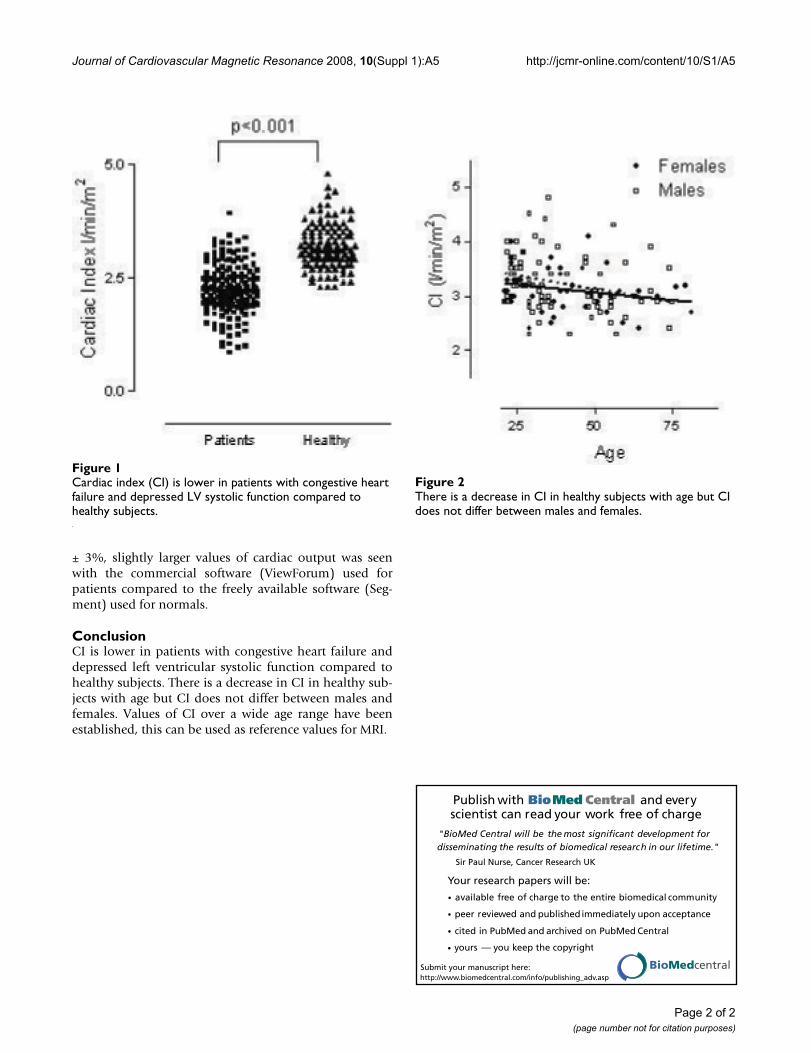
ResultsCI in patients with congestive heart failure (2.2 ± 0.5 l/min/m2) was lower compared to the healthy population(3.2 ± 0.5 l/min/m2, p < 0.001), Fig 1. In patients, CI wasweakly related to decreasing EF (r2 = 0.11, p < 0.001). Inthe normal population there was a slight decrease of CIwith age (8 ml/min/m2 per year, r2 = 0.07, p < 0.01), Fig 2.CI in normals aged 20–29 years was 3.3 ± 0.4 l/min/m2,in 30–39 years 3.2 ± 0.6 l/min/m2, in 40–49 years 3.1 ±0.5 l/min/m2 and above 50 years CI was 3.0 ± 0.4 l/min/m2. Cardiac index in males (3.2 ± 0.5 l/min/m2) andfemales (3.1 ± 0.4 l/min/m2) did not differ (p = 0.24). Theinterobserver variability of cardiac output measurementswas 0.2 ± 0.2 l/min or 3 ± 4%. The intermethod variabilityof cardiac output measurements was 0.4 ± 0.1 l/min or 6
from 11th Annual SCMR Scientific SessionsLos Angeles, CA, USA. 1–3 February 2008
Published: 22 October 2008
Journal of Cardiovascular Magnetic Resonance 2008, 10(Suppl 1):A5 doi:10.1186/1532-429X-10-S1-A5
<supplement> <title> <p>Abstracts of the 11<sup>th </sup>Annual SCMR Scientific Sessions - 2008</p> </title> <note>Meeting abstracts – A single PDF containing all abstracts in this Supplement is available <a href="http://www.biomedcentral.com/content/files/pdf/1532-429X-10-s1-full.pdf">here</a>.</note> <url>http://www.biomedcentral.com/content/pdf/1532-429X-10-S1-info.pdf</url> </supplement>
This abstract is available from: http://jcmr-online.com/content/10/S1/A5
© 2008 Carlsson et al; licensee BioMed Central Ltd.
Page 1 of 2(page number not for citation purposes)
Journal of Cardiovascular Magnetic Resonance 2008, 10(Suppl 1):A5 http://jcmr-online.com/content/10/S1/A5
Publish with BioMed Central and every scientist can read your work free of charge
"BioMed Central will be the most significant development for disseminating the results of biomedical research in our lifetime."
Sir Paul Nurse, Cancer Research UK
Your research papers will be:
available free of charge to the entire biomedical community
peer reviewed and published immediately upon acceptance
cited in PubMed and archived on PubMed Central
yours — you keep the copyright
Submit your manuscript here:http://www.biomedcentral.com/info/publishing_adv.asp
BioMedcentral
± 3%, slightly larger values of cardiac output was seenwith the commercial software (ViewForum) used forpatients compared to the freely available software (Seg-ment) used for normals.
ConclusionCI is lower in patients with congestive heart failure anddepressed left ventricular systolic function compared tohealthy subjects. There is a decrease in CI in healthy sub-jects with age but CI does not differ between males andfemales. Values of CI over a wide age range have beenestablished, this can be used as reference values for MRI.
There is a decrease in CI in healthy subjects with age but CI does not differ between males and femalesFigure 2There is a decrease in CI in healthy subjects with age but CI does not differ between males and females.
Cardiac index (CI) is lower in patients with congestive heart failure and depressed LV systolic function compared to healthy subjectsFigure 1Cardiac index (CI) is lower in patients with congestive heart failure and depressed LV systolic function compared to healthy subjects.
Page 2 of 2(page number not for citation purposes)