11. dr. s n senapati
TRANSCRIPT
.
1
IMAGING FOR BRACHYTHERAPY
PLANNING
DR S.UMIT GOYAL
ASSISTANT PROF
DEPT OF RADIATION ONCOLOGY,
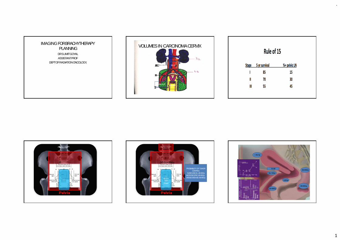
CX,V,MP
PA.LN
ARA,C.LN
VOLUMES IN CARCINOMACERVIX
II
I
II
50 Gy+10Gy
I
45GY+
HDR
7Gyx3
45-50Gy+
5-10Gy III
PROBABILITYOF TUMOR
CONTROL
SUBCLINICAL:-45-50Gy
MICROSCOPIC:-50-60Gy
GROSSDISEASE:-80-85Gy
60-65Gy
60-65Gy
80-90Gy
< 45 Gy
65-70Gy
120Gy
75-90

.
2
Brachytherapy Timeline
1932:
The Manchester dosage
System
(Point Aintroduced)
1970’s: Caesium
Treatments
Selectron LDR
(afterloading)
1990’s
Iridium-192
HDR/PDR
Technology
Dosimetricsystems
Empiricalprescription
3DImage
guided,
CT/MRI
Discovery of
Radioactivity
18961915:
Radium
Treatment
Point basedsystemNot formalisedPresent
Volume based
Limitations – 2D imaging
POINT-A• THEORETICAL REFERENCEPOINT
• RELATES TOANATOMICAL POINT
GENERICNOT
INDIVIDUALTUMOUR
• RELATIONSHIP WITH THETUMOUR ISUNKNOWN
• VARIABLE DISTANCE TO TARGET AND OAR
RELATIONSHIPS ASSESS WITH
• DOSE-EFFECT DIFFICULT TO POINT DOSES– POINT A WITH LOCALCONTROL
– ICRU BLADDER DOSE WITH BLADDERMORBIDITY
2DPLANNING
• LIMITATION OF POINT A DOSIMETRY
• INADEQUATE INFORMATION ABOUT
TARGET VOLUMEASSESSMENT
• NO VOLUMETRIC INFORMATION ON
ORGAN ATRISK
The Problems
• ONE SIZE FITSALL?
• UNDER TREAT LARGE
TUMOURS
• OVER TREAT SMALL
TUMOURS
Fallacies of ICRU Bladder PointTandem Ovoid Application

.
3
Fallacies of ICRU Bladder PointFunction of Bladder Filling
True B Max lies 1.5-3 cm cranial and lateral to ICRU point.
B Max is often 2-3 times higher than ICRU point dose
Watchner Gerstner R and O 2003
Actual B max Higher than ICRU
Point in Distended Bladder
Good Correlation in empty bladder
Rectum Bladder
ICRU Rectal point dose best correlated with 2 cc volume
ICRU Bladder Point Dose correlated with 13 cc volume. GrossUnderreporting of Bladder doses.
Pelloski IJROBP 2005
On comparing 2D with 3D, doses of OARs differed by 4-12%.
Rectal doses are 1-1.6 times than 2D Planning and bladder doses 1.4-2.2 times
2D ICRU Point vs 3D CT based dosimetry
Spatial Mismatch may still be there
Comparison between CT-based volumetric calculations and
ICRU reference-point estimates of radiation doses delivered
to bladder and rectum during intracavitary radiotherapy for
cervical cancerChristopher E. Pelloski, ., Matthew Palmer, B.S., Gregory M. Chronowski, , Anuja
Jhingran,., John Horton, Patricia J. Eifel,
• Compared CT-based volumetric calculations and ICRU reference-point
estimates of radiation doses to the bladder and rectum in patients with
carcinoma cervix treated with definitive low-dose-rate intracavitary
radiotherapy
• The minimal doses delivered to the 2 cm3 of bladder and rectum receiving
the highest dose (DBV2 and DRV2, respectively) were determined from dose-
volume histograms, and these estimates were compared with two-
dimensionally derived estimates of the doses to the corresponding ICRU
reference points.
• CONCLUSION : . ICRU rectal point may be a reasonable surrogate
for the DRV2
• In contrast, the dose to the ICRU bladder point does not
appear to be a reasonable surrogate for the DBV2.
RECENT IMAGING
MRI,CT,USG,PETDEV IN TREATMENT
PLANNINGSYSTEMNEWERAPPLICATORS
VOLUM
E
BASED
WHYIMAGING
IMAGING PROCEDURES FOR DELINEATION OF THE GTV,CTV,PTV FOR DIFFERENT SITES OF BRACHYTHERAPY
SITE 1STCHOICE 2ND CHOICE
MOBILE TONGUE MRI CT
BUCCAL MUCOSA MRI,CT,US
OROPHARYNX MRI,ES CT
NASOPHARYNX ES,MRI CT
CERVIX MRI CT, US (ENDO)
ENDOMETRIUM MRI ES, CT, US(ENDO)
BREAST MAMMOGRAPHY,MRI CT,US
BLADDER ES, MRI,CT US
PROSTATE MRI US (ENDO),CT
ANORECTAL ES, MRI, US(ENDO) CT
OESOPHAGUS ES,ESOPHAGOGRAM CT,MRI,US (ENDO)
BILEDUCT CHOLANGIGRAM,ES CT, US,MRI
SOFT TISSUESARCOMA MRI CT
BRONCHUS ES,CT,CHEST X-RAY MRI
BRAIN MRI CT

.
4
CARCINOMA CERVIX
WHY IMAGE ASSISTED?
➢ TUMOR REGRESSION
➢ ORGAN MOTION
➢ INTER AND INTRA FRACTION VARIATION
➢ TARGET VOLUME LOCALIZATION
➢ CONTOURING
➢ APPLICATOR RECONSTRUCTION
WHY IMAGING IN BRACHYTHERAPY
CARCINOMACERVIX
➢ DIAGNOSTIC IMAGING
➢ LOCALISATION IMAGING WITH APPLICATOR IN
PLACE
➢ DELINEATION OF GTV, HR/IR CTVAND
➢ DOSE VOLUMEPARAMETERS
IMAGING
IMAGEASSISTED TREATMENT
• IMAGING AFTER APPLICATION FOR
DEFINITIVE TREATMENT PLANNING
(CR, US, CT, MRI) TO DERIVING
TUMOR,ORGAN AT
RISK,GTV,HIGH RISK CTV,INT
RISK CTV
IMAGE GUIDED APPLICATION
IMAGED GUIDED APPLICATION,THE
APPLICATION IS DONE UNDER
GUIDANCE OF AN IMAGE
PROCEDURE.
CR, MRI, CT, US,ES
3D DELINEATION OF THE PTV, INDICATING WIDTH, LENGTH AND
THICKNESS AND AS FAR AS POSSIBLE THE TOPOGRAPHY RELATED TO
SURROUNDING STRUCTURES.
“INTERNAL” TOPOGRAPHY OF THE TUMOUR
“EXTERNAL” POSITIONING OF THE
PATIENT IS NOT AS IMPORTANT AS IN
EXTERNAL BEAM THERAPY
ORGANS AT RISK ISCRUCIAL
SOMEDEFINATIONCTVcoverage: evaluated using V100, D90
• D90: Dose covering 90% of the volume
• V100: Volume that receives the prescribeddose
• High dose volume in CTV was estimated usingV200.
For organs at risk (OARs): D0.1cc, D1cc, and D2cc (i.e., minimum dose received by 0.1-, 1-, and 2-cm3 tissue volume) were calculated.
• GTVD, CTVD : Dose received at the time of external beam radiotherapy
• GTVB, CTVB : Dose received at the timeof brachytherapy
(GYN) GEC ESTRO Working Group (II)
3D dose volume parameters
Assessed DVH parametersfor
• Tumour– GTV, HR CTV, IR CTV, D90, D100
• Organs at risk– Minimum dose in the most irradiatedvolume
– D0.1cc, D1cc, D2cc
• Express dose in EQD2(Gy)– Total dose (EBRT and BT) calculated with time and half life
corrections in 2Gy equivalent (α/β ratio=10 target, α/β ratio=3 OAR)
Potter et al. Radiother Oncol 2006;78:67

.
5
Express dose in EQD2(Gy)
• Total dose (EBRT and BT)
calculated with time and
half life corrections in2Gy
equivalent (α/β ratio=10
target, α/β ratio=3 OAR)
IMAGING MODALITIES
• ULTRASOUND,
• COMPUTEDTOMOGRAPHY,
• MAGNETIC RESONANCE
• PET CTSCAN
CT SCAN INBRACHYTHERAPY
• WIDELY ACCEPTIBLE DUE TO EASILY ACCESSIBLE
• BLADDER, RECTUM WELLVISUALIZED
• METALARTIFACT
• POOR DIFFERENTIATION BETWEEN UTERUS,CERVIX,TUMOR,PARACERVICALAREA
CT SCAN IN INTERSTITIALIMPLANT
Conformal Brachytherapy Planning for Cervical Cancer Using Transabdominal
UltrasoundSylvia Van Dyk, D.App.Sc. ,Kailash Narayan, Franzcr.,Richard Fisher,., David Bernshaw, Franzcr
• Seventy-one patients with locoregionally advanced cervix cancer were included.
• The protocol consisted of US-assisted tandem insertion and conformal US-based
planning
• Retrospectively, individual standard , US, and MRI plans were extrapolated for five
fractions and superimposed onto the two-dimensional sagittal MRI images for
comparison
• USplan assessed on two-dimensional MRI image was comparable for target
volume (p = 0.11), rectal point (p = 0.8), and vaginal mucosa (p = 0.19). Local
control was90%.Latebowel morbidity (G3, G4)was <2%.
Conclusions
Transabdominal ultrasound offers an accurate, quick, accessible, and cost-effective
method of conformal brachytherapy planning.
MR BASED BRACHYTHERAPY
• MULTIPLANNARIMAGE
• GOOD SOFT TISSUE CONTRAST
• DIFFERNTIATION BETWEEN UTERUS,CERVIX,TUMOR
• RECTUM,BLADDER,RECTOSI GMOID WELLVISUALISED
• SPECIAL MR COMPATIBLE APPLICATOR
• NOT AVAILABLE IN MANY CENTRES

.
6
(PARA)SAGITTAL (PARALLEL), PARACORONAL (PARALLEL) AND
PARATRANSVERSE (90°) IMAGES (MRI) RELATED TO THE AXIS OF THE CERVIX
CANAL AND THE UTERINE CAVITY IN UTERINE CANCER
.
IMAGING PRINCIPLE
CTIMAGES• CT COMPATIBLEAPPLICATORS
• IV AND RECTAL CONTRAST : TO
BETTER OPACIFY THE BLADDER AND
RECTOSIGMOID.
• IF ORALCONTRAST:-SCAN SHOULD
BEPERFORMED 30 MINUTES AFTER
ADMINISTRATION
• FOLEYCATHETERBULBSHOULDBE
INFLATEDWITHAIR
• SLICESTHICKNESS:- 3 MM ORLESS
• EXTEND OF SCAN:FROM ATLEAST1
CM ABOVETHETIP OFTHETANDEM
TOTHEBOTTOMOFTHEISCHIUM.
MRI• A NON-FERROMAGNETIC
APPLICATOR
• T2 WEIGHTED MRI
• WHEN MOVING A PATIENT BETWEEN
THE IMAGING UNIT AND THE
TREATMENT ROOM: FIXATION OF THE
APPLICATOR ISREQUIRED.
• INFUSE 50 CC OF WATER INTO THE
BLADDER
• INFUSE 7CC OF WATER IN FOLEY
BULB
MRI IMAGES CACERVIX
• Best possible imaging modality : MRI
• T2: hyperintense mass relative to normalstroma
• T1: isointense to normal stroma and may not be detectable
• T1: optimal sequencefor assessment of lymphadenopathy.
• Fat saturation sequence can be used to differentiate between hemorrhage and fat
• With fat saturation techniques, fat appears dark (fat-saturated) and hemorrhage remains high in signal intensity.
Example images for comparison
• 2.5mm slice has more clearly defined boundaries due to reduction in partial volumeeffects
• Uterus typically curves out of image plane so edge boundary becomes more poorly defined
with greater slicethickness
• Applicator demonstrates more partial volume in 4mmslice
4mm (1mm gap) 2.5mm(2.5mm gap)
CONTD.
• FAST SPIN ECHO (FSE): REDUCE MOTIONARTIFACT
• GADOLINIUM IS ROUTINELY USED IN THE EVALUATION OF ENDOMETRIAL AND OVARIAN DISEASE.
• GADOLINIUM IS NOT ROUTINELY USED IN THE STAGING OF CERVICAL CANCER AS IT HAS NOT BEEN SHOWN TO IMPROVE OVERALL STAGINGACCURACY
HELP DIFFERENTIATE VIABLE TUMOR FROM DEBRIS AND AREAS OF NECROSIS, AND ASSESS FOR BLADDER OR RECTALINVOLVEMENT.
Inter and Intra fraction movement
• Fundus of uterus – maxdocumented
movement 48mmAP
• Uterus changes from antevertedto
retroverted in 11%
• Cervix movement recorded
• AP
• Sup-inf
• Lat
2.3-16mm
2.7-8mm
0.3-10mm
• Nodal movement –5-9mm
• Intrafraction movement
• cervix 0.1-3mm
• >5mm in <5% of the time
• Less with VMAT and Rapid
Arc techniquesTip of the fundus movement/bladder fullvs
empty
Jadon et al. Clin Oncol 2014:26;185

.
7
CHEMORADIATION VS RADIATION ALONE
MRI PROCEDURES
MRI PROCEDURES
• MRI:-1.5 TESLAMAGNET
• BOWEL MOTION :-FAST FOR 4-6HOURS
• BUSCOPAN 20 MG IV OR IM JUST BEFORE THE EXAMINATION.
• THE AREA OF COVERAGE:-AORTIC BIFURCATION DOWN THROUGH THE INTROITUS WITH 24-28 CM FIELD OFVIEW
• 5MM SLICE THICKNESS WITH 1 MM GAP, 16 KHZ BANDWIDTH, 256-521 X 256 MATRIX AND NEX OF 2. AN ECHO TRAIN LENGTH (ETL) OF 8 WILL BE USED FOR THE FSE T2SEQUENCES.
MRI SEQUENCE
• LOCALIZER, SAGITTALPLANE
• SAGITTAL PLANE OF SECTION, FSE T2-WEIGHTED IMAGE.
• AXIAL PLANE OF SECTION, FSE T2-WEIGHTED IMAGE.
• AXIAL PLANE OF SECTION, T1-WEIGHTEDIMAGE; SCAN TO RENAL HILUM IF PELVICLYMPHADENOPATHY PRESENT.
• ONLY IF CANCER EXTENSION TO THE URINARY BLADDER OR RECTUM IS SUSPECTED AFTER REVIEWING THE NON-CONTRAST IMAGES, GADOLINIUM WILL BE ADMINISTERED AND A SAGITTAL PLANE OF SECTION, POSTCONTRAST T1 WILL BE OBTAINED.
• SEQUENCES REQUIRED FOR ASSESSING POSITION OF APPLICATOR: (B) AND (C) AS ABOVE WITH APPLICATOR IN PLACE.CAN USE SINGLE SHOT FSE T2, IFADEQUATE.
INFORMATION TO BE GATHERED
FROMMRI
TUMOR
WIDTH,HEIGHT,THIC
KNES
BOTH THEPARAMETRIUM
TYPE OFINFILTRATION
UTERUS,VAGINA
VAGINA ANT WALL
VAGINA POSTWALL
PARAMETRIAL IINV
VAGINALEXTESION
BLADDEREXTENSION
RECTUM INVOLVEMENT
APPLICATORS VSIMAGING
VISIBILITY,
RELIABILITY
REPRODUCIBILITY OF THE
APPLICATOR
IMAGE QUALITY OF THE
TUMOUR AND THE
ORGANS ATRISK
• ULTRASOUND BY ECHOGENIC NEEDLE TIPS (FOR EXAMPLE FORPROSTATE)
• CT BY APPLICATORS NOT PRODUCING METALLICARTIFACTS
• MRI BY NON-METALLIC APPLICATORS ANDNEEDLES
• (FOR EXAMPLE FORGYNAECOLOGY)
TARGET VOLUMEDELINEATION

.
8
Target volume delineationGTV D Vs GTVB
GTVD GTVBT
GTVD: High signal mass on T2 FSE images+ clinical examination
GTV B: High signal intensity mass on T2FSE at the time of BT +examination
HRCTV: IRCTV:
• EXTENT OF GTV AT THE TIME OF BRACHY
• WHOLECERVIX
• PRESUMEDTUMOR• RESIDUAL GREY ZONE ON MRI
• GRAY ZONE:-PATHOLOGICAL RESIDUAL DISEASE AS DEFINED BY PALPABLE
INDURATION
• EXTENT OF GTV AT THE TIME OF
DIAGNOSIS
• HRCTV
• SAFETYMARGIN
• CRANIAL:-1 TO 1.5 CM
• ANT-POST:-.5CM
• LATERAL:- 1CM
NOPTV
GTV
Bladder
HRCTV
Rectum
IRCTV
Sigmoid Bowel
.
9
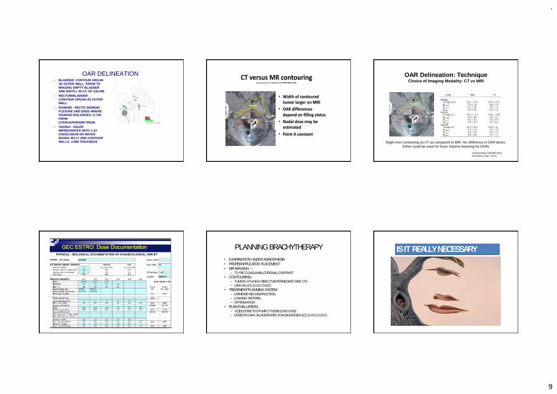
OAR DELINEATION✓ BLADDER: CONTOUR ORGAN
AS OUTER WALL. PRIOR TO
IMAGING EMPTY BLADDER
AND INSTILL 50 CC OF SALINE
✓ RECTUM/BLADDER :
CONTOUR ORGAN AS OUTER
WALL
✓ SIGMOID : RECTO SIGMOID
FLEXURE AND ENDS WHERE
SIGMOID DISLODGES >2 CM
FROM
UTERUS/PARAMETRIUM.
✓ VAGINA : GAUZE
IMPREGNATED WITH 1:10
GADOLINIUM OR WATER
BASED JELLY AND CONTOUR
WALLS. 4 MM THICKNESS Slight over-contouring on CT as compared to MRI. No difference in OAR doses.
Either could be used for Dose Volume reporting for OARs
Vishwanathan IJROBP 2012
Krishnatry, Patel JJCO,
OAR Delineation: TechniqueChoice of Imaging Modality: CT vs MRI
PLANNINGBRACHYTHERAPY
• EXAMINATION UNDERANAESTHESIA
• PROPER APPLICATOR PLACEMENT
• MR IMAGING :-– T2 FSE,T1,SAG,AXIAL,CORONAL,CONTRAST
• CONTOURING:-– TUMOR:-GTV,HIGH RISK CTV,INTERMEDIATE RISKCTV
– OAR:-D0.1CC,D-1CC,D-2CC
• TREATMENT PLANNINGSYSTEM– CATHETERRECONSTRUCTION,
– LOADING PATTERN,
– OPTIMISATION
• PLANEVALUATION:-– EQD2,DOSE TO GTV,HRCTV(D90,D100,V100)
– DOSES TO OAR:-BLADDER,RECTUM,SIGMOD(D0.1CC,D-1CC,D-2CC)
IS IT REALLYNECESSARY

.
10
CLINICAL IMPACT OF MRI ASSISTED DOSE VOLUME
ADAPTATION AND DOSE ESCALATION IN BRACHYTHERAPY OF
LOCALLY ADVANCED CERVIXCANCER.
CONV BRACHY
1998-2000
MRI BASED BRACHY
2001-2003
p
D90 81 GY 90 GY
OVERALL
SURVIV
AL
53% 64% 0.03
CSS 71% 90% 0.13
>5 CM
LOCALCONTROL
64% 82% 0.09
OS 28% 58% 0.003
CSS 40% 62% 0.07
LATEMORBIDITY 10% 2%
Potter et al. Radiother Oncol 2007:83;148
Reference tolerance doses
• For D2cc (Gy)
– Rectum, bowel, sigmoid – 75Gy
– Bladder – 95Gy

.
11
Dose effect relationship for late side effectsand
IGABT -Rectum
• Probability of EQD2 for G2-4 side effects (Gy) for the incidencerates
shown (95%CI)
Dose volume 5% 10% 20% p value
D2cc 67 (30-79) 78 (66-110) 90 (78-171) 0.0178
D1cc 71 (0-89) 87 (69-209) 104 (87-443) 0.0352
D0.1cc 83 132 186 0.1364
Georg et al. IJROBP 2012;82(2):653
Dose effect relationship for late side effectsand
IGABT -Bladder
• Probability of EQD2 for G2-4 side effects (Gy) for the incidencerates
shown (95%CI)
Dose
volume5% 10% 20% p value
D2cc 70 (0-95) 101 (29-137) 134 (110-371) 0.0274
D1cc 71 (0-107) 116 (17-169) 164 (129-498) 0.0268
D0.1cc 61 (0-155) 178 (0-368) 305 (129-498) 0.0369
Georg et al. IJROBP 2012;82(2):653
Comparative Study of LDR (Manchester System) and HDR Image-
guided Conformal Brachytherapy of Cervical Cancer: Patterns of
Failure, Late Complications, and SurvivalKailash Narayan, F.R.A.N.Z.C.R.correspondenceemail, Sylvia van Dyk, Dip App Sci,
David Bernshaw, F.R.A.N.Z.C.R., Chrishanthi Rajasooriyar, M.D.
, SrinivasKondalsamy-Chennakesavan,
• Retrospective study of 217 patients of advanced carcinomacervix.
• 90 patients received LDR and 123 patients received HDR
brachytherapy
• Conclusion of the study-- Image-guided HDR planning led to alarge
decrease in late radiation effects in patients treated by HDR.
Patterns of failure and survival were similar in patients treated
either by LDR orHDR.

.
12
TAKE HOME MESSAGE
IMAGE ASSISTEDBRACHYTHERAPY
• BRACHY THERAPY ISAN INTEGRAL PART OF TREATMENT
• MRI IS THE BEST MODALITY OFIMAGING
• T2 FSE IMAGING TO DELINEATE THETUMOR
• CONTOURING:-
– TUMOR:-GTV,HIGH RISK CTV,INTERMEDIATE RISK CTV
– OAR:-0.1CC,1CC,2CC
– BETTER TUMOR CONTROL AND LESSMORBIDITY