1.1 introduction, objectives and scope of the...
TRANSCRIPT

17
1.1 INTRODUCTION, OBJECTIVES AND SCOPE OF THE
INVESTIGATION
A nanoparticle is a particle having one or more dimensions of the
order of 100nm or less. Novel particles that differentiate nanoparticles
form the bulk material typically develop at a critical length scale of under
100nm1.
In nanotechnology, a particle is defined as a small object that
behaves as a whole unit in terms of its transport and properties.
In terms of diameter, fine particles cover a range between 100 and
2500nm, while fine particles on the otherhand are sized between 1 and
1000nm. Nanoparticles may or may not exhibit size related properties
that differ significantly from those observed in fine particles or bulk
materials. (Wikipedia, the free Encyclopedia). Protein nanoparticles are of
gelatin, albumin, gliadin and legumin3.
Nanoparticle research is currently an area of intense scientific
research due to a wide variety of potential applications in biomedical,
optical and electronic fields.
Nanoparticles form an effective bridge between bulk materials and
atomic or molecular structures2. The properties of materials change as
their size approaches the nano sale and as the percentage of atoms of the
surface of a material becomes significant.
Suspensions of nanoparticle are possible because the interaction
of the particle surface with the solvent is strong enough to overcome

18
differences in density which usually result in a material either sinking or
floating in a liquid. Nanoparticles often have unexpected visible
properties because they are small enough to confine their electrons and
produce quantum effects4.
Nanoparticles have a very high surface area to volume ratio. This
provides a tremendous driving force for diffusion especially at elevated
temperatures. The large surface area to volume ratio also reduces the
incipient melting temperatures of nanoparticles.
The unique size dependent properties of nanomaterials make them
very attractive for pharmaceutical applications. Cytotoxic effects of
certain engineered nanomaterials towards malignant cells form the basis
for nanomedicine. It is inferred that size, three dimensional shape,
hydrophobicity and electronic configuration make nanoparticles an
appealing subject in medicinal chemistry. The unique structure of
nanoparticles coupled with immense shape for derivatization forms a
base for exciting developments in therapeutics. Solid Lipid Nanoparticles
(SLN) forms an alternative colloidal carrier system for controlled drug
delivery. Because of their versatility and wide range of properties,
biodegradable polymeric nanoparticles are being used as novel drug
delivery systems. Further, this class of carrier holds tremendous
promise in the areas of cancer therapy and controlled delivery of vaccines

19
1.1.2 Classification:
Nanoparticles are often referred to cluster, nanospheres, nano –
rods, nano – fibers and nano – caps. Nanoparticles are made of semi
conducting materials may also be labeled quantum dots if they are small
enough (typically about 10nm) their quantization electronic energy level
occurs. Such nanoparticles are used in biomedical applications as drug
carriers or imaging agents
A prototype – nanoparticle of semi – solid nature is the liposome.
Various types of liposome nanoparticles are currently used clinically as
delivery systems for anti – cancer drugs and vaccines.
1.1.3 Characterisation:
It is done by Transmission Electron Microscopy/Scanning Electron
Microscopy, (TEM/SEM), Atomic Force Microscopy (AFM), Dynamic Light
Scattering (DLS), X-ray Photoelectron Spectroscopy (XPS), Powder X-ray
Diffractometry (XRD), Fourier Transform Infrared spectroscopy (FTIR),
Matrix Assisted Laser. Desorption Time of Flight mass spectrometry
(MALD –TOF) and Ultra – Violet Visible spectroscopy
1.2 CONTROLLED DRUG DELIVERY SYSTEMS6
1.2.1 INTRODUCTION:
The controlled drug delivery systems are gaining greater attention
in recent years owing to their importance and manifold advantages.
These systems are designed to release one or more drugs continuously in
a predetermined pattern for a fixed period of time either systematically or

20
to a specified target organ. Drug release from these systems should be at
a designed predictable reproduceable rate. By employing this system, the
safety, improved efficiency of drugs and patients compliance could be
assumed. Through, better control of plasma drug level and less frequent
dosing, the objectives of controlled drug delivery system can be fully
achieved. Though, these systems have been designed for oral, parenteral,
implantations and transdermal routes, oral routes are considered to be
the most convenient and common modes of administration Oral route
includes systems in the form of coated pellets, matrix tablets, poorly
soluble drug complexes and ion exchange resin complexes. Osmotic
preparations are known to release drug over an extended period of time
either in a continuous manner (sustained release) or as a series of pulses
(timed release). Among the various approaches, microencapsulation and
microcapsules have been accepted as reliable methods to achieve
controlled release.
1.2.2 MICROENCAPSUALTION
It is a process in which small, discrete solid particles or liquid
droplets are surrounded and enclosed by an intact shell and the
resulting materials are microcapsules. The capsule shells can be
designed to release their contents at specific rates under specific set of
conditions. Though, a variety of wall materials are used for the above,
polymeric substances having film forming properties are most suited for
microencapsulation.

21
1.3 AMOXICILLIN 7, 8
Heptane – 2 carboxylic acid. 6 [(Amino (4-hydroxyphenyl) acetyl)]
Amino – 3.3- dimethyl -7oxo-trihydrate.
D (-) - Amino –p-hydroxy benzyl Penicillin C16 H19 N3 O5 S. 3H2O
Preparation: - By Acetylation of 6 – Amino penicillanic acid with D (-) – 2
– (p–hydroxylphenyl glycine)
Properties: The solubility is at 1g in 370 ml water and 2000 ml alcohol.
It is a fine white to off-white crystalline powder with bitter taste. High
humidity and temperatures over 37ºC adversely affects its stability.
By the oral route, 75-90% is absorbed. An oral dose of 250mg will
provide a peak plasma concentration of about 4µg/ml. 50-72% is
eliminated by renal tubular secretion. The half life is about 1hr when
renal function is normal and 8-16hr in renal failure.
Uses: It is chemically p.hydroxyampicllin and has an antibacterial
spectrum similar to that of ampicillin drug except that it is less active
against Clostridium, Salmonella, Streptococcus and Shigella. Like
ampicillin, it is destroyed by β – lactamases and hence, it cannot be used
to treat infections caused by resistantstrain in bacteria of the β –

22
lactamase producing typhi. It cannot be given parent orally. It is the drug
of choice for infections by penicillinase producing Staphylococcus.
1.4 CIPROFLOXACIN HYDROCHLORIDE9, 10
3-Quinoline carboxylic acid
1-cyclopropyl – 6 fluro-1, 4 dihydro-4-oxo-7–(1 – piperazinyl) monohydro
chloride, monohydrate
C17H18FN3O3·HCl·H2O 385.82
Preparation: It is a pale yellow amphoteric crystal prepared from 3-
chloro-4-fluroamiline by condensation with ethyl ethoxy methylene
malonate to form the imine which is thermocycized to ethyl 7-chloro-6-
fluro-4 hydroxyguinoline-3-carboxyl-N-alkylation with cyclopropyl iodide
followed by nucleophilic displacement of the 7 chlorogroup by N-methyl
piperazine and hydrolysis of the ester affords the product.
Properties: It is soluble at 1g in 25ml water. The oral bioavailability is
about 70-80%. A dose of 0.5g yields plasma concentration 12hrs after
administration of about 0.2μg.Urinary excretion accounts for the
elimination of 40-50% of the dose. 20-35% is eliminated in feces. There
is hepatic biotransformation of four known metabolites which account for
15%. The halflife is about 4hrs.

23
Uses: It is used in the treatment of bone and joint infections caused by
certain microbes. Further, it is an unlabelled but authoritatively
alternate drug for the treatment of gonorrhea and salmonella infections.
The molecular weight value of ciprofloxacin-loaded PEBCA
nanoparticles was shown to be reduced as compared with unloaded
nanoparticles. Drug release from the colloidal carrier in medium
containing esterase was found to be very slow (a maximum of 51.5% after
48hrs) suggesting that this release resulted from bioerosion of the
polymer matrix. F-NMR analysis demonstrated that ciprofloxacin
entrapped into nanoparticles was only in its neutral form. Ciprofloxacin
HCl-loaded nanoparticles of chitosan, lipid, (SLNs) albumin and gelatin
showed sustained drug release avoiding burst effect of the free drugs.
Further, ciprofloxacin nanoparticles and SLNs can act as promising
carriers for sustained ciprofloxacin release.
AMPICILLIN
C16H14N3O4S (6R)-6(alpha phenyl-D-glycyl amino) pencillanic acid
It is an antibacterial agent,effective against various types of
bacteria.The daily dose is 2-6 g in frequent intervals.It is a crystalline
white substance sparingly soluable in water but insoluable in

24
ethanol,chloroform and fixed oils; soluable in acids and alkali
hydroxides.It should be stored in well closed containers in a cool and dry
place
OFLOXACIN
Ofloxacin inhibits an enzyme called DNA gyrase that is an essential
component of the mechanism that passes genetic information onto
daughter cells when a cell divides.
9-fluoro-2,3-dihydro-3-methyl-10-(4-methyl-1-piperazinyl)-7-oxo-
7H-pyrido[1,2,3-de]-1,4-benzoxazine-6-carboxylic acid hemihydrate
1.5 Sepia officinalis – A SOURCE OF NATURAL POLYMER OF DRUG
DELIVERY106, 107, 108
Sepia melanins are negatively charged pigments that are
hydrophobic, containing phenolic or indolic compounds.
These melanins are of the following types
Eumelanins – black or brown in colour
Pheomelanin – yellow or reddish in colour
Pyomelanin – brownish in colour

25
Sepia melanins are dark in colour and they are used in the
preparation of UV-absorbing optical lenses and in cosmetic
creams.
They are conductive to electricity.
Melanins are mainly used in pharmaceutical formulations and
drug delivery systems in nanotechnology.
In human physiology, melanins play main role in imparting
pigmentation to hair, skin and eyes, as a free radical scavenger
and increases the speed of nerve and brain messages.
Melanins are synthesized by free-living microbes, even facultative
microbes like “Cryptococcus neoformans”8 in soils. Melanin
production in these offers an advantage of survival from
environmental predators which produce hydrolytic enzymes. It is
due to sequestration of enzymes on melanin or stearic hindrance.
Melanins offer protection from UV-light and prevent photoinduced
damage.
1.5.1 Evidence for melanins bind to drugs in-vitro
(i) Isotherm analysis of adsorption of drugs by melanin:
Binding of Gentamicin, Methotrexate and Chlorpromazine to melanins
is explained by Isotherm binding equations to characterize the
adsorption of drugs to synthetic and sepia melanins.
Best fit Freundlich equation for Gentamicin6.

26
[q = qo (KC) 1/n] dm3.g-1
where, q = amount absorbed (m.mol.g-1)
qo = adsorption capacity
K = energy of absorption
C = equilibrium solution concentration of solute and
heterogeneity index 1/n (between 0 and 1)
(ii) Scatchard plot analysis of drug binding by melanin:
This method involves usage of radio-labelled compounds to
demonstrate the presence of heterologous binding sites.
Aminoglycoside antibiotics like Gentamicin and Kanamycin7 have
„2‟ binding sites on synthetic DOPA melanin.
For Kanamycin, association constants for strong and weak binding
sites were 3 X 10⁻⁵ and 4 X 10⁻³ m-1 respectively.
0.64µm Kanamycin is required to saturate binding sites in 1 mg
melanin.
Scatchard plot type analysis with melanins reveals that high
and low affinity binding sites for cocaine, amphetamines and
anti-arrythmics quinidine, disopyramide and metoprolol.
1.5.2 Absorption studies with Anti-fungals:
Amphotecin-B and caspofungin bind to melanin which uses 2
methods. They are

27
The melanin produced by C.neoplasms and synthetic melanin to bind
to these anti-fungal drugs was interfered from experiments,
incubating melanins with various compounds and anti-fungal activity
of solution was determined.
Testing of anti-fungal solutions in MIC and time-kill studies were
performed by removing melanin particles by centrifugation and
testing.
1.5.3 Binding of compounds by melanin in human’s in-vivo
Binding of drugs to host melanin damages certain tissues and causes
pathogenecity. For example in Parkinson‟s disease, there is a loss of
pigment in melanonic dopaminergic neurons in substantia nigra of
the brain.
In Parkinson‟s disease, 1-methyl 4-phenyl 1, 2, 3, 6 - tetrahydro
pyridine (MPTP)9 caused damage of substantia nigra neurons which
are concentrated with melanin.
Phenothiazines caused parkinsonian symptoms and secondary which
are reversible. The specific retention of other drugs which concentrate
at the pigmented tissues causes the damage of cells like skin, eye and
inner eye.
The complex interactions depend on diverse factors like cysteine
content, pH and ionic Interactions7.
Chloroquine accumulates in dermal melanocytes and hair follicles
where it causes irreversible hearing loss, tinnitus and dizziness.

28
Hearing loss is due to effect on 8th cranial nerve. Quinine accumulates
in melanin in the Stria nascularis of cochlea and causes cellular
degeneration.
Aminoglycosides8 become positively charged at the physiological
pH. Because of its high molecular weight, its penetration in the
tissues is reduced. Further, administration of this drug can cause
permanent vestibular and auditory ototoxicity.
Aminoglycosides when administered as intravitreal injection,
caused ocular pigmentation can partially protect retina turn damage.
Thioureylenes when incorporated into melanin like propylinic
uracil cause a loss or depigmentation of hair.
Ravuconazole, which is similar to voriconazole is effective against
Aspergillus fumigates and Aspergillus flavus.
1.5.4 Cuttlefish ink (sepia)
Cuttlefishes are the ink producing marine invertebrates and they
belong to the Phylum Mollusca and Class Cephalopoda which include
similar ink producing animals such as octopus and squid. The
cuttlefishes are soft bodied swimming animals provided with a large head
ringed by tentacles and an internal cuttle bone made of chiefly calcium
carbonate. These animals possess an ink pouch (sac) in which, a
brownish black fluid called „sepia‟ secreted by them is stored.
Cuttlefishes are known to display natural camouflage. In order to escape
from predators at the times of emergency, cuttlefishes darken the

29
environment by ejecting a gelatinous and mildly narcotic dark brown ink
to stun attackers and this defensive response gives them time to escape.
Further, the melanin particles of sepia are easily miscible in sea
water and remain dispersed in solution for more than 14 days.
Factors controlling ink production: The ink production and ejection in
cuttlefishes are affected and modulated by N-methyl-d-aspartate (NMDA)
- nitric oxide (NO)-cyclic GMP (cGMP) signaling pathway, Glutamate
NDMA receptor and NO synthase, the enzyme which is responsible for
the synthesis of NO has been detected in immature ink gland cells.
1.5.5 Extraction of sepia
The crude ink obtained from the ink sac is boiled with caustic
soda, filtering the extract and then adding HCl for precipitating the
colouring matter. The liquid ink may also be dried by combining with
lactose and then ground.
Characteristics of sepia: The liquid of cuttlefish ink has a grainy texture
and is alkaloid. Hence, it is not preferred by predators especially fish.
The ink is not poisonous and acts solely as a decoy device. The main
constituents of the ink are melanin and mucus. Melanin is a natural
melanoprotein containing 10 – 15% protein. The melanin binding protein
through aminoacid containing sulphur which is sistein.
The ink gland contains a variety of melanogenic enzymes including
tyrosinase, a peculiar dopachrome rearranging enzyme (which catalyses
the rearrangement of dopachrome to 5, 6 – dihydroxyindole) and a

30
peroxidase presumably involved in later stages of melanin biosynthesis.
The ink is also believed to contain dopamine and L-DoPA and small
amounts of aminoacids, including taurine, aspartic acid, glutamic acid,
alamine and lysine.
Human use: While the flesh of cuttlefish is used as a food source, its ink
finds applications in food colouring and in the preparation of pastries
and sauces. As an important dye, cuttlefish ink has been used for
centuries by humans for writing, drawing and in photographic works.
Sepia ink is available in Italy as hard dark chips. These are smelly
and hard to grind small enough to form an ink. They do not dissolve
readily in water. Mixing sepia powder with gum Arabic water to make
little cakes letting them dry and rubbing them up water when an ink is
needed.
1.5.6 Protective and therapeutic uses:
Sepia is a long standing homeopathic remedy for females because
it is effective for all menstrual and menopausal complaints. It also helps
combat persistent sadness and depression. That is sepia can lift the
mood of melancholy people urging them to take a more positive approach
to their lives. Vaginal discharge and even severe pain from endometriosis,
the growth of uterine cells in the abdominal cavity may be greatly
relieved by sepia. Migraines, liver weakness constipation, hair loss,
exhaustion and poor circulation with its resulting chillness can also be
treated with sepia remedy.

31
Sepia is known to soothe disturbances of the metabolism and ANS.
It also helps restore hormonal balance in women positively affecting
uterus and ovaries. Further, sepia improves blood circulation in the
organs especially those in abdominal cavity.
1.5.7 Bioactive properties:
The bioactive properties of ink gland of cuttlefish have been
studied for antibacterial, antiviral and anticancer agents. Purified cuttle
fish ink with a mixture containing mainly of a conjugated glucide (in
which agar, protein and lipid units are combined) ink may be effective in
fighting cancer. It was tested on 15 mice which were implanted with
tumours. The compound present in ink works by activating
macrophages, a type of WBC near the site of tumour. This would
increase the body‟s immune response to the tumour cells rather than
fighting the cancer cells directly.
Cytotoxicity: An uronic acid with rich peptidolglycon isolated from the
ink of cuttlefish Sepia pharaonis showed cytotoxicity against human
cervical cancer.
1.5.8 Radio-protective effect:
Irradiation leads to immunosuppression, haemopoiesis injury as
well as subhealth of human being. The protective and therapeutic effects
of cuttlefish ink on haemopoiesis in 60 Co gamma radiated model mice
were investigated. The results showed that the cuttlefish ink showed
significant effect on granulopoiesis. It is suggested that the increases of

32
antioxidant level in mice, the improvement of bone marrow
haematopoietic micro environment and the inducement of cellular
factors promoted the proliferation and differentiation of CFU–S (colony
forming unit in spleen) and CFU–GM (colony forming unit of granulocyte
and monocyte) and thus enhance the defensive system of organism.
1.6 CHITOSAN11, 12, 13
It occurs naturally in fungi, yeasts, marine invertebrates and arthropods.
Chitosan is the principal component of exoskeletons of marine
crustaceans from which supplements are often derived.
SYNONYMS: Chitosan hydrochloride or 2-Amino–2-deoxy-(1, 4) – β – D-
gluco pyranosoamine or β-1, 4 – poly-D-glucosamine or poly – (1, 4 – β –
D – gluco pyranosoamine).
Chemical name: Poly-β-(1, 4) – 2 – Amino – 2 – deoxy – D – Glucose.
Empirical Formula
Partial deacetylation of chitin results in the production of chitin
which is a polysaccharide comprising copolymers of glucosamine and N-
acetylglucosamine. The degree of deacetylation necessary to obtain a
soluble product must be greater than 80-85%. Chitosan is available with
different molecular weights (10000 to 1000000).

33
Structural Formula:
Uses: Chitosan is widely used as an excipient in oral and other
pharmaceutical formulations. It is used as a coating agent, disintegrant,
film-forming agent, mucoadhesive, tablet binder and viscosity increasing
agent.
1.6.1 Application in Pharmaceutical Formulations:
The suitability and performance of chitosan for drug delivery
applications have been investigated. It is used in controlled drug delivery
application as a component mucoadhesive dosage forms and rapid
release dosage forms in improved peptide delivery and for gene delivery.
Chitosan has been processed into several pharmaceutical forms
including gels, films, microspheres tablets and coatings for liposome.
Furthermore, chitosan may be processed into drug delivery systems
using several techniques including spray drying, coacervation, direct
compression and conventional granulation processes.
Although, the carriers are of the same size (200nm), drug loading
capacity of chitosan is 20 times higher for nanoparticle than for
liposome. Polysaccharide based nanoparticles of chitosan are prepared

34
by covalent cross linking, ionic cross linking polyelectrolyte complex and
the self assembly hydrophobically modified polysaccharides. Chitosan is
non-toxic, biocompatible and biodegradable and these properties make
chitosan a good candidate for conventional and novel drug delivery
systems. Chitosan forms colloidal particles and entraps bioactive
molecules through a number of mechanisms including chemical cross
linking, ionic cross linking and ionic complexation. Because of high
affinity of chitosan for cell membrane, it has been used as a coating
agent for liposome formulations. Chitosan is only soluble in acidic
solution with <pH6 and loses its change in >pH6. Therefore, it will be
insoluble in aqueous media. Synthesis of quaternary derivatives of
chitosan to improve solubility in wide pH range for increasing its
potential as an enhancer has been investigated. A number of factors
such as degree of polymerization, level of deacetylation, types of
quarternisation, installation of various hydrophobic substances, metal
complexation and combination with other agents influence the structure
characteristics of chitosan. Biodegradable, non-toxic and non-allergic
nature of chitosan encourages its potential use as a carrier for drug
delivery systems in all targets.
1.6.2 Properties:
Chitosan occurs as odourless white or creamy powder or flakes.
Fibre formation is quite common during precipitation and the chitosan

35
may look „cotton like‟. Chitosan is a cationic polyamine with a high
change density at pH <6.5. It is a linear polyelectrolyte with reactive
hydroxy and aminogroups. The presence of a number of aminogroups
allows chitosan to react chemically with anionic systems which results in
alteration of physico – chemical characteristics of such combinations.
The nitrogen in chitosan is mostly in the form of primary aliphatic
aminogroups. Chitosan therefore, undergoes reactions typical of amines.
All functional properties of chitosan depend on the chain length, chain
density and charge distribution. Further, salt form, molecular weight,
degree of deacetylation and pH are known to influence chitosan in
pharmaceutical applications. Particle size distribution is < 30μm.
Chitosan is sparingly soluble in water and is practically insoluble in
ethanol (95%), other organic solvents and neutral or alkali solution at pH
> 6.5. Chitosan dissolves readily in dilute and concentrated organic
acids and to some extent in inorganic acids (except phosphoric and
sulphuric acids). Upon dissolution, aminogroups of the polymer become
protonated resulting in a positively charged polysaccharides and
chitosan salts (chloride, glutamate, etc.) that are soluble in water.
Solubility of chitosan is affected by the degree of deacetylation. Solubility
is also greatly influenced by the addition of salt to the solution.

36
1.6.3 Stability and storage:
Chitosan powder is a stable material at room temperature
although, it is hygroscopic after drying. Hence, it should the stored at a
temperature of 2 - 8ºC.
1.6.4 Preparation:
Chitosan is prepared by chemically treating the shells of
crustaceans such as shrimps and crabs. The basic preparatory process
involves the removal of protein by treatment with alkali and of minerals
such as calcium carbonate and calcium phosphate by treatment with
acid. Before these treatments, the shells are ground to make them more
accessible. The shells are initially deproteinized by treatment with an
aqueous sodium hydroxide 3.5% solution. The resulting product is
neutralized and calcium is removed by aqueous HCl 3.5% solution at
room temperature to precipitate chitin. The chitin is dried so that, it can
be stored as a stable intermediate for deacetylation to chitosan at a latter
stage. N-deacetylation of chitin is achieved by treatment with an
aqueous sodium hydroxide (40-45%) solution at elevated temperature
(110ºC) and the precipitate is washed with water.
The crude sample is dissolved in 2% acetic and the insoluble
material is removed. The resulting clear supernatant solution is
neutralized with an aqueous sodium hydroxide solution to give a purified
white precipitate of chitosan. The product can then be further purified
and ground to a fine powder or granules.

37
Chitosan, the deacetylated polymer of N-acetyl-D-glucosamine (chitin)
is water soluble and chemically similar to cellulose.
1.6.5 Pharmaceutical Uses:
Chitosan is believed to affect cholesterol levels and weight
because it has positively charged aminogroups at the same pH as the
gastrointestinal tract. These aminogroups are believed to bind to
negatively charged molecules such as lipids and bile preventing their
absorption and storage by the body. The action of chitosan in cholesterol
management may be explained by the theory that ingested chitosan salts
react with fatty acids and binds lipids because of hydrophobic
interactions; these bound lipids are extracted rather than absorbed.
Animal studies in rats, mice and chickens indicate that chitosan
decreases very low density lipoprotein-cholesterol levels while increasing
high density-lipoprotein (HDL)-cholesterol levels. In vitro studies have
also shown that O-carboxy methyl chitosan beads absorb low-density
lipoprotein (LDL) cholesterol.
Chitosan acts as a „Fat Blocker‟. Chitosan is the only edible fibrin
with positive charge in nature. The resulting molecule called chitosan -
fat polymer is too large to be absorbed through the intestinal wall and
therefore excreted via feces without digestion.

38
1.7 Biopharmaceutics:
It deals with the inter-relationships of physicochemical properties of
the drug in dosage form in which the drug is given and the route of
administration on the rate and extent of systemic drug absorption. The
factors which influence biopharmaceutics include:
(i) protection of the activity of the drug within the drug product;
(ii) the release of the drug from a drug product;
(iii) the rate of dissolution of the drug at the absorption site and
(iv) the systemic absorption of drug.
The dynamic relationships existing in biopharmaceutics are shown
hereunder.
Studies in biopharmaceutics use both in-vitro and in-vivo methods. In-
vitro methods are procedures employing test approaches and equipments
without involving laboratory animals and humans. In-vivo methods on
the otherhand involve human subjects and laboratory animals14

39
1.7.1 Pharmacokinetics:
It involves the kinetics of drug absorption, distribution and
elimination (i.e. excretion and metabolism). The drug distribution and
elimination are together often termed as „drug disposition‟. The study of
pharmacokinetics involves both experimental and theoretical
approaches. The experimental aspects of pharmacokinetics involve the
development of biological sampling techniques, analytical methods for
the measurement of drugs and metabolites, the procedures that facilitate
data collection and manipulation. The theoretical aspect of
pharmacokinetics involves the development of pharmacokinetic models
that predict drug disposition after drug administration.
1.7.2 Bioavailability:
It refers to the measurement of the rate and extent of active drug that
reaches the systemic circulation and is available at the site of action.
Physicochemical characteristics of the drug:
The physicochemical properties of the solid drug particles not only
affect dissolution kinetics, but are important considerations in designing
the dosage form.
Solubility, pH and drug absorption:
The solubility – pH profile is a plot of the solubility of drug at
different pH values. While a basic drug is more soluble in acidic medium
forming a soluble salt, an acid drug is more soluble in the intestine
forming a soluble salt at more alkaline pH. The solubility pH profile gives

40
a rough estimation of the completeness of dissolution for a dose of drug
in the stomach or in intestine. Solubility may be improved with the
addition of an acidic / basic excipient15.
Stability, pH and drug absorption:
The pH – stability profile is a plot of the reaction rate constant for
drug degradation versus pH. If drug decomposition occurs by an acid or
base catalysis, some precision of the degradation of the drug in the
gastrointestinal tract may be made.
Particle size and drug absorption:
The effective surface area of the drug is measured enormously by a
reduction in the particle size. Because dissolution takes place at the
surface of solute (drug), the greater surface area the more rapid the rate
of drug dissolution. The geometric shape of the particle also affects the
surface area and during dissolution, the surface is constantly changing.
Particle size and particle size distribution studies are important for
drugs that have low water solubility. Many hydrophilic drugs are very
active intravenously but are not very effective when given orally due to
poor absorption. Smaller particle size results in an increase in the total
surface area of the particles thus enhancing water penetration into the
particles and increases the dissolution rates.
Polymorphic crystals, solvates and drug absorption: 16
Polymorphism refers to the arrangement of a drug in various
crystal forms or polymorphs which have the same chemical structure,

41
but different physical properties such as solubility, density, hardness,
and compression characteristics. Some polymorphic crystals have much
lower aqueous solubility than the amorphous forms causing a product to
incompletely absorb. A drug that exists as an amorphous form generally
dissolves more rapidly than the same drug in a more structurally rigid
crystalline form. Some polymorphs are metastable and may get converted
into more stable forms overtime.
Polymeric drugs:
Polymers have been used to prolong drug release in controlled
release dosage forms. The basic components of site-specific polymer
carriers are:
(i) The polymeric backbone,
(ii) A site specific component for recognizing the target (horning
device),
(iii) The drug covalently attached to the polymer chain and
(iv) Functional chains to enhance the physical characteristics of the
carrier system.
The molecular weight of the polymer carrier is an important
consideration in designing the dosage forms. Generally large molecular
weight polymers have longer residence time and diffuse more slowly.
Insoluble polymers are used either as regular carriers or formulated into
microparticles and nanoparticles.

42
Polymeric backbone
1.7.3 Bioavailability17:
These studies are performed for both approved active drug
ingredients and therapeutic moieties not yet approved for marketing by
FDA. Further, these studies are used to define the effect of changes in
the physicochemical properties of drug substance and the effect of the
drug product (dosage form) on the pharmacokinetics of the drug.
Relative and Absolute availability: The area under the drug
concentration-time curve (AUC) is used as a measure of the total amount
of drug that reaches the systemic circulation. The AUC is dependent on
the total quantity of available drug FDo divided by elimination rate
constant „K‟ and the apparent volume of distribution VD. F is the fraction
of the dose absorbed. After IV administration, F is equal to unity because
the entire dose is placed into systemic circulation. Therefore, the drug is
considered to be completely available after IV administration. After oral
administration of the drug, F may vary from 0(no drug absorption) to
1(complete drug absorption).

43
Relative Availability (Apparent availability):
It is the availability of a drug from its product as compared to a
recognized standard. The availability of drug in the formulation is
compared to the availability of the drug in a standard dosage
formulation, usually a solution of the pure drug evaluated in a crossover
study. The relative availability of two drug products gives at the same
dosage level and by the same route of administration can be obtained
with the following equation.
Relative availability = (AUC)A_
(AUC)B
where drug product B is the recognized reference standard. This fraction
may be multiplied by 100 to give percent relative availability. When
different doses are administrated, a correction for the size of dose is
made as in the following equation.
Relative availability = (AUC) A/dose A
(AUC)B/dose B
Urinary drug excretion data may also be used to measure relative
availability, as long as the total amount of the intact drug, excreted in
the urine is collected. The percent relative availability using urinary
excretion data can be determined as follows:
Percent relative availability = (Du) (Ax) X 100
(Du) (Bx)
Here (Du) is the total amount of drug excreted in the urine
44
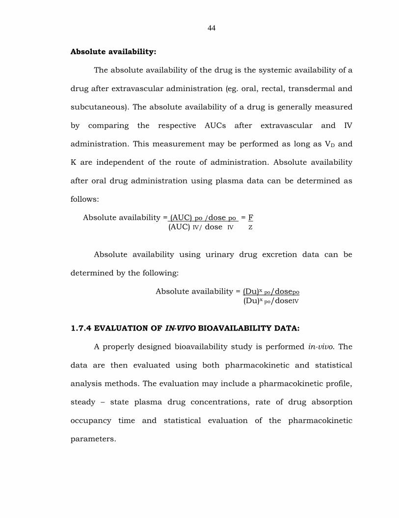
Absolute availability:
The absolute availability of the drug is the systemic availability of a
drug after extravascular administration (eg. oral, rectal, transdermal and
subcutaneous). The absolute availability of a drug is generally measured
by comparing the respective AUCs after extravascular and IV
administration. This measurement may be performed as long as VD and
K are independent of the route of administration. Absolute availability
after oral drug administration using plasma data can be determined as
follows:
Absolute availability = (AUC) po /dose po = F
(AUC) IV/ dose IV Z
Absolute availability using urinary drug excretion data can be
determined by the following:
Absolute availability = (Du)x po/dosepo
(Du)x po/doseIV
1.7.4 EVALUATION OF IN-VIVO BIOAVAILABILITY DATA:
A properly designed bioavailability study is performed in-vivo. The
data are then evaluated using both pharmacokinetic and statistical
analysis methods. The evaluation may include a pharmacokinetic profile,
steady – state plasma drug concentrations, rate of drug absorption
occupancy time and statistical evaluation of the pharmacokinetic
parameters.

45
Pharmacokinetic Profile: Plasma drug concentrations versus time
curve define the bioavailability of the drug from the dosage form. The
bioavailability data should include a profile of the fraction of a drug
absorbed and it should rule out dose dumping or lack of a significant
food effect. The bioavailability data should also demonstrate the
controlled -release characteristics of the dosage form compared to the
reference or immediate release drug products.
Steady -state plasma drug concentration:
The fluctuation between the C∞max (peak) and C∞min (trough)
concentration may be calculated as follows.
Fluctuation = C∞max - C∞min
C∞av
where C∞av is equal to (AUC)/T
An ideal extended release dosage form should have minimum
fluctuations between Cmax and Cmin. A true zero-order release will have no
fluctuations. In practice, the fluctuation in plasma drug levels after the
extended release dosage form should be less than the fluctuation after
the same drug given more immediate release dosage
Rate of drug absorption:
The rate of drug absorption from the conventional or immediate
release dosage form is generally of first order, whereas, the drug
absorption after the extended release dosage form may be zero order,
first order or an intermediate order. For many controlled release dosage

46
forms, the rate of drug absorption is of first order with an absorption rate
constant Ka which is smaller than the elimination rate constant k the
pharmacokinetic models when ka<k is termed flip–flop
pharmacokinetics.
Occupancy Time: For drugs for which the therapeutic window is known,
the plasma drug concentrations should be maintained above the
minimum effective drug concentration (MEC) and below the minimum
toxic drug concentration (MTC). The time required for the maintenance of
the plasma drug levels within the therapeutic window is known as
occupancy time
1.7.5 Bioequivalence Studies: 18
Bioequivalent drug products that have the same systemic drug
bioavailability will have the same predictable drug response. However,
variable clinical responses among individuals that are unrelated to
bioavailability may be due to differences in the pharmacodynamics of the
drug. Differences in pharmacodynamics i.e. the relationship between
drug and receptor site may be due to difference in receptor sensitivity to
the drug. Bioequivalence is established if the in-vivo bioavailability of a
test drug product does not differ significantly in the product‟s rate and
extent of drug absorption. A drug product that differs from the reference
material in its rate of absorption, but not in it‟s extent of absorption may
be considered bioavailable if the difference in the rate absorption is
intentional and appropriately reflected in the labeling and the rate of
47
absorption is not detrimental to the safety and effectiveness of the drug
product.
1.7.6 Statistical Evaluation:
Variables subjected to statistical analysis generally include plasma
drug concentrations at each collection time, AUC (from zero to last
sampling time), AUC (from zero to infinity), Cmax, tmax and elimination half
life t1/2. Statistical testing may include an analysis of variance (ANOVA)
computation of 90% and 95% confidence intervals on the difference in
formulation means and the power of ANOVA to detect a 20% difference
from the reference mean
1.7.7 Pharmacokinetics of oral absorption: 19
The systemic absorption of a drug from the G.I. tract or any other
extravascular site is dependent on the physicochemical properties of the
drug, the dosage form, and the anatomy and physiology of absorption
site. Further, surface area of gut, stomach emptying rate, G.I mobility
and blood flow to the absorption site may affect the rate and extent of
drug absorption. The overall rate of drug absorption may be described
mathematically as a first order or zero order input process. Most
pharmacokinetic models assume first order absorption unless an
assumption of zero order absorption improves the model significantly
and it has been verified experimentally.
The rate of change in the amount of drug in the body dDB/dt is
dependent on the rates of drug absorption and elimination.

48
The rate of drug accumulation in the body at any time is equal to
the rate of drug absorption less the rate of drug elimination.
dDB = dDGI - dDe
dt dt dt
During the absorption phase of a plasma level time curve, the rate of
drug absorption is greater than the rate of drug elimination.
dDGI > dDe
dt dt
At the time of peak drug concentration in the plasma which corresponds
to the time of peak absorption, the rate of drug absorption just equals
the rate drug elimination and there is no change in the amount of drug
in the body.
dDGI = dDe dt dt
1.7.8 Model of drug absorption and elimination:
Immediately after the time of peak drug absorption, some drug
may still be at the absorption site (i.e., in the GI tract). However, the rate
of drug elimination at this time would be faster than the rate of
absorption

49
dDaI < dDe
dt dt
When the drug at the absorption site becomes depleted, the rate of
drug absorption approaches zero or of DGI/dt =0. The elimination phase
of the curve then represents only the elimination of drug from the body
usually a first order process. Therefore, during the elimination phase, the
rate of change in the amount of drug in the body is described as a first
order process.
dDB = -kDB
dt where, k is the first order elimination rate constant
Zero – order absorption Model:
In this model drug in the GI tract DGI is absorbed systemically at a
constant rate ko. Drug is eliminated from the body by a first order rate
process with a first order rate constant k.
The rate of elimination at any time by first order process is equal to
DBk. The rate of input is ko. Therefore, the change per unit time in the
body can be expressed as
dDB = ko - kDB
dt
DGI
ko
DBVD
ko

50
One compartment model for zero – order drug absorption and first order
drug elimination
Integration of this equation with substitution of VD Cp for DB produces.
Cp = ko (1- e–kt)
VDk
The rate of drug absorption is constant and it continues until the
entire amount of drug in gut DGI is depleted. The time at which drug
absorption is continuous is equal to DGI/ko. After this time, the drug is
no longer available for absorption from the gut. The drug concentration
in the plasma will decline in accordance with first order elimination rate
process.
First order absorption model:
This model assumes a first order impact across the gut wall and
first order elimination from the body. This model applies mostly to the
oral absorption of drugs in solution or rapidly dissolving dosage
(immediate release) forms such as tablets, capsules and suppositories. In
addition, drugs given by intramuscular aqueous injections may also be
described using a first order process.
After administration, the drug is absorbed from the absorption site
by a first order process. In the case of a drug given orally, the drug
dissolves in the fluids of GI tract and is absorbed into the body according
to a first order process. The rate of disappearance of drug from the GI
tract is described by the following

51
dDGI = ka DGI F
dt
where, ka is the first order absorption constant from GI tract, F is
the fraction absorbed and DGI is the amount of drug in solution in GI
tract at anytime.
Integration of the above differential equation gives
DGI = Doe-kat
where, Do is the dose of drug. The rate of drug elimination is described
by a first order rate process for most drugs and is equal to -kDB. The rate
of drug change in the body dDB/dt is therefore the rate of drug in, minus
the rate of drug out as given by the following differential equation.
dDB = Rate in – Rate out dt
dDB/dt=Fka
where, F is the fraction of drug systemically absorbed
1.7.9 One compartment model for first order absorption and first
order elimination:20
F may vary from 1 for a fully absorbed drug to zero for a drug
completely unabsorbed. The maximum concentration is cmax and the
time needed to reach maximum concentration is tmax. The time needed to
reach maximum concentration is independent of dose and is dependent
on the rate constants for absorption (ka) and elimination k.

52
tmax = Inka – Ink = In (Ka/k)
ska – k ka – k
The time for maximum drug concentration tmax is dependent only
on the rate constants ka and k. The rate of drug excretion after a single
oral dose of drug is given with the following formula
dDu = Fke kaDo
dt
where, dDu/dt = rate of urinary drug excretion
K = fraction of dose absorbed
F = first order renal excretion constant
1.8 Biopharmaceutic considerations:
The prime considerations in the design of a drug product are safety
and efficiency. The drug product must effectively relieve the active drug
at an appropriate rate and amount to the targeted site, so that, the
intended therapeutic effect is achieved. The finished dosage form should
not produce any additional side effects or discomfort due to the drug
and/or the excipient. Ideally, all the excipients in the drug producer
should be inactive ingredients above or in combination in the final
dosage form.
The finished drug product is a compromise of various factors
including therapeutic objectives, pharmacokinetics, physical and
chemical properties, manufacturing cost, and patient acceptance. Most
importantly the drug product should meet the therapeutic objective by

53
delivering the drug with maximum bioavailability and minimum or nil
adverse effects.
Biopharmaceutical considerations in drug product design
Pharmacodynamic considerations21
Therapeutic objectives
Toxic effects
Adverse reactions
Drug considerations
Physical and chemical properties of drugs.
Drug product considerations
Pharmaceutics of drug
Bioavailability of drug
Route of drug administration
Designed drug dosage form
Designed dose of drug
Patient considerations
Compliance and acceptability of drug product cost
Manufacturing considerations
Cost
Availability of raw materials
Stability
Quality control.