1111111111111111111imuuuu - nasa
TRANSCRIPT
1111111111111111111imuuuu ~
(12) United States Patent Tai et al.
(54) MICROFLUIDIC DEVICE
(71) Applicant: California Institute of Technology, Pasadena, CA (US)
(72) Inventors: Yu-Chong Tai, Pasadena, CA (US); Siyang Zheng, Pasadena, CA (US); Jeffrey Chun-Hui Lin, Pasadena, CA (US); Harvey Kasdan, Chatsworth, CA (US)
(73) Assignee: California Institute of Technology, Pasadena, CA (US)
(*) Notice: Subject to any disclaimer, the term of this patent is extended or adjusted under 35 U.S.C. 154(b) by 0 days.
(21) Appl. No.: 14/296,199
(22) Filed: Jun. 4, 2014
(65) Prior Publication Data
US 2014/0377742 Al Dec. 25, 2014
Related U.S. Application Data
(62) Division of application No. 12/062,808, filed on Apr. 4, 2008, now abandoned.
(60) Provisional application No. 60/922,296, filed on Apr. 6, 2007.
(51) Int. Cl. GOIN15/06 (2006.01) GOIN33150 (2006.01)
(Continued)
(52) U.S. Cl. CPC ....... GOIN3315094 (2013.01); BOIL 31502753
(2013.01); BOIL 31502761 (2013.01); BOIL 220010636 (2013.01); BOIL 220010647
(2013.01); BOIL 230010681 (2013.01); BOIL 230010816 (2013.01);
(Continued)
(lo) Patent No.: US 9,029,158 B2 (45) Date of Patent : May 12, 2015
(58) Field of Classification Search USPC ............ 422/50, 58, 68.1, 100, 102, 502, 503;
436/174, 180, 43, 56 See application file for complete search history.
(56) References Cited
U.S. PATENT DOCUMENTS
4,233,029 A 11/1980 Columbus
4,376,820 A 3/1983 Giannini et al.
(Continued)
FOREIGN PATENT DOCUMENTS
WO W001/68238 A2 9/2001 WO WO 2006/055816 5/2006
(Continued)
OTHER PUBLICATIONS
Groselj-Gren, et al. Neutrophil and monocyte CD64 and CD163 expression in critically ill neonates and children with sepsis: com-
parison of fluorescence intensities and calculated indexes. Mediators
Inflamm. (2008), vol. 2008, p. 1-10; article ID 202646; doi: 10.11551 2008/202646.
(Continued)
Primary Examiner Brian 7 Sines (74) Attorney, Agent, orFirm Hema Vakharia-Rao; Nixon Peabody LLP
(57) ABSTRACT
Described herein are particular embodiments relating to a microfluidic device that may be utilized for cell sensing, counting, and/or sorting. Particular aspects relate to a micro-fabricated device that is capable of differentiating single cell types from dense cell populations. One particular embodi-ment relates a device and methods of using the same for sensing, counting, and/or sorting leukocytes from whole, undiluted blood samples.
19 Claims, 10 Drawing Sheets
270
230
:
2100
O e®
200 e=
220
2
240

US 9,029,158 B2 Page 2
(51) Int. Cl. GOIN33158 (2006.01)
BOIL 3100 (2006.01)
GOIN15110 (2006.01)
GOIN15114 (2006.01)
(52) U.S. Cl. CPC . BOIL 230010822 (2013.01); BOIL 230010864
(2013.01); BOIL 230010877 (2013.01); BOIL 24001086 (2013.01); COIN 1511056 (2013.01);
GOI N 1511484 (2013.01); GOI N 201511006 (2013.01); GOIN201511486 (2013.01); COIN 20151149 (2013.01); GOIN331582 (2013.01)
(56) References Cited
U.S. PATENT DOCUMENTS
4,400,370 A 8/1983 Kass 4,882,284 A 11/1989 Kirchanski et al. 5,304,487 A 4/1994 Wilding et al. 5,716,852 A 2/1998 Yager et al. 5,837,115 A 11/1998 Austin et al. 5,932,100 A 8/1999 Yager et al. 5,972,710 A 10/1999 Weigletal. 6,136,610 A 10/2000 Polito et al. 6,372,516 B1 4/2002 Sun 6,426,230 B1 7/2002 Feistel 6,551,841 B1 4/2003 Wilding et al. 6,635,163 B1 10/2003 Han etal. 6,637,463 B1 10/2003 Lei etal. 6,674,525 B2 1/2004 Bardell et al. 6,852,284 B1 2/2005 Holl et al. 7,105,355 B2 9/2006 Kurabayashi et al. 7,192,560 B2 3/2007 Parthasarathy et al. 7,247,274 B1 7/2007 Chow 7,347,976 B2 3/2008 Parthasarathy et al. 7,553,453 B2 6/2009 Gu et al. 7,718,421 B2 5/2010 Chen et al. D669,191 S 10/2012 Handique 8,318,109 B2 11/2012 Saltsman et al. 8,364,418 B2 1/2013 Davis et al. 8,518,705 B2 8/2013 Chan et al.
2001/0008760 Al 7/2001 King et al. 2002/0031255 Al 3/2002 Kasdan et al. 2003/0002037 Al 1/2003 Kasdan et al. 2003/0073089 Al 4/2003 Mauze et al. 2003/0170881 Al 9/2003 Davis et al. 2004/0126008 Al 7/2004 Chapoulaud et al. 2005/0105077 Al 5/2005 Padmanabhan et al. 2006/0011862 Al 1/2006 Bernstein 2007/0227890 Al 10/2007 Ramsey et al. 2007/0253868 Al 11/2007 Beebe et al. 2007/0292941 Al 12/2007 Handique et al. 2008/0101993 Al 5/2008 Andersson et al. 2008/0212102 Al 9/2008 Nuzzo et al. 2009/0042241 Al 2/2009 Yu-Chong et al. 2009/0117605 Al 5/2009 Davis et al. 2010/0051124 Al 3/2010 Imran 2010/0093019 Al 4/2010 Ditcham et al. 2011/0184537 Al 7/2011 Kasdan et al. 2012/0071342 Al 3/2012 Lochhead et al. 2012/0266986 Al 10/2012 Wimberger-Friedl et al. 2012/0275972 Al 11/2012 Schoen et al. 2013/0137135 Al 5/2013 Tai et al. 2013/0230867 Al 9/2013 Davis et al.
FOREIGN PATENT DOCUMENTS
WO WO 2006/118586 A2 11/2006 WO WO 2008/121828 10/2008 WO WO 2008/124589 A2 10/2008 WO WO 2009/144660 12/2009 WO WO 2011/094577 A2 8/2011 WO WO 2011/128893 A3 10/2011
WO WO 2012/092010 7/2012 WO WO 2014/097286 6/2014 WO WO 2014/097287 6/2014
OTHER PUBLICATIONS
Notice of Allowance dated Aug. 21, 2014 for U.S. Appl. No.
14/296,317. U.S. Appl. No. 60/922,296, filed Apr. 6, 2007, Tai et al. Adams, et al. Fluorometric characterization of six classes of human leukocytes. Acta Cytol. Sep.-Oct. 1974; 18(5): 389-391. Adams, et al. Machine characterization of human leukocytes by acridine orange fluorescence. Acta Cytol. May-Jun. 1971; 15(3): 289-291. Altendorf, et al. Differential Blood Cell Counts Obtained Using a Microchannel Based Flow Cytometer. Transducers. Jun. 1997 16-19; 1: 531-534. Assicot, et al. High serum procalcitonin concentrations in patients with sepsis and infection. Lancet. Feb. 27, 1993; 341(8844): 515-518. Aulesa, et al. Validation of the Coulter LH 750 in a hospital reference laboratory. Lab Hematol. 2003; 9(1): 15-28. Ault, Kenneth A. Flow cytometric measurement of platelet function and reticulated platelets. Annals of the New York Academy of Sci-ences. Mar. 20, 1993; 677: 293-308. Bhattacharya, eta]. Studies on Surface Wettability ofPoly(Dimethyl) Siloxane (PDMS) and Glass Under Oxygen-Plasma Treatment and Correlation With Bond Strength. J. Microelectromechan. Syst. Jun. 2005; 14: 590-597. Blajchman, et al. Bacterial detection of platelets: current problems and possible resolutions. Transfusion medicine reviews. Oct. 2005;19(4):259-272. Bodensteiner, David C. A flow cytometric technique to accurately measure post-filtration white blood cell counts. Transfusion. Sep. 1989; 29(7): 651-653. Cheson, et al. National Cancer Institute-sponsored Working Group guidelines for chronic lymphocytic leukemia: revised guidelines for diagnosis and treatment. Blood. 1996; 87(12): 4990-4997. Christ-Crain, et al. Effect of procalcitonin-guided treatment on anti-biotic use and outcome in lower respiratory tract infections: cluster-randomised, single-blinded intervention trial. Lancet. Feb. 21, 2004; 363(9409): 600-607. Cristofanilli, et al. Circulating tumor cells, disease progression, and survival in metastatic breast cancer. N Engl J Med. Aug. 19, 2004; 351(8): 781-791. Davis, et al. Neutrophil CD64 is an improved indicator of infection or sepsis in emergency department patients. Arch Pathol Lab Med. May 2006; 130(5): 654-661. Dieye, et al. Absolute CD4 T-cell counting in resource-poor settings: direct volumetric measurements versus bead-based clinical flow cytometry instruments. J Acquir Immune Defic Syndr. May 1, 2005; 39(1): 32-37. Divers, et al. Quantitation of CD62, soluble CD62, and lysosome-associated membrane proteins 1 and 2 for evaluation of the quality of stored platelet concentrates. Transfusion. Apr. 1995; 35(4): 292-297. Drexler, et al. Diagnostic value of immunological leukemia phenotyping. Acta Haematol. 1986; 76(1): 1-8. Dziegiel, et al. Detecting fetomaternal hemorrhage by flow cytometry. Curr Opin Hematol. Nov. 2006; 13(6): 490-495. Fischer, et al. Reducing costs in flow cytometric counting of residual white blood cells in blood products: utilization of a single platform bead free flow rate calibration method. Transfusion. Jul. 2011; 51(7): 1431-1438. Fujimoto, Keiji. Principles of Measurement in Hematology Analyz-ers Manufactured by Sysmex Corporation. Sysmex Journal Interna-tional. 1999; 9(1): 31-44. Gawad, et al. Micromachined impedance spectroscopy flow cytometer for cell analysis and particle sizing. Lab Chip. Sep. 2001; 1(1): 76-82. Graff, et al. Close relationship between the platelet activation marker CD62 and the granular release of platelet-derived growth factor. J Pharmacol Exp Ther. Mar. 2002; 300(3): 952-957.

US 9,029,158 B2 Page 3
(56) References Cited
OTHER PUBLICATIONS
Guerti, et al. Performance evaluation of the PENTRA 60C+ auto-mated hematology analyzer and comparison with the ADVIA 2120. Int J Lab Hematol. Apr. 2009; 31(2): 132-141. Hawkins, Robert C. Laboratory turnaround time. The Clinical Bio-chemist Reviews. Nov. 2007; 28(4): 179-194. Hershman, et al. Monocyte HLA-DR antigen expression character-izes clinical outcome in the trauma patient. Br. J. Surg. Feb. 1990; 77(2): 204-207. Hilfrich, et al. Prognostic relevance of human papillomavirus L1 capsid protein detection within mild and moderate dysplastic lesions of the cervix uteri in combination with p16 biomarker. Anal Quant Cytol Histol. Apr. 2008; 30(2): 78-82. Hillier, et al. A case-control study of chorioamnionic infection and histologic chorioamnionitis in prematurity. N. Engl. J. Med. Oct. 13, 1988; 319(15): 972-978. Hoffmann, Johannes JML. Neutrophil CD64 as a sepsis biomarker. Biochem Med (Zagreb). 2011; 21(3): 282-290. Holmes, et al. High throughput particle analysis: combining dielectrophoretic particle focussing with confocal optical detection. Biosens Bioelectron. Feb. 15, 2006; 21(8): 1621-1630. Hughes-Jones, et al. Differential white cell counts by frequency distribution analysis of cell volumes. J. Clin. Pathol. Aug. 1974; 27(8): 623-625. IPRP and WO in PCT/US2008/059408, dated Oct. 6, 2009. Jackson, JE Supravital blood studies, using acridine orange fluores-cence. Blood. May 1961; 17: 643-649. Kass, L. Identification of lymphocyte subpopulations with a polymethine dye. J. Histochem. Cytochem. Jul. 1988; 36(7): 711-715. Kass, L. Staining of granulocytic cells by Chlorazol black E. Am J. Clin. Pathol. Dec. 1981; 76(6): 810-812. Kibe, et al. Diagnostic and prognostic biomarkers of sepsis in critical care. J Antimicrob Chemother. Apr. 2011; 66 Suppl 2: ii33-40. Larosa, et al. Biomarkers: the future. Crit. Care Clin. Apr. 2011; 27(2): 407-419. Lee, et al. A flow-rate independent counter using a fixed control volume between double electrical sensing zones. Proceedings of the 18th IEEE International Conference on Micro Electro Mechanical Systems (MEMS). 2005.678-681. Lee, et al. Micromachine-based multi-channel flow cytometers for cell/particle counting and sorting. J. Micromechanics and Microengineering. 2005; 15(3): 447-454. Liu, et al. Improved quantitative Apt test for detecting fetal hemo-globin in bloody stools of newborns. Clin. Chem. Nov. 1993; 39(11 Pt 1): 2326-2329. Lotan, et al. Bladder cancer screening in a high risk asymptomatic population using a point of care urine based protein tumor marker. J Urol. Jul. 2009; 182(1): 52-57. Masse, et al. Validation of a simple method to count very low white cell concentrations in filtered red cells or platelets. Transfusion. Jul.-Aug. 1992; 32(6): 565-571. Matic, et al. Whole blood analysis of reticulated platelets: improve-ments of detection and assay stability. Cytometry. Oct. 15, 1998; 34(5): 229-234. McDonald, et al. Use of a solid-phase fluorescent cytometric tech-nique for the detection of bacteria in platelet concentrates. Transfus Med. Jun. 2005; 15(3): 175-183. Michelson, Alan D. Flow cytometry: a clinical test of platelet func-tion. Blood. Jun. 15, 1996; 87(12): 4925-4936. Miller, et al. Proteomics in Microfluidic Devices. In Encyclopedia of Micro- and Nanofluidics; Li, D. Q., Ed.; Springer: Heidelberg, Ger-many, 2008; 3: 1749-1758.
Morgan, et al. High speed simultaneous single particle impedance and fluorescence analysis on a chip. Curr. Appl. Phys. 2006; 6: 367-370. Moriyama, et al. Acridine Orange as a Fluorescent Probe for Lysosomal Proton Pump3. J. Biochem. 1982; 92: 1333-1336. Moro, et al. A new broad-spectrum cancer marker. Vitro Diagnostic Technology. Jun. 1, 2005; 1-3. Niehren, et al. An All-Solid-State Flow Cytometer for Counting Fluorescent Microspheres. Anal. Chem. 1995; 67(15): 2666-2671. OA in CN 200880015296.7, dated Sep. 20, 2012. Oberjat, et al. Rapid and reliable differential counts on dilute leukocyte suspensions. J. Lab. Clin. Med. Sep. 1970; 76(3): 518-522. Perry, et al. Is low monocyte HLA-DR expression helpful to predict outcome in severe sepsis? Intensive Care Med. Aug. 2003;29(8):1245-1252. Ramakumar, et al. Comparison of screening methods in the detection of bladder cancer. J Urol. Feb. 1999; 161(2): 388-394. Rawstron, et al. Quantitation of minimal disease levels in chronic lymphocytic leukemia using a sensitive flow cytometric assay improves the prediction of outcome and can be used to optimize therapy. Blood. Jul. 1, 2001; 98(1): 29-35. Rodriguez, et al. A microchip CD4 counting method for HIV moni-toring in resource-poor settings. PLoS Med. Jul. 2005; 2(7): e182. Rylatt, et al. An immunoassay for human D dimer using monoclonal antibodies. Thromb Res. Sep. 15, 1983; 31(6): 767-778. Sacks, et al. Guidelines and recommendations for laboratory analysis in the diagnosis and management of diabetes mellitus. Clin Chem. Mar. 2002; 48(3): 436-472. Satake, et al. A sensor for blood cell counter using MEMS technol-ogy. Sensors and Actuators B: Chemical. 2002; 83(1): 77-81. Segal, et al. Accuracy of platelet counting haematology analysers in severe thrombocytopenia and potential impact on platelet transfu-sion. Br. J. Haematol. Feb. 2005; 128(4): 520-525. Shapiro, et al. Combined blood cell counting and classification with fluorochrome stains and flow instrumentation. J Histochem Cytochem. Jan. 1976; 24(1): 396-411. Shapiro, et al. Cytomat-R: a computer-controlled multiple laser source multiparameter flow cytophotometer system. J Histochem Cytochem. Jul. 1977; 25(7): 836-844. Sheehan, et al. An improved method of staining leucocyte granules with Sudan black B. J Pathol Bacteriol. Jan.-Apr. 1947; 59(1-2): 336-337. Simonnet, et al. High-throughput and high-resolution flow cytometry in molded microfluidic devices. Anal Chem. Aug. 15, 2006; 78(16): 5653-5663. Stein, et al. D-dimer for the exclusion of acute venous thrombosis and pulmonary embolism: a systematic review. Ann Intern Med. Apr. 20, 2004; 140(8):589-602. Steinkamp, et al. Multiparameter Cell Sorting: Identification of Human Leukocytes by Acridine Orange Fluorescence. Acta Cytol. 1973; 17: 113-117. Sutherland, et al. The ISHAGE guidelines for CD34+ cell determinaton by flow cytometry. J Hematother. Jun. 1996; 5(3): 213-226. Tatsumi, et al. Principle of blood cell counter-development of elec-tric impedance method. Sysmex J. Int. 1999; 9(1): 8-20. Tibbe, et al. Optical tracking and detection of immunomagnetically selected and aligned cells. Nat Biotechnol. Dec. 1999; 17(12): 1210-1213. Van Dilla, et al. Volume distribution and separation of normal human leucocytes. Proc. Soc. Exp. Bio. Med. Jun. 1967; 125(2):367-370. Wang, et al. "Reticulated platelets predict platelet count recovery following chemotherapy." Transfusion. Mar. 2002; 42(3): 368-374. Weigl, et al. Design and rapid prototyping of thin-flim laminate-based microfluidic devices. Biomed Microdev. 2001; 3: 267-274. Yang, et al. A cell counting/sorting system incorporated with a microfabricated flow cytometer chip. Meas. Sci. Technol. 2006; 17: 2001-2009.

U.S. Patent May 12, 2015 Sheet I of 10 US 9,029,158 B2
,--,I N N'-,
a N~
FIG. 1
270
230 290
280 210
I
250 240
200
220
260
FIG. 2

U.S. Patent May 12, 2015 Sheet 2 of 10 US 9,029,158 B2
cl

U.S. Patent May 12, 2015 Sheet 3 of 10 US 9,029,158 B2
FIGAA FIGAB FIGAC
FICAD
F'l C _ 4 E
Fl G.4 li-"
N RIM ALJ -'ED
INTENSITY
0 ,:) , .1
0.9
0,8
0"/,
TME, (SEC ,C)ND)
FIG.5

TWE ("SECONIC))
FIG s 7
U.S. Patent May 12, 2015 Sheet 4 of 10 US 9,029,158 B2
FIG ~ 6A t" I G, 6 C
FIG - 6B FIG ~H
0,74
it 7 3
VOLTAGE (-)\ ~-T) -8
0. l6i 7
0 - 6 11",
M4

U.S. Patent May 12, 2015 Sheet 5 of 10
US 9,029,158 B2
FIG-, A
FIG 8B

------- -- --
i iN CD
Mi LLJ
L It
C I
U.S. Patent May 12 2015 Sheet 6 of 10 US 9,029,158 B2

"OUNT
FIGAO
Fl G,t 1
U.S. Patent May 12, 2015 Sheet 7 of 10 US 9,029,158 B2

U.S. Patent May 12 , 2015 Sheet 8 of 10 US 9,029,158 B2
:~,-~
? .... V

U.S. Patent May 12, 2015 Sheet 9 of 10 US 9,029,158 B2
FIG . 12B

U.R. Patent May 12 2013 Sheet 10 of 10 US 9,029,138 G2
' ©/J\/
\ \
51-310 &~ .
\, ~\ .
~ Z ^ } yz ,
////— _zz\ |
FIG ƒG > 1 A
I,,-,t440
\ / }/ .& .
~ \ \ ~ Jee ¥z C
—/4 0
; y
! ~
9 A 5i
1I! G .ƒ3

US 9,029,158 B2 2
crate more waste than the systems developed using microde-vices. Furthermore, for analysis of certain cell types, such as leukocytes, accuracy and speed of counting, differentiation, and/or sorting is important for determining disease state and
5 treatment.
MICROFLUIDIC DEVICE
CROSS-REFERENCE TO RELATED APPLICATIONS
This present application is a divisional of U.S. Ser. No. 12/062,808, filed Apr. 4, 2008, currently pending, which claims the benefit of priority of U.S. provisional application No. 60/922,296, filed Apr. 6, 2007, the contents of each which are herein incorporated by reference in their entirety.
STATEMENT OF GOVERNMENT INTEREST
This work was supported in part by the National Space Biomedical Research Institute through NASA, grant number NCC 9-58-317. The United States government has certain rights in this invention.
FIELD OF THE INVENTION
The present disclosure relates to fabricated microfluidic devices that can be utilized as cell sensors and/or actuators. In certain embodiments, the microfluidic device may be used for labeling, sensing differentiating, and/or sorting targets, par-ticularly cell populations.
BACKGROUND OF THE INVENTION
Standard cell sensors or actuators are generally based on flow cytometry and employ one or a combination of electrical impedance sensing, light scattering measurement, and chemi-cal or immunostaining followed by optical sensing.
For differentiation of blood cells by electrical impedance sensing, red blood cells are removed by lysing in order to reduce the blood volume. Lysing is generally done through the use of saponin or surfactants. During the lysing process, the leukocyte cell volume changes depending on cell type, due to the leakage of cytoplasm contents and cell nucleus shrinkage in varying amounts. Fujimoto, Sysmex J. Int. 9 (1999). Thus, normally 2-part (lymphocytes versus granulo-cytes) or even 3-part (lymphocytes, neutrophils, and other leukocytes) differential can be achieved by simple electrical impedance measurement of particle volume. Hughes-Jones, et al., J. Clin. Patol. 27; 623-625 (1974); Oberjat, et al., J. Lab. Clin. Med. 76; 518 (1970); Vandilla, et al., Proc. Soc. Exp. Biol. Med. 125; 367 (1967); Maeda, et al., Clin. Pathol. 27; 1117-1200 (1979); Maeda, et al., Clin. Pathol. 9; 555-558 (1982). Combining direct current and alternating current impedance, special acidic hemolysis in basophile channel and alkali hemolysis in eosinophil channel, a 5-part leukocyte differential can be achieved. Tatsumi, et al., Sysmex J. Int. 9; 9-20 (1999).
Alternative optical methods are based on light scattering and fluorescence staining of organelles, granules, and nuclei. Generally, low-angle scattered light contains information on cell size and high-angle scattered light can be used to probe internal composition of the cell. To achieve 5-part differen-tial, certain leukocyte populations, such as eosinophils, require special stain to change its scattering characteristics from other granulocytes, and basophils typically need to be counted separately following the differential lysis of other leukocytes. McKenzie, Clinical Laboratory Hematology, Prentice Hall, 2004; Fujimoto, Sysmex J. Int. 9 (1999).
In general, conventional automated cell analyzers are bulky, expensive, and mechanically complex, which restricts their locations to hospitals or central laboratories. Conven-tional cell analyzers require larger sample volumes and gen-
BRIEF DESCRIPTION OF THE SEVERAL VIEWS OF THE DRAWING(S)
10 FIG. 1A shows the molecular structure of acridine orange. FIG. 1B shows leukocyte staining results with acridine
orange. FIG. 2 shows the top view of one embodiment of a novel
15 fabricated microfluidic apparatus. FIG. 3 shows one embodiment of the optical system setup. FIG. 4A shows erythrocyte concentration in whole blood. FIG. 4B shows leukocyte staining in whole blood with
acridine orange at a concentration of 100 ng/mL. 20 FIG. 4C shows leukocyte staining in whole blood with
acridine orange at a concentration of 1 µg/mL. FIG. 4D shows leukocyte staining in whole blood with
acridine orange at a concentration of 10 µg/mL. FIG. 4E shows leukocyte staining in whole blood with
25 acridine orange at a concentration of 100 µg/mL. FIG. 4F shows leukocyte staining in whole blood with
acridine orange at a concentration of 1 mg/mL. FIG. 5 shows fluorescent signal bleaching from a single
leukocyte in one embodiment. 30 FIG. 6A shows an image of background and a 5 M bead
with focused laser illumination flow taken by CCD camera with long pass emission filter, according to one embodiment.
FIG. 6B shows an image of background and a 5 µm bead 35 with laser illumination flow taken by CCD camera with long
pass emission filter, according to one embodiment. FIG. 6C shows an image of background and a 5 µm bead
with diffused laser illumination flow taken by CCD camera with long pass emission filter, according to one embodiment.
40 FIG. 6D shows an image of background and the trace of a 5 µm bead with diffused laser illumination flow taken by CCD camera with long pass emission filter, according to one embodiment.
FIG. 7 shows a graph of detection of 5 µm fluorescent beads 45 detection with photodiode detector with long pass emission
filter. FIG. 8A shows background image of focused laser beam
from video taken by CCD camera with long pass emission filter, according to one embodiment.
50 FIG. 8B shows a signal from a leukocyte from diluted whole blood testing from video taken by CCD camera with long pass emission filter, according to one embodiment.
FIG. 9 shows the time trace of amplified photodiode signal of acridine orange stained undiluted whole blood with green
55 emission filter centered at 525 mu, and peaks labeled, accord-ing to one embodiment.
FIG. 10 shows a histogram of signal intensity from photo-diode detector with green emission filter centered at 525 mu.
60 FIG. 11 shows a histogram of signal intensity from photo- diode detector with red emission filter centered at 650 mu.
FIG. 12A shows an illustration of a handheld detection box instrument according to one embodiment.
FIG. 12B shows an assembled detection box instrument 65 according to one embodiment.
FIG. 13A shows the top view of an apparatus according to one particular embodiment.

US 9,029,158 B2 3
FIG. 13B shows the top view of an apparatus according to one particular embodiment.
SUMMARY OF THE INVENTION
Certain embodiments disclosed herein include a microflu-idic apparatus comprising a substrate having a first channel having a definedphysical feature, wherein said first channel is in fluid communication with at least one inlet for receiving a fluid, wherein said first channel leads to a restrictive access, io and wherein said first channel is in fluid communication with a second channel having a defined physical feature, wherein said second channel is in fluid communication with at least one fluid flow outlet; and a fluid biological sample. In certain embodiments, said defined physical feature is a depression or 15
protrusion. In particular embodiments, said fluid biological sample comprises blood. In certain embodiments, the microf-luidic apparatus further comprises a detection zone, and/or a filter array, each in fluid communication with said channel and said fluid flow outlet. 20
A microfluidic apparatus comprising a substrate having at least one first channel having a defined physical feature; at least one first inlet formed in said first channel for receiving a first fluid; wherein said first channel is in fluid communica-tion with a bifurcated channel, wherein said bifurcated chan- 25
nel is in fluid communication with a third channel detection zone; at least one second inlet for receiving a second fluid, wherein said second inlet is in fluid communication with a branched channel; a filter structure in fluid communication with a reservoir, wherein said reservoir is in fluid communi- 30
cation with said third channel detection zone; at least one fluid flow outlet formed in said third channel; and a fluid sample; wherein the ratio of the cross-sectional area of said second channel compared to the cross-sectional area of said first channel is 1:10. In certain embodiments, the defined physical 35
feature is a depression or a protrusion. In certain embodiments, said filter structure comprises a
filter array, said first fluid comprises sheath fluid and said second fluid comprises blood.
Certain embodiments disclosed herein relate to a detection 40
system comprising microfluidic apparatus and further com-prising a light source; a lens assembly; a filter assembly; and an image capture device. In some embodiments, the detection system further comprises at least one display unit or at least one recording unit. In certain particular embodiments, said 45
excitation source comprises a laser, particularly an argon laser. In particular embodiments, said filter assembly com-prises an excitation filter, and at least one emission filter. In certain embodiments, said filter assembly further comprises at least one aperture and at least one neutral density filter. In 50
particular embodiments, said filter assembly further com-prises at least one glass polarizer.
In certain embodiments, the lens assembly of the detection system comprises at least one condenser lens, at least one objective lens, and at least one beamsplitter. In particular 55
embodiments, said image capture device comprises at least one CCD camera, CMOS device, photodiode, or photomul-tiplier tube. In certain embodiments, said filter assembly comprises at least two emission filters and said image capture device comprises at least one photomultiplier tube. In certain 60
embodiments, said display unit comprises a computer and said recording unit comprises an oscilloscope. In particular embodiments, said excitation source comprises an argon laser; said lens assembly comprises a condenser lens, an objective lens, and a beamsplitter; said filter assembly com- 65
prises an excitation filter, a pinhole aperture and a neutral density filter, and at least one emission filter; said image
4 capture device comprises a CCD camera and a photodiode, and said display unit comprises a personal computer, and further comprising an amplifier.
In particular embodiments, said excitation source of the detection system comprises an argon laser; said lens assem-bly comprises a condenser lens, an objective lens, and a beamsplitter, said filter assembly comprises an excitation fil-ter, at least one emission filter; said image capture device comprises a photomultiplier tube, and said display unit com-prises a personal computer.
Other embodiments disclosed herein relate to a method for identifying a target comprising providing a fluid sample to at least one microfiuldic apparatus, wherein said fluid sample contains at least one dye; providing an excitation source to induce at least one fluorescent signal in a target; detecting the fluorescent signal using as sensor in the apparatus; and iden-tifying the target based in part on the analysis of the fluores-cent signal. In certain embodiments, said target is selected from the group consisting of: cells, organelles, nuclei, gran-ules, DNA, and RNA. In other embodiments, said target comprises a cell selected from the group consisting of a monocyte, a granulocyte, a macrophage, a neutrophil, an eosinophil, abasophil, or other leukocyte. In specific embodi-ments, said target comprises a leukocyte and said dye com-prises acridine orange. Particular embodiments of the method further comprising counting or sorting the target in the sample by analysis of the fluorescent signal. In certain embodiments, said fluid sample comprises blood.
DETAILED DESCRIPTION OF THE INVENTION
The present disclosure relates to fabricated microfluidic devices that can be utilized as cell sensors and/or actuators. In certain embodiments, the microfluidic device may be used for labeling, sensing, differentiating, and/or sorting cell popula-tions.
Microfluidic cell sensors and actuators can provide cell sensing and counting for a more accurate outcome and a lower cost. Particle counting (including bead, erythrocyte, and cultured cell) has been demonstrated, for example, by electrical impedance sensing, light scattering detection, and fluorescent sensing. Gawad, et al. Lab Chip 1; 76 (2001); Lee, et al. Proceedings of the 18 th IEEE International Conference on Micro Electro Mechanical Systems (MEMS) 678-681 (2005); Satake et al. Sens. Actuators B: Chem. 83; 77 (2002); Morgan, et al. Curr. Appl. Phys. 6, 367-370 (2006); Lee et al., J. Micromech. Microeng. 15; 447-454 (2005); Altendorf, et al. Proceedings of the International Conference on Solid State Sensors and Actuators (Transducers '97) v.1, p. 531, Chicago, Ill. (1997); Holmes et al., Biosens. Bioelectron. 21; 1621-1630 (2006); Yang et al., Meas. Sci. Technol. 17; 2001-2009 (2006); Simonnet et al., Anal. Chem. 78; 5653-5663 (2006); Niehren, et al., Anal. Chem. 67; 2666-2671 (1995).
In the area of optical sensing, microfabrication has allowed development of microdevices to replace glass capillary-based flow chambers, and to integrate compact optics and provide on-chip sample transport.
Cell sensing and counting, particularly of leukocytes, is cumbersome due in part to the cell population numbers. For leukocyte differential in microdevices based on optical sens-ing, a V-groove microchannel was fabricated by anisotropic wet etching of a silicon substrate and 3 -part leukocyte differ-ential was demonstrated for diluted blood without sheath flow by two-parameter light scattering. Altendorf, Proceedings of the Int'l Conference on Solid State Sensors and Actuators, v. 1, p. 531 (1997).

US 9,029,158 B2 5
6
However, until the instant embodied disclosure, it was phils, and basophils) has been demonstrated with hypotonic
necessary to dilute cell samples for cell sensors and actuators
dilution and fresh Acridine orange-stained leukocyte
for many reasons. One reason dilution has been necessary is samples. Adams and Kamentsky, Acta Cytol. 15, 289 (1971);
in order to prevent the coincidence effect in which multiple
Adams and Kamentsky, Acta Cytol. 18, 389-391 (1974); cells appear in the detection zone simultaneously. In human 5 Steinkam et al., Acta Cytol, 17, 113-17 (1973).
blood, the ratio of erythrocytes, or red blood cells, to leuko- Other dyes can be utilized with certain embodiments
cytes is on the order of about a thousand to one, a dilution
described in the instant disclosure, such as ethidium bromide,
factor of from about one hundred to several tens of thousands three-dye combinations (ethidium bromide, brilliant sulfafla-
is typically required to avoid erythrocyte interference for vine, and stilbene disulfonic acid derivative); oxazine dyes, electrical impedance or light scattering detection. Further- io basic orange 21, and a polymethine dye. Shapiro, et al., His-
more, for counting leukocytes in samples where leukocytes tochem. Cytochem. 24, 396-411 (1976); Shapiro, et al., J.
are specifically fluorescently labeled, a dilution of at least ten
Histochem. Cytochem. 25, 836-844 (1977); U.S. Pat. No.
times is usually required. Sheenan and Storey, J. Pathol. 4,376,820; U.S. Pat. No. 4,882,284; Tibbe, et al., Nat. Bio-
Bacteriol. 59; 336 (1947); Kass, J. Clin. Pathol. 76; 810-12
technol 17,1210-1213 (1999); U.S. Pat. No. 4,400,370; Kass, (1981); Weigl et al., Biomed Microdeu 3; 267-274 (2001). 15 J. Histochem. Cytochem, 36, 711-715 (1988).
Dilution is also often required in order to avoid clogging
Apparatus
sample chambers, and also in order to remove erythrocytes
One embodiment of the instant disclosure relates to a
that are lysed prior to running the sample, particularly for
device or apparatus for cell counting and/or differentiating. In
electrical impedance or light scattering detection. Some of
particular embodiments, the device or apparatus comprises a these protocols also require an additional fixation buffer. 20 substrate formed from a material, such as silicon, glass, plas- Dyes tic, metal, or other material. One particular embodiment of
In the present disclosure, a dye, such as Acridine orange the instant disclosure was fabricated using soft lithography.
(3,6-dimethylamineoacridine, FIG. 1), can be used to differ- Quake, Science 290, 1536-40 (2000). Other photolitho-
entiate a target, such as cells, organelles, granules, nuclei, graphic or etching techniques could also be used, according to molecules (including double or single stranded nucleic acids, 25 specific embodiments.
such as DNA, or RNA, chromosomes, and also including
One embodiment of the device was microfabricated using
synthetic forms). In one particular embodiment, leukocytes two parts of PDMS (polydimethylsiloxane) (Sylgard 184,
may be detected, counted, or sorted without need for lysing
Dow Corning, Mich., USA) mixed vigorously in 10:1 ratio.
erythrocytes or fixing the cell sample. Certain dyes, such as
After degassing in vacuum for about 30 minutes, the mixture Acridine orange, are also desirable due to the fast diffusion 30 was poured onto DRIE-etched silicon mold, that had been
into cells, easy commercial availability, and excitation and
pretreated with HMDS (hexamethyldisilazane) for easy sepa-
emission wavelength compatibility with common light ration after baking. The molds were baked at 80° C. for 30
sources (i.e. argon laser and other broad spectrum light minutes. The hardened PDMS was separated from the silicon
sources in visible range) and optical filters. Kosenow, Acta mold, and PDMS sheet was cut into pieces and fluidic access Haematol. 7, 217 (1952); Schiffer, Blood, 19, 200 (1962); 35 holes were punctured on each piece with a Luer stub adapter
Jackson, Blood, 17, 643 (196 1); Hallermann et al., PerhDeut- (Becton Dickinson, N7, USA). Each PDMS piece was care- sch Gies Inn Med. 70, 217 (1964). fully placed on a cleaned glass slide and baked overnight at
Acridine orange is a pH-sensitive fluorescent cationic dye
80° C. In some cases, oxygen plasma treatment (300 m Torr,
that binds to double-stranded DNA by electrostatic attraction
25 W, 30 s) was used for PDMS and glass slides in order to and intercalation of the Acridine orange between base pairs. 40 improve adhesion between them, particularly with devices
Upon binding, the excitation maximum becomes 502 nm and
that were intended to be reused. Bhattacharya et al., J. Micro-
the emission maximum becomes 525 nm (green). Acridine electromechan. Syst. 14, 590-97 (2005).
orange also binds to RNA and single-stranded DNA, with a
In one particular embodiment, the channel structure was
shifted excitation maximum of 460 nm and an emission maxi- molded on a 1 cmxI cm PDMS block, with the thickness of mum of 650 nm (red). Adams and Kamentsky, Acta Cytol. 15, 45 the PDMS block at less than 3 mm. In one particular embodi-
289 (1971); Adams and Kamentsky Acta Cytol. 18, 389-91
ment the channel depth was 16 µm in order to accommodate
(1974); Steinkam et al., Acta Cytol. 17,113-17 (1973). Acri- large leukocyte sizes.
dine orange is also desirable in that it is hydrophobic in
One exemplary embodiment of the device is shown in FIG.
neutral pH, and can easily diffuse through the cell membrane
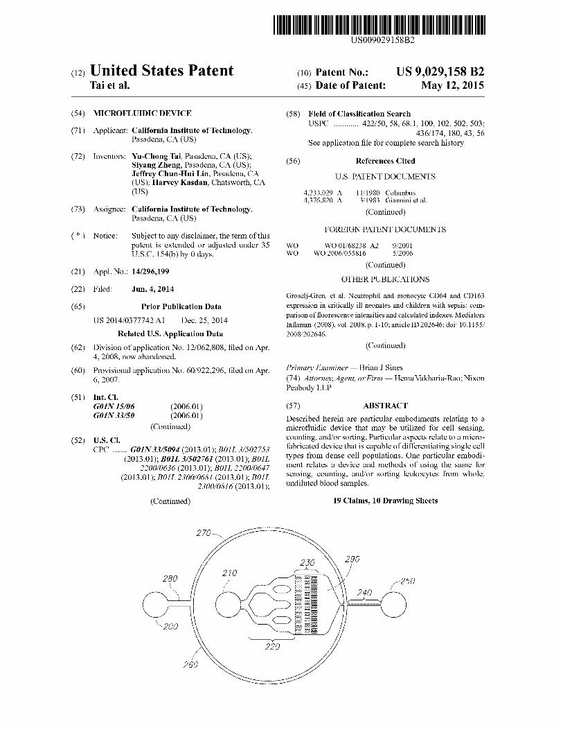
2. For this particular embodiment, a first fluid flow inlet 200 and cell nuclear membrane to bind to RNA and DNA. In 5o allows for deposition of, for example sheath flow fluid, and is
living cells, Acridine orange is protonated in the acidic envi- in fluid communication with a bifurcated channel with a first
ronment of lysosomes, which makes it cationic, and prevents channel arm 260 and a second channel arm 270 that both
the dye from leaking out of lysosome membranes. Moriyama converge at a junction of a reservoir 290 and the detection
et al., J. Biochem. 92; 1333-36 (1982). WhenAcridine orange zone 240. In this particular embodiment, the apparatus further is used for leukocyte analysis, the cell nucleus is stained green 55 comprises a second fluid flow inlet 210 that allows for depo-
with slightly mixed red, a result of double-stranded DNA and
sition of, for example, a sample fluid, such as blood, that is in
single-stranded RNA while the cell cytoplasm is stained red
fluid communication with a filter array structure 230, byway
due to the RNA and lysosomes. Thus, leukocyte counting can of a branched sample flow zone channel 220 and a fluid flow
be achieved easily by using the strong signal from the green outlet 250. In this particular exemplary embodiment, 2 -D fluorescent channel. Leukocyte differentiation can be 6o hydrodynamic focusing was adopted to control the particle
achieved by analyzing the signal from the red fluorescence position of the cell sample in the detection zone 240. Accord- channel. ing to the embodiment shown in FIG. 2, the ratio of cross-
For fresh-stained leukocytes, a 3 -part differential (lympho- sectional area of sheath flow to core sample flow was 10:1,
cytes, monocytes, and granulocytes) can be achieved by and the channel width of the detection zone 240 was 50 µm, studying the red fluorescent signal of an Acridine orange 65 with the width of the focused sample flow preferably 5 µm or
stained diluted blood sample, whereas a 5 -part differential
less. In particular embodiments, the channels comprise a
leukocytes (lymphocytes, monocytes, neutrophils, eosino- physical feature, such as a depression or a protrusion.
US 9,029,158 B2 7
One other exemplary embodiment of the device is shown in FIG. 13A. For this particular embodiment, the fluid flow inlet 1340 allows for deposition of a sample fluid, such as a bio-logical sample, or other fluid sample containing a target. In one particular embodiment, the biological sample includes a 5
cell sample, such as blood. In this exemplary embodiment, the fluid inlet is in fluid communication with a first channel 1330 which contains a restrictive access 1320 that is juxta-posed to a second channel 1310 which comprises the detec-tion zone which is also in fluid communication with the fluid io flow outlet 1300. In certain embodiments, the height of the first and/or second channels is approximately 5 µm, approxi-mately 8 µm, approximately 10 µm, approximately 12 µm, approximately 15 µm, approximately 20 µm, approximately 25 µm, approximately 30 µm, approximately 35 µm, approxi- 15
mately 40 µm, or any value therebetween. In certain embodi-ments the width of the second channel is approximately 5 µm, 10 µm, approximately 15 µm, approximately 20 µm, approxi-mately 25 µm, approximately 30 µm, approximately 35 µm, approximately 40 µm, approximately 45 µm, approximately 20
50 µm, or any value therebetween. In the exemplary embodi-ment shown in FIG. 13A, the second channel width was approximately 20 µm in size.
One other exemplary embodiment of the device is shown in FIG. 13B. For this particular embodiment, the fluid flow inlet 25
1440 allows for deposition of a sample fluid, such as a bio-logical sample, or other fluid sample containing a target. In one particular embodiment, the biological sample includes a cell sample, such as blood. In this exemplary embodiment, the fluid inlet is in fluid communication with a first channel 30
1430 which contains a restrictive access 1420 that is juxta-posed to a second channel 1410 which comprises the detec-tion zone which is also in fluid communication with the fluid flow outlet 1400. In certain embodiments, the height of the first and/or second channels is approximately 5 µm, approxi- 35
mately 8 µm, approximately 10 µm, approximately 12 µm, approximately 15 µm, approximately 20 µm, approximately 25 µm, approximately 30 µm, approximately 35 µm, approxi-mately 40 µm, or any value therebetween. In certain embodi-ments, the width of the second channel is approximately 5 40
µm, 10 µm, approximately 15 µm, approximately 20 µm, approximately 25 µm, approximately 30 µm, approximately 35 µm, approximately 40 µm, approximately 45 µm, approxi-mately 50 µm, or any value therebetween. In the exemplary embodiment shown in FIG. 1313, the second channel width 45
was approximately 30 µm in size. Certain embodiments of the device use a focused laser
source for illumination, since cell focusing in the detection zone 240 is highly desirable. However, other embodiments included in the present disclosure use a more uniform dif- 50
fused light source and a slit aperture. Such embodiments utilize straight channel geometry without cell focusing. In one embodiment, the channel length of the detection zone 240 is 1000 µm. A filter structure 230 upstream of the sample flow zone 220 may also be included in certain embodiments, 55
which filtered out contaminants, including erythryocyte rou-leaux, and other large particle aggregates to prevent clogging in the detection zone 240. In certain embodiments, the size of the rectangular pillar structure components of the filter struc-ture 230 was 200 µmx40 µm. The spacing between the pillars 60
in each of the three rows was 40 µm, 30 µm, and 20 µm respectively, which allows for even the largest leukocytes to pass through the filter region 230. System
The optical system was setup on an optical bench as shown 65
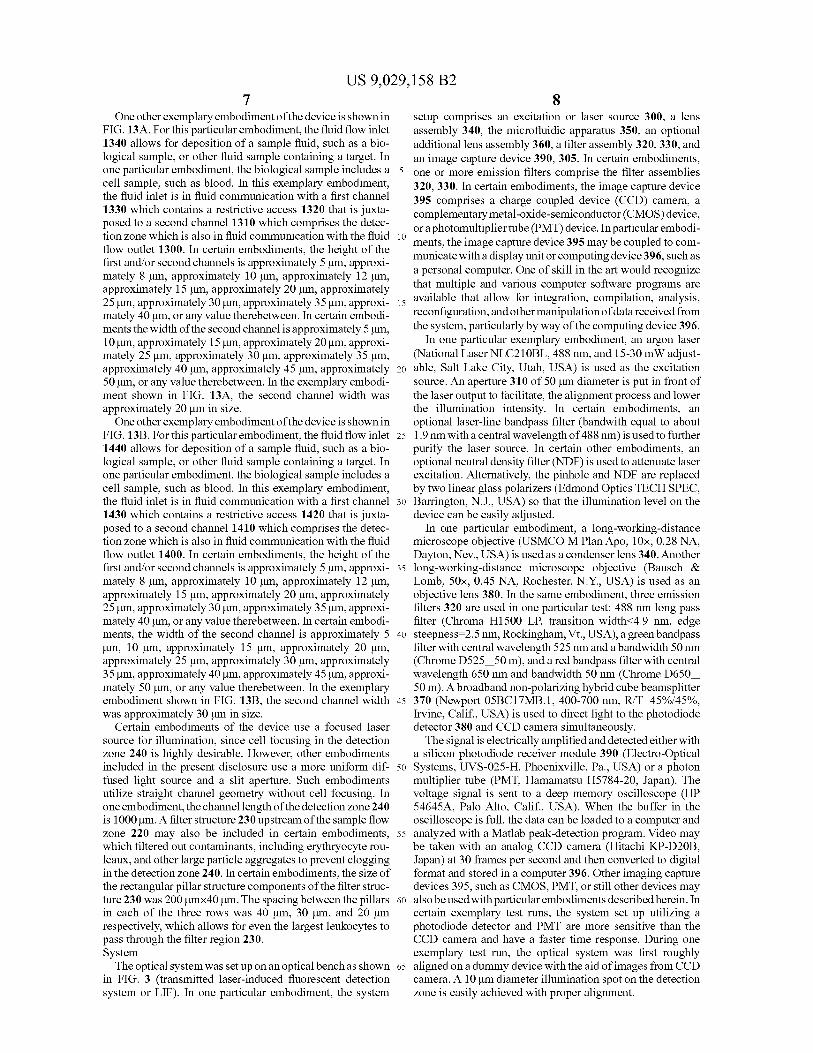
in FIG. 3 (transmitted laser-induced fluorescent detection system or LIF). In one particular embodiment, the system
8 setup comprises an excitation or laser source 300, a lens assembly 340, the microfluidic apparatus 350, an optional additional lens assembly 360, a filter assembly 320, 330, and an image capture device 390, 305. In certain embodiments, one or more emission filters comprise the filter assemblies 320, 330. In certain embodiments, the image capture device 395 comprises a charge coupled device (CCD) camera, a complementary metal-oxide-semiconductor (CMOS) device, or a photomultiplier tube (PMT) device. In particular embodi-ments, the image capture device 395 may be coupled to com-municate with a display unit or computing device 396, such as a personal computer. One of skill in the art would recognize that multiple and various computer software programs are available that allow for integration, compilation, analysis, reconfiguration, and other manipulation of data received from the system, particularly by way of the computing device 396.
In one particular exemplary embodiment, an argon laser (National Laser NLC2I OBL, 488 nm, and 15-30 mW adjust-able, Salt Lake City, Utah, USA) is used as the excitation source. An aperture 310 of 50 µm diameter is put in front of the laser output to facilitate, the alignment process and lower the illumination intensity. In certain embodiments, an optional laser-line bandpass filter (bandwith equal to about 1.9 mn with a central wavelength of 488 mu) is used to further purify the laser source. In certain other embodiments, an optional neutral density filter (NDF) is used to attenuate laser excitation. Alternatively, the pinhole and NDF are replaced by two linear glass polarizers (Edmond Optics TECH SPEC, Barrington, N.J., USA) so that the illumination level on the device can be easily adjusted.
In one particular embodiment, a long-working-distance microscope objective (USMCO M Plan Apo, 10x, 0.28 NA, Dayton, Nev., USA) is used as a condenser lens 340. Another long-working-distance microscope objective (Bausch & Lomb, 50x, 0.45 NA, Rochester, N.Y., USA) is used as an objective lens 380. In the same embodiment, three emission filters 320 are used in one particular test: 488 nm long pass filter (Chroma H1500 LP, transition width<4.9 mu, edge steepness=2.5 mu, Rockingham, Vt., USA), a greenbandpass filter with central wavelength 525 mn and a bandwidth 50 mn (Chrome D52550 m), and a red bandpass filter with central wavelength 650 nm and bandwidth 50 nm (Chrome D650 50 m). A broadband non-polarizing hybrid cube beamsplitter 370 (Newport 05BC17MB.1, 400-700 mu, R/T=45%/45%, Irvine, Calif., USA) is used to direct light to the photodiode detector 380 and CCD camera simultaneously.
The signal is electrically amplified and detected either with a silicon photodiode receiver module 390 (Electro-Optical Systems, UVS-025-H, Phoenixville, Pa., USA) or a photon multiplier tube (PMT, Hamamatsu H5784-20, Japan). The voltage signal is sent to a deep memory oscilloscope (HP 54645A, Palo Alto, Calif., USA). When the buffer in the oscilloscope is full, the data can be loaded to a computer and analyzed with a Matlab peak-detection program. Video may be taken with an analog CCD camera (Hitachi KP-D20B, Japan) at 30 frames per second and then converted to digital format and stored in a computer 396. Other imaging capture devices 395, such as CMOS, PMT, or still other devices may also be used with particular embodiments described herein. In certain exemplary test runs, the system set up utilizing a photodiode detector and PMT are more sensitive than the CCD camera and have a faster time response. During one exemplary test run, the optical system was first roughly aligned on a dummy device with the aid of images from CCD camera. A 10 µm diameter illumination spot on the detection zone is easily achieved with proper alignment.

US 9,029,158 B2 9
As shown in FIG. 12A and FIG. 12B, the instant apparatus may be incorporated into a hand-held unit comprising a laser source such as a laser emitting diode or LED 120), at least one lens 190, at least one filter assembly with optional beamsplit-ter 195, a microfluidic apparatus as described herein on a microchip or other substrate 185, an input/output port 130, at least one image capture device 100, 110, which may be a photomultiplier tube. In certain embodiments, the hand-held unit may be assembled and enclosed by an outer casing or casings 150, 180, and rivets or bolts 140, 160. Cell Detection
One aspect of the instant disclosure relates to methods of counting and/or differentiating cells, particularly leukocytes, from undiluted cell samples, such as human or other animal blood, by utilizing microfabricated devices. In one exemplary embodiment, cell detection was conducted utilizing Acridine orange and fresh whole human blood.
In one exemplary embodiment, fresh human blood was obtained from healthy donors and used within 3 days of collection. EDTA was added to the blood samples in order to prevent coagulation. For Acridine orange staining, the stock solution was added to obtain a final dye concentration of 10 µg/mL in Ficoll-Paque Plus. Ficoll-Paque Plus was also used as the sheath flow solution. Fluorescent polystyrene beads (5 µm green fluorescent beads) were purchased from Duke Sci-entific Corporations, Fremont, Calif., USA. Cell nucleus stain Acridine orange was obtained from Molecular Probes, Eugene, Oreg., USA, and dissolved in water to achieve a 10 mg/mL stock solution. Blood diluent Ficoll-Paque Plus was purchased from Amersham Biosciences, Sweden. Phosphate buffered saline (10xPBS) was obtained fromAmbion (9625), Austin, Tex., USA.
Staining results were observed under a fluorescent micro-scope (Nikon E800, Japan) with a triple and filter block DAPI-FITC-TRITC, which has excitation filter wavelengths of 385-400 mu, 475-490 mu, and 545-565 mu, and emission filter wavelengths of 450-465 mu, 505-535 nm and 580-620 mu. Images were taken with a cooled CCD camera (RT-KE color 3-shot, Diagnostic Instruments, Sterling Heights, Mich., USA). Rough count of leukocytes was made with a hemactyometer (Hausser Scientific, Horsham, Pa., USA). When necessary, blood or fluorescent beads were diluted with Ficoll-Paque Plus (specific gravity 1.077 g/mL) to match the specific gravity of the solvent to leukocytes. All fluids were pumpedinto the devices using syringepumps (HarvardAppa-ratus Pico Plus, Holliston, Mass., USA).
In this particular embodiment, an analog CCD camera was used for video recording at a matched camera frame rate of 3 nL/min sample flow rate and 30 nL/min sheath flow rate. For photodiode detection, a 0.1 µL/minute sample flow rate and a 1 µL/minute sheath flow rate were used. A 1 µL/minute sample flow and a 10 µL/minute sheath flow were used with the photon multiplier tube instrument.
In order to achieve a high signal-to-noise ratio, the maxi-mal concentration for cell staining was established using routine methods in the art. Adams and Kamentsky, Acta Cytol. 15; 289 (1971). As shown in FIG. 4, whole blood samples were analyzed with different Acridine orange con-centrations. The optimal concentration for leukocyte staining as determined to be approximately in the range of 1 µg/mL. In the particular exemplary embodiment utilized in FIG. 4, the distance between the coverslip and the grid surface was approximately 100 µm. As can be seen in FIG. 4A, an abun-dance of erythrocytes were present under the field of view, yet these cells did not interfere with the fluorescent signal from the leukocytes, as shown in FIG. 413-F.
10 As can be seen in FIG. 5, the exemplary embodiment
utilized in cell detection did not experience any significant photobleaching. The signal was fitted as a first-order expo-nential decay with time constant of 6.4±0.7 seconds. Two
5 more tests confirmed that the photobleaching time constant for one particular embodiment was between 1 second and 10 seconds. The photobleaching time constant for one particular embodiment was characterized by filling the device with Acridine orange-stained whole blood. The channel was
io scanned by the laser spot and the illumination was set to be the same as that used in testing. The entire process was recorded with a CCD camera. Whenever a fluorescing leukocyte was observed with fluorescent emission clearly distinct from the background, we stopped moving the laser spot and waited
15 until the leukocyte was photobleached to background level. The images were extracted from the video, converted to 8-bit gray scale images, and analyzed with a Matlab program. The data was fitted to a single time-constant exponential decay.
Additionally, green fluorescent beads were tested at a con- 20 centration of about 2x10 3/µL, as observed by CCD camera,
and shown in FIG. 6. Sample flow rate was set at about 3 nL/min, and sheath flow was about 30 nL/min. In one exem-plary test run, a hydrodynamic focused laser beam, as shown in FIG. 6A, created an enlarged light circle as shown in FIG.
25 6B. Only a single bead normally appeared in each image. With diffused laser illumination, as shown in FIG. 6C, the trace of the bead could be identified, as shown in FIG. 6D. Hydrodynamic focusing limits the cross-sectional area of the detection zone without shrinking the channel diameter, thus
30 the signal-to-noise ratio may be improved without increasing the risk of clogging the channel. Also, the reduction of the cross-section of the core flow reduces the coincidence effect. Finally, enclosing the core sample flow with sheath flow minimizes fluorescent dye absorption in the device walls,
35 thus reducing background noise. As indicated in FIG. 7, bead signals from the photodiode detector could easily be identi-fied.
As shown in FIG. 8, using both red and green emission filters, images extracted from video taken by the CCD camera
40 show the signal identified from a leukocyte stained withAcri-dine orange, as well as the signal obtained from the fluores-cent control bead. For photodiode detection, the expected leukocyte detection rate would average about 4-11 cells per second for a normal individual.
45 In one exemplary embodiment, a time trace over 50 sec-onds of an undiluted blood sample stained with Acridine orange using a green emission filter, and a throughput of up to about 1000 leukocytes per second was attained. Maxima sig-nal intensity (peak height as in FIG. 9) from the green fluo-
5o rescent channel with 525 nm emission filter was studied by plotting its histogram, as shown in FIG. 10. As expected, the lower-intensity portion is likely contributed mainly by lym-phocytes, while the higher-intensity portion is likely mainly from monocytes, with the center-region is likely mostly from
55 granulocytes, Steinkam et al., Acta Cytol, 17; 113-117 (1973).
In one exemplary embodiment, a time trace over 50 sec- onds of an undiluted blood sample stained with Acridine orange using a red fluorescent channel with 650 nm emission
60 filter was conducted. As shown in FIG. 11, two peaks were identified, the lower intensity is dominated by lymphocytes and the higher-intensity peak is largely monocytes and granu- locytes. The time between the start of staining the cells to photodiode recording was typically greater than 15 minutes.
65 In both exemplary studies, the maximal throughput was about 1000 leukocytes per second utilizing one embodiment of the PMT detector. By using undiluted blood, minimal

US 9,029,158 B2 11
12 sample volume was maintained, which increases the through- this specification and/or listed in the Application Data Sheet, put. Since sample throughput is proportional to volume flow are incorporated herein by reference, in their entirety. rate, but is limited by the maximal pumping rate and response
The invention claimed is: time of the sensing system a 3 nL/minute core flow rate was
1. A method for analyzing leukocytes in a whole blood used with the CCD camera detection. Under this flow rate, a 5 sample, the method comprising: typical leukocyte traveled through the detection zone in a) introducing a modified blood sample comprising fluo- approximately 30 milliseconds, which roughly equals the rescently labelled leukocytes and fluorescently labeled CCD frame acquisition time. control beads, to an inlet of a first microfluidic channel
How rates for varying embodiments may be suitable for a that gradually narrows and is directly coupled to a sec-
range from approximately I nL/minute, approximately 2 10 and microfluidic channel that has fixed dimensions, nL/minute, approximately 3 nL/minute, approximately 4
wherein the fluorescently labeled leukocytes are nL/minute, approximately 5 nL/minute, approximately 6
labelled with a fluorophore different than a fluorophore
nL/minute, approximately 7 nL/minute, approximately 8
label of the control beads, and wherein the second chan- nL/minute, approximately 9 nL/minute, approximately 10
nel having the fixed dimensions of approximately 40 nL/minute, approximately 20 nL/minute, approximately 30 15 microns by approximately 50 microns, and wherein the nL/minute, approximately 40 nL/minute, approximately 50
first channel and the second channel are obstacle-free;
nL/minute, approximately 60 nL/minute, approximately 70
b) flowing the modified blood sample from the inlet of the nL/minute, approximately 80 nL/minute, approximately 90
first microfluidic channel, to the outlet of the second
nL/minute, approximately 100 nL/minute, approximately microfluidic channel, whereby decreasing the cross-sec- 110 nL/minute, approximately 120 nL/minute, approxi- 20 tional area increases a core flow rate of the modified mately 130 nL/minute, approximately 140 nL/minute, blood sample, such that the core flow rate is from approximately 150 nL/minute, or any value therebetween for approximately 5 microliters/minute at a detection zone photodiode detection. Likewise, for PMT detection, flow
located in the second microfluidic channel; rates for varying embodiments may be suitable for a range c) detecting a level of fluorescence of the fluorescently from approximately 200 nL/minute, approximately 300 25 labelled leukocytes and a level of fluorescence of the nL/minute, approximately 400 nL/minute, approximately
fluorescently labelled control beads, as they pass 500 nL/minute, approximately 800 nL/minute, approxi- through the detection zone in the second microfluidic mately 700 nL/minute, approximately 800 nL/minute, channel, by exciting the fluorophores of the fluores- approximately 900 nL/minute, approximately I µL/minute, cently labelled leukocytes and of the fluorescently approximately 2 µL/minute, approximately 3 µL/minute, 30 labeled control beads, wherein the detection of the leu- approximately 4 µL/minute, approximately 5 µL/minute, or
kocytes is performed on individual leukocytes; and any value therebetween. d) using the level of fluorescence to thereby provide a result
In one exemplary embodiment, the time response of the concerning the blood sample. photodiode receiver module under low sensitivity setting was
2. The method of claim 1, wherein the method further 0.16 milliseconds, and 0.6 milliseconds under high sensitiv- 35 comprises identifying the fluorescently labelled leukocytes. ity, while the time response of the PMT detector in one
3. The method of claim 1, wherein the method further exemplary run was about 16 microseconds. comprises sorting the fluorescently labelled leukocytes.
Furthermore, by decreasing the cross-sectional area, the 4. The method of claim 1, wherein the method further
linear flow velocity of the core flow is increased, which comprises counting the fluorescently labelled leukocytes.
requires faster sensing, and reduces the coincidence effect by 40 5. The method of claim 1, further comprising recording increasing the average distance between cells in the detection measurement on a recording unit. zone. 6. The method of claim 1, further comprising recording the
Thus, by utilizing particular embodiments disclosed herein result on a recording unit. relating to a microfluidic device, leukocyte sensing, counting, 7. The method of claim 1, wherein the method can detect and sorting can be achieved one-by-one in a micro flow 45 leukocytes at arate of up to about 1000 leukocytes per second. cytometer system. Furthermore, dense cell suspensions, such
8. The method of claim 1, further comprising using an
as whole, undiluted blood may be utilized in certain embodi- excitation source to excite the fluorophores of the fluores- ments described herein, which provides for reduced sample cently labelled leukocytes and of the fluorescently labeled and waste volume, reduced processing time, and completely control beads. eliminates on-chip mixing and buffer storage. In particular 50 9. The method of claim 8, wherein the excitation source is aspects, leukocytes can be sensed one-by-one in a micro flow a laser or a LED. cytometer system. 10. The method of claim 1, wherein the excitation source
As described herein, certain embodiments of the device further comprises a lens assembly.
can be implemented in various sizes and conformations, 11. The method of claim 1, further comprising measuring including but not limited to a bench-top device, a handheld 55 the fluorescence of the fluorescently labeled leukocytes and device (such as is shown in FIG. 12), an implantable device, of the fluorescently labeled beads using a light sensor. a nanotechnology device, or other size or conformation. In the
12. The method of claim 11, wherein the light sensor com- smaller exemplary conformations, high-illumination LED is prises a photomultiplier. used for excitation and a minipump is used to manipulate the
13. The method of claim 1, further comprising manipulat- sample in suction mode, while fluorescent signals from green 60 ing the fluorescence measurement using a computer software and red channels can be detected simultaneously. inscribed on a computing device.
14. The method of claim 13, further comprising displaying INCORPORATION BY REFERENCE
a result of the manipulation on a display unit.
15. The method of claim 1, wherein the fluorescently All of the above U.S. patents, U.S. patent application pub- 65 labeled beads are polymeric beads.
lications, U.S. patent applications, foreign patents, foreign
16. The method of claim 1, wherein the fluorescently patent applications and non-patent publications referred to in
labeled beads are polystyrene beads.

US 9,029,158 B2
13 14 17. The method of claim 1, wherein the measurement of the
fluorescence comprises using an image capture device. 18. The method of claim 1, wherein the flow of the modi-
fied blood sample is performed by pumping the fluid sample
through the microfluidic device. s 19. The method of claim 1, wherein the modified blood
does not comprise a sheath fluid.