115 fracture toughness of veneering ceramics for fused to ...resistance to fracture was assessed by...
TRANSCRIPT
1. Introduction
Over the last 15 years, zirconia has seen use as a sub-structure material for the fabrication of fixed prostho-dontic restorations, i.e., crowns and bridges. Zirconia isregarded as offering superior strength, toughness andreliability over other ceramic materials due to the trans-formation toughening mechanisms of its microstructure[1-3]. Clinical studies are now available that supportzirconia’s performance potential, with indications ofexpanded functionality compared with other ceramics,such as use in long span bridges [4-8]. The primaryissues noted in such studies were not related to frame-work integrity, but rather chipping, wear, and fracture ofthe veneering ceramic [9, 10]. A recent study suggeststhat such chipping could also be an issue aroundendodontic access openings for all-ceramic crowns [11].
These observations prompted thinking about differ-ences between porcelain fused to metal (PFM) veneeringceramics, which have been used for over 40 years[12], and the more recently developed veneeringceramics intended for zirconia. The veneers exhibitsome compositional and microstructural differences,but are manufactured to identical internationalstandards in terms of mechanical properties [13].Veneer compositions are adjusted so that theirthermal expansions are optimized for the frameworkmaterials they are designed to be used with. Hence,PFM veneers are different than those for zirconia.Based on clinical observations, as well as thein-vitro materials data, the question arises whetherzirconia veneering ceramics are more susceptible tochipping than PFM veneering ceramics.
Volume 115, Number 5, September-October 2010Journal of Research of the National Institute of Standards and Technology
343
[J. Res. Natl. Inst. Stand. Technol. 115, 343-352 (2010)]
Fracture Toughness of Veneering Ceramicsfor Fused to Metal (PFM) and Zirconia
Dental Restorative Materials
Volume 115 Number 5 September-October 2010
Janet B. Quinn, George D.Quinn,
American Dental AssociationFoundation,Paffenberger Research Center,National Institute of Standardsand Technology,Gaithersburg, MD
and
Veeraraghaven Sundar
Dentsply Prosthetics,550 West College Avenue,York, PA
[email protected]@dentsply.com
Veneering ceramics designed to beused with modern zirconia frameworkrestorations have been reported tofracture occasionally in vivo. Thefracture toughness of such veneeringceramics was measured and comparedto that of conventional feldspathicporcelain veneering ceramics formetal framework restorations. Thefracture toughness of the leucite freeveneer was measured to be0.73 MPa m ± 0.02 MPa m, whichis less than that for the porcelainfused to metal (PFM) veneeringceramic: 1.10 MPa ± 0.2 MPa.(Uncertainties are one standarddeviation unless otherwise noted.) The surface crack in flexure (SCF)method was suitable for bothmaterials, but precrack identificationwas difficult for the leucite containingfeldspathic porcelain PFM veneer.
Key words: dental restorations; fracturetoughness; hardness; PFM; surface crackin flexure; veneering ceramic; zirconia.
Accepted: September 10, 2008
Available online: http://www.nist.gov/jres
In a companion paper, the fracture resistance of therespective veneers were measured by their edge chipresistance [14]. Bilayer PFM and PFZ (porcelain fusedto zirconia) test coupons were subjected to edgeloading by a sharp conical indenter near the edge.Resistance to fracture was assessed by measuring theforce necessary to flake a chip off the side of the testcoupon. Surprisingly, there was very little differencein edge chip resistance between the PFM and PFZveneers.
To further examine potential differences in thefracture resistance of the respective veneers, the
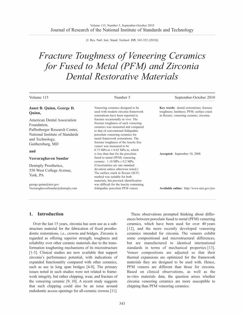
fracture toughness, KIc , of each type of veneer wasmeasured using the surface crack in flexure (SCF)method. In this method, which is schematically illus-trated in Figs. 1-3, a Knoop indentation is positionedin the center of each flexure test bar at sufficient load tocause cracks to form. Crack orientation may be con-trolled by orienting the Knoop indentation axis. Afterindentation, 4.5 to 5 times the depth of the indent isremoved from the surface of each bar. This is an impor-tant step, for it eliminates the residual stresses resultingfrom the indentation, and leaves a stress-free semi-elliptical crack in the bar surface. The bars are then
Volume 115, Number 5, September-October 2010Journal of Research of the National Institute of Standards and Technology
344
Fig. 1. The surface crack in flexure (SCF) method. An aligned Knoop indentation is used tocreate a semielliptical precrack on a bend bar.
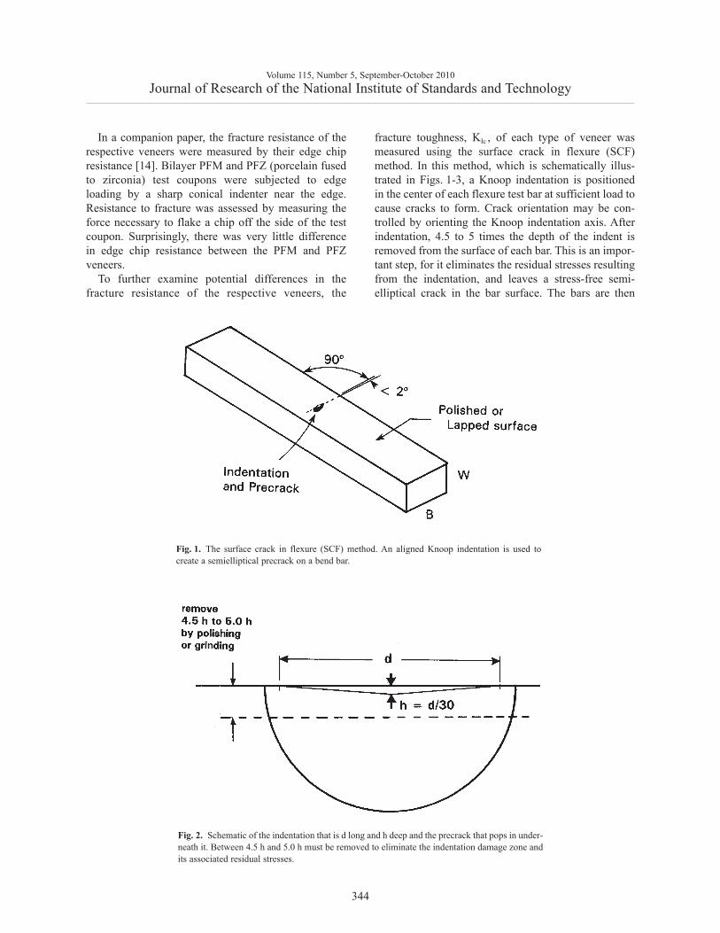
Fig. 2. Schematic of the indentation that is d long and h deep and the precrack that pops in under-neath it. Between 4.5 h and 5.0 h must be removed to eliminate the indentation damage zone andits associated residual stresses.

tested in flexure and break at the semi-elliptical crack ifit is large enough. The size of the crack is measured onthe fracture surface of each specimen, and substitutedinto formulae for calculating KIc , along with thespecimen size and break stress. One advantage of theSCF method is that it gives a fracture toughness resultgermane to small cracks, of the order of size of thenaturally occurring flaws in a brittle material. Thismethod has been used previously for dental restorationceramics [15] and has been standardized by ASTM Int.[16, 17], the International Organization for Standardi-zation (ISO) [18], and the European Committee forStandards (CEN) [19]. It was the focus of a majorVersailles Advanced Materials and Standards(VAMAS) international round robin [20-22]. It alsowas one of the core methods used to prepare StandardReference Material (SRM) 2100, the world’s onlyreference material for the property fracture toughness,KIc , [23, 24]. SCF results were identical to within0.01 MPa√m to those from chevron notch in bendingand single-edge precracked beam experiments. In otherwords, the three methods produced virtually identicalresults on the reference material, which has a very finegrain size and had a flat R-curve (such that fracturetoughness was independent of the crack size). Althoughthe method has been widely used to measure fracture
toughness of a wide range of ceramics (e.g., [25]), itdoes have limitations. The SCF method will not workon all ceramic materials. The following criteria must bemet:
1. The material must be hard and brittle.
2. It must be possible to detect the precracks on thefracture surface after fracture.
3. The precrack size should be larger than thenatural flaws in the material.
Difficulties arise if the material is coarse-grained,porous, too tough, or too soft. It is difficult to detectthe precracks if the material is coarse grained orporous. Also, if the material is porous or soft, crackswill not form under the indentation. If the materialhas too great a fracture toughness, then only smallprecracks form and they may be removed when theindentation damage zone is removed in the polishingstep.
The goals of this work were to ascertain whether theSCF method was suitable for dental veneering ceramicsand whether a conventional PFM veneer had a differentresistance to fracture than a new PFZ veneer.
Volume 115, Number 5, September-October 2010Journal of Research of the National Institute of Standards and Technology
345
Fig. 3. Schematic of the beam cross section with a semielliptical surface crack. The precrack sizeis exaggerated in this view for clarity.
2. Materials and Methods2.1 Materials
A vacuum dental furnace (model: Centurion VPC,Dentsply Prosthetics, York, PA) was used to fire six2.8 mm × 3.6 mm × 42 mm long beam specimens inrectangular molds. The PFM veneer was Ceramco 3(Dentsply Prosthetics, York, PA)1, a feldspathic veneer-ing porcelain containing about 0.30 volume fractionleucite. The veneer for the zirconia, Ceramco PFZ(Dentsply Prosthetics, York, PA), was designed to beused with a 3 mole % yttria stabilized zirconia (Cercon)made by the same manufacturer. This veneer is a felds-pathic porcelain with negligible leucite. The Ceramco 3veneer for the alloy had a heat up rate of 55 °C/min toa peak firing temperature of 960 °C and 0 min hold.The Ceramco PFZ veneer for the zirconia had a heat uprate of 60 °C/min to a peak temperature of 900 °C anda short 15 s hold on the first firing and 890 °C and0 min hold on the second firing. The specimens wererough-polished on the four long surfaces to eliminateany bowing or small surface lumps. The two 3.6 mmwide surfaces were more carefully polished with suc-cessively smaller grits down to 1200 grit. A companionpaper has additional information including edge chipresistance and Knoop hardness measurements for bothveneering ceramics [14].
2.2 Methods: SCF Fracture Toughness
Knoop indentations with a load of 39.2 N (4 kg masson the indenter) were placed on one of the wide 3.6 mmsurfaces. The indentation and the residual stress-creating damage zone associated with the indentationwere removed to a depth of 4.5 to 5.0 times the depthof the indentation, h, by hand grinding with 180 gritsilicon carbide abrasive papers. The Knoop indentationlengths, d, were of the order of 370 μm for the zirconiaveneer so that the indentation depths (≈ 1/30th of thediagonal length) were about 12 μm deep. Thus, about60 μm of material was removed by the hand grinding.Lateral cracks around the indentation were detected forthe PFZ veneer, but they were removed by the grinding.The indentations were about 440 μm long for the PFMveneer, so about 73 μm was removed by hand grindingfor each bar.
Fractographic techniques are used to detect andmeasure the precrack on the fracture surface after testspecimen fracture [26]. The precrack size was meas-ured for each test specimen. The precracks were veryeasy to detect on the zirconia veneer. A stereopticalmicroscope (Leica MZ16, Wetzlar, Germany) with atraversing stage with a resolution of 1 μm was used tomeasure the precrack width, 2c, and depth, a, on thefracture surfaces. The precacks were far more difficultto measure in the PFM veneer since the fracture surfacewas much rougher. One half of each specimen wascoated with a thin sputter applied gold coating (such asis done for scanning electron microscope examination)in order to facilitate viewing with the stereopticalmicroscope. The coating cut down on internal lightscattering in this translucent material and improvedcontrast.
Bars were broken in three-point flexure with a20 mm span, taking care that the precrack was wellcentered under the middle loading roller. The 42 mmlong bars could have been tested in four-point loadingwith 20 mm and 40 mm fixture spans, but the shorterspan fixture was used so that two breaks could beobtained with some of the test pieces. Four-pointloading is usually recommended for the SCF method[16-21, 23-25], to ensure that the precrack is within aconstant stress region, obviating the need for meticu-lous alignment in three-point testing. All testing wasdone in laboratory ambient conditions at a crossheadrate of 0.5 mm/ min.
Fracture toughness (KIc ) was calculated from theformula for a semicircular or semielliptical surfacecrack in tension or flexure:
(1)
where Y is the stress intensity shape factor, σ is theflexure strength of the specimen (MPa), and a is thecrack depth (m). Y is dimensionless and is a function ofthe crack size and shape and was individually calculat-ed for each precrack. The solutions by Newman andRaju [27] were used. The maximum Y value fromaround the crack front periphery was used to computeKIc . For the precracks in this study, this was oftenwhere the precrack intersected the tensile surface, butusually the Y values varied by less than 10 % aroundthe precrack periphery. One surprise about the SCFmethod is that computed fracture toughness values arenot especially sensitive to the measurement of the pre-crack size. This is due in part to the square root depend-ence of KIc on the crack size, but also due to an offset-ting influence of Y on the crack size measurement. For
Volume 115, Number 5, September-October 2010Journal of Research of the National Institute of Standards and Technology
346
1 Commercial products and equipment are identified only to specifyadequately experimental procedures and does not imply endorsementby the authors, institutions or organizations supporting this work, nordoes it imply that they are necessarily the best for the purpose.
KIc = Y σ √a
example, as discussed in Scherrer et al., [15], multipleobservers using different photos and measurements ob-tained KIc values that agreed on average to within0.01 MPa √ m for a dental feldspathic porcelain.Additional details about Y and the SCF methodmay be found in [15-21, 25].
3. Results
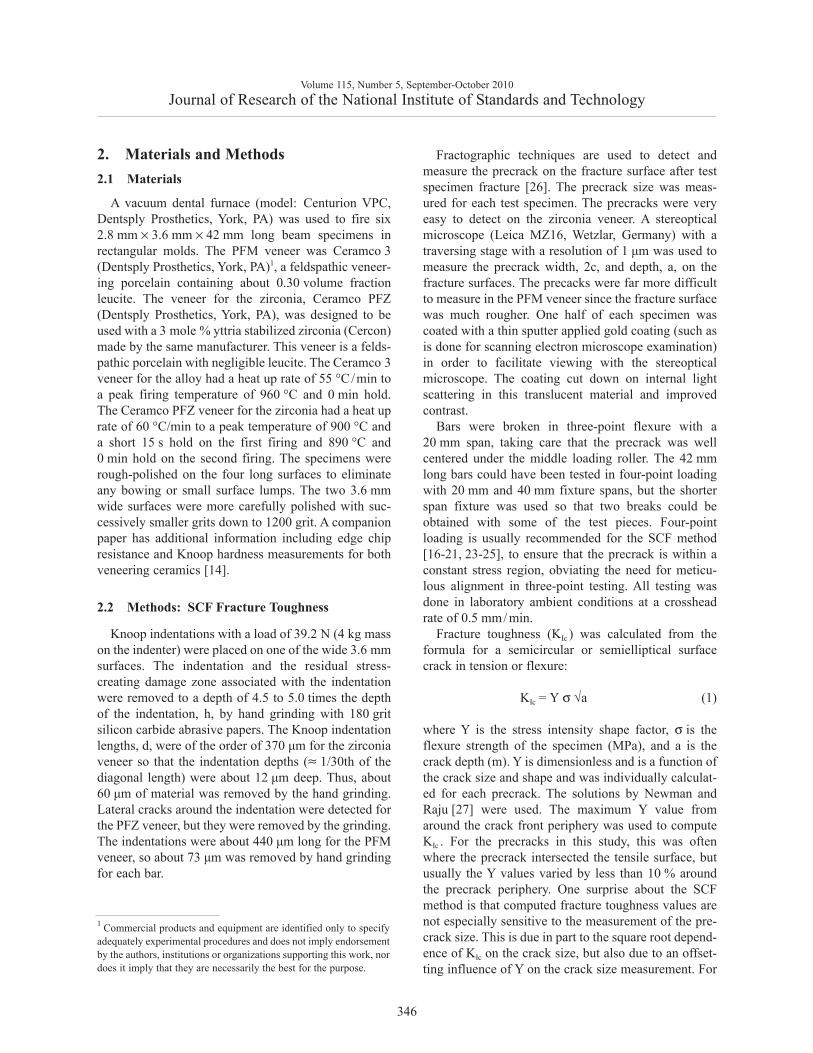
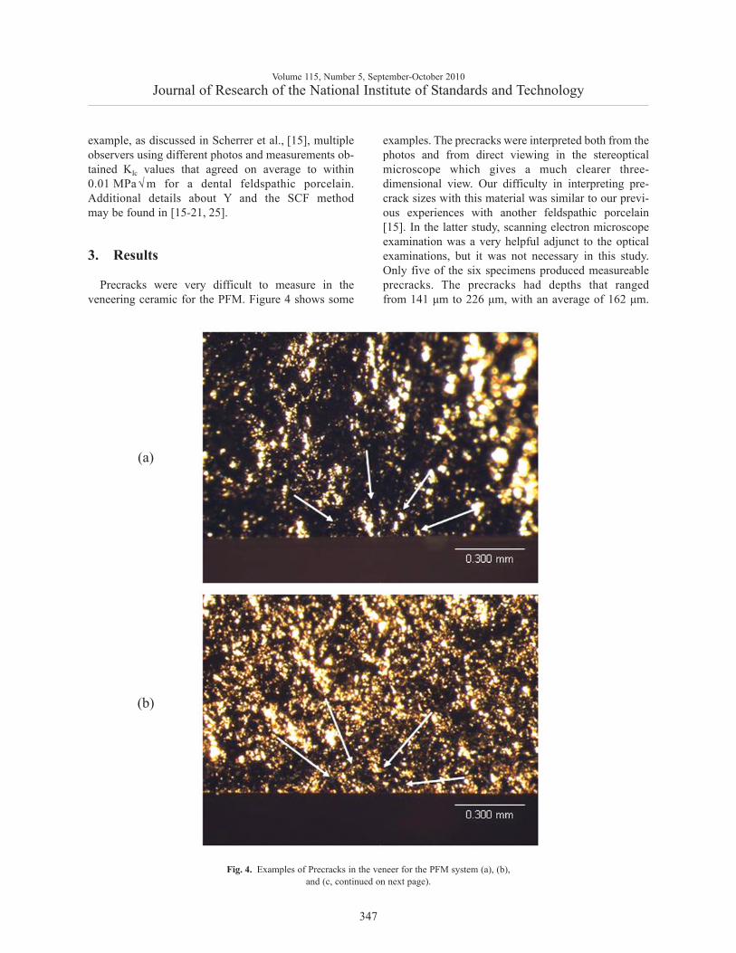
Precracks were very difficult to measure in theveneering ceramic for the PFM. Figure 4 shows some
examples. The precracks were interpreted both from thephotos and from direct viewing in the stereopticalmicroscope which gives a much clearer three-dimensional view. Our difficulty in interpreting pre-crack sizes with this material was similar to our previ-ous experiences with another feldspathic porcelain[15]. In the latter study, scanning electron microscopeexamination was a very helpful adjunct to the opticalexaminations, but it was not necessary in this study.Only five of the six specimens produced measureableprecracks. The precracks had depths that rangedfrom 141 μm to 226 μm, with an average of 162 μm.
Volume 115, Number 5, September-October 2010Journal of Research of the National Institute of Standards and Technology
347
Fig. 4. Examples of Precracks in the veneer for the PFM system (a), (b), and (c, continued on next page).
(a)
(b)
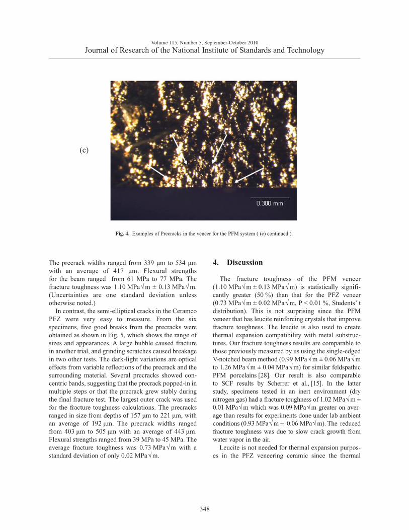
The precrack widths ranged from 339 μm to 534 μmwith an average of 417 μm. Flexural strengthsfor the beam ranged from 61 MPa to 77 MPa. Thefracture toughness was 1.10 MPa√m ± 0.13 MPa√m.(Uncertainties are one standard deviation unlessotherwise noted.)
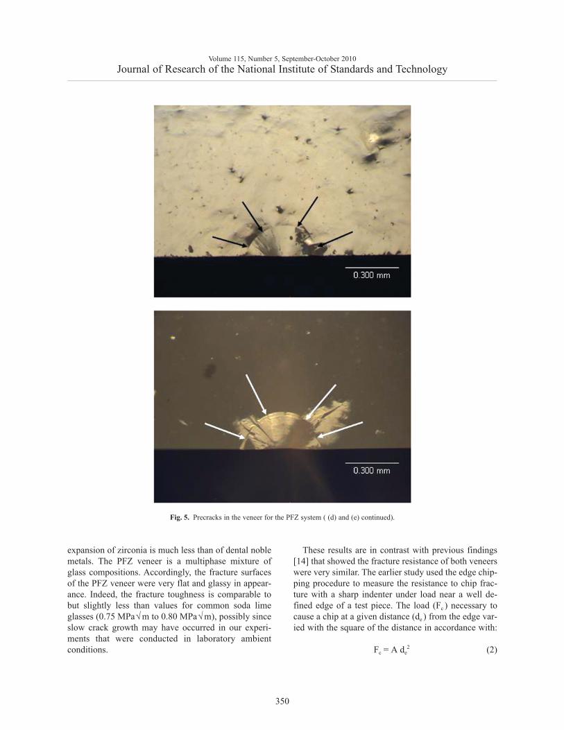
In contrast, the semi-elliptical cracks in the CeramcoPFZ were very easy to measure. From the sixspecimens, five good breaks from the precracks wereobtained as shown in Fig. 5, which shows the range ofsizes and appearances. A large bubble caused fracturein another trial, and grinding scratches caused breakagein two other tests. The dark-light variations are opticaleffects from variable reflections of the precrack and thesurrounding material. Several precracks showed con-centric bands, suggesting that the precrack popped-in inmultiple steps or that the precrack grew stably duringthe final fracture test. The largest outer crack was usedfor the fracture toughness calculations. The precracksranged in size from depths of 157 μm to 221 μm, withan average of 192 μm. The precrack widths rangedfrom 403 μm to 505 μm with an average of 443 μm.Flexural strengths ranged from 39 MPa to 45 MPa. Theaverage fracture toughness was 0.73 MPa√m with astandard deviation of only 0.02 MPa√m.
4. Discussion
The fracture toughness of the PFM veneer(1.10 MPa√m ± 0.13 MPa√m) is statistically signifi-cantly greater (50 %) than that for the PFZ veneer(0.73 MPa√m ± 0.02 MPa√m, P < 0.01 %, Students’ tdistribution). This is not surprising since the PFMveneer that has leucite reinforcing crystals that improvefracture toughness. The leucite is also used to createthermal expansion compatibility with metal substruc-tures. Our fracture toughness results are comparable tothose previously measured by us using the single-edgedV-notched beam method (0.99 MPa√m ± 0.06 MPa√mto 1.26 MPa√m ± 0.04 MPa√m) for similar feldspathicPFM porcelains [28]. Our result is also comparableto SCF results by Scherrer et al., [15]. In the latterstudy, specimens tested in an inert environment (drynitrogen gas) had a fracture toughness of 1.02 MPa√m ±0.01 MPa√m which was 0.09 MPa√m greater on aver-age than results for experiments done under lab ambientconditions (0.93 MPa√m ± 0.06 MPa√m). The reducedfracture toughness was due to slow crack growth fromwater vapor in the air.
Leucite is not needed for thermal expansion purpos-es in the PFZ veneering ceramic since the thermal
Volume 115, Number 5, September-October 2010Journal of Research of the National Institute of Standards and Technology
348
Fig. 4. Examples of Precracks in the veneer for the PFM system ( (c) continued ).
(c)

Volume 115, Number 5, September-October 2010Journal of Research of the National Institute of Standards and Technology
349
Fig. 5. Precracks in the veneer for the PFZ system (a), (b), (c), and ((d) and (e), continued on next page).
(a)
(b)
(c)
expansion of zirconia is much less than of dental noblemetals. The PFZ veneer is a multiphase mixture ofglass compositions. Accordingly, the fracture surfacesof the PFZ veneer were very flat and glassy in appear-ance. Indeed, the fracture toughness is comparable tobut slightly less than values for common soda limeglasses (0.75 MPa√m to 0.80 MPa√m), possibly sinceslow crack growth may have occurred in our experi-ments that were conducted in laboratory ambientconditions.
These results are in contrast with previous findings[14] that showed the fracture resistance of both veneerswere very similar. The earlier study used the edge chip-ping procedure to measure the resistance to chip frac-ture with a sharp indenter under load near a well de-fined edge of a test piece. The load (Fc ) necessary tocause a chip at a given distance (de ) from the edge var-ied with the square of the distance in accordance with:
(2)
Volume 115, Number 5, September-October 2010Journal of Research of the National Institute of Standards and Technology
350
Fig. 5. Precracks in the veneer for the PFZ system ( (d) and (e) continued).
Fc = A de2
where A was a proportionality constant. A was407 N/mm2 for the zirconia veneer and 440 N/mm2 forthe PFM veneer, an 8 % difference. Although somestudies have shown that edge toughness scales withfracture toughness, our new results suggest fracturetoughness and edge toughness are two different indicesof resistance to fracture.
The SCF fracture toughness technique was success-ful in these instances, but interpretation of the pre-cracks was very difficult in the case of the veneer forthe PFM system. A simple dye penetration procedure tohighlight the precracks would be a welcome step tofacilitate interpretation, but past efforts have had mixedsuccesses. Dyes are effective in some materials, butineffective in others due to variations in wetabilty andthe tightness of the rather small Knoop precracks.Although we were able to obtain valid fractures inthree-point loading, four-point is much preferred andshould be used in the future due to the strong stress gra-dients in the former. Future testing of such oxideceramics should be done in inert environmental condi-tions to minimize possible interferences from slowcrack growth.
The reduced fracture toughness of the veneeringceramic for the zirconia system could be an importantfactor in the difference in the clinical behavior. It hasalso been reported recently that residual stresses in thezirconia veneering ceramic may also contribute to theirincreased propensity to fracture [29, 30].
5. Conclusion
The fracture toughness of the leucite containingfeldspathic porcelain veneering ceramic intended foruse with PFMs was 1.10 MPa√m ± 0.13 MPa√m. It isgreater than that for a veneering ceramic designedfor zirconia: 0.73 MPa√mm ± 0.02 MPa√m. The SCFmethod was suitable for both materials, but precrackidentification was difficult for the leucite containingfeldspathic porcelain PFM veneering ceramic.
Acknowledgements
This work was supported by National Institutefor Standards and Technology, American DentalAssociation Foundation and National Institute ofHealth with grant NIH R01-DE17983.
6. References
[1] E. C. Subbarao, Zirconia: an overview, in Advances inCeramics, vol. 3. Science and Technology of Zirconia, eds. A.H. Heuer, L. W. Hobbs (Elsevier, Amsterdam) (1981).
[2] C. Piconi and G. Maccauro, Zirconia as a ceramic biomaterial,Biomater. 20, 1-25 (1999).
[3] J. Tinschert, D. Zwez, R. Marx, and K. J. Anusavice, Structuralreliability of alumina-, feldspar-, leucite-, mica- and zirconia-based ceramics, J. Dent. 28 [7], 529-35 (2000).
[4] F. Beuer, H. Aggstaller, T. Fishcher, K. Spiegl, J. Schweiger,and W. Gernet, Clinical behavior of zirconia based bridges:Two-years results, J. Dent. Res. 86, (Spec Iss A), Abst. No.0901 (2007).
[5] S. Wolfart, S. Eschbach, and M. Kern, Outcome of posteriorFPDs of veneered zirconia ceramic (Cercon), J. Dent. Res. 86(Spec Iss A), Abst. No. 0292 (2007).
[6] P. Vult Von Steyern, P. Carlson, and K. Nilner, All-ceramicfixed partial dentures designed according to the DC-Zirkon®technique: A 2-year clinical study, J. Oral Rehab. 32 [3], 180-187 (2005).
[7] J. Tinschert, G. Natt, P. Latzke, K. A. Schulze, N. Heussen,and H. Spiekermann, 5-Jahres-Ergebnisse—Bewährung vonvoll-keramischen Brücken aus DC-Zirkon®, DeutscheZahnartzeblatt 1 [16], 116-19 (2007).
[8] F. P. Nothdurft, P. R. Rountree, and P. R. Pospiech. Clinicallong-term behavior of zirconia-based bridges (LAVA): Fiveyears results, J. Dent. Res. 85 (Spec Iss C), Abst. No. 0312(2006).
[9] I. Sailer, H. Lüthy, A. Feher, M. Schumacher, P. Schärer, andC. Hämmerle, 3-year results of zirconia posterior fixed partialdentures made by Direct Ceramic Machining (DCM), J. Dent.Res. 82 (Spec Iss B), Abst. No. 0074 (2003).
[10] R. Hickel, Trends in materials science from the point of view ofa practicing dentist, J. Eur. Cer. Soc. 29, 1283-1289 (2009).
[11] K. C. Wood, D. W. Berzins, Q. Luo, G. A. Thompson,J. M. Toth, and W. W. Nagy, Resistance to fracture of two all-ceramic crown materials following endodontic access,J. Prosthet. Dent. 95 [1], 33-41 (2006).
[12] K. F. Leinfelder, Porcelain esthetics for the 21st century, J. Am.Dent. Assoc. 131 [1], 47S-51S (2000).
[13] ISO 6872. Dentistry—Ceramic Materials, InternationalOrganization for Standardization, Geneva, SW (2008).
[14] J. B. Quinn, V. Sundar, E. E. Parry, and G. D. Quinn,Comparison of Edge Chipping Resistance of PFM andVeneered Zirconia Specimens, Dental Materials. 26 [1], 13-20(2010).
[15] S. Scherrer, J. R. Kelly, G. D. Quinn, and K. Xu. FractureToughness (KIc ) of a Dental Porcelain Determined byFractographic Analysis, Dental Materials. 15 [5], 342-348(1999).
[16] ASTM C 1421-99, Standard Test Method for Determination ofFracture Toughness of Advanced Ceramics at AmbientTemperature. Annual Book of Standards, Vol. 15.01, ASTM,West Conshohocken, PA, (1999).
[17] J. A. Salem, G. D. Quinn, M. G. Jenkins, Measuring the RealFracture Toughness of Ceramics—ASTM C 1421, pp. 531- 554in Fracture Mechanics of Ceramics, Vol. 14, R. C. Bradt, D.Munz, M. Sakai, and K. W. White, eds., 2005, Springer, USA,(2005).
Volume 115, Number 5, September-October 2010Journal of Research of the National Institute of Standards and Technology
351

[18] ISO 18756, Fine Ceramics (Advanced Ceramics, AdvancedTechnical Ceramics)— Determination of Fracture Toughness ofMonolithic Ceramics at Room Temperature by the SurfaceCrack in Flexure (SCF) Method, ISO Geneva, 2003.
[19] EN 14425, European Standard, Advanced Technical Ceramics -Monolithic Ceramics—Fracture Toughness–Parts 1-5,European Committee for Standardization, Brussels, 2003.
[20] G. D. Quinn, R. J. Gettings, and J. J. Kübler, FractureToughness of Ceramics by the Surface Crack in Flexure (SCF)Method: Results of the VAMAS Round Robin, CeramicEngineering and Science Proceedings, 15 [5], 846-855 (1994).
[21] G. D. Quinn, J. J. Kübler, and R. J. Gettings, FractureToughness of Advanced Ceramics by the Surface Crack inFlexure (SCF) Method: A VAMAS Round Robin, VAMASTechnical Report #17, National Institute of Standards andTechnology, Gaithersburg, MD 20899, (1994).
[22] G. D. Quinn, The Fracture Toughness Round Robins inVAMAS: What We Have Learned, pp. 107-126 in FractureResistance Testing of Monolithic and Composite BrittleMaterials, ASTM STP 1409, J. A. Salem, G. D. Quinn, M. G.Jenkins, eds., (ASTM, West Conshohocken, PA) (2002).
[23] SRM 2100, Fracture Toughness of Ceramics, StandardReference Material Office, National Institute of Standards andTechnology, Gaithersburg, MD, (1999).
[24] G. D. Quinn, K. Xu, R. Gettings, J. A. Salem, and J. J. Swab,SRM 2100: Fracture Toughness of Ceramics, pp. 499–530 inFracture Mechanics of Ceramics, Vol. 14, R. C. Bradt, D.Munz, M. Sakai, and K. W. White, eds., Springer, USA (2005).
[25] G. D. Quinn, R. J. Gettings, and J. J. Kübler, FractureToughness of Ceramics by the Surface Crack in Flexure (SCF)Method, pp. 203–218 in Fracture Mechanics of Ceramics,Vol.11, R. C. Bradt, D. P. H. Hasselman, D. Munz, M. Sakai,and V. Yashevchenko, eds., (Plenum, NY) (1996).
[26] G. D. Quinn, Guide to Practice for Fractography of Ceramicsand Glasses, NIST Special Publication SP 960-16, May 2007.
[27] J. C. Newman, Jr., and I. S. Raju, An Empirical Stress-IntensityFactor Equation for the Surface Crack, Eng. Fract. Mech. 15[1-2], 185-92 (1981).
[28] J. B. Quinn, V. Sundar, and I. K Lloyd, Influence of micro-structure and chemistry on the fracture toughness of dentalceramics, Dental Materials 19 (7), 603-11 (2003).
[29] J. Fischer, B. Stawarzcyk, A. Trottmann, and C. H. F.Hämmerle, Impact of thermal misfit on shear strength ofveneering ceramic/zirconia composites, Dental Materials 25[4], 419-423 (2009).
[30] M. V. Swain, Unstable cracking (chipping) of veneeringporcelain on all-ceramic dental crowns and fixed partialdentures, Acta Biomater. 5, 1668-1677 (2009).
About the authors: Janet B. Quinn was a staff scien-tist in the Paffenbarger Research Center (PRC) of theAmerican Dental Association Foundation (ADAF) untilher untimely death in July 2008. George D. Quinn wasa research ceramic engineer in the Ceramics Divisionof MSEL in NIST from 1990 to 2009. He is now aResearch Associate for MSEL and a consultant to thePRC-ADAF. Veeraraghavan Sundar is Manager,Professional Service and Clinical Education, forDentsply Prosthetics, York, PA. The National Instituteof Standards and Technology is an agency of theU.S. Department of Commerce.
Volume 115, Number 5, September-October 2010Journal of Research of the National Institute of Standards and Technology
352