12-o-tetradecanoylphorbol-1,3-acetate … · final form accepted: ... (leica tcs sp2 se). ... an...
TRANSCRIPT
CELLULAR & MOLECULAR BIOLOGY LETTERS http://www.cmbl.org.pl
Received: 30 October 2009 Volume 15 (2010) pp 377-394 Final form accepted: 10 April 2010 DOI: 10.2478/s11658-010-0014-4 Published online: 29 April 2010 © 2010 by the University of Wrocław, Poland
* Author for correspondence. e-mail: [email protected], [email protected], tel.: +86 5925921461, fax: +86 5922188680
Abbreviations used: BrdU – 5-bromo-2’-deoxyuridine; PI3K – phosphoinositide-3-OH kinase; PDK1/PDK2 – 3-phosphoinositide-dependent kinase; PI – propidium iodide; PKB – protein kinase B; PKC – protein kinase C; TPA – 12-O-tetradecanoylphorbol-1,3-acetate; WGA – wheat germ agglutinin
Research article
12-O-TETRADECANOYLPHORBOL-1,3-ACETATE INDUCES THE NEGATIVE REGULATION OF PROTEIN KINASE B BY PROTEIN
KINASE Cα DURING GASTRIC CANCER CELL APOPTOSIS
BING ZHANG1 and CHUN XIA2* 1Medical School, Xiamen University, Xiamen 361005, Fujian Province, China
2Zhongshan Hospital, Xiamen University, Xiamen 361004, Fujian Province, China
Abstract: The PKB signaling pathway is essential for cell survival and the inhibition of apoptosis, but its functional mechanisms have not been fully explored. Previously, we reported that TPA effectively inhibited PKB activity and caused PKB degradation, which was correlated with the repression of PKB phosphorylation at Ser473. In this study, we focus on how PKB is regulated by TPA in gastric cancer cells. One of the TPA targets, PKCα, was found to mediate the inhibition of PKB phosphorylation and degredation caused by TPA. Furthermore, TPA induced the import of PKCα into the nucleus, where PKCα exerted an inhibitory effect on PKB expression and phosphorylation. As a result, cancer cell proliferation was arrested. Our study characterizes a novel function of PKCα in mediating the negative regulation of PKB by TPA, and suggests a potential application in the clinical treatment of gastric cancer. Key words: Protein kinase B (PKB), Protein kinase Cα (PKCα), Translocation, Growth inhibition, TPA

Vol. 15. No. 3. 2010 CELL. MOL. BIOL. LETT.
378
INTRODUCTION PKB is a 60-kDa serine-theronine kinase functioning downstream of phosphatidylinositol 3-kinase (PI-3 kinase) in response to mitogen or growth factor stimulation. It has emerged as a crucial regulator of diverse cellular processes, including survival, proliferation, differentiation, apoptosis and metabolism [1]. It is activated by a variety of stimuli through a phosphorylation mechanism [2-4]. The phosphorylation of threonine 308 and serine 473 of PKB are prerequisites to its activation [2, 5]. In most situations, the phosphorylation of PKB at Ser473 occurs in tandem with that at Thr308. However, a number of studies have shown that phosphorylation can occur independently at the two sites [6, 7]. For example, Ser473 phosphorylation can be stimulated by insulin in the absence of Thr308 phosphorylation [8]. Conversely, the attenuation of PI3K activation results in a rapid dephosphorylation at Ser473 and a slower one at Thr308, accompanied by a reduction in PKB activity [9]. The inactivation of PKB by ceramide and osmotic stress occurs predominantly via Ser473 dephosphorylation by an okadaic acid-sensitive phosphatase [10, 11]. We also found that the inhibition of PKB by TPA occurs mainly via the attenuation of Ser473 phosphorylation [12]. PKC is also a subfamily of serine/threonine kinases that play a variety of regulatory roles in proliferation, differentiation, apoptosis, membrane transportation, and signal transduction [13, 14]. Based on their structural features and cofactor requirements, PKC isoforms are classified into three categories: classical PKCs (α, βI, βII and γ); novel PKCs (δ, ε, η, θ and μ); and atypical PKCs (ξ, τ and λ) [15]. Each PKC isoform has unique specific functional characteristics, even within one cell line. For example, in gastric cell lines, each PKC isoform takes on a different role in the regulation of apoptosis [16-18]. Overexpression of PKCδ enhances cisplatin-induced cytotoxicity correlated with p53 in the gastric cancer cell line MKN28 [16]. PKCδ also participates in the modulation of anti-apoptosis by endogenous IAP expression in the human gastric cancer cell line MKN45 [17]. Inhibiting the PKCβ1-mediated overexpression of p21 (waf1/cip1) partially reduces the anti-apoptotic effect of PKCβ1 in the gastric adenocarcinoma cell line AGS [18]. The down-regulation of PKCβ1 provides an explanation for the COX-independent apoptotic effects of the specific COX-2 inhibitor in the gastric cancer cell line AGS. Moreover, PKCβ1 acts as a survival mediator in gastric cancer, and its down-regulation by the COX-2 inhibitor SC-236 may offer a new method for the treatment of gastric cancer [19]. There are reports of cross-talk between PKB and PKC. The overexpression of PKC stimulates PKB activity and suppresses cytokine-dependent apoptosis [20]. Conversely, the phorbol ester phorbol 12-myristate 13-acetate (PMA), an activator of PKC, down-regulates growth factor-induced PKB activation, and specific isoforms of PKC directly act as negative regulators of PKB [20, 21]. PKCβII can regulate PKB activity by directly phosphorylating the critical

CELLULAR & MOLECULAR BIOLOGY LETTERS
379
residue Ser473 in FcεRI-stimulated mast cells [22]. By contrast, PKCβ is not required for PKB phosphorylation at Ser473 in mast cells stimulated with stem cell factor or IL-3, in serum-stimulated fibroblasts, or in antigen receptor-stimulated T or B cells [22]. Thus, PKC might be a potential positive or negative regulator for PKB. We previously found that TPA, an activator for PKCα activity [23], induced PKB degradation, which was associated with the inhibition of PKB phosphorylation at Ser473 but not at Thr308 [12]. In this study, we investigated the possible effect of PKCα on the regulation of PKB in BGC-823 gastric cancer cells. We observed that this functional role of TPA was mediated by PKCα, since the diminishing of PKCα activity by its inhibitor and siRNA could abolish the inhibition of TPA-induced phosphorylation and degradation of PKB. The key point for PKCα to mediate TPA’s effect on PKB was that TPA could activate and consequently induce PKCα translocation from the cytoplasm to the nucleus, where PKCα could directly inhibit nuclear PKB phosphorylation and expression. Therefore, PKCα might function as an important upstream factor for negatively regulating PKB activity. Taken together, our results indicate a novel mechanism by which PKCα negatively controls the TPA-regulated PKB signaling pathway in gastric cancer cells. MATERIALS AND METHODS Cell culture and transfection The human embryonic kidney 293T cell line (obtained from ATCC, USA) was maintained in DMEM medium, and the gastric cancer cell line BGC-823 (purchased from the Cell Biology Institute, Shang-hai, China) was cultured in RPMI-1640 medium that contained 10% fetal bovine serum, 1 mM glutamine and 100 μg/ml penicillin. The cells were transfected with different expression vectors using the Ca3(PO4)2 sedimentation method in all the experiments except for the siRNA transfection, which used liposomal transfection reagent (Roche, Fugene 6), as previously described [24, 25]. BrdU assay Cells were transfected with different expression vectors as required, and then incubated with 5-bromo-2’-deoxyuridine (20 μM, Sigma) for 2 h. After harvesting, the cells were fixed in 4% paraformaldehyde for 30 min at 4ºC as previously described [12]. Finally, the cells were analyzed by flow cytometer (Beckman Coulter). Immunoprecipitation Cells were treated with TPA (100 ng/ml) for 24 h, then harvested. The cells were lysed in a lysis buffer, and the lysates were incubated with the appropriate antibody for 1 h, and subsequently incubated with protein A-sepharose beads

Vol. 15. No. 3. 2010 CELL. MOL. BIOL. LETT.
380
(Sigma) for 1 h. The protein-antibody complexes that were recovered on the beads were subjected to Western blot analysis as described below. Western blot analysis Protein extracts were electrophoresed on 8-10% denaturing gel and electroblotted onto nitrocellulose membrane. The membrane was incubated with various antibodies as required at 4ºC overnight, followed by the addition of the corresponding secondary antibody at room temperature for 3 to 4 h. An ECL kit (Pierce) was used to detect the antibody reactivity. For the preparation of the cytoplasmic and nuclear fractions, cells were suspended in 2 ml MS buffer, and then homogenized using a Dounce homogenizer. As previously described, the cytoplasmic and nuclear fractions were obtained by centrifuging [12]. Confocal microscopic observation Cells were fixed in 4% paraformaldehyde after harvesting. To stain the endogenous PKB and PKCα proteins, the cells were incubated with anti-PKB or anti-PKCα antibody (Santa Cruz) followed by FITC- or Texas Red-conjugated secondary antibodies (Santa Cruz). The cells were stained with propidium iodide (PI, Sigam, 50 μg/ml) to visualize the nuclei simultaneously. The stained cells were finally visualized under a confocal microscope (Leica Tcs Sp2 SE). Specific silencing of the PKCα gene by siRNA PKCα-siRNA (AAGCACAAGUUCAAAAUCCAC) was introduced into pSuper plasmid [26]. BGC-823 cells were transfected with 2 μg of PKCα-siRNA or Scrambled-siRNA (control) using liposomal transfection reagent. The effect of PKCα-siRNA on the silencing of PKCα expression was examined via Western blotting. Treatment of cells with wheat germ agglutinin The Chariot protein delivery system (Active Motif, CA, USA) was used to transfect WGA into cells [27]. Briefly, WGA was mixed with Chariot at room temperature for 30 min, and then the Chariot/WGA complex was added to the cells. After 3 h incubation, the cells were harvested for further use [27]. Data analysis The results shown represent the means ± SEM for the number (n) of independent experiments performed. Duplicate or triplicate values were obtained for each parameter measured. Student’s two-tailed t tests were used to evaluate the statistical differences between the means of paired sets of data.
CELLULAR & MOLECULAR BIOLOGY LETTERS
381
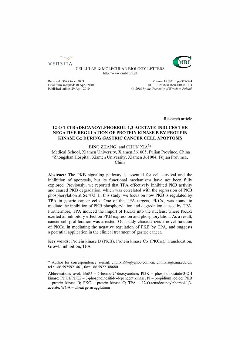
RESULTS PKCα mediates the inhibition of PKB caused by TPA We previously demonstrated that the TPA-induced inhibition of PKB phosphorylation at Ser473 is associated with the degradation of PKB [12]. Since PKCα is the direct target of TPA action [23], we analyzed the role of PKCα in TPA-induced PKB degradation. Gö6976, a specific inhibitor for the activities of both PKCα and PKCβI [28], was used to treat BGC-823 cells, and the expression level of PKB was examined in the presence of TPA. Although
Fig. 1. The effect of Gö6976 on PKB expression and phosphorylation. BGC-823 cells were pre-treated with Gö6976 for 2 h, followed by TPA treatment for 24 h. The levels of PKB expression and phosphorylation were determined via Western blotting, respectively against the PKB antibody or specific phosphor-PKBSer473 antibody. PKCα levels were detected by western blotting against the PKCα antibody. The ratios (PKB, PKCα or P-PKB/S473 to β-actin) are presented as the means ± SEM for three to five independent experiments. *p < 0.01, #p < 0.05, when compared with the control group.
Vol. 15. No. 3. 2010 CELL. MOL. BIOL. LETT.
382
Gö6976 alone did not affect PKB, it caused a significantly greater repression of TPA-induced PKB degradation than TPA treatment alone (Fig. 1, left panel). Furthermore, Gö6976 was able to block the inhibitory effect of TPA on the PKB phosphorylation at Ser473 (Fig. 1, right panel). These results strongly suggest that the PKCα signal is involved in the TPA-induced phosphorylation inhibition and degradation of PKB.
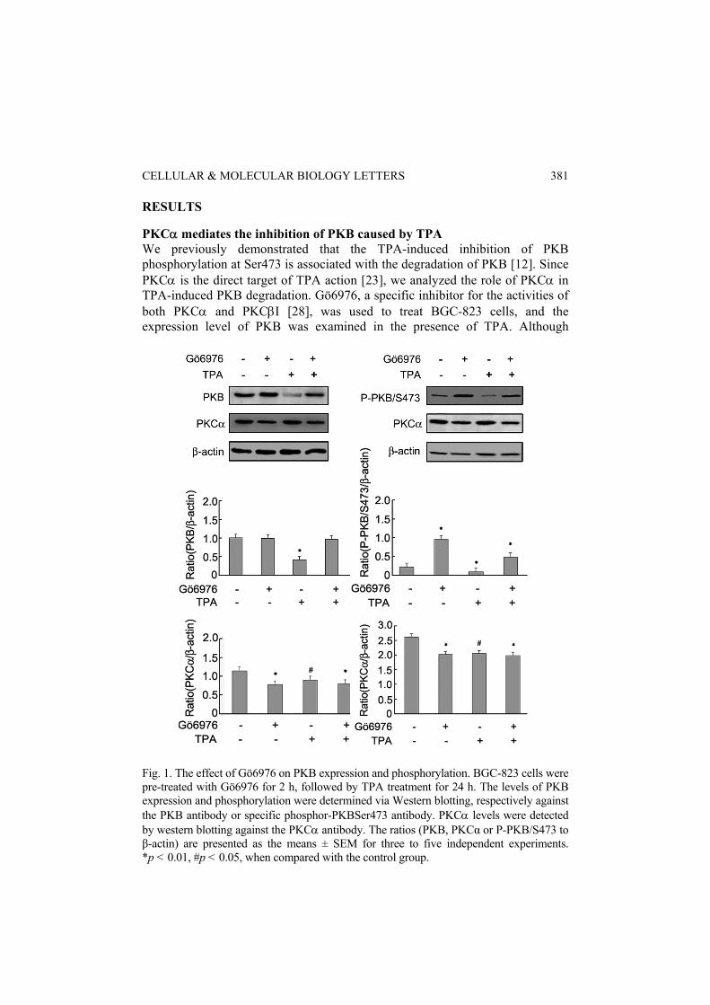
Fig. 2. PKCα inhibits the endogenous and exogenous expression of PKB. The Myc-PKCα expression vector, with or without Myc-PKB, was transfected into BGC-823 or 293T cells. Endogenous and exogenous PKB levels were determined by Western blotting, respectively against the PKB or Myc antibodies. The ratios (PKB, Myc-PKCα or Myc-PKB to β-actin) are presented as means ± SEM for four independent experiments. *p < 0.01, when compared with the control group.

CELLULAR & MOLECULAR BIOLOGY LETTERS
383
The above results suggest that PKCα may act as the upstream regulating factor for PKB and mediate the repression of PKB by TPA. To verify this possibility, an increasing amount of PKCα was transfected into BGC-823 cells, and the expression level of endogenous PKB was examined via Western blotting. As expected, PKCα could inhibit the endogenous PKB expression in a dose-dependent manner (Fig. 2, left panel). In addition, the co-expression of PKCα and PKB in 293T cells showed a similar result: PKCα inhibited PKB expression (Fig. 2, right panel). These results support a negative regulatory role of PKCα in PKB expression. Conversely, we used the siRNA approach to inhibit the expression level of endogenous PKCα (Fig. 3), and found that the inhibitory effect of TPA on PKB phosphorylation and expression was partially attenuated (Fig. 3). Thus, PKCα is a critical factor in mediating the TPA-induced inhibition of PKB expression. We further investigated whether PKCα also affected PKB phosphorylation at Ser473. Indeed, PKCα down-regulated PKB expression in PKB/T308A- but not in PKB/S473A-expressing 293T cells (Fig. 4). Moreover, PKCα inhibited PKB phosphorylation at Ser473 but not at Thr308 in BGC-823 cells, as detected by the specific PKB phosphor-antibody (Fig. 5). These results further confirmed a negative regulatory role for PKCα in PKB phosphorylation and degradation in gastric cancer cells.
Fig. 3. The effect of siRNA against PKCα on the TPA-induced inhibition of PKB expression and phosphorylation. The pSuper expression vector containing PKCα-siRNA or Scrambled-siRNA (Ctrl-siRNA) was transfected into BGC-823 cells. After transfection, the cells were treated with TPA for 24 h. The endogenous levels of PKB and PKCα were determined by Western blotting. The phosphorylation of PKB at Ser473 was detected as described in Fig. 1.

Vol. 15. No. 3. 2010 CELL. MOL. BIOL. LETT.
384
Fig. 4. PKCα is involved in the degradation and phosphorylation of PKB. Myc-PKCα, with or without various HA-PKB point mutants, was transfected into 293 T cells as indicated. The levels of expression and phosphorylation of PKB (or its mutants) were determined as described in Fig. 1. The ratios (Myc-PKCα, HA-PKB/S473A or HA-PKB/ T308A to Tubulin) are presented as means ± SEM for four independent experiments. *p < 0.01, #p < 0.05, when compared with the control group. The TPA-induced inhibition of PKB mediated by PKCα mainly occurred in the nucleus of BGC-823 cells Interestingly, cellular fractional analysis via Western blotting showed that after BGC-823 cells were treated with TPA for 48 h, PKB was barely detected in the nucleus, but almost maintained the same levels in the cytoplasm (Fig. 6A, left panel). Confocal microscopic analysis further supported this finding. As shown in Fig. 6B, PKB was visualized in both the cytoplasm and nucleus of BGC-823 cells prior to the TPA treatment. However, nuclear PKB was largely reduced after the cells were treated with TPA for 24 h, and totally disappeared when the treatment was extended to 48 h (Fig. 6B, left panel), suggesting that the nuclear PKB is degraded upon the TPA treatment in BGC-823 cells.

CELLULAR & MOLECULAR BIOLOGY LETTERS
385
Fig. 5. PKCα is involved in the degradation and phosphorylation of PKB. Myc-PKCα, with or without various HA-PKB point mutants, was transfected into the BGC-823 cells as indicated. The levels of expression and phosphorylation of PKB (or its mutants) were determined as described in Fig. 1. The ratios (Myc-PKCα, P-PKB/S473 or P-PKB/T308 to Tubulin) are presented as means ± SEM for four independent experiments. *p < 0.01, when compared with the control group. The results of a previous study showed that TPA treatment can induce PKCα translocation from the cytoplasm to the nucleus in BGC-823 cells [29]. As shown in Fig. 6B (right panel), TPA treatment gradually induced PKCα translocation

Vol. 15. No. 3. 2010 CELL. MOL. BIOL. LETT.
386
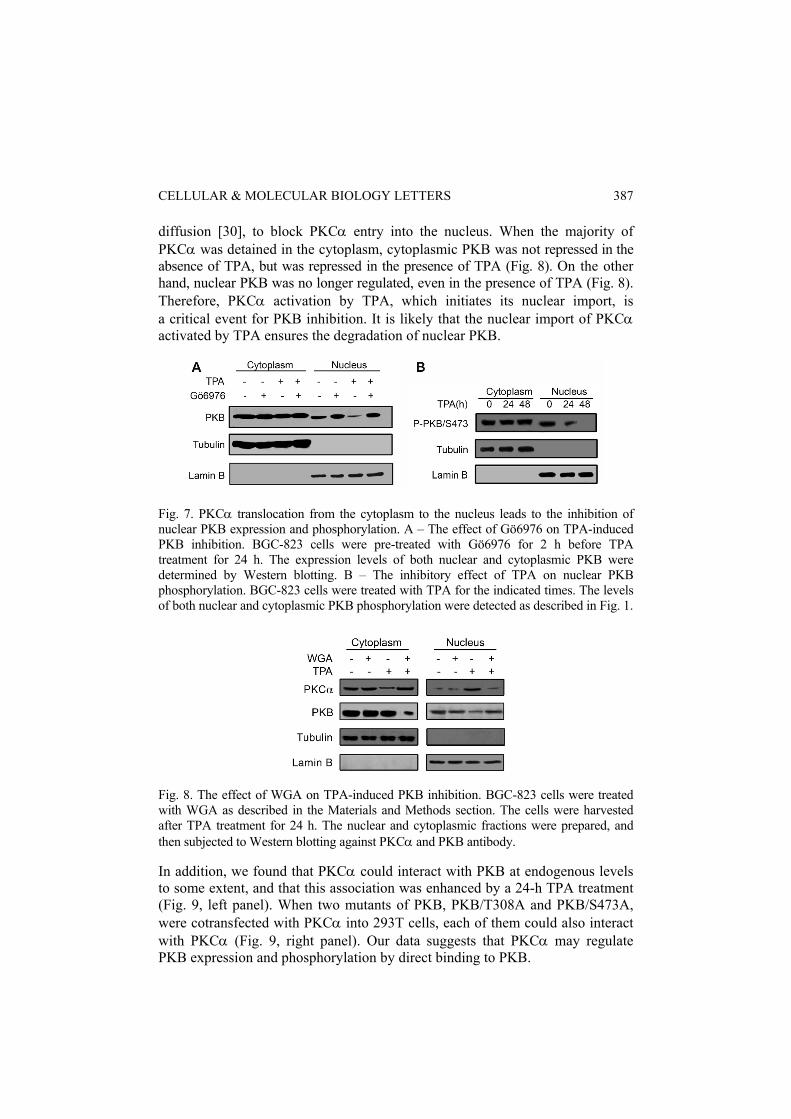
Fig. 6. The subcellular localization of PKB and PKCα in BGC-823 cells treated with TPA. A – TPA induces PKCα translocation and inhibits nuclear PKB expression. BGC-823 cells were treated with TPA for the indicated times, and then the cytoplasmic and nuclear fractions were prepared. The expression levels of PKB and PKCα were determined via Western blotting. Tubulin and LaminB were used to indicate the amount of cytoplasmic and nuclear proteins used. B – The subcellular localization of PKB and PKCα in BGC-823 cells. The cells were treated with TPA for the indicated times, and then immunostained with PKB- or PKCα-antibody, followed by FITC-conjugated secondary antibody. The nuclei were simultaneously indicated by PI staining. The images were observed under a confocal microscope. from the cytoplasm to the nucleus in BGC-823 cells, but did not impair its expression level (data not shown). When the treatment was continued for 48 h and the nuclear PKB had reached a barely detectable level, the cytoplasmic PKCα had almost completely translocated into the nucleus (Fig. 6B). Analysis of the PKCα subcellular localization via Western blotting also confirmed such PKCα translocation in response to TPA (Fig. 6A, right panel). Moreover, when the PKCα activity was inhibited by Gö6976, both the nuclear and cytoplasmic PKB levels remained the same, even in the presence of TPA (Fig. 7A). These results suggest that the inhibition of PKB by PKCα mainly occurred in the nucleus of the BGC-823 cells. Consistently, the TPA-induced inhibition of PKB phosphorylation at Ser473 was also restricted in the nucleus (Fig. 7B). We further used wheat germ agglutinin (WGA), a lectin that binds to nuclear pore complexes and inhibits protein import into the nucleus without affecting passive
CELLULAR & MOLECULAR BIOLOGY LETTERS
387
diffusion [30], to block PKCα entry into the nucleus. When the majority of PKCα was detained in the cytoplasm, cytoplasmic PKB was not repressed in the absence of TPA, but was repressed in the presence of TPA (Fig. 8). On the other hand, nuclear PKB was no longer regulated, even in the presence of TPA (Fig. 8). Therefore, PKCα activation by TPA, which initiates its nuclear import, is a critical event for PKB inhibition. It is likely that the nuclear import of PKCα activated by TPA ensures the degradation of nuclear PKB.
Fig. 7. PKCα translocation from the cytoplasm to the nucleus leads to the inhibition of nuclear PKB expression and phosphorylation. A – The effect of Gö6976 on TPA-induced PKB inhibition. BGC-823 cells were pre-treated with Gö6976 for 2 h before TPA treatment for 24 h. The expression levels of both nuclear and cytoplasmic PKB were determined by Western blotting. B – The inhibitory effect of TPA on nuclear PKB phosphorylation. BGC-823 cells were treated with TPA for the indicated times. The levels of both nuclear and cytoplasmic PKB phosphorylation were detected as described in Fig. 1.
Fig. 8. The effect of WGA on TPA-induced PKB inhibition. BGC-823 cells were treated with WGA as described in the Materials and Methods section. The cells were harvested after TPA treatment for 24 h. The nuclear and cytoplasmic fractions were prepared, and then subjected to Western blotting against PKCα and PKB antibody. In addition, we found that PKCα could interact with PKB at endogenous levels to some extent, and that this association was enhanced by a 24-h TPA treatment (Fig. 9, left panel). When two mutants of PKB, PKB/T308A and PKB/S473A, were cotransfected with PKCα into 293T cells, each of them could also interact with PKCα (Fig. 9, right panel). Our data suggests that PKCα may regulate PKB expression and phosphorylation by direct binding to PKB.

Vol. 15. No. 3. 2010 CELL. MOL. BIOL. LETT.
388
Fig. 9. The interaction of PKCα with PKB or its point mutants. BGC-823 cells were treated with TPA for 24 h. Cell lysates were immunoprecipitated with PKB antibody, and then subjected to Western blotting with PKCα antibody (left panel). 293T cells were transfected with different expression vectors as indicated. Cell lysates were immunoprecipitated with Myc antibody to precipitate PKCα, and then subjected to Western blotting with HA antibody to indicate either PKB/T308A or PKB/S473A (right panel). IgG expression served as a control for indicating similar proteins in each lane. The same lysates were applied to ascertain the position and expression of PKCα and PKB by Western blotting (Input).
Fig. 10. The effect of TPA on the mitogenic activity of BGC-823 cells. The PKCα and PKB expression vectors were transfected into cells as indicated. The untransfected or transfected cells were treated with TPA for the indicated times (upper) or 24 h (lower), then maintained in BrdU-containing medium for 2 h, and finally identified by flow cytometry. The percentages indicate the cells that showed BrdU uptake, showing the mean values of BrdU uptake for each sample.
CELLULAR & MOLECULAR BIOLOGY LETTERS
389
The effects of PKCα and PKB on cell proliferation in response to TPA Next, we analyzed the effects of PKB and PKCα on cell proliferation in response to TPA. The BrdU incorporation assay is often used to identify DNA-synthesizing cells, and thereby identify reflexing cell populations [31, 32]. We found that TPA exhibited strong inhibition upon BrdU incorporation into BGC-823 cells in a time-dependent manner (Fig. 10, upper panel). However, the PKCα inhibitor Gö6976 neutralized the effect of TPA on cell proliferation inhibition (Fig. 3, left of lower panel). By contrast, the transfection of PKCα blocked PKB-mediated cell proliferation, comparably to the TPA treatment (Fig. 10, right of lower panel). Therefore, PKCα, activated by TPA, contributes to the inhibition of BGC-823 cell proliferation through the repression of PKB function. DISCUSSION TPA effectively inhibited PKB activity and caused PKB degradation, which correlated with the repression of PKB phosphorylation at Ser473 [12]. In this study, we found that the effect of TPA on PKB was mainly mediated by PKCα. Upon TPA treatment, PKCα was translocated from the cytoplasm to the nucleus, where it exerted inhibitory effects on nuclear PKB phosphorylation and expression, which finally contributed to cancer cell death via the inhibition of DNA synthesis. In BGC-823 cells, we observed that PKB displayed a higher kinase activity and was constitutively phosphorylated at both Ser473 and Thr308 [12], consistent with the viewpoint that the state of phosphorylation of PKB is an indication of its activation [3]. Interestingly, the PKB phosphorylation at Ser473 was largely attenuated, but that at Thr308 did not change after TPA treatment [12] or PKCα transfection (Fig. 5). Ser473 of PKB is believed to be phosphorylated by the kinase PDK2 [33, 34], while the phosphorylation of Thr308 is catalyzed by PDK1 [35]. To phosphorylate Thr308, both PDK1 and PKB must bind with PI(3,4,5)P3 to induce a conformational change, thereby providing PKB access for PDK1 [11]. Our finding that phosphorylation at Thr308 was not affected by TPA [12] suggested that PKB might bind to other interaction partners in addition to PI(3,4,5)P3. This hypothesis is supported by our finding that PKCα interacted with PKB in vivo, even with PKB/S473A or PKB/T308A (Fig. 9). Therefore, PKCα could be one of the binding partners to regulate PKB expression (Fig. 2) and phosphorylation (Figs 4, 5). One possibility is that the binding of PKC to PKB may protect Thr308 but not Ser473 from being phosphorylated by PDK1. Accumulated evidence has shown that the regulation of PKB phosphorylation by PKCs, whether negatively or positively, mainly occurs at Ser473 [11, 22, 36-38]. A putative PDK2 is believed to phosphorylate PKB at Ser473 [39]. PKCβII has a PDK2 activity that up-regulates PKB activity by directly phosphorylating Ser473 in vitro and in IgE/antigen-stimulated mast cells [22]. In endothelial cells, PKCα may also function like PDK2 and activate PKB activity via Ser473

Vol. 15. No. 3. 2010 CELL. MOL. BIOL. LETT.
390
phosphorylation in response to IGF-1 [38]. By contrast, in the current case, PKCα activation by TPA in BGC-823 cells inhibits PKB phosphorylation at Ser473 (Figs 1, 4, 5 and 7B), suggesting that PKCα may not possess the same properties as PDK2 in gastric cancer cells. Since we found that PKCα binds to either PKB/S473A or PKB/T308A (Fig. 9), it is tempting to speculate that PKCα binding may cause a conformation change of PKB, leading to variability in Ser473 phosphorylation while Thr308 phosphorylation remains unchanged. Similarly, PKCζ has been reported to act as a negative regulator of PKB by direct binding [21]. Therefore, depending on the cell types and the stimuli used, PKCα activation may have controversial effects on PKB phosphorylation [20, 36, 38, 40]. PKB activation is believed to occur near the cell membrane, where PI3 products are produced as a result of receptor stimulation. However, recent studies have shown that PKB can also be activated in the nucleus [41], where it may be phosphorylated at the endogenous level. On the other hand, PKC is thought to reside in the cytoplasm in an inactive conformation, translocating to the plasma membrane or cytoplasmic organelles upon cell activation by different stimuli [42]. A wealth of evidence has also shown that PKC is capable of translocating to the nucleus to regulate multiple biological processes as important as cell proliferation and differentiation, gene expression, neoplastic transformation, and apoptosis [43]. Wu also previously demonstrated that the translocation of PKCα from both the mitochondria and cytosol to the nucleus in gastric cancer BGC-823 cells is accompanied by the induction of apoptosis [29]. Therefore, the nucleus could be another site for PKCα function in addition to the cytoplasmic location. The observation that PKCα is translocated into the nucleus following TPA treatment (Fig. 6A) led us to investigate whether PKCα translocation is correlated with its activation. In the presence of Gö6976, PKCα activity was repressed, and TPA no longer exhibited an inhibition of cytoplasmic and nuclear PKB expression in BGC-823 cells (Fig. 7A). However, during the treatment of WGA, PKCα translocation was blocked, but its activity remained in the presence of TPA. Under this circumstance, TPA inhibited cytoplasmic but not nuclear PKB (Fig. 8). This phenomenon implies that PKCα activity is important for the suppression of PKB. Once activated by TPA, PKCα would exert its inhibitory function on PKB in the nucleus [this study], the plasma membrane or cytoplasmic perinuclear region [44, 45]. Together, these findings reveal a novel physiological function of PKCα in the negative regulation of PKB activity. Acknowledgments. We would like to thank Dr. Qiao Wu (Xiamen University, Xiamen, Fujian, China) for the generous supply of PKB plasmid. This study was supported by the Natural Science Foundation of Fujian Province, China (No.C0910644).

CELLULAR & MOLECULAR BIOLOGY LETTERS
391
REFERENCES 1. Brazil, D.P. and Hemmings, B.A. Ten years of protein kinase B signalling:
a hard Akt to follow. Trends Biochem. Sci. 26 (2001) 657-664. 2. Kohn, A.D., Kovacina, K.S. and Roth, R.A. Insulin stimulates the kinase
activity of RAC-PK, a pleckstrin homology domain containing ser/thr kinase. EMBO J. 14 (1995) 4288-4295.
3. Alessi, D.R., Andjelkovic, M., Caudwell, B., Cron, P., Morrice, N., Cohen, P. and Hemmings, B.A. Mechanism of activation of protein kinase B by insulin and IGF-1. EMBO J. 15 (1996) 6541-6551.
4. Andjelkovic, M., Jakubowicz, T., Corn, P., Ming, X.F., Han, J.W. and Hemmings, B.A. Activation and phosphorylation of a pleckstrin homology domain containing protein kinase (RAC-PK/PKB) promoted by serum and protein phosphatase inhibitors. Proc. Natl. Acad. Sci. USA 93 (1996) 5699-5704.
5. Vanhaesebroeck, B. and Alessi, D.R. The PI3k-PDK1 connection: more than just a road to PKB. Biochem. J. 346 (2000) 561-576.
6. Alessi, D.R., James, S.R., Downes, C.P., Holmes, A.B., Gaffney, P.R., Reese, C.B. and Cohen, P. Characterization of a 3-phosphoinositide-dependent protein kinase which phosphorylates and activates protein kinase B alpha. Curr. Biol. 7 (1997) 261-269.
7. Kroner, C., Eybrechts, K. and Akkerman, J.W. Dual regulation of platelet protein kinase B. J. Biol. Chem. 275 (2000) 27790-27798.
8. Hill, M.M., Andjelkovic, M., Brazil, D.P., Ferrari, S., Fabbro, D. and Hemmings, B.A. Insulin-stimulated protein kinase B phosphorylation on Ser-473 is independent of its activity and occurs through a staurosporine-insensitive kinase. J. Biol. Chem. 276 (2001) 25643-25646.
9. Andjelkovic, M., Maira, S.M., Cron, P., Parker, P.J. and Hemmings, B.A. Domain swapping used to investigate the mechanism of protein kinase B regulation by 3-phosphoinositide-dependent protein kinase 1 and Ser473 kinase. Mol. Cell Biol. 19 (1999) 5061-5072.
10. Chen, D., Fucini, R.V., Olson, A.L., Hemmings, B.A. and Pessin, J.E. Osmotic shock inhibits insulin signaling by maintaining Akt/protein kinase B in an inactive dephosphorylated state. Mol. Cell Biol. 19 (1999) 4684-4694.
11. Schubert, K.M., Scheid, M.P. and Duronio, V. Ceramide inhibits protein kinase B/Akt by promoting dephosphorylation of serine 473. J. Biol. Chem. 275 (2000) 13330-13335.
12. Zhang, B. and Xia, C. The expression of protein kinase B in gastric cancer cell apoptosis induced by 12-O-tetradecanoylphorbol-1, 3-acetate. Cell. Mol. Biol. Lett. 14 (2009) 466-480.
13. Moore, E.D., Ring, M., Scriven, D.R., Smith, V.C., Meloche, R.M. and Buchan, A.M. The role of protein kinase C isozymes in bombesin-stimulated

Vol. 15. No. 3. 2010 CELL. MOL. BIOL. LETT.
392
gastrin release from human antral gastrin cells. J. Biol. Chem. 274 (1999) 22493-22501.
14. Kelly, M.L., Tang, Y., Rosensweig, N., Clejan, S. and Beckman, B.S. Granulocyte-macrophage colony-stimulating factor rescues TF-1 leukemia cells from ionizing radiation-induced apoptosis through a pathway mediated by protein kinase Calpha. Blood 92 (1998) 416-424.
15. Shao, R.G., Cao, C.X. and Prommier, Y. Activation of PKCalpha downstream from caspases apoptosis induced by 7-hydroxystaurosporine or the topoisomerase inhibitors, camptothecin and etoposide, in human myeloid leukemia HL60 cells. J. Biol. Chem. 272 (1997)31321-31325.
16. Iioka, Y., Mishima, K., Azuma, N., Tsuchida, A., Takagi, Y., Aoki, T. and Saito, I. Overexpression of protein kinase Cdelta enhances cisplatin-induced cytotoxicity correlated with p53 in gastric cancer cell line. Pathobiology 72 (2005) 152-159.
17. Endo, K., Kohnoe, S., Tsujita, E., Watanabe, A., Nakashima, H., Baba, H. and Maehara, Y. Modulation of anti-apoptosis by endogenous IAP expression in MKN45 human gastric cancer cells. Anticancer Res. 25 (2005) 2713-2717.
18. Zhu, G.H., Wong, B.C., Slosberg, E.D., Eggo, M.C., Ching, C.K., Yuen, S.T., Lai, K.C., Soh, J.W., Weinstein, I.B. and Lam, S.K. Overexpression of protein kinase C-beta1 isoenzyme suppresses indomethacin-induced apoptosis in gastric epithelial cells. Gastroenterology 118 (2000) 507-514.
19. Jiang, X.H., Lam, S.K. Lin, M.C., Jiang, S.H., Kung, H.F. Slosberg E.D., Soh, J.W., Weinstein, I.B. and Wong, B.C. Novel target for induction of apoptosis by cyclo-oxygenase-2 inhibitor SC-236 through a protein kinase C-beta(1)-dependent pathway. Oncogene 21 (2002) 6113-6122.
20. Li, W., Zhang, J., Flechner, L., Hyun, T., Yam, A., Franke, T.F. and Pierce, J.H. Protein kinase C-alpha overexpression stimulates Akt activity and suppresses apoptosis induced by interleukin 3 withdrawal. Oncogene 18 (1999) 6564-6572.
21. Doornbos, R.P., Theelen, M., Van Der Hoeven, P.C., Van Blitterswijk, W.J., Verkleij, A.J. and Van Bergen En Henegouwen, P.M. Protein kinase Czeta is a negative regulator of protein kinase B activity. J. Biol. Chem. 274 (1999) 8589-8596.
22. Kawakami, Y., Nishimoto, H., Kitaura, J., Maeda-Yamamoto, M., Kato, R.M., Littman, D.R., Leitges, M., Rawlings, D.J. and Kawakami, T. Protein kinase C betaII regulates Akt phosphorylation on Ser-473 in a cell type- and stimulus-specific fashion. J. Biol. Chem. 279 (2004) 47720-47725.
23. Aicher, B., Lerch, M.M., Muller, T., Schilling, J. and Ullrich, A. Cellular redistribution of protein tyrosine phosphatases LAR and PTPsigma by inducible proteolytic processing. J. Biol. Chem. 138 (1997) 681-696.
24. Wu, Q., Lin, X.F., Zhang, B., Xie, Z. and Su, W.J. Ubiquitinated or sumoylated retinoic acid receptor alpha determines its characteristic and

CELLULAR & MOLECULAR BIOLOGY LETTERS
393 interacting model with retinoid X receptor alpha in gastric and breast cancer cells. J. Mol. Endocrinol. 32 (2004) 595-613.
25. Zhao, B.X., Chen, H.Z., Lei, N.Z., Li, G.D., Zhao, W.X., Zhan, Y.Y., Liu, B., Lin, S.C. and Wu, Q. p53 mediates the negative regulation of MDM2 by orphan receptor TR3. EMBO J. 25 (2006) 5703-5715.
26. Partovian, C., Zhuang, Z., Moodie, K., Lin, M., Ouchi, N., Sessa, W.C., Walsh, K. and Simons, M. PKCalpha activates eNOS and increases arterial blood flow in vivo. Circ. Res. 97 (2005) 482-487.
27. Chipuk, J.E., Kuwana, T., Bouchier-Hayes, L., Droin, N.M., Newmeyer, D.D., Schuler, M. and Green, D.R. Direct activation of Bax by p53 mediates mitochondrial membrane permeabilization and apoptosis. Science 303 (2004) 1010-1014.
28. Lu, D., Huang, J. and Basu, A. Deregulation of PKB influences antiapoptotic signaling by PKC in breast cancer cells. Int. J. Oncol. 25 (2004) 671-676.
29. Wu, Q., Liu, S., Ding, L., Ye, X. and Su, W. PKC alpha translocation from mitochondria to nucleus is closely related to induction of apoptosis in gastric cancer cells. Science in China 45 (2002) 237-244.
30. Bunn, C.F., Neidig, J.A., Freidinger, K.E., Stankiewicz, T.A., Weaver, B.S., Mcgrew, J. and Allison, L.A. Nucleocytoplasmic shuttling of the thyroid hormone receptor alpha. Mol. Endocrinol. 15 (2001) 512-533.
31. Hermann, A., Maisel, M., Wegner, F., Liebau, S., Kim, D.W., Gerlach, M., Schwarz, J., Kim, K.S. and Storch, A. Multipotent neural stem cells from the adult tegmentum with dopaminergic potential develop essential properties of functional neurons. Stem Cells 24 (2006) 949-964.
32. Kolluri, S.K., Bruey-Sedano, N., Cao, X., Lin, B., Lin, F., Han, Y.H., Dawson, M.I. and Zhang, X.K. Mitogenic effect of orphan receptor TR3 and its regulation by MEKK1 in lung cancer cells. Mol. Cell Biol. 23 (2003) 8651-8667.
33. Balendran, A., Casamayor, A., Deak, M., Paterson, A., Gaffney, P., Currie, R., Downes, P.S., and Alessi, D.R. PDK1 acquires PDK2 activity in the presence of a synthetic peptide derived from the carboxyl terminus of PRK2. Curr. Biol. 9 (1999) 393-404.
34. Delcommenne, M., Tan, C., Gray, V., Rue, L., Woodgett, J. and Dedhar, S. Phosphoinositide-3-OH kinase-dependent regulation of glycogen synthase kinase 3 and protein kinase B/AKT by the integrin-linked kinase. Proc. Natl. Acad. Sci. USA 95 (1998) 11211-11216.
35. Peterson, R.T. and Schreiber, S.L. Kinase phosphorylation: Keeping it all in the family. Curr. Biol. 9 (1999) R521-R524.
36. Li, L., Sampat, K., Hu, N., Zakari, J. and Yuspa, S.H. Protein kinase C negatively regulates Akt activity and modifies UVC-induced apoptosis in mouse keratinocytes. J. Biol. Chem. 281 (2006) 3237-3243.
37. Munteanu, A., Taddei, M., Tamburini, I., Bergamini, E., Azzi, A. and Zingg, J.M. Antagonistic effects of oxidized low density lipoprotein and alpha-tocopherol on CD36 scavenger receptor expression in monocytes:

Vol. 15. No. 3. 2010 CELL. MOL. BIOL. LETT.
394
involvement of protein kinase B and peroxisome proliferator-activated receptor-gamma. J. Biol. Chem. 281 (2006) 6489-6497.
38. Partovian, C. and Simons, M. Regulation of protein kinase B/Akt activity and Ser473 phosphorylation by protein kinase Calpha in endothelial cells. Cell Signal. 16 (2004) 951-957.
39. Galetic, I., Andjelkovic, M., Meier, R., Brodbeck, D., Park, J. and Hemmings, B.A. Mechanism of protein kinase B activation by insulin/insulin-like growth factor-1 revealed by specific inhibitors of phosphoinositide 3-kinase--significance for diabetes and cancer. Pharmacol. Ther. 82 (1999) 409-425.
40. Wen, H.C., Huang, W.C., Ali, A., Woodgett, J.R. and Lin, W.W. Negative regulation of phosphatidylinositol 3-kinase and Akt signalling pathway by PKC. Cell. Signal. 15 (2003) 37-45.
41. Wang, R. and Brattain, M.G. AKT can be activated in the nucleus. Cell. Signal. 18 (2006) 1722-1731.
42. Newton, A.C. Protein kinase C: structural and spatial regulation by phosphorylation, cofactors, and macromolecular interactions. Chem. Rev. 101 (2001) 2353-2364.
43. Martelli, A.M., Evangelisti, C., Nyakern, M. and Manzoli, F.A. Nuclear protein kinase C. Biochim. Biophys. Acta 1761 (2006) 542-551.
44. Eliyahu, E. and Shalgi, R. A role for protein kinase C during rat egg activation. Biol. Reprod. 67 (2002) 189-195.
45. Carter, C.A., Parham, G.P. and Chambers, T. Cytoskeletal reorganization induced by retinoic acid treatment of human endometrial adenocarcinoma (RL95-2) cells is correlated with alterations in protein kinase C-alpha. Pathobiology 66 (1998) 284-292.