128039 1 128039 1 enteropathies of...
TRANSCRIPT

9/12/2013
1
ENTEROPATHIES OF INFANCY
ASCP Chicago Sept 20th 2013
Pierre Russo MD
Director, Division of Anatomic Pathology
The Children’s Hospital of Philadelphia
Speaker Disclosure
In the past 12 months, I have not had a
significant financial interest or other
relationship with the manufacturer(s) of the
product(s) or provider(s) of the service(s) that
will be discussed in my presentation.
Pathophysiology of diarrhea
• Inadequate absorption of water, due to:
– Congenital transport defect
– Lumenal fermentation of unabsorbed solute
– Diffuse mucosal injury
• Acute
– Usually infectious, + stool cult, rarely biopsied
• Chronic
– > 2-3 weeks, often associated with FTT
– may require biopsy, nutritional support, etc.
– Congenital diarrheas
• Osmotic - unabsorbed solute
- disorder of digestion or transport
- remits with removal of solute or NPO
• Secretory - diffuse mucosal disease or injury
- does not remit when NPO
Causes of chronic diarrhea and malabsorption
in infancy and childhood
• Congenital transport and enzymatic deficiencies
• Severe (Primary) Enteropathies of Infancy
• Allergic enteropathies
• Metabolic disorders (GSD I, Wolman’s, MPS)
• Motility disorders (Hirschsprung disease)
• Infections (bacterial overgrowth, Giardia, HIV)
• Anatomical Disorders (malrotation, short gut, lymphangiectasia)
• Tumors (direct infiltration, secretion of VIP)
• Pancreatic disorders
• Endocrine disorders – hyper/hypothyroidism
Outline of topics covered in this lecture
• Congenital Transport and Enzymatic
Deficiencies
• Primary Enteropathies
• Autoimmune Enteropathy
• Allergic/eosinophilic Enteritides
TABLE 1. EVOLVING ETIOLOGIES OF SEVERE PROTRACTED
DIARRHEA IN CHILDREN IN ITALY
1977-1993 (N=38) 1993-1996 (N=32) 1997-2001 (N=61)
ETIOLOGY n(%) n(%) n(%)
Enteric infection 18 (48) 4 (12) 2 (3)
Food intolerance 8 (22) 3 (10) 10 (17)
Autoimmune enteropathy 2 (5) 8 (25) 7 (12)
Structural enterocyte defects 2 (5) 7 (22) 16 (26)
Celiac disease 1 (2.5) 0 (0) 0 (0)
Eosinophilic enteropathy 1 (2.5) 1 (3) 0 (0)
Lymphangiectasia 1 (2.5) 1 (3) 2 (3)
Motility disorders 2 (5) 3 (9) 16 (26)
Munchausen syndrome by proxy 0 (0) 0 (0) 1 (15)
Unknown 3 (7.5) 5 (16) 7 (11.5)
From: Guarino A and DeMarco G. Persistent Diarrhea, chapter 10, in Pediatric Gastrointestinal Disease, 4th edition (2004)
9/12/2013
2
Disease Gene Location Function
Disaccharidase Deficiency
Congenital lactase deficiency LCT 2q21 Lactase-phlorizin hydrolase activity
Sucrase-isomaltase deficiency EC
3.2.1.48
3q25-q26 Isomaltase-sucrase
Maltase-glucoamylase deficiency MGAM 7q34 Maltase-glucoamylase activity
Ion and Nutrient Transport Defects
Glucose-galactose malabsorption SGLT1 22q13.1 Na+/glucose contransporter
Fructose malabsorption GLUT5 1p36 Fructose transporter
Fanconi-Bickel syndrome GLUT2 3q26 Basolateral glucose transporter
Cystic fibrosis CFTR 7q31.2 cAMP-dependent CL- channel
Acrodermatitis enteropathica SLC39A4 8q24.3 Zn2+ transporter
Congenital chloride diarrhea DRA 7q22-q31.1 CL-/base exchanger
Congenital sodium diarrhea SPINT2* 19q13.1 Serine-protease inhibitor
Lysinuric protein intolerance SLC7A7 14q11 Hydrolyzes endo-/exopeptidases Amino acid
basolateral transport
Congenital bile acid diarrhea ABAT 13q3 Ileal Na+/bile salt transporter
Pancreatic Insufficiency
Enterokinase deficiency PRSS7 21q21 Proenterokinase
Trypsinogen deficiency PRSS1 7q35 Trypsinogen synthesis
Pancreatic lipase deficiency PNLIP 10q26.1 Hydrolyzes triglycerides to fatty acids
Lipid Trafficking
Abetalipoproteinemia MTP 4q22 Transfer lipids to apolipoprotein
Hypobetalipoproteinema APOB 2p24 Apolipoprotein that forms chylomicrons
Chylomicron retention disease SAR1B 5q31.1 Intracellular chylomicron trafficking
Molecular basis of disorders of digestion, absorption and transport
Canani, R.B., et al., Congenital Diarrheal Disorders: Journal of Pediatric Gastroenterology & Nutrition, 2010. 50(4, April 2010).
Primary epithelial defects – gene, location and function
Disease Gene Chromosome Function
Microvillous Inclusion MYO5B 18q21 Distribution of
endosomes
Tufting enteropathy EpCAM 2p21 Cell adhesion
Syndromic diarrhea (THE) TTC37 5q14 Thespin; function
unknown
Enteroendocrine deficiency Neurog 3 10q21 Enteroendocrine
development
IPEX FOXP3 Xp11.23-q13.3 Scurfin – Treg
development
IPEX-like unknown
Autoimmune polyglandular
syndrome
AIRE 21q22 Autoimmune regulator
Intestinal Biopsy Findings in Enteropathies
• Normal villous morphology• congenital chloride diarrhea
• carbohydrate malabsorption
• sucrose isomaltase deficiency
• Villous atrophy +/- inflammation• Autoimmune enteropathy and IPEX
• Microvillous inclusion disease
• Epithelial dysplasia (“tufting”)
• Gluten-sensitive enteropathy
• Eosinophilic gastroenteritis and dietary protein-induced enteropathy
• Congenital immunodeficiency disorders
• Specific or characteristic features• Fat-filled enterocytes (abetalipoproteinemia, chylomicron retention)
• Infectious agents
• Absence of plasma cells – immunodeficiency
• Lymphangiectasia
• Metabolic storage disorders
Congenital Transport and Enzymatic
Deficiencies
• Normal or slightly abnormal biopsy
– Carbohydrates
– Aminoacids
– Electrolytes and trace metals
– Vitamins
• Abnormal biopsy
– Lipids
Lipids
• Fat malabsorption
• Low levels of serum lipids
• Failure to thrive
• Neurologic and visual problems
1. Abeta/hypobetalipoproteinemia
2. Chylomicron retention disease
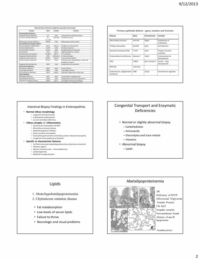
Abetalipoproteinemia
• AR
• Deficiency of MTTP
• (Microsomal Triglyceride
• Transfer Protein)
• Chr 4q22
• Irregular vacuoles
• Non-membrane bound
• Absence of apo-B
lipoproteins
Acanthocytosis

9/12/2013
3
Chylomicron Retention Disease
AR
SAR1B, chr 5q31
(Sar1-ADP-ribosylation)
Codes for GTPase
Chylomicron trafficking
Primary enteropathies of infancy
Epithelial defects
Microvillous inclusion disease
Tufting enteropathy
Enteroendocrine cell deficiency
Autoimmune enteropathies
Others
Microvillus Inclusion Disease (MVID),
Microvillous Atrophy (MVA)• Described in 1978 by Davidson
et al
•Severe secretory diarrhea during
1st week of life
•Absence of infectious/enzymatic
etiology
• Villous atrophy without
significant inflammation
•Abnormal mucosal staining by
PAS, CD10
•Abnormal mucosal ultrastructure
•MYO5B gene; 18q21
•Encodes myosin Vb, regulates
distribution of endosomesNormal
PAS stain
Microvillous inclusion disease
normal Microvillous Inclusion Disease
18 day-old boyCD10
PAS
Microvillous inclusion disease “Tufting” Enteropathy
•Severe diarrhea 1st week
•Dysmorphic features in some infants
Choanal atresia
Esophageal/rectal atresia
–+/- “tufting” in colon
•AR
•EpCAM gene mutations (chr 2p21)
•TPN dependent
•Clusters of cases described in families
from Malta and from the Gulf states

9/12/2013
4
Tufting enteropathy
EpCAm MOC 31 + controlEpCAm MOC 31 - case
Enteroendocrine cell dysgenesis – Neurogenin-3
mutation
• NEUROG 3 is a protein involved in gut and pancreatic endocrine development
• Pts with Neurogenin 3 mutations present with congenital diarrhea and eventually develop type I diabetes
• TPN-dependent; bowel transplantation
Duodenal biopsy, 5 months
Immunostaining for chromogranin
Enteroendocrine cell dysgenesis – Neurogenin-3
mutation
• No enteroendocrine cells per IHC for chromogranin
• Neurogenin-3 -/- mice lack endocrine cells in pancreas and intestine, death from diabetes in the first days of life control
case
Enteropathy with dysmorphic features
“Syndromatic diarrhea”
Chronic diarrhea in first year of life
Facial dysmorphism
Trichorrhexis nodosa
Immunodeficiency
Poor prognosis
Similar patients described as tricho-hepato-enteric syndrome
Gene TTC37 (5q14) - Thespin
Autoimmune Enteropathy
• Most common severe enteropathy of childhood
• Rarely observed in adults, but may account for some cases
of refractory celiac disease
• Heterogenous entity
• Severe early-onset diarrhea, male preponderance
• Concomitant colitis and gastritis present in majority
• Circulating gut-autoantibodies
• Autoimmune phenomena in majority of cases
• Favorable response to immunosuppression (Tacrolimus)
• BMT attempted in some cases
Autoimmune enteropathy -
extra-intestinal manifestations
• Insulin-dependent DM
• Nephrotic syndrome, membranous GN with granular IgG
deposits or interstitial nephritis
• Thyroid insufficiency
• Acute or chronic hepatitis
• Coombs –positive hemolytic anemia and thrombocytopenia
• Diffuse pulmonary interstitial infiltrates
• Autoantibodies: ASMA, AMA, ANA, anti-parietal cell antibodies

9/12/2013
5
Autoimmune enteropathy (AE) –clinical
conditions
• IPEX Immunodysregulation / polyendocrinopathy / enteropathy / X-linked.
– Mutation in FOXP3 gene, Xp11.23-q13.3
– FOXP3 codes for a protein called Scurfin which is predominately expressed in CD4+/CD25+ regulator T-cells
• 50% of patients with clinical features of IPEX have normal FOXP3 gene – “IPEX-like”
– A few cases reported with specific CD 25 deficiency
Autoimmune Enteropathy
• Severe villous atrophy
• Marked inflammatory destruction of crypts
• Increased apoptosis
• Loss of Paneth and goblet cells
• Concomitant colitis and gastritis
• Few surface intraepithelial lymphocytes
Gut Autoantibodies
Anti-Enterocyte Antibodies
• Linear fluorescence pattern along the
apex or brush border of enterocytes
• Also anti-goblet abs
• Predominantly IgG but IgA and IgM have
been described
Autoimmune enteropathy in adults
• Protracted diarrhea, weight loss, malnutrition
• Absence of response to gluten-free diet and/or absence of typical celiac antibodies and/or characteristic HLA immunotype
• AEA + variety of autoantibodies
• T-cell rearrangement studies neg
• Good response to immunosuppression
Autoimmune enteropathy -
differential diagnosis
• Celiac disease
• Enteritis in congenital immunodeficiencies
• Food allergy
• IBD
• GVHD
• Collagenous or lymphocytic enterocolitis
• Autoimmune polyglandular syndrome
IgA deficiency - 6 yr-old boy with chronic diarrhea and FTT
Duodenum – celiac-like
Colon –focal marked lymphoid hyperplasia
Colon- focal crypt architectural distorsionIgA

9/12/2013
6
Common Variable Immunodeficiency
Increased basal crypt apoptotic activity, or
extensive loss of goblet and Paneth cells
GVHD-like or IPEX-like histology
Crohn’s-like enteritis, especially in younger age
Microvillus
inclusion
disease
Tufting
enteropathy
Enteroendocrine
cell dysgenesis
Syndromic
(THE)
Autoimmune
enteropathy
Presentation First 2
weeks
First 2 weeks First 2 weeks First months After 1 month
Gene defect MYO5b
(18q21)
EpCAM (2p21) NEUROG 3
(10q21.3)
TTC37
(5q14)
FOXP3 (Xp11.23) in
IPEX syndrome
Extraintestinal
disease
Low GGT
cholestasis
post bowel
transplanta
tion
Dysmorphism
keratitis
arthritis
Insulin-
dependent
diabetes
Dysmorphism
Trichorrhexis
nodosa
Polyendocrinopathy
Anti-
enterocyte
antibodies
no no no no yes
Villous
atrophy
yes variable variable variable variable
Surface
epithelium
Absent
brush
border
Tufting and
desquamation
Normal Normal Normal or atrophic
Inflammation Minimal Variable Variable Minimal Increased
Differentiating features of severe diarrhea of early infancy
Food AllergyThe New Epidemic
• Food hypersensitivity reactions affect up to 8% of children under 3 years of age and approximately 2.5% of the general population
• 3x increase in the prevalence of food allergies over the past 20 years– Changes in environment
– Changes in the processing of foods
– Alteration of immunologic recognition
– Use of antibiotics
• Food intolerance (non-allergic food hypersensitivities) are adverse responses caused by metabolic or enzymatic disorder (lactose)
Immunopathology of Food Allergic Disorders
� IgE mediated (Immediate hypersensitivity)
• Oral allergy
• Urticaria
• Anaphylaxis
� Cell mediated (delayed onset/chronic)
• Dietary protein enteropathy/enterocolitis
• Dietary protein-induced proctocolitis
• Gluten-sensitive enteropathy
� Mixed IgE and Cell mediated (delayed onset/chronic)
• Atopic dermatitis
• Eosinophilic gastrointestinal disease
From Sicherer, S. H. Annu Rev Med 2009. 60:261-77
Spectrum of disease or unique diseases
Allergic proctocolitis Eosinophilic esophagitis
Eosinophilic gastroenteritis
Colon Esophagus
Eosinophilic GastroenteropathiesThe New Epidemic
“Normal” Number of Eosinophils
Lowichik and Weinberg Mod Pathol 1995;9:110-4 DeBrosse et al. Pediatr Dev Pathol. 2006;9:210-8

9/12/2013
7
Allergic proctocolitis: key features
• Usually presents by 6 months of life
• Blood streaked, loose stools +/- diarrhea in otherwise well-appearing infants
• Some may present with constipation, mimicking HD
• Usually occurs in breast-fed (50-60%) or cow/soy milk formula-fed infants
• Diagnosis is via clinical history; food prick skin tests
negative
• Treatment via protein elimination; resolution of symptoms in 48–72 h
• Tolerance to allergen usually occurs by 1 yr of life
Adapted from Maloney, J. Pediatr Allergy Immunol 2007:18:360-367
EndoscopyColon
Normal
Allergic Proctocolitis
Eosinophilic infiltrate, frequently patchy
and rather variable in severity.
Neutrophilic cryptitis can also be seen but
usually not to the extent seen in
infectious colitis.
Chronic mucosal changes are not seen.
Occasional cases may cause constipation,
mimicking Hirschsprung disease clinically
and radiographically
Allergic Eosinophilic Gastroenteritis in children:
Key features
• Usually occurs from infancy through adolescence
• Chronic symptoms of poor appetite, poor weight gain or weight loss, emesis, diarrhea, occult blood in stool
• Endoscopy and biopsy helpful in diagnosis with usually marked eosinophilic infiltration of mucosa and submucosa
• > 90% of cases have involvement of gastric antrum
• Approximately, 50% are atopic; 50% have peripheral blood eosinophilia
• Resolution of symptoms with removal of causal food within 6 wk
• Most common foods: cow's milk, egg, soy, cereals, fish
• Excellent response to amino-acid-based formula
• Responsive to steroids
• Typically prolonged; natural history not well understood
Adapted from Maloney, J. Pediatr Allergy Immunol 2007:18:360-367
Normal
Antrum
Eosinophilic
Gastroenteritis Allergic Eosinophilic Gastroenteritis
Mucosal type Mural typeMucosal disease Mural disease

9/12/2013
8
Eosinophilia in the GI Tract
• Allergy ≠ Eosinophilia
• Eosinophilia ≠ Allergy
Dietary protein-induced
enteropathy/enterocolitis
• Dietary protein-induced enteropathy/enterocolitis
– Infancy to school age
– Cow’s milk, soy, wheat, rice, chicken and fish
– Malabsorption and osmotic diarrhea
– Biopsy : flat villi, +/-increased eosinophils; diff dx: celiac disease
2 month-old with malabsorption; improved
on elemental formula
Eosinophilic Gastroenteritis – Differential
Diagnosis
• Infections, particularly parasitic – Stool ova and parasite study may be diagnostic
• Drug reactions– Check drug history – Azathioprine, NSAIDS, tacrolimus
• Crohn’s disease– May primarily show eosinophilic abscesses
– Typically more of a focal lesion
• Some primary immunodeficiencies
• Connective tissue disorders– Consider lupus, polyarteritis, and Wegeners.
– Are fibrinoid changes present in vessels?
• Inflammatory fibroid polyps– Check configuration of lesion on endoscopy
• Hypereosinophilic syndrome.– Are tumorous lesions present, particularly in soft tissue?
• Post-transplant eosinophilic gastroenteritis– Check transplant history, immunomodulatory drugs
Post-Liver Transplant Eosinophilic
Gastroenteritis
2yr-old girl 18 months post-OLT with weight loss, food refusal, peripheral eosinophilia