1.3.2 congenital abnormalities - barbados …...2019/10/01 · cri du chat syndrome • also known...
TRANSCRIPT

1.3.2 congenitalabnormalities
• 1.3.2 congenital abnormalities:
• principles, • patterns of anomalies,
• maternal illness,
• drug abuse, • medications, • infectious agents,
• malnutrition

Malformation - definition
Congenital malformation are structuraldefects present at birth.
They may be gross or microscopic, on thesurface of the body or within it, familiar orsporadic, hereditary or nonhereditary, single ormultiple
A major congenital anomaly is one that isincompatible with survival, is life-threatening, or seriously compromises anindividual´s capacity to function normally insociety)

Malformation is a primary structural defectresulting from a localized error ofmorphogenesis
Disruption is specific abnormality that resultsfrom disruption of normal developmentalprocesses It depends on time not on agent
Deformation is an alteration in shape / structureof previously normally formed part
Syndrome is a recognized pattern ofmalformations with a given etiology.

Birth defects
3% of all live-born infants have an majoranomaly
Additional anomalies are detected duringpostnatal live – about 6% at 2 year-olds,8% in 5year-olds, other 2% later
Single minor anomalies are present inabout 14% of newborns

Birth defects
Major anomalies are more common inearly embryos (up to 15%) than they are innewborns (3%).
Most severely malformed embryos arespontaneously aborted during first 6 to8 weeks.

Causes of congenital anomalies

• Figure 3.9 Sources of Congenital Defects

Inherited defects may be due to• 1 Chromosomal Abnormalities may be due to
a change in chromosome number i.e toomany or too few chromosomes
• 2 Chromosomal Abnormalities may be due toa change in chromosome structure i.ebroken or damaged chromosomes
• 3 Genetic abnormalities• Recessive genes for a disorder• Dominant genes for a disorder• 4 Genetic mutations

Anomalies caused by geneticfactors
Chromosomal aberrations are common and arepresent in 6 to 7% of zygotes – (result =abort)
Numerical chromosomal abnormalities – usually non-disjunction- error in cell division
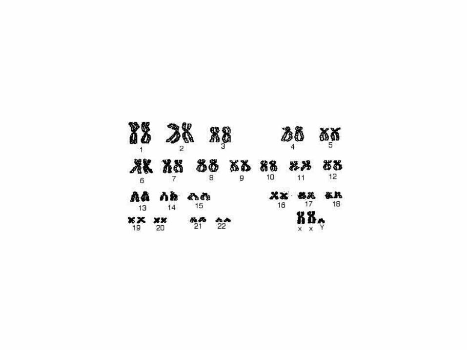
Down syndrom (21) Edwards (18) Patau (13)Turner (X0), Klinenfelter (XXY)
Structural chromosomal abnormalities –chromosome breaks = translocation, deletion (cridu chat syndrome), duplication, inversion.
Mutant genes – achondroplasia, fragile-Xsyndrome

Table 12-1, p. 196
Stepped Art

• ALL OF THE CONDITIONS THAT WEWILL BRIEFLY MENTION IN THISINTRODUCTORY SESSION WILL BEDISCUSSED IN SOME DETAIL INSUBSEQUENT SESSIONS IN THISCOURSE,,,,,,,,,,,,,,,,,,,,,,
• BUT WE WILL SAY A FEW WORDSTODAY ABOUT SOME OF THESEMALADIES
ChromosomalAbnormalities
– may be too manyor too few
chromosomes
Chromosomal & Genetic Factors
• Numerical Abnormalities– Trisomy 21 (Down syndrome)– Trisomy 18– Trisomy 13– Klinefelter Syndrome– Turner Syndrome– Triple X Syndrome
• Structural Abnormalities• Mutant Genes

Chromosomal Abnormalities• May be numerical or structural• Important causes of congenital
malformations & spontaneousabortions
• Estimated that 50% of all conceptions endin spontaneous abortion & 50% of thesehave major chromosome abnormalities
• Most common chromosomeabnormalities in aborted fetuses is:– Turner syndrome (45,X)– triploidy– trisomy 16
Numerical Abnormalities• Normal gametes are haploid (n =23)• Normal human somatic cell contains 46
chromosomes; Diploid (2n = 46)• Euploid-Exact multiple of n• Aneuploid-Any chromosome # that is noneuploid
– Additional chromosome– Missing chromosome
• Most common cause is nondisjunction duringeither meiosis to mitosis
– Risk of meiotic nondisjunction with maternal age

Changes in ChromosomeStructure or Number
• On rare occasions, a chromosome mayundergo a large-scale, permanent changein its structure, or the number ofautosomes or sex chromosomes maychange
• In humans, such changes usually result ina genetic disorder

Heritable Changes in the Chromosome Number
• Occasionally, new individuals end up with thewrong chromosome number– Consequences range from minor to lethal
• Euploids – Normal number of chromosomes
Aneuploidy– Too many or too few copies of one chromosome– Extra or missing chromosomes
• Polyploidy– Extra sets of chromosomes (triploids, tetraploids)– Three or more copies of each chromosome– Spindle fails during mitosis

• Organisms with more than two complete sets ofchromosomes, have undergone polypoidy.
• This may occur when a normal gamete fertilizesanother gamete in which there has beennondisjunction of all its chromosomes.– The resulting zygote would be triploid (3n).
• Alternatively, if a 2n zygote failed to divide afterreplicating its chromosomes, a tetraploid (4n)embryo would result from subsequentsuccessful cycles of mitosis.

• Polyploids are more nearly normal inphenotype than aneuploids.
• One extra or missing chromosomeapparently upsets the genetic balanceduring development more than does anentire extra set of chromosomes.

Aneuploids• Abnormalities usually prevent embryo
development
• Exception in humans is Downsyndrome– Three copies of chromosome 21 (trisomy
21)– Physical and learning difficulties– Frequency of nondisjunction increases as
women age

Aneuploidyof Sex
Chromosomes
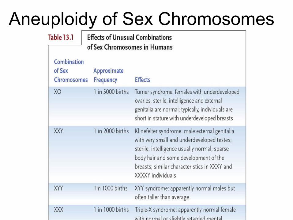
Aneuploidy of Sex Chromosomes

Polyploids
• Uncommon in animals– Usually has lethal effects during
embryonic development

Numerical abnormalities
• euploidy - normal 46 (2n)• polyploidy (3n or 4n) - spontaneous abortion• aneuploidy• trisomy (2n+1) - 47 - compatible with life• monosomy (2n-1) - autosomal - incompatible
with life• - sex chromosomal -
compatible with life

Alterations of chromosome number orstructure cause some genetic disorders
• Nondisjunction occurs when problems withthe meiotic spindle cause errors in daughtercells.– This may occur if
tetrad chromosomes do not separate properly during meiosis I.
– Alternatively, sister chromatids may fail to separate during meiosis II.

• As a consequence of nondisjunction, somegametes receive two of the same type ofchromosome and another gamete receives nocopy.
• Offspring results from fertilization of a normal gametewith one after nondisjunction will have an abnormalchromosome number or aneuploidy.– Trisomic cells have three copies of a particular
chromosome type and have 2n + 1 totalchromosomes.
– Monosomic cells have only one copy of a particularchromosome type and have 2n - 1 chromosomes.
• If the organism survives, aneuploidy typically leads toa distinct phenotype.

Nondisjunction
• Changes in chromosome number can becaused by nondisjunction, when a pair ofchromosomes fails to separate properlyduring mitosis or meiosis
• Affects the chromosome number atfertilization– Monosomy (n-1 gamete)– Trisomy (n+1 gamete)

Nondisjunction

Nondisjunction (1)• Failure of homologous pair separation during Meiosis I
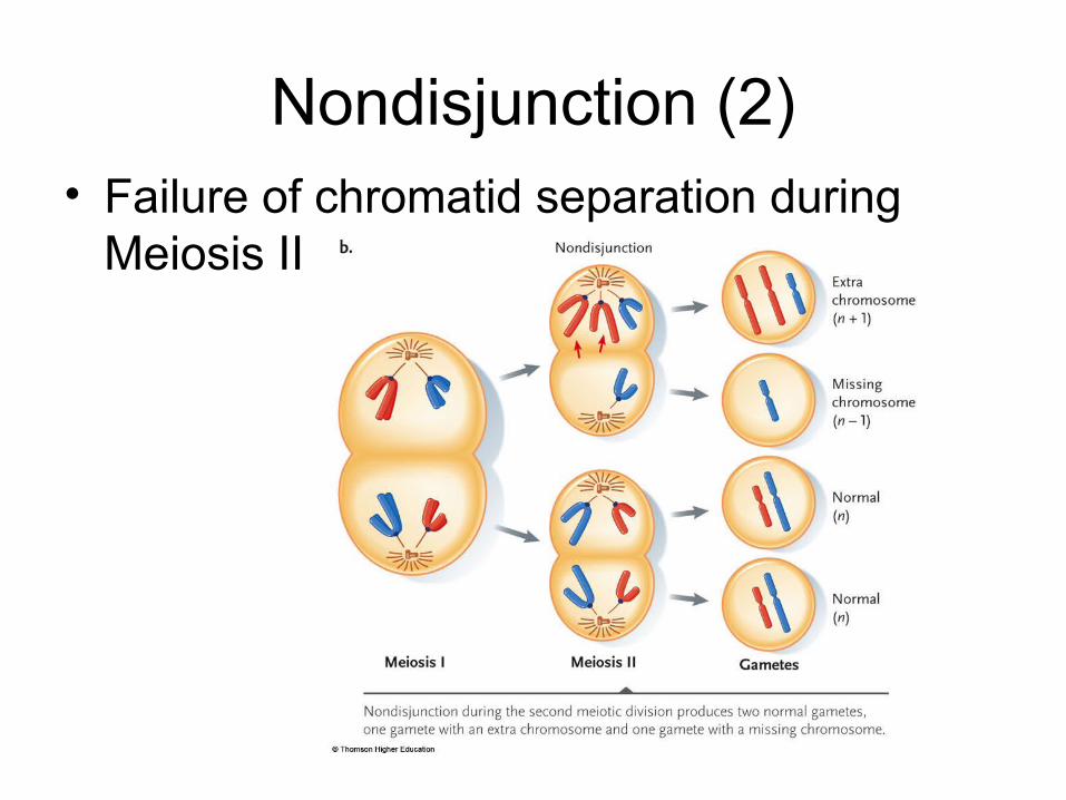
Nondisjunction (2)• Failure of chromatid separation during
Meiosis II

Autosomal Change and DownSyndrome
• Only trisomy 21 (Down syndrome) allowssurvival to adulthood– Characteristics include physical appearance,
mental impairment, and heart defects
• Incidence of nondisjunction increases withmaternal age
• Can be detected through prenatal diagnosis

Trisomy 21

Fig. 12-13b, p. 194
n + 1
n + 1
n − 1
n − 1
chromosomealignments atmetaphase I
NONDISJUNCTIONAT ANAPHASE I
alignments atmetaphase II
CHROMOSOMENUMBER
IN GAMETESanaphase II

• (a) A case of nondisjunction. Thiskaryotype reveals the trisomic 21 conditionof a human female.
• (b) One example of how nondisjunctionarises.
• Of the two pairs of homologouschromosomes shown here, one fails toseparate during anaphase I of meiosis.
• The chromosome number is altered in thegametes that form after meiosis.

Down’s Syndrome
• Caused by non-disjunction of the21st chromosome.
• This means that theindividual has atrisomy (3 – 2lstchromosomes).
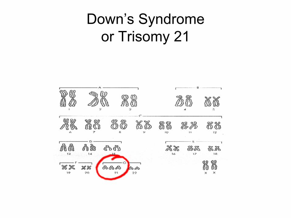
Down’s Syndromeor Trisomy 21

Down Syndrome and Maternal Age

Down Syndrome

Change in Sex ChromosomeNumber
• Changes in sex chromosome number mayimpair learning or motor skills, or beundetected
• Female sex chromosome abnormalities– Turner syndrome (XO)– XXX syndrome (three or more X chromosomes)
• Male sex chromosome abnormalities– Klinefelter syndrome (XXY)– XYY syndrome
Kleinfelter’s syndrome(or Klinefleter’s)
• Disorder occurring due tonondisjunction of the X chromosome.
• The Sperm containing both X and Ycombines with an egg containing the X,results in a male child.
• The egg may contribute the extra Xchromosome.


Turner Syndrome• XO (one unpaired
X chromosome)– Usually caused by
nondisjunction inthe father
– Results in femaleswith undevelopedovaries
Broken or damagedchromosomes

Structural abnormalities• breakage followed by loss or rearrangement• deletion, translocationGenerally: • loss of chromosomal material is more
dangerous than gain• abnormalities of sex chromosomes are better
tolerated than autosomal• abnormalities of sex chromosomes
sometimes symptomatic in adult age (e.g.infertility)
• usually origin de novo (both parents andsiblings are normal)

Heritable Changes in Chromosome Structure
• On rare occasions, a chromosome’sstructure changes; such changes are usuallyharmful or lethal, rarely neutral or beneficial
• A segment of a chromosome may beduplicated, deleted, inverted, or translocated

Chromosomal Alterations
• Deletion: brokensegment lostfromchromosome
• Duplication:broken segmentinserted intohomologouschromosome

• Breakage of a chromosome can lead to fourtypes of changes in chromosome structure.
• A deletion occurs when a chromosomefragment lacking a centromere is lost during celldivision.– This chromosome will be missing certain genes.
• A duplication occurs when a fragmentbecomes attached as an extra segment to asister chromatid.

Chromosomal Alterations (2)• Translocation:
broken segmentattached tononhomologouschromosome
• Inversion:broken segmentreattached inreversedorientation

• An inversion occurs when a chromosomalfragment reattaches to the originalchromosome but in the reverse orientation.
• In translocation, a chromosomal fragmentjoins a nonhomologous chromosome.– Some translocations are reciprocal, others are
not.

Deletion• Loss of some portion of a
chromosome; usually causes seriousor lethal disorders– Example: Cri-du-chat

Deletions• When homozygous, most deletions are lethal, because
most genes are necessary for life and a homozygousdeletion would have zero copies of some genes.
• When heterozygous, the genes on the normal homologueare hemizygous: there is only 1 copy of those genes, andthus they are expressed even if recessive (like genes on theX in male mammals).
• Heterozygous deletions are aneuploid, because thegenes in the deleted region are present in only 1 copyinstead of the normal two copies. Some genes need tobe present in two copies, so heterozygous deletionssometimes give rise to defects in the affectedindividual, especially if the deletions are large.

Cri du chat syndrome
• also known as chromosome 5p deletionsyndrome, 5p minus syndrome orLejeune’s syndrome, is a rare geneticdisorder due to a missing part ofchromosome 5.
• Its name is a French term (cat-cry or callof the cat) referring to the characteristiccat-like cry of affected children.

Deletion: Cri-du-chat
Infant 4 years old

• For most genes it is a reasonableassumption that a specific allele will have thesame effect regardless of whether it wasinherited from the mother or father.
• However, for some traits in mammals, it doesdepend on which parent passed along thealleles for those traits.– The genes involved may or may not lie on the X
chromosome.– Involves “essential” silencing of one allele during
gamete formation
The phenotypic effects of some mammalian genesdepend on whether they were inherited from the
mother or the father (genomic imprinting).
• Two disorders, Prader-Willi syndrome andAngelman syndrome, with different phenotypiceffects are due to the same cause, a deletion ofa specific segment of chromosome 15.– Individuals with Prader-Willi syndrome are
characterized by mental retardation, obesity, shortstature, and unusually small hands and feet.
– These individuals inherit the abnormal chromosomefrom their father.
– Individuals with Angelman syndrome exhibitspontaneous laughter, jerky movements, and othermotor and mental symptoms.
– This is inherited from the mother.
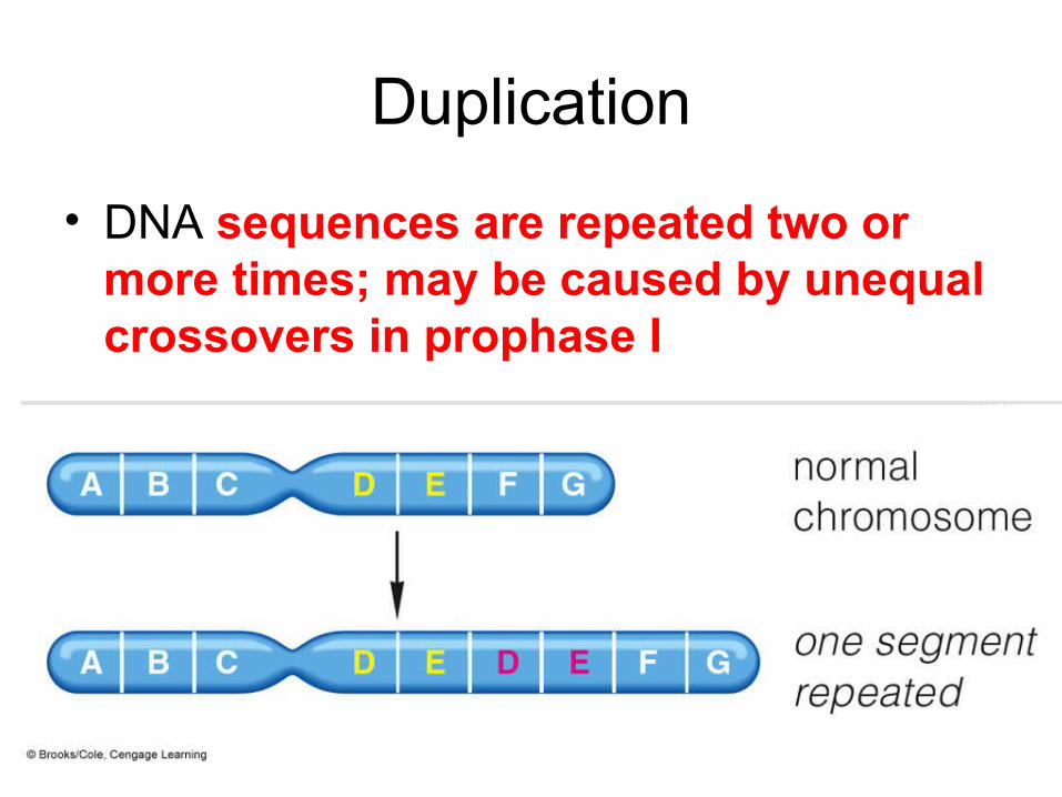
Duplication
• DNA sequences are repeated two ormore times; may be caused by unequalcrossovers in prophase I

Duplications• Genes are duplicated if there is
more than one copy present in thehaploid genome.
• Some duplications are “dispersed”,found in very different locations fromeach other.
• Other duplications are “tandem”,found next to each other.

Translocation• Typically, two broken chromosomes
exchange parts (reciprocal translocation)

Translocations• In a translocation, two different, non-homologous chromosomes are
broken and rejoined to each other. All the genes are present, so anindividual with a translocation can be completely normal. However, anindividual who is heterozygous for a translocation and a set of normalchromosomes can have fertility problems
• The problem occurs during meiosis 1, as the result of confusion about howthe chromosomes should segregate to opposite poles.
• During prophase and metaphase of M1, the homologous chromosomes pairup. Because translocations have pieces of two different chromosomesattached together, they pair up in a cross-shaped configuration, so allthe pieces have a partner. This structure is three-dimensional, not flat,and there is ambiguity about which centromeres are attached to whichpole of the spindle.
• When anaphase occurs, two main possibilities exist: alternate segregation,where centromeres on opposite sides of the cross go to the same pole, andadjacent segregation, where centromeres on the same side of the cross goto the same pole.

Translocational Down Syndrome• Most cases of Down syndrome, trisomy-21, are spontaneous. They are
caused by non-disjunction which gives an egg or sperm with two copies ofchromosome 21.
• However, about 5% of Down’s cases are caused by a translocationbetween chromosome 21 and chromosome 14. These translocationalDown’s cases are heritable: several children in the same family can havethe disease.
• Both chromosome 14 and chromosome 21 are acrocentric, and the short armscontain no essential genes.
• Sometimes a translocation occurs that joins the long arms together on onecentromere and the short arms on another centromere. In this case the shortarm chromosome is usually lost. The individual thus has a normal chromosome14, a normal chromosome 21, and a translocation chromosome, called t(14;21).
• During meiosis, one possible gamete that occurs has both the normal 21 and thet(14;21) in it. When fertilized, the resulting zygote has 2 copies of the importantparts of chromosome 14, but 3 copies of chromosome 21: 2 normal copies plusthe long arm on the translocation. This zygote develops into a person withDown syndrome.

Inversion
• Part of the sequence of DNA becomesoriented in the reverse direction, with nomolecular loss

Inversions• An inversion is when a segment of a chromosome
is removed and then replaced backwards. • The problem with inversions occurs in meiosis,
when a chromosome containing an inversion isheterozygous with a normal chromosome. Acrossover within the inverted region results inaneuploidy and death of the resulting embryo.
• One consequence of this is that crossing over isapparently suppressed.
• Inversions can be either paracentric, where thecentromere is NOT in the inverted region, orpericentric, where the inversion is in the invertedregion.
Genetic abnormalities
Recessive genesfor a disorder

Dominant genes for adisorder

68
Medical GeneticsWhen studying rare disorders, 6 general
patterns of inheritance are observed:
• Autosomal recessive• Autosomal dominant• X-linked recessive• X-linked dominant• Codominant • Mitochondrial
69
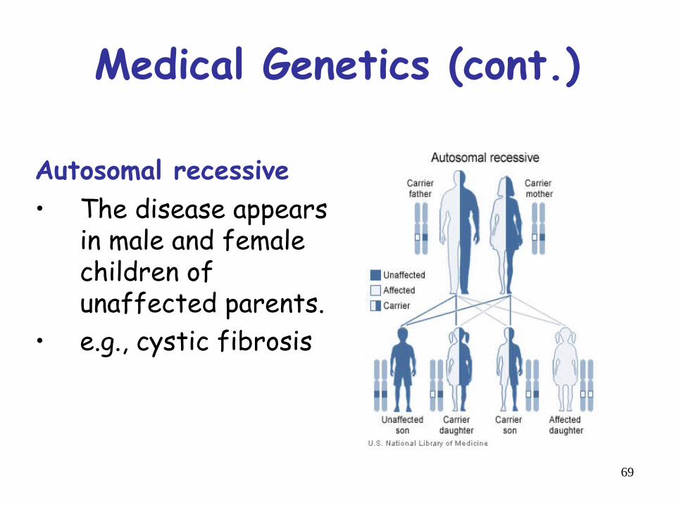
Medical Genetics (cont.)
Autosomal recessive • The disease appears
in male and femalechildren ofunaffected parents.
• e.g., cystic fibrosis

70
Medical Genetics (cont.)
Autosomal dominant• Affected males and
females appear in eachgeneration of thepedigree.
• Affected mothers andfathers transmit thephenotype to both sonsand daughters.
• e.g., Huntington disease.
71
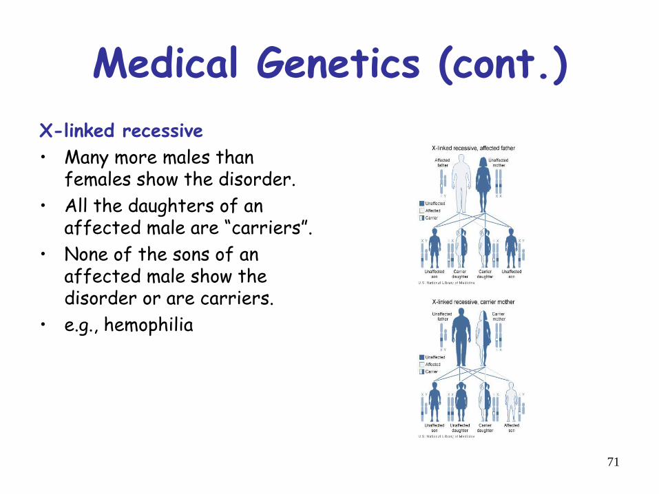
Medical Genetics (cont.)X-linked recessive• Many more males than
females show the disorder.• All the daughters of an
affected male are “carriers”.• None of the sons of an
affected male show thedisorder or are carriers.
• e.g., hemophilia

72
Medical Genetics (cont.)X-linked dominant • Affected males pass the
disorder to all daughters butto none of their sons.
• Affected heterozygousfemales married tounaffected males pass thecondition to half their sonsand daughters
• e.g. fragile X syndrome

73
Medical Genetics (cont.)Codominant inheritance
• Two different versions(alleles) of a gene can beexpressed, and each versionmakes a slightly differentprotein
• Both alleles influence thegenetic trait or determinethe characteristics of thegenetic condition.
• E.g. ABO locus

74
Medical Genetics (cont.)Mitochondrial inheritance• This type of inheritance
applies to genes inmitochondrial DNA
• Mitochondrial disorders canappear in every generation ofa family and can affect bothmales and females, butfathers do not passmitochondrial traits to theirchildren.
• E.g. Leber's hereditary opticneuropathy (LHON)

Genetic mutations

• Genetic Abnormalities– Many passed to children by parents who are
carriers of recessive alleles– Some are caused by dominant alleles– Some result from mutations – changes in
structure of one or more genes• Spontaneous
• Environmental hazards

Defining Genetic Disorders and Abnormalities
• Genetic abnormality– A rare or uncommon version of a trait; not
inherently life threatening
• Genetic disorder– An inherited condition that causes mild to
severe medical problems, characterized by aspecific set of symptoms (a syndrome)

Recurring Genetic Disorders
• Mutations that cause genetic disordersare rare and put their bearers at risk
• Such mutations survive in populationsfor several reasons– Reintroduction by new mutations– Recessive alleles are masked in
heterozygotes– Heterozygotes may have an advantage in a
specific environment
Mutations• Gene mutations can be either inherited from a
parent or acquired. • A hereditary mutation is a mistake that is
present in the DNA of virtually all body cells. • Hereditary mutations are also called germ line
mutations because the gene change exists inthe reproductive cells and can be passed fromgeneration to generation, from parent tonewborn. Moreover, the mutation is copiedevery time body cells divide

• Mutations occur all the time in every cell inthe body.
• Each cell, however, has the remarkableability to recognize mistakes and fix thembefore it passes them along to itsdescendants. But a cell's DNA repairmechanisms can fail, or beoverwhelmed, or become less efficientwith age. Over time, mistakes canaccumulate.

Disease MutationsSingle mutations Fragile X
Sickle Cell Anemia
Common mutations Deafness Hemochromatosis
Panel of mutations Cystic Fibrosis
Private mutations Breast Cancer
Colorectal cancer
Mutations
A mutation may be defined as a permanent change in the
DNA.
Mutations that affect the germ cells are transputted to the
progeny and may give rise to inherited diseases.
Mutations thar aries in somatic cells are important in the
genesis of cancers and some congeital malformations.

Mutations may be classified into three catagories:
Genome mutations – involve loss or gain of whole
chromosomes (giving rise to monosomy or trisomy)
Chromosome mutations – result from rearrangement of
genetic material and give rise to visible structural changes
in the chromosome.
Gene mutations – may result in partial or complete
deletion of a gene or, more often, affect a single base. For
example, a single nucleotide base may be substituted by
a different base, resulting in a point mutation.

Autosomal dominant disorders (neurofibromatosis,tuberous sclerosis, polycystic kidney disease, familiarpolyposis coli, hereditary spherocytosis, Marfansyndrome, osteogenesis imperfecta, achondroplasia,familiar hypercholesterolemia)
Autosomal recessive disorders (cystic fibrosis,phenylketonuria, homocystinuria, hemochromatosis, sicklecell anemia, thalassemias, alkaptonuria, neurogenicmuscular atrophies)
X-linked disorders (glucose-6-phosphate dehydrogenasedeficiency)

Biochemical and molecular basis ofsingle-gene disorders
1) Enzyme defects and theirconsequences
2) Defects in receptors and transportsystems
3) Alterations in structure, function orquantity of nonenzyme proteins
4) Genetically determined adversereactions to drugs.

1-Disorders associated with defectsin structural proteins
Marfan syndrome
A disorder of the connective tissues of the body, manifestedprincipally by changes in the skeleton, eyes, and cardiovascularsystem.
Ehlers-Danlos syndromes
A clinically and genetically heterogeneous group of disorders thatresult from some defect in collagen synthesis or structure (otherdisorders resulting from mutations affecting collagen synthesisinclude osteogenesis imperfecta, Alport syndrome, epidermolysisbullosa)

2-Disorders associated with defectsin receptor proteins
Familiar hypercholesterolemiaA disease that is the consequence of a mutationin the gene encoding the receptor for low-density lipoprotein (LDL), which is involved inthe transport and metabolism cholesterol. More than 150 mutations, including insertions,deletions, and missense and nonsensemutations, involving the LDL receptor genehave been identified.

• These can be classified into five groups: Class I mutations -uncommon, they lead to a complete failure of synthesis of thereceptor protein.
• Class II mutations - common, they encode receptor proteinsthat accumulate in the endoplasmic reticulum because theycannot be transported to the Golgi complex.
• Class III mutations - affect the LDL-binding domain of thereceptor.
• Class IV mutations - encode proteins that are synthesized andtransported to the cell surface efficiently, they bind LDHnormally, but the bound LDL is not internalized.
• Class V mutations - encode proteins that are expressed on thecell surface, can bind LDL, and can be internalized, however,the acid-dependent dissociation of the receptor and the boundLDL fails to occur.
3-Disorders associated with defectsin enzymes
Lysosomal storage diseases: Lysosomes containdifferent types of hydrolytic enzymes, which can cleavevarious substrates in the acid milieu and can be secreted.
With an inherited deficiency of a functional lysosomalenzyme, catabolism of its substrate remains incomplete,leading to the accumulation of the partially degradedinsoluble metabolite within the lysosomes.
These organells become large and numerous giving rise tothe lysosomal storage disorders.
These disorders result exclusively from mutations thatlead to reduced synthesis of lysosomal emzymes

• There are also other defects: Synthesis of acatalytically inactive proteins that cross-reactimmunologically with normal enzymes, so theenzyme level appear to be normal.,.defects inpost-translational processing of enzymes(example is a failure of mannose-6-phosphatereceptor), lack of an enzyme activator orprotector protein, lack of a substrate activatorprotein, lack of transport protein.

• The lysosomal storage disorders can bedivided into
• (1) glycogenoses, • (2) sphingolipidoses (lipidoses),
• (3) mucopolysaccharidoses, and
• (4) mucolipidoses. • Examples follow:

Disorders associated with defects inenzymes
Tay-Sachs disease – GM2 gangliosidosis, hexosaminidase -subunitdeficiency,GM2 ganglioside accumulates in heart, liver, spleen etc.,destruction of neurons, proliferation of microglia and accumulation oflipids in phagocytes within the brain.
Niemann-Pick disease – types A and B, two related disorders withlysosomal accumulation of sphingomyelin, deficiency ofsphingomyelinase, 80% of all cases repreents type A – the severeinfantile form with neurologic involvement, visceral accumulation ofsphingomyelin and early death within the first 3 years of life.
Gaucher disease – a cluster of autosomal recessive disordersresulting from mutations in the gene encoding glucocerebrosidase, themost common lysosomal storage disorder, accumulation ofglucocerebrosides, types I-III, the glucocere¨brosides accumulatewithin phygocytes (Gaucher cells) throughout the body – spleen, liver,bone marrow, lymph nodes, tonsils thymus etc.

Disorders associated with defects inenzymes
Mucopolysaccharidoses (MPS) – the deficiencies of lysosomalenzymes involved in the degradation of mucoplysaccharides(glycosaminoglycans), several clinical variants classified from MPS I(Hurler syndrome) to MPS VII, each resulting from the deficiency of onespecific enzyme, all the MPS except one are autosomal recessivedisorders, the exception (Hynter syndrome) is an X-linked recessivedisorder, involvement of multiple organs including liver, spleen, heart,blood vessels, joint stiffness, mental retardation.
Glycogen storage diseases – resulting from a hereditary deficiency ofone of the enzymes involved in the synthesis or sequential degradation ofglycogen, 3 forms: hepatic, myopathic, miscellaneous (deficiency of -glucosidase and lack of branching enzymes, type II – Pompe diseaseand type IV, death early in life.

Disorders associated with defects inenzymes
Alkaptonuria (Ochronosis) – an autosomal recessive disorder in whichthe lack of homogentisic oxidase blocks the metabolism ofphenylalanine-tyrosine at the level of homogentisic acid, homogentisicacid accumulates in the body, it selectively binds to collagen inconnective tissues, tendons, and cartilage, these tissues have a blue-black pigmentation (ochronosis) most evident in the ears, nose, andcheeks, the deposits of the pigment in the articular cartilages cause thecartilage to lose its normal structure and function resulting inosteoarthritis.

4-Disorders associated with defectsin proteins that regulate cell growth
Neurofibromatosis: types 1 and 2 – two autosomal dominant
disorders, neurofibromatosis type 1 previously called von
Recklinghausen disease, neurofibromatosis type 2 previously
called acoustic neurofibromatosis. Although there is some
overlap in clinical features, these two entities are genetically
distinct.

Disorders associated with defects inproteins that regulate cell growth
Neurofibromatosis-1: The neurofibromatosis 1 gene (NF-1) has been mapped
to chromosome 17q11.2. It encodes a protein called neurofibromin, which
down-regulates the function of the p21ras oncoprotein. NF-1 therefore belongs
to the family of tumor-suppressor genes. Three major features of disorder –
multiple neural tumors (neurofibromas) dispersed anywhere on or in the body,
numerous pigmented skin lesions, and pigmented iris hamartomas, also called
Lisch nodules. A wide range of associated abnormalities has been reported in
these patients – skeletal lesions like erosive defects, scoliosis, intraosseous
cystic lesions, subperiosteal bone cysts, pseudoarthrosis of the tibia. Patients
have also a twofold to fourfold greater risk of developing other tumors (Wilm´s
tumor, rhabdomyosarkoma, meningioma, optic glioma, pheochromocytoma,
chronic myeloid leukemia). There is also tendency for reduced intelligence.
Whem neurofibromas arise within gastrointestinal tract, intestinal obstruction or
bleeding may occur. A frequency about 1 in 3000.

Disorders associated with defects inproteins that regulate cell growth
Neurofibromatosis-2: an autosomal dominant disorder in which
patients develop a range of tumors – bilateral acoustic schwannomas,
multiple meningiomas, gliomas, ependymomas of the spinal cord,
and/or non-neoplastic lesions – nodular ingrowth of Schwann´s cells
into the spinal cors, meningiomatosis, glial hamartia. Pigmented (café
au lait) spots like NF-1 are present, but Lisch nodules are not found.
The NF-2 gene, located on chromosome 22q12, is also a tumor-
suppressor gene, the product of this gene called merlin shows structural
similarity to a series of cytoskeletal proteins, but is function remains
uncertain. An frequency about 1 in 45,000.

5 Disorders with multifactorial inheritanceDown syndrome (trisomy 21): The incidence in newborns is about 1 in 700,the most common cause is meiotic nondisjunction of genetic material,symptoms: the mental retardation (IQ of 25 to 50), 40% congenital heartmalformations, atresias of esophagus and small bowel, 10-fold to 20-foldincreased risk of developing acute leukemia, 100% patients after 40 years ofage haveneuropathologic changes, Alzheimer disease, a degenerative changesof brain, abnormal immune responses.Edwards syndrome (trisomy 18), Patau syndrome (trisomy 13): like Down sy.,however, the malformations are much more severe and wide-ranging. Theseinfants only rarely survive beyond the first year of life.DiGeorge syndrome (chromosome 22q11 deletion – a small deletion of band11 on the long arm of chromosome 22): Thymic hypoplasia, congenital heartdefects, abnormalities of the palate, facial dysmorphism, developmental delay,and variable degrees of T-cell immunodeficiency and hypocalcemia. Themolecular basis of this syndrome is not known. The similar clinical andcytogenetic feature has velocardiofacial syndrome, which includes facialdysmorphism (prominet nose, retrognathia), cleft palate, cardiovascularanomalies, and learning disabilities, the immunodeficiency is less frequent.

Disorders with multifactorial inheritance
Klinefelter syndrome (2 or more X chomosomes and 1 or more Ychromosomes): male hypogonandism, eunuchoid body habitus,infertility, cryptorchidism, hypospadias, skeletal changes.
XYY syndrome: Individuals are excessively tall, may be susceptible tosevere acne, the intelligence is in the normal range, only 1-2% ofindividuals exhibit deviant behavior.
Turner syndrome (complete or partial monosomy of the Xchromosome): hypogonandism with female phenotype, short body,webbing of neck, heart anomalies, infertility, amenorrhea, pigmentednevi, peripheral lymphedema at birth.

6- Single-gene disorders withnonclassic inheritance
Diseases caused by triplet-repeat mutations (fragile X chromosomesyndrome): The mutation which is characterized by a long repeatingsequence of three nucleotides CGG. It is the second most commongenetic cause of mental retardation after Down sy. The affected malesare mentally retarded (IQ 20-60) with a long face and large mandibule,large everted ears, and large testicles (macro-orchidism). 50% ofaffected females have mental retardation.
Diseases caused by mutations in mitochondrial genes (leber hereditaryoptic neuropathy)
Diseases associated with genomic imprinting (Prader-Willi syndrome)
Diseases associated with gonadal mosaicism (germ line mosaicism,gonadal mosaicism)

CongenitalMalformations/abnormalities
• Causes– Genetic/chromosomal– Envirornmental
• Incidence– 2-3% of newborn (4-6% by age 5)– In 40-60% of all birth defects cause is unknown
• Genetic/chromosomal– 10%-15%
• Environmental– 10%
• Multifactorial (genetic & environmental)– 20%-25%

Types of Anomalies
• Malformations– Occur during formation of structures
• Complete or partial absence• Alterations of its normal configuration
• Disruptions– Morphological alterations of structures
after formation• Due to destructive processes
– Vascular accidents bowel atresias

Types of Anomalies (cont.)• Deformations
– Due to mechanical forces that mold a part offetus over a prolonged period of time
• Clubfeet due to compression in the amniotic cavity
• Often involve the musculoskeletal system & maybe reversible postnatally
• Syndromes– Group of anomalies occuring together with a
specific common etiology• Diagnosis made & risk of recurrence is known

Syndrome examples
• CHARGE– Colobomas– Heart defects– Atresia of the choanae
– Retarded growth
– Genital anomolies
– Ear anomalies
• VACTERI– Verterbral anomalies (A)– Anal A– Cardiac A
– Tracheoesophageal A
– Renal A
– Limb A

• 1.3.2 congenital abnormalities: • principles, • patterns of anomalies, • maternal illness,
• drug abuse, ----chemical agents, hormones• medications, • infectious agents, • malnutrition/nutritional deficiencies
Congenital malformations
• structural defects present at birth - somemay become apparent later!
• etiology is either genetic or environmental• viral infections (rubella, CMV) - during first 3M• other infectious (toxoplasmosis, syphilis, HIV)• drugs (thalidomide, alcohol, cytostatics)• irradiation• in 40-60% is the cause unknown!
Anomalies caused by environmentalfactors
Teratogens are exogeneous agents that may causedevelopmental defects:
Drugs ( warfarin, valproic acid, phenytoin, vitamin A,thalidomide, cytostatic drugs – cyclophosphamide, lithiumcarbonate)
Chemicals (PCBs, methylmercury, alcohols) Infections (rubella, cytomegalovirus, herpes, toxoplasma,
syphilis) Ionizing radiation (RTG) Maternal factors (diabetes mellitus, hyperthermia,
phenylketonuria, hyper-/hypo-thyreosis)

Perinatal infections
• ascending (transcervical) - in utero orduring birth (HSV, HIV)
• transplacental - syphilis,toxoplasmosis, rubella, CMV

Maternal Disease• Disturbances in CHO metabolism
(diabetic mothers)– High incidence of stillbirth, neonatal deaths– Abnormally large infants– Congenital malformations
risk 3-4X• Cardiac, Skeletal, CNS Anomalies• Caudal dysgensis
– Partial or complete agenesis of sacral vertebrae inconjuction with hindlimb hypoplasia
– Hypoglycemic episodes teratogenic (why?)– Oral hypoglycemic agents maybe
teratogenic
Maternal Disease (cont.)
• Phenylketonuria (PKU)– Enzyme phenylalanine hydroxylase is
deficient phenylalanine (PA)concentrations
• Mental retardation
• Microcephaly
– Risk can be with low PA diet

Chemical agents/Drugs
• Role of chemical agents & drugs inproduction of anomalies is difficult to assess– Most studies are retrospective
• Relying on mother’s memory
– Large # of pharmaceutical drugs used bypregnant women
• NIH study – 900 drugs taken by pregnant women– Average of 4/woman during pregnancy– Only 20% of women use no drugs during pregnancy
– Very few drugs have been positivelyidentified as being teratogenic

Recreational drugs
• PCP angel dust– Possible malformations & behavioral
disturbances
• Cocaine-vasoconstrictor hypoxia– Spontaneous abortion– Growth retardation– Microcephaly– Behavioral problems– Urogenital anomalies– gastroschisis

Alcohol• Relationship between alcohol consumption
& congenital abnormalities• Fetal alcohol syndrome
– Craniofacial abnormalities• Short palpebral fissures• Hypoplasia of the maxilla
– Limb deformities• Altered joint mobility & position
– Cardiovascular defects• Ventricular septal abnormalites
– Mental retardation– Growth deficiency

Alcohol (Ethanol)
Ethanol is the causative agent of FetalAlcohol Syndrome (FAS). FAS is seen in approximately 2 in 1000 livebirths, depending upon culture andsocioeconomic status. For instance, there is an occurrences ofFAS in 19.5:1000 live births in AmericanNative Indian culture verses a rate of1.9:1000 in middle class Caucasianfamilies. FAS does seem to be dose dependantin that greater amounts of alcoholconsumed increases the chances ofhaving an FAS child.

Fetal alcohol syndrome

FAS was formally defined in 1970 as containing a combination ofthe malformations seen below:
Growth deficiencies Maxillary hypoplasia Decreased philtrumsize
Microphthalmia Microcephally Narrow upper lip
Cardiovasculardisorders
Short palpebralfissures Low nose bridge
Small brain size
Fetal alcohol effects (FAE)

Neural crest cells are particularly sensitive toalcohol-induced injury and cell death
Alcohol interfere with development of neurotransmitter Systems
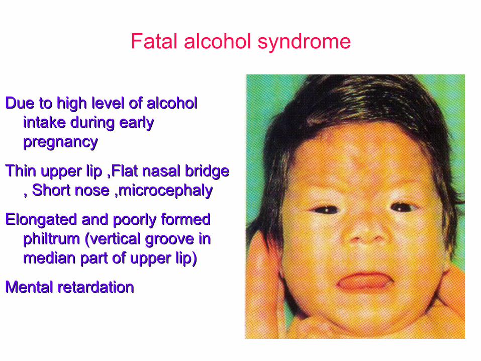
Fatal alcohol syndrome
Due to high level of alcoholDue to high level of alcoholintake during earlyintake during earlypregnancypregnancy
Thin upper lip ,Flat nasal bridgeThin upper lip ,Flat nasal bridge, Short nose ,microcephaly, Short nose ,microcephaly
Elongated and poorly formedElongated and poorly formedphiltrum (vertical groove inphiltrum (vertical groove inmedian part of upper lip)median part of upper lip)
Mental retardationMental retardation

Cigarette Smoking
• Has not been linked to major birth defects– Smoking does contribute to intrauterine
growth retardation & premature delivery– Some evidence that is causes behavioral
disturbances

Drugs
• Thalidomide– Antinauseant & sleeping pill– Found to cause amelia & meromelia
• Total or partial absence of the extremities
– Intestinal atresia– Cardiac abnormalities– Many women had taken thalidomide early in
pregnancy (in Germany in 1961)

Amelia and meromelia
A) Quadruple ameliaA) Quadruple amelia(comlete absence of the(comlete absence of theupper and lower limbs).upper and lower limbs).
B, C) meromelia (partialB, C) meromelia (partialabsence) of the upperabsence) of the upperlimb .limb .
caused by thalidomidecaused by thalidomide..

Various types of meromelia
Partial absence of limbs (disturbancePartial absence of limbs (disturbanceof growth of limb)of growth of limb)
A) absence of hands and most of fore-A) absence of hands and most of fore-armsarms
B) absence of the digitsB) absence of the digits
C) absence of the handC) absence of the hand
D) absence of the fourth and fifthD) absence of the fourth and fifthdigits, syndactyly of the second anddigits, syndactyly of the second andthird digitthird digit
E) absence of third digit, cleft handE) absence of third digit, cleft hand
F) absence of second and third toes,F) absence of second and third toes,syndactyly of fourth and fifth toessyndactyly of fourth and fifth toes

Meromelia
Limb reductionLimb reduction
Caused by thalidomideCaused by thalidomideingested during criticalingested during criticalperiod of limbperiod of limbdevelopment development

Drugs (cont.)
• Aminopterin– Antagonist of Folic Acid– Antineoplastic agent which inhibits
mitosis– Defects
• Anencephaly• Meningocele
• Hydrocephalus
• Cleft lip & palate

Aminopterin-induced abnormalities
Is an anti-metabolite drugIs an anti-metabolite drug
A) meroanencephaly (partialA) meroanencephaly (partialabsence of the brain)absence of the brain)
B) intrauterine growthB) intrauterine growthretardation, large head,retardation, large head,small mandibles,small mandibles,deformed ears,deformed ears,clubhands and clubfootsclubhands and clubfoots

Drugs (cont.)• Anticonvulsants (to treat epilepsy)
– Diphenylhydantoin (phenytoin)• Craniofacial defects
• Nail & digital hypoplasia
• Growth abnormalities• Mental deficiency• The above pattern is know as “fetal hydantoin
syndrome”
– Valproic acid• Neural tube defects• Heart defects• Craniofacial & limb anomalies

Drugs (cont.)
• Trimethadione (syndrome)– Malformed ears– Cleft palate– Cardiac defects– Urogenital anomalies– Skeletal anomalies

Drugs (cont.)• Antipsychotic drugs (major
tranquilizers) – Phenothiazine & lithium
• Suspected teratogenic agents
• Antianxiety drugs (minor tranquilizers)– Meprobamate, chlordiazepoxide,
• Severe anomalies in 11-12% of offspring wheremothers were treated with the above compared to2.6% of controls
– diazepam (valium)• Fourfold in cleft lip with or without cleft
palate

Drugs (cont.)• Anticoagulants
– Warfarin (A.K.A cumadin or cumarol)• Teratogenic• Hypoplasia of nasal cartilage• Chondrodysplasia• Central nervous system defects
– Mental retardation– Atrophy of the optic nerves
• Antihypertensive agents– angiotensin converting enzyme (ACE) inhibitor
• Growth dysfunction, renal dysfunction,oliogohydramnios, fetal death

Drugs (cont)• Propylthiouracil
– Goiter– Mental retardation
• Potassium iodide– Goiter– Mental retardation
• Streptomycin– deafness
• Sulfonamides– kernicterus
• Imipramine(antidepr.)– Limb deformaties
• Tetracyclines– Bone & tooth anomalies
• Amphetamines– Oral clefts– CV abnormalities
• Quinine– Deafness
• Aspirin– Potentially harmful in
large doses

Drugs (cont.)
• Isotretinoin (13-cis-retinoic acid) – Analogue of vitamin A– Drug is prescribed for treatment of cystic acne
& other chronic dermatoses– Highly teratogenic
• Reduced & abnormal ear development• Flat nasal bridge• Cleft palate• Hydrocephaly• Neural tube defects• Heart anomalies

Hormones• Androgenic Agents
– Synthetic progestins were used frequently to preventabortion
• Ethisterone & norethisterone– Have considerable androgenic activity
» Masculinization of female genitalia
• Diethylstilbesterol– Commonly used in the 1940’s & 1950’s to prevent
abortion; in 1971 determined that DES causedincreased incidence of vaginal & cervical cancer inwomen who had been exposed to DES in utero
– In addition high % suffered from reproductivedysfunction
• Oral Contraceptives– Low teratogenic potential, discontinue if pregnancy
suspected• Cortisone-cleft palate in mice (not humans)

Infectious Agents• Rubella (German Measles)
– Malformations of the eye• Cataract (6th week) • Microphthalmia
– Malformations of the ear (9th week)• Congenital deafness
– Due to destruction of cochlea
– Malformations of the heart (5th -10th week)• Patent ductus arteriosis• Atrial septal defects
• Ventricular septal defects

Infectious Agents (cont.)• Rubella (German measles)
– May be responsible for some brain abnormalities• Mental retardation
– Intrauterine growth retardation– Myocardial damage– Vascular abnormalites– Incidence
• 47%- during 1st four weeks• 22% - 5th – 8th weeks• 13% - 9th – 16th week

Infectious Agents (cont.)• Rubella (cont.)
– Lab tests permit detection of virus– Antibody levels can be determined– In one study 85 % of women tested were
immune (n = 600)– Virus infects fetus via the placenta
• Infection of the child may persist after birth for anumber of years
– Infection can be transmitted to hospital personnel
– Vaccines are considered safe & effective
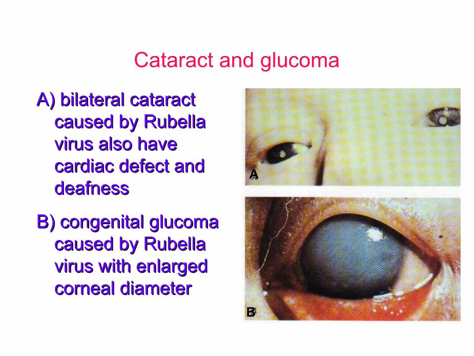
Cataract and glucoma
A) bilateral cataractA) bilateral cataractcaused by Rubellacaused by Rubellavirus also havevirus also havecardiac defect andcardiac defect anddeafnessdeafness
B) congenital glucomaB) congenital glucomacaused by Rubellacaused by Rubellavirus with enlargedvirus with enlargedcorneal diametercorneal diameter
AA
BB

Infectious Agents (cont.)• Cytomegalovirus
– Disease is often fatal early on– Malformations
• Microcephaly
– Cerebral calcifications– Blindness
• Chorioretinitis
– Kernicterus (a form of jaundice)– multiple petechiae of skin– Hepatosplenomegaly– Mother asymptomatic

Infectious Agents (cont.)• Herpes Simplex Virus
– Intrauterine infection of fetus occasionally occurs– Usually infection is transmitted close to time of
delivery– Abnormalities (rare)
• Microcephaly• Microphthalmos• Retinal dysplasia• Hepatosplenomegaly• Mental retardation
– Usually child infected by mother at birth• Inflammatory reactions during first few weeks

Infectious Agents (cont.)• Varicella (chickenpox)
– Congenital anomalies • 20% incidence following infection in 1st trimester• Limb hypoplasia• Mental retardation• Muscle atrophy
• HIV/AIDS– Microcephaly– Growth retardation– Abnormal facies (expression or appearance of the
face)
Infectious Agents (cont.)• Toxoplamosis
– Protozoa parasite (Toxoplama gondii)• Sources
– Poorly cooked meat– Domestic animals (cats)– Contaminated soil with feces
• Syphilis– Congenital deafness– Mental retardation– Diffuse fibrosis of organs (eg. liver & lungs)
• In general most infections are pyrogenic– Hyperthemia can be teratogenic
• Fever• Hot tubs & Saunas

Peds 141
TORCHS Infections• Toxoplasmosis
– Rash, seizures, microcephaly/microphthalmia
• Rubella– Heart defects, deafness, neurologic abnormalities
• Cytomegalovirus– IUGR, purpura, chorioretinitis, microcephaly
• Herpes simplex virus– Disseminated or localized HSV (CNS, skin, eye)
• Syphilis– ”Snuffles,” rash of trunk, palm and soles

Environmental factors
• Radiation
• Hypoxia

Radiation• Teratogenic effect of ionizing radiation well
established– Microcephaly– Skull defects– Spina bifida– Blindness cleft palate– Extremity defects
• Direct effects on fetus or indirect effects ongerm cells
• May effect succeeding generations• Avoid X-raying pregnant women

Radiation• Studies of offspring of Japanese women
who were pregnant at the time of theatomic bomb explosions over Hiroshima &Nagasaki who survived the blast– 28% aborted– 25% gave birth to children who did not survive
their first year– 25% of the surviving children had
abnormalities of CNS• e.g. Microcephaly & mental retardation

Hypoxia
• Associated with congenital malformationsin a great variety of experimental animals– In humans ???
• Maybe smaller babies e.g. offspring at high altitude

Environmental Chemicals
• Mercury– Fish, seed corn sprayed with mercury
containing fungicide• Multiple neurological symptoms
• Lead abortions– Growth retardation– Neurological disorders
Prevention of birth defects
• Good prenatal care• Iodine supplementation eliminates mental
retardation & bone deformities– Prevent cretinism
• Folate/Folic Acid supplementation incidence of neural tube defects
• Avoidance of alcohol & other drugsduring all stages of pregnancy incidence of birth defects

• REVIEW IN YOUR EMBRYOLOGY TEXTSTHE TOPIC TERATOLOGY
• Note the maternal illness,drug abuse,----chemical agents, hormones,medications, infectiousagents,malnutrition/nutritionaldeficiencies that cause congenitalabnormalities.