1,4-dhp-lipid parameters and rod like micellae · journal of biophysical chemistry. 1,4-dhp-lipid...
TRANSCRIPT

Vol.2, No.4, 386-394 (2011)doi:10.4236/jbpc.2011.24044
Copyright © 2011 SciRes. Openly accessible at http://www.scirp.org/journal/JBPC/
Journal of Biophysical Chemistry
1,4-DHP-lipid parameters and rod like micellae
Inta Liepina1*, Cezary Czaplewski2, Adam Liwo2, Gunars Duburs1
1Latvian Institute of Organic Synthesis, Riga, Latvia; 2Faculty of Chemistry, University of Gdansk, Gdansk, Poland; *Corresponding Author: [email protected]
Received 6 July 2011; revised 14 August 2011; accepted 26 August 2011.
ABSTRACT
The cationic amphiphilic lipid type compound 1,1’-{[3,5-bis(dodecyloxycarbonyl)-4-phenyl-1,4- dihydropyridin-2,6-diyl]dimethylene} bispyridini- um dibromide (1,4-DHP lipid) (charge +2), is a gene transfection agent. The electronic struc- ture of 1,4-DHP lipid molecule was investigated by ab initio quantum mechanics, the charges were derived, and the supramolecular structure formed by 1,4-DHP lipid molecules in water was investigated by means of molecular dynamics simulation, f99 force field, version AMBER 8.0. During the molecular dynamics simulations of 10 ns of MD 1,4-DHP lipid formed a worm-like mi- cellae, which was preserved during the time course of 300 ns of MD simulations. Result was confirmed with the electron microscopy show-ing extended, rod-like structures as one of the 1,4-DHP lipid supramolecular structures, which are expected to be formed at high 1,4-DHP lipid concentrations.
Keywords: Lipid; Gene Transfection; Rod-Like Structure; Worm-Like Micellae; Tubular Micellae; Molecular Dynamics
1. INTRODUCTION
New biotechnological drugs, such as DNA, RNA and proteins, require the nanostructure delivery technologies which would pack a DNA or a protein, move through the membrane and release macromolecules inside the cell [1, 2]. Delivered in such a way, a drug targeted at tumors, acts directly in the proper place, and the toxic effect to the whole organism is substantially reduced. For such nanomedicine approach new, different drug delivery agents are required.
DNA delivery into the cell nucleus can be performed by viral carriers, where the guest-DNA is inserted into a virus, or by cationic polymers, cationic peptide or cati- onic lipid carriers that cap the DNA with the help of elec-
trostatic interactions. Synthetic cationic lipids [3] be- come increasingly popular as gene transfection agents due to their minimal toxicity, absence of immunological problems, possible large scale production. A large num- ber of lipids with variations in the hydrophilic and hy- drophobic regions were generated. Unfortunately, they still have less efficiency when compared with viral vec- tors. Recently, we synthesized a novel group of gene transfection agents comprising partially hydrogenated cyclic heterocyclic 1,4-dihydropyridine moiety as active linker. 1,4-DHP derivatives are well-known antihyper-tensive agents, cardiovascular drugs [4], they have pepti- domimetic properties, and their structure has been inves- tigated in our earlier studies [5]. Appropriately decorated with substituents, they may possess bioprotective proper- ties [6]. The cationic amphiphilic lipid type compound 1,1’-{[3,5-bis(dodecyloxycarbonyl)-4-phenyl-1,4-dihydropyridin-2,6-diyl]dimethylene} bispyridinium dibromide (1,4-DHP lipid, charge q = +2) (Figure 1) has gene trans-fection activity [7,8]. This compound is more active than DOTAP and PEI 25 (liposomal and polymeric gene transfection agents). The sharp increase of the 1,4-DHP cycle N-H acidity (up to pKa ≈ 7 - 8) is due to 2- and 6-pyridiniomethyl groups and conjugated alkoxycar-bonyl substituents in positions 3 and 5 of the 1,4-DHP cycle. N-H acidity is the basis for its buffering activity in this novel type of gene transfection agents.
The electronic structure of 1,4-DHP lipid molecule was investigated by ab initio quantum mechanics, and the supramolecular structure formed by 1,4-DHP lipid molecules was investigated by means of molecular dy- namics simulation.
2. METHODS
1,4-DHP lipid molecule was minimized by Sybyl, with the Tripos force field in vacuo [9] (Figure 1(a)). As it was expected that the fatty acid tails must be not in the fully extended state (Figure 1(a)) in water or lipid bilayer, but close to each other (Figures 1(b)-(d)), the struc- ture with bent fatty acid tails was chosen as the starting structure for lipid supramolecular structure calculations

I. Liepina et al. / Journal of Biophysical Chemistry 2 (2011) 386-394
Copyright © 2011 SciRes. Openly accessible at http://www.scirp.org/journal/JBPC/
387
(a) (b) (c) (d)
Figure 1. 1,4-DHP lipid molecule in a space fill and in the stick models: (a) the low energy conformation in vacuo; (b) starting structure for lipid bilayer, front view; (c) starting structure for lipid bilayer, side view; (d) starting structure for lipid bilayer, top view. Minimised by Sybyl, Tripos force field [9].
and also as the starting structure for ab initio quantum mechanics calculations.
The 1,4-DHP lipid structure was calculated by Re- stricted Hartree-Fock (RHF) ab initio quantum mecha- nics with the 6-31G* basis set, by using the program GAMESS [10-13], to obtain the charges for molecular dynamics using the RESP algorithm (molecular electro- static potential based method using charge restrains for determining atom-centered charges).
72 molecules of 1,4-DHP-lipid initially were put in a periodic lipid bilayer-water box, with 10 Å water over the solute and with a small amount of excessive water on the lipid edges to ensure the mobility of lipid molecules (Figures 2(a)-(b)). There were 72 DHP-lipid molecules, 144 counterions of chlorine ions and 4401 water mole- cules in the system DHP-lipid-water box. The total num- ber of atoms in the system is 22491.
The 1,4-DHP-lipid-water box were subjected to mo- lecular dynamics, (AMBER 8.0, f99 force field, version 8.0 [14-16], NTP protocol (constant number of particles, con-stant temperature, constant pressure, the derived parameters for 1,4-DHP-lipid are presented in Supplements Table S2).
The temperature was increased gradually from T = 10 K by step of 10 degrees till 300 K. Calculations were performed for 326212 ps.
Results were proceeded with VMD [17], visualization were made with RASMOL [18].
3. RESULTS AND DISCUSSION
1,4-DHP lipid were calculated by Restricted Hartree-
(a) (b)
Figure 2. 1,4-DHP lipid initial state of molecular dyna- mics (a) front vie; (b) side view.
Fock (RHF) ab initio quantum mechanics, 6-31G* bases set, and the quantum minimized structure are depicted in Figure 3. The quantum mechanics calculations show a shallow boat conformation for 1,4-DHP lipid dihydro- pyridine moiety (Figure 3(b)). The distance between pyridinium nitrogens is 5.9 Å, and the distance between pyridinium carbon atoms in the fourth position is 8 Å, thus the pyridinium rings are upon narrow angle towards each other (Figure 3(a)). The distances between oxygen of the carbonyl group and pyridinium hydrogen are of 2.17 Å - 2.18 Å, and the distances between oxygen of the carbonyl group and the hydrogen atoms of the methylene is of 2.16 and 2.18 Å denoting that the oxygen of the car-bonyl group simultaneously interacts with the both hy-drogens (Figure 10(a)). The geometric parameters: bond
(a) (b)
Figure 3. Ab intito minimized 1,4-DHP-lipid structure— (a) front view; (b) side view. The dihydropyridine ring is in a boat conformation.

I. Liepina et al. / Journal of Biophysical Chemistry 2 (2011) 386-394
Copyright © 2011 SciRes. Openly accessible at http://www.scirp.org/journal/JBPC/
388
length and valence angles are shown in Figure S1. The lipid head has a +2 charge. The molecular electro-
static potential surfaces were calculated and used to de- rive the atom point charges for molecular dynamics with RESP algorithm. The representative charges together with derived Amber 8.0 atom types are shown in Figure 4. The full set of charges and atom types for molecular dy- namics are presented in Table S1, the corresponding atom numbering is presented in Figure S2.
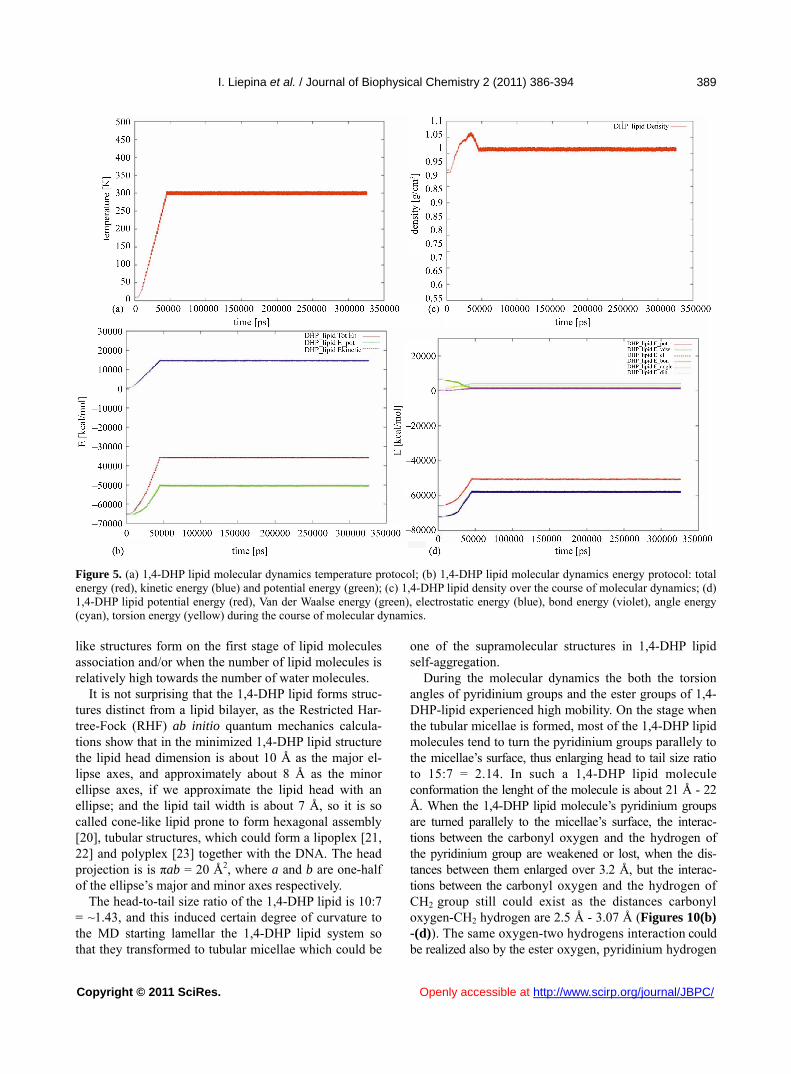
Molecular dynamics simulations were started from the temperature T = 10 K and risen gradually till 300 K. The temperature changes are represented of Figure 5(a), showing the stepwise rising of the temperature from T = 10 K till T = 300 K, by step of 10 degrees. The total en- ergy with its components of kinetic and potential energy is represented of Figure 5(b), showing that system be- come stable at 49 ns.
Also the density becomes stable during 49 ns, and fur- ther is oscillating around 1.01 g/cm3 (Figure 5(c)).
On Figures 6(a)-(b), representing the MD snapshot at 14725 ps, T = 60 K, we observe that the system still keeps the initial bilayer organization, (6(a) atom type co- loring to represent the positions of the charged head groups), but the fatty acid tails of one bilayer squeeze into another bilayer (6(b), molecules are represented in different colors to show the fatty acid tail movement).
On Figures 6(c)-(d) is represented the MD snapshot at the temperature 220 K, 35696 ps of MD run, Figure 6(c) atom type coloring, shows that system still keeps the initial bilayer organization, on Figure 6(d) the different molecule coloring shows that the fatty acid tails of one bilayer squeeze into another bilayer.
After 35 ns of the MD simulation few lipid molecules turned with their charged heads to the side of the lipid bi- layer. It is represented on Figures 6(e)-(f), showing 1,4- DHP-lipid at the temperature 300 K 48849 ps of MD run. At this stage of MD run the lipid heads have turned to the sides of the periodic box (Figure 6(e)) and the lipid molecules continue to squeeze from one lipid layer into the other one (Figure 6(f)). After 100 ns of MD simula- tions a profound worm-like micelle structure began to form. Figures 6(g)-(h) represents a snapshot at 110673 ps of MD run, T = 300 K, and on Figure 6(g) the system, colored by atom type, shows that the 1,4-DHP-lipid charged heads have turned to the sides of the periodic box, and. at this stage of MD run the worm-like micellae has formed. Figure 6(h), where the separate lipid mole- cules are colored differently shows that molecules from one lipid layer squeeze into the other one; the lipid layers are interwinded.
On Figure 7 is shown the snapshot of 1,4-DHP-lipid- water box at the t = 159582 ps, T = 300 K. The worm- like micelle structure is stable and becomes more perfect
Figure 4. Charge distribution calculated for DHP-lipid molecule and the derived AMBER atom types CA, CM, CT, NA, O, OS, HA, HC, H1, H.
during the course of simulation of 300 ns (Figure 8).
The graphics on Figure 5(d) show that in the supra- molecular organization the DHP lipid molecules are in relaxed state, as the bond energy, angle energy and tor- sion energy increments to the potential energy are small, close to zero, in comparison with electrostatic energy, which is clearly dominating. During the first 50 ns of MD simulations only E-vdw is reduced because of better packing of molecules, while all other components of E_ pot are growing because the system is heated.
The size of the periodic box has changed from the di- mensions of x = 70 Å, y = 55 Å, z = 103 Å at the start of molecular dynamics, to x = 102 Å, y = 46 Å, z = 48 Å at 318039 ps (Figure 9). The worm-like micellae axes—the longer dimension, is in the x-axes direction, but y-axes and z-axes dimensions are similar, which is explainable with the top view of the worm-like micellae, having ap- proximately the same diameter in all directions of the yz-plane (Figure 8(b)).
The results of MD simulation were confirmed by elec- tron microscopy, showing the interwinding worm-like structure [19]. The diameter of thinner worm-like struc- tures is around 4 - 5 nm, which corresponds to approxi- mate estimation of the calculated worm-like structure be- ing of 3.8 - 4.5 nm. It could be expected that such worm-
I. Liepina et al. / Journal of Biophysical Chemistry 2 (2011) 386-394
Copyright © 2011 SciRes. http://www.scirp.org/journal/JBPC/
389
Figure 5. (a) 1,4-DHP lipid molecular dynamics temperature protocol; (b) 1,4-DHP lipid molecular dynamics energy protocol: total energy (red), kinetic energy (blue) and potential energy (green); (c) 1,4-DHP lipid density over the course of molecular dynamics; (d) 1,4-DHP lipid potential energy (red), Van der Waalse energy (green), electrostatic energy (blue), bond energy (violet), angle energy (cyan), torsion energy (yellow) during the course of molecular dynamics. like structures form on the first stage of lipid molecules association and/or when the number of lipid molecules is relatively high towards the number of water molecules.
Openly accessible at
It is not surprising that the 1,4-DHP lipid forms struc- tures distinct from a lipid bilayer, as the Restricted Har- tree-Fock (RHF) ab initio quantum mechanics calcula- tions show that in the minimized 1,4-DHP lipid structure the lipid head dimension is about 10 Å as the major el- lipse axes, and approximately about 8 Å as the minor ellipse axes, if we approximate the lipid head with an ellipse; and the lipid tail width is about 7 Å, so it is so called cone-like lipid prone to form hexagonal assembly [20], tubular structures, which could form a lipoplex [21, 22] and polyplex [23] together with the DNA. The head projection is is πab = 20 Å2, where a and b are one-half of the ellipse’s major and minor axes respectively.
The head-to-tail size ratio of the 1,4-DHP lipid is 10:7 = ~1.43, and this induced certain degree of curvature to the MD starting lamellar the 1,4-DHP lipid system so that they transformed to tubular micellae which could be
one of the supramolecular structures in 1,4-DHP lipid self-aggregation.
During the molecular dynamics the both the torsion angles of pyridinium groups and the ester groups of 1,4- DHP-lipid experienced high mobility. On the stage when the tubular micellae is formed, most of the 1,4-DHP lipid molecules tend to turn the pyridinium groups parallely to the micellae’s surface, thus enlarging head to tail size ratio to 15:7 = 2.14. In such a 1,4-DHP lipid molecule conformation the lenght of the molecule is about 21 Å - 22 Å. When the 1,4-DHP lipid molecule’s pyridinium groups are turned parallely to the micellae’s surface, the interac-tions between the carbonyl oxygen and the hydrogen of the pyridinium group are weakened or lost, when the dis-tances between them enlarged over 3.2 Å, but the interac-tions between the carbonyl oxygen and the hydrogen of CH2 group still could exist as the distances carbonyl oxygen-CH2 hydrogen are 2.5 Å - 3.07 Å (Figures 10(b) -(d)). The same oxygen-two hydrogens interaction could be realized also by the ester oxygen, pyridinium hydrogen

I. Liepina et al. / Journal of Biophysical Chemistry 2 (2011) 386-394
Copyright © 2011 SciRes. Openly accessible at http://www.scirp.org/journal/JBPC/
390
Figure 6. (a) Snapshot of the 1,4-DHP-lipid-water box at the temperature 60 K, 14725 ps of MD run, space fill representa-tion, colored by atom type; (b) Snapshot of the 1,4-DHP-lipid – water box at the temperature 60 K, 14725 ps of MD run, space fill representation, lipid molecules are colored differently to show that molecules from one lipid layer squeeze into the other one; (c)-(d) snapshot at the temperature 220 K, 35696 ps of MD run; (e)-(f) snapshot at the temperature 300 K 48849 ps of MD run. At this stage of MD run the lipid heads have turned to the sides of the periodic box; (g)-(h) snapshot at the tempera-ture 300 K, 110673 ps of MD run. At this stage of MD run the worm-like micellae has formed.
Figure 7. The snapshot of 1,4-DHP-lipidwater box at the t = 159582 ps of MD run.
(a) (b)
Figure 8. 1,4-DHP lipid worm-like micellae is stable during the course of MD simulations. Snapshot at the 300 K, t = 301 ns. (a) Side view; (b) top view.
Figure 9. The course of 1,4-DHP lipid periodic box dimensions during the molecular dynamics: x—red, y—green, z—blue. The worm-like micellae axes corresponding to the longer di-mension (102 Å), is in the x-axes direction, but the y-axes and z-axes dimensions are similar (46 Å - 48 Å) corresponding to the same diameter of worm-like micellae in the both directions.
(a) (b)
(c) (d)
Figure 10. 1,4-DHP lipid molecule ab intio calculations mini-mization (a) and the snapshots of three 1,4-DHP lipid mole-cules during at 300 ns of MD (b)-(d).

I. Liepina et al. / Journal of Biophysical Chemistry 2 (2011) 386-394
Copyright © 2011 SciRes. http://www.scirp.org/journal/JBPC/
391
and CH2 group hydrogen (Figures 10(b)-(d)).
Openly accessible at
3. CONCLUSIONS
We have performed the Restricted Hartree-Fock (RHF) ab initio quantum mechanics calculations on a 1,4-DHP lipid molecule, as well as 300 ns MD of 72 1,4-DHP li- pid molecules starting from a lipid bilayer structure. Re- sults show that the gene transfection agent 1,4-DHP lipid in natural conditions does not form a lipid bilayer, but one of its structures is a worm-like, rod-like micellae, which are expected to be formed at high 1,4-DHP lipid concentrations. We could expect that such the micellaes are capable to form a lipoplex for the DNA transfection. During the molecular dynamics 1,4-DHP lipid molecules tend to turn the piridinium groups parallely to the micel- lae’s surface.
4. ACKNOWLEDGMENTS
This work was supported by ESF project 2009/0197/1DP/1.1.1.2.0
/09/APIA/VIAA/014, Calculations were performed on computers of the
Gdansk Academic Computer Center TASK.
REFERENCES
[1] Hughes, G.A. (2005) Nanostructure-mediated drug deli- very. Nanomedicine: Nanotechnology, Biology, and Me- dicine, 1, 22-30. doi:10.1016/j.nano.2004.11.009
[2] Basarkar, A. and Singh, J. (2007) Nanoparticulate sys- tems for polynucleotide delivery. International Journal of Nanomedicine, 2, 353-360.
[3] Ma, B.-C., Zhang, S.-B., Jiang, H.-M., Zhao, B.-D. and Lv, H.-T. (2007) Lipoplex morphologies and their influ-ences on transfection efficiency in gene delivery. Journal of Controlled Release, 123, 184-194. doi:10.1016/j.jconrel.2007.08.022
[4] Triggle, D.J. (2006) L-type calcium channels. In: Rampe, D. and Zheng, W., Eds., Voltagegated Ion Channels as Drug Targets, Wiley-VCH Verlag GmbH & Co. KGaA, Weinheim, 100-122.
[5] Liepina, I., Blanco, M., Duburs, G. and Liwo, A. (1997) Spatial structure of dihydropyridines and similarity of dihydropyridines with some amino acids. Molecular En-geneering, 7, 401-427. doi:10.1023/A:1008272311650
[6] Duburs, G., Vigante, B., Plotniece, A., Krauze, A., Sobo- levs, A., Briede, J., Klusa, V. and Velena, A. (2008) Dihy- dropyridine derivatives as bioprotectors. Chemica OGGI/ Chemistry Today, 26, 68-70.
[7] Hyvonen, Z., Plotniece, A., Reine, I., Chekavichus, B., Duburs, G. and Urtti, A. (2000) Novel cationic amphi-philic 1,4-dihydropyridine derivatives for DNA delivery. Biochimica et Biophysica Acta, 1509, 451-466. doi:10.1016/S0005-2736(00)00327-8
[8] Hyvonen, Z., Ronkko, S., Toppinen, M.-R., Jaaskelainen, I., Plotniece, A. and Urtti, A. (2004) Dioleoylphosphati Dylethanolamini and PEG-lipid conjugates modify DNA delivery mediated by 1,4-dihydropyridine amphiphiles. Journal of Controlled Release, 99, 177-190.
doi:10.1016/j.jconrel.2004.06.019 [9] SYBYL 8.0, Tripos International, 1699 South Hanley Rd.,
St. Louis, Missouri, 63144, USA. [10] Guest, M.F., Bush, I.J., van Dam, H.J.J., Sherwood, P.,
Thomas, J.M.H., van Lenthe, J.H., Havenith, R.W.A. and Kendrick, J. (2005) The GAMESS-UK electronic stru- cture package: Algorithms, developments and applica- tions. Molecular Physics, 103, 719-747. doi:10.1080/00268970512331340592
[11] Zarrabian, S. and Harrison, R.J. (1989) Chemical Physics Letters, 81.
[12] Saunders, V.R. and van Lenthe, J.H. (1983) Molecular Physics, 48, 923. doi:10.1080/00268978300100661
[13] Buenker R.J. (1982) Studies in Physical and Theoretical Chemistry, 21, 17.
[14] Pearlman, D.A., Case, D.A., Caldwell, J.W., Ross, W.S., Cheatham, T.E. III, DeBolt, S., Ferguson, D., Seibel, G. and Kollman, P. (1995) AMBER, a package of computer programs for applying molecular mechanics, normal mode analysis, molecular dynamics and free energy cal-culations to simulate the structural and energetic proper-ties of molecules. Computer Physics Communications, 91, 1-41. doi:10.1016/0010-4655(95)00041-D
[15] Case, D.A., Cheatham, T., Darden, T., Gohlke, H., Luo, R., Merz, K.M. Jr., Onufriev, A., Simmerling, C., Wang, B. and Woods, R. (2005) The Amber biomolecular simu-lation programs. Journal of Computational Chemistry, 26, 1668-1688. doi:10.1002/jcc.20290
[16] Ponder, J.W. and Case, D.A. (2003) Force fields for pro-tein simulations. Advances in Protein Chemistry, 66, 27- 85. doi:10.1016/S0065-3233(03)66002-X
[17] Humphrey, W., Dalke, A. and Schulten, K. (1996) VMD- Visual Molecular Dynamics. Journal of Molecular Gra- phics, 14, 33-38. doi:10.1016/0263-7855(96)00018-5
[18] Sayle, R. and Milner-White, E.J. (1995) RasMol: Bio-molecular graphics for all. Trends in Biochemical Sci-ences (TIBS), 20, 374. doi:10.1016/S0968-0004(00)89080-5
[19] Liepina, I., Czaplewski, C., Ose, V., Danne, R. and Du- burs, G. (2008) 1,4-DHP-lipid forms a tubular micellae. In: Hansman. H. E., Meinke, Jan H., Mohanty, S., Nadler W. and Zimmermann, O., Eds., Proceedings of NIC Workshop from Computational Biophysics to System Bi-ology, Julich, 19-21 May 2008, 305-307.
[20] Gajria, S., Neumann, T. and Tirrell, M. (2011) Self-as- sembly and applications of nucleic acid solid-state films. Wiley Interdisciplinary Reviews: Nanomedicine and Na- nobiotechnology. Early View, Article First Published Online: 2 June 2011. doi:10.1002/WNAN.148
[21] Ma, B., Zhang, S., Jiang, H., Zhao, B. and Lv, H. (2007) Lipoplex morphologies and their influences on trans- fection efficiency in gene delivery. Journal of Controlled Release, 123, 184-194. doi:10.1016/j.jconrel.2007.08.022
[22] Ulrich, A.S. (2002) Biophysical aspects of using lipo- somes as delivery vehicles. Bioscience Reports, 22, 129- 150. doi:10.1023/A:1020178304031
[23] Elouahabi, A. and Ruysschaert, J.-M. (2005) Formation and intracellular trafficking of lipoplexes and polyple- xes. Molecular Therapy, 11, 336-347. doi:10.1016/j.ymthe.2004.12.006

I. Liepina et al. / Journal of Biophysical Chemistry 2 (2011) 386-394
Copyright © 2011 SciRes. Openly accessible at http://www.scirp.org/journal/JBPC/
392
Supplements
Figure S1. Geometric parameters: valence bond length (black), valence angles (red) of 1,4-DHP lipid molecule ab initio calcu-lations.
Figure S2. Atom numeration of 1,4-DHP lipid molecule.

I. Liepina et al. / Journal of Biophysical Chemistry 2 (2011) 386-394
Copyright © 2011 SciRes. Openly accessible at http://www.scirp.org/journal/JBPC/
393
Table S1. Atom charges of DHP-lipid.
1 HA 0.1589 44 HC 0.0237 87 HC –0.0062
2 CA 0.0024 45 H1 0.0820 88 HC –0.0062
3 NA 0.1031 46 H1 0.0820 89 HC –0.0162
4 CT –0.034 47 CT –0.0944 90 HC –0.0162
5 CM 0.2158 48 HC 0.0798 91 HC –0.0095
6 CM –0.077 49 CA 0.0777 92 HC –0.0095
7 CE 0.5528 50 CA –0.0926 93 HC –0.006
8 O –0.4789 51 HA 0.1036 94 HC –0.006
9 OS –0.2964 52 CA –0.2108 95 HC 0.0030
10 CT 0.0373 53 HA 0.1537 96 HC 0.0030
11 CT 0.0107 54 CA –0.0635 97 HC 0.0237
12 CT 0.0398 55 HA 0.1581 98 HC 0.0237
13 CT 0.0193 56 CA –0.2108 99 H1 0.0820
14 CT –0.0005 57 HA 0.1537 100 H1 0.0820
15 CT 0.0609 58 CA –0.0926 101 CM 0.2157
16 CT –0.0002 59 HA 0.1036 102 CT –0.034
17 CT 0.0513 60 CM –0.077 103 NA 0.1031
18 CT –0.001 61 CE 0.5528 104 CA 0.0024
19 CT 0.0246 62 O –0.4789 105 HA 0.1589
20 CT 0.0572 63 OS –0.2964 106 CA –0.1456
21 CT –0.1137 64 CT 0.0373 107 HA 0.1884
22 HC 0.0273 65 CT 0.0107 108 CA 0.0052
23 HC 0.0273 66 CT 0.0398 109 HA 0.1933
24 HC 0.0273 67 CT 0.0193 110 CA –0.1456
25 HC –0.006 68 CT –0.0005 111 HA 0.1884
26 HC –0.006 69 CT 0.0609 112 CA 0.0024
27 HC –0.0056 70 CT –0.0002 113 HA 0.1589
28 HC –0.0056 71 CT 0.0513 114 HC 0.1156
29 HC –0.0114 72 CT –0.001 115 HC 0.1156
30 HC –0.0114 73 CT 0.0246 116 NA –0.7587
31 HC –0.0156 74 CT 0.0572 117 H 0.3826
32 HC –0.0156 75 CT –0.1137 118 HC 0.1156
33 HC –0.0062 76 HC 0.0273 119 HC 0.1156
34 HC –0.0062 77 HC 0.0273 120 CA 0.0024
35 HC –0.0162 78 HC 0.0273 121 HA 0.1589
36 HC –0.0162 79 HC –0.006 122 CA –0.1456
37 HC –0.0095 80 HC –0.006 123 HA 0.1884
38 HC –0.0095 81 HC –0.0056 124 CA 0.0052
39 HC –0.006 82 HC –0.0056 125 HA 0.1933
40 HC –0.006 83 HC –0.0114 126 CA –0.1456
41 HC 0.0030 84 HC –0.0114 127 HA 0.1884
42 HC 0.0030 85 HC –0.0156
43 HC 0.0237 86 HC –0.0156

I. Liepina et al. / Journal of Biophysical Chemistry 2 (2011) 386-394
Copyright © 2011 SciRes. http://www.scirp.org/journal/JBPC/Openly accessible at
394
Table S2. Derived AMBER parameters for 1,4-DHP lipid.
BOND LENGTH NA-CT 337.0 1.475 CM-NA 418.0 1.388 CM-CE 410.0 1.444 TORSION ANGLE CA-NA-CT 70.0 120.00 CA-NA-CA 70.0 120.00 CM-NA-CM 70.0 120.00 CM-NA-H 30.0 118.00 NA-CA-CA 63.0 120.00 NA-CT-CM 63.0 114.00 NA-CT-HC 50.0 109.50 NA-CA-HA 35.0 120.00 CT-CM-NA 70.0 119.70 CM-CM-CE 63.0 120.70 CM-CM-NA 70.0 121.20 CE-CM-CT 70.0 119.70 CM-CE-O 80.0 125.30 CM-CE-OS 80.0 125.30 CM-CT-CA 63.0 114.00 CM-CT-CM 63.0 114.00 DIHEDRAL ANGLE X-NA-CT-X 6 0.00 0.0 2.0 X-CM-NA-X 4 6.00 180.0 2.0 X-CM-CE-X 4 0.00 0.0 2.0 IMPROPER TORSIONS CA-CA-NA-CT 1.1 180. 2.0 CT-NA-CM-CM 1.1 180. 2.0 CE-CM-CM-CT 1.1 180. 2.0 CE-CT-CM-CM 1.1 180. 2.0 CM-NA-CM-CT 1.1 180. 2.0 CT-CA-NA-CA 1.1 180. 2.0