1472-6750-9-82_integrating human stem cell
TRANSCRIPT
8/7/2019 1472-6750-9-82_Integrating human stem cell
http://slidepdf.com/reader/full/1472-6750-9-82integrating-human-stem-cell 1/14
BioMed Central
Page 1 of 14(page number not for citation purposes)
BMC Biotechnology
Open AccesResearch article
Integrating human stem cell expansion and neuronal differentiationin bioreactors
Margarida Serra1,2
, Catarina Brito1,2
, Eunice M Costa1,2
, Marcos FQ Sousa2
and Paula M Alves*1,2
Address: 1Instituto de Tecnologia Química e Biológica, Universidade Nova de Lisboa, Av. da República, 2780-157 Oeiras, Portugal and 2IBET, Apartado 12, 2781-901 Oeiras, Portugal
Email: Margarida Serra - [email protected]; Catarina Brito - [email protected]; Eunice M Costa - [email protected];Marcos FQ Sousa - [email protected]; Paula M Alves* - [email protected]
* Corresponding author
Abstract
Background: Human stem cells are cellular resources with outstanding potential for cell therapy.
However, for the fulfillment of this application, major challenges remain to be met. Of paramount
importance is the development of robust systems for in vitro stem cell expansion and differentiation.
In this work, we successfully developed an efficient scalable bioprocess for the fast production of
human neurons.Results: The expansion of undifferentiated human embryonal carcinoma stem cells (NTera2/cl.D1
cell line) as 3D-aggregates was firstly optimized in spinner vessel. The media exchange operation
mode with an inoculum concentration of 4 × 10⁵ cell/mL was the most efficient strategy tested,
with a 4.6-fold increase in cell concentration achieved in 5 days. These results were validated in a
bioreactor where similar profile and metabolic performance were obtained. Furthermore,
characterization of the expanded population by immunofluorescence microscopy and flow
cytometry showed that NT2 cells maintained their stem cell characteristics along the bioreactor
culture time.
Finally, the neuronal differentiation step was integrated in the bioreactor process, by addition of
retinoic acid when cells were in the middle of the exponential phase. Neurosphere composition
was monitored and neuronal differentiation efficiency evaluated along the culture time. The results
show that, for bioreactor cultures, we were able to increase significantly the neuronal
differentiation efficiency by 10-fold while reducing drastically, by 30%, the time required for the
differentiation process.
Conclusion: The culture systems developed herein are robust and represent one-step-forward
towards the development of integrated bioprocesses, bridging stem cell expansion and
differentiation in fully controlled bioreactors.
Published: 22 September 2009
BMC Biotechnology 2009, 9:82 doi:10.1186/1472-6750-9-82
Received: 17 April 2009Accepted: 22 September 2009
This article is available from: http://www.biomedcentral.com/1472-6750/9/82
© 2009 Serra et al; licensee BioMed Central Ltd.This is an Open Access article distributed under the terms of the Creative Commons Attribution License (http://creativecommons.org/licenses/by/2.0),which permits unrestricted use, distribution, and reproduction in any medium, provided the original work is properly cited.

8/7/2019 1472-6750-9-82_Integrating human stem cell
http://slidepdf.com/reader/full/1472-6750-9-82integrating-human-stem-cell 2/14
BMC Biotechnology 2009, 9:82 http://www.biomedcentral.com/1472-6750/9/82
Page 2 of 14(page number not for citation purposes)
BackgroundMany neurodegenerative disorders, such as Parkinson'sdisease, are caused by the impairment or death of neuronsin the central nervous system [1]. In the future, it is hopedthat large numbers of stem cell-derived neurons will be
produced in culture with the purpose of being used inclinical applications [2]. Hampering the faster implemen-tation of the ambitious stem cell therapy technology,there is still the need of efficient, robust and scalable bio-processes for cell expansion and/or differentiation in vitro.
During the last five years, substantial progress has beenmade towards this goal [3,4]. Stirred suspension systemshave been pioneered, by others and ourselves, as a prom-ising in vitro system for stem cell expansion [5,6], embry-oid body cultivation [7,8] and stem cell differentiationinto specific cell types [9]. These systems offer attractiveadvantages of scalability and relative simplicity; stirring
provides a more homogenous culture environment andallows the measurement and control of extrinsic factorssuch as nutrient and cytokine concentration, pH and dis-solved oxygen (pO2) [10].
Aiming to improve the yields of specific stem cell stages,several culture parameters have been optimized, includ-ing the agitation rate, cell inoculum concentration andmedium composition [3,4,11], and different culturing approaches have been developed such as the use of micro-carrier supports [5] and cell encapsulation [11]. Perfusionand frequent feeding operation modes have been shownto increase the expansion of mesenchymal stem cells [11],
embryonic stem cells [12,13] and mammary epithelialstem cells [14], without compromising their stem cell per-formance.
Computer-controlled bioreactors are particular advanta-geous for process development by allowing the onlinemonitoring and control of specific culture parameters(temperature, pH and pO2), ensuring a fully controlledenvironment for stem cell cultivation. Oxygen-controlledbioreactors have been used for culture of mouse andhuman ESC-derived cardiomyocytes [7,15]. Gilbertson et al [16] were the first group to use controlled conditionsfor neural precursor cell culture as aggregates; the authors
report the successful expansion of mouse neural stem cellsin 500 mL bioreactors (temperature, pH and pO2 control)
while retaining the cell multilineage potential [16]. Morerecently, this system was applied to the culture of humanneural precursor cells [17]. The expansion of varioushuman stem cell types in bioreactors under defined andcontrolled conditions remains to be addressed. Futurechallenges also include the combination of expansion anddirected differentiation steps in an integrated bioprocessthat will ultimately result in scale-up of well differentiatedcells to clinically relevant numbers.
Within this context, the present work focused the develop-ment of a reproducible scalable system for the productionof human neurons derived from expanded and differenti-ated stem cells. The human embryonal carcinoma cell lineNTera-2/cl.D1 (NT2) was the cellular system used because
it is a valuable model for both undifferentiated humanembryonic stem cells (hESCs) [18] and human neuronaldifferentiation in vitro [19]. In addition, the neuronsderived from this cell line have been successfully used intransplantation studies in several mouse models and inhuman stroke patients [20], providing also promising material for cell therapy investigations in central nervoussystem.
Herein, undifferentiated NT2 cells were cultivated as 3D-aggregates in controlled stirred suspension conditions. Inorder to improve the yields of stem cells, two parameters
were studied: (i) the inoculum concentration, as it has
been shown to be critical in enhancing cell aggregationand culture profile [6], and (ii) the culture operationmode, since it has been demonstrated that the feeding strategy affects cell metabolism and consequently couldimprove cell culture performance [11,15,21]. At the end,the expansion of undifferentiated NT2 cells, followed by directed neuronal differentiation were integrated instirred bioreactors with temperature, pH and pO2 control,in an effort to develop a promising model system for theproduction of human stem cell derivatives.
Results With the goal of developing a robust and scalable system
for NT2 neuronal differentiation, both expansion and dif-ferentiation steps were integrated in a fully controlled bio-reactor process. Firstly, different strategies for expansionof undifferentiated NT2 cells as 3-D aggregates werescreened in stirred spinner vessels; two parameters werestudied (i) the inoculum concentration and (ii) the cul-ture operation mode, i.e., medium replenishing strategies.Having the expansion of pluripotent NT2 cells optimizedand well characterized, the neuronal differentiation strat-egy previously developed by our group [9], was integratedand the overall bioprocess combined in the bioreactor.Figure 1 summarizes the experimental outline used for expansion and differentiation processes.
Effect of inoculum concentration in NT2 expansion
Three different cell inoculum concentrations were testedin batch culture mode, using 125 mL spinners: 0.4, 1 and4 × 105 cell/mL (SP-0.4B, SP-1B and SP-4B, respectively).
During the first 24 h of SP-1B and SP-4B cultures, cellsassembled into small 3D-aggregates (Figure 2 A) ranging from 40 to 65 μm. After this period, cells started to divideand aggregate size increased up to 150 μm. The growthcurve and the calculated apparent growth rates are shown

8/7/2019 1472-6750-9-82_Integrating human stem cell
http://slidepdf.com/reader/full/1472-6750-9-82integrating-human-stem-cell 3/14
BMC Biotechnology 2009, 9:82 http://www.biomedcentral.com/1472-6750/9/82
Page 3 of 14(page number not for citation purposes)
Figure 1 (see legend on next page)

8/7/2019 1472-6750-9-82_Integrating human stem cell
http://slidepdf.com/reader/full/1472-6750-9-82integrating-human-stem-cell 4/14
BMC Biotechnology 2009, 9:82 http://www.biomedcentral.com/1472-6750/9/82
Page 4 of 14(page number not for citation purposes)
in Figure 2B and Table 1, respectively. SP-1B exhibited ahigh apparent growth rate (0.51 ± 0.01 day -1) and thehighest FI in cell concentration (7.14 ± 0.86). Neverthe-less, maximum cell density 6.64 (± 1.57) × 105 cell/mL
was only reached 6 days after inoculation, whereas in SP-4B, a maximum of 8.48 (± 0.11) × 105 cell/mL wasachieved at day 3. From day 4 onwards of SP-4B culture,cells started to detach from the aggregates (Figure 2 A),resulting in cell death (data not shown). Similar behavior
was observed for SP-1B culture upon day 7 of cultivation.
Concerning the SP-0.4 culture, cell aggregates were rareand small throughout cultivation time (Figure 2 A). Infact, no effective cell growth was observed (Figure 2B) andcell viability was low (data not shown).
Aiming to develop an efficient bioprocess for the fast pro-
duction of human neurons, cell number and culture time were the parameters preferentially used to select the best strategy. For SP-4B, the time needed to achieve X max was 2times lower than for SP-1B, reaching similar X max values(Table 1). Based on these results, SP-4B was chosen to be
further optimized and integrated with the neuronal differ-entiation step.
Impact of operation mode in NT2 cell expansion
In all batch cultures there was a rapid decrease in cell den-sity after the culture reached its maximum concentration
value (Figure 2 A). Although no complete depletion of nei-ther glucose nor glutamine was observed (Figure 3 A, C),this profile could be correlated to the exhaustion of other essential nutrients and/or the progressive accumulation of toxic metabolic waste products such as lactate and ammo-nia (Figure 3B, D). In SP-4B, by the 4th day of cultivation,the lactate and ammonia concentrations were already 21.9 mM and 3.1 mM, respectively (Figure 3B, D). In SP-1B, these values were also high at day 7 of culture (27.2mM and 4.2 mM for lactate and ammonia concentration,respectively).
Aiming at prolonging the exponential growth phase andimprove the cell expansion, two additional operationmodes were tested. The first strategy consisted of a glucosefed-batch operation mode (SP-4FB). In this strategy, cul-ture was initiated at low concentration of glucose (1.4mM) and the feeding was performed twice a day assuring the maintenance of low levels of glucose throughout cul-tivation time (see Methods section). The second strategy (SP-4ME) was designed to simulate a perfusion system, in
which cells are kept in culture and the media is renovatedregularly. This was achieved by performing a daily partialmedia exchange (50%) from the 3rd cultivation day
onwards, as this time point corresponded to the growthpeak in the batch culture (Figure 2B, SP-4B).
For SP-4ME and SP-4FB cultures, the exponential growthphase was extended until day 5 (Figure 3F), with a signif-icant increase in X max , when compared to SP-4B (Table 1).
These differences are also reflected in cell metabolism, asshown by the nutrient consumption and metabolite pro-duction profiles (Figure 3E). The SP-4FB culture presentedthe lowest specific rates of glucose consumption and lac-tate production. The lower accumulation of lactate (16.5
Experimental outline for NT2 cell sampling and characterization during expansion (A) and differentiation (B) in fully controlledbioreactorsFigure 1 (see previous page)Experimental outline for NT2 cell sampling and characterization during expansion (A) and differentiation (B)in fully controlled bioreactors. (A) In expansion runs, cells were harvested from days 0 (inoculum), 3 and 6 and immedi-ately characterized by flow cytometry. Harvested cells were plated on glass coverslips and processed for immunofluorescencemicroscopy analysis after 2 days or plated in tissue culture flasks for induction of neuronal differentiation. For this, cultureswere treated with retinoic acid (RA) for 5 weeks, splitted and further cultured in mitosis inhibitory (MI) conditions. After 12days in MI, the neurons were harvested, identified by immunofluorescence microscopy using neuronal markers and neuronaldifferentiation efficiencies were calculated. (B) In differentiation runs, the addition of RA was initiated at day 3 of bioreactorculture and prolonged for 3 weeks. Neurospheres were harvested at day 9, 16 and 23. The latest were analyzed by cryosectionimmunofluorescence microscopy. All neurosphere harvested were plated in static culture flasks and cultured in MI conditions.After 3 days, cultures were characterized by immunofluorescence microscopy and after 7 days and neuronal differentiation effi-ciencies were calculated.
Table 1: Growth kinetics of NT2 cell expansion as 3D-aggregates
using different culture strategies.
Strategy μ (day-1) FI Xmax (×105 cell/mL)
SP-0.4B n. a. n. a. 0.63 ± 0.11 *
SP-1B 0.51 ± 0.01 7.14 ± 0.86 * 6.64 ± 1.57
SP-4B 0.39 ± 0.02 2.12 ± 0.03 8.48 ± 0.11
SP-4FB 0.52 ± 0.06 4.30 ± 0.33 * 17.19 ± 1.30 *
SP-4ME 0.41 ± 0.06 4.56 ± 0.04 * 18.25 ± 0.18 *
BR-4ME 0.37 ± 0.03 4.10 ± 0.41 16.25 ± 0.16
Apparent growth rate (μ), fold increase (FI) and maximum cellconcentration values (Xmax) of NT2 cells cultured in spinner vessel(SP) or in bioreactor (BR); with inoculum densities of 0.4 × 10⁵ (SP-0.4) 1 × 10⁵ (SP-1) or 4 × 10⁵ cell/mL (SP-4, BR-4); in batch (B), fed-batch (FB) and media-exchange (ME) culture operation mode. Resultsare expressed as mean ± SEM from n = 2 independent experiments. n.a. - not applicable. *Indicates significant statistical difference (p-value <0.05) from the SP-4B mean values of μ, FI and Xmax by the one-wayANOVA analysis with a Scheffé post-hoc multiple comparison test.

8/7/2019 1472-6750-9-82_Integrating human stem cell
http://slidepdf.com/reader/full/1472-6750-9-82integrating-human-stem-cell 5/14
BMC Biotechnology 2009, 9:82 http://www.biomedcentral.com/1472-6750/9/82
Page 5 of 14(page number not for citation purposes)
Effect of inoculum concentration in NT2 cell expansion as 3D-aggregatesFigure 2Effect of inoculum concentration in NT2 cell expansion as 3D-aggregates. Cells were cultured in spinner vesselswith inoculum concentrations of 0.4 (SP-0.4B, squares), 1 (SP-1B, circles) and 4 (SP-4B, triangles) ×105 cell/mL. Phase contrastphotomicrographs of cultures samples visualized by day 1, day 3 and day 6 of cultivation. Scale bar: 100 μm (A). Growth curvesexpressed in terms of cell concentration; error bars denote standard deviation of average from 2 independent experiments(B).

8/7/2019 1472-6750-9-82_Integrating human stem cell
http://slidepdf.com/reader/full/1472-6750-9-82integrating-human-stem-cell 6/14
BMC Biotechnology 2009, 9:82 http://www.biomedcentral.com/1472-6750/9/82
Page 6 of 14(page number not for citation purposes)
Effect of culture operation mode on NT2 cell expansion as 3D-aggregatesFigure 3Effect of culture operation mode on NT2 cell expansion as 3D-aggregates. Cells were cultured in spinner vessels(SP) or in bioreactors (BR), with inoculum concentration of 4 × 10⁵ cell/mL, using different operation modes: batch (SP-4B,black line and triangles), fed-batch (SP-4FB, dashed line and white triangles) and media exchange (SP-4ME, dashed line and black triangles, and BR-4ME, grey line and triangles). Concentrations of glucose (A), lactate (B), glutamine (C) and ammonia (D)presented in media during culture time. Specific rates of glucose consumption and lactate production shown over the course of exponential growth phase (E) (day 2- white bars, day 3- grey bars, day 4-striped bars, day 5- black bars). Growth curvesexpressed in terms of cell concentration; error bars denote standard deviation of average from 2 independent experiments(F).

8/7/2019 1472-6750-9-82_Integrating human stem cell
http://slidepdf.com/reader/full/1472-6750-9-82integrating-human-stem-cell 7/14
BMC Biotechnology 2009, 9:82 http://www.biomedcentral.com/1472-6750/9/82
Page 7 of 14(page number not for citation purposes)
mM at day 6, Figure 3B) in SP-4FB contributed to the highapparent growth rate of this strategy (0.52 ± 0.06 day -1,
Table 1). Nevertheless, there was still a steeply decrease incell concentration after day 6 (Figure 3F) that may result from the accumulation of other toxic metabolites, such as
ammonia, which reached values as high as in SP-4B (4.0mM and 4.2 mM for SP-4FB and SP-4B cultures, respec-tively, at day 6 of cultivation, Figure 3D).
Cell viability was calculated in term of cell lysis, translatedby the specific release rates of the intracellular enzymeLDH (qLDH). For SP4-ME, the qLDH achieved were lower (fold increase of 9.1) than those obtained for SP-4B andSP-4FB (fold increase of 20.5 and 19.4, respectively)throughout 6 days of cultivation, indicating that a lower percentage of cell lysis occurred in the SP-4ME culture.Despite no complete depletion of either glucose or glutamine was observed in the strategies tested, cells in
SP4-ME were not continuously subjected to the accumu-lation of toxic metabolites, which probably had a positiveeffect on cell viability (Figure 3 A-D).
Expansion and characterization of undifferentiated NT2
cells in a bioreactor
From the results shown above, SP-4ME was the most promising culture strategy for expansion of undifferenti-ated stem cell. The next step was the implementation of this strategy in a fully controlled 125 mL bioreactor, BR-4ME.
The growth curve obtained for the bioreactor run BR-4ME
was comparable to the one obtained for the mediumexchange operation mode in spinner SP-4ME; similar apparent growth rates and maximum concentrations wereobtained (Figure 3F, Table 1). NT2 cells expanded in thebioreactor for 6 days were characterized in terms of pluripotency, undifferentiated phenotype and differentia-tion potential. The expression of stem cell markers (Oct-4,
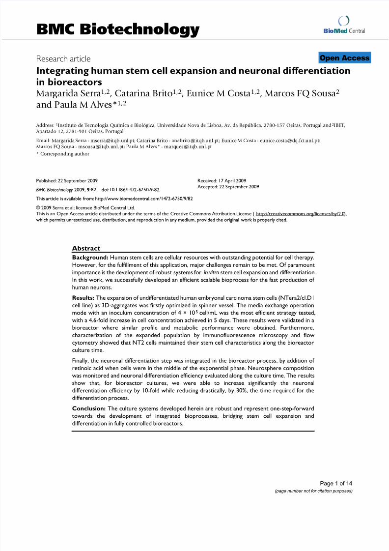
TRA-1-60, SSEA-4) and nestin, an intermediate filament protein associated with undifferentiated phenotype of NT2 cells [22], was detected during exponential growthphase (day 3) and at day 6 (Figure 4 A). This labeling pat-tern was similar to the cell inoculum (day 0).
Moreover, in addition to the expression of stem markersanalysis, the expanded cells ability to differentiate intoneurons was also confirmed. For that purpose, cells werecollected at 3 time points (day 0, 3 and 6) and induced todifferentiate into neurons using the standard static differ-entiation protocol [23]. After treatment with RA and fur-ther cultivation in MI medium, the neuronaldifferentiation efficiency (defined as the ratio between thenumber of neurons obtained and the number of cells har-
vested from the bioreactor, see Methods section) was sim-ilar for all culture samples, presenting values in the range
typically obtained for the static differentiation protocol(3.3 ± 0.2%) [9]. The differentiated neurons were identi-fied by βIII-Tub and MAP2 positive staining (Figure 4B).
Overall, these results showed that NT2 cells maintained
their pluripotency, undifferentiated phenotype, and dif-ferentiation potential along expansion in the bioreactor.
Integrating expansion and neuronal differentiation of NT2
cells in the bioreactor
Once the expansion of pluripotent NT2 cells was adaptedand characterized in the bioreactor system, we further integrated the neuronal differentiation step according toSerra et al [9]. Neuronal differentiation was induced by RA addition when cells achieved the middle of the exponen-tial growth phase at day 3 (Figure 3C). Flow cytometry analysis of cell populations showed that the levels of Oct-4 (94.8% positive cells) and Tra-1-60 (88.7% positive
cells) obtained for the inoculum were kept at day 3 of thebioreactor culture (97.2% and 94.6% Oct-4 and Tra-1-60positive cells, respectively), confirming that the stem cellpopulation was maintained at this time point.
Throughout differentiation, the aggregate size increased,reaching average diameters of 150 ± 40, 309 ± 94 and 458± 44 μm after 1, 2 and 3 weeks of RA treatment, respec-tively (Figure 5 A, B, C, Table 2). The aggregate shapebecame uniform, forming compact and spherical struc-tures (Figure 5B, C). Immunofluorescence microscopy of aggregate cryosections showed that these were neuro-spheres, composed of precursors (nestin-positive) and
differentiated neurons (βIII-Tub-positive), the latest dis-tributed preferentially at the surface (Figure 5C1).
After 9, 16 and 23 days of bioreactor culture (1, 2 and 3 weeks of neuronal differentiation, respectively), neuro-spheres were harvested and cultured for 7 days, on PDL-MG coated flasks, in MI medium, to allow cell migrationand inhibit cell proliferation. One day post-seeding, thepresence of neurites surrounding the neurospheres wasmore pronounced on cultures harvested at day 23 (Figure5F), while on neurospheres harvested earlier, cells withflattened morphology predominated (Figure 5D). Threedays post-seeding, the cell culture composition was ana-
lyzed by immunofluorescence microscopy (Figure 5G, H,I). Cultures derived from neurospheres harvested at day 23 were richer in neurons (βIII-Tub-positive staining) andpresented more developed neuritic networks than theneurospheres harvested at day 16 (Figure 5H, I). A reduced number of βIII-Tub-positive cells was detected incultures derived from neurospheres collected at day 9, in
which nestin-positive cells predominated (Figure 5G). The estimated neuronal differentiation efficiency was 0.13± 0.06% and 17.2 ± 2.2% for cultures derived from neuro-spheres harvested at day 9 and 16 (Table 2). The results
8/7/2019 1472-6750-9-82_Integrating human stem cell
http://slidepdf.com/reader/full/1472-6750-9-82integrating-human-stem-cell 8/14
BMC Biotechnology 2009, 9:82 http://www.biomedcentral.com/1472-6750/9/82
Page 8 of 14(page number not for citation purposes)
Characterization of NT2 cells expanded as 3D-aggregatesFigure 4Characterization of NT2 cells expanded as 3D-aggregates. Immunofluorescence images of cells from the inoculum(day 0) and collected from the bioreactor culture (day 3 and day 6). Immunolabeling of Oct-4, TRA-1-60, SSEA-4 (green) andnestin (red). Nuclei are labeled with DAPI (blue) (A). Immunofluorescence images of differentiated cultures derived from theinoculum (day 0) and from the bioreactor culture (day 3 and day 6). Neurons labeled with βIII-Tub and MAP2 (green) (B).Nuclei were stained with DAPI (blue). Scale bars: 100 μm.

8/7/2019 1472-6750-9-82_Integrating human stem cell
http://slidepdf.com/reader/full/1472-6750-9-82integrating-human-stem-cell 9/14
BMC Biotechnology 2009, 9:82 http://www.biomedcentral.com/1472-6750/9/82
Page 9 of 14(page number not for citation purposes)
obtained until day 16 were similar to the ones describedfor the spinner culture [9], both in culture profile and dif-ferentiation efficiency, proving that the integrated culturestrategy was successfully implemented in the bioreactor.Moreover, by extending the RA treatments for an addi-tional week, a significant increase in the yield of neuronal
differentiated cells was obtained (neuronal differentiationefficiency of 37.4 ± 0.9%, Table 2).
Discussion To fully fulfill the expectations raised by cell therapy it isurgent to develop robust and totally controlled culturesystems, specially designed for the production of highnumbers of differentiated and well characterized cells,expanded as fast and pure as possible. In the present study, we successfully developed a bioprocess for therapid production of human neurons using fully control-led stirred tank bioreactors (125 mL). This was accom-plished by integrating human NT2 cell expansion and
differentiation in a two-step bioprocess.
In this particular study, an ideal expansion strategy shouldassure the fast production of high numbers of stem cells
without compromising their potential. We demonstratedthat, along expansion as 3-D aggregates, NT2 cells main-tained their pluripotent and undifferentiated phenotypeas well as the ability to differentiate into neurons. Differ-ent bioreaction parameters, including cell inoculum con-centration and culture operation mode were studied. Theresults indicate 4 × 10⁵ cell/mL as the most adequate inoc-ulum strategy to be integrated with the differentiationstep, as it allowed higher cell densities in less culture time
contributing to a fast overall process. However, the feasi-bility of starting the cultures with inoculum concentra-tions as lower as 1 × 10⁵ cell/mL looks promising for specific clinical applications in which the starting materialis a limiting factor. Although lower inoculation concentra-tions have been used to expand undifferentiated murineembryonic stem cells as aggregates [6,24], NT2 cell prolif-eration could not be achieved when 4× 10⁴ cell/mL wereused. This difference in cell behavior may reflect the dis-tinct cell origins, as NT2 are pluripotent human embryo-nal carcinoma stem cells, derived from teratocarcinomas
[23], that closely resemble the human embryonic stemcells derived from the blastocyst inner cell mass [25].
By using a fed-batch strategy, where low levels of glucose were maintained in culture, it was possible to enhancedglucose metabolism efficiency with a concomitant
improvement of the FI in cell concentration and increaseof culture lifespan. This strategy may have minimized thetoxicity effect associated with lactate accumulation, asreported previously for several animal cell cultures[21,26]. Nevertheless, the accumulation of other toxic metabolites, including ammonia, resulted in an increasein cell death. The possible depletion of nutrients (othersthan glucose and glutamine) as well as the exhaustion of essential small molecules, namely growth factors, not replenished in the glucose fed-batch strategy, may havecontributed to arrest cell growth. The media exchangemode overcame these drawbacks, being the most efficient strategy to enhance undifferentiated stem cell cultivation,
as shown by the higher cell densities and higher culture viability obtained throughout the cultivation time. There-fore this strategy was chosen for implementation in thecontrolled bioreactor in which stem cell expansion wassuccessfully reproduced, confirming the robustness of theprocess. Media exchange and perfusion strategies havebeen used previously for adult stem cell cultivation [3,9]and human embryoid bodies [13]. In order to achievehigher expansion ratios, as those obtained for the expan-sion process as aggregates of murine embryonic stem cell[6,24] and human neuronal precursor cell [17], serial pas-sage with addition of fresh media can be further included.
By incorporating both expansion and differentiation stepsin an integrated bioprocess, this strategy also assures thefeasibility of expanding human differentiated neuronsderived from a continuous source of pluripotent stemcells. The system described herein allows for obtaining
well differentiated neurons after 2 weeks of differentia-tion, as well as higher yields of neurons for a later culturetime. Importantly, when compared to well establishedstatic differentiation protocols, this methodology drasti-cally enhanced the neuronal differentiation efficiency of NT2 cells and reduced the time needed for differentiation
Table 2: Characterization of NT2 neurospheres cultured in a fully controlled bioreactor.
Neurospheres
Time of harvesting (day) 9 16 23
Duration of retinoic acid treatment (week) 1 2 3
Neurosphere size (μm) 150 ± 40 309 ± 94 458 ± 44Differentiation efficiency 0.13 ± 0.06 17.2 ± 2.2 37.4 ± 0.9
Neurosphere size and neuronal differentiation efficiency are expressed as mean ± SEM from n = 2 independent bioreactor experiments.

8/7/2019 1472-6750-9-82_Integrating human stem cell
http://slidepdf.com/reader/full/1472-6750-9-82integrating-human-stem-cell 10/14
BMC Biotechnology 2009, 9:82 http://www.biomedcentral.com/1472-6750/9/82
Page 10 of 14(page number not for citation purposes)
process; for a differentiation time of 23 days in the biore-actor culture a 10-fold improvement in yield was observedover the static culture protocols lasting 35 days [23].
In this work, the expansion and differentiation of NT2cells was successful validated in computer-controlled bio-reactors. In future, further optimizations can be attemptedaiming to determine the optimal conditions (pH, pO2 and
temperature) to grow and differentiate NT2 cells. So far,some studies have demonstrated that low pO2 decreasesthe rate of stem cell differentiation and enhances stem cellproliferation [27]. Nieruebuegge et al. also reported a sig-nificant increase in final cell number as well as animprovement of cardiac-enriched genes in hEBs culturesunder hypoxic conditions (pO2 = 4%) [7]. A recent study reports that rat mesenchymal stem cell differentiation is
Neuronal differentiation of NT2 cells in a fully controlled bioreactorFigure 5Neuronal differentiation of NT2 cells in a fully controlled bioreactor. Neuronal differentiation was induced by addi-tion of retinoic acid (RA) from day 3 onwards (RA treatments). Phase contrast photomicrographs of neurospheres harvestedat day 9 (A), day 16 (B) and day 23 (C) of the bioreactor culture. By day 23, neurosphere composition was analyzed by cryo-section immunofluorescence microscopy - double labeling of nestin (red) and βIII-Tub (green) (C1). Harvested neurosphereswere further cultured in mitotic inhibitory (MI) conditions, on poly-D-lysine and Matrigel-coated surfaces. Cultures were visu-alized by phase contrast microscopy 1 day after plating (D,E,F) and characterized by immunofluorescence microscopy 3 daysafter plating (G,H,I). Double labeling of nestin (red) and βIII-Tub (green). Phase contrast and immunofluorescence images of
cultures derived from neurospheres harvested at day 9 (D,G), day 16 (E,H) and day 23 (F,I). Scale bars: 100 μm.

8/7/2019 1472-6750-9-82_Integrating human stem cell
http://slidepdf.com/reader/full/1472-6750-9-82integrating-human-stem-cell 11/14
BMC Biotechnology 2009, 9:82 http://www.biomedcentral.com/1472-6750/9/82
Page 11 of 14(page number not for citation purposes)
enhanced at lower temperatures (32°C) than in 37°Cconditions [28].
ConclusionIn this work, a scalable and efficient two-step bioprocess
for the generation of human NT2-derived neurons wasdeveloped in a fully controlled bioreactor, allowing con-tinuous monitoring, non-invasive sampling and charac-terization. By integrating a fast expansion step with anefficient differentiation process, this strategy significantly reduced the time and improved the yields of the neuronaldifferentiation, when compared to the standard static dif-ferentiation protocols.
The controlled bioprocess developed herein can be adapt-able to other cell types, including hESCs and iPS, repre-senting a strong and promising starting point for thedevelopment of novel technologies for the production of
differentiated derivatives from pluripotent cells.
MethodsCell culture
NTERA-2/cl.D1 cells (NT2) were obtained from theCNDR, University of Pennsylvania School of Medicine.Undifferentiated NT2 cells were routinely cultivated instandard tissue culture flasks (Nunc) and maintained inOptiMEM medium (Invitrogen) supplemented with 5%(v/v) of fetal bovine serum (FBS, Hyclone) and 100 U/mL of penicillin- streptomycin (P/S, Invitrogen), according tomethod described at Brito et al.[29].
Stirred suspension cultureUndifferentiated NT2 cell expansion in spinner vessels
Undifferentiated NT2 cells (passage 60-62) were culturedas 3D-aggregates in 125-mL spinner vessels (Wheaton)equipped with a ball impeller and maintained at 37°Cand 5% CO2 for up to 7 days. The agitation rate wasincreased during cultivation in order to avoid aggregateclumping and to control aggregate size (day 0 to 2 - 60rpm, day 2 to 3 - 70 rpm, day 3 to 4 - 80 rpm, day 4upwards - 90 rpm). Two independent experiments wereperformed for each expansion strategy.
Inoculum Concentration Experiments
Cells were cultured in a batch operation mode in Dul-becco's Modified Eagle's Medium- High Glucose (DMEM-HG, 25 mM glucose) (Invitrogen) supplemented with10% (v/v) FBS and 100 U/mL of P/S (complete DMEM-HG). The cell inoculum concentrations evaluated were:0.4 × 10⁵, 1 × 10⁵ and 4 × 10⁵ cell/mL; for an easier reading the nomenclature used was SP-0.4B, SP-1B and SP-4B,respectively. In SP-0.4B and SP-1B, cells were cultured in75 mL of medium at 50 rpm during the first 4-8 h, to pro-mote cell aggregation.
Culture Operation Mode Experiments
Glucose fed-batch and medium exchange culture opera-tion modes were performed using an inoculum cell den-sity of 4 × 10⁵ cell/mL; the nomenclature used for theseexperiments were SP-4FB (SP- spinner, FB- fed-batch) and
SP-4ME (SP- spinner, ME- media exchange), respectively.In SP-4FB, the culture medium was DMEM-Base (Sigma)supplemented with 10% (v/v) FBS, 4 mM of glutamine(Invitrogen), 100 U/mL P/S and 1.4 mM of glucose(Merck). During culture time, glucose concentration wasmonitored twice a day and maintained at lower levels(<1.4 mM); refeeds were performed accordingly to theconsumption rates (calculated from 2 consecutive sam-ples). SP-4ME was cultured in similar conditions to thosedescribed for SP-4B, except that medium was partially exchanged daily from the day 3 onwards as follows: fifty percent of culture media was collected in sterile condi-tions and centrifuged at 200 × g for 5 min; the supernatant
was discarded and the recovered cell aggregates gently resuspended in an equivalent volume of pre-warmedcomplete DMEM-HG.
For all spinner cultures, sampling (2.5 mL) was performed4 h after inoculation and daily from then on. Cell aggre-gates were monitored under an inverted microscope(Leica DM IRB). Cell concentration, metabolite concen-tration and lactate dehydrogenase activity were analyzedas described below.
NT2 culture in a fully controlled bioreactor
To ensure fully controlled cell culture environment, a
stirred tank bioreactor [30] equipped with ball impeller and pH and dissolved oxygen (pO2) measuring probes(Mettler-Toledo) was used for the expansion and differen-tiation of NT2 cells. The pH was kept at 7.2 by injection of CO2 and addition of base (NaOH, 0.2 M). The pO2 wasmaintained at 25% via surface aeration. The temperature
was kept at 37°C by water recirculation in the vessel jacket controlled by a thermocirculator module. Data acquisi-tion and process control were performed using MFCS/WinSupervisory Control and Data Acquisition (SCADA) soft-
ware (Sartorius-Stedim, Germany).
NT2 cell expansion
The SP-4ME experiment was reproduced in the bioreactor system, using undifferentiated NT2 cells with 60-62 pas-sages in static conditions. Moreover, cells used for theinoculum (day 0) and at days 3 and 6 of cultivation in thebioreactor, were characterized using immunofluorescencetools and the neuronal differentiation potential evaluated(see below).
NT2 neuronal differentiation
Undifferentiated NT2 cells with up to 62 passages in static conditions were expanded in the bioreactor, in complete

8/7/2019 1472-6750-9-82_Integrating human stem cell
http://slidepdf.com/reader/full/1472-6750-9-82integrating-human-stem-cell 12/14
BMC Biotechnology 2009, 9:82 http://www.biomedcentral.com/1472-6750/9/82
Page 12 of 14(page number not for citation purposes)
DMEM-HG, using an inoculum concentration of 4 × 10⁵
cell/mL. Differentiation was initiated in the middle of theexponential phase (day 3), following the differentiationprotocol developed by Serra et al [9]. Briefly, neuronal dif-ferentiation was induced by addition of retinoic acid (RA,
Sigma) to the culture media, at a final concentration of 10μM. A 50% media exchange was performed 3 times a week on a regular basis for up to 24 days. Two bioreactor inde-pendent experiments were performed.
Samples were collected from the bioreactor at 3 timepoints: day 9, 16 and 23 (corresponding to 1, 2 and 3
weeks of differentiation process). Cell concentration andneurosphere size were determined and culture was charac-terized using immunofluorescence microscopy. Neuro-spheres harvested at the referred time points weretransferred to coverslips or culture flasks (5 × 10⁴ cell/cm2) coated with poly-D-lysine (PDL, Sigma) and
Matrigel (MG, Becton-Dickinson) and cultured for up to 7days in mitosis inhibitor (MI) medium: DMEM-HG sup-plemented with 5% FBS, 100 U/mL of P/S, 1 μM cytosinearabinosine (Sigma), 10 μM fluorodeoxyuridine (Sigma)and 10 μM uridine (Sigma). Neurons were selectively trypsinized [22,23] using a 0.015% Trypsin-EDTA solu-tion (prepared from Trypsin-EDTA 1X, liquid 0.05%
Trypsin, Invitrogen), counted and transferred to coverslipscoated with PDL and MG for characterization by immu-nocytochemistry. Neuronal differentiation efficiency wasdefined as the ratio between the number of neuronsobtained after 7 days of culture in MI medium and thetotal amount of cells harvested at the 3 different harvest-
ing times.
Analytical methods
Cell concentration determination
Cell aggregates were dissociated by a 2 min incubation with Trypsin-EDTA (0.05%) at 37°C followed by cellresuspension in complete DMEM-HG. Cell density wasassessed using a Fuchs-Rosenthal haemocytometer (Brand, Wertheim, Germany) and cell viability estimatedby the standard trypan blue exclusion test.
Aggregate diameter
Aggregate size in each culture sample was determined
using a micrometer coupled to an inverted microscope(Leica, DM IRB). Two perpendicular diameters of a mini-mum of 15 aggregates were measured and the averagediameter was calculated. Aggregates less than 20 μm indiameter (generally cell doublets or triplets) were not con-sidered for calculations as they represent a small percent-age of the total cell number in culture.
Lactate dehydrogenase activity
Lactate dehydrogenase (LDH) activity from the culturesupernatant was determined as an indirect way of assess-
ing cell death. LDH activity was determined by following spectrophotometrically (at 340 nm) the rate of oxidationof NADH to NAD+ coupled with the reduction of pyruvateto lactate. The specific rate of LDH release (qLDH, U.day -1.cell-1) was calculated for every time interval using the
following equation: qLDH = ΔLDH/(Δt Δ X V ), where ΔLDH(U) is the change in LDH activity over the time period Δt (day) and Δ Xv (cell) is the average of total cells during thesame time period. The cumulative value qLDHcum was esti-mated by qLDHcum i+1 = qLDH i + qLDH i+1. The fold increaseof the specific LDH release rates achieved throughout 6days of cultivation were determined by calculating theratio between the values of qLDHcum obtained at day 6 andday 0. These values indirectly represent the fold increasein cell lysis obtained within 6 days of culture.
Metabolite analysis
Glucose (GLC), lactate (LAC) and glutamine (GLN) con-
centrations in the culture medium were analyzed using an YSI 7100MBS (YSI Incorporated, USA). Ammonia wasquantified enzymatically using a commercially availableUV test (Roche, Germany).
The specific metabolic rates (qMet ., mol.day -1.cell-1) werecalculated using the equation: qMet . =ΔMet /(Δt Δ X v ), whereΔMet (mol) is the variation in metabolite concentrationduring the time period Δt (day) and Δ X v (cell) the averageof adherent cells during the same time period.
Apparent growth rate and fold increase in cell expansion
Apparent growth rates and fold increase parameters were
calculated for all expansion cultures. Apparent growthrates (μ, day -1) were calculated using a first order kinetic model for cell expansion: dX/dt = μ X, where t (day) is theculture time and × (cell) is the value of viable cells for aspecific t. The μ values were estimated applying the modelto the slope of the curves during the exponential phase.
The fold increase in cell expansion (FI) was defined as theratio X MAX /X 0, where X MAX is the peak cell density (cell/mL) and X 0 is the inoculation cell density (cell/mL).
Differentiation potential
To assess the neuronal differentiation potential along theexpansion assays, 2.3 × 10⁶ cells were collected from the
suspension cultures and plated in a T75 flask (Nunc) (Fig-ure 1). NT2 cells were differentiated into post-mitotic neu-rons according to Pleasure et al [23]. Briefly, cells werecultured for 5 weeks in complete DMEM-HG supple-mented with 10 μM RA. Cells were splitted at 1:4.5 ratioand cultured in MI medium for 12 days. After this period,neurons were selectively trypsinized, as described above,counted and transferred to coverslips coated with PDL and MG for characterization by immunocytochemistry.Neuronal differentiation efficiency was defined as theratio between the number of neurons obtained after cul-

8/7/2019 1472-6750-9-82_Integrating human stem cell
http://slidepdf.com/reader/full/1472-6750-9-82integrating-human-stem-cell 13/14
BMC Biotechnology 2009, 9:82 http://www.biomedcentral.com/1472-6750/9/82
Page 13 of 14(page number not for citation purposes)
ture in MI medium and the total amount of cells harvestedafter RA treatments.
Immunofluorescence microscopy
In expansion cultures, cell aggregates were collected at day
3 and 6, dissociated using Trypsin-EDTA (0.05%) at 37°Cfollowed by cell resuspension in complete DMEM-HG,and transferred to glass coverslips. Three days after plat-ing, cultures were characterized. In differentiation assaysneurospheres were harvested from the bioreactor culturesat day 9, 16 and 23, and processed for cryosection or transferred to coverslips coated with PDL and MG (seeFigure 1).
Cells in coverslips were washed in PBS with 0.5 mMMgCl2 and fixed in 4% (w/v) paraformaldehyde solutionin PBS with 4% (w/v) sucrose, for 20 min. For cryosection,neurospheres were washed in PBS, transferred to a tissue
protecting compound (Tissue Teck, OCT™ Compound)and frozen at -80°C. Ten μm sections, obtained using acryostat (Leica), were rehydrated with PBS and fixed inmethanol, at -20°C, for 10 min. After fixation, the sameprocedure was followed for cryosections and coverslips.
For staining intracellular epitopes, cells were permeabi-lized with 0.1% (w/v) Triton X-100 (TX-100) in PBS, for 15 min. After 1 h in blocking solution (0.2% (w/v) fishskin gelatin in PBS), cells were incubated with primary antibody for 2 h. The coverslips were washed 3 times withPBS and overlaid with secondary antibody for 1 h. Pri-mary and secondary antibodies were diluted in 0.125%
(w/v) fish skin gelatin in PBS with 0.1% (w/v) TX-100.Samples were mounted in ProLong mounting medium(Molecular Probes), supplemented with DAPI for nucleusstaining. For surface epitopes staining, cells were not per-meabilized with TX-100. Samples were visualized using afluorescence microscope (Leica DMRB).
Primary antibodies used were: mouse anti-tumor relatedantigen-1-60 (Tra-1-60) (Santa Cruz Biotechnology),mouse anti-stage specific embryonic antigen-4 (SSEA-4)(Santa Cruz Biotechnology), mouse anti-Oct-4 (SantaCruz Biotechnology), mouse anti-nestin (Chemicon),mouse anti-type III β-tubulin (βIII-Tub) (Chemicon),
mouse anti-microtubule associated protein 2A and 2B(MAP2) (Chemicon). The secondary antibodies were goat anti-mouse IgM-AlexaFluor488, goat anti-mouse IgG-
AlexaFluor 594, goat anti-mouse IgG-AlexaFluor 488 andrabbit anti-mouse IgG-AlexaFluor 594 (Invitrogen).
Flow cytometry
Cells used for the inoculum (day 0) and from day 3 of thebioreactor expansion culture were dissociated into singlecells and analyzed by flow cytometry (Figure 1). Samples
were fixed in CytofixCytoperm reagent (BD Pharmigen)
for 10 min, blocked with 1% BSA in PBS at 4°C for 30 minand, in the case of intracellular antigens, permeabilized
with 1% TX-100 for 10 min. Primary antibodies weremouse anti-Tra-1-60 and anti-Oct-4. Secondary antibod-ies were anti-mouse IgM-AlexaFluor488 and anti-mouse
IgG-AlexaFluor488. Ten thousand events were registeredper sample with a CyFlow® space (Partec) instrument,using the appropriate scatter gates to avoid cellular debrisand aggregates. A cell was considered to be positively stained if the measured fluorescence intensity exceededthe signal obtained by cells incubated with an isotypecontrol antibody (Santa Cruz Biotechnology).
Statistical analysis
For each spinner and bioreactor assays, two independent experiments were performed. The results were expressedas the mean ± standard deviation. The statistical test used,One-way ANOVA, was performed in SPSS 13.0 for Win-
dows for a level of confidence of 95% (a = 0.05) followedby the Scheffé multiple comparison test.
List of abbreviationsBSA: bovine serum albumin; DAPI: 4',6-diamidino-2-phe-nylindole; DMEM-HG: Dulbecco's modified Eagle'smedium-high glucose; FBS: foetal bovine serum; FI: foldincrease; hESC: human embryonic stem cells; iPS cells:induced pluripotent stem cells; LAC: lactate; LDH: lactatedehydrogenase; MAP2: microtubule-associated protein 2;MG: Matrigel; MI: mitosis inhibitors; NT2: NTera2/cl.D1;P/S: penicillin-streptomycin; PDL: poly-D-lysine; pO2:dissolved oxygen; qGLC: specific rate of glucose consump-
tion; qLAC: specific rate of lactate production; qLDHcum:cumulative value of specific LDH release rate; RA: retinoic acid; SSEA-4: stage specific embryonic antigen-4; Tra-1-60: tumor related antigen-1-60; TX-100: triton X-100; βIII-tub: type III β-tubulin; μ: apparent growth rate.
Authors' contributionsMS participated in the spinner and bioreactor experimentsand in the collection of Flow Cytometry data; carried out the growth kinetics and metabolic profile analyses and thecryosection immunofluorescence microscopy; contrib-uted to the conception and design of the study and draftedthe manuscript. CB participated in the spinner and biore-
actor experiments and immunological assays; carried out the assessment of the neuronal differentiation potential;contributed to the conception, design and coordination of the study and helped to draft the manuscript. EC wasinvolved in spinner experiments and immunofluores-cence microscopy analysis. MFQS participated in bioreac-tor experiments. PA participated in the conception, designand coordination of the study and gave final approval of the version to be published. All authors read andapproved the final manuscript.

8/7/2019 1472-6750-9-82_Integrating human stem cell
http://slidepdf.com/reader/full/1472-6750-9-82integrating-human-stem-cell 14/14
Publish with BioMedCentral and everyscientist can read your work free of charge
"BioMed Central will be the most significant development for
disseminating the results of biomedical research in our lifetime."
Sir Paul Nurse, Cancer Research UK
Your research papers will be:
available free of charge to the entire biomedical community
peer reviewed and published immediately upon acceptance
cited in PubMed and archived on PubMed Central
yours — you keep the copyright
Submit your manuscript here:
http://www.biomedcentral.com/info/publishing_adv.asp
BioMedcentral
BMC Biotechnology 2009, 9:82 http://www.biomedcentral.com/1472-6750/9/82
AcknowledgementsThe authors are grateful to Prof. Virginia Lee and Prof. John Trojanowski
(CNDR, University of Pennsylvania School of Medicine, USA) for the kind
gift of NT2 cells; Sofia B. Leite for her support in the bioreactor protocol
for neuronal differentiation; António Roldão for thoughtful discussions and
useful support in statistical analysis.
The authors acknowledge the financial support received from FCT (PTDC/
BIO/72755/2006) and from the European Commission (NMP4-CT-2004-
500039 and LSHB-CT-2006-018933). MS, EC and CB are recipients of BD
(SFRH/BD/42176/2007, SFRH/BD/35382/2007) and BPD (SFRH/BPD/
34622/2007) fellowships, respectively, from FCT, Portugal.
References1. Storch A, Schwarz J : Neural stem cells and neurodegeneration.
Curr Opin Investig Drugs 2002, 3:774-781.2. Jones JM, Thomson JA: Human embryonic stem cell technol-
ogy. Semin Reprod Med 2000, 18:219-223.3. King JA, Miller WM: Bioreactor development for stem cell
expansion and controlled differentiation. Curr Opin Chem Biol 2007, 11:394-398.
4. Ulloa-Montoya F, Verfaillie CM, Hu WS: Culture systems for
pluripotent stem cells. J Biosci Bioeng 2005, 100:12-27.5. Serra M, Brito C, Leite SB, Gorjup E, von Briesen H, Carrondo MJ,
Alves PM: Stirred bioreactors for the expansion of adult pan-creatic stem cells. Ann Anat 2009, 191:104-115.
6. Cormier JT, zur Nieden NI, Rancourt DE, Kallos MS: Expansion of undifferentiated murine embryonic stem cells as aggregatesin suspension culture bioreactors. Tissue Eng 2006,12:3233-3245.
7. Niebruegge S, Bauwens CL, Peerani R, Thavandiran N, Masse S,Sevaptisidis E, Nanthakumar K, Woodhouse K, Husain M, KumachevaE, Zandstra PW: Generation of human embryonic stem cell-derived mesoderm and cardiac cells using size-specifiedaggregates in an oxygen-controlled bioreactor. Biotechnol Bio-eng 2009, 102:493-507.
8. Cameron CM, Hu WS, Kaufman DS: Improved development of human embryonic stem cell-derived embryoid bodies bystirred vessel cultivation. Biotechnol Bioeng 2006, 94:938-948.
9. Serra M, Leite SB, Brito C, Costa J, Carrondo MJ, Alves PM: Novelculture strategy for human stem cell proliferation and neu-ronal differentiation. J Neurosci Res 2007, 85:3557-3566.
10. Zandstra PW, Nagy A: Stem cell bioengineering. Annu Rev Biomed Eng 2001, 3:275-305.
11. Zhao F, Ma T: Perfusion bioreactor system for human mesen-chymal stem cell tissue engineering: dynamic cell seedingand construct development. Biotechnol Bioeng 2005, 91:482-493.
12. Fong WJ, Tan HL, Choo A, Oh SK: Perfusion cultures of humanembryonic stem cells. Bioprocess Biosyst Eng 2005, 27:381-387.
13. Come J, Nissan X, Aubry L, Tournois J, Girard M, Perrier AL,Peschanski M, Cailleret M: Improvement of Culture Conditionsof Human Embryoid Bodies Using a Controlled Perfused andDialyzed Bioreactor System. Tissue Eng Part C Methods 2008,14:289-298.
14. Youn BS, Sen A, Behie LA, Girgis-Gabardo A, Hassell JA: Scale-up of breast cancer stem cell aggregate cultures to suspensionbioreactors. Biotechnol Prog 2006, 22:801-810.
15. Bauwens C, Yin T, Dang S, Peerani R, Zandstra PW: Developmentof a perfusion fed bioreactor for embryonic stem cell-derived cardiomyocyte generation: oxygen-mediatedenhancement of cardiomyocyte output. Biotechnol Bioeng 2005,90:452-461.
16. Gilbertson JA, Sen A, Behie LA, Kallos MS: Scaled-up productionof mammalian neural precursor cell aggregates in compu-ter-controlled suspension bioreactors. Biotechnol Bioeng 2006,94:783-792.
17. Baghbaderani BA, Mukhida K, Sen A, Hong M, Mendez I, Behie LA:Expansion of Human Neural Precursor Cells in Large-ScaleBioreactors for the Treatment of Neurodegenerative Disor-ders. Biotechnol Prog 2008, 24:859-870.
18. Andrews PW: From teratocarcinomas to embryonic stemcells. Philos Trans R Soc Lond B Biol Sci 2002, 357:405-417.
19. Przyborski SA, Christie VB, Hayman MW, Stewart R, Horrocks GM:Human embryonal carcinoma stem cells: models of embry-onic development in humans. Stem Cells Dev 2004, 13:400-408.
20. Kondziolka D, Wechsler L: Stroke repair with cell transplanta-tion: neuronal cells, neuroprogenitor cells, and stem cells.Neurosurg Focus 2008, 24:. E13(1-6)
21. Xie L, Wang DI: Fed-batch cultivation of animal cells using dif-
ferent medium design concepts and feeding strategies. 1994.Biotechnol Bioeng 2006, 95:270-284.22. Pleasure SJ, Lee VM: NTera 2 cells: a human cell line which dis-
plays characteristics expected of a human committed neuro-nal progenitor cell. J Neurosci Res 1993, 35:585-602.
23. Pleasure SJ, Page C, Lee VM: Pure, postmitotic, polarized humanneurons derived from NTera 2 cells provide a system forexpressing exogenous proteins in terminally differentiatedneurons. J Neurosci 1992, 12:1802-1815.
24. zur Nieden NI, Cormier JT, Rancourt DE, Kallos MS: Embryonicstem cells remain highly pluripotent following long termexpansion as aggregates in suspension bioreactors. J Biotech-nol 2007, 129:421-432.
25. Henderson JK, Draper JS, Baillie HS, Fishel S, Thomson JA, Moore H,Andrews PW: Preimplantation human embryos and embry-onic stem cells show comparable expression of stage-specificembryonic antigens. Stem Cells 2002, 20:329-337.
26. Cruz HJ, Freitas CM, Alves PM, Moreira JL, Carrondo MJ: Effects of
ammonia and lactate on growth, metabolism, and produc-tivity of BHK cells. Enzyme Microb Technol 2000, 27:43-52.27. Gibbons J, Hewitt E, Gardner DK: Effects of oxygen tension on
the establishment and lactate dehydrogenase activity of murine embryonic stem cells. Cloning Stem Cells 2006, 8:117-122.
28. Stolzing A, Scutt A: Effect of reduced culture temperature onantioxidant defences of mesenchymal stem cells. Free Radic Biol Med 2006, 41:326-338.
29. Brito C, Escrevente C, Reis CA, Lee VM, Trojanowski JQ, Costa J:Increased levels of fucosyltransferase IX and carbohydrateLewis(x) adhesion determinant in human NT2N neurons. JNeurosci Res 2007, 85:1260-1270.
30. Sa Santos S, Fonseca LL, Monteiro MA, Carrondo MJ, Alves PM: Cul-turing primary brain astrocytes under a fully controlled envi-ronment in a novel bioreactor. J Neurosci Res 2005, 79:26-32.