149 - handout - cancer and the eye.ppt · 3 cancer epidemiology it is worth screening for prostate,...
TRANSCRIPT
1
Cancer and the Eye
Course Title:
DISCLOSURE STATEMENT
Please silence all mobile devices.
No financial disclosures
Lecturer: Brad Sutton, OD, FAAOClinical ProfessorIU School of Optometry
Cancer factoids
� Can affect any tissue or organ at any age
� All cancers begin with a defect in a single cell (monoclonal)
� This is followed by unrestrained growth
Cancer factoids
� A one cm. tumor contains one billion cells
� One trillion cells usually means a lethal tumor
� BENIGN tumors may damage local tissue by occupying space but they do not spread
� MALIGNANT tumors invade surrounding tissue and may metastasize
Cancer factoids
� Cell division is controlled by genes that promote it and genes that suppress it
� Cancer is the result of some combination of defects in this genetic functioning
Recent study
� Study of a 43 year old with skin cancer found 23,000 genetic mutations
� Study of a 55 year old with lung cancer found 33,000 genetic mutations
Cancer types
� General types of cancer include……..
� Adenocarcinoma: Glandular tissue
� Melanomas (melanin)
� Sarcomas (connective tissue)
� Carcinomas (epithelial tissue)
� Leukemias (bone marrow)
� Lymphomas (lymphoid tissue)
2
Leading cancers in US
� Men…
� 1) Prostate
� 2) Lung
� 3) Colon
� 4) Urinary
� 5) Skin melanoma
� But the most common malignancy in humans is………….Basal Cell Carcinoma!
� Women….
� 1) Breast
� 2) Lung (90% use tob.)
� 3) Colon
� 4) Uterine
� 5) NH lymphoma
� P,B,L, and C make up 50% of all non-skin cancer
� 25% of people affected during their lifetime
Skin cancer
� In the US 50% of all cancer is skin cancer…………..and 80% of skin cancer is BCC
� Did you know……….that patients that have had a kidney transplant are 20X more likely to develop ocular squamous cell carcinoma?
2013 American Cancer Society Report
Overall in 2013
� 1,660,290 new cases of cancer excluding SCC and BCC
� 13.7 million living Americans with a history of cancer (active or remission)
� 580,350 deaths: 1600/day
Combined M & F Rank
� Prostate (238,590)
� Breast (234,580)
� Lung (228,190)
� Colon (102,480)
� Lymphoma (79,030)
� Skin melanoma (76,690)
Cancer mortality in the US (combined M & F)
� 1) lung
� 2) colorectal
� 3) breast
� 4) pancreatic (# 12 in frequency)
� 5) prostate
Cancer in the future
� By 2020, the number of cancer patients and cancer survivors will increase by over 50% to an estimated 18.2 million
� There will be a projected shortage of 4000 oncologists
Cancer epidemiology
� Cancer screenings are driven by several factors…………
� 1) Is it a common cancer ?
� 2) Can we detect it ?
� 3) Can we treat it ?
� 4) How much does the screening cost?

3
Cancer epidemiology
� It is worth screening for prostate, colon, breast, cervical, and skin cancer
� Cancer increases with age due to increased length of exposure to carcinogens
Cancer screenings
� 1) Blood tests
� 2) Bone scans
� 3) Biopsies
� 4) X-ray, CT, MRI and other imaging
� 5) Observation
Cancer
� Leading cause of death worldwide
� It accounts for ¼ of deaths in the US…….Only heart disease kills more.
� Economic toll of $895 billion…….not counting the cost of treatment!
Cancer treatment
� CHEMOTHERPY:
� Drugs that interfere with cell division
� Multiple drugs available, oral and IV
� RADIATION:
� Damages cellular DNA
� SURGERY:
� Removes the tumor
Cancer treatment
� Chemotherapy targets all cells in the body that are actively dividing…………abnormal and normal!
� Hair follicles > loss of hair
� Intestinal mucosa > diarrhea, vomiting, nausea
� Bone marrow > anemia
Cancer treatment
� Radiation results in the damage of cellular DNA > cell death
� Need to be focal with treatment because all cells are affected
� Both radiation and chemotherapy can lead to retinal complications
4
Cancer treatment
� Treatment often consists of a combination of surgery, radiation and chemotherapy
� This depends on many factors (location, metastases, etc.)
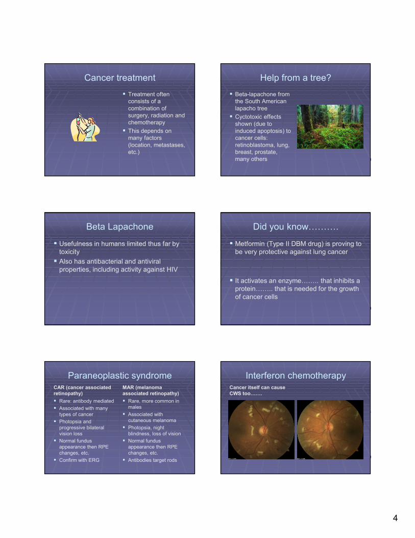
Help from a tree?
� Beta-lapachone from the South American lapacho tree
� Cyctotoxic effects shown (due to induced apoptosis) to cancer cells: retinoblastoma, lung, breast, prostate, many others
Beta Lapachone
� Usefulness in humans limited thus far by toxicity
� Also has antibacterial and antiviral properties, including activity against HIV
Did you know……….
� Metformin (Type II DBM drug) is proving to be very protective against lung cancer
� It activates an enzyme…….. that inhibits a protein…….. that is needed for the growth of cancer cells
Paraneoplastic syndromeCAR (cancer associated
retinopathy)
� Rare: antibody mediated
� Associated with many types of cancer
� Photopsia and progressive bilateral vision loss
� Normal fundusappearance then RPE changes, etc.
� Confirm with ERG
MAR (melanoma
associated retinopathy)
� Rare, more common in males
� Associated with cutaneous melanoma
� Photopsia, night blindness, loss of vision
� Normal fundusappearance then RPE changes, etc.
� Antibodies target rods
Interferon chemotherapyCancer itself can cause
CWS too…….
5
Case Report
� A 55 year-old white female reported to our clinic with a complaint of blurry vision in her left eye for about one month
� She also complained of dizziness, nausea, and a “pressure” behind her left ear
� Her medical history was significant only for a family history of colon cancer
Case Report
� An eye exam performed in our clinic three years prior was remarkable only for refractive error
� Entering acuity with correction was 20/20 OD, 20/40 OS and BCVA was 20/20 OD, 20/25 “-” OS
� Entrance testing was unremarkable as was the anterior segment OU
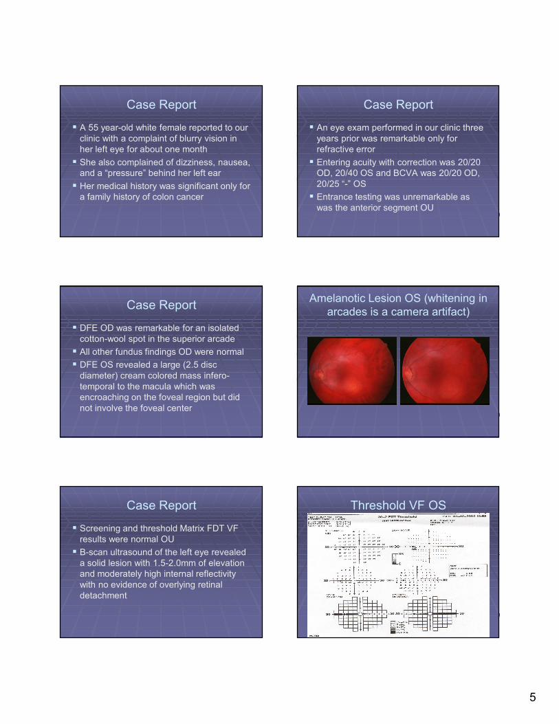
Case Report
� DFE OD was remarkable for an isolated cotton-wool spot in the superior arcade
� All other fundus findings OD were normal
� DFE OS revealed a large (2.5 disc diameter) cream colored mass infero-temporal to the macula which was encroaching on the foveal region but did not involve the foveal center
Amelanotic Lesion OS (whitening in arcades is a camera artifact)
Case Report
� Screening and threshold Matrix FDT VF results were normal OU
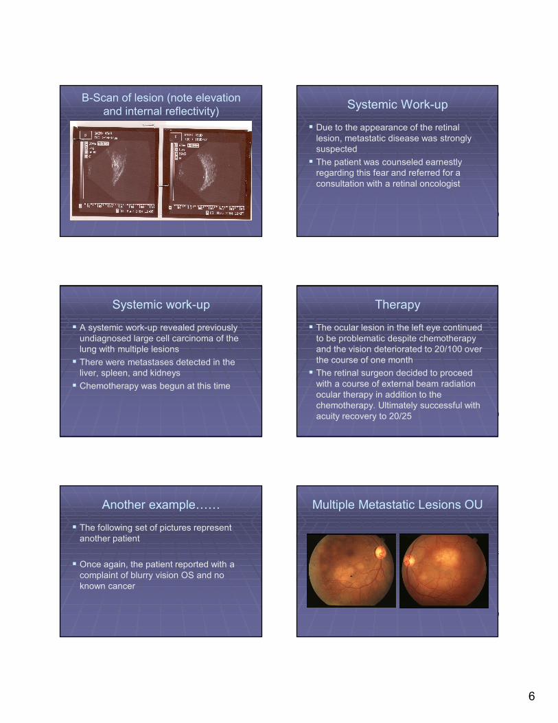
� B-scan ultrasound of the left eye revealed a solid lesion with 1.5-2.0mm of elevation and moderately high internal reflectivity with no evidence of overlying retinal detachment
Threshold VF OS
6
B-Scan of lesion (note elevation and internal reflectivity)
Systemic Work-up
� Due to the appearance of the retinal lesion, metastatic disease was strongly suspected
� The patient was counseled earnestly regarding this fear and referred for a consultation with a retinal oncologist
Systemic work-up
� A systemic work-up revealed previously undiagnosed large cell carcinoma of the lung with multiple lesions
� There were metastases detected in the liver, spleen, and kidneys
� Chemotherapy was begun at this time
Therapy
� The ocular lesion in the left eye continued to be problematic despite chemotherapy and the vision deteriorated to 20/100 over the course of one month
� The retinal surgeon decided to proceed with a course of external beam radiation ocular therapy in addition to the chemotherapy. Ultimately successful with acuity recovery to 20/25
Another example……
� The following set of pictures represent another patient
� Once again, the patient reported with a complaint of blurry vision OS and no known cancer
Multiple Metastatic Lesions OU
7
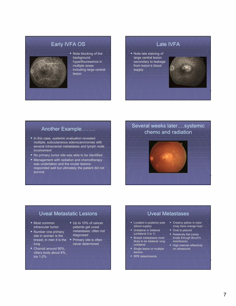
Early IVFA OS
� Note blocking of the background hyperflouresence in multiple areas including large central lesion
Late IVFA
� Note late staining of large central lesion secondary to leakage from lesion’s blood supply
Another Example……..
� In this case, systemic evaluation revealed multiple, subcutaneous adenocarcinomas with several intracranial metastases and lymph node involvement
� No primary tumor site was able to be identified
� Management with radiation and chemotherapy was undertaken and the ocular lesions responded well but ultimately the patient did not survive
Several weeks later….systemic chemo and radiation
Uveal Metastatic Lesions
� Most common intraocular tumor
� Number one primary site in women is the breast, in men it is the lung
� Choroid around 90%, ciliary body about 8%, iris 1-2%
� Up to 10% of cancer patients get uveal metastases: often not diagnosed
� Primary site is often never determined
Uveal Metastases
� Located in posterior pole (blood supply)
� Unilateral or bilateral (unilateral 3 to 1)
� Breast metastases most likely to be bilateral: lung unilateral
� Single lesion or multiple lesions
� RPE detachments
� Creamy yellow in color (may have orange hue)
� Oval to placoid
� Relatively flat (rarely break through Bruch's membrane)
� High internal reflectivity on ultrasound
8
Management of Metastatic Tumors
� Metastatic lesions tend to be detected sooner because their posterior pole location leads to earlier symptoms
� Systemic work-up is critical
� Average survival time of 9 months after Dx
� Systemic chemotherapy
� Radiation via external beam (outpatient) or plaque (hospital)
� PBT (protons)
� Photocoagulation
� Enucleation
� Anti-VEGF injections
� Must consider life expectancy
Differential Diagnoses of Metastatic Tumors
� Primary uveal melanomas
� Hemangiomas
� Osteomas
� Posterior scleritis
� Inflammatory disorders
Other Examples of Ocular Neoplasms
� Choroidal nevi
� Primary Choroidal / CB Melanomas
� Melanocytomas
� Iris melanomas
Choroidal nevi
� Possibly present in up to 30% of general population (? clinically) 6.5% of whites in 2011 st.
� Flat or minimally elevated (< 1.5 mm)
� < 6 mm in diameter: 95% are less than 2dd
� Melanotic or amelanotic
� Overlying drusen: usually indicate longstanding inactivity: lipofuscin?
� Possible overlying serous RD
� RPE disturbance / atrophy over time
� Conversion to uveal melanoma : 1 in 4000-8000
� 10% will grow without undergoing malignant conversion. Recent 2011 study showed growth in 31% over 15 years.
Choroidal Nevi
� Photodocument
� B-scan
� If small follow annually
� If suspicious, more frequent observation
� Significant elevation rare with nevi
Choroidal Nevi
9
Choroidal nevus Peripheral choroidal nevus
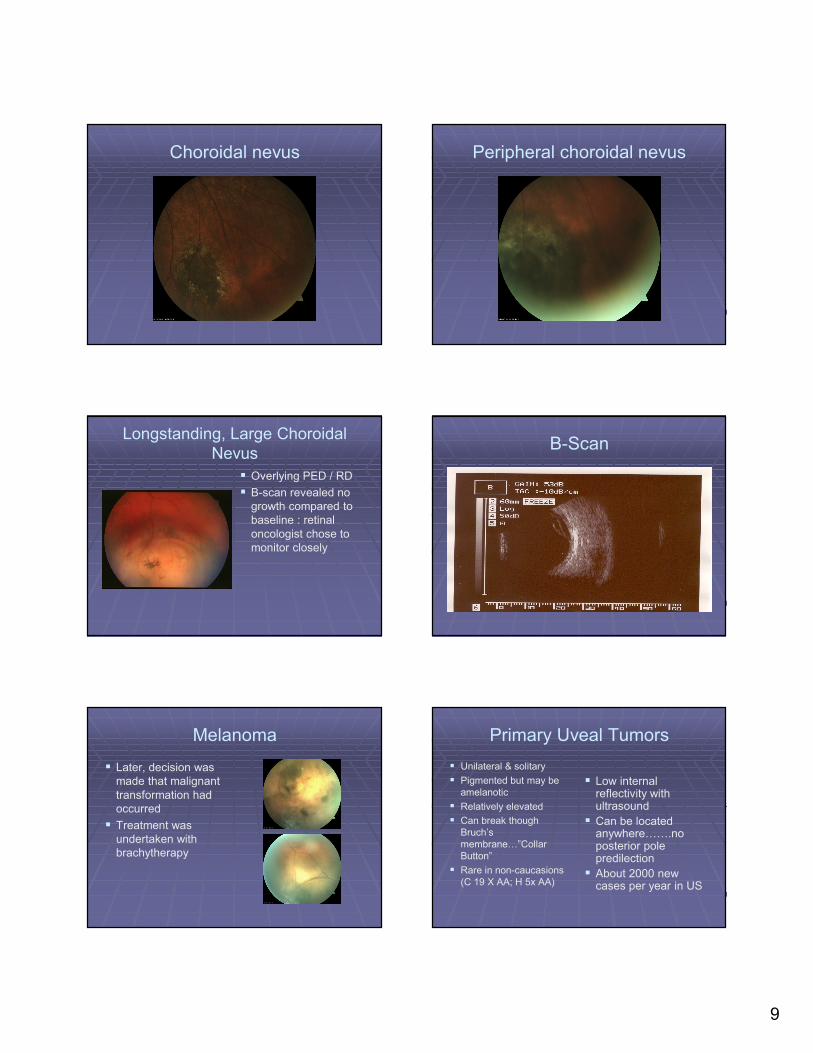
Longstanding, Large Choroidal Nevus
� Overlying PED / RD
� B-scan revealed no growth compared to baseline : retinal oncologist chose to monitor closely
B-Scan
Melanoma
� Later, decision was made that malignant transformation had occurred
� Treatment was undertaken with brachytherapy
Primary Uveal Tumors
� Unilateral & solitary
� Pigmented but may be amelanotic
� Relatively elevated
� Can break though Bruch’s membrane…”Collar Button”
� Rare in non-caucasions(C 19 X AA; H 5x AA)
� Low internal reflectivity with ultrasound
� Can be located anywhere…….no posterior pole predilection
� About 2000 new cases per year in US
10
Melanoma vs. nevus
� Important risk factors for possible malignant transformation….
� Thickness > 2mm
� Symptoms
� Orange Pigment
� New onset of subretinal fluid / serous RD, especially in the absence of drusen
� Ultrasound hollowness / no halo
� Location within 3mm of ONH
� Diameter of 12mm or more
Shields
� “To find small ocular melanomas”
� Thickness > 2mm
� Fluid
� Symptoms
� Orange Pigment
� Margin near ONH (3mm)
EDI SD-OCT feature of small melanomas vs. nevi
� Shaggy photoreceptors
� Loss of PIL
� Disruption of IPL, GCL
� Loss of ELM
Primary uveal tumors
� Can metastasize, but rarely have by the time they are detected in the eye
� Gene mutation that causes metastasis has been discovered (Dr. Harbour, Washington University, and others. Dr. Harbour now at Bascom Palmer)
� Systemic work-up a must, but not common to find metastases at time of diagnosis
� Most frequent site……..75%.........is the liver
� 2X risk of colon cancer compared to general population
Radiation risk
� For a 50 year old diagnosed with an ocular melanoma, the cancer risk from 10 years of radiation exposure with abdominal scans is…..
� CT scan around 1% lifetime risk
� CT plus PET scan around 2% lifetime risk
� This is average for both sexes, women slightly higher risk
Pathology
� Three main tumor types based upon cell morphology….
� Spindle (relatively benign)
� Epithelioid (most large tumors)
� Mixed
11
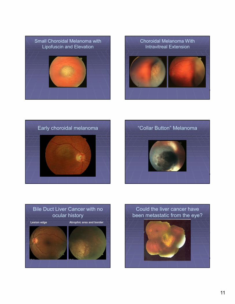
Small Choroidal Melanoma with Lipofuscin and Elevation
Choroidal Melanoma With Intravitreal Extension
Early choroidal melanoma “Collar Button” Melanoma
Bile Duct Liver Cancer with no ocular history
Lesion edge Atrophic area and border
Could the liver cancer have been metastatic from the eye?
12
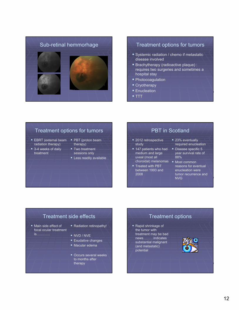
Sub-retinal hemmorhage Treatment options for tumors
� Systemic radiation / chemo if metastatic disease involved
� Brachytherapy (radioactive plaque) : requires two surgeries and sometimes a hospital stay
� Photocoagulation
� Cryotherapy
� Enucleation
� TTT
Treatment options for tumors
� EBRT (external beam radiation therapy)
� 3-4 weeks of daily treatment
� PBT (proton beam therapy)
� Two treatment sessions only
� Less readily available
PBT in Scotland
� 2012 retrospective study
� 147 patients who had medium and large uveal (most all choroidal) melanomas
� Treated with PBT between 1993 and 2008
� 23% eventually required enucleation
� Disease specific 5 year survival rate of 88%
� Most common reasons for eventual enucleation were tumor recurrence and NVG
Treatment side effects
� Main side effect of focal ocular treatment is…………
� Radiation retinopathy!
� NVD / NVE
� Exudative changes
� Macular edema
� Occurs several weeks to months after therapy
Treatment options
� Rapid shrinkage of the tumor with treatment may be bad news………indicates substantial malignant (and metastatic) potential
13
COMS and other studies
� Five year survival rates for……….
� Small melanomas (< 10 mm) : 94%
� Medium melanomas (10-15 mm): 70-90%
� Large melanomas ( > 15 mm): 40-60%
� Enucleation does NOT appear to increase metastatic risk
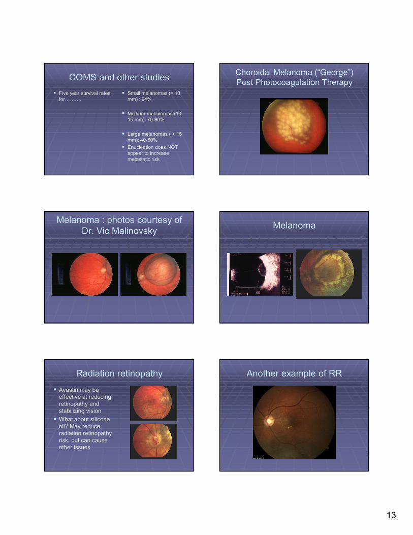
Choroidal Melanoma (“George”) Post Photocoagulation Therapy
Melanoma : photos courtesy of Dr. Vic Malinovsky
Melanoma
Radiation retinopathy
� Avastin may be effective at reducing retinopathy and stabilizing vision
� What about silicone oil? May reduce radiation retinopathy risk, but can cause other issues
Another example of RR
14
What if……….
� Fine needle biopsies of melanomas are yielding amazing info with RNA transcriptomic profiling
� Essentially two types of tumors that can be identified with over 90% accuracy
� Class one signature: almost never metastasizes: Class two almost always does
� What are the implications of this?
Possible intervention
� New research by Dr. Harbour indicates that a certain class of seizure drugs……HDAC inhibitors……..may help
� Cancer cells that have metastasized from the eye to other sites are inhibited and made less aggressive by these drugs. May be able to keep disease “at bay” for an extended period
More genetics…….
� 80% of uveal melanoma patients have mutations in either GNA11 or GNAQ
� But……………this mutation alone does not result in melanoma formation. Must have mutation plus other factors (as of now not known)
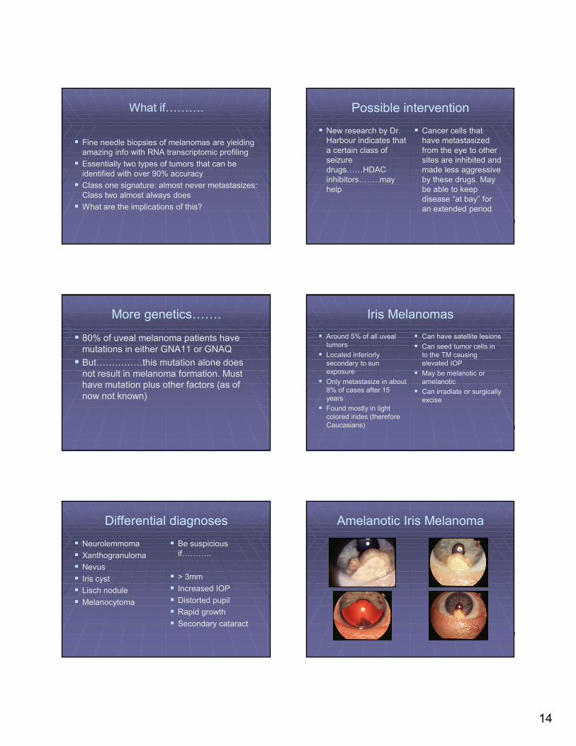
Iris Melanomas
� Around 5% of all uveal tumors
� Located inferiorly secondary to sun exposure
� Only metastasize in about 8% of cases after 15 years
� Found mostly in light colored irides (therefore Caucasians)
� Can have satellite lesions
� Can seed tumor cells in to the TM causing elevated IOP
� May be melanotic or amelanotic
� Can irradiate or surgically excise
Differential diagnoses
� Neurolemmoma
� Xanthogranuloma
� Nevus
� Iris cyst
� Lisch nodule
� Melanocytoma
� Be suspicious if………..
� > 3mm
� Increased IOP
� Distorted pupil
� Rapid growth
� Secondary cataract
Amelanotic Iris Melanoma
15
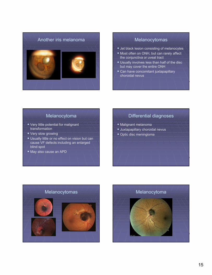
Another iris melanoma Melanocytomas
� Jet black lesion consisting of melanocytes
� Most often on ONH, but can rarely affect the conjunctiva or uveal tract
� Usually involves less than half of the disc but may cover the entire ONH
� Can have concomitant juxtapapillary choroidal nevus
Melanocytoma
� Very little potential for malignant transformation
� Very slow growing
� Usually little or no effect on vision but can cause VF defects including an enlarged blind spot
� May also cause an APD
Differential diagnoses
� Malignant melanoma
� Juxtapapillary choroidal nevus
� Optic disc meningioma
Melanocytomas Melanocytoma
16
Melanocytoma Multiple CHRPE / Bear tracks
A ticket for………….. Colonoscopy!
Familial AdenomatousPolyposis
FAP
� 1 / 8000 people
� Associated with RPE hypertrophy
� Colon polyps with a chance for malignant transformation
Gardner’s Syndrome
� A variant of FAP, but also has….
� Osteomas of the jaw
� Soft tissue benign tumors
� Dental abnormalities
� Polyps have nearly 100% chance of malignant transformation
� 1 / 1,000,000 people
What’s this?
Conjunctival Intraepithelial Neoplasia (CIN)
Or this? Melanocytoma
17
How about this? OsteomaPhoto Courtesy Dr. Mark
Dunbar OCT
Or this one? Syphliticchorioretinitis
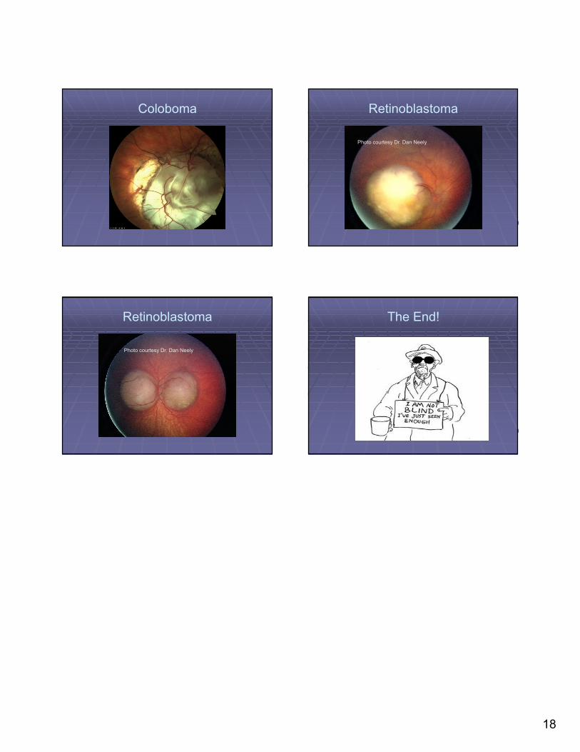
Retinoblastoma
� Malignant, congenital tumor
� Derived from retinoblasts
� Most common intraocular tumor in infants / children
� 70 % unilateral
� 1 / 15,000 children
� 200-300 US cases/yr
� No racial or sexual predilection
� Two types……
� 1) Inherited (AD). Less than 10% of cases. Frequently multifocal and bilateral
� 2) Sporadic. Usually unilateral
Retinoblastoma
� Can metastasize and be fatal if detected too late
� Survival rate 90+% if detected early (typical age of diagnosis is around 18 months)
� LEUKOCORIA
� Strabismus
� Poor VA
� Involvement of ONH is ominous
� Many treatment options depending on multiple factors
� IV chemo?
Genetics
� RB survivor with inherited type: 50% chance of transmitting to their children
� Healthy parents: one child with RB; 6% chance of another : two or more children with RB; 50% of another
Leukocoria differentials
� Retinoblastoma
� Coat’s disease
� Toxocariasis
� Toxoplasmosis
� Congenital cataract
� PHPV
� coloboma
18
Coloboma Retinoblastoma
Photo courtesy Dr. Dan Neely
Retinoblastoma
Photo courtesy Dr. Dan Neely
The End!