14th annual scholars symposium · 2020-01-06 · greetings from the dean michelle robinson, dmd, ma...
TRANSCRIPT

Scholars Symposium14th Annual
March 6, 2019
Optimizing Oral Health in Alabama and Beyond
Greetings from the Dean
Michelle Robinson, DMD, MA Interim Dean, UAB School of Dentistry
I am delighted to welcome you to our 14th Annual Scholars Symposium. The event highlights one of our school’s four foundational pillars, Research.
Each year, Scholars Symposium provides a venue for our faculty, residents, students, staff, and guests to discuss a broad range of topics associated with the methods and outcomes of oral health, dental and craniofacial research. It fosters interactions between the School of Dentistry and other schools across campus, as well as UAB’s interdisciplinary centers.
Scholars Symposium is also a yearly celebration of our rich history and current accomplishments, showcasing the achievements of students, residents, and faculty as we prepare the next generation of academicians and scientists to impact the oral health of our nation. We are proud of our presenters whose dedication to scholarship is on display today. Their excellence is recognized throughout our nation and around the world.
Through robust partnerships and from the classroom, to the chairside, to the community, UAB School of Dentistry continues to build on a strong tradition of scientific discovery. Our work in the area of oral health and pregnancy outcomes has been featured in the university’s new research and innovation campaign, “UAB. Powered by will.” The campaign highlights UAB research, innovation and entrepreneurship at the frontiers of medicine, technology, and other fields. Also, the school has two projects that are strong contenders in the UAB Grand Challenge. The projects, “Re-engineering Equal Access to Comprehensive Healthcare (REACH)” and “Opioid Overdose Prevention and Treatment Using Precision Medicine,” are part of UAB’s ambitious but achievable goal to harness science, technology, policy and innovation to solve an important local, state, national or global problem.
The strength of our research pillar centers on our various areas of nationally and internationally recognized scientific expertise including the oral microbiome, biomaterials, oral head and neck cancer, and the epidemiology of caries. It is also underscored by our leadership of the National Dental Practice Based Research Network (NDPBRN), housed at the UAB School of Dentistry. Fittingly, the emphasis of this year’s symposium is Dental Public Health/Epidemiology which highlights current clinical outcomes issues in oral health and the mission of the NDPBRN.
In keeping with this year’s emphasis, it is a special honor to welcome keynote speaker, Dr. Michael Glick. Dr. Glick is Professor and William M. Feagans Chair at the University of Buffalo School of Dental Medicine. Well-known for his medicine-oriented approach to dental care, he served as chair for the Vision 2020, World Dental Federation (FDI), and is presently co-chairing the FDI Think Tank. In addition to his many leadership positions, he has received numerous awards for his entrepreneurship, community and professional activities, clinical acumen, teaching excellence and writings. Dr. Glick’s keynote address is made possible through the generosity of Dr. Robert Taylor. Dr. Taylor established the Robert E. and Ann S. Taylor Endowed Lectureship in Oral Biology and his wonderful gift supports our keynote speaker each year.
In addition, we deeply appreciate the contributions of so many, including the generous financial support of the Hinman Dental Society, Dentsply International, the American and International Colleges of Dentists, Omicron Kappa Upsilon National Dental Honor Society, and the University of Alabama at Birmingham School of Dentistry Alumni Association. Also, thanks to the following UAB University Wide Interdisciplinary Research Centers (UWIRC): Global Center for Craniofacial and Oral Dental Disorders (GC-CODED), Comprehensive Arthritis, Musculoskeletal, Bone, and Autoimmunity Center (CAMBAC), and the Microbiome Center. We are also grateful for the support of the Alabama Chapter of the American Association for Dental Research (AADR), and the UAB Chapter of the AADR National Student Research Group. Also, kind thanks to the SOD Research Advisory Committee, the SOD Office of Development & Alumni Relations, and our many dedicated staff volunteers for making Scholars Symposium a success.

2
Michael Glick, DMD
Professor and William M. Feagans Chair School of Dental Medicine, University at Buffalo
State University of New York. Editor, The Journal of the American Dental Association
Dr. Glick received his DMD from the Hebrew University School of Dental Medicine in Israel and Temple University Dental School in Philadelphia. He completed residency training in Oral Medicine from the University of Pennsylvania School of Dental Medicine. Dr. Glick started his academic career as an Assistant Professor of Oral Medicine at the Temple University School of Dentistry. He then joined the University of Pennsylvania School of Dental Medicine in 1994, as an Associate Professor of Oral Medicine. He later moved to the University of Medicine and Dentistry, New Jersey. There he served as Director of the postgraduate training program in Oral Medicine and later as Chair, Department of Diagnostic Sciences. In 2007, he joined A.T. Still University, School of Osteopathic Medicine as Associate Dean for Oral and Medical Sciences. In 2009, Dr. Glick became the Dean of School of Dental Medicine, University at Buffalo, SUNY. He concluded his service as Dean in 2015. Dr. Glick is Diplomate, American Board of Oral Medicine. Dr. Glick has received numerous awards for his entrepreneurship, community and professional activities, clinical acumen, teaching excellence and writings. He founded one of the first dental clinics in the US dedicated to treating patients infected with HIV. Dr. Glick served as chair for the Vision 2020, World Dental Federation and has presented more than 300 continuing education courses. He has been an invited speaker across the United States and in over 30 countries. Dr. Glick has authored more than 250 journal publications, book chapters and editorials, and has edited and co-edited 10 textbooks on dental management and oral medicine. He is the recipient of over six million dollars of grant support. Dr. Glick is past-President of the American Board of Oral Medicine and since 2005 serves as the Editor of The Journal of the American Dental Association (JADA).
Scholars Symposium Keynote Speaker
3
Gregg H. Gilbert, DDS, MBA, FAAHD, FACD, FICD Distinguished Professor/Chair
Behavioral & Population Sciences, UAB School of Dentistry Dr. Gilbert is Chair of the Department of Clinical & Community Sciences at the UAB School of Dentistry and Distinguished University Professor, a campus-wide rank that recognizes international accomplishments. His research interests are in the field of oral health clinical research, including practice-based research, oral epidemiology, and dental behavioral sciences research. He serves as National Network Director for The National Dental PBRN. Dr. Gilbert is the recipient of the Distinguished Scientist Award from the International Association for Dental Research. He is a Fellow of the American Association of Hospital Dentists and a Fellow of the American and International Colleges of Dentists. In recognition of his vision and efforts to bridge research, education, and practice, he received the William J. Gies Award from the ADEA-Gies Foundation for outstanding vision in dental education. He has authored over 220 peer-reviewed journal articles, scientific abstracts, and non-peer-reviewed publications. Dr. Gilbert has distinguished himself professionally by sustained significant contributions to oral health research that bridge investigation between the population and clinical levels. His work has changed the oral health research model by bringing everyday clinicians and patients to the discussion table – end users who have an immense amount of practical knowledge and wisdom that previously had been largely ignored. He has received grant funding as PI on numerous NIH, VA, and foundation grants. As of 2018, he had received $94 million in research funding for his work as Principal Investigator, with an additional $12+ million as co-PI or Investigator. Dr. Gilbert has served on numerous grant application review committees for the NIH, VA, and research foundations.
Scholars Symposium Faculty Presentation
4
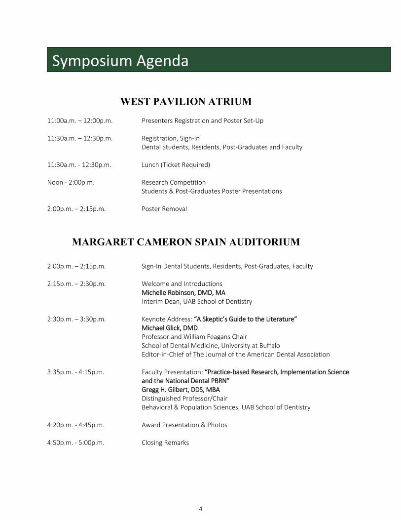
WEST PAVILION ATRIUM
11:00a.m. – 12:00p.m. Presenters Registration and Poster Set-Up 11:30a.m. – 12:30p.m. Registration, Sign-In Dental Students, Residents, Post-Graduates and Faculty 11:30a.m. - 12:30p.m. Lunch (Ticket Required) Noon - 2:00p.m. Research Competition
Students & Post-Graduates Poster Presentations 2:00p.m. – 2:15p.m. Poster Removal
MARGARET CAMERON SPAIN AUDITORIUM
2:00p.m. – 2:15p.m. Sign-In Dental Students, Residents, Post-Graduates, Faculty 2:15p.m. – 2:30p.m. Welcome and Introductions
Michelle Robinson, DMD, MA Interim Dean, UAB School of Dentistry
2:30p.m. – 3:30p.m. Keynote Address: “A Skeptic’s Guide to the Literature”
Michael Glick, DMD Professor and William Feagans Chair School of Dental Medicine, University at Buffalo Editor-in-Chief of The Journal of the American Dental Association
3:35p.m. - 4:15p.m. Faculty Presentation: “Practice-based Research, Implementation Science
and the National Dental PBRN” Gregg H. Gilbert, DDS, MBA Distinguished Professor/Chair Behavioral & Population Sciences, UAB School of Dentistry
4:20p.m. - 4:45p.m. Award Presentation & Photos 4:50p.m. - 5:00p.m. Closing Remarks
Symposium Agenda

5
Thank you for judging the poster competition.
Basic Science Judges
Olga Beliaeva, PhD Quamarul Hassan, PhD Jenny Katz, DDS, PhD Mohammed Kass, PhD Suzanne M Michalek, PhD Champion Deivanayagam, PhD Sarah Peters, PhD
Heather Ray, PhD Jessica Scoffield, PhD Yang Yang, PhD Nabiha Yusuf, PhD Jue Wang, PhD Ping Zhang, PhD
Clinical Science Judges
Warren Arrasmith, DMD Celin Arce Urena, DDS, MS Edward Bradford, Jr., DDS, MPH James Broome, DDS Ramakiran Chavali, BDS, MS Kyounga Cheon, DMD, MS Noel Childers, DDS, PhD, MS Terpsithea Christou, DDS, MS Diane Feagin, DMD, MSPH George Ford, DDS Chin-Chuan Fu, DDS, MS Nicolaas Geurs, DDS, MS Yung-Tsung Hsu, DMD Janice Jackson, DMD Michael Kase, DMD
Maninder Kaur, BDS, MPH, MS Shandra Keith, DMD Nathaniel Lawson, DMD, PhD Perng-Ru Liu, DMD Jocelyn S McClelland, DDS Carly McKenzie, PhD Lillian Mitchell, DDS Toni Neumeier, DMD, MS Somsak Sittitavornwong, DMD, MS Timothy Smith, DMD, MBA Nada Souccar, DDS, MS Alvin Stevens, DMD Christos Vlachos, DMD, DDS, MS Wen-Chou Wu, DDS
Poster Competition Judges
6
Dr. Robert Taylor for the Robert E. and Ann S. Taylor Endowed Lectureship in Oral Biology
Omicron Kappa Upsilon National Dental Honor Society
Office of Development & Alumni Relations
Alabama Chapter
Global Center for Craniofacial, Oral and Dental Disorders
Microbiome Center Comprehensive Arthritis, Musculoskeletal, Bone and Autoimmunity Center
Sponsors

7
Volunteers
Thank you to all who volunteered to make the
14th Annual Scholar’s Symposium a success.
Sheila Akers
Maria Bird
Sheila Blake
Bright Chang
Teresa Creel
Robert Dean
Renee Holifield
Shirley Jackson
Lynne Jarreau
Nancy Parsons
Angela Rembert
LaTara Rogers
Nannozi Ssenkoloto
Kelly Stinson
Sylvia Strothers
Adam Stoves
Rosie Turner
Sheila Turner
Abby Vinson
8
Basic Science / Pre-Doctoral
101 – Tanner Godfrey
BAF45A, a Critical Regulator of Osteoblast Development and Activity
T. Godfrey, M. Rehan, B. Wildman, T. Busby, Y. Chen, L. Matalka, M. Hassan
Objective: Two key processes affecting the quantity of bone during development and maturity are the differentiation and maintenance of bone building cells, osteoblasts. Osteoblast differentiation and maintenance requires remodeling of the DNA-chromatin structure. This is accomplished through the Brg1 Associated Factors (BAF) complex. The objective of this work is to determine the role of a specific BAF factor, BAF45A, in the determination and maintenance of the osteoblast cell type required for bone formation and maintenance.
Method: The osteoblast-specific subset of BAF factors required for maturation was investigated using immuno-precipitation mass spectrometry (IP-MS) of BAF from differentiating mouse calvarial cells. Osteoblast-specific deletion of Baf45a was completed using a Baf45a floxed-allele mouse (Baf45afl/fl) crossed with a Prrx1-Cre (pre-differentiation) or osteocalcin-Cre (post-differentiation) mouse model. Femurs were analyzed at 2 and 6 months of age by Micro-CT (uCT), histomorphometry, 3-point-bend test, and RTqPCR. Changes in DNA-chromatin structure with deletion of Baf45a were assessed in calvarial cells isolated from Baf45afl/fl mice crossed with an inducible Cag-Cre model. These cells were differentiated (D3, D13) and analyzed by ATAC sequencing, ChIP-qPCR and RT-qPCR. Results: IP-MS analysis of the osteoblast BAF complex revealed a subset of BAF factors unique to osteoblasts. Deletion of Baf45a prior to differentiation (Prrx1-Cre) revealed that BAF45A is required for normal osteoblast differentiation, resulting in decreased bone in 2-month-old femurs. Deletion of Baf45a post-differentiation (Osteocalcin-Cre) demonstrated that BAF45A is required for osteoblast cell type maintenance. Here we observed decreased bone at 2 and 6 months of age, with large increases in bone marrow adiposity at 6 months. Analysis of DNA-chromatin structure and gene expression revealed that loss of Baf45a results in repressed osteoblast specific genes.
Conclusions: These data demonstrate that Baf45a is critical in the development and maintenance of osteoblasts. Loss of Baf45a results in repressed osteoblast specific genes, ultimately decreasing bone levels during synthesis and maintenance.
102 – Victoria Matkins
Altered Differentiation of Mesenchymal Stromal Cells during Inflammation
V. Matkins, V. Camacho, A. Hoang, S. Patel, R. Welner
Objective: The bone marrow microenvironment (BMM) is a complex network of blood and non-hematopoietic cells. These cells form the stem cell niche to aid in regulation of hematopoietic stem cell self-renewal and differentiation. Within the non-hematopoietic compartment, subpopulations of mesenchymal stromal cells (MSCs) give rise to osteoblasts and adipocytes. These cells communicate with the hematopoietic system through adhesion molecules and cytokines to maintain homeostasis. Inflammation’s disruptive impact on the hematopoietic system has been greatly studied; but how inflammation impacts the BMM is poorly understood. During inflammatory conditions bone loss has been noted; therefore, we hypothesize an increase in the MSC population to compensate for the defect in bone differentiation.
9
Method: Chronic infection using LCMV or direct Toll-like receptor stimulation followed by flow cytometry to assess phenotypic changes in BMM subpopulations, and tri-lineage differentiation. Additionally, we use lineage-tracing models for an unbiased assessment of altered stromal lineages. The lineage-specific Cre models mark stroma (Prrx1), adipocytes (AdipoQ), and early (Osx) and late (OCN) stages of osteoblasts.
Results: Our data shows increased MSCs with decreased osteoblasts and adipocytes just days after Toll-like receptor stimulation. One week later, the osteoblasts are still decreased, while the MSCs are now unchanged. However, differences in stromal cell maturation potential persist from in vivo stimulated versus in culture stimulated when differentiated into their respective lineages. In vivo stimulated cells show increased osteoblasts differentiation while cells stimulated in culture have the opposite impact on bone formation. During adipocyte differentiation, there is no change in the number of adipocytes present; however, the adipocytes are more mature from in vivo stimulation and less mature in culture.
Conclusions: Likely, the hematopoietic system impacts the stromal compartment leading to these alterations in differentiation bias. Understanding changes in the BMM during inflammation will allow for therapeutic intervention in inflammatory diseases such as arthristis, osteomyelitis, and leukemia.
103 – Edwin Rojas
Diadenylate Cyclase: A promising Target for Biofilm Inhibition in S. mutans
E. Rojas, H. Wu
Objective: To present a novel target for the prevention of dental cavities.
Method: Biofilm Inhibition Assay, Biofilm Dispersion Assay, Coralyne Assay, High-throughput in silico screening, organic synthesis.
Results: The lead compound up to this point, known as PB8, shows to have an IC50 value of 25 micromolar, and the coralyne assay shows that it is preventing the synthesis of cyclic di-AMP.
Conclusions: PB8 analogs are currently being tested for SAR studies, and other lead compounds are being ordered from NCI database for in vitro testing.
104 – Joshua Mieher
Structural and Functional Characterization of Streptococcus intermedius Surface Antigen PAS
J. Mieher, N. Schormann, M. Patel, S. Purushotham, C. Deivanayagam
Objective: Subgingival dental plaque can lead to periodontal disease; the infection can lead to tooth loss as well as leaving the host susceptible to bacterial invasion of the blood stream. PAS is an AgI/II-family surface antigen on Streptococcus intermedius, which is a contributor to subgingival dental plaque and is implicated in extraoral infections. Given the ability of AgI/II family proteins to adhere to glycoprotein 340 (Gp340), this study aims to characterize the structure of PAS and its interactions with Gp340 as a factor in its attachment to host surfaces.
Method: The PAS domains, VPas and C123Pas, were purified using affinity and ion exchange. Crystal data was collected at the NE-CAT beamline at the Advanced Photon Source at the Argonne National Laboratory. Surface Plasmon resonance was used to quantify the affinity of the interaction between PAS domains, FLPas, VPas, and C123Pas, and immobilized Gp340, SRCR1 or SRCR123.
10
Results: The crystal structure of VPas and C123Pas adopt folds similar to that of AgI/II, indicating high similarity in these structures. Like other V-regions of AgI/II, GbpC and SspB it has a conserved calcium binding site. Surface plasmon resonance studies show every construct of PAS, FLPas, VPas and C123Pas interacted with high nanomolar affinity to SRCRs and Gp340. Comparatively, the C-regions of AgI/II and SspB were typically lower than the V-region.
Conclusions: Here we present the crystal structure of the V- and C-regions of Pas, and its adherence characteristics to SRCR domains of Gp340. The nanomolar affinities were not much different from its binding affinities compared to AgI/II and SspB, which is line with the structures showing a high degree of similarity.
105 – Theodore Busby, III
The Role of Mammalian SWI/SNF (BAF) Chromatin Remodeling in Tooth Cells
T. Busby, T. Godfrey, B. Wildman, M. Rehan, Q. Hassan
Objective: Cellular proliferation, differentiation, and commitment are regulated in part by the chromatin landscape of the cell. The mammalian SWI/SNF (BAF) chromatin remodeling complex contributes to gene activation by sliding nucleosomes into an open conformation around gene promoters. Cell specific regulation arises from the homolog composition of BAF subunits. However, little is known about BAF complex regulation in mineralized tissue. Our lab has identified BAF45A as an essential BAF subunit for differentiation in osteoblasts, the primary mineral depositing cells of the bone. Similar in function to osteoblasts, odontoblasts are the primary mineralizing cells of the tooth with mesenchymal origin like osteoblasts. This objective of this study is to characterize the molecular function of BAF45A and its homologs BAF45B/C/D during differentiation and mineralization of both bone and tooth cells.
Method: Based on our preliminary studies, we hypothesize that stage specific deletion of BAF45A will impair tooth formation. To test this, BAF45Afl/fl was deleted in mice in the limb bud mesenchyme by Prrx1-Cre. MicroCT was performed on 2-month-old WT and BAF45A KO mice to analyze mineral density of the molars. To characterize the functional role of BAF45A in vitro, we subjected the OD-21 pre-odontoblast cell line differentiation conditions. We analyzed the changes in expression of genes specific to odontoblast and mineralized tissue at the stages of differentiation. We also performed ChIP QPCR to assess the changes in BAF complex occupancy at these tissue specific loci.
Results: Here we show that BAF45A and BAF45D are preferentially expressed in both osteoblasts and odontoblasts compared to BAF45B and BAF45C. BAF45D has been reported to be ubiquitously expressed across tissue types as part of the canonical BAF complex. Thus, we focused on the function of BAF45A as part of the Polybromo-BAF (PBAF) complex in odontogenesis and osteogenesis. Deletion of BAF45A in mouse leads to defects in craniofacial development. Furthermore, in vivo and in vitro data suggests that BAF45A not only promotes expression of mineralized tissue specific genes, but also prevents alternative phenotypes. Here we investigate BAF45A dependent chromatin regulation and how this promotes the deposition of active histone modifications, including H3K27 acetylation.
Conclusions: BAF45A is an important BAF complex member for the development of mineralized tissue, both tooth and bone. BAF45A, in addition to BAF45D, are the primary homologs expressed in these tissues and it appears at this time that the expression of BAF45B and C are negligible for differentiation and development.
11
106 – Benjamin Wildman
EZH2 is Regulated by the MiR-23a Cluster to Maintain Bone Mass In Vivo
B. Wildman, T. Godfrey, M. Rehan, Y. Chen, T. Busby, Q. Hassan
Objective: Differentiation of pre-osteoblasts is critical to controlling in-vivo development and growth of bone. Recent studies highlight the importance of epigenetic regulation in directing osteoblast commitment and function. Here we show that the microRNA-23a cluster (miR-23a, 27a, and 24-2) controls bone mass in-vivo through a previously unknown EZH2 mediated epigenetic mechanism.
Method: First, we knocked down the miR-23a cluster in mouse pre-osteoblasts (MC3T3-E1) cells with an anti-microRNA cassette (miRZIP). Next, we created a mouse model that inducibly expresses the miRZIP cassette to knock-down the microRNA cluster (miR-23aClZIP) in-vivo. Luciferase and Chromatin Immunoprecipitation (ChIP) assays along with RNA sequencing were performed to elucidate the mechanism of miR-23a cluster action in maturing osteoblasts.
Results: MiR-23a cluster knockdown increased the intensity of Alkaline Phosphatase staining in MC3T3-E1 cells. Additionally, it upregulated mRNA expression of osteogenic marker genes such as Runx2 and Osteocalcin.
Micro-CT analysis of 2 month old femurs showed that trabecular bone volume and trabecular number significantly increased in miR-23aClZIP mice as compared to controls. Additionally, connective density and trabecular thickness were significantly greater while trabecular space was significantly decreased. Supporting this increased bone mass, Runx2 expression levels were significantly upregulated while the levels of a potent epigenetic repressor Ezh2 were significantly reduced in whole bone RNA sequenced from miR-23aClZIP mice.
Mechanistically, we found that the miR-23a cluster inhibits RUNX2 translation by binding to the 3’ UTR of Runx2 mRNA transcripts. Furthermore, ChIP assays revealed RUNX2 binds to the Ezh2 promoter inhibiting transcription in MC3T3-E1 cells. Additional ChIP experiments in miR-23aClZIP mouse primary calverial pre-osteoblasts showed that miR-23a cluster knockdown results in decreased binding of the epigenetic repressor EZH2 to osteogenic gene promoters such as Osteocalcin and Runx2, resulting in a more osteogenic transcription program.
Conclusions: We developed a novel microRNA cluster knockdown mouse model allowing us to decipher how the miR-23a cluster orchestrates bone mass maintenance in-vivo.
107 – Parul Sarwalia
miRNAs In The Maternal Recognition of Pregnancy
P. Sarwalia, P.Dubey, A. Kumar
Objective: The inability of an embryo to get implanted is the leading cause of pregnancy failure. Non- coding RNAs are the key players during the critical event of adhesion of blastocyst to the uterus. This study was undertaken to find out the role played by miRNAs during implantation of the embryo to establish the maternal-fetal crosstalk.
Method: Blastocysts produced after IVF were allowed to hatch and were cultured in a trophoectoderm growth specific medium for 21 days. The purity of cultured TE cells was confirmed by cdx2, a transcription factor specific to TE cells. The spent media were collected during the course of this culture and miRNAs were isolated on day 6, day 12, day 17 and day 21. Seven miRNAs, including one novel

12
miRNA reported earlier from our lab NGS data, were selected for this study and their expression profiles were analyzed to find out the miRNA which revealed characteristically enhanced expression on day 21. Being a novel candidate, mir-1246 was chosen for transfecting its mimic in cultured endometrial epithelial cells. For transfection studies, a primary culture of endometrial epithelial cells from luteal phase uterus was established. Morphological characterization of endometrial epithelial cells and their distinction from stromal cells was confirmed by various characterization strategies. Prior to transfection the cultured EECs were primed with IFN-T to simulate the in vivo physiological conditions during pregnancy establishment. The transfection protocol was optimized using pAcGFP1-N1 vector by lipofection. We used miR-1246 mimic which was transfected into cultured EECs to investigate its effect on the expression of surface adhesion molecules of EECs. Three target candidate molecules viz. mucin 1(MUC1), integrin beta-8(ITGB-8) and osteopontin-1(SPP1) involved in implantation were chosen, as predicted by in silico target prediction tools, for their expression profiles in transfected cells vis-à-vis the control group.
Results: Three miRs viz. miR-1246, miR-let-7a and miR-let-7b, displayed a very typical expression pattern with highest expression on day 21. The expression of mucin1 decreased significantly in transfected endometrium epithelial cells which is described as crucial for successful implantation as it plays role in providing a scaffold for selectin ligands that potentially could support blastocyst interactions via selectins at the maternal-fetal interface extracellular matrix.
Conclusions: We conclude that miR-1246 is most likely to play an important role in making the endometrium receptive for implantation and could be a potential marker for early detection of successful pregnancy.
Clinical Science / Pre-Doctoral
108 – Marikit Magkalas
Effect of Restoration Size on the Clinical Performance of Posterior Composites
M. Magkalas, N. Lawson Objective: To determine if material fracture, margin adaptation, margin discoloration, or post-operative sensitivity are related to the bucco-lingual dimensions of posterior composite restorations.
Method: The primary author completed IRB training and was added to an ongoing clinical trial comparing three posterior restorative materials after 1 year of clinical service. Photographs from the clinical trial were obtained of the preparation of 142 posterior (Class I or II) restorations. Using Microsoft PowerPoint, the primary author measured the bucco-lingual dimensions of the restored tooth from the location of the buccal cusp tip to the lingual cusp tip. For molars, a line was drawn between both lingual cusp tips and both buccal cusp tips and the distance between the centers of those lines was recorded as the bucco-lingual dimension of the tooth. Then the bucco-lingual dimension of the cavity preparation was estimated based on the most buccal and most lingual extension of the preparation. Finally, a percentage of intercuspal dimension was determined by dividing the bucco-lingual dimension of the cavity preparation by the intercuspal dimension of the tooth. Some preparations with buccal or lingual extensions were greater than 100% of the intercuspal dimension. These percentages of intercuspal dimension were then compared to four different FDI criteria that had been previously assigned to the restoration in the clinical trial using a Kruskal-Wallis test. FDI scoring ranges from a value of 1, representing clinically ideal, to a value of 5, which requires replacement.
13
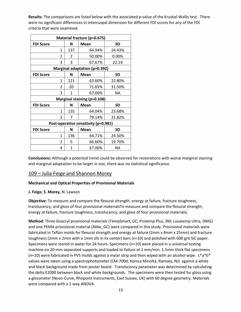
Results: The comparisons are listed below with the associated p-value of the Kruskal-Wallis test. There were no significant differences in intercuspal dimension for different FDI scores for any of the FDI criteria that were examined.
Material fracture (p=0.675) FDI Score N Mean SD
1 137 64.94% 24.43% 2 2 50.00% 0.00% 3 3 67.67% 22.19
Marginal adaptation (p=0.392) FDI Score N Mean SD
1 121 63.60% 22.80% 2 20 71.65% 31.50% 3 1 67.00% NA
Marginal staining (p=0.108) FDI Score N Mean SD
1 135 64.04% 23.68% 2 7 79.14% 31.82%
Post-operative sensitivity (p=0.981) FDI Score N Mean SD
1 136 64.71% 24.50% 2 5 66.60% 19.70% 4 1 67.00% NA
Conclusions: Although a potential trend could be observed for restorations with worse marginal staining and marginal adaptation to be larger in size, there was no statistical significance.
109 – Julia Feige and Shannon Morey
Mechanical and Optical Properties of Provisional Materials
J. Feige, S. Morey, N. Lawson
Objective: To measure and compare the flexural strength, energy at failure, fracture toughness, translucency, and gloss of four provisional materialsTo measure and compare the flexural strength, energy at failure, fracture toughness, translucency, and gloss of four provisional materials.
Method: Three bisacryl provisional materials (TempSmart, GC; Protemp Plus, 3M; Luxatemp Ultra, DMG) and one PEMA provisional material (Alike, GC) were compared in this study. Provisional materials were fabricated in Teflon molds for flexural strength and energy at failure (2mm x 4mm x 25mm) and fracture toughness (2mm x 2mm with a 1mm slit in its center) bars (n=10) and polished with 600 grit SiC paper. Specimens were stored in water for 24 hours. Specimens (n=10) were placed in a universal testing machine on 20-mm separated supports and loaded to failure at 1 mm/min. 1.5mm thick flat specimens (n=10) were fabricated in PVS molds against a mylar strip and then wiped with an alcohol wipe. L*a*b* values were taken using a spectrophotometer (CM-700d; Konica Minolta, Ramsey, NJ) against a white and black background made from poster board. Translucency parameter was determined by calculating the delta E2000 between black and white backgrounds. The specimens were then tested for gloss using a glossmeter (Novo-Curve, Rhopoint Instruments, East Sussex, UK) with 60 degree geometry. Materials were compared with a 1-way ANOVA.
14
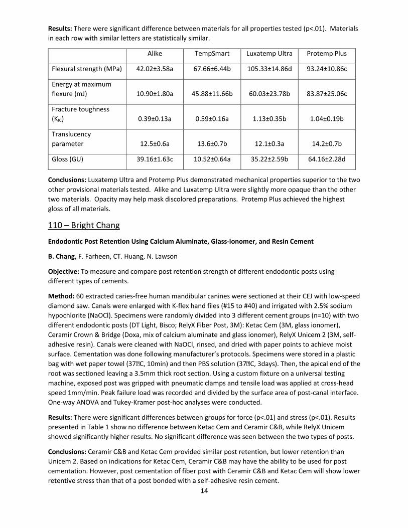
Results: There were significant difference between materials for all properties tested (p<.01). Materials in each row with similar letters are statistically similar.
Alike TempSmart Luxatemp Ultra Protemp Plus
Flexural strength (MPa) 42.02±3.58a 67.66±6.44b 105.33±14.86d 93.24±10.86c
Energy at maximum flexure (mJ) 10.90±1.80a 45.88±11.66b 60.03±23.78b 83.87±25.06c
Fracture toughness (KIC) 0.39±0.13a 0.59±0.16a 1.13±0.35b 1.04±0.19b
Translucency parameter 12.5±0.6a 13.6±0.7b 12.1±0.3a 14.2±0.7b
Gloss (GU) 39.16±1.63c 10.52±0.64a 35.22±2.59b 64.16±2.28d
Conclusions: Luxatemp Ultra and Protemp Plus demonstrated mechanical properties superior to the two other provisional materials tested. Alike and Luxatemp Ultra were slightly more opaque than the other two materials. Opacity may help mask discolored preparations. Protemp Plus achieved the highest gloss of all materials.
110 – Bright Chang
Endodontic Post Retention Using Calcium Aluminate, Glass-ionomer, and Resin Cement
B. Chang, F. Farheen, CT. Huang, N. Lawson
Objective: To measure and compare post retention strength of different endodontic posts using different types of cements.
Method: 60 extracted caries-free human mandibular canines were sectioned at their CEJ with low-speed diamond saw. Canals were enlarged with K-flex hand files (#15 to #40) and irrigated with 2.5% sodium hypochlorite (NaOCl). Specimens were randomly divided into 3 different cement groups (n=10) with two different endodontic posts (DT Light, Bisco; RelyX Fiber Post, 3M): Ketac Cem (3M, glass ionomer), Ceramir Crown & Bridge (Doxa, mix of calcium aluminate and glass ionomer), RelyX Unicem 2 (3M, self-adhesive resin). Canals were cleaned with NaOCl, rinsed, and dried with paper points to achieve moist surface. Cementation was done following manufacturer’s protocols. Specimens were stored in a plastic bag with wet paper towel (37C, 10min) and then PBS solution (37C, 3days). Then, the apical end of the root was sectioned leaving a 3.5mm thick root section. Using a custom fixture on a universal testing machine, exposed post was gripped with pneumatic clamps and tensile load was applied at cross-head speed 1mm/min. Peak failure load was recorded and divided by the surface area of post-canal interface. One-way ANOVA and Tukey-Kramer post-hoc analyses were conducted.
Results: There were significant differences between groups for force (p<.01) and stress (p<.01). Results presented in Table 1 show no difference between Ketac Cem and Ceramir C&B, while RelyX Unicem showed significantly higher results. No significant difference was seen between the two types of posts.
Conclusions: Ceramir C&B and Ketac Cem provided similar post retention, but lower retention than Unicem 2. Based on indications for Ketac Cem, Ceramir C&B may have the ability to be used for post cementation. However, post cementation of fiber post with Ceramir C&B and Ketac Cem will show lower retentive stress than that of a post bonded with a self-adhesive resin cement.

15
111 – Meghana Sthanam and Drew Patchett
Evaluating the effect of Enamel Microabrasion on Varying Levels of Discoloration
M. Sthanam, D. Patchett, A. Robles
Objective: To demonstrate the effectiveness of enamel microabrasion therapy on mild white spot lesions.
Method: Screenings for candidates were performed using clinical presentation followed by close inspection of potential lesions under Valo UV ultraviolet light. Candidates were approved based off of the amount of definition of prospective lesions under trans illumination. Approved candidates underwent current standard for micro abrasion therapy. This means each tooth’s facial surface is coated in 6.6% hydrochloric acid imbedded with silicon carbide micro particles. Each coated surface was abraded using a hard bristled prophy cup at 500 rpm under constant pressure for 60 seconds per tooth per application. This process was completed during each case a number of times between 1 and 5 dependent on the severity of the case, water rinse was applied between each abrasion. Results were documented using digital photography before and after each session.
Results: A marked decrease was found in clinical white spot presentation for “superficial” spots as defined in this study.
Conclusions: Microabrasion is an appropriate cosmetic treatment for select cases of clinically present white spots on enamel.
Undergraduate
112 – Evalynn Ngamau
Detection of Bacteriocin Activity of Streptococcus mutans on Oral Streptococci
E. Ngamau, S. Momeni, N. Childers
Objective: Streptococcus mutans (Sm) is one of the main etiological agents in dental caries, the most common chronic childhood disease. With the increase of antimicrobial resistance, there is increased interest in the discovery of novel natural antimicrobials. Bacteriocins, ie., antimicrobial peptides, can provide an ecological-edge for Sm colonization and caries development. The aim of this study was to determine bacteriocin production in Sm from caries/caries-free subjects.
Method: Forty representative Sm strains from a longitudinal epidemiological study of over 14,000 strains were analyzed by agar plate bacteriocin assay in addition to 10 control strains. Each strain was used as both an indicator and a producer. Whole genome sequencing was performed, and SPAdes was used for de novo assembly. Comparative genomics was performed using Sybil.
Results: Four strains were highly inhibitive (ie., inhibited all strains tested) including 3 clinical strains G02, G11, G13 and one control strain LM7, with zones of inhibition ranging 9-29.5mm (median 16mm). Seven strains were moderately inhibitive (ie., inhibited more than half the strains or inhibited all strains, but with less intensity) with zones of inhibition ranging 5-18.5mm (median 6mm). A Fisher’s Exact test was used to determine the association between highly inhibitive or inhibitive strains and the presence of caries. No statistically significant difference was found between Sm strain inhibition and caries status, possibly due to small sample size.

16
Conclusions: The discovery of the 4 strains that were highly inhibitive against oral streptococci offers potentially new natural antimicrobial compounds that may be used to inhibit, treat or prevent dental caries. Furthermore, identification of new antimicrobial compounds from Sm may be useful against other pathogens. Further study is needed to identify the exact metabolites responsible for the inhibition.
113 – Nellie Baghaei
NFI-C Downstream Targets Involved in Root Elongation and Resorption
N. Baghaei, O. Mamaeva, D. Crossman, M. MacDougall, E. Lamani Objective: NFI-C, a transcriptional factor, is a master regulator of root formation. Previously, we identified a NFI-C mutation associated with a novel autosomal recessive form of Radicular Dentin Dysplasia (AR-RDD). Objectives: 1) To identify down-stream target genes regulated by NFI-C critical for (a) root elongation, focusing on Msx2 and WNT signaling pathway, and (b) resorption; 2) to evaluate the potential role of NFI-C in the cellular response to orthodontic compressive forces in vitro.
Method: AR-RDD patient and unaffected control periodontal ligament (PDL) cells were grown and harvested for gene profiling (Affymetrix U133-Plus-2.0 chip). Bioinformatics was used to identify predicted NFI-C binding sites in the promoters of potential targets and to design flanking primers to these cis-elements. Chromatin immunoprecipitation (ChIP) was performed using a NFI-C antibody and target cis-element primers to investigate protein-chromatin interactions with product confirmation by DNA sequencing. Confluent PDL cells (RDD/control) were subjected to compressive forces (24hrs) and qRT-PCR analysis was used to evaluate the effect on cellular response to mechanical stimulus.
Results: Through transcriptomics, altered gene expression profiles in AR-RDD PDL cells were identified belonging to signaling pathways involved in root elongation and resorption. WNT5A, TCF4, TLR3, CTSK, OPG, IL6, IL1-RN, MMP2, MMP3, MMP12, were upregulated >2 fold (p<0.05), while MSX2, WNT2, DKK1, TIMP3 were downregulated >2 fold (p<0.05). ChIP revealed MSX2, associated with root elongation and eruption, is directly regulated by NFI-C. Force-dependent changes in RANKL expression were seen in AR-RDD PDL versus control cells.
Conclusion: Our data supports NFI-C’s role in root development and suggests an important function related to pathways critical for root elongation and resorption. However, since Msx2 has been shown to modulate RANK signaling; the response to mechanical stimulus identified may be indirect of NFI-C.
114 – Aubrey Johnson and Henry Kendrick
Patient-derived Xenografts of Ameloblastoma for In vivo Imaging
A.O. Johnson, H.S. Kendrick, L.S. Moore, J.M. Warram, A.B. Morlnadt, H.M. Amm
Introduction: Ameloblastomas demonstrate locally aggressive and destructive behavior, primarily in the posterior mandible. Wide variability in surgical treatment has been advocated leading to residual disease and wide ranges of disease recurrence (3-62%). It has been previously demonstrated that fluorescently labeled epidermal growth factor receptor (EGFR) antibodies can successfully identify microscopic tumors in multiple in vivo preclinical models of human cancers with limited toxicity. Objective: The objective is to demonstrate the specificity and sensitivity of fluorescently labeled anti-EGFR antibody, cetuximab-IRDye800CW, to ameloblastoma tumors in vivo using a patient-derived xenograft (PDX) model.
17
Method: Surgical tissue specimens of ameloblastoma were implanted subcutaneously into the flanks of immunocompromised mice and were imaged following tail vein injection of cetuximab-IRDye800CW or IgG-IRDye800CW.
Results: Specific binding of cetuximab-IRDye800CW to ameloblastoma cells was demonstrated by positive staining, with little to no staining seen with the negative control IgG-IRDye800CW. PDXs were embedded into immunocomprised mice and allowed 6 weeks to establish. After tumors were established, animals were injected with cetuximab-IRDye800CW or IgG-IRDye800CW. Fourteen days post-injection, the skin over the PDX was removed to represent a pre-resection state. Tumor imaging revealed the tumor-to-background ratios (TBRs) produced by cetuximab were significantly higher than those produced by IgG in samples from three ameloblastoma patients (AB-20, AB-33, AB-34). Excised tissues were paraffin-embedded to used confirm the presence of tumor by H&E staining of each PDX tumor.
Conclusions: Fluorescently labeled anti-EGFR demonstrates specificity and sensitivity for ameloblastoma cells and PDX tumor xenografts. Next, we are developing a bone-orthotopic model to better represent the ameloblastoma tumor microenvironment and determine the ability to detect tumor within bone. This will give surgeons technology to more confidently remove ameloblastomas by accurately assessing tumor margins to improve long-term local tumor control and reduce recurrence in this patient population.
115 – Joshua Holsey
Radiolabeled Anti-EGFR for Imaging Ameloblastomas in vivo
J.W. Holsey, A. Massicano, J. Warram, Y. Ying, A. Morlandt, S. Lapi, H. Amm
Objective: Ameloblastomas demonstrate locally aggressive and destructive behavior primarily in the posterior mandible. The ability to accurately assess tumor margins with specific, non-invasive imaging could result in the preservation of healthy tissue and improve long-term local tumor control, thereby reducing the risk of recurrence and providing appropriate reconstructive therapies with minimal morbidity. We hypothesize that epidermal growth factor receptor (EGFR) expression in ameloblastomas may be used to specifically visualize tumors intraosseously, which may be used to assess tumor margins intraoperatively. Objective: The aims of this study are designed to measure the specificity of radiolabeled 89Zr-panitumumab (an EGFR antibody) in vivo using patient-derived tumor models of ameloblastoma and positron emission tomography/computed tomography (PET/CT) scans.
Method: Patient-derived xenografts (PDX) of ameloblastoma were implanted subcutaneously into the flanks of immunocompromised mice. Following tumor establishment, mice receive 89Zr-panitumumab and are imaged 120 hours post-injection by PET/CT.
Results: In PDX of ameloblastomas from two patients (AB-36, AB-37), the biodistribution of 89Zr-panitumumab was measured 120 hours post-injection and was reported as the injected dose per gram of tissue (%ID/g). The average tumor uptake was ~40 %ID/g for AB-36 and ~65 %ID/g for AB-37. The radiolabeled %ID/g was significantly greater in tumors of 89Zr-panitumumab-treated mice that did not receive unlabeled panitumumab as a blocking control. MicroPET/CT imaging showed high uptake of 89Zr-panitumumab in the ameloblastoma tumors compared to other areas of the mouse, including low uptake in the bone.
Conclusion: Radiolabeled anti-EGFR demonstrates specificity for ameloblastoma PDX tumor xenografts. We believe 89Zr-panitumumab is an attractive target for imaging EGFR-expressing tumors. With this

18
technology, we believe we can more accurately assess neoplastic margins for the surgical removal of ameloblastomas, thus improving patient outcomes.
116 – Courtney Barkley
Transforming Growth Factor Beta Signaling Regulates Afferent Tooth Innervation
C. Barkley, S. Peters, K. Nguyen, R. Serra
Objective: Teeth are densely innervated with afferent fibers to maintain tooth function and vitality. Dental pulp (DP) cells use paracrine signals to guide trigeminal nerve ganglia (TG) axonal extensions into and throughout the DP around postnatal day 2-3 (P2-3). Transforming growth factor beta (Tgfb) is an important signaling pathway in bone and tooth development. Secreted phosphoprotein 1 (Spp1) is transcriptionally regulated by Tgfb in multiple tissues and is reported to evoke neurite outgrowth. The objective of this project was to investigate the role of Tgfb signaling in regulating afferent innervation of the DP during postnatal development.
Method: Our lab deleted Tgfb receptor 2 (Tgfbr2cko) in bone and tooth mesenchymal cells using an Osterix promoter driven Cre recombinase system. These mice survived several weeks of postnatal development and exhibited bone and tooth defects, including short tooth roots. To investigate pathways regulating tooth development, we performed mRNA sequence analysis of control and Tgfbr2cko P7 DP. To observe dental pulp innervation, we performed immunofluorescence on control and Tgfbr2cko P7 first molars and imaged afferent axons with confocal microscopy. We then co-cultured wild type TG neuronal cells on a transwell inserts overlying primary Tgfbr2f/f or Spp1-/- mice DP cells to study the role of paracrine signals from the DP in neurite outgrowth. Tgfbr2 was knocked down in Tgfbr2cko DP cells using Adenovirus-Cre recombinase-GFP. The media from culture was analyzed with proteomics analysis to investigate any alterations in protein expression.
Results: The mRNA Seq Analysis and gene ontology revealed several neuronal genes and pathways regulated by Tgfbr2 despite the fact that Tgfbr2 was only deleted in the dental mesenchyme. Confocal images demonstrated a significant reduction of DP innervation in Tgfbr2cko mice. Co-culture experiments indicated a significant increase of TG neurite outgrowth in the presence of DP cells, which was significantly reduced when Tgfbr2 was knocked down in Tgfbr2cko DP cells. Spp1-/- DP cells did not induce neurite outgrowth when co-cultured with TG cells. Proteomics analysis of co-cultures indicated a reduction of Spp1 expression in Tgfbr2 KD co-cultures.
Conclusion: These results suggest Tgfb signaling in DP guides tooth innervation.
117 – Maria Kolettis
Treatment Decisions Among National Dental PBRN Dentists: Suspicious Occlusal Caries
M. Kolettis, S. Makhija, M. Litaker, G. Gilbert
Objective: A suspicious occlusal carious lesion (SOCL) can be defined as an occlusal tooth surface with suspected caries, but no cavitation or radiographic radiolucency. Online case-based scenarios (vignettes) about the treatment of SOCLs were completed by dentists participating in the National Dental Practice-Based Research Network (“network”). The objective was to determine the differences in treatment decisions through the vignettes before and after participating in a clinical study on patients with SOCLs.
Method: Thirty network dentists were given vignettes containing a series of 16 questions, each with an image of a lesion and description of the patient. The 30 dentists were part of a 3 arm randomized clinical

19
trial (no device, Spectra®, and DIAGNOdent®). This study was limited to those who were not assigned a caries detecting device. The image and patient description provided information of the four clinical cues for determining the severity of a lesion: luster, color, roughness, and risk. Dentists were asked to choose a method of treatment for the patient after creating their own assessment of the likelihood that the lesion is in dentin. Following the completion of the initial vignettes, the dentists took part in a clinical study on SOCLs, enrolling approximately 40 patients who presented with an SOCL. The dentists then completed the same vignettes after the clinical study. For each image, the percentage of dentists who chose an invasive approach (tooth structure was altered) vs. non-invasive (no tooth structure was altered) for each vignette before and after completing the clinical study was analyzed using the McNemar test.
Results: While some fluctuation in the dentists’ chosen treatment decision can be observed, these changes were not great enough to be statistically significant on any of the 16 vignettes.
Conclusion: Participating in a clinical study on SOCLs did not change the practitioner’s treatment decision-making based on clinical vignettes.
118 – Chloe Cater
Fluoride Recharge of Three Different Restorative Materials
C. Cater, M. Badahman, N. Lawson
Objective: Fluoride recharge of three different restorative materials
Method: A mold was used to make disc-shaped specimens (size 12x12x2mm). The materials tested in this study (n=10) were resin-modified glass ionomer (RMGI; Fuji II LC), resin composite (Filtek Supreme), and an experimental “bioactive” material. The materials were light cured for 20sec using Bluephase 20i curing light (Irradiance=1100 mW/cm2). The specimens were stored in a test tube of deionized water at 37C for 3 months prior to testing to deplete the specimens of fluoride. The initial fluoride concentration (Day 1) was then was measured using an ion specific electrode attached to a MM340 GLP pH/ISE Laboratory Meter (HACH Company). Prior to testing, calibration curves were created with standard ion solutions. All measurements were taken with 5 ml of PBS solution mixed with 1ml TISAB III. The specimens were then immersed for a period of 10 minutes in a stirred sodium fluoride slurry (1450 ppm F). After recharge, specimens were rinsed with deionized water, air dried and placed in to a container with 15ml of fresh PBS at 37C. Two days after recharging (Day 3) fluoride was remeasured and specimens were placed into new 37C PBS. The next day (Day 4), specimens were remeasured, immediately recharged, and placed into new 37C PBS. Two days later (Day 6) and three days later (Day 7), the specimens were remeasured. The data were analyzed with a 2-way ANOVA and Tukey post-hoc analysis. Results: The 2-way ANOVA found statistical differences for factors material and time and their interaction (p<.01). Groups in each column with similar letters are statistically similar.
Conclusions: The RMGI recharged significantly more fluoride than the resin composite or “bioactive” material in a PBS solution
119– Kayla Holcombe
External Apical Root Resorption in Orthodontic Patients
K. Holcombe, C. Browne, C. Kau, E. Lamani
20
Objective: External apical root resorption (EARR), which occurs in about one third of patients as a consequence of orthodontic treatment, is a serious complication that results in permanent loss of radicular structures. Individuals vary in their susceptibility to EARR and a number of treatment-related and patient-related factors have been implicated in its pathology. Objective: To evaluate the prevalence of EARR in orthodontic patients and identify patient related factors associated with an increased risk of developing this disorder
Method: Orthodontic records of 73 Caucasian patients (27 males and 46 females) were analyzed. Patients were selected based on age at initial records (13-35 years old) and completion of orthodontic treatment within 30 months. No patients with craniofacial syndromes, history of trauma or prior orthodontic treatment were included. Patients with radiographic evidence of open apices in the incisors and first molars were also excluded. Dolphin Imagine System was used to measure root lengths and crown heights of maxillary and mandibular incisors and mandibular first molars. Root resorption was recorded when 2mm or more of root structure loss was observed on final radiographs. Chi-square statistics were used to evaluate the effects of patient and treatment related factors on root resorption.
Results: The overall EARR prevalence in this cohort was 60.27%. Maxillary laterals were the most affected teeth (29% had EARR) followed by maxillary centrals (25%). Mandibular incisors were the least affected teeth in the study (10%). We found no significant correlations between EARR and treatment related factors. Of the patients related factors significant association with development of EARR was only seen with maxillary incisor proclination (p=0.000032).
Conclusion: Our assessment of EARR prevalence indicates a greater risk for orthodontic patients than previously reported. Better understanding of the factors associated with this disorder will enhance treatment and reduce patients’ risk of root resorption.
Scholarly Activity
120 – Angel Chen and Lisa Djernes
Robotics and Traditional Animal Behavioral Therapy Implications on Cognitive and Mental Health Status in Geriatric Populations
A. Chen, L. Djernes, L. Mitchell
Objective: Animal assisted behavioral therapy (AAT) impacts cognitive and mental health disabilities in geriatrics populations and plays a role in the medical and dental setting. When AAT is not feasible, technology has allowed gerontology and robotics to collaborate on an alternative- robotic pets. This article reviews the many benefits AAT may have on the cognitive and mental health status in geriatric populations.
Method: A PubMed search was conducted with the following parameters. Date limits set from 2004 to present. The search was limited to reviews, meta-analyses, and randomized control trials. MeSH terms searched included “geriatric” or “elderly” and “animal-assisted therapy” or “robotics” and “cognitive” or “social” or “mental” or “dementia”. A census search was conducted for demographic information in March 2018.
Results: 165,540 studies met the parameters set. Using best match algorithm, 4 randomized control trials, 4 reviews, and 1 meta-analysis were chosen.

21
Conclusions: Whether in the medical or dental setting animals can be utilized more to help decrease anxiety and agitation among geriatric patients. Results from research show hope of implementing more AAT in the future. Research is continuing with the use of animal therapy either via live animals or robots and is providing non-conventional promising options for geriatric patients suffering from cognitive impairment or other mental health issues.
121 – Joseph Glover
Kidney Transplantation in the Elderly Population: Increased Longevity and Overall Quality of Life
J. Glover, L. Mitchell
Objective: The life expectancy of an individual is steadily increasing as medicine and technology evolve to provide more advanced forms of treatment. This trend has led to an increase in the older adult population that is rising and projected to continually increase. This increase in life expectancy can be accredited to advancement in medial technologies, discovery of new and useful medications, and a immense improvement in personal health behavior. The baby boomer generation is also a contributor to this rising trend, and these individuals are beginning to age and require medical treatment. With these rising numbers of the elderly population there has also been a substantial increase in the need to treat systemic disease. In particular, the treatment of End-Stage Renal Disease (ESRD), which is very prevalent in the adult population. In 1990, there were 21,479 new cases of ESRD in United States for individuals over the age of 65; this number has steadily increased and was reported at 53,842 in the year 2006. This data corresponds with an increase in the need ESRD treatment. Methods include traditional dialysis but sufficient and well-supported data exist for Kidney Transplantation in improving the longevity and quality of life of the recipient.
Method: It has been reported that ESRD patients who undergo kidney transplantation experience an improved quality of life encompassing improved personal health perception, physical as well as social functioning, and vitality. One study conducted by Huang et al analyzed elderly patient quality of life in transplant versus continued dialysis regimen patients using the nationally standardized quality of life questionnaire. This study measured physical status, pain level, social interaction, mental health, role limitation due to emotional or physical health problems, vitality, and general health perception. The results of this study showed that transplantation is associated with an increased quality of life score of significant magnitude. This data shows that the transplantation of a donor kidney to an elderly recipient has more than a physical means of improving well-being. Patient perception of health status, confidence, social interaction, mental stability, and motivation are all improved. The intellectual capabilities of these recipients are increased as well and the desire to live has more meaning.
Results: The timeline of when the transplant is performed is also an important factor to consider for a successful outcome. Data collected shows lower 5 and 10 years survival rates for recipients who directly underwent transplantation compared to those who were treated with dialysis before receiving a donor kidney. Therefore, it can be concluded that individuals with the shortest waiting time will receive the greatest survival benefits from transplantation. This is a tough consideration to present to patients because the thought of organ transplantation is a hesitant decision for most patients and therefore the pros and cons must be clearly explained. The data supports the findings that transplantation sooner rather than later gives the recipient the best quality of life and predicted life expectancy. Healthcare providers must consider this when treating patients with ESRD. With elderly patients being the fastest growing population in need of renal transplantation, the number of waitlisted elderly patients needing a transplant has also risen. In a 2009 study done in the United States, patients above the age of 65 made up >15% of the waitlisted patients compared to 7% in 1997. Despite these findings, there has been a big

22
push for kidney allocation in many countries and this practice is focused mainly around the age of the patient. It was once thought that elderly adults would not have a good transplant success rate and therefore, donor kidneys should be allocated for younger recipients who in turn can live longer. However, this unethical and subjective belief toward the elderly population has been proven wrong through multiple longitudinal studies that found a high level of evidence supporting Kidney Transplantation as an effective form of treatment in ESRD elderly patients. In individuals over the age of 65, the number of transplants performed has tripled between the years of 1998 and 2011. These increasing numbers are a result of the decreased likelihood of host rejection in the elderly population and therefore increased transplant success rate. This is due to the immunosuppressed state of an elderly individual and these recipients do not require the use of potent immunosuppressive agents to suppress the recipient’s immune system. Transplant loss due to immunologic mediators is more prevalent in the younger population who possess more active immune systems. This trend has led to an increase in the number of donor kidneys allocated to individuals over the age of 65 and in 2006, 34.3% of these were given to the 65 years and older population. With increasing success in transplants for the elderly population this trend will continue and a greater number of donor kidneys will be allocated to the elderly population.
Conclusions: In summary, elderly patients suffering from ESRD get a significant survival advantage by undergoing kidney transplantation versus conventional dialysis treatment. When considering a patient for transplant, age should not be a significant barrier for treatment. However, health status can be considered when deciding whether or not a patient is an ideal recipient for a donor kidney. Transplantation is safe and effective in the elderly population and should be considered the standard of care for these patients who undergo comprehensive medical assessment to be considered a good candidate. In patients who choose to continue dialysis, a two-fold increase in mortality rate is seen compared to transplant recipients. Numerous longitudinal studies have been done and all present a common finding that the mortality rate of transplant recipients is lower than those who remain on dialysis.
122 – Juan Maura-Pessagno and Ross Stanley
Oral Health in Parkinson's Disease Patients
J. Maura-Pessagno, R. Stanley, L. Mitchell
Objective: Parkinson’s disease is a progressive movement disorder that affects approximately 1% of the US population over 60 years of age. Initially in the disease process movement impairment is minor, however as parkinson’s develops skeletal movement becomes severely impaired with almost complete loss of fine motor dexterity. This poster is a scholarly review of articles concerning oral health care in the parkinson’s patient population and will answer the questions of how parkinson’s disease affects a patient’s oral health and the best treatments for a parkinson’s patient.
Method: A PubMed search was utilized via the UAB portal link granting the research team access to a large number of articles. An initial search of “Parkinson’s Oral Health Care” was made with over 60 articles listed. To narrow down the number of articles and research areas the team met with our academic advisor and focused our search on the following areas dry mouth, swallowing, drooling, rate of decay, and periodontal status. The new narrowed search led to the finding of 5 quality (Meta Analysis /Case Study/Basic Research) articles listed as reviewed for the team’s research poster.
Results: Review of the scholarly articles revealed many areas where a parkinson’s patient typically has different oral health outcomes from the general population at large when controlled for age and
23
lifestyle choices. Due to the nature of parkinson’s as a disease primarily of the skeletal muscles the two primary causes of oral health decreases can be attributed to poorer than average oral hygiene and limited ability to control the muscles needed for swallowing, however other issues where noted that could affect oral health care such as depression and autonomic abnormalities. A decreased ability to perform proper oral hygiene daily in a parkinson’s patient led to on average an increase in DMFT score, increased average periodontal pocket depth, increase in number of mobile teeth, and a decrease in the ability for a patient to properly use a removable appliance. A parkinson’s patients reduced control of the muscles needed for proper swallowing led to an increase in aspiration pneumonia and an increase in drooling. A Parkinson’s patients increase in drooling led to a higher rate of angular cheilitis infection.
Conclusions: The following recommendations are made concerning the dental treatment of patients with parkinson’s disease. First, a group approach with many different medical specialties and caregivers should be taken when treating a patient. A patient’s dental needs are only one part of a patients overall treatment with primary emphasis given to proper training of caregivers to aid in daily oral hygiene. Second, the use of removable appliances to treat tooth loss should be limited in the later stages of the disease. Third, dental treatment can be completed on a parkinson’s patient with the following considerations. In Parkinson’s patients with severe disease with limited gag reflex/swallowing control it is recommended that the patient receive dental treatment in an operating room due to a much higher risk of aspiration. For patients with mild to moderate parkinson’s disease with acceptable gag reflex/swallowing control the dentist should keep the patient at a 45 degree angle or greater, monitor vitals during the procedure, minimize epinephrine use due to potential adverse reactions with medications, and perform the procedure 1 hour prior to levodopa administration preferably in the morning. Fifth, increased dental visits for cleanings and fluoride application should be initiated. Finally, the choice of dental materials to restore carries should focus on filling materials that limit cariogenic activity with increased fluoride release such as glass ionomers. Parkinson’s disease has many effects on a patient’s body, however with the following recommendations the adverse dental effects can be limited.
123 - Collena Robertson
Investigating New Patient Adherence to Future Dental Appointments at UAB School of Dentistry
C. Robertson, C. McKenzie
Objective: This study explores new UAB School of Dentistry patients’ perceptions of benefits, barriers, behaviors, and social norms related to oral health.
Method: This study employs a survey design targeting new patients at UAB School of Dentistry. Survey items investigate oral health attitudes, beliefs, and behaviors as well as social norms and perceived barriers to treatment. Basic demographic information is collected via both surveys and chart review. Appointment scheduling and attendance records serve as behavior intention and execution, respectively. Statistical tests such as chi-square and ANOVA investigate relationships among variables.
Results: Participants completed 50 surveys (response rate of 50). Descriptive statistics characterize the sample’s attributes. Future follow-up analyses incorporating patient records will inform predictive analyses.
Conclusions: Survey results show that new patients view finances and office location as primary barriers to pursuing dental treatment at UAB. On average, 90% of this patient population reports dental care to be important when compared to competing investments. UAB Dentistry should consider proactively addressing these primary perceived barriers to increase appointment adherence and patient retention.

24
124 – Roshni Varia
Affected Dentin: Friend or Foe?
R. Varia, A. Robles
Objective: The study aimed to review the current literature regarding validity of conserving affected dentin during caries excavation using three parameters: bacterial load, bonding ability and remineralization capacity.
Method: A systematic search of the publications in the PubMed/Medline and Embase databases was performed with no publication year or language limit. Reviews, methodology and animal studies were excluded to reduce the risk for bias and clinical irrelevance. Manual screening was conducted on full texts and bibliographies of remaining papers to identify relevant articles.
Results: 331 articles were found. 21 articles met the criteria and were therefore included. The results show that: 1. Caries infected dentin contained six times more CFU/ml (colony forming units per milliliter) than caries affected dentin. 2. The lower hardness and ultimate tensile strength of caries-affected dentin as compared to sound dentin may not be a clinical problem, if there is surrounding normal dentin and/or enamel that can provide high bond strength to the adhesives. 3. There is a mineral gain by the affected dentin after IPC, regardless of the protective base material.
Conclusions: Strong evidence suggests the advisability of leaving the caries-affected dentin while extending the bond to peripheral sound dentin.
125 – Nehal Patel and Mili Patel
Techniques of Fabricating Surgical and Radiographic Stents for Dental Implants
N. Patel, M. Patel, C. Fu
Objective: To facilitate the ideal placement of implants, which can establish the favorable forces on the implant and the prosthetic components as well as to ensure the esthetic outcome.
Method: Comparing the usage of different kinds of surgical guides and the result outcome. For anterior implant placement, case I shows fabrication of surgical guide with vacuum-form matrix using diagnostic wax-up and case II presents fabrication of radiographic and surgical guide from existing provisional restorations. For posterior implant placement, case III shows fabrication of surgical guide with vacuum-form matrix using diagnostic wax-up and case IV demonstrates fabrication of radiographic and surgical guide using barium sulfate.
Results: This case report demonstrates a simple solution for fabrication of a radiographic guide and surgical guide at the same time. A careful surgical and restorative interdisciplinary planning is the key to the success of the implant restorations.
Conclusions: Precise surgical guide ensures the appropriate implant placement, which will result in a functional and esthetically pleasing restoration. The time spent during diagnostic phase is worth to ensure long term esthetic and functional implant restoration.
126 – Michelle Abouhaidar and Mary Katherine Cleveland
Temporary Anterior Esthetics: A Case Study
M. Abouhaidar, M. Cleveland, C. Fu
25
Objective: This presentation will include two case studies of individual patients that have sought esthetic, temporary solutions for replacing anterior teeth. These unique solutions to replace edentulous areas will be discussed, as well as the creation of the prosthesis and challenges associated with fabrication. The techniques in each case will be compared to Essix retainers and temporary partial dentures. The goal of these case studies is to display newer techniques and prostheses to help patients achieve these goals.
Method: In Case I, orthodontic first molar bands were utilized to make a prosthesis that was semi-permanent. A wire was connected to the molar bands, run along the palatal surface, and bent towards the anterior region. In the anterior section, pink acrylic was cold cured around resin denture teeth #7-#10. Duralon was used as cement to attach the orthodontic bands to both maxillary first molars. In Case II, when #7 was extracted, a temporary implant was placed and a temporary screw retained abutment was utilized. The temporary bridge replacing #6-#9 was fabricated using white acrylic.
Results: Overall, both patients struggled through removable appliances and requested a semi-fixed or fixed solution. The results from Case I illustrate a solution that resembles a Nance appliance: the appliance is bonded to both first molars and extends to the anterior region to replace #7-#10. Case II shows a temporary bridge that is supported on a temporary implant at site #7 and a tooth at site #9. The temporary bridge spans from #6-#9.
Conclusions: The patients in both Case I and II were satisfied with the esthetic results and both plan to undergo implant treatment in the future for a more permanent solution. Both patients noted high levels of satisfaction with these semi-permanent solutions versus the use of Essix retainers or anterior flippers.
127 – Omar Almakky
Combined Surgical Orthodontic Treatment of a Patient with Retrognathic Mandible and Bilateral Total Temporomandibular Joint Prostheses: A Case Report
O. Almakky, C. Kau
Objective: The treatment included a combined orthodontic surgical approach (1) Bimaxillary orthognathic surgery: a surgical procedure on the Mandible to reposition the prosthetic joints and correct the mandible position, and a segmental LeFort to expand the maxilla, (2) post-surgical orthodontics treatment to finish the occlusion.
Method: The treatment included a combined orthodontic surgical approach (1) Bimaxillary orthognathic surgery: a surgical procedure on the Mandible to reposition the prosthetic joints and correct the mandible position, and a segmental LeFort to expand the maxilla, (2) post-surgical orthodontics treatment to finish the occlusion.
Results: At the end of the treatment good esthetic and functional results were obtained with the cooperation of two specialties.
Conclusions: Idiopathic Condylar Resorption and other progressive condylar resorption cases are difficult to manage, and the underlying etiology is still poorly understood. The complication of those cases requires careful planning and comprehensive approach. Optimum results cannot be obtained without a multidisciplinary approach and a good communication between the different specialties involved in the treatment. In this case report, the proper management of the case required combined orthodontic treatment with bimaxillary orthognathic surgery and repositioning of the total joint prostheses.
26
128 – Zack Eades and Payton Sittason
Dental Implication of Substance Abuse in the Geriatric Population
Z. Eades, P. Sittason, L. Mitchell
Objective: Substance abuse in the geriatric population is on the rise due to the aging baby boomer population. During their coming of age in the 1960s and 1970s, the attitudes towards drug and alcohol use shifted drastically. Consequently, the substance abuse prevalence, in this population, has remained high as they have aged. The abuse of these drugs has been coined Substance Use Disorder (SUD) and has many implications on the health of this aging geriatric population. Among the numerous effects of substance abuse on general health, there are specific dental implications associated with SUD. Drugs that may cause harm in the oral cavity include opiates, cannabis, and stimulants. Opioid drug users are prone to cervical caries, periodontal disease, and salivary hypo function. Marijuana use can lead to an increased risk of carries, oral cancer, xerostomia, and periodontitis. Stimulants such as methamphetamine and cocaine can have several adverse effects on the oral cavity. Cocaine snorting can lead to perforation of the nasal septum and palate. Meth abusers usually present with bruxism, xerostomia, and rampant caries that is commonly referred to as “meth mouth”. Drug users that are addicts have a tendency to have poor oral hygiene and have a low priority for oral health. The volume of people affected warrants creating oral healthcare programs to improve barriers to care as well as emphasizing the importance of the role of dental professionals. It is imperative that they have the tools to be cognizant of early signs of drug use and provide appropriate counsel to get them into rehabilitation centers in its early stages. This systematic review was done to bring to light the seriousness of oral health problems among drug abusers.
Method: A literature review of publications in PubMed and the National Center for Biotechnology Information.
Results: There are many illicit drugs that can cause dental complications; the issue is increasing in the aging Baby Boomer population.
Conclusions: Treatment for illicit substance abuse in the elderly is an area that is sorely lacking in data and a big limitation going forward. Some potential suggestions have been following the alcoholic anonymous’ model where rehabilitation would involve both old and young peers as it has been successful, but there is no current evidence to show it would work with illicit substance use. In the future, a good screening process that could be potentially detect elderly use and abuse and in turn help them receive proper treatment could be helpful. It is a dentist responsibility to recognize these oral signs of drug use to get the patient the care they need and to treat their oral needs accordingly. The dentist can also help by not contributing to the problem and prescribing drugs of less abuse potential.
129 – Kandis Carter
Perceived Stress and Bruxism in Veterans with Gulf War Illness
K. Carter, C. McKenzie
Objective: This study explores perceived stress and experience with bruxism among veterans with Gulf War Illness (GWI).
Method: An online survey of GWI veterans (n = 28, 27.7% response rate) assessed perceived general stress and self-reported behaviors, symptoms, and outcomes associated with bruxism. Survey questions

27
also collected basic demographic data and past military experience. Statistical analyses utilized both ANOVA and linear regression techniques.
Results: This sample of GWI veterans reported higher levels of perceived stress (M = 20.2, SD = 7.0) than general population males (M = 12.1, SD = 5.9). A majority of GWI veterans reported both grinding (77.8%) and clenching (85.2%) teeth on a weekly or daily basis. Grinding frequency did not predict perceived stress scale values (F = 2.38, p = 0.11). Clenching frequency did significantly predict perceived stress scale values (F = 4.07, p = 0.03). Those who reported daily clenching had significantly higher perceived stress scores (M = 22.17, SD = 5.87) than did those who reported never clenching (M = 12.00, SD = 5.35). Length of military service did not significantly predict perceived stress or bruxism experience.
Conclusions: GWI Veterans reported higher levels of perceived stress in comparison to that of general population males. Both the high frequency of teeth grinding and clenching in these patients is a potential physical manifestation of the high perceived stress levels reported. It is imperative that both military and civilian dentists and physicians are aware of the potential for increased stressed and consequently bruxism in this patient population as it can have negative impacts on oral and mental health. Treatment of these patients can include but is not limited to behavior modification, stress reduction, and the fabrication of mouth guards. The dental and medical implications of bruxism and stress in veterans with GWI should be further investigated.
130 – Chelsea Killingsworth
The Physical, Mental, and Social Effects of Video Games on Elderly Adults: A review
C. Killingsworth, L. Mitchell
Objective: Video games and gaming has become a staple of entertainment in the modern society. It has branched into a wide variety of applications, helping to improve physical fitness, mental acuity, and socializing. This article reviews how different types of video games can benefit the quality of life of the geriatric population.
Method: A PubMed search was conducted with the following parameters. Date limits set from 2010 to present. The search was limited to reviews, meta-analyses, and randomized control trials. MeSH terms searched included “geriatric” or “elderly” and “video game” or “WiiFit” and “physical” or “mental” or “cognitive” or “social”. A census search was conducted for demographic information in March 2018.
Results: 181,858 studies met the parameters set. Using a best match algorithm, 3 randomized control trials, 4 reviews, and 2 meta-analysis were chosen.
Conclusions: Interactive games requiring physical movement can help increase balance, mobility, and overall increase the amount of activity of adults over the age of 65, including those with debilitating conditions such as stroke or Parkinson’s disease. Those games focusing on mental acuity have shown an increase in processing speed, attention span, and memory of visual stimuli in adults over the age of 65. Online gaming has been shown to reconnect older adults to younger people and family members, decreasing feelings of isolation and depression. Research is continuing in using virtual reality systems for the benefit of older adults.
131 – Caroline Maddox and Robby Spruiell
The Impact of Decreased Ability to Perform Activities of Daily Living and Its Effects on Oral Health in Geriatric Patients
28
C. Maddox, R. Spruiell, L. Mitchell
Objective: America’s elder population is currently the fastest growing segment of the population. In addition to this, more and more elder adults are keeping their teeth for longer. All of this may be attributed to the advances in science and medicine that are making us more educated and able to stay healthy for longer. However, an important aspect to this increase in lifetime is keeping the quality of life on the same upward trajectory. It is essential that, if we as a population are living longer, we are able to enjoy that life fully. Part of achieving that higher quality of life is maintaining independence. Therefore, measuring activities of daily living (ADLs) and working with elderly patients to maintain the ability to perform these has become a focus of current research and therapy. One such daily activity is the ability to perform oral hygiene and properly use dental prosthetics. The aim of this paper is to explore the correlation between independence through ADL ability and oral health status.
Method: Databases used to research this area were PubMed as well as government-contracted sites which were used for statistics. Exclusion criteria for articles included those for which full-text was not offered by University of Alabama Birmingham, or English translation was not offered.
Results: When determining care needs for the elderly, the main concern is the inability to perform ADLs. Not only does it correlate to declines in physical capacity, but it also contributes to psychological issues as well, namely depression. It is not clear which predicates the other as there are multiple studies that contradict each other, though there is probably a reciprocal component to them that compounds the effects. Therefore, loss of independence in this form contributes to both physical and psychological decline—an obvious degeneration of quality of life in these individuals. It is essential to assist these patients with both a psychological therapy as well as physical therapy in order to maintain their quality of life. As elderly patients lose their independence, their oral health-related quality of life begins to decline as well. It has been shown that ADLs and oral-health-related quality of life are significantly related. One study even showed that being dependent for just one ADL was enough to increase their scores on the Oral Health Impact Profile. As oral health declines, overall health and nutrition decline and significantly worsens overall quality of life. It is important to maintain a high level of oral health in aging patients. It is easy for oral health to become neglected as patients and physicians alike begin to focus on the other, sometimes more obvious needs of the patient. It is easy to focus on cancer, dementia, or diabetes as they are major problems, but this can come at the expense of oral health, which may exacerbate their issues further. Those receiving long term care may not be able to perform their own oral hygiene, and unfortunately, staff may let this fall by the wayside when more seemingly pressing matters present themselves. It is important to highlight these issues. As the older population grows, it is important to be mindful of their health and quality of life. Improvements may be made just through oral health education and through treatment of these individuals. Because so many elderly adults are keeping more of their natural teeth for longer, there are plenty of patients (and teeth) to be treated and maintained. When elderly adults are given access to dental treatment, it has been shown to significantly alter their quality of life. In one study, oral health and functional independence assessments were given before and after dental treatment was rendered to geriatric patients. Improvements were seen in both areas, with an overall improved perception of quality of life. This could be attributed to cessation of dental pain, dramatic improvement in eating habits, advances in communication ability, and recovering a small amount of independence through improvement in ADL abilities. Providing elderly adults with dental treatment must be emphasized. It is an unfortunate truth that this population has the lowest reimbursement rates for dental care, and the highest out-of-pocket expenses. This is a major barrier to their dental care along with the perception that it is “too late” to get dental

29
care. As this population grows, it is important to educate them on the importance of the maintenance of their teeth on their overall health, as well as to lower the cost of their dental out-of-pocket expenses. If they do not have access to care, their oral health will rapidly decline. If elderly adults are experiencing more and more ADL disability, it will cause both their overall and their oral health-related quality of life to decline. As their independence is lost, they will become more and more unable to perform oral hygiene or take themselves to the dentist. We have shown that simple dental care is enough to change this outcome entirely. It was shown that ADLs and instrumental activities of daily living (IADLs) can even be significantly improved by economic growth and technological advancement. As the economy grows, improved living standards may be accomplished, introducing technology that may improve accessibility for elders. It may also improve the availability and quality of elder services. This should be an issue that is kept in mind as our population ages. It is increasingly important to help our older generations maintain their independence and quality of life. It seems we have found the ability to extend the length of life further than ever, but if we do not pay mind to its quality, it is all for naught.
Conclusions: In summation, our focus should be on improvement of ADL ability and independence so that quality of life is maintained, and oral health is achieved. Providing access to our elders is of increasing importance as our population ages and natural teeth are kept for longer. If we can accomplish this, quality of life can be maintained, and our elderly population will not only live longer, they will thrive.
132 – Noopur Shah and Sanjna Mehta
Platelet Rich Fibrin in Dentistry
N. Shah, S. Mehta, R. Abou-Arraj
Objective: The use of Platelet Rich Fibrin (PRF) has been growing with several applications in dentistry. PRF is prepared from the patient’s own blood and is used alone or in combination with biomaterials in regenerative procedures. The aim of this review is to discuss evidence-based applications and clinical advantages of PRF use.
Method: A critical review of PRF-associated literature was performed by evaluating randomized controlled trials, systematic reviews and meta-analyses studies conducted over the last 5 years. A total of 89 papers were initially considered and 10 were selected for the final review.
Results: The use of PRF has been most investigated for the treatment of periodontal intra bony defects and gingival recessions. Very few studies evaluated PRF in guided bone regeneration, sinus floor elevation and extraction sockets in a randomized controlled design. Endodontic applications of PRF have been reported in case reports and case series. Overall, the majority of periodontal studies have demonstrated favorable results in soft tissue management and repair with moderate evidence. While PRF has been shown to be advantageous in other surgical and endodontic applications, such clinical benefits could not be demonstrated with the same level of evidence.
Conclusions: This literature review highlights the various application of PRF as an autologous source of growth factors in dentistry. The most recent evidence demonstrates that PRF has some clinical advantages in select periodontal applications. Additional research with randomized controlled trials is needed to validate PRF benefits as reported in other surgical and endodontic applications.
30
133– Elizabeth Hughes and Ericka Hauber
The Link Between Periodontal Disease and Alzheimer's Disease
E. Hughes, E. Hauber, L. Mitchell Objective: Alzheimer’s disease is the leading cause of dementia in the elderly with a prevalence of almost 50% in those over the age of 85. Research in the field of modifiable risk factors to prevent or slow the progression of cognitive impairment has become extensive. The purpose of this paper is to discuss the possible association between periodontal disease and Alzheimer’s disease in the elderly.
Method: PubMed was the primary resource for research using the following MeSH terms: Periodontitis, Alzheimer’s, dementia, gingivitis, gingival inflammation, plaques, memory, mental deterioration, infection, and elderly. Studies published from 2002 to 2019 were included in our search. Published papers that were not peer-reviewed or not in English were excluded.
Results: Studies have demonstrated periodontal disease and the concomitant inflammation associated with it does not necessarily cause Alzheimer’s disease, but that the inflammation exacerbates cognitive impairment. It is known that inflammation and inflammatory mediators, along with blood vessel damage and oxidative stress are all causes of neurodegeneration. Lipopolysaccharide (LPS)/endotoxin rich Gram-negative periodontal bacteria (P. gingivalis, T. forsythia, A. actinomycocetemcomitans, and T. denticola) invades the blood stream and/or peripheral nerves leading to neuroinflammation.
Conclusions: With evidence linking systemic inflammation to further brain deterioration in Alzheimer’s patients, prevention of periodontal inflammation to avoid further cognitive impairment is suggested. While this is not proven as a cure for Alzheimer’s disease, maintaining a healthy gingival environment has been proven to support healthy systemic outcomes in several circumstances more ways than one. Furthermore, in the elderly where both Alzheimer’s and periodontal disease is prevalent in over half of the population, any modifiable risk factor for prevention or slowing progression should be taken. More research including long term clinical studies are warranted to further investigate this relationship for concrete evidence linking these two diseases.
134 – Hayden Rathel and Connor Kelley
Mini review: The response of TNF-alpha in Orthodontic Therapy and Orthodontic Therapy Performed in Conjunction with Micro-osteoperforation.
H. Rathel, C. Kelley, N. Souccar
Objective: In an attempt to accelerate orthodontic therapy, orthodontists have begun to explore micro-osteoperforation (MOPs) as a mode to increase the rate of orthodontic tooth movements. Micro-osteoperforations function by promoting an increased aseptic inflammatory response via proinflammatory mediators such as cytokines. The goal of this paper is to evaluate TNF-alpha’s response to both orthodontic therapy and orthodontic therapy performed in conjunction with micro-osteoperforations.
Method: We have studied the expression of TNF-alpha using in vitro primary literature articles published after the year 2000 which focus on both orthodontic therapy and orthodontic therapy performed in conjunction with micro-osteoperforations.
Results: We have found the response of TNF-alpha is critical to the rate of orthodontic tooth movement in both orthodontic therapy and orthodontic therapy associated with micro-osteoperforations. In

31
addition, increased response of TNF-alpha is noted in orthodontic therapy combined with micro-osteoperforations compared to orthodontic mechanotherapy performed without MOPs.
Conclusions: The response of TNF-alpha is vital for both orthodontic tooth movements and orthodontic tooth movements performed in conjunction with micro-osteoperforations. Due to micro-osteoperforations, the rate limiting step of orthodontic therapy is increased.
135 – Emerson Boogaard and Henry Jackson
An Investigation into the Relationship Between Periodontal Disease and Alzheimer's Disease
E. Boogaard, H. Jackson, L. Mitchell
Objective: Alzheimer’s Disease (AD) is a progressively debilitating and degenerative disease, usually of advanced age, which is highly prevalent, and has an increasing incidence. It is poorly understood, has detrimental effects to individuals and caretakers, and is potentially preventable. While AD is almost surely multifactorial, some factors may be modifiable, as there appears to be links to sequellae from other treatable diseases. In this case, the link between pathogens common to Periodontal Disease (PD), which is also a degenerative disease, and AD are explored in currently available published research.
Method: A search in high quality medical and dental journals was conducted to find the most recent and up to date research into this question. Relevant article with high levels of evidence were used in this literature review.
Results: Current research shows strong evidence for a link between PD and AD, but much more research is needed to establish the nature of that link, the mechanism of transfer of PD pathogens to the brain, and how best to control the etiological factors, thereby reducing the incidence, and later the prevalence of AD.
Conclusions: The evidence to suggest a direct relationship is present, and similar research has been conducted for similar links to diseases such as Diabetes, Colon Cancer, Breast Cancer, and many others. If resources can be directed to studying this link in more detail, there is potential to reduce the incidence, and later the prevalence of this devastating disease.
136 – Andrea Johnson and Lindsay Thorn
Spousal Relationships Among the Elderly in Oral and Systemic Health
A. Johnson, L. Thorn, L.M. Mitchell
Objective: The aim of this study is to elucidate if there is a link in oral and systemic health between spousal pairs. Spouses whose partner reports poor health are three times more likely to report poor health themselves. Negative health outcomes accumulate within households due to shared unhealthy behaviors, environment, and educational attainment. Therefore, it is important to determine if there is a relationship in oral and systemic health in spousal pairs.
Method: A literature review was performed using MeSH terms “marriage” and/or “spouse” and/or “health status” and/or “correlation”. Ten studies were included in this review.
Results: The included studies revealed spouses show correlations in health early on in the relationship. Further, the correlations are stable throughout the relationship and do not increase over time. Spouses whose partner report lack of health are three times more likely to report lack of health as well. This is seen in many health outcomes: cardiovascular disease risk, BMI, diastolic blood pressure, triglycerides,
32
total and low-density lipoprotein, cholesterol, weight, waist/hip ratio, dementia, hip fracture, tooth loss/retention, root caries index, etc. There are many theories as to why correlations in health exist including assortative mating, similar education attainment, similar environment, and similar behaviors. It has been shown that if one spouse makes a healthy lifestyle change, the other is more likely to implement a healthy lifestyle change as well. This is seen more drastically than if one spouse has consistently lived a healthy lifestyle and the other has lived an unhealthy lifestyle for a long period.
Conclusions: So, it seems reasonable to conclude that the best way to treat and manage the elderly adult population is to include the patient’s spouse in treatment. In regards to improving oral health outcomes for individual patients, this review demonstrates the need to involve the patient’s spouse in nutritional counseling, oral hygiene measures, and any other lifestyle change implemented.
137– Dorothy Crowley
Animal Assisted Therapy for Elderly Patients with Dementia and Related Mental Illnesses
D. Crowley, L. Mitchell
Objective: Though animals have long aided humans, it has only been in recent years that health benefits of pet ownership and animal interaction have been discovered. Unsurprisingly, these health benefits have reached beyond the realm of physical health and into realm of mental health, particularly in the elderly population. Animal assisted therapy (AAT), a structured form of interaction, can help to alleviate loneliness and depression, improve symptoms of dementia, and potentially aid in reducing symptoms of other mental illnesses in older adults. This paper will focus on the benefits of AAT as a preventative measure and treatment for dementia and related mental illnesses in elderly people and discuss the potential drawbacks and need for future research in this area.
Method: A PUBMED search of “animal assisted therapy dementia” was used. This search was filtered to only include articles from the last 10 years in English. The goals of this search were to determine how much information is readily available on the topic and to broadly evaluate the benefits of animal assisted therapy in elderly patients suffering from dementia and related mental illnesses.
Results: Studies found that results of animal assisted therapy are generally favorable and that there are many possible psychological benefits. They found that AAT could improve quality of life in institutionalized people with dementia and can reduce agitation, promote social skills, improve mood and self-esteem, and help patients be more cooperative. One case-control trial found that AAT could help delay neuropsychiatric symptoms and deemed it a promising therapy for the treatment of depression and agitation in such patients.
Conclusions: A general consensus of articles surveyed is that more research is necessary. There are many limitations to studies of the use of AAT in elderly patients with dementia and related mental illness. Many of the studies use only dogs, an animal that may prevent some patients from participating in studies because of fear or allergic response. It would be worthwhile to know whether the use of other types of therapy animals changed or even improved outcomes in patients with mental illness. Furthermore, the long-term effects of animal assisted therapy for patients with mental illness must be explored, as many studies only evaluate benefits for a period of a few weeks. This is important because the duration, dose response, and cumulative effects of animal assisted therapy have not been widely studied. The best mode of AAT (residential animal vs. visiting animal) and the effect of handler patient-handler interactions need also be studied further. Finally, more research must be done so that widely accepted guidelines may be developed so that AAT may be successfully incorporated into patient treatment plans.

33
138 – John Haywood and Jessie Still
Dental Treatment in Patients with Dementia and Dementia-Related Diseases
J. Haywood, J. Still, L. Mitchell
Objective: With the rising average age of the general population, the dental profession must be able to adapt to the changing patient population in order to meet its needs. Declining cognitive function represents a unique barrier to providing care in a patient centered manner. Found in nearly 20% of the elderly population over the age of 85, dementia is a chronic disorder that leads to progressive loss of memory and cognitive ability. This literature review will summarize treatment modifications and considerations when treating the elderly who may exhibit reduced cognitive functions.
Method: We reviewed journal articles from the NCBI database using "geriatric", "dental", and "dementia" as key words that spanned from the years 1980-2019.
Results: The loss of ability to perform activities of daily living, executive functions, and memory impairment are critical when providing dental care to someone with dementia. In addition to home care problems, treatment of patients with dementia present a problem with both how to provide treatment and how to obtain informed consent for the patient. Increased patience, thorough explanations, and treatment modifications to suit the patient's mental and physical demands are some practices that can be adopted to achieve the best results.
Conclusions: As the geriatric communities are increasing in the general population due to longer lifespans, it is critical to evolve dental practices to stay up-to-date on issues where dementia may influence treatment decisions and informed consent may not be achievable.
139– Renee Prochazka and Allison Pratt Trending: Dentistry Without A Dentist
R. Prochazka, A. Pratt, G. Ford
Objective: The aim of this literature review is to evaluate the body of research available for at-home dental procedures, namely the use of mail-order orthodontic aligners and charcoal-based products.
Method: PubMed and Google Scholar searches were performed to evaluate the degree of clinical research available regarding the following topics: at-home orthodontics and charcoal-based oral health products. Informal consumer reviews (Amazon, YouTube, and Facebook) were also surveyed to grasp the public opinion and knowledge of these products.
Results: Charcoal and charcoal-based products claim various oral health benefits, including reduced caries, tooth whitening, and oral detoxification. The articles surveyed do not validate these claims. However, few studies report adverse effects related to the use of charcoal toothpaste, such as enamel abrasion or increased tooth decay.
SmileDirectClub offers tooth alignment through digital screening and mail-order clear aligners. Treatment plans are obtained and carried out without a dental appointment. Numerous search terms returned no published reports on the use of SmileDirectClub aligners.
Conclusions: Limited information has been published regarding the use of charcoal products or SmileDirectClub aligners. More evidence is needed to support or refute the use of these popular, commercially available products.
34
140 – Ah Young Hong
Review: Therapeutic Usage of Botox in Dentistry
A. Young, A. Azam, S. Manchanda, S. Thotapalli, S. Kotha, E. Bradford
Objective: Summarized the therapeutic usage of Botox in dentistry.
Method: The botulinum neurotoxin type A (BTA) is prepared from toxins of Clostridium botulinum with other complex. Each vial contains 100U of BTA complex. Before injection, 4ml of 0.9% saline must be added to a BTA refrigerated vial (2-4°). The preparation is injected to different facial region for therapy. The preparation must be used within 4 hours.
Results: BTA can achieve clinical results in safe, quick, and conservative way in many dental conditions including bruxism, trigeminal neuralgia, gummy smile, masseter hypertrophy and black triangle between the teeth. BTA can achieve immediate results in one appointment, the result can be last within 4-8 months. BTA needs to be administered 2-3 times a year depending upon the therapeutic effects of BTA and declination of its effect.
Conclusions: BTA is effective adjunct therapy in many dental conditions. Dentists are absolutely necessary to receive proper hands-on training to administer the BTA injection to face and oral cavity; even dentists are trained and knowledgeable in head and neck area. The potential side effects of BT can be occurred but these complications are transient and resolve within a couples of weeks.
141– Dwight King
Is Arthrocentesis just as good as Arthroscopy?
D. King, R. Gutta, A. Bartolucci, P. Louis
Objective: To determine the modality of choice between arthrocentesis and arthroscopy when treating TMJ disorders.
Method: To examine sample patients that are placed in separate groups to receive different treatment options. In arthroscopy, there are 3 categories: (1) was with lavage and lysis; (2) anterior release with posterior scarification; (3) anterior release with suture plication; (4) the final group is those that received routine arthrocentesis.
Results: In the 1st study relating to arthrocentesis, 15 patients were examined at random that had TMJ discomfort. The range of age was 17-60 years of age, the duration of pain ranged from 1-187 months. The pre-op pain ranged from 4-9, and pre MIO range was from 27-45 mm. The post op pain ranged from 0-8 and post MIO ranged from 26-45 mm. In the second study concerning arthroscopy, 83 patients underwent an arthroscopy procedure and were classified into 3 categories. The first category (Group I) consisted of arthroscopy with lysis and lavage. The second category (Group II) consisted of arthroscopy with anterior release and posterior scarification. The third category (Group III) consisted of arthroscopy with anterior release and posterior suturing. The range of age was 16-67 years of age and 118 joints were examined and treated. 31 joints were placed in Group I, with a pre op MIO range of 18-53 mm. The pain scale ranged from 1-10. The post op MIO range was 20-36 mm and a pain scale from 0-5. 41 joints were placed in Group II with a pre op MIO range of 5-50 mm. The pain scale ranged from 3-10. The post op MIO range was 10-55 mm and a pain scale from 0-6. 36 joints were placed in Group III with a pre op MIO range of 5-70 mm. The pain scale ranged from 4-10. The post op MIO range was 10-42 mm and a pain scale from 0-10.
35
Conclusions: The results concluded that both modalities are beneficial to the treatment of TMJ disorders and that no significant difference has been documented between the two procedures. However, more research and a larger sample size would need to be done to have more detailed results for arthrocentesis.
142– Colson Smith and Patrick Young
New Periodontal Classification System
C. Smith, P. Young, R. Abou-Arraj, N. Souccar
Objective: The 1999 Classification of Periodontal Diseases and Conditions was revered as the standard in diagnosing periodontal conditions until the 2017 World Workshop on the Classification of Periodontal and Peri-implant Diseases and Conditions. Experts from the American Academy of Periodontology (AAP) and European Federation of Periodontology (EFP) introduced a new classification model after reviewing the strongest available scientific evidence. This scholarly activity aims to analyze, compare and contrast the old and new systems, focusing on the diagnosis of periodontitis.
Method: In 2018, the World Workshop released 4 consensus papers supported by a total of 18 review publications that discuss the newest research and findings that led to the changing of the periodontal classification system. For this activity, the consensus papers were thoroughly read and evaluated. In addition, the old and new classification systems were compared and contrasted in order to properly convey the similarities and differences between the two systems. Opportunities for the new classification in optimizing patient care will be discussed.
Results: Among the significant changes, periodontitis is no longer classified as chronic or aggressive and its diagnosis has acquired a multidimensional approach. Similar to cancer, periodontitis is assigned a stage based on severity and a grade based on rate of progression. Staging and grading also incorporate the complexity of disease management and the influence of known risk factors on disease progression, respectively. This new approach in diagnosing one of the most prevalent oral diseases, i.e. periodontitis, interfaces greatly with precision medicine as it allows for individualized treatment based on the patient’s presentation and associated systemic risk factors. Challenges in implementing the new classification relate to dissemination among the dental community and clinicians’ calibration.
Conclusions: The new classification system introduced several periodontal nomenclature changes. The adoption of staging and grading in the diagnosis of periodontitis was undoubtedly the most impactful change. This new system is intended to optimize patient communication and care. Its complete adoption in dental school curricula is anticipated to take time. Practitioners should make it their goal to incorporate the new system in their diagnoses and treatment decisions.
143– Vivek Brahmbhatt and Prabdeep Sekhon
V. Brahmbhatt, P. Sekhon, M. Kaur
Dental Implant Placement in Patients with Osteogenesis Imperfecta: Literature Review
Objective: This literature review aids in recognizing the clinical approach of placing implants to improve function and esthetics in Osteogenesis imperfecta patients.
Method: Five articles reviewed from PubMed and Google Scholar. Key Words: “dental implants” “Osteogenesis imperfecta” Success

36
Results: Reviewed articles; Prabhu et al - 11 implants successful 1 failed, Payne et al- 11 successful 0 failure, Bringer et al- 5 successful 0 failure, Zola M- 16 successful 3 failure.
Conclusions: Dental implants placed in patients with OI showed minimal deviation if not equal survival rates compared to patients without OI. There is still limited scientific evidence and research to support long term survival of implants in OI patients.
144– Ryan Seeley and Lee Maniscalco
A Retrospective Analysis of Dental Implants Placed at the UAB School of Dentistry Oral and Maxillofacial Surgery Clinic
R. Seeley, L. Maniscalco, P. Louis
Objective: We reviewed the electronic dental record (EDR) to review the success of implants placed in the University of Alabama at Birmingham School of Dentistry Oral and Maxillofacial Surgery Clinic by residents over a 5 year period. The objective of the study is to compare the medical problems and follow up rates of our patient population with the national averages. We also analyze the outcome of dental implant success at an academic setting.
Method: A review of the electronic dental record was performed to find all dental implants placed by residents at the UAB School of Dentistry Oral & Maxillofacial Clinic between 7/2013 and 12/2017. Clinical records, radiographs, and restorative records were reviewed and recorded. Patients with incomplete documentation and those who never returned after initial implant placement were excluded. Medical history was reviewed in the EDR and histories of tobacco use, bisphosphonate use, osteoporosis, diabetes mellitus (DM), head and neck radiation, postmenopausal estrogen use, other autoimmune diseases, gastroesophageal reflux (GERD), selective serotonin reuptake inhibitors,(SSRI) and serotonin norepinephrine reuptake (SNRI) inhibits were recorded. Follow up rates and radiographic review was performed and placed implants into three categories based crestal bone level at year one to year five compared with bone levels at the time of implant placement. Class I, II, and III were defined as bone level within 0-2 mm, greater than 2-3mm, and >3mm of bone level from the time of implant placement respectively.
Results: 463 Patients were identified as having implants placed at the school of dentistry. 52 patients were excluded who had incomplete records in the EDR. 19 of the remaining 411 patients had implants placed and never returned to the School of Dentistry for follow up after the day of placement. The Final 392 patients (217 female, 55%; 175 male, 45%) had 722 Implants placed between 7/2013 and 12/2017. The Average age was 62 years old (range: 20-90 years old). 48 implants (6.65%) were loaded or had healing abutments placed at the time of implant placement. Of the remaining implants, uncovery ranged from 0.4 - 17 months with an average of 5 months. Biohorizon was the primary implant placed (680, 94.2%), followed by Nobel Biocare (21, 2.9%) and Biomet 20, 2.7%, and Straumann (1, 0.14%). Implants were restored by dental students, residents, faculty, and outside dentists and were made up of two implant supported overdenture (347, 48.06%), crowns (304, 42.11%), Fixed Hybrid (48, 6.65%), Fixed Partial dentures (17,2.35%), implant supported removable partial dentures (7, 0.97%). Adjunctive procedures were needed in 34 patients (8.6%) and consisted most commonly of Guided bone regeneration (12, 34.28%), Direct sinus lift (11, 31.43%), Guided tissue regeneration (6, 17.14%), indirect sinus lift (6, 17.14%). Our patient population had the following medical problems: GERD (91, 23.27%), DM (83, 21.23%), Current Smokers (70, 17.65%), SSRI use (42, 10.49%), Bruxism (40, 10.23&), Post-menopausal estrogen use (29, 7.16%), Other Autoimmune diseases (19, 5.96%), SNRI use (23, 5.88%), Inhaled steroid use (22,

37
5.63%), Osteoporosis (21, 5.37%), Rheumatoid Arthritis (14, 3.58%), smokeless tobacco use (11, 2.81%), Oral Steroid use (11, 2.81%), Head and Neck Radiation history (6, 1.53%). Bisphosphonate use (7, 1.79%) was limited to patients on oral bisphosphonates and IV use of less than 1 year. Following implant placement 158 patient (40.3%) did not have radiographic follow-up after uncovery. The remaining 234 patients were categorized into having less than 6 month radiographic follow-up (11, 4.7%), 6-12 months radiographic follow-up (148, 63.2%), 48 month radiographic follow up (10, 4.27%), 36 month radiographic follow-up (47, 20%), and 60 month radiographic follow-up (8, 3.4%). 224 (95.7%) patients had less than 2mm of bone loss on all postoperative radiographs. Amongst this patient population and date range, 25 patients (6.3%) and 34 implants failed (4.71%). Of these 25 patients, medical history was significant for: current smoker (7, 28%), DM (5, 20%), bruxism (3, 12%), SSRI use (3, 12%), post-menopausal estrogen use (3, 12%), GERD (2, 8%), smokeless tobacco use (1, 4%).
Conclusions: The implant success at the UAB School of Dentistry Oral & Maxillofacial Clinic was found to be similar to the reported implant success rate by multiple studies regarding implant success. Our patient population’s follow up rates are low, which can prevent early detection of problems. Additionally, the dental school setting is poorly set up to routinely follow implant patients. Lack continuity of care in the dental student clinic (due to graduation and patient reassignments) and no protocol for following or checking dental implants on the student level further complicate the situation. Further discussion should be had to establish a long term clinical and radiographic follow up and maintenance protocol with licensed practitioners to increase continuity of care.
145 – Riddhi Kshatriya and Amanda Densmore
Non-traditional Uses of Silver Diamine Fluoride
R. Kshatriya, A. Densmore, F. Perlis, L. Leonard, W. Wanninger, B. Gill, J. Pignataro
Objective: Silver diamine fluoride (SDF) is known for its use in pediatric settings and it is also approved by the Food and Drug Association as a desensitizing agent. However, SDF is used throughout the dental field for numerous off label uses including an antimicrobial root canal irrigant, interim caries-arresting medicament and treatment of root caries. The application of this dental material has the benefit of being a cost and time effective treatment and can serve a diverse group of patients including geriatrics, the medically compromised, physically and cognitively disabled, and head and neck radiation patients. Medically complex and elderly patients often suffer from xerostomia and recession leading to a higher incidence in root caries.
Method: Extensive research accomplished with the use of PubMed regarding nontraditional uses of silver diamine fluoride. Articles discussing silver diamine used as an alternative to pediatric restorative treatment was excluded.
Results: SDF reduces caries through multiple pathways. The silver component has antimicrobial properties and reduces the bacterial load. The fluoride component of SDF reacts in solution with the hydroxyapatite forming fluorohydroxyapatite and creating a resistant tooth surface. The science of this medicament is often overshadowed by some of its pitfalls including discoloration of the tooth structure, altered bond strengths of composites and the required number of applications. Newer properties have been incorporated into SDF to combat these issues.
Conclusions: There is significant data supporting the effectiveness of Silver Diamine Fluoride in the prevention and management of dental caries in medically complex and elderly patients. SDF is a practical and beneficial treatment option for patients who are unable to tolerate or afford restorative care.

38
146 – Ishita Johal
Biomechanical Rationale for Post Placement
I. Johal, J. Broome, R. Chavali
Objective: With the advent of endodontic techniques that focus on the preservation of internal tooth structure, it is now possible for more cases, especially those following endodontic re-treatment or with flared canals, to be successfully treated using newer materials and methods for the restoration of biological and mechanical harmony. This poster aims to shed light on the available modalities to restore endodontically treated teeth, their indications and pros and cons.
Method: A review of the advantages and disadvantages of available post systems will be drawn from published literature.
Results: Newer post materials have been better able to mimic the modulus of elasticity of dentin. The goal of creating a "monoblock" complex has led to a focus on adhesion of the post in the root canal system. In order to optimize the economic and clinical outcome, we need to use techniques that substitute low strength, high shrinkage materials with high strength, low shrinkage materials.
Conclusions: Due to the availability of a plethora of post materials and systems, it is imperative for the practitioner to be aware of the biomechanical rationale behind each, and make informed treatment decisions.
147 – Maggie Misch
An In-Vitro Analysis of Implant Surfaces Following Various Implantoplasty Protocols
M. Misch, D. Givan, J. Lemons
Objective: Although dental implants are predictable to replace missing teeth, complications such as peri-implantitis may arise, resulting in loss of integration and exposure of implant surfaces. Implantoplasty is a treatment option that utilizes a series of dental rotary instruments to progressively smooth the exposed surface. The purpose of this in vitro study was to evaluate the microtopography and roughness of implant surfaces before and after simulated implantoplasty treatments with a series of different burs. Generated debris, which could be embedded into the implants and/or tissue, were also characterized.
Method: Twelve titanium alloy dental implants with a microtextured surface (BioHorizons, Tapered Internal Implant RBT resorbable blast media surface, 4.6x12 mm) and four unalloyed titanium implants (Camlog, Screw-Line, Promote abrasive blasted surface, acid-etched surface, 4.3x11 mm) donated from Biohorizons were utilized. A control and four treatment groups were employed for each implant type, Each implant group were treated with two smoothing protocols: (I) fine diamond (football) followed by an extra fine diamond (football) (II) diamonds + Arkansas stone (tapered), (III) tungsten 12-blade carbide (football), and (IV) carbide + Arkansas stone (Brasseler). Surface topography imaging was obtained (500x) on each implant with a VHX-6000 digital light microscope (Keyence) and 3D image analysis quantified surface roughness. SEM of implant surfaces and collected debris at 50x and 500x were extended to well as energy dispersive spectrographic (EDAX) analysis. Differences in roughness were assessed by two-way ANOVA with Tukey’s post hoc tests (p < 0.05).
Results: In vitro implantoplasty resulted in significant changes of the implant micro-topography and elemental composition compared to the original surfaces. However, there were no significant
39
differences between the treatment groups, regardless of implant, showing that all treatments achieved smoother surfaces. Although not statistically significant, trends for 3 of the 4 groups using an Arkansas stone demonstrated a higher magnitude roughness. SEM-EDAX analysis showed embedded debris including carbon, oxygen, sodium, chlorine, aluminum, silicon, titanium, and vanadium. Optical and SEM images demonstrated irregular implant surface features and shapes of the multielemental debris particles.
Conclusions: Simulated implantoplasty resulted in multidirectional micrometer surface features and grooves. Embedded debris on modified implant surfaces from the rotating instrumentation were present which might influence tissue compatibility. Generated debris were muticompositional and irregular in shape. Further investigation into the role(s) of these altered surface conditions and debris products on tissue reactions and implant biomechanical strengths are indicated.
148 – Joshua Rabbit
Dental Findings in a Child with NBAS Deficiency: A Review of the Literature and Case Report
J. Rabbit, J. Jackson
Objective: The aim of the present report was to present a review of the literature on NBAS Deficiency, to describe a case report concerning an affected 6-year-old male, and to review the main implications and precautions in pediatric dental management.
Method: A literature search was conducting using PubMed database. Search headings included: NBAS, acute liver failure, and pediatric liver failure.
Results: A 6-year-old male with acute liver failure during a febrile illness presented with gross decay, acute abscesses, eruption cyst, and hypodontia.
Conclusions: In summary, patients with NBAS deficiency require early, intensive, and supportive care at the onset of febrile illnesses to prevent liver crisis.
Basic Science / Post-Doctoral
149 – Yuechuan Chen
LncRNA Dio3os: A Missing Link Between Hormone Metabolism and Skeletogenesis
Y. Chen, B. Wildman, T. Godfrey, T. Busby III, M. Rehan, Q. Hassan
Objective: Mesenchymal osteoblast precursors translate into mature osteoblast and osteocyte through the program osteoblastogenesis. This program is defined by multiple genetic and epigenetic events at stages of development, formation and maintenance of bone tissue. LncRNAs are classified as a unique class of long RNA transcripts which lacks the translational potential, and influence the chromatin state of protein-coding genes. A few of these lncRNAs have been shown to regulate osteoblastogenesis. However, the mechanism of lncRNA function in osteoblastogenesis is not clear yet. Here we identified a lncRNA named Dio3os in the proximity of iodothyronine deiodinase 3 (Dio3), coded by the opposite strand of Dio3 locus, was significantly decreased during osteoblast differentiation similar to those of Dio3. More important, Thyroid hormone T4 induce both of LncRNA Dio3os and Dio3. Based on these findings, We tried to identify that lncRNA Dio3os can positively regulates neighbor coding gene Dio3 and negatively regulates osteoblast formation and differentiation.

40
Method: First, we established the stable overexpression lncRNA Dio3os by lentivirus and knock out lncRNA Dio3os by CRISPR/CAS9 in MC3T3-E1 cell line to confirm the function of lncRNA Dio3os on bone formation and differentiation. In addition to this, we treated MC3T3-E1 cells with or without T4 in osteogenic media. We also performed biotin RNA pull down assay for the endogenous bait protein(s) of interest followed by the mass spectrometry for the identification of the interacting partners. RNA-IP was applied to confirm the lncRNA Dio3os interacting partners.
Results: LncRNA Dio3os positively controls the neighbor coding gene Dio3, but inhibits the osteoblast formation and differentiation. More importantly, when lncRNA Dio3os was knocked out it aborted the T4 function on osteoblastogenesis. RNA pull down assay and RNA-IP shows that lncRNA Dio3os interacts with NuRD complex.
Conclusions: Our studies have identified lncRNA Dio3os positively control the neighbor coding gene Dio3 in cis but negatively regulates the osteogenesis in trans. In addition, lncRNA Dio3os may interacts with NuRD complex to control the thyroid hormone metabolism on bone.
150 – Zhaofei Li
Osteoclast Precursors and Age-associated Periodontal Bone Loss
Z. Li, Z. Chen, Y. Zhao, J. Katz, S. Michalek, Y. Li, P. Zhang
Objective: We have recently shown that infection of mice with the periodontal pathogen Porphyromonas gingivalis (Pg), induces the expansion and increased osteoclastogenesis of osteoclast precursors (OCP) in bone marrow and periphery. Although evidence exists that human aging is associated with increased prevalence and severity of periodontitis, the impact of aging on host susceptibility to periodontal bone loss is poorly understood at the mechanistic level. The objective of this study was to determine whether aging renders greater susceptibility to periodontal bone loss and if such is associated with an increased frequency and function of OCP.
Method: Alveolar bone loss was analyzed in three different age groups (8-12 weeks, 12 months, and > 18 months) of C57BL/6 mice. BM and spleen cells were analyzed for their osteoclastogenic potential and for the percentage of CD11b+c-fms+Ly6Chi OCP. RANKL expression on CD4+, CD8+ T cells and on B cells was also assessed. In addition, using a calvarial infection model, we analyzed Pg-induced bone loss and the induction of OCP in the three groups of mice.
Results: In comparison with young mice, non-infected aging mice displayed significantly increased alveolar bone loss, as well as a higher percentage of OCP in BM and spleen. In addition, RANKL expression on CD4+, CD8+ T cells and on B cells was significantly higher in aging mice as compared with younger mice. Moreover, Pg infection significantly increased the percentage of OCP in BM and spleen compared with non-infected controls; yet no significant difference was observed among the three groups of mice following Pg infection. Increased expression of RANKL was not observed on CD4+, CD8+ T cells or on B cells following calvarial infection with Pg as compared to non-infected controls.
Conclusions: Our data suggest that changes in RANKL expression and frequency of OCP may explain in part age-associated periodontal bone loss.
151 – Harunur Rashid
Runx2 Loss in Hypertrophic Chondrocytes Leads to Poor Cartilage Turnover and Increase Bone Mass
H. Rashid, R. Koski, H. Chen, A. Javed
41
Objective: The Runx2 transcription factor is essential for skeletogenesis. Global deletion of the Runx2 gene in mice results in complete failure of mineralized tissue development. Deletion of Runx2 in chondroprogenitors disrupts chondrocyte differentiation and endochondral ossification. Expression of Runx2 progressively increases from immature chondrocytes and reaches the maximal level in hypertrophic chondrocytes (HC). However, Runx2 role after chondrocyte hypertrophy for cartilage and bone development and homeostasis is unknown.
Method: Runx2 gene was deleted in hypertrophic chondrocytes by Col10-Cre and skeletal development evaluated by molecular, histological and micro-CT analysis.
Results: Runx2HC/HC homozygous mice were born alive, have a well-developed skeleton but poorly mineralized extremities. Compared to wild-type littermates, the overall length of long bones was shorter in homozygous mice. Histologic analysis of growth plate in the tibia and femur showed the length of HC zone was double in Runx2HC/HC mice. Importantly, the cartilage template was present up to the mid-diaphysis of all long bones. Lack of cartilage resorption was coupled with a significant decrease in the expression of cartilage matrix degrading enzymes by HC. Surprisingly, micro-CT analysis and von Kossa staining revealed a 3-fold increase in the trabecular bone. Mutant bones exhibited no change in trabecular thickness but a significant increase in trabecular number, and a concomitant decrease in trabecular space. However, the cortical bones were comparable among the littermates. To better understand the failure of cartilage resorption and increased trabecular bone in Runx2HC/HC mice, we evaluated osteoclasts activity. TRAP staining revealed a 30% decrease in the number of osteoclasts in Runx2HC/HC bones. Analysis of 2.5-month old littermates revealed a locally osteopetrotic phenotype in the Runx2 mutant bones. Interestingly, mutant bone showed increased marrow adiposity. Lineage tracing of td+ HCs indicated Runx2 deficient hypertrophic chondrocytes transdifferentiate to adipocytes.
Conclusions: Runx2 plays an essential role in cartilage for the development and homeostasis of trabecular bone that is independent of hypertrophic maturation of chondrocytes.
152– Yanfang Zhao
Characterization of Osteoclast Precursors Following Porphyromonas gingivalis Infection
Y. Zhao, Z. Li, L. Su, J. Katz, S. Michalek, P. Zhang
Objective: Inflammation and immune system dysfunction can contribute to bone damage by inducing the differentiation of osteoclasts. Bone destruction in periodontitis is almost exclusively mediated by osteoclasts, these cells originate from precursor cells of the myeloid lineage, termed osteoclast precursors. In contrast to the ample knowledge that we currently have on mature osteoclasts, little is known about osteoclast precursors and their regulation during bacterial infection. Therefore, the objective of this study was to identify and characterize osteoclast precursors following systemic infection with the periodontal pathogen Porphyromonas gingivalis (Pg).
Method: We used a micro-osmotic pump to continually release Pg subcutaneously in a murine model. After two weeks, spleen and bone marrow cells were collected and a portion of the cells were cultured in the presence of RANKL and MCSF to induce osteoclasts, and the rest of the cells were used in flow cytometry analysis. Different myeloid populations were FACS-sorted and cultured for the induction of osteoclasts. TRAP, F-actin ring and WGA staining were used to identify differences in osteoclast differentiation and bone resorption function. The expression of osteoclast genes was also analyzed.
Results: Pg infection significantly enhanced the osteoclastogenic potential of BM cells and spleen cells. In addition, the percentage distribution of the myeloid populations significantly changed after Pg
42
infection, with significantly elevated CD11b+CD115+Ly6Chi cells in the BM and spleen. Furthermore, splenic CD11b+CD115+Ly6Chi cells from Pg-infected mice showed the strongest osteoclastogenic potential, compared to other subpopulations from control and Pg-infected mice.
Conclusions: Our study identified a subpopulation of myeloid cells that showed increased osteoclastogenic potential following Pg infection, which may be used as biomarker and target for diagnosis and therapeutic intervention for infection-mediated bone loss diseases. Further investigations are needed to determine the mechanisms underlying the expansion and the increased osteoclastogenic potential of osteoclast precursors following Pg infection.
153 – Khandaker Ahmed
Oral Nitrate Reductase Activity Declines with Age: Implications for Age-Associated Decrease in Vascular Nitric Oxide Bioavailability
K. Ahmed, K. Kim, M. Bamman, W. Pol, E. Lefkowitz, G. Fisher, C. Morrow, R. Patel
Objective: Recent data suggest ingestion of nitrate-rich foods improves cardiovascular function by increasing nitric oxide (NO) bioavailability. The proposed mechanism involves salivary nitrate reduction to nitrite by lingual nitrate-reductase expressing bacteria; nitrite then mediates systemic NO-signaling leading to lower blood pressure and prevention of coagulative and inflammatory pathologies. However, little is known about how oral nitrate-reductase (NR) activity is regulated, and whether this is a modifiable factor that could affect cardiovascular disease risk. In this study, we developed methods to screen oral NR activity on human tongue swabs and tested how this activity varied as a function of age.
Method: Volunteers were recruited into three age groups; Group 1: 20~30 years, n=12; Group 2: 31-50 years, n=8 and Group 3: 51-75 years, n=9. Tongue swabs were collected from the posterior tongue and NR activity measured ex vivo by following nitrate-dependent nitrite formation. Colony-forming units (CFU) were also determined to assess bacterial number. Oral NR activity was indexed by calculating the initial nitrate-dependent nitrite formation rate (INNFR activity). In addition, microbe composition was determined using Microbiome Analysis with 16S rRNA gene sequencing.
Results: NR activity was significantly higher in Group 2 relative to Group 1 or 3 (both p<0.001). Although, there was a Gaussian non-linear correlation between age and INNFR (R2 = 0.27), the NR activity significantly increases with CFU (spearman r = 0.54 and p < 0.01, suggesting that higher bacterial number may play a role in higher oral NR activity in Group 2 adults. Microbiome analysis revealed that the relative abundance of nitrate reductase expressing bacteria Actinomyces sp. significantly correlated (r= 0.72, p= 0.0162) with NR activity in the 20-30 yrs age group. Interestingly, genus Veillonella, which is positive for both nitrate reductase and nitrite reductase genes, negatively correlated with NR activity in both age group 2 (Veillonella sp 2: r= -0.74, p< 0.05) and group 3 (Veillonella dispar : r = -0.8, p< 0.05), suggesting a metabolic switch (i.e. from nitrate reducer to nitrite reducer) may occur in certain populations. Other nitrate reductase gene producing bacteria such as Prevotella sp. (r=-0.38, p<0.05), Haemophilus parainfluenzae (r=-0.37, p< 0.05) decreased significantly with the age.
Conclusions: Decrease in oral NR activity in the older age group may contribute to age-associated decreases in NO-bioavailability. Moreover, it is clear that, the overall nitrate reductase activity in different age group is not mediated by a single species; rather it is a social interaction between several nitrate and nitrite reducing bacteria. Therefore, understanding the complete microbiome network toward the modulation of nitrate vs nitrite reductase activity through metagenome analysis is warranted.
43
154 – Burthia Booker
Evaluation and Characterization of Hedgehog Signaling in Normal Oral Epithelium and Oral Fibroblasts
B. Booker, L. Buchan, J. Floyd, H. Amm
Objective: Keratocystic odontogenic tumors (KCOT) are benign yet locally aggressive lesions with a high incidence of recurrence in the mandible. Previous research in our lab identified a single nucleotide polymorphism of the Patched-1 (PTCH1) receptor at Pro1315Leu, occurring in 75% of KCOT patient samples tested (n = 4). Our overall goal is to elucidate the effect of this polymorphism on the Hedgehog (HH) signaling pathway and its role in tumorigenicity. To characterize normal oral epithelial and fibroblast cells for the presence of HH signaling components and PTCH1 receptor mutations.
Method: qRT-PCR was used to determine the expression of HH signaling molecules: PTCH1, GLI1, GLI2, smoothened (SMO), sonic hedgehog (SHH). Bi-directional sequencing was used to detect PTCH1 mutations in exon 23 of PTCH1.
Results: Expression of HH signaling components have been confirmed in human embryonic kidney 293 cells (HEK), human oral keratinocytes (HOK), human oral fibroblasts (HOrF), and human esophageal fibroblasts (HEF). The expression of HH-related transcription factors, GLI1 and GLI2, was similar in all of the cells tested. We are currently performing PCR-directed sequencing of exon 23, which is where the Pro1315Leu polymorphism occurs. The polymorphism has been confirmed in an additional three KCOT patient samples. We are determining if the polymorphism occurs in normal oral samples.
Conclusions: Thus far we have been able to evaluate expression levels of hedgehog signaling molecules in normal cells. We have confirmed the expression of Pro1315Leu in additional KCOT samples. In the future, we are generating mutants for both the Pro1315Leu and the introduction of a STOP codon to delete the PTCH1 c-terminus for the observation of any biological and tumorigenic effects.
155 – Mohammad Rehan
Brg-1 Associated Factor (BAF) Complex in Osteoblasts
M. Rehan, T. Godfrey, B. Wildman, T. Busby III, Y. Chen, Q. Hassan
Objective: Chromatin remodeling by the SWI/SNF (BAF) complex is vital for regulation of gene expression by sliding the nucleosomes. BAF45A is a subunit of the BAF complex that promotes chromatin accessibility for tissue specific gene expression. Our preliminary data suggest that BAF45A regulates osteogenesis. The current objective of this study is to identify additional osteoBAF complex subunits that are critical for synthesis and maintenance of the bone mass.
Method: Immunoprecipitation followed by masspec analysis was used in calvarial osteoblasts and pre-osteoblast cell line MC3T3-E1 to identify the osteoblast specific BAF complex subunits. In addition, Biotin proximity ligation (BIOID) experiment was performed to identify the proximal interacting partners of BAF45A within the osteoBAF complex.
Results: BioID proximity ligation experiment revealed the interaction between BAF45A and Brg-1. Co-Immunoprecipitation assays performed on cell lysate from mouse calvarial cells showed that Brg-1, BAF155, BAF57 BAF180 and BAF200 interact with each other. We further confirmed these findings with western blot analysis.
Conclusions: This study has confirmed the BAf45A is important and specific for osteoblasts and has identified additional osteoBAF subunits in osteoblast. This work will give us insights into the osteoblast

44
specific complex composition, assembly and chromatin remodeling mechanism in bone tissue synthesis and maintenance.
156 – Stephanie Momeni
Biosynthetic Gene Cluster Analysis of Streptococcus mutans Inhibiting Oral Streptococci
S. Momeni, J. Baker, A. Edlund, N. Childers, H. Wu
Objective: Streptococcus mutans (Sm), commonly associated with initiation of dental caries, produce secondary metabolites that are important in colonization, caries development and communication. Analysis of biosynthetic genome clusters (BGCs) related to antimicrobial activity is a potentially valuable resource of natural products to treat or prevent disease. The purpose of this study was to characterize the inhibitive effect and BGCs composition of a diverse collection of Sm.
Method: Representative Sm strains previously determined as unique by rep-PCR were selected. Stab plate inhibition assay was performed using each Sm strain (n=40) as producer and indicator and against 10 oral streptococci controls. Resulting zones of inhibition were measured. For highly inhibitive strains, additional strains (n= 9) of the same rep-PCR strain type were subsequently analyzed to determine if inhibition was consistent within strains type. Whole genome sequencing of the 40 Sm genomes was performed using Illumina MiSeq. Sm genomes were assembled de novo using SPAdes. Sm BGCs were identified using antiSMASH and BiG-SCAPE, and subsequently evaluated for expression in health compared to disease by mapping publically available transcriptomics data.
Results: Three Sm representative strains (G02, G11, G13) were highly inhibitive against all Sm and controls analyzed. Additional strain analysis found some G02 (3/10) and all G11 (10/10) were highly inhibitive against control oral bacteria. In 40 Sm genomes, 246 BGCs were identified of which one secondary metabolite was unique to the current study population. Mapping of transcriptomic reads indicated that expression of 1 bacteriocin, 1 transatpks, 2 lantipetides, and the secondary metabolite were significantly increased in caries versus health.
Conclusions: The identification of 3 highly inhibitive Sm strains offers a unique opportunity to develop new therapeutic targets. Additional comparative genomics and RNA expression profiling may identify additional metabolite(s) responsible for inhibition. The identification of 5 up-regulated BGCs associated with caries individuals provides new areas for experimental characterization.
Clinical Science / Post-doctoral
157 – Caroline Spruill
Survival of Sodium Hypochlorite Primary Pulpotomies: A Ten-Year Review
C. Spruill, K. Martin, S. Mitchell
Objective: Retrospective assessment of the survival of sodium hypochlorite therapeutic pulpotomies (TP) performed on primary molars between 2008-2018.
Method: Alabama Medicaid Agency reported on TP performed on primary molars in patients under 11 years of age between 2008-2016 by the UAB Department of Pediatric Dentistry. Data through 2018 were included to provide a minimum of 24m follow-up. TP-treated teeth requiring additional treatment were identified and categorized as clinical failures. The time elapsed between treatments was recorded. TP

45
teeth were also categorized by tooth type (mandibular or maxillary molars; 1st or 2nd molars) and coronal fill materials (ZOE vs. Tempit). Overall and 24m failure rates were calculated and compared to other characteristics (year, tooth type, coronal fill) via the Chi squared test. Differences in time to TP failure were assessed using Kaplan-Meier survival curves.
Results: A total of 1,583 TP on 952 patients with a mean age of 4.5 ± 1.7 years were performed. Number of annual TP decreased 79% between 2009 (261) and 2016 (55). Extraction was the only additional treatment performed. Survival at 24m averaged 92% and ranged from 84% (2011) to 100% (2016). Overall survival rate was 76%. Survival based on tooth type differed by 3% (90-93%) at 24m and 7% (74-81%) overall. Coronal fill materials (ZOE vs. Tempit) did not show a statistically significant difference in survival of TP.
Conclusions: Survival of TP was 92% after 24 months and 76% over 10 years. Tooth type and coronal fill material did not impact survival. Annual TP performed decreased significantly.
158 – Po-Hsu Chen
Retention of Zirconia Copings Luted with Self-Adhesive Resin Cements
P. Chen, C. Huang, J. Burgess, N. Lawson Objective: To measure the tensile strength of zirconia copings cemented with different cements following thermocycling.
Method: 60 extracted non-carious mandibular premolar teeth were mounted in acrylic filled cylinders. The teeth were prepared to uniform dimensions (20° total taper) and 3.5mm preparation height using a flat-end tapered diamond bur (846.11.025HP, Brasseler). The surface area of the prepared surface was calculated with digital microscopy. The teeth were scanned with a True Definition Scanner (3M). Zirconia crowns (Lava Plus, 3M) were milled and sintered following manufacturers recommendations. The intaglio surfaces were sandblasted with 30 micron alumina at 2 bar for 10 seconds. The crowns (n=10) were then cemented with either an experimental cement (3M), Unicem 2 (3M), Panavia SA (Kuraray), Maxcem (Kerr), Calibra Universal (Dentsply), or SpeedCEM (Ivoclar) cement. No primers were used on the crowns or teeth. Crowns were allowed to self-cure under a 2.5 kg weight, stored in a moist bag for 24 hours at 37°C and then thermocycled for 10,000cycles from 5-500C with a 30 second dwell time. The specimens were placed in a custom fixture in a universal testing machine and loaded in tension at a crosshead speed of 5mm/min until debonding. The retention strength (MPa) at debonding was calculated using the maximum recorded tensile force and surface area of the preparation. Data were compared with a 1-way ANOVA and Tukey analysis (alpha=0.05).
Results: Significant differences in retention strength (MPa) between cements were noted with 1-way ANOVA (p<.01). Materials can be categorized into significantly different groups with Tukey analysis as represented by the letters in the chart below. 1. Experimental (3M) 6.54±1.30a 2. Unicem 2 (3M) 5.76±1.07a,b 3. Panavia SA (Kuraray) 6.55±1.79a 4. Maxcem (Kerr) 3.16±0.95c 5. Calibra Universal (Dentsply Sirona) 3.20±.079c 6. SpeedCEM (Ivoclar Vivadent) 4.03±1.32b,c

46
Conclusions: It is possible that the experimental cement, Unicem 2, and Panavia SA performed better than the other cements due to the addition of MDP to these cements which allows a superior bond to zirconia.
159 – Amin Ehsan
Interdisciplinary Approach in Treating Pediatric Patients With Oligodontia
A. Ehsan, I. Sooudi, K. Kinderknecht, D. Givan
Objective: Oligodontia is the agenesis of 6 or more teeth, excluding third molars. Oral Rehabilitation in pediatric patients with Oligodontia and limited Prosthetic space. A multidisciplinary approach; with orthodontic and a combination of fixed and removable prosthesis. Psychological implications of oligodontia and psychological changes and behavior of pediatric patients after treatment.
Treatment of pediatric patients with oligodontia, with limited Prosthetic space and uneven plan of occlusion. Using Bonded Crowns to increase VDO, and orthodontics in order to create Prosthetic space for removable Prosthesis. Monitoring integrity and reliability of restorations, and the psychological effects that such treatments have on pediatric patients.
Method: Orthodontics Treatment was rendered in order to close the diastema between the maxillary central incisors to establish esthetics, and create more Prosthetic space, Onlay Crowns were fabricated using Lava ultimate material. The crowns were bonded on the patient’s maxillary molars in order to increase the Vertical dimension of occlusion and create Vertical stops, thus allowing for more Prosthetic space. Interim removable Partial dentures were fabricated for maxillary and mandibular arches. Oral rehabilitation was done in three Phases: with the patient wearing the maxillary interim partial denture for a week, followed by bonding maxillary onlay crowns, and finally placing the mandibular Interim partial denture in order to allow the patient to adapt functionally and psychologically to the drastic changes made intraorally.
Results/Conclusions: 1. Bonded crown were maintained throughout a 1 year follow up. 2. Removable interim Partial dentures were maintained throughout a 1 year follow up with no complications. 3. Patient adapted very well to changes in vertical dimension of occlusion. 4. Psychologically the patient and his mother were very happy and thrilled with the results.
160 – Vinita Ved
Removable Prosthodontics for a Patient with Cerebral Palsy – A Patient Study
V. Ved, W. Wu, K. Kinderknecht
Objective: 1) To discuss the various restorative treatment considerations when treating patients with Cerebral Palsy 2) To discuss a patient case with cerebral palsy rehabilitated with overdenture prosthesis. 3) To understand the necessary measures to be taken when treating patients with Cerebral Palsy, and understand the occurrence of dental findings in such patients.
Method: A 39-year-old woman with Diplegic Cerebral Palsy was referred to Graduate Prosthodontics clinic at UAB having issues with the way her jaws bite together, and compromised esthetics. A thorough examination of the patient was done at the clinic. The patient reported with a history of Bruxism and GERD. The diagnosis after the comprehensive evaluation included reduced Vertical Dimension of occlusion, lack of restorative space and high caries risk. A comprehensive treatment plan was presented
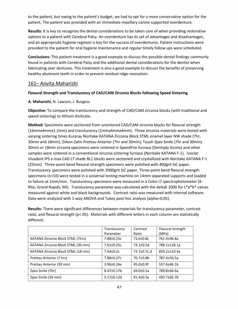
47
to the patient, but owing to the patient’s budget, we had to opt for a more conservative option for the patient. The patient was provided with an immediate maxillary canine supported overdenture.
Results: It is key to recognize the dental considerations to be taken care of when providing restorative options to a patient with Cerebral Palsy. An overdenture has its set of advantages and disadvantages, and an appropriate hygiene regimen is key for the success of overdentures. Patient instructions were provided to the patient for oral hygiene maintenance and regular timely follow ups were scheduled.
Conclusions: This patient treatment is a good example to discuss the possible dental findings commonly found in patients with Cerebral Palsy and the additional dental considerations for the dentist when fabricating over dentures. This treatment is also a good example to discuss the benefits of preserving healthy abutment teeth in order to prevent residual ridge resorption.
161– Anvita Maharishi
Flexural Strength and Translucency of CAD/CAM Zirconia Blocks following Speed Sintering
A. Maharishi, N. Lawson, J. Burgess
Objective: To compare the translucency and strength of CAD/CAM zirconia blocks (with traditional and speed sintering) to lithium disilicate.
Method: Specimens were sectioned from unsintered CAD/CAM zirconia blocks for flexural strength (16mmx4mmx1.2mm) and translucency (1mmx4mmx4mm). Three zirconia materials were tested with varying sintering times Kuraray Noritake KATANA Zirconia Block STML enamel layer NW shade (7hr, 30min and 18min), Zirkon Zahn Prettau Anterior (7hr and 30min), Tosoh Zpex Smile (7hr and 30min). 30min or 18min zirconia specimens were sintered in SpeedFire furnace (Dentsply Sirona) and other samples were sintered in a conventional zirconia sintering furnace (Noritake KATANA F-1). Ivoclar Vivadent IPS e.max CAD LT shade BL1 blocks were sectioned and crystallized with Noritake KATANA F-1 (25min). Three-point bend flexural strength specimens were polished with 400grit SiC paper. Translucency specimens were polished with 2000grit SiC paper. Three-point bend flexural strength specimens (n=10) were tested in a universal testing machine on 14mm separated supports and loaded to failure at 1mm/min. Translucency specimens were measured in a Color-i7 spectrophotometer (X-Rite, Grand Rapids, MI). Translucency parameter was calculated with the deltaE 2000 for L*a*b* values measured against white and black backgrounds. Contrast ratio was measured with internal software. Data were analyzed with 1-way ANOVA and Tukey post-hoc analysis (alpha=0.05).
Results: There were significant differences between materials for translucency parameter, contrast ratio, and flexural strength (p<.05). Materials with different letters in each column are statistically different.
Translucency Parameter
Contrast Ratio
Flexural strength (MPa)
KATANA Zirconia Block STML (7hrs) 7.88±0.25c 72.6±0.8c 761.4±96.8a
KATANA Zirconia Block STML (30 min) 7.61±0.25c 74.1±0.5d 788.1±128.1a
KATANA Zirconia Block STML (18 min) 7.64±0.2c 73.7±0.7c,d 859.2±110.4a
Prettau Anterior (7 hrs) 7.88±0.27c 70.7±0.8b 787.4±50.5a
Prettau Anterior (30 min) 3.96±0.26e 85.0±0.9f 557.6±46.1b
Zpex Smile (7hr) 8.47±0.17b 69.0±0.5a 789.8±66.6a
Zpex Smile (30 min) 5.17±0.12d 81.4±0.5e 493.7±65.7b
48
IPS e.max CAD LT (25 min) 9.33±0.56a 68.3±1.9a 471.2±87.6b
Conclusions: Two of the zirconia materials (Prettau Anterior and Zpex Simle) became less translucent and less strong using a shortened sintering time. All zirconia materials were stronger but less translucent than IPS e.max CAD with traditional sintering times. With shortened sintering times, only KATANA Zirconia Block STML retained a higher strength than IPS e.max CAD. The translucencies of the different zirconia should not be compared as the Zpex Smile did not have color pigments.
162 – Amir Koujan
A Modified Injectable Composite Resin Technique – Clinical Reports
A. Koujan, F. Mourad, K. Kinderknecht
Objective: The injectable resin composite technique as presented by Dr. Douglas Terry is a special and indirect/direct process of predictably translating a diagnostic wax-up into composite restorations. There are many applications for this technique using a highly filled flowable resin composite. These include repairing fractured teeth, fabricating provisional restorations, transitional composite restorations veneers, and can be used to establish the vertical dimension of occlusion prior to final restorations.
Method: A clear vinyl Poly-Siloxane (VPS) impression material is used to replicate the diagnostic wax- up. The clear matrix can be placed intraorally over the unprepared teeth and used as a transfer vehicle for the flowable composite resin to be injected and cured. An adjustment was applied on this technique to make the transitional restoration works as a definitive one by building up the body of the restoration first, followed by the injectable resin composite to finish the restoration and give it the final mold that matches the wax-up. The composite restorations can then be modified to satisfy the functional and aesthetic needs of the patient.
Results: We got more esthetic results with this modified injecting technique.
Conclusions: This procedure can reduce the potential for patient dissatisfaction without teeth preparation, increases the potential for a more conservative preparation design, and produce direct definitive restorations.
163– Harry Haring
Treatment Planning for Full Arch Implant Supported Prosthesis Using Radiopaque Acrylic Material – A Patient Study.
H. Haring, W. Wu, K. Kinderkinecht
Objective: Establishing proper tooth position is one of the most critical steps while treatment planning patients for implant supported/retained prostheses. Denture tooth set ups, diagnostic wax ups, and using the patients existing dentition are traditional methods for establishing the desired tooth position. Esthetic try – ins and fabrication of long – term provisionals allow the practitioner to properly diagnose the proposed tooth position by evaluating patient phonetics, function and esthetics. Radiographic stents based off the verified tooth position are then fabricated and placed prior to cone beam computed tomography, allowing the practitioner to plan for prosthetically driven implant placement. The purpose of this case study is to present a method that combines the esthetic try – in, functional and phonetic evaluation appointment and radiographic guide placement using a full arch implant supported radiopaque methacrylate – based provisional material. Implications of this case study attempt to

49
influence further investigations toward an optimally esthetic radiopaque material for use as a long – term provisional prosthesis.
Method: Laboratory fabricated implant supported screw - retained acrylic mock up using Lang Jet XR opaque acrylic on non-engaging titanium temporary copings. 2 Nobel Conical and 3 Straumann BLT implants used in conjunction with other abutment teeth as temporary anchors. Mock up tried in the mouth for esthetic try in, seating of copings verified radiographically thereby confirming accurate master cast. The patient was then sent for CBCT with radiopaque mock up in place. Guided CBCT DICOM file imported into 3shape Implant Studio software and planning for implant placement at site #9 and #11 completed. Mock up then modified at lingual contours of #9 and #11 to become a screw retained implant supported surgical guide for implant placement. Laboratory fabricated provisional made according to mock up with regards to changes made at try in and placed at the day of surgery.
Results: Mock up evaluated chair side with patient acceptance, photographs taken and adjustments made chair side. Using this technique allowed a realistic evaluation of VDO since the provisional was screw retained, as well as proper position of the teeth in regards to esthetics/phonetics/occlusal scheme. Radiographically verifying seating of each abutment coping allowed verifying of the master cast. Implant placement done successfully with immediately load of temporary implant at #8. Grafting of sites #9, 11 completed at surgery and placement of implants #9, 11 to be done at later date.
Conclusions: Radiopaque mock ups are a relevant tool that can be used while treatment planning implant surgery. The proposed tooth position can be evaluated chair side and modified prior to guided CBCT scans. Esthetic properties of the radiopaque acrylic allow the prosthesis to serve as a mock up for patient acceptance. Improving the esthetic and polish - ability of these prosthesis would allow them to be used as long term provisionals in the esthetic zone.
164 – Chan-Te Huang
Retention of Lithium Disilicate Crowns Cemented with Glass Ionomer, RMGI, Bioactive and Resin Cements
C.T. Huang, J. Burgess, N. Lawson
Objective: To measure the retention strength of lithium disilicate copings cemented with glass ionomer, RMGI, resin, and bioactive cements.
Method: 50 extracted human premolars were mounted in acrylic and prepared to uniform dimensions (20° total taper and 3.5mm preparation height) with a flat-end tapered diamond bur in a lathe. The surface area of the prepared surface was calculated with digital microscopy. The teeth were scanned with a True Definition Scanner (3M). Lithium disilicate crowns (e.max CAD, Ivoclar) were milled and crystallized following manufacturers recommendations. The intaglio surfaces were etched with 5% hydrofluoric acid for 20 seconds. The crowns (n=10) were then cemented with either a glass ionomer (Ketac Cem, 3M), RMGI (FujiCEM 2, GC or Rely X Luting Plus, 3M), calcium aluminate “bioactive” (Calibra Bio, Dentsply), or self-adhesive resin (Calibra Universal) cement. A silane primer was used with the Calibra Universal cement. Crowns were allowed to self-cure under a 2.5 kg weight, stored in a moist bag for 24 hours at 37°C and then thermocycled for 10,000 cycles from 5-500C with a 30 second dwell time. The specimens were placed in a custom fixture in a universal testing machine and loaded in tension at a crosshead speed of 5mm/min until debonding. The tensile strength (MPa) at debonding was calculated using the maximum recorded tensile force and surface area of the preparation. Data were compared with a 1-way ANOVA and Tukey analysis (alpha=0.05).
50
Results: Significant differences between cements were noted with 1-way ANOVA (p<.01). Materials can be categorized into significantly different groups with Tukey analysis as represented by the letters in the chart below.
Conclusions: The self-adhesive resin cement demonstrated the greatest retention. The “bioactive” calcium aluminate cement and one of the RMGI cements (Rely X Luting Plus) achieved greater retention than the glass ionomer cement. The other RMGI cement (FujiCEM 2) performed similarly as the glass ionomer cement.
165 – Luis Guzman
Correlation of Preoperative Analysis of CBCT Gray Value with Implant Primary Stability
L. Guzman, P. Liu, R. Abou-Arraj, N. Geurs, D. Givan, A. Javed
Objective: This study aims to investigate the correlation between the gray values (GV) of Cone Beam Computed Tomography (CBCT) analyzed by two commonly used implant planning soft-ware (coDiagnostiX, and Simplant®) and the primary stability of dental implants by measuring insertion torque value (ITV) and Resonance Frequency Analysis (ISQ).
Method: Thirty-eight patients planned to receive a total of 102 dental implants (Straumann Bone Level Tapered) were enrolled in the study. At the time of implant placement, ITV in Ncm with a Bien-Air iChiropro implant console, resonance frequency analysis (ISQ) with an Osstell device, and the surgeon’s estimated torque of placement were recorded. A versatile and highly reliable workflow was utilized to merge the postoperative position of implant placement with the preoperative virtual implant plans by using InVesalius 3.1, and Autodesk MeshMixer. The relationship of ITV, CBCT gray value of bone density in coDiagnostiX, Outer Simplant, Inner Simplant, ISQ, and surgeon estimated torque were assessed using Spearman correlations. Also, correlations were analyzed according to other variables such as implant length, implant diameter, implant site, type of bone, type of graft, and time of healing. Furthermore, multivariable analysis was conducted to identify independent predictors of ITV
Results: The workflow was examined with intraclass correlation coefficient (0.999, 0.995, 0.999, and 1) showing a high degree of correlation p<.0001. Spearman correlation for all variables (coDiagnostiX, outer Simplant, inner Simplant, ISQ, estimated torque, and measured ITV) showed positive and statistically significant correlations with GV in coDiagnostiX (r=0.39, p<0.0001), outer Simplant (r=0.37, p=0.0001), inner Simplant (r=0.33, p=0.0008), ISQ (r=0.52, p<.0001), and surgeon’s estimated torque value (r=0.48, p<.0001) with ITV. The generalized estimated equation (GEE) showed ISQ (p<0.032), surgeon estimated torque (p<0.002), and implant diameter (p<0.020) as independent predictors of measured ITV.
Conclusions: Overall, preoperative CBCT GV in coDiagnostiX and Simplant had statistically significant correlations with ITV, suggesting that treatment planning for immediate or early loading protocols can be optimized through virtual planning and GV assessment at potential implant sites. The used methodology for implant position registration into a preoperative scan was validated and found highly predictable and repeatable, and therefore, could be useful in future studies.
166 – Jennifer Vu
Pulp Revascularization: Management of a Non-Vital Immature Permanent Tooth in a Pediatric Patient
J. Vu, D. Clanton, J. Jackson
51
Objective: To report the outcome of revascularization technique on a necrotic immature permanent incisor in a pediatric patient, and to discuss the importance of patient selection and behavior management.
Method: A non-vital immature permanent incisor was treated with pulp revascularization. The canal system was accessed and disinfected then bleeding was initiated for a scaffold at a separate appointment. A cone beam CT scan was taken preoperatively, and at 12-month recall. Clinical and radiographic evaluation was conducted at 3-month intervals for twelve months. A combination of behavior guidance techniques were utilized to manage the child’s behavior during treatment.
Results: At 3-month recall, pulp vitality from cold testing and osseous healing of the periapical lesion was evident; however, staining was visible at the cervical margin of the treated tooth. At 9-month recall, pulp vitality was confirmed by electric pulp testing, and at 12-month recall, apical closure was apparent on cone beam CT. The patient was cooperative throughout treatment and during all follow-up appointments.
Conclusions: Pulp regeneration on non-vital developing permanent tooth is a good alternative to apexification as it encourages apexogenesis with the possibility of continued root development and thickening of the dentinal wall. Patient selection and behavioral management are important considerations as children are predominately the patients who will benefit from this type of treatment.
167 – Yasko Darkoue
Surface Treatment- Shear-bond Strength and Biaxial Flexural Strength of Zirconia
Y. Darkoue, J. Burgess, N. Lawson, D. Givan, C. Fu, E. McLaren
Objective: Mechanical and chemical surface treatments are required to achieve a durable bond to zirconia. This study compared the effect of particle type and pressure on shear bond strength and biaxial flexural strength of three generations of zirconia.
Method: 420 sintered zirconia discs of Lava (Frame, Plus and Esthetics) were produced and divided into 14 groups (n=10). These groups were tested for either shear bond strength or biaxial flexural strength as a control or surface-treated groups. The specimens in the control group received no mechanical treatment and was tested as sintered. All surface treated-groups were air abraded on one surface by 50u alumina or glass beads at 10mm distance for 10 seconds using three different pressures (15, 30, and 45 psi), then sonicated for 2 minutes in water, dried with compressed air for 30 seconds and a tube filled with PANAVIA SA Cement Plus (Kuraray Noritake) was placed onto the surface and light cured from top and two sides for 10 seconds each surface. The specimens were stored in water in an incubator at 370C for 24 hours and then placed into Instron and shear bond strength was calculated from the bonding area and the peak failure load. The thickness of each specimen for biaxial flexural strength test was measured. Each specimen was placed in a fixture mounted on Instron and the test was performed according to ISO standard 6872. Data were analyzed with ANOVA.
Results: Significant differences were seen between surface treatments (p<.01), materials (p<.01) and their interaction (p<.01). Materials in each column with different letters are statistically different.
Conclusions: Alumina produced a significantly better combination of bond strength while maintaining the biaxial strength of all zirconia. 15 psi is recommended.
52
168 – Mohammed Badahman
Bioactive materials: Fluoride Release, Recharge, Wear, Gloss & Hydroxyapatite Formation
M. Badahman, J. Burgess, N. Lawson, J. Lemon, A. Javed
Objective: Dental caries is one of the most prevalent chronic diseases 1. Unfortunately, there have been numerous failures with the treatment rendered and the materials used and how it was used. One significant cause for dental restoration failure is secondary caries and the need to replace the existing restoration. It has been reported that more than half of the restorations placed have been replaced because of recurrent caries. New materials and techniques have been developed to enhance adhesives in dentistry and improve the bonded interface however that interface still remains a weakest component of composite restorations. Marginal degradation and the inert nature of resin restoration have led to the introduction of bioactive material.
Method: Experimental (Activa 2), Fuji LC II and Filtek Supreme Ultra. Why were these materials selected? Which are the controls? Firstly, measurement of fluoride release in deionized water from these materials were recorded with ion electrode at 24 hours, 1 week, and 1 and 3-month intervals. Secondly, fluoride recharge was measured after 3 months storage in deionized water. Measurement of recharge was measured 2 days after storage. Thirdly, Gloss developed during polishing was measured using a glossmeter. Fourth, hydroxyapatite formation was measured using 3 samples of each material and was immersed in phosphate solution for 21 days. Sections from specimens were examined with the scanning electron microscope (SEM). Fifth, wear test was conducted by mounting 8 specimens in a UAB wear machine. Human premolar teeth were collected, and the lingual cusp was refined using a cone shaped bur to act as an antagonist. After 400,000 wear cycles the volumetric wear was measured with non-contact profilometer (Proscan). The data obtained was analyzed using an ANOVA, repeated ANOVA and post-hoc Tukey statistical tests.
Results: Fluoride release, fluoride recharge, gloss and hydroxyapatite formation from Activa 2 is not significantly (P>.05) different from Filtek Supreme Ultra. The wear of Activa 2 were significantly different (P<.05) than Filtek Supreme Ultra. Fuji II LC showed significant (P<.05) increase in fluoride recharge, fluoride release and hydroxyapatite formation with significant difference. However, Fuji II LC showed significant (P>.05) poor mechanical properties in wear and gloss when compared to Activa 2 and Filtek Supreme Ultra.
Conclusions: There were no statistically significant difference between Activa 2 and Filtek Supreme Ultra material in gloss, hydroxyapatite, fluoride release and fluoride recharge there were statistical significant in the wear. There were statistical significant between Fuji II LC and rest of the groups in all the completed tests.
Faculty
169 – Mi Young Kim
Provisionals with a Reference Seating Guide
MY Kim, D. Givan
When planning esthetic zone treatment, a diagnostic wax-up should be the first step to visualize desired outcomes. With digital workflow, a diagnostic wax up can directly transfer to a provisional restoration.
53
Conventional protocols for FP-1 implant provisional prosthetics often use the shell technique. A primary disadvantage of this technique is the difficulty in the accurate positioning of the shell during a pick-up of the temporary cylinder. This is due to the lack of a reference point and also working on the surgical site if it is for an immediate loading and immediate placement of an implant. An extended seating guide in the provisional shell will provide an accurate positioning of the provisional restorations and an effective assessment of restorative contours. This patient presentation demonstrates a simplified method using a milled PMMA provisional restorations with a seating guide to direct transfer of a diagnostic wax-up into an implant site to supported FP-1 provisional prosthesis. This would be advantageous for edentulous patients with high esthetic demands or with an implant placement and immediate loading protocol.
Verified master casts were mounted in a semi-adjustable articulator using facebow records. Diagnostic wax-up was created and scanned to fabricate milled PMMA provisional restorations for the edentulous site. The diagnostic wax up provides esthetic and hygienic assessment of the interproximal contours, embrasure locations, emergence profiles, or the need for pink porcelain. When designing a milled PMMA provisional shell, extended coverage on the cusps of adjacent teeth were added. Non-engaging titanium cylinders were placed on #7 and 10. The height of the cylinder was reduced to the desired occlusal plane. The milled PMMA shell with a seating guide was placed onto #6 and 11. Verified complete seating of the PMMA Provisionals. Jet acrylic was then injected between the cylinders and the shell to form core structures. Then, conventional picking up, modify the contour and polishing were performed. With this technique, FP-1 provisional prosthesis planning was easily transferred to the patient with accuracy.