15 - acquiring energy - feeding, digestion and metabolism
TRANSCRIPT
I n order to grow, maintain itself, and reproduce, every an- imal requires both raw materials and energy. These ma-
terials and the energy used in their metabolism come from food, but what actually constitutes a food item varies greatly between animals, ranging from individual molecules ab-
- -
sorbed across the general body surface to living prey swal- lowed whole. Regardless of its origin, which can be plant, animal, or inorganic sources, food is used as material for production of new tissue, for the repair of existing tissue, and for reproduction. Food also serves as an energy source for ongoing processes, such as movement and metabolism.
The chemical energy contained within food ultimately is derived from the sun (see Figure 3-3). Chlorophyll- containing plants are photosynthetic, autotrophic (self- nourishing) organisms that harness radiant energy to syn- thesize complex carbon compounds from simple precur- sors-C02 and H20. These compounds are repositories of chemical energy that can be released and utilized through coupled reactions to drive energy-consuming processes in living tissue. Almost all organisms are heterotrophic, de- pending on energy-yielding carbon compounds derived from the ingestion of other plants or animals, and ulti- mately on the photosynthesizers, which gather in the sun's energy. The exception of the relatively recently discovered "deep-sea vent" invertebrates, deriving their nutrition from mineral-rich vent waters, only highlights the normal de- pendency of animal life on the energy of the sun.
The flow of energy from the sun through a photosyn- thetic autotroph to a molecule of ATP in a heterotrophic animal is shown in Figure 15-1. Monosaccharides such as glucose are synthesized by green plants from CO, and H20. These elementary carbon compounds occur at the be- ginning of the food chain, which represents a series of or- ganisms linked together by the fact that each "link" of the chain serves as a food item for the next. Each group of or- ganisms represents a trophic level. In a short food chain with only two trophic levels, green plants are eaten by a large heterotroph, such as an elephant. This heterotroph, having no natural predators except humans, is at the end of that food chain until it dies and is consumed by bacteria
and carrion-eating scavengers. In a longer chain, a repre- sentative succession would be phytoplankton > zooplank- ton > small fish > medium fish > large fish, and the nu- trient flow is generally more complex (see Figure 3-3).
Usable material and free energy are lost in passing from one trophic level to another in a food chain. The grain pro- duced in a 1-hectare (ha; 2.47-acre) field of wheat contains more material and energy directly available for human con- sumption than it does if that same grain is used as cattle feed, converted to beef, and then consumed by humans. For ex- ample, a 1-ha grain field produces on average 5 times more protein than does a hectare devoted to beef production, while a hectare of legumes produces 10 times more. A cow must be fed more than 20 kg of plant protein to produce just 1 kg of protein for human consumption. Humans are at the top trophic level of that food chain. At each level of feeding, digestion, and incorporation along the food chain, there is considerable energy loss due to the energetic cost of tissue maintenance and of food digestion and reassembly into new molecules to be incorporated into tissue. Consequently, a shorter food chain generally conserves greater amounts of photosynthetically captured energy for the top consumer than a long one does, if the efficiency of transfer from each trophic level to the next one is approximately equal.
We will now consider how various animals acquire food items.
FEEDING METHODS
Obtaining food adequate in both quantity and quality oc- cupies much of the routine behavior of most animals. Cer- tainly an animal's physiology and morphology are the re- sult of natural selection that favors effective acquisition of energy from food while avoiding becoming someone else's meal. The complexity and sophistication of the nervous and muscular systems, for example, attest to the power of the selective forces acting on the organism. As these vary, so do the variety of methods by which animals feed. Sessile (non-mobile) bottom-dwelling species commonly resort to surface absorption, filter feeding, or trapping. Mobile

I N T E G R A T I O N O F P H Y S I O L O G I C A L SYSTEMS . . . . . . . . . . . . . . . . . . . . . . . . . . . . . . . . . . . . . . . . . . . . . . . . . . . . . . . . . . . . . . . . . . . .
Sunl~ght
c- - - #-- ---- Photosynthetic &-- -- autotroph
CO, + H,O 4c---%& % *_ -..-- , 3-
Heterotroph
Figure 15-1 Two trophic levels occur in this generalized flow diagram of in metabolism. Chemical energy content is at its peak following the pho- chemical energy through a food chain. The flowchart begins with the tosynthetic production of sugars. When the plant material is consumed photosynthetic formation of high-energy-content molecules (sugars) by a heterotroph, some chemical energy IS converted to heat and thus from low-energy-content raw materials (CO, and H,O) in plants. Oxida- is lost as a direct source of energy for driving biological processes. This tion of carbon compounds yields free energy coupled to the synthesis of food chain has only two trophic levels, but most food chains have many high-energy compounds, such as ATP, used as common energy currency more intervening levels.
animals follow a more active sequence, which in the ex- treme of many carnivores (meat eaters) includes searching, stalking, pouncing, capturing, and killing.
Food Absorption through Exterior Body Surfaces
The feeding method that is least dependent on specialized capture and digestive organs involves absorption of nutri- ents directly across the body wall. Certain protozoans, en- doparasites (animals that live within other animals), and aquatic invertebrates are able to take up nutrient molecules from the surrounding medium directly through their soft body wall. Endoparasites such as parasitic protozoans, tapeworms, flukes, and certain mollusks and crustaceans are surrounded by host tissues or by alimentary canal flu- ids, both of which are high in nutrients. Tapeworms, which may be many meters long, lack even a rudimentary diges- tive system. Tapeworms evolved from a primitive flatworm that lacked a body cavity (i.e., was acoelomic). However, some endoparasites appear to have secondarily lost the di- gestive apparatus present in their ancestors. For example, parasitic crustaceans, which belong to the cirripeds (bar- nacle group), lack an alimentary canal, but they appear to have evolved from nonparasitic ancestors possessing a gut.
Some free-living protozoans and invertebrates derive part of their nutrients by direct surface uptake from the sur- rounding medium. Small molecules such as amino acids are taken up from dilute solution by transport mechanisms (de- scribed in Chapter 4), against what can often be a huge concentration gradient. In some of these organisms, larger molecules or particles are taken up by a bulk process such as phagocytosis, which is described next.
Endocytosis
Endocytosis represents a more active form of "feeding" than passive absorption directly across the body wall. Like direct nutrient absorption, however, it occurs at the local cellular rather than tissue or organismal level. Endocytosis includes two processes, phagocytosis ("cell eating") and pinocytosis ("cell drinking"). In phagocytosis, pseudopod- like protuberances extend out and envelope relatively large nutrient particles. Pinocytosis occurs when a smaller par- ticle binds to the cell surface and the plasma membrane in- vaginates (folds inward) under it, forming an endocytotic cavity. Whether captured by phagocytosis or pinocytosis, the morsel is then engulfed in a membrane-enclosed vesicle that pinches off from the bottom of the cavity.

ACQUIRING ENERGY: FEEDING, DIGESTION, AND METABOLISM 629 . . . . . . . . . . . . . . . . . . . . . . . . . . . . . . . . . . . . . . . . . . . . . . . . . . . . . . . . . . . . . . . . . . . . . . . . . . . . . . .
The vesicle (or food vacuole in Protozoa) fuses with Oscu~um
lysosomes, organelles containing intracellular digestive en- zymes, whereafter it is called a secondary vacuole. After digestion, the contents of the vacuole pass through the vac- uole wall into the cytoplasm. The remaining undigested material is excreted externally by exocytosis, essentially a reverse process of pinocytosis. Feeding by pinocytosis and phagocytosis is familiar in protozoans such as Para- ath of water
mecium, but also occurs in the lining of the alimentary drawn into flagellated
canals and other tissues of many multicellular animals. chambers
Filter Feeding
Many aquatic animals use filter feeding, also called sus- pension feedmg, to capture food. Food items (usually phy- toplankton or zooplankton) are carried to specialized en- trapment devices either on the body surface or within it.
Most marine filter feeders are small, sessile animals, such as sponges, brachiopods, lamellibranchs, and tuni- cates. Food items are carried along on water currents that either occur naturally or are generated by the movements of body parts of the filter-feeding animal itself, such as cilia or flagella. Brachiopods respond behaviorally to currents, rotating on their pedal stalks to present the most efficient hydrodynamic orientation for capturing the water current. A number of other sessile animals located in moving wa- ter make use of Bernoulli's effect (i.e., a drop in fluid pres- sure as fluid velocity increases) to increase the rate of water flowing through the entrapment sites, at no energy cost to themselves. An example of such passively assisted filter feeding is seen in sponges (Figure 15-2). The flow of water across the large terminal opening causes a drop in pressure (Bernoulli's effect) outside the osculum. As a result, water is drawn out of the sponge through the osculum, and is drawn in through the numerous ostia (mouth-like open- ings) in the body wall. The drop in pressure is facilitated by the shape of the sponge's exterior, which causes the water over the osculum to flow with greater velocity than the wa- ter flowing past the ostia. Food particles, swept into the os- tia of the sponge along with the water, are engulfed by choanocytes, the flagellated cells lining the body cavity. The flagella of the choanocytes also create internal water cur- rents within spongocoel, the hollow water-filled interior. Some sponges living in moving water "pump" a volume of water equivalent to up to 20,000 times their body volume per day.
Mucus, a sticky mixture of mucopolysaccharides, often plays an important role in filter feeding. Waterborne mi- croorganisms and food particles are trapped in a layer of mucus that covers a ciliated epithelium. The mucus is then transported to the oral parts by beating cilia. The cilia pro- pel water through sessile animals not only to capture sus- pended food but also to aid in respiration. This is of great- est importance in still water. In mollusks such as the mussel, Mytilus, the cilia on the surface of the ctenidium draw a stream of water through the inhalant siphon, passing the water between the gill filaments (Figure 15-3). These cilia are also responsible for keeping mucus traveling down
Figure 15-2 Water flows in an organized fashion through syconoid sponges. In this diagrammatic section, red arrows indicate water flow. A significant proportion of this flow results from the reduction in hydrosta- tic pressure at the osculum due to the Bernoulli effect, produced by the transverse water currents (black arrows) flowing over the osculum at ele- vated velocity. Water flow is also generated from the activity of flagellated choanocytes that line the flagellated chambers (and give them their name). The choanocytes are found in the regions of the flagellated cham- bers lined in red. Water enters the sponge through the ostia, passes through the flagellated chambers, and ends up in the inner cavity, the spongocoel. Nutrients are then taken up into individual cells by endocy- tosis. [Adapted from Hyman, 1940; Vogel, 1978.1
along the filaments (i.e., 90" to the water flow) to the tip of the gill, where it travels in a special groove under ciliary power toward the mouth in a rope-like string of mucus. Sand and other indigestible particles are sorted out and re- jected (presumably on the basis of texture), passing out with the water leaving the exhalant siphon.
Non-sessile animals filter-feed by various mechanisms. A number of fishes are planktivorous, using modified gill
Ctenidium Adduct,or muscle
Path of mucus and food particles
+--- Path of water flow
Figure 15-3 Lamellibranch mollusks employ ciliary feeding. Side view of generalized lamellibranch with right valve removed. Red arrows show paths of inhaled water with food particles in the inhalant siphon and, over the surfaces of the gills (ctenidium). After passing down the gills, sand and other indigestible materials are eliminated out the exhalant siphon, while food particles are passed by cilia to the mouth.

rakers to strain food out of the flow of water passing through the mouth and over the gills. Juveniles of the pad- dlefish, Polyodon spathula, swim rapidly and continuously both to ventilate their gills and to filter out food items (see Chapter 16). Filter feeding is also very common in am- phibian larvae. In Xenopus laevis, the South African clawed toad, the branchial chamber contains gills bearing branchial filter plates that entrap suspended organic mate- rial. The material becomes entrapped in mucus, which is then swept by cilia into the esophagus to be swallowed. In Xenopus, branchial respiration and food ingestion may present functional conflicts. As the gill filter plates load up with suspended food items, the resistance to water flow through the gills rises sharply. Indeed, in larval Xenopus, gill ventilation decreases in proportion to food density in inspired water, presumably maintaining a constant rate of
- food ingestion. Increased cutaneous and pulmonary respi- ration apparently can compensate for thelack of branchial gas exchange when optimal conditions for filter feeding re- sult in reduced branchial water flow.
The largest filter feeders are the baleen whales, such as the right whale. Horny baleen plates bear a frlnge of par- allel filaments of halr-like keratin that hang down from the upper and lower jaws and act as strainers analogous to the gill rakers of fishes or larval amphibians (Figure 15-4A). These whales swim with jaws open into schools of pelaglc
Figure 15-4 Convergent evolution has led to filter-feeding mechanisms
in both (A) the black right whale, Eubalaena glacialis, and (B) the lesser
flamingo, Phoen~conaias minor. Both the baleen in the whale? mouth and the fringe along the edge of the flamingo's bill act as strainers. [Adapted
from Milner, 1981.)
crustaceans such as krill, engulfing vast numbers suspended in tons of water. As the jaws close, the water is squeezed back out through the baleen strainers with the help of the large tongue, and the crustaceans, left behind inside the mouth, are swallowed. Clearly, filter feeding can be a very effective form of food capture and can support an animal of huge dimensions.
Birds such as flamingos also use filter feeding to capture small animals and other morsels they find in the muddy bottoms of their freshwater habitat (Figure 15-4B). The flamingo and the right whale exhibit remarkable conver- gent evolution: they both have a deep-sided lower jaw, a re- curved rostrum, fibrous-fringe filters suspended from the upper jaw, and a large, fleshy tongue. Both feed by filling the mouth cavity with water, and then using the tongue as a piston to force water out through the filters, trapping and retaining waterborne food particles.
Fluid Feeding
Fluid feeding involves a variety of structures and mecha- nisms, including piercing and sucking, and cutting and licking.
Piercing and sucking Feeding by piercing a prey or food item and sucking flu- ids from it occurs among the platyhelminths, nematodes, annelids, and arthropods. Leeches, among the annelids, are true bloodsuckers, using an anticoagulant in their saliva to prevent clotting in their prey's blood. In fact, the anticoagulants in leeches have been chemically isolated and are being used clinically. Leeches themselves are still used for medicinal purposes to reduce swelling by remov- ing extracellular fluid following cosmetic and other forms of surgery. Some free-living flatworms seize their inverte- brate prey by wrapping themselves around it. They then penetrate the body wall with a protrusible pharynx that sucks out the victim's body fluids and viscera. Penetration by the pharynx and liquefaction of the victim's tissues are facilitated by proteolytic enzymes secreted by the muscu- lar pharynx.
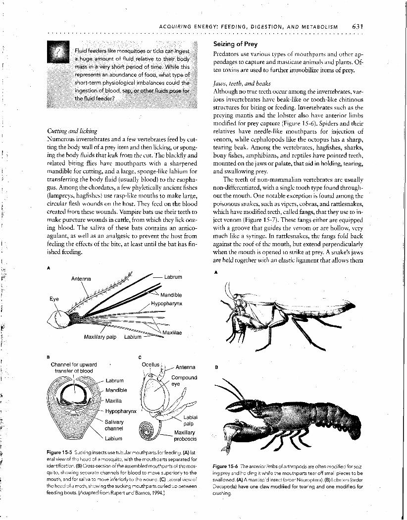
Large numbers of arthropods feed by piercing and sucking. Most familiar and irksome of these to humans are mosquitoes, fleas, bedbugs, and lice, which can be vectors of disease. The majority of sucking arthropods victimize animal hosts. However, especially among the Hemiptera (true bugs) are species that pierce and suck plants, from which they draw sap. Sucking insects generally possess fine piercing mouthparts in the form of a proboscis (Figure 15-SA). Often, the two maxillae are shaped so that they make up two canals that run to the tip of the proboscis (Figure 15-5B and C). One of these, the dorsal canal, is the passage for blood or sap sucked from the host. The other, the ventral canal, carries saliva, containing anticoagulants or enzymes, from the salivary glands into the host. Sucking occurs by the action of a muscular pharynx. After feeding, most insects are able to fold the proboscis back out of the way.
ACQUIRING ENERGY: FEEDING, DIGESTION, A N D METABOLISM 631 . . . . . . . . . . . . . . . . . . . . . . . . . . . . . . . . . . . . . . . . . . . . . . . . . . . . . . . . . . . . . . . . . . . . . . . . . . . . .
Cutting and licking Numerous invertebrates and a few vertebrates feed by cut- ting the body wall of a prey item and then licking, or spong- ing the body fluids that leak from the cut. The blackfly and related biting flies have mouthparts with a sharpened mandible for cutting, and a large, sponge-like labium for transferring the body fluid (usually blood) to the esopha- gus. Among the chordates, a few phyletically ancient fishes (lampreys, hagfishes) use rasp-like mouths to make large, circular flesh wounds on the host. They feed on the blood created from these wounds. Vampire bats use their teeth to make puncture wounds in cattle, from which they lick ooz- ing blood. The saliva of these bats contains an antico- agulant, as well as an analgesic to prevent the host from feeling the effects of the bite, at least until the bat has fin- ished feeding.
Channel for upward transfer of blood
Labrum
Mandible
Maxilla
Hypophatynx
Salivary channel
Labium
Figure 15-5 Sucking insects use tubular mouthparts for feeding. (A) lat-
eral view of the head of a mosquito, with the mouthparts separated for identification. (6) Cross-section of the assembled mouthparts of the mos-
quito, showing separate channels for blood to move superiorly to the mouth, and for saliva to move inferiorly to the wound. (C) Lateral view of
the head of a moth, showing the sucking mouthparts curled up between feeding bouts. [Adapted from Rupert and Barnes, 1994.1
Seizing of Prey
Predators use various types of mouthparts and other ap- pendages to capture and masticate animals and plants. Of- ten toxins are used to further immobilize items of prey.
Jaws, teeth, and beaks Although no true teeth occur among the invertebrates, var- ious invertebrates have beak-like or tooth-like chitinous structures for biting or feeding. Invertebrates such as the preying mantis and the lobster also have anterior limbs modified for prey capture (Figure 15-6). Spiders and their relatives have needle-like mouthparts for injection of venom, while cephalopods like the octopus has a sharp, tearing beak. Among the vertebrates, hagfishes, sharks, bony fishes, amphibians, and reptiles have pointed teeth, mounted on the jaws or palate, that aid in holding, tearing, and swallowing prey.
The teeth of non-mammalian vertebrates are usually non-differentiated, with a single tooth type found through- out the mouth. One notable exception is found among the poisonous snakes, such as vipers, cobras, and rattlesnakes, which have modified teeth, called fangs, that they use to in- ject venom (Figure 15-7). These fangs either are equipped with a groove that guides the venom or are hollow, very much like a syringe. In rattlesnakes, the fangs fold back against the roof of the mouth, but extend perpendicularly when the mouth is opened to strike at prey. A snake's jaws are held together with an elastic ligament that allows them
Figure 15-6 The anterior limbs of arthropods are often modified for seiz- ing prey and holding it while the mouthparts tear off small pieces to be swallowed. (A) A mantispid insect (order Neuroptera). (B) Lobsters (order
Decapoda) have one claw modified for tearing and one modified for
crushing.

Figure 15-7 Rattlesnakes have modified teeth, known as fangs, which they use to inject venom into their prey. These side views of a rattlesnake skull show (A) a non-striking position, with the jaws only partially open and the hinged fangs folded into the roof of the mouth, and (B) a striking position, in which the jaws are open wide and the fangs extended. The extraordinary flexibility of the lower jaws allows the snake to swallow prey whole after injecting it with deadly venom. [Adapted from Parker, 1963.1
to spread apart during swallowing. This enables the snake to swallow animals larger than the diameter of its head (see Figure 15-7). Swallowing prey whole is relatively common, and very evident in prey capture and consumption in snakes.
Mammals use their teeth for seizing and masticating their prey. Their teeth have developed very different shapes during evolution (Figure 15-8). Chisel-like incisors are used for gnawing, especially by rodents and rabbits. In the ele- phants (and before them, mammoths), the incisors are modified into a pair of tusks. Pointed, dagger-like canines are used by the carnivores, insectivores, and primates for piercing and tearing food. In some
Figure 15-8 Mammalian dentition is specialized for food type. (A) Teeth of a generalized placental mammal, showing the major divis~ons of den- tition. (B) Squirrel, showing incisors enlarged for gnawing. (C) African lion, with carnassial teeth modified for shearing bone and tendon. (D) Ox, with extensive molarsfor gr~nd~ng plant material. [Adapted from Romer, 1962; Cornwall, 1956.1
Generalized placental mammal
S
n African

and walruses, the canines are elongated as tusks, which are used for prying and fighting. Most complex and interesting in their form are the molars of some herbivorous groups such as cattle, including oxen, pigs, hippopotamuses, and horses and zebras. These teeth, which are used in a side-to- side grinding motion, are composed of folded layers of enamel, cement, and dentine, all of which differ in hardness and in rate of wearing. Because the softer dentine wears rather quickly, the harder enamel and cement layers form ridges that enhance the effectiveness of the molars for chewing grass and other tough vegetation. Many mam- mals, such as the cats (the domestic cat and the great cats, such as the lion) use limbs equipped with sharp claws to supplement the teeth as food-capturing structures.
Instead of teeth, birds have horny beaks, in a multitude of shapes and sizes, evolved to adapt to each species' unique food sources and methods of obtaining them. For instance, beaks may have finely serrated edges, sharp, hook-like up- per bills, or sharp, wood-pecking points (Figure 15-9). Seed-eating birds eat their food whole (perhaps after re- moving the outer hull), but may grind the swallowed seed in a muscular crop or gizzard containing pebbles that act like "millstones." Raptorial birds (hawks, eagles), endowed with excellent vision and flight mobility, capture prey with their talons as well as their beaks.
Herbivores
Filter-feeding (goose)
Seed-eating / (crossbill)
Toxins A large number of animals from different phyla use toxins either to subdue prey or to fend off predators. Most of these toxins act at synapses in the nervous system. Surpris- ingly simple animals can use sophisticated arrays of venom- producing cells. Among the coelenterates (hydras, jellyfish, anemones, corals) for example, there is extensive use of ne- matocysts (stinging cells). Concentrated in large numbers on the tentacles, the nematocysts inject paralytic toxins into prey and immobilize it while the tentacles carry it to the mouth (Figure 15-10). Many nemertine worms paralyze their prey by injecting venom through a stiletto-like pro- boscis. Venoms are also used by annelids, gastropod mol- lusks (including one species of octopus), and a wide variety of arthropods.
Among the last group, scorpions and spiders are most notorious for their toxins, which are usually highly specific chemicals that bind to specific receptor types. After grab- bing its prey with its large chelae (pincer-like organs), a scorpion will arch its tail and then plunge its sting into its prey (Figure 15-1 1). The scorpion then injects the victim with a poison containing a neurotoxin that interferes with the proper firing of nerve impulses. Spider poisons also contain neurotoxins. The venom from the black widow spi- der contains a substance that induces massive release of
Carnivores
Omnivores
(heron)
( Flesh-eatlng (hawk)
Figure 15-9 Bird beaks are adapted to suit herbivory, ornnlvoty, and carnivoty. [Adapted from Marshall and Hughes, 1980.1

634 INTEGRATION OF PHYSIOLOGICAL SYSTEMS . . . . . . . . . . . . . . . . . . . . . . . . . . . . . . . . . . . . . .
Neyatocysts \\k ~y stalk
Entrapped prey / 1 Tentacles k' "
Figure 15-10 Tentacles bearing stinging nematocysts dangle from around the mouth ofthe hydra. Small prey items (generally zooplankton) are stung, paralyzed, and then transferred to the mouth for ingestion. [Adapted from Rupert and Barnes, 1994.1
Figure 15-1 1 The scorpion Androctonus captures prey and then injects poison to subdue it. As the prey item is grabbed by the chela, the tail is arched over the scorpion's head to bring the sting into position. The prey is then impaled by the sting, which injects the rapidly acting toxin. [Adapted from Jennings, 1972.1
neurotransmitter at the motor endplate in muscle. A neu- rotoxin, a-bungarotoxin (see Spotlight 6-3), found in the venom of the cobra-like krait, binds to nicotinic acetyl- choline (ACh) receptors, thereby blocking neuromuscular transmission in vertebrates. The venoms of various species of rattlesnake contain hemolytic (blood cell-destroying) substances.
Toxins, although highly effective, are generally expen- sive to produce. Usually carefully measured doses of toxins are delivered during a bite or sting. Toxins also must be specially stored before administration to avoid self- poisoning. Toxins are generally proteins and, as such, are rendered harmless by the proteolytic enzymes of the preda- tor's digestive system when it ingests its poisoned prey.
Herbivory and Grazing to Collect Food
Herbivores often have mouth parts specialized for feeding on plant material. Many gastropods use a rasp-like structure termed a radula to scrape algae from rock surfaces or to rasp through vegetation (Figure 15-12). Vertebrate herbi- vores have bony plates (some fish and reptiles) or teeth primarily in the form of molars with wide flat surfaces mod-
A
Radula sac Radula membrane \ Radula teeth / .Salivary gland
Radula retractor Radula protractor muscle muscle
Figure 15-12 The head of a gastropod mollusk contains a powerful radula. (A) The rasp-like radula, revealed in sagittal section, is used for grazing on vegetation. (B) Protraction of the radula. (C) Retraction of the radula. [Adapted from Rupert and Barnes, 1994.1

ified for grinding plant material. Plants (especially some grasses) contain relatively large amounts of silicates, and can be tremendously abrasive. Consequently, the molars of her- bivores often are coated in especially tough enamel to resist wear. Alternatively, some herbivores such as small rodents (microtines) have continuously growing, rootless teeth.
OVERVIEW OF ALIMENTARY SYSTEMS
Alimentary systems play an essential role in providing nour- ishment through digesting and absorbing food, and remov- ing from the body indigestible materials and toxic by-prod- ucts of digestion. The most primitive "alimentary system" is the plasma membrane of unicellular organisms, in which mi- croscopic food particles are engulfed, undigested, by endo- cytosis directly into the cell itself. Once in the cell, food par- ticles undergo intracellular digestion by acids and enzymes. More complex multicellular animals rely primarily on ex- tracellular digestion carried out by true alimentary systems.
From an anatomical perspective, there are myriad designs of alimentary systems. However, from a physiolog- ical perspective, alimentary systems fall into one of three categories on the basis of how they process food in a "re- actor," or site of chemical digestion. So-called batch reac- tors are blind tubes or cavities that receive food and elimi- nate wastes in a pulsed fashion; that is, one batch is processed and eliminated before the next one is brought in
(Figure 15-13, left). Coelenterates, for example, have a blind tube or cavity, the coelenteron, which opens only at a "mouth" that serves also for the expulsion of undigested remains. In all phyla higher than the flatworms, ingested material passes through a hollow, tubular cavity-the alimentary canal-extending through the organism and open at both ends. Processing goes on continuously, rather than in pulses, with new food being ingested while older food is still being processed. Some alimentary canals can be modeled as ideal continuous-flow, stirred-tank reactors, in which food is continually added and mixed into a homogeneous mass, and the products of digestion are continuously eliminated, overflowing from the reactor (Figure 15-13, middle). An example of such a reactor is the forestomach of ruminants. The third way of processing food is in a plug-flow reactor, in which a bolus (a discrete plug or collection) of food is progressively digested as it winds its way through a long, tube-like digestive reactor (Figure 15-13, right). Unlike the stirred-tank reactor, its composition varies according to its position along the re- actor tube. The small intestine of many vertebrates func- tions as a plug-flow reactor. It is important to recognize that many animals combine features of both continuous- and plug-flow reactors. As you will see below, in many animals chemical digestion begins in the stomach, configured as a continuous-flow, stirred-tank reactor, and then continues on into the small intestine, configured as a plug-flow reactor.
Ideal batch reactor Ideal continuous-flow Ideal plug-flow reactor stirred-tank reactor
Pulsed output Contents mixed Contents mixed Axial gradlent
In composition
Composition uniform In cross-section Composition changes with time Composition unchanging with at steady state, unchanging with
time at steady state time at any point along reactor.
Gastrovascular cavity
Third compartment
Hydra Ruminant forestomach Small intestine
Figure 15-13 Digestive systems are functionally classified according to tine of many vertebrates acts as a plug-flow reactor, which may function the type of chemical reactor they form. (Left) Batch reactors are found in in addition to the stomach. [From Hume, 1989; adapted from Penry and simple organisms like Hydra. (Middle) Ruminants have a continuous-flow, Jurnars, 1987.1 stirred-tank reactor in the form of a forestomach. (RightjThe small intes-

636 INTEGRATION OF PHYSIOLOGICAL SYSTEMS . . . . . . . . . . . . . . . . . . . . . . . . . . . . . . . . . . . . . . . . . . . . . . . . . . . . . . . . . . . . . . . . . . . . . . . . . . . . . . .
A High-quality food B Low-quality food Figure 15-14 The qual~ty of food will greatly
Max~mum rate Influence the tlme requ~red for d~gest~on In a
of digest~on Maximum rate cont~nuous-flow d~gest~ve reactor (A) A hlgh-
of digest~on qual~ty food requlres m~n~mal energy to cap- ture and eat and, once eaten, 1s qulckly dl- gested to release large amounts of energy The maximum rate of d~gest~on occurs at the polnt of the curved l~ne w~th the steepest slope (B) A low-qual~ty food requlres consld- erable energy to capture and eat, and takes a
) long per~od of d~gest~on to yleld only low amounts of energy [Adapted from Hume, 1989, Sibly, 1981 ]
Mean retention t~me Mean retention time of food of food
It is critically important that the design of the alimen- (4) absorbing water and defecating. Representative alimen- tary canal and the reactors it contains matches well with the tary canals from the different invertebrate and vertebrate quality of the food that the animal routinely eats. A high- classes are illustrated in Figures 15-16 and 15-17, respectively. quality food can release maximum amounts of energy with -.
minimal time spent in the digestive reactor, whatever its Ingestion
type (Figure 15-14). A lower-quality food, on the other hand, requires a longer period of digestion to release its en- Headgut ergy. This in turn requires longer periods spent in the reac- tor and longer transit times through the alimentary canal. As also indicated in Fig. 15-14, the amount of energy spent \ Conducting in capturing a particular food must also be factored into Foregut
consideration of food quality. A generalized alimentary canal, or digestive tract, is il-
lustrated in (Figure 15-15). The lumen of this alimentary canal is topologically external to the body. Sphincters and other devices guard the entrance to and exit from the canal, preventing uncontrolled exchange between the lumen and the external environment. Ingested material is subjected to Midgut various mechanical, chemical, and bacterial treatments as it passes through this canal, and digestive juices (primarily en- zymes and acids) are mixed with the ingested material at ap- (Acidic secretions) propriate regions in the alimentary canal. As the ingested material is first mechanically broken down and then chemi- cally digested, nutrients undergo absorption and are then transported into the circulatory system. Undigested, non-ab- sorbed material is stored briefly until it, along with bacter- ial remains, is expelled as feces by the process of defecation.
The overall tubular organization of the alimentary Absorption-+Assimilation
canal is efficient because it allows ingested material to travel (Basic secretions) in one direction, passing through different regions that can then be specialized for particular digestive tasks. For ex- ample, the alimentary canal near the point of ingestion is often specialized for acid secretion, while more distant re- gions are alkaline. This regional specialization allows both Hindgut
acid and base secretion to occur at the same time and to Storage of waste . ,
permit different types of digestive action. In general, alimentary canals can be divided on a struc-
tural and functional basis into four major divisions (see -' Defecation
Figure 15-15): (1) headgut, (2) foregut, (3) midgut and Figure 15-1 5 A digestive tract with one-way passage of food allows si-
(4) These are 'pecialized for ('1 receiving in- multaneous operation of sequential stages in the processing of food and gested material, (2) conducting, storing, and digesting in- reduces mixing of digested and undigested matter. The dashed outline gested material, (3) digesting and absorbing nutrients, and represents a "crop," a storage region found in some animals.
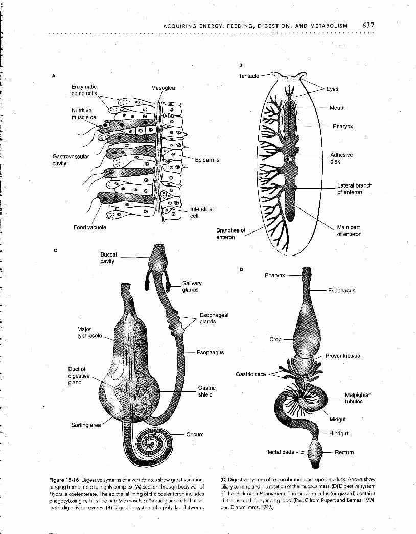
ACQUIRING ENERGY: FEEDING, DIGESTION, AND METABOLISM 637 . . . . . . . . . . . . . . . . . . . . . . . . . . . . . . . . . . . . . . . . . . . . . . . . . . . . . . . . . . . . . . . . . . . . . . . . . . . .
Gastro cavity
Enzymatic gland cells
Mesoglea /
Food vacuole Branch6 enteron
B
Tentacle
Gastric
Rectal pads Rectum
Figure 15-16 Digestive systems of invertebrates show great variation, (C) Digestive system of a prosobranch gastropod mollusk. Arrows show ranging from simple to highly complex. (A) Section through body wall of ciliary currents and the rotation of the mucous mass. (D) Digestive system Hydra, a coelenterate. The epithelial lining of the coelenteron includes of the cockroach Periplaneta. The proventriculus (or gizzard) contains phagocytosing cells (called nutritive muscle cells) and gland cells that se- chitinous teeth for grinding food. [Part C from Rupert and Barnes, 1994; Crete digestive enzymes. (B) Digestive system of a polyclad flatworm. part D from Imms, 1949.1

638 INTEGRATION OF PHYSIOLOGICAL SYSTEMS . . . . . . . . . . . . . . . . . . . . . . . . . . . . . . . . . . . . . . . . . . . . . . . . . . . . . . . . . . . . . . . . . . . . . . . . . . . . . . .
Cyclostome Elasmobranchs (hagfish) (shark)
Aves
(pigeon)
Teleost f ishes (bass)
Mammals (rabbit)
Amphibians (frog)
Mammals (human)
Figure 15-17 The tube-l~ke digestive system of vertebrates has a basic G, gallbladder; L, liver; LI, large intestine; P, pancreas; PA, pyloric appen- organizational plan, with common elements of esophagus, stomach, in- dices; SG, spiral gut; SI, small intestine; St, stomach. [From Florey, 1966; testine, and colon. B, bladder; C, cecum; Cr, crop (gizzard); E, esophagus; adapted from Stempell, 1926.1

ACQUIRING ENERGY: FEEDING, DIGESTION, A N D METABOLISM 639 . . . . . . . . . . . . . . . . . . . . . . . . . . . . . . . . . . . . . . . . . . . . . . . . . . . . . . . . . . . . . . . . . . . . . . . . . . . . . . .
Headgut: Food Reception
The headgut is the anterior (cranial) region of the alimen- tary canal, providing an external opening for food entry (see Figure 15-15). It consists of organs and structures for feeding and swallowing, including the mouthparts, buccal cavity, pharynx, and associated structures such as bills, teeth, tongue, and salivary glands. Where a common path- way exists leading to both the alimentary canal and the pas- sageway (e.g., the trachea) ending in the organ of internal gas exchange, there may be additional sphincter- or valve- like structures that control and divert the flow of ingested material and inspired water or air into their respective channels.
Other than in small-particle feeders such as coelenter- ates, flatworms, and sponges, the headgut of most meta- zoans has salivary glands, the secretions of which aid in- gestion and the mechanical (and often chemical) digestion -
of food. The primary function of the salivary secretion, saliva, is lubrication to assist swallowing. The lubrication is provided in many cases by a slippery mucus of which the chief constituent is a type of mucopolysaccharide named mucin. The saliva often contains additional agents, such as -
digestive enzymes, toxins, and anticoagulants (in blood- lapping or blood-sucking animals such as vampire bats and leeches). (See Chapter 8 for a discussion of salivary glands.)
Tongues, an innovation of the chordates, assist in the mechanical digestion and swallowing of food. In some an- imals tongues are used to grasp food. They are also used in chemoreception, bearing gustatory receptors called taste buds (see Figure 7-16A). Snakes use their forked tongues to take olfactory samples from the air and the substratum, re- tracting the tongue to wipe the samples in Jacobson's or- gan, which consists of a pair of richly innervated chemosen- sory pits located in the roof of the buccal cavity. Jacobson's organs are found in other reptiles and some amphibians.
Foregut: Food Conduction, Storage, and Digestion
In most species the foregut consists of an esophagus, a tube that leads from the oral region to the digestive region of the alimentary canal, and a stomach (see Figure 15-15).
Esophagus The esophagus conducts food from the headgut to the di- gestive areas, usually the stomach (see below). In chordates and some invertebrates, the esophagus conducts the bolus, or mass of chewed food mixed with saliva, by peristaltic movement (see Chapter 11) from the buccal cavity or phar- ynx. In some animals, this conducting region contains a sac-like expanded section, the crop, which is used to store food before digestion. The presence of a crop, generally found in animals that feed infrequently, allows quantities of food to be stored for digestion at a later time. Leeches, for example, feed very infrequently, with weeks or months between feeding periods. However, they ingest large quan- tities of blood at a "sitting," storing the blood for many weeks and digesting it in small amounts between their rare
feedings. In some animals crops are also used to ferment or digest foods for purposes other than their immediate di- gestion. Parent birds prepare food in this way to be regur- gitated for their nestlings.
Stomach In vertebrates and some invertebrates digestion takes place primarily in the stomach and the midgut. The stomach serves as a storage site for food, and in many species begins the initial stages of digestion. In most vertebrates, for ex- ample, the stomach initiates protein digestion by secreting the enzyme pepsinogen (later converted to pepsin) and hy- drochloric acid, which provides the highly acidic environ- ment required for pepsin activation. Contraction of the muscular walls of the stomach also provides mechanical mixing of food, saliva, and stomach secretions.
Stomachs are classified as monogastric or digastric, ac- cording to the number of chambers they possess. A mono- gastric stomach consists of a single strong muscular tube or sac. Vertebrates that are carnivorous or omnivorous char- acteristically have a monogastric stomach (Figure 15-18). Instead of a stomach, some invertebrates, such as insects (see Figure 15-16D), have outpouchings termed gastric ceca (singular, cecum), which are lined with enzyme-secreting cells, as well as phagocytic cells that engulf partially di- gested food and continue the process of digestion. In these alimentary systems the processes of digestion and absorp- tion are completed in the ceca, and the remainder of the al- imentary canal is concerned primarily with water and elec- trolyte balance and defecation.
Some birds have a tough, muscular gizzard, or crop, or both (see Figure 15-17). Sand, pebbles, or stones are swal- lowed and then lodge in the gizzard, where they aid in the grinding of seeds and grains. The proventriculus of insects and the stomach of decapod crustaceans, comparable to the bird's gizzard, contain grinding apparatuses for chew- ing swallowed food. Some fish such as mullets also have gizzards. On the other hand, some fish and larval toads lack stomachs altogether, with material from the esophagus en- tering into what is functionally the midgut.
Multichambered digastric stomachs (Figure 15-19) are found in the mammalian suborder Ruminantia (deer, elk, giraffe, bison, sheep, cattle, etc.). Somewhat similar digas- tric stomachs occur outside this suborder, in particular in the suborder Tylopoda (camel, llama, alpaca, vicuaiia). Mi- croorganisms in the first division of the stomach carry out fermentation, the anaerobic conversion of organic com- pounds to simpler compounds, yielding energy as ATP. All of the above-named groups carry out rumination, in which partially digested food is regurgitated (transported back to the mouth) for remastication (additional chewing). This process allows the ruminant (a gazelle on the open savanna, for example) to swallow food hastily while grazing and then to chew it more thoroughly later when at rest in a place of relative safety from predators. After the regurgi- tated food is chewed, it is swallowed again. This time it passes into the second division of the digastric stomach and

640 INTEGRATION OF PHYSIOLOGICAL SYSTEMS . . . . . . . . . . . . . . . . . . . . . . . . . . . . . . . . . . . . . . . . . . . . . . . . . . . . . . . . . . . . . . . . . . . . . . . . . . . . . . . . . A
Esophagus
\ / ~ ~ ~ $ $ h i n c t e r
Duodenum
, .
py lo r~ r
Abomasum
Figure 15-19 The digastricstomach of ruminants has multiple chambers for storing and digesting food materials. This sheep stomach, character- istic of the ruminants, has two divisions made up of four chambers. The rumen and reticulum make up the fermentative division. The omasum and abomasum (true stomach) make up the digestive division.
B I \ \ act as a fermentation vat that receives grazed vegetation.
Bacteria and protozoans in these chambers thrive on the vegetation, causing extensive digestive breakdown by fer- mentation of carbohydrates to butyrate, lactate, acetate,
Gastric
pit and propionate. These products of fermentation, along with some peptides, amino acids, and short-chain fatty acids, are absorbed into the bloodstream from the rumen
Goblet fluid. Symbiotic microorganisms grown in the rumen, cells along with undigested particles, are passed into the oma-
Parietal sum (absent in the Tylopoda) and then into the abomasum. cells Only the latter secretes digestive enzymes and is homolo-
gous to the monogastric stomach of non-ruminants. Fermentation in the stomach is not limited to ruminat-
Gastric, ing animals. It is found in other animals in which the pas- gland
sage of food in the stomach is delayed, allowing the growth of symbiotic microorganisms in a zone anterior to the di-
Chief cells gestive stomach, as in the kangaroo and the crops of galli-
form (chicken-like) birds.
Gastric wall Midgut: Chemical Digestion and Absorption Figure 15-1 8 The monogastric stomach is a single chamber lined with a In vertebrates, the rnidnut is the site for the chemi- - specialized epithelium. (A) Major parts of the mammalian stomach. cal digestion of proteins, fats, and carbohydrates. once di- (B) Detail of fundic, or gastric, glands lining a single gastric pit. The in- gested to their component molecules, these materials are ner layer of the stomach is lined with thousands of gastric pits, into which the gastric glands open and dump their digestive juices. The epithelium then in the midgut and away of the gastric gland contains chief (pe~sinogen-secreting) and parietal the alimentary canal in the blood. AS food is ready to pass (HCI-secreting) cells as well as goblet (mucus-secreting) cells. on from the vertebrate stomach, it is released into the
begins the second stage of digestion. In this stage hydroly- sis takes place with the assistance of digestive enzymes se- creted by the stomach lining.
The digastric stomach of the Ruminantia (see Figure 15-19) has four chambers, separated into two divi- sions. The first division consists of the rumen and reticulum chambers; the second division comprises the omasum and the abomasum (true stomach). The rumen and reticulum
midgut through the pyloric sphincter, which relaxes as the peristaltic movements of the stomach squeeze the acidic contents into the duodenum, the initial segment of the small intestine (see Figure 15-1 8A). Digestion continues in the small intestine, generally in an alkaline environment.
General structure and function of the rnidgut Among the vertebrates, carnivores have shorter and simpler intestines than do herbivores, reflecting the shorter time re- quired to digest meat than vegetation. For example, a tad-

ACQUIRING ENERC . . . . . . . . . . . . . . . . . . . . . . . . . . . . . . . . . . . . . . . . .
pole, which is almost always herbivorous, has a longer in- testine than the adult frog, which is carnivorous.
The vertebrate midgut or small intestine is typically divided into three distinct portions. The first, rather short, section is the duodenum, the lining of which secretes mu- cus and fluids and receives secretions carried by ducts from the liver and pancreas. Next is the jejunum, which also secretes fluid and is involved in digestion and ab- sorption. The most posterior section, the ileum, acts pri- marily to absorb nutrients digested previously in the duo- denum and jejunum, although some secretion occurs from the ileum.
As just noted, the secretory functions of the vertebrate duodenal epithelium are supplemented by secretions from the liver and pancreas. The cells of the liver produce bile salts, which are carried in the bile fluid to the duodenum through the bile duct. Bile fluid has two important func- tions. It emulsifies fats, and it helps neutralize acidity in- troduced into the duodenum from the stomach. The pan- creas, an important exocrine organ described in Chapter 9, produces pancreatic juice, which contains many of the pro- teases, lipases, and carbohydrases essential for intestinal di- gestion in vertebrates. Pancreatic juice is released into the pancreatic duct and, like bile, is important in neutralizing gastric acid in the intestine.
The intestine of most animals contains large numbers of bacteria, protozoans, and fungi. These multiply, con- tributing enzymatically to digestion, and are usually, in turn, digested themselves. An important function of some intestinal symbionts is the synthesis of essential vitamins.
The midgut region varies greatly not only in structure, but also in function in different animal groups. In many in- vertebrates, especially those with extensive ceca and diver- ticula (blind outpouchings of the alimentary canal), the in- testine serves no digestive function. In some air-breathing fishes (e.g., the weather loach, Misgurnus anguillicaudatus), the midgut is modified into a gas exchange organ where 0, from gulped air is exchanged with CO, from the cells, with residual gas then being expelled out the anus.
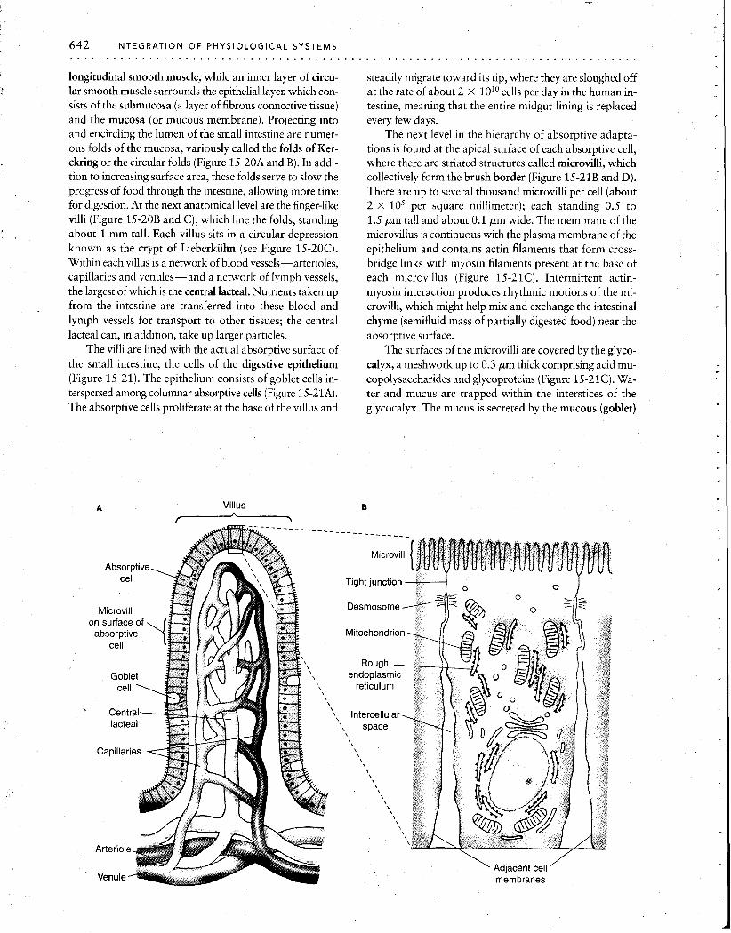
Intestinal epithelium The vertebrate small intestine has adaptations at every anatomical level, from its gross anatomy to the organelles of individual cells, all designed to amplify the surface area ' available for absorption of nutrients. In humans, the lumen of the small intestine has a gross cylindrical surface area of only about 0.4 m2, or about 7- 8 pages of this book. How- ever, because of the enormous elaboration of absorptive surfaces provided by this hierarchy of structures, the true area is increased at least 500 times, to a total of 200 to 300m2, or about the size of a doubles tennis court. Since the rate of absorption is generally proportional to the area of the apical surface membrane of the cells lining the epithe- lium, this huge increase in surface area greatly aids absorp- tion of digested substances from the fluid within the intes- tine. We will now examine this remarkable system of valleys and peaks, peninsulas and inlets.
The general organization of the vertebrate small intes- tine is shown in Figure 15-20A. The outermost layer is the serosa, which is the same tissue that covers the visceral or- gans of the abdomen. The serosa overlies an outer layer of
~ i r c u ~ a r k u s c ~ e '0'4,," \\ Submucosa , , \
Submucosa 'w
Villus
Central lacteal
Figure 15-20 The anatomy of the small intestine is dominated by spe- cializations for increased surface area. (A) Overall plan. (B) Intestinal folds of the mucosa are covered by (C) finger-like villi. [From "The Lining of the Small Intestine," by F. Moog. Copyright 0 1981 by Scientific American, Inc. All rights resewed.]
642 INTEGRATION OF PHYSIOLOGICAL SYSTEMS . . . . . . . . . . . . . . . . . . . . . . . . . . . . . . . . . . . . . .
longitudinal smooth muscle, while an inner layer of circu- lar smooth muscle surrounds the epithelial layel; which con- sists of the submucosa (a layer of fibrous connective tissue) and the mucosa (or mucous membrane). Projecting into and encircling the lumen of the small intestine are numer- ous folds of the mucosa, variously called the folds of Ker- ckring or the circular folds (Figure 15-20A and B). In addi- tion to increasing surface area, these folds serve to slow the progress of food through the intestine, allowing more time for digestion. At the next anatomical level are the finger-like villi (Figure 15-20B and C), which line the folds, standing about 1 mm tall. Each villus sits in a circular depression known as the crypt of Lieberkiihn (see Figure 15-20C). Within each villus is a network of blood vessels-arterioles, capillaries and venules-and a network of lymph vessels, the largest of which is the central lacteal. Nutrients taken up from the intestine are transferred into these blood and lymph vessels for transport to other tissues; the central lacteal can, in addition, take up larger particles.
The villi are lined with the actual absorptive surface of the small intestine, the cells of the digestive epithelium (Figure 15-21). The epithelium consists of goblet cells in- terspersed among columnar absorptive cells (Figure 15-21A). The absorptive cells proliferate at the base of the villus and
Villus -
steadily migrate toward its tip, where they are sloughed off at the rate of about 2 x 101° cells per day in the human in- testine, meaning that the entire midgut lining is replaced every few days.
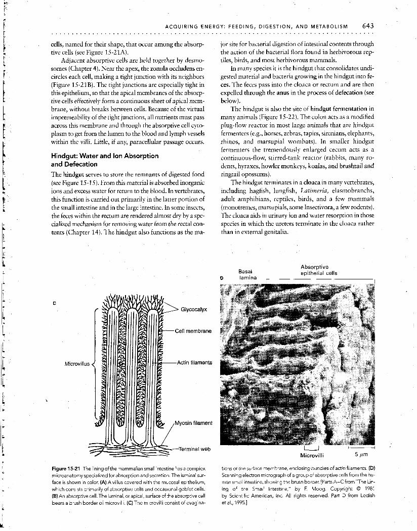
The next level in the hierarchy of absorptive adapta- tions is found at the apical surface of each absorptive cell, where there are striated structures called microvilli, which collectively form the brush border (Figure 15-21B and D). -
There are up to several thousand microvilli per cell (about 2 X lo5 per square millimeter); each standing 0.5 to 1.5 pm tall and about 0.1 pm wide. The membrane of the microvillus is continuous with the plasma membrane of the epithelium and contains actin filaments that form cross- bridge links with myosin filaments present at the base of each microvillus (Figure 15-21C). Intermittent actin- myosin interaction produces rhythmic motions of the mi- crovilli, which might help mix and exchange the intestinal chyme (semifluid mass of partially digested food) near the absorptive surface.
The surfaces of the microvilli are covered by the glyco- calyx, a meshwork up to 0.3 pm thick comprising acid mu- copolysaccharides and glycoproteins (Figure 15-21C). Wa- ter and mucus are trapped within the interstices of the glycocalyx. The mucus is secreted by the mucous (goblet)
- - - - - - -_ -___
Microvilli
Tight junction -----I 0
\ Adjacent cell ' membranes
ACQUIRING ENE6 . . . . . . . . . . . . . . . . . . . . . . . . . . . . . . . . . . . . . . . .
cells, named for their shape, that occur among the absorp- tive cells (see Figure 15-21A).
Adjacent absorptive cells are held together by desmo- somes (Chapter 4). Near the apex, the zonula occludens en- circles each cell, making a tight junction with its neighbors (Figure 15-21B). The tight junctions are especially tight in this epithelium, so that the apical membranes of the absorp- tive cells effectively form a continuous sheet of apical mem- brane, without breaks between cells. Because of the virtual impermeability of the tight junctions, all nutrients must pass across this membrane and through the absorptive cell cyto- plasm to get from the lumen to the blood and lymph vessels within the villi. Little, if any, paracellular passage occurs.
Hindgut: Water and Ion Absorption and Defecation
The hindgut serves to store the remnants of digested food (see Figure 15-15). From this material is absorbed inorganic ions and excess water for return to the blood. In vertebrates, this function is carried out primarily in the latter portion of the small intestine and in the large intestine. In some insects, the feces within the rectum are rendered almost dry by a spe- cialized mechanism for removing water from the rectal con- tents (Chapter 14). The hindgut also functions as the ma-
Micro
Cell membrane
Actin filaments
Figure 15-21 The lining ofthe mammalian small intestine has a complex microanatomy specialized for absorption and secretion. The luminal sur- face is shown in color. (A)Avillus covered with the mucosal epithelium, which consists primarily of absorptive cells and occasional goblet cells. (B) An absorptive cell. The luminal, or apical, surface of the absorptive cell bears a brush border of microv~lli. (C) The microvilli consist of evagina-
jor site for bacterial digestion of intestinal contents through the action of the bacterial flora found in herbivorous rep- tiles, birds, and most herbivorous mammals.
In many species it is the hindgut that consolidates undi- gested material and bacteria growing in the hindgut into fe- ces. The feces pass into the cloaca or rectum and are then expelled through the anus in the process of defecation (see below).
The hindgut is also the site of hindgut fermentation in many animals (Figure 15-22). The colon acts as a modified plug-flow reactor in most large animals that are hindgut fermenters (e.g., horses, zebras, tapirs, sirenians, elephants, rhinos, and marsupial wombats). In smaller hindgut fermenters the tremendously enlarged cecum acts as a continuous-flow, stirred-tank reactor (rabbits, many ro- dents, hyraxes, howler monkeys, koalas, and brushtail and ringtail opossums).
The hindgut terminates in a cloaca in many vertebrates, including hagfish, lungfish, Latimeria, elasmobranchs, adult amphibians, reptiles, birds, and a few mammals (monotremes, marsupials, some Insectivora, a few rodents). The cloaca aids in urinary ion and water resorption in those species in which the ureters terminate in the cloaca rather than in external genitalia.
Absorptive Basal epithelial cells
D lam,ina ,
u - Microvil l i 5 ~m
tions ot the suriace membrane, enclosing bundles of actin filaments. (D) Scanning electron micrograph of a group of absorptive cells from the hu- man small intestine, showing the brush border. [Parts A-CfromnThe Lin- ing of the Small Intestme," by F. Moog. Copyright 0 1981 by Scientific American, Inc. All rights resewed. Part D from Lodish et al., 1995.1
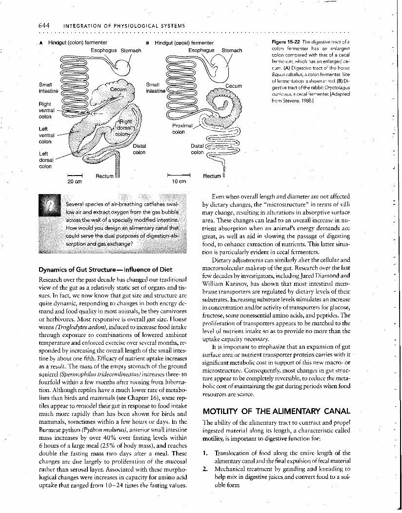
A Hindgut (colon) fermenter B Hindgut (cecal) fermenter Figure 15-22 The digestive tract of a
Esophagus Stomach Esophagus Stomach
H Rectum 1 Rectum U 20 cm 10 crn
Dynamics of Gut Structure-Influence of Diet
Research over the past decade has changed our traditional view of the gut as a relatively static set of organs and tis- sues. In fact, we now know that gut size and structure are quite dynamic, responding to changes in both energy de- mand and food quality in most animals, be they carnivores or herbivores. Most responsive is overall gut size. House wrens (Troglodytes aedon), induced to increase food intake through exposure to combinations of lowered ambient temperature and enforced exercise over several months, re- sponded by increasing the overall length of the small intes- tine by about one-fifth. Efficacy of nutrient uptake increases as a result. The mass of the empty stomach of the ground squirrel (Spermophilus tridecemlineatus) increases three- to fourfold within a few months after rousing from hiberna- tion. Although reptiles have a much lower rate of metabo- lism than birds and mammals (see Chapter 16), some rep- tiles appear to remodel their gut in response to food intake much more rapidly than has been shown for birds and - .
mammals, sometimes within a few hours or days. In the Burmese python (Python molurus), anterior small intestine mass increases by over 40% over fasting levels within 6 hours of a large meal (25% of body mass), and reaches double the fasting mass two days after a meal. These changes are due largely to proliferation of the mucosal rather than serosal layer. Associated with these morpho- logical changes were increases in capacity for amino acid uptake that ranged from 10-24 times the fasting values.
colon fermenter has an enlarged colon compared with that of a cecal fermenter, which has an enlarged ce- cum. (A) Digestive tract of the horse Equus caballus, a colon fermenter. Site
of fermentation isshown in red. (B) Di- gestive tract of the rabbit Oryctolagus cuniculus, a cecal fermenter. [Adapted
from Stevens, 1988.1
Even when overall length and diameter are not affected by dietary changes, the "microstructure" in terms of villi may change, resulting in alterations in absorptive surface area. These changes can lead to an overall increase in nu- trient absorption when an animal's energy demands are great, as well as aid in slowing the passage of digesting food, to enhance extraction of nutrients. This latter situa- tion is particularly evident in cecal fermenters.
Dietary adjustments can similarly alter the cellular and macromolecular makeup of the gut. Research over the last few decades by investigators, including Jared Diamond and William Karasov, has shown that most intestinal mem- brane transporters are regulated by dietary levels of their substrates. Increasing substrate levels stimulates an increase in concentration andlor activity of transporters for glucose, fructose, some nonessential amino acids, and peptides. The
- -
proliferation of transporters appears to be matched to the level of nutrient intake so as to provide no more than the uptake capacity necessary.
It is important to emphasize that an expansion of gut surface area or nutrient transporter proteins carries with it significant metabolic cost in support of this new macro- or microstructure. Consequently, most changes in gut struc- ture appear to be completely reversible, to reduce the meta- bolic cost of maintaining the gut during periods when food resources are scarce.
MOTILITY OF THE ALIMENTARY CANAL The ability of the alimentary tract to contract and propel ingested material along its length, a characteristic called motility, is important to digestive function for:
1. Translocation of food along the entire length of the alimentary canal and the final expulsion of fecal material
2. Mechanical treatment by grinding and kneading to help mix in digestive juices and convert food to a sol- uble form
3. Mixing of the contents so that there is continual re- newal of material in contact with the absorbing and secreting surfaces of the epithelial lining
Muscular and Ciliary Motility
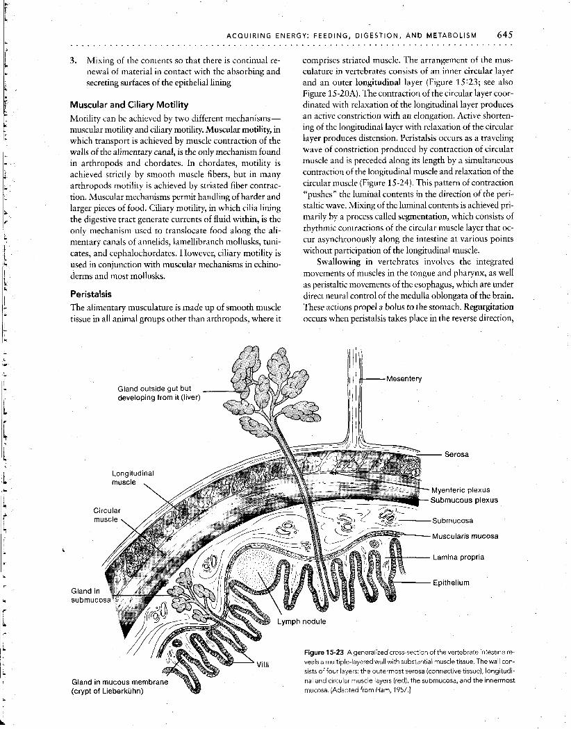
Motility can be achieved by two different mechanisms- muscular motility and ciliary motility. Muscular motility, in which transport is achieved by muscle contraction of the walls of the alimentary canal, is the only mechanism found in arthropods and chordates. In chordates, motility is achieved strictly by smooth muscle fibers, but in many arthropods motility is achieved by striated fiber contrac- tion. Muscular mechanisms permit handling of harder and larger pieces of food. Ciliary motility, in which cilia lining the digestive tract generate currents of fluid within, is the only mechanism used to translocate food along the ali- mentary canals of annelids, lamellibranch mollusks, tuni- cates, and cephalochordates. However, ciliary motility is used in conjunction with muscular mechanisms in echino- derms and most mollusks.
Peristalsis The alimentary musculature is made up of smooth muscle tissue in all animal groups other than arthropods, where it
comprises striated muscle. The arrangement of the mus- culature in vertebrates consists of an inner circular layer and an outer longitudinal layer (Figure 15'23; see also Figure 15-20A). The contraction of the circular layer coor- dinated with relaxation of the longitudinal layer produces an active constriction with an elongation. Active shorten- ing of the longitudinal layer with relaxation of the circular layer produces distension. Peristalsis occurs as a traveling wave of constriction produced by contraction of circular muscle and is preceded along its length by a simultaneous contraction of the longitudinal muscle and relaxation of the circular muscle (Figure 15-24). This pattern of contraction "pushes" the luminal contents in the direction of the peri- staltic wave. Mixing of the luminal contents is achieved pri- marily by a process called segmentation, which consists of rhythmic contractions of the circular muscle layer that oc- cur asynchronously along the intestine at various points without participation of the longitudinal muscle.
Swallowing in vertebrates involves the integrated movements of muscles in the tongue and pharynx, as well as peristaltic movements of the esophagus, which are under direct neural control of the medulla oblongata of the brain. These actions propel a bolus to the stomach. Regurgitation occurs when peristalsis takes place in the reverse direction,
Gland outside gut but developing from it (liver)
Myenteric plexus
Submucous plexus
Muscularis mucosa
Figure 15-23 A generalized cross-section of the vertebrate intestine re- veals a multiple-layered wall with substantial muscle tissue. The wall con- sists of four layers: the outermost serosa (connective tissue), longitudi- nal and circular muscle layers (red), the submucosa, and the innermost
(crypt of Lieberkiihn) mucosa. [Adapted from Ham, 1957.1
646 INTEGRATION OF PHYSIOLOGICAL SYSTEMS . . . . . . . . . . . . . . . . . . . . . . . . . . . . . . . . . . . . . . .
Figure 15-24 Coordinated contraction of the gastrointestinal tract pro-
pels material through its lumen. (A) Peristalsis occurs as a traveling wave of contraction of circular muscle preceded by relaxation. This produces longitudinal movement of the bolus. (6) Segmentation occurs as alter- nating relaxat~ons and contractions, primarily of circular muscle. The re-
sult is a kneading and mixing of the intestinal contents.
moving the luminal contents back into the buccal cavity. Ruminants regularly use regurgitation to bring up the unchewed food for further chewing, and other vertebrates use it during emesis (vomiting).
Normal peristalsis in the vertebrate stomach occurs with the ring of contraction only partially closed. Conse- quently there is a mixing action in which the contents are
squeezed backward (opposite to the direction of the wave) centrally through the partially open ring and forward pe- ripherally in the direction of peristalsis as the partially closed ring of contraction moves from the cardiac to the py- loric end of the stomach.
Control of Motility
The coordinated contractions of circular and longitudinal smooth muscle layers that provide alimentary canal motil- ity in vertebrates are regulated by a combination of distinct mechanisms.
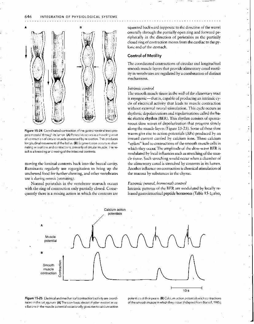
Calcium action ,
/ potentials
Intrinsic control The smooth muscle tissue in the wall of the alimentary tract is myogenic-that is, capable of producing an intrinsic cy- cle of electrical activity that leads to muscle contraction without external neural stimulation. This cycle occurs as rhythmic depolarizations and repolarizations called the ba- sic electric rhythm (BER). This rhythm consists of sponta- neous slow waves of depolarization that progress slowly along the muscle layers (Figure 15-25). Some of these slow waves give rise to action potentials (APs) produced by an inward current carried by calcium ions. These calcium "spikes" lead to contractions of the smooth muscle cells in which they occur. The amplitude of the slow-wave BER is modulated by local influences such as stretching of the mus- cle tissue. Such stretching would occur when a chamber of the alimentary canal is stretched by contents in its lumen. Another influence on contraction is chemical stimulation of the mucosa by substances in the chyme.
Extrinsic (neural, hormonal) control Intrinsic patterns of the BER are modulated by locally re- leased gastrointestinal peptide hormones (Table 15-1; also,
0 Smooth g
Figure 15-25 Electrical and mechanical (contraction) activity are coordi- potentials at their peaks. (B) Calcium action potentials elicit contractions
nated in the cat jejunum. (A) The slow basic electric rhythm evident as 0s- of the smooth muscle in which they occur. [Adapted from Bortoff, 1985.1 cillations in the muscle potential occasionally gives rise to calcium action

ACQUIRING ENERGY: FEEDING, DIGESTION, AND METABOLISM 647 . . . . . . . . . . . . . . . . . . . . . . . . . . . . . . . . . . . . . . . . . . . . . . . . . . . . . . . . . . . . . . . . . . . . . . . . . . . . . . .
see Spotlight 9-1). Thus, a chemical stimulant in the chyme can cause the release of a local hormone, and this, in turn, can modulate the motility of the muscle tissue.
In addition to local stimuli, intestinal motility is influ- enced by diffuse innervation from the sympathetic, parasympathetic, and peptidergic (purinergic) divisions of the autonomic nervous system (see Chapter 9). Sympa- thetic and parasympathetic postganglionic neurons form
networks dispersed throughout the smooth muscle layers (Figure 15-26). The parasympathetic network made up of cholinergic neurons is divided into the myenteric plexus and the submucosal plexus. These plexi, which receive their parasympathetic input primarily via branches of the vagus nerve, mediate excitatory actions (i.e., increased motility and gastrointestinal secretion) of the digestive tract. In con- trast, the innervation from the sympathetic division is
A Sympathetic Longitudinal Circular I Muscularis \ \
ganglia Mucosa muscle muscle mucosae
B Vaaal Myenteric Submucosal
plexus plexus I
Splnal cord Postgangl~onic (cholinerglc Endocrine cells
and adrenergic)
Figure 15-26 The gastro~ntesttnal tract has r~ch sympathet~c and nal target tlssues (muscle, glands) are postgangl~ontc [Adapted from parasympathet~c lnnervatlon (A) Efferent sympathet~c lnnervatlon Davenport, 1977 ] (B) Parasympathettc tnnervatlon All nerve endlngs on the gastrotntestl-
648 I N T E G R A T I O N O F P H Y S I O L O G I C A L SYSTEMS . . . . . . . . . . . . . . . . . . . . . . . . . . . . . . . . . . . . . . . . . . . . . . . . . . . . . . . . . . . . . . . . . . . . . . . . . . . . . . .
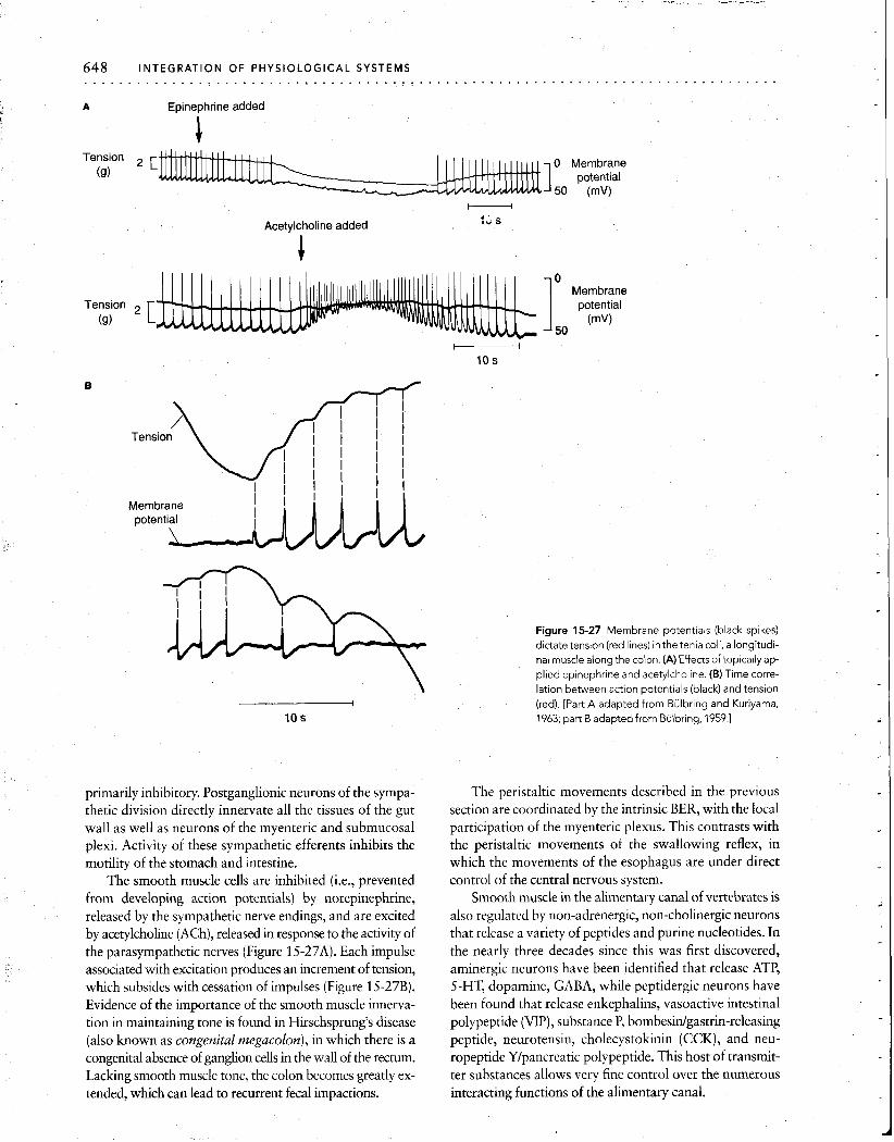
A Epinephr~ne added
Tension 2 [I 0 Membrane
(9) potential
Acetylcholine added :G s
1: fvlez;ne
1 potential
(9)
Membrane
Figure 15-27 Membrane potent~als (black sp~kes) d~ctate tenston (red Ihnes) In the tenla col~, a long~tudl- nal muscle along the colon (A) Effects of top~cally ap- pl~ed ep~nephr~ne and acetylchol~ne (B) T~me corre- lat~on between aalon potent~als (black) and tenston
I (red) [Part A adapted from Bulbr~ng and Kurtyama, 10 S 1963, part B adapted from Bulbring, 1959 ]
primarily inhibitory. Postganglionic neurons of the sympa- thetic division directly innervate all the tissues of the gut wall as well as neurons of the myenteric and submucosal plexi. Activity of these sympathetic efferents inhibits the motility of the stomach and intestine.
The smooth muscle cells are inhibited (i.e., prevented from developing action potentials) by norepinephrine, released by the sympathetic nerve endings, and are excited by acetylcholine (ACh), released in response to the activity of the parasympathetic nerves (Figure 15-27A). Each impulse associated with excitation produces an increment of tension, which subsides with cessation of impulses (Figure 15-27B). Evidence of the importance of the smooth muscle innerva- tion in maintaining tone is found in Hirschsprung's disease (also known as congenital megacolon), in which there is a congenital absence of ganglion cells in the wall of the rectum. Lacking smooth muscle tone, the colon becomes greatly ex- tended, which can lead to recurrent fecal impactions.
The peristaltic movements described in the previous section are coordinated by the intrinsic BER, with the local participation of the myenteric plexus. This contrasts with the peristaltic movements of the swallowing reflex, in which the movements of the esophagus are under direct control of the central nervous system.
Smooth muscle in the alimentary canal of vertebrates is also regulated by non-adrenergic, non-cholinergic neurons that release a variety of peptides and purine nucleotides. In the nearly three decades since this was first discovered, aminergic neurons have been identified that release ATP, 5-HT, dopamine, GABA, while peptidergic neurons have been found that release enkephalins, vasoactive intestinal polypeptide (VIP), substance P, bombesidgastrin-releasing peptide, neurotensin, ch~lec~stokinin (CCK), and neu- ropeptide Ylpancreatic polypeptide. This host of transmit- ter substances allows very fine control over the numerous interacting functions of the alimentary canal.
ACQUIRING ENERGY: FEEDING, DIGESTION, AND METABOLISM 649 . . . . . . . . . . . . . . . . . . . . . . . . . . . . . . . . . . . . . . . . . . . . . . . . . . . . . . . . . . . . . . . . . . . . . . . . . . . . . . .
TABLE 15-1 Action o f some enzymes secreted in the mouth, stomach, pancreas, and small intestine o f mammals
Enzyme Site of adion Substrate Products of action
Mouth Salivary a-amylase Mouth
Stomach Pepsinogen + pepsin Stomach
Pancreas Pancreatic a-amylase
Trypsinogen + trypsin
Chymotrypsin
Small intestine
Small intestine
Small intestine
Starch Disaccharides (few)
Proteins Large peptides
Starch
Proteins
Proteins
Disaccharides
Large peptides
Large peptides
Elastase Small intestine Elastin Large peptides
Carboxypeptidases
Aminopeptidases
Lipase
Nucleases
Small intestine Enterokinase
Small intestine
Small intestine
Small intestine
Small intestine
Small intestine
Large peptide
Large peptide
Triglycerides
Nucle~c acids
Trypsinogen
Small peptides (oligopeptides)
Oligopeptides
Monoglycerides, fatty acids, glycerol
Nucleotldes
Trypsin
Disaccharidases Small ~ntestine* Disaccharides Monosacchar~des
Peptidases Small intestine* Oligopeptides Amino acids
Nucleotldases Small ~ntest~ne* Nucleot~des Nucleosidases, phosphoric acid
Nucleosidases Small intestine* Nucleosides Sugars, purines, pyrimidines
*Intracellular
GASTROINTESTINAL SECRETIONS
The alimentary canal of most animals produces both en- docrine and exocrine secretions. In fact, the alimentary canal in many animals has been described as the "largest endocrine and exocrine gland of the body." As explained in Chapters 8 and 9, hormones are produced in the alimen- tary canal by cells of endocrine glands and are liberated di- rectly into the bloodstream, acting as messengers to recep- tor molecules in target tissues, which usually include other tissues of the alimentary canal.
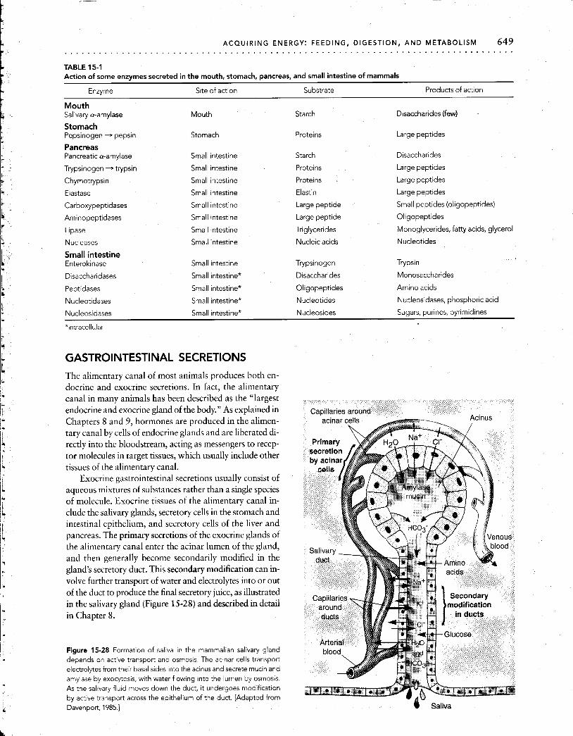
Exocrine gastrointestinal secretions usually consist of aqueous mixtures of substances rather than a single species of molecule. Exocrine tissues of the alimentary canal in- clude the salivary glands, secretory cells in the stomach and intestinal epithelium, and secretory cells of the liver and pancreas. The primary secretions of the exocrine glands of the alimentary canal enter the acinar lumen of the gland, and then generally become secondarily modified in the gland's secretory duct. This secondary modification can in- volve further transport of water and electrolytes into or out of the duct to produce the final secretory juice, as illustrated in the salivary gland (Figure 15-28) and described in detail in Chapter 8.
Figure 15-28 Formation of saliva in the mammalian salivary gland depends on active transport and osmosis. The acinar cells transport electrolytesfrom their basal sides into the acinus and secrete mucin and amylase by exocytosis, with water flowing into the lumen by osmosis. As the salivary fluid moves down the duct, i t undergoes modification by active transport across the epithelium of the duct. [Adapted from Davenport, 1985.1 @ Saliva

Exocrine Secretions of the Alimentary Canal the osmotic flow of water into the acinus. There is subse-
~h~~~ are large variations in the composition of the secre- quent secondary modification of this ultrafiltrate by active
tions from different regions of the alimentary canal. H ~ ~ - or passive transport across the epithelium lining the ducts
ever, these mixtures usually consist of some of as the fluid passes along the exocrine ducts toward the ali-
water, ions, mucus, and enzymes. mentary canal.
Water and electrolytes The exocrine glands of the alimentary canal typically se- crete large quantities of water-based fluids bearing digestive enzymes and other chemicals into the alimentary canal lu- men (Figure 15-29). Most of this water is reabsorbed in the distal portions of the gut.
In aqueous solution, the mucus produced in the goblet cells of the stomach and intestine (see Figures 15-18 and 15-21) provides a slippery, thick lubricant that helps pre- vent mechanical and enzymatic injury to the lining of the gut. The salivary glands and pancreas secrete a thinner mu- coid solution.
Secretion of inorganic constituents of digestive fluids generally occurs in two steps. First, water and ions are se- creted into the lumen of the gland either by passive ultrafil- tration due to a hydrostatic pressure gradient across the lu- minal epithelium, or by active (energy-requiring) processes from the interstitial fluid bathing the basal portions of the acinar cells. The latter is believed usually to entail active transport of ions by these cells, which is then followed by
Bile and bile salts The vertebrate liver does not produce digestive enzymes. However, it does secrete bile, a fluid essential for digestion of fats. Bile consists of water and a weakly basic mixture of cholesterol, lecithin, inorganic salts, bile salts, and bile pigments. The bile salts are organic salts composed of bile acids manufactured by the liver from cholesterol and conjugated with amino acids complexed with sodium (Figure 15-30). The bile pigments derive from biliverdin and bilirubin, which are products of the breakdown of hemoglobin spilled into plasma from old, ruptured red blood cells. Bile produced in the liver is transported via the hepatic duct to the gallbladder, where it is concentrated and stored. Water is removed osmotically, following active transport of Na+ and C 1 from the bile across the gallblad- der epithelium.
Bile serves numerous functions important to digestion. First, its high alkalinity is important in the terminal stages of digestion because it buffers the high acidity provided by the gastric juice secreted earlier in the digestive process.
Region Secretion Daily
amount (L) PH Composition*
Rectum
Buccal cavity
'F salivary Saliva 1+ 6.5 Amylase, bicarbonate glands
Esophagus
Stomach Gastric juice 1-3 1.5 Pepsinogen, HCI, rennln in infants, "lntrlnsic factor"
Pancreatic juice 1 7- 8 Trypslnogen, chymotrypsinogen,
*Exclud~ng mucus and water, which together make up some 95% of the actual secretion.
Gall- bladder
Figure 15-29 Important digestwe secretions occur a t all polnts along the human alimentary canal The approx~mate volume and pH of each secretion IS shown on the right.
carboxy- and aminopept~dase, Ilpase, amylase, maltase, nucleases, bicarbonate
Bile 1 7- 8 Fats and fatty acids, bile salts and pigments, cholesterol
'Succus entericus' 1 7- 8 Enterokinase, carboxy- and aminopept~dases, maltase, lactase, sucrase, Ilpase, nucleases

0 0 I I II C-NH-CH,-C-0- Na+
Glycine
Second, bile salts facilitate enzymatic fat digestion by breaking down fat into microscopic droplets that collec- tively have a much higher surface area. The ability of the bile salts to disperse fatty, water-insoluble substances de- rives from their amphipathic nature. That is, the bile salt molecule contains a lipid-soluble bile acid together with a water-soluble amino acid. Thus, it acts as a detergent for the emulsification of fat droplets, dispersing them in aque- ous solution for more effective attack by digestive enzymes. Ultimately, the bile salts are removed from the large intes- tine by highly efficient active transport and returned to the bloodstream. The bile salts then become bound to a plasma carrier protein and are returned to the liver to be recycled. Bile salts also disperse lipid-soluble vitamins for transport in the blood.
Third, bile fluid contains waste substances removed from the blood by the liver, such as hemoglobin pigments, cholesterol, steroids, and drugs. These substances are either digested or excreted in the feces.
Digestive enzymes An animal must first digest food before it can be used for tissue maintenance and growth and as a source of chemical energy. Digestion is primarily a complex chemical process in which special digestive enzymes catalyze the hydrolysis of large foodstuff molecules into simpler compounds that
Figure 15-30 Sodium glycholate is the mam- malian bile salt. Cholic acid (colored area) is con- jugated with the amino acid glycine and sodium.
are small enough to cross cell membranes of the intestinal barrier. For example, starch, a long-chain p~l~saccharide, is degraded to much smaller disaccharides and monosac- charides; proteins are hydrolyzed into polypeptides and then into tripeptides, dipeptides, and amino acids.
All digestive enzymes carry out hydrolysis, adding H+ to one residue and OH- to the other (Figure 15-31). Hy- drolysis of the anhydrous bonds frees the constituent residues (e.g., monosaccharides, amino acids, monogly- cerides) from which the polymer is formed, making them small enough for absorption from the alimentary canal into the circulating body fluids and for subsequent entry into cells to be metabolized.
Digestive enzymes, like all enzymes, exhibit substrate specificity and are sensitive to temperature, pH, and certain ions (see Chapter 3). Corresponding to the three major types of foodstuffs are three major groups of digestive en- zymes: proteases, carbohydrases, and lipases.
Proteases Proteases are proteolytic enzymes, categorized as either endopeptidases or exopeptidases. Both types of en- zymes attack peptide bonds of proteins and polypeptides (Figure 15-31A, Table 15-2). They differ in that endopep- tidases confine their attacks to bonds well within (endo, "within") the protein molecule, breaking large peptide chains into shorter polypeptide segments. These shorter
Peptidase ' \
C Peptide Water Acid Amine
B
Galactose unit Glucose unit Galactose Glucose
Lactose Water
Figure 15-31 Pept~des (A) and d~sacchar~des (B) are broken down by hy- res~dues as shown, break~ng the covalent bond holdlng the resldues
drolys~s. Under enzyme catalys~s, a molecule of water 1s added to the two together.

TABLE 15-2 Some gastrointestinal peptide hormones
Hormone Tlssues of orlgln Target tlssue Primary actlon St~mulus to secretion
Gastrln Stomach and Secretory cells and HCl product~on and Vagus nerve actlvlty, duodenum muscles of stomach secret~on, st~mulat~on pept~des and prote~ns
of gastric mot~l~ty in stomach
Cholecystokinin Upper small Gallbladder (ccK)* intestine
Pancreas
Secretin*
Contraction of gallbladder
Pancreatic juice secretion
Duodenum Pancreas, secretory Water and NaHCO, cells, and muscles of secretion; inhibition stomach of gastric motility
Fatty acids and amino acids in duodenum
Food and strong acid in stomach and small intestine
Gastrlc Upper small Gastrlc mucosa and lnh~b~t~on of gastrlc Monosacchar~des ~nh~b~tory lntestlne musculature secretion and mot~l~ty and fats ~n peptlde (GIP) duodenum
Bulbogastrone Upper small Stomach intestine
lnhibition of gastric secretion and motility
Acid in duodenum
Vasoact~ve Duodenum Stomach, intestine Increase of blood flow; Fats ~n duodenum ~ntest~nal secretion of thln peptlde (VIP)* pancreat~cflu~d, rnh~blt~on
of gastrlc secretlon
Enteroglucagon
Enkephalln*
Duodenum
Small intestine
Jejunum, pancreas Inhibition of motility and Carbohydrates in secretion duodenum
Stomach, pancreas, Stimulation of HCI Basic conditions in intestine secretion; inhibition of stomach and
pancreatic enzyme intestine secretion and intestinal motility
Somastostatin* Small intestine Stomach, pancreas, Inhibition of HCI Acid in lumen of intestines, splanchnic secretion, pancreatic stomach arterioles secretion, intestinal
motility, and splanchic blood flow
*Peptides marked with an asterisk are also found in central nervous tissue as neuropeptides. Neuropeptides not listed here, but identified in both brain and gut tissue, include substance P, neurotensin, bombesin, insulin, pancreatic polypeptide, and ACTH.
segments provide a much greater number of sites of action for the exopeptidases. The exopeptidases attack only pep- tide bonds near the end (exo, "outside") of a peptide chain, providing free amino acids, plus dipeptides and tripeptides. Some proteases exhibit marked specificity for particular amino acid residues located on either side of the bonds they attack. Thus, the endopeptidase trypsin attacks only those peptide bonds in which the carboxyl group is provided by arginine or lysine, regardless of where they occur within the peptide chain. The endopeptidase chymotrypsin attacks peptide bonds containing the carboxyl groups of tyrosine, phenylalanine, tryptophan, leucine, and methionine.
In mammals, protein digestion usually begins in the stomach by the action of the gastric protease pepsin. There are different forms of this enzyme, but the most powerful form functions best at a low pH value of around 2. The ac- tion of pepsin is aided by secretion of gastric HC1 and re- sults in the hydrolysis of proteins into polypeptides and some free amino acids. In the mammalian intestine, several proteases produced by the pancreas continue the prote- olytic process, yielding a mixture of free amino acids and small peptide chains. Finally, proteolytic enzymes inti- mately associated with the epithelium of the intestinal wall hydrolyze the polypeptides into oligopeptides, which con-
sist of residues of two or three amino acids, and then fur- ther break these down into individual amino acids.
Carbohydrases Carbohydrases can be divided functionally into polysaccharidases and glycosidases. Polysaccharidases hydrolyze the glycosidic bonds of long-chain carbohydrates such as cellulose, glycogen, and starch. The most common polysaccharidases are the amylases, which hydrolyze all but the terminal glycosidic bonds within starch and glyco- gen, producing disaccharides and oligosaccharides. The glycosidases, which occur in the glycocalyx attached to the surface of the absorptive cells (see Figure 15-21C), act on disaccharides such as sucrose, fructose, maltose, and lactose by hydrolyzing the remaining alpha-1,6 and alpha-1,4 glycosidic bonds. This breaks these sugars down into their constituent monosaccharides for absorption (see Figure 15-31B). Amylases are secreted in vertebrates by the salivary glands and pancreas and in small amounts by the stomach, and in most invertebrates by salivary glands and intestinal epithelium. Many herbivores consume large amounts of plant cell walls, containing cellulose, hemicel- lulose, and lignin. Cellulose, which is in greatest abun- dance, consists of glucose molecules polymerized via beta- 1,4 bonds. Cellulase, an enzyme that digests cellulose and
hemicellulose, is produced by symbiotic microorganisms in the gut of host animals as diverse as termites and cattle, which themselves are incapable of producing cellulase. In termites, cellulase is liberated into the intestinal lumen by the symbiont and functions extracellularly to digest the in- gested wood. In cattle, the symbiotic microbes take up cel- lulose molecules (from ingested grass, etc.), digesting them intracellularly and passing some digested fragments into the surrounding fluid. These symbiotic gut bacteria, in turn, multiply and are themselves subsequently digested. Were it not for these symbiotic microorganisms, cellulose (the ma-, jor nutritional constituent of grass, hay, and leaves) would be unavailable as food for grazing and browsing animals. Only a few animals, such as the shipworm Toredo (a wood- boring clam), Limnoria (an isopod), and the silverfish (an insect), can secrete cellulase without the help of symbionts.
Lipases Fats are water-insoluble, which presents a special problem for their digestion. Fats must undergo a special, two-stage treatment before they can be processed in the aqueous contents of the digestive tract. First, fats are emul- sified-that is, they are rendered water-soluble by dispers- ing them into small droplets through the mechanical churn- ing of the intestinal contents produced by segmentation (see Figure 15-24). The process of emulsification is aided by the chemical action of detergents such as bile salts and the phospholipid lecithin under conditions of neutral or alka- line pH. Bile salts have a hydrophobic, fat-soluble end and a hydrophilic, water-soluble end. Lipid attaches to the hy- drophobic end, while water attaches to the hydrophilic end, dispersing the fat in the water-based fluid of the digestive tract. The overall effect is comparable to making mayon- naise, in which salad oil is dispersed in vinegar and egg yolk.
The second step, in vertebrates, is the formation of mi- celles (see Figure 2-16), aided by bile salts. Micelles are ex- ceedingly small spherical structures formed from molecules which have polar hydrophilic groups at one end and non- polar hydrophobic groups at the other end and which are assembled so that their polar ends face outward into the aqueous solution. The lipid core of each micelle is about
times the size of the original emulsified fat droplets, greatly increasing the surface area available for pancreatic lipase digestion. Enzymatic degradation then results from the action of intestinal lipases (in invertebrates) or pancre- atic lipases (in vertebrates), producing fatty acids plus monoglycerides and diglycerides. In the absence of suffi- cient bile salts, fat digestion by the lipase is incomplete, and undigested fat is allowed to enter the colon.
Proenzymes Certain digestive enzymes, in particular pro- teolytic enzymes, are synthesized, stored, and released in an inactive molecular form known as a proenzyme, or zymo- gen. Proenzymes require activation, usually by hydrochlo- ric acid in the lumen of the gastric gland, before they can carry out their degradative functions. Initial packaging of the enzyme in an inactive form prevents self-dgestion of the
enzyme and its tissue container while it is stored in zymo- gen granules. The proenzyme is activated by the removal of a portion of the molecule, either by the action of another enzyme specific for this purpose or through a rise in ambi- ent acidity. Trypsin and chymotrypsin are good examples of enzymes originally constituted as proenzymes. The proenzyme trypsinogen, a 249-residue polypeptide, is inert until a 6-residue segment is cleaved from the NH,-terminal end. This cleavage is achieved either by the action of an- other trypsin molecule or by enterokinase, an intestinal proteolytic enzyme. Trypsin also activates chymotrypsino- gen through hydrolytic active form, chymotrypsin.
Other hgestive enzymes In addition to the major classes of digestive enzymes just described, there are others playing a less important role in digestion. Nucleases, nucleotidases, and nucleosidases, as their names imply, hydrolyze nucleic acids and their residues. Esterases hydrolyze esters, which include those fruity-smelling compounds characteristic of ripe fruit. These and other minor digestive enzymes are not essential for nutrition, but they enhance the efficient use of ingested food.
Control of Digestive Secretions
Among vertebrates, the primary stimulus for secretion of digestive juices in a given part of the digestive tract is the presence of food there or, in some instances, elsewhere in the tract. The presence of food molecules stimulates chemo- sensory endings, which leads to the reflex activation of au- tonomic efferents that activate or inhibit motility and ex- ocrine secretion. Appropriate food molecules also directly stimulate epithelial endocrine cells by contact with their re- ceptors, causing reflex secretion of gastrointestinal hor- mones into the local circulation. These reflexes permit se- cretory organs outside the alimentary tract proper (the liver and pancreas, for example) to be properly coordinated with the need for digestion of food passing along the digestive tract. Gastrointestinal secretion is largely under the control of gastrointestinal peptide hormones secreted by endocrine cells of the gastric and intestinal mucosa. Several of these hormones turn out to be identical with neuropeptides that act as transmitters in the central nervous system. This sug- gests that the genetic machinery for producing these bio- logically active peptides has been put to use by cells of both the central nervous systerl~ and the gastrointestinal tract. Some gastrointestinal hormones are listed in Table 15-2.
Often ignored in the control of digestive secretion in an- imals is the role of cognition or thought processes. Cephalic influences such as mental images of food as well as learned behaviors also stimulate digestive secretion, at least in mammals (Spotlight 15-1). However, none of these neural and hormonal mechanisms regulating secretion is under simple voluntary control.
The characteristics of digestive secretion (rate of secre- tion, quantity of secretion) depends on several interacting features, including: (1) whether secretion is neurally or hor- monally controlled, (2) where in the alimentary canal
CONDITIONING IN
FEEDING AND DIGESTION
The experiments of the Russian physiologist Ivan Pavlov figure
prominently in the histories of both psychology and physiology.
Pavlov, nearly a century ago, demonstrated reflexive secretion of
saliva in dogs. A dog was given food following the sounding of
a bell. Normally a dog will salivate in response to the sight or
taste of food, but not in response to a bell. However, after sev-
eral presentations of the bell (conditioned stimulus) together
with food (unconditioned stimulus), the bell alone elicited sali-
vation. This was the first recognition of a conditioned reflex.
These experiments became important for the development of
theorles of an~mal behavlor and psychology In the context of th~s
chapter, Pavlov's experiments demonstrated that some secre-
tions of the digestlve tract are under cephalic control (~.e., con-
secretion occurs, and (3) how long food is normally present in the region being stimulated. For example, salivary secre- tion is very rapid and entirely under involuntary neural control, gastric secretions are under hormonal as well as neural control, and intestinal secretions are slower and are primarily under hormonal control. As in other systems, neural control predominates in rapid reflexes, whereas en- docrine mechanisms are involved in reflexes that develop over minutes or hours.
Compared with vertebrates, very little is known about the control of digestive secretions in the invertebrates. Fil- ter feeders evidently maintain a steady secretion of digestive fluids while they continuously feed. Other invertebrates se- crete enzymes in response to the presence of food in the al- imentary canal, but the precise control mechanisms have yet to be intensively studied. The formidable variety of in- vertebrate types further precludes generalizing about their digestive systems.
Salivary and gastric secretions Mammalian saliva contains water, electrolytes, mucin, amylase, and antimicrobial agents such as lysozyme and thiocyanate (see Figure 15-28). In the absence of food, the salivary glands produce a slow flow of watery saliva. Se- cretion of saliva is stimulated by the presence of food in the mouth, or, indeed, by any mechanical stimulation of tissues within the mouth, via cholinergic parasympathetic nerves to the salivary glands. Cognitive awareness of food has an identical effect (see Spotlight 15-1). The amylase in saliva mixes with the food during chewing and digests starches. The mucin and watery fluid conditions the food bolus to help it slide smoothly toward the stomach by the peristaltic movements of the esophagus.
trolled by the brain). Thus, in vertebrates, neural control of
digestive secretions consists of two categories. In the first, secre-
tomotor output to gland tissue occurs by an unconditioned re-
flex elicited directly by food in contact with chemoreceptors.
In the second, secretomotor output is evoked indirectly by
association of a conditioned stimulus with an unconditioned
stimulus.
Another example of cephalic control of secretions is the re-
flexive secretion of salivary and gastricfluids evoked by the sight,
smell, or anticipation of food. This reaction is based on past ex-
perience (i.e., associative learning). Closely related to this is the
discovery that some animals exhibit one-trial avoidance learning
of noxious food. Thus, a meal will be rejected even before it is
tasted if it looks or smells like something previously sampled that
proved to be noxious. Insect-eating birds have been found to
avoid a particular species of bad-tasting insect prey on the basis
of a one-trial experience with that prey. Examples of avoidance
of noxious foods by one-trial learning have also been described
in several mammalian species.
A major secretion of the stomach lining is hydrochloric acid (HCI), which is produced by the parietal, or oxyntic, cells located in the gastric mucosa. The secretion of HC1 is stimulated by:
Vagal motor discharges.
The action of the gastric hormone gastrin, in conjunc- tion with histamine, a local hormone with paracrine ac- tions synthesized in the mast cells of the gastric mucosa. (Both hormones are required for HCI secretion because they bind to different receptors on the parietal cell membrane, both of which must be filled for HCl secre- tion to occur.)
Secretatgogues in food, such as caffeine, alcohol, and ~ ~
the active ingredients of spices.
The secreted HC1 helps break the peptide bonds of pro- teins, activates some gastric enzymes, and kills microor- ganisms that enter with the food. In some animals, the amount of H+ used to produce secreted HCl is so great that blood and other extracellular fluids may actually become alkalotic for hours or days after ingestion of a large meal. This so-called alkaline tide can result in a rise in blood pH of 0.5 or even 1.0 pH unit in crocodiles, snakes, and other predators that have large, infrequent meals.
The parietal cells produce a concentration of hydrogen ions in the gastric juice 106 times greater than in plasma (Figure 15-32). They do this with the aid of the enzyme car- bonic anhydrase, which catalyzes the reaction of water with carbon dioxide:
CO, + H 2 0 - carbon~c H2C03
anhydrase

ACQUIRING ENERGY: FEEDING, DIGESTION, AND METABOLISM 655 . . . . . . . . . . . . . . . . . . . . . . . . . . . . . . . . . . . . . . . . . . . . . . . . . . . . . . . . . . . . . . . . . . . . . . . . . . . . . . .
Gastric lumen
Stomach wall
Protein - Peptides
~asoiateral membrane
Apical membrane
Figure 15-32 The secretion of hydrochloric acid (HCI) by gastric parietal cells employs primary active transport of H+ produced from the break- down of water. ACI-/HCO, exchange pump res~des on the basolateral membrane and a C I channel on the ap~cai membrane.
The HC0,- resulting from the dissociation of H,CO, is ex- ported from the parietal cell into the plasma in exchange for C1- via a HC0,-ICI antiporter in the basolateral mem- brane. The imported C1- diffuses to the apical membrane, where it exits via a C1- channel and enters the lumen of the gastric gland. The H+ created by carbonic anhydrase is ac- tively secreted by the apical cell membrane into the lumen of the gastric gland. These processes of importing and export- ing ions allow the parietal cell to maintain a constant pH while providing the stomach with a highly acidic solution.
Figure 15-33 The powerful proteolytic enzyme pepsin is secreted In an inactive form (pepsinogen), which is then activated by HCI. The chief (zygomatic) cell secretes the pepsinogen, while the parietal cell secretes the HCI, as well as intrinsic factor.
Pepsin is the major enzyme secreted by the stomach. This proteolytic enzyme is secreted in the form of the proenzyme pepsinogen by the exocrine cells called chief cells, or zygomatic cells (Figure 15-33). Chief cell secre- tion is under vagal control and is also stimulated by the hormone gastrin, which arises from the gastric wall (Figure 15-34). The inactive pepsinogen, of which there are several variants, is converted to the active pepsin by a low-pH-dependent cleavage of a part of the peptide chain. Pepsin, an endopeptidase, selectively cleaves inner peptide bonds that occur adjacent to carboxylic side groups of large protein molecules.
Figure 15-34 Vertebrate gastro~ntest~nal hor- mones rnfluence secretlon and mechan~cal ac- tlv~ty of the d~gest~ve tract Gastr~n from the lower stomach st~mulates the flow of both HCI and pepsln from stomach secretory cells as well as the churn~ng actlon of the muscular walls Gastr~n IS secreted In response to lntragastrlc proteln, stomach d~stens~on, and ~nput from the vagus nerve Gastr~c ~nh~brtory pept~de (GIP), Ilb- erated from the small lntestlne In response to h~gh levels of fatty ac~ds, rnh~b~ts these actlvltles Neutral~zat~on and d~gest~on of the chyme IS ac- compl~shed by pancreatrc secretions st~mulated by cholecystok~n~n (CCK), whlch also Induces contraction of the gallbladder, l~berat~ng thefat- emulslfylng b~ le Into the small lntest~ne CCK IS
secreted In response to the presence of amlno and fatty ac~ds In the duodenum Secretln stlm- ulates pancreat~c secretlon, but ~ n h ~ b ~ t s gastric act~v~ty The plus and mlnus slgns ~nd~cate stlm- ulat~on and rnh~blt~on, respectively

656 INTEGRATION OF PHYSIOLOGICAL SYSTEMS . . . . . . . . . . . . . . . . . . . . . . . . . . . . . . . . . . . . . . . . .
Goblet cells in the lining of the stomach secrete a gas- tric mucus containing various mucopolysaccharides. The mucus coats the gastric epithelium, protecting it from di- gestion by pepsin and HCI. HC1 can penetrate the layer of mucus but is neutralized by alkaline electrolytes trapped within the mucus.
In some young mammals, including bovine calves (but not human infants), the stomach secretes rennin, an en- dopeptidase that clots milk by promoting the formation of calcium caseinate from the milk protein casein. The curdled milk is then digested by proteolytic enzymes, including rennin.
Gastric secretion in mammals occurs in three distinct phases: the cephalic, the gastric, and the intestinal phase. In the cephalic phase, gastric secretion occurs in response to the sight, smell, and taste of food, or in response to condi- tioned reflexes (see Spotlight 15-1). This phase is mediated by the brain (hence the term cephalic) and is abolished by section of the vagus nerve. In the gastric phase, mediated by the hormone gastrin and the compound histamine, the se- cretion of HC1 and pepsin is stimulated directly by the pres- ence of food in the stomach, which stimulates both chemo- receptors and mechanoreceptors. The intestinal phase is controlled by gastrin, as well as the hormones secretin, va- soaaive intestinal peptide (VIP), and gastric inhibitory pep- tide (GIP) (see Table 15-2). GIP, for instance, is liberated by endocrine cells in the mucosa of the upper small intestine in response to the entry of fats and sugar into the duodenum (see Table 15-2 and Figure 15-34).
Understanding how the gastric phase of secretion is reg- ulated was determined by use of the "Heidenhain pouch," which is a denervated pouch surgically constructed within an animal from part of the stomach. Secretions from the pouch are directed outside the body wall, where they can be collected and their volume measured. The pouch's only contact with the rest of the stomach is indirect, through the circulation. Since it is not innervated, the pouch can exhibit no cephalic phase secretion. However, it does secrete gas- tric juice in response to food placed into the stomach proper. The researchers correctly interpreted this finding as evidence that a hormonal messenger is released into the bloodstream when food is in the stomach. The hormone was named gastrin (see Table 15-2) and was later found to be a polypeptide. Gastrin is secreted from endocrine cells of the pyloric mucosa of the stomach in response to gastric chyme containing protein arid to distension of the stomach. It stimulates stomach motility by binding to smooth mus- cle, and it induces a strong secretion of HC1 and moderate secretion of pepsin by binding to secretory cells in the stom- ach lining. When the pH of the gastric chyme drops to 3.5 or below, gastrin secretion slows, and at pH 1.5 it stops. As already noted, secretion of histamine by the gastric mucosa also stimulates secretion of HCl, as does mechanical dis- tension of the stomach.
The intestinal phase of gastric secretion is more com- plex (see Figure 15-34). As food enters the duodenum of the small intestine, partially digested proteins in acidic
chyme directly stimulate the duodenum's mucosa to secrete enteric gastrin (also called intestinal gastrin). Enteric gas- trin has the same action as stomach gastrin, stimulating the gastric glands to increase their rate of secretion. In humans, at least, the intestinal phase is thought to play a relatively small role in overall regulation of gastric secretion.
The secretion of gastric juices can be reduced both by the absence of stimulating factors and by reflex inhibition. The enterogastric reflex, which inhibits gastric secretion, is triggered when the duodenum is stretched by chyme pumped from the stomach, and when this chyme contains partially digested proteins or has particularly low pH. Gas- tric secretion can also be inhibited by strong activation of the sympathetic nervous system. Action potentials in the sympathetic nerves terminating in the stomach release nor- epinephrine, which inhibits both gastric secretion and gas- tric emptying.
Intestinal and pancreatic secretions The epithelium of the mammalian small intestine secretes intestinal juice, or succus entericus, which is a mixture of two fluids. Brunner's glands in the first part of the duode- num between the pyloric sphincter and the pancreatic duct secrete a viscous, enzyme-free, alkaline mucoid fluid that enables the duodenum to withstand the acidic chyme com- ing from the stomach until it can be neutralized by the al- kaline pancreatic and biliary secretions coming from the pancreatic duct. A thinner, enzyme-rich alkaline fluid arises in the crypts of Lieberkiihn (see Figure 15-20) and mixes with duodenal secretions. The secretion of intestinal juice is regulated by several hormones, including secretin, gastric inhibitory peptide (GIP), and gastrin, and additionally is under neural control. Distension of the wall of the small in- testine elicits a local secretory reflex. Vagal innervation also stimulates secretion.
The large intestine secretes no enzymes. However, it does secrete a thin alkaline fluid containing bicarbonate and potassium ions plus some mucus that binds the fecal matter together.
In addition to its endocrine secretion of insulin from the islets of Langerhans (see Chapter 9), the pancreas contalns exocrine tissue that produces several digestive secretions that enter the small intestine through the pancreatic duct. The pancreatic enzymes, including alpha-amylase, trypsin, chymotrypsin, elastase, carboxypeptidases, aminopepti- dases, lipases, and nucleases, are delivered in an alkaline, bicarbonate-rich fluid that helps neutralize the acid chyme formed in the stomach. This buffering is essential, since the pancreatic enzymes require a neutral or slightly alkaline pH for optimum activity.
Exocrine secretion by the pancreas is controlled by the peptide hormones produced in the upper small intestine. Acid chyme reaching the small intestine from the stomach stimulates the release of secretin and VIP, both produced by endocrine cells in the upper small intestine (see Table 15-2). These peptides are transported in the blood, reaching the duct cells of the pancreas and stimulating them to produce

ACQUIRING ENERGY: FEEDING, DIGESTION, A N D METABOLISM 657 . . . . . . . . . . . . . . . . . . . . . . . . . . . . . . . . . . . . . . . . . . . . . . . . . . . . . . . . . . . . . . . . . . . . . . . . . . . . . . .
its thin bicarbonate fluid. Peptide hormones only weakly stimulate secretion of pancreatic enzymes, however. Gas- trin secreted from the stomach lining also elicits a small flow of pancreatic juice in anticipation of the food that w d enter the duodenum.
Secretion of pancreatic enzymes is elicited by another upper intestinal hormone-the peptide cholecystokinin (see Table 15-2)-secreted from epithelial endocrine cells in response to fatty acids and amino acids in the intestinal chyme. Cholecystokinin is now known to be identical with pancreozymin, and so both are currently referred to as cholecystokinin (CCK). It stimulates pancreatic secretion of enzymes as well as contraction of the smooth muscle wall of the gallbladder, forcing bile into the duodenum (see Figure 15-34).
The neuropeptides somatostatin and enkephalin have also been identified in endocrine cells of the upper intesti- nal mucosa in vertebrate guts. Both hormones have a vari- ety of actions on gastrointestinal function. Somatostatin, which normally acts through paracrine effects, inhibits gas- tric acid secretion, pancreatic secretion, and intestinal motility, as well as blood flow. The enkephalins inhibit gas- tric acid secretion, stimulate pancreatic enzyme secretion,
Nutrient Uptake in the Intestine
The carbohydrate-rich filaments composing the glycocalyx covering the microvilli arise from, and are continuous with, the surface membrane of the microvillus itself. The fila- ments of the glycocalyx appear to be the carbohydrate side chains of glycoproteins embedded in the membrane. Fur- ther, the brush border (microvilli plus glycocalyx) has been found to contain digestive enzymes for the final digestive stages of various small foodstuff molecules. These enzymes are membrane-associated glycoproteins having carbohy- drate side chains protruding into the lumen. The enzymes found associated with the brush border include disacchari- dases, aminopeptidases, and phosphatases. Thus, some of the terminal stages of digestion are carried out at the ab- sorptive cell membrane, close to the sites of uptake from the lumen into the absorptive cells.
Several transfer processes are involved in absorption. These include passive diffusion, facilitated diffusion, cotransport, countertransport, and active transport (see Chapter 4), and endocytosis. The type of transfer mecha- nism used depends on the type of molecule being trans- ported during the absorption process.
and inhibit intestinal motility. Simple diffusion The com~osition of pancreatic secretions can be mod- Simple diffusion can take place across the lipid bilayer (pro-
ified in some species by the content of the diet. Thus, a diet viding the diffusing substance has a high lipid solubility) or high in carbohydrates over several weeks will result in an through water-filled pores. Substances that diffuse across increase in the amylase content of pancreatic enzymes. Sim- the brush border membrane of the intestine include fatty ilar correlations have been noted between protein and Pro- acids, monoglycerides, cholesterol, and other fat-soluble teases, and fat and lipases. substances. Substances that pass through water-filled pores
include water, certain sugars, alcohols, and other small, watcr-\olublt. mulecule\. tor ~ionclcctrol\ tc\, net J~ffus~on
What effect would a d~m~n~shed flow of secre- rate 1s proport~on'd to thc~r ~I ie~n~ca l ~on~entratlon gradl- tlons from the pancreatic duct have on dlges- ent. tor electrolytes, ~t 15 proport~onal to the electro~hernl- tlon? Why does blockage of the pancreatlc duct cal gradlent. In passnic d~ffus~on, nct transfer 1s always of mammals potent~ally lead to rapld death? "do\\ nh~ll," Llslng thc energy of the ionientrdtlon gradlent.
w , - c ,V . . 'r' s x rs* - , + w 2" F I
ABSORPTION
The breakdown products of digestion (amino acids from proteins, sugars from carbohydrates, etc.) are transported from the gut to the animal's tissues and cells. In a unicellu- lar organism, the products of digestion leave the food vac- uole to enter the surrounding cytoplasm. In a multicellular animal, these products must be transported across the ab- sorptive epithelium into the circulation, and then move from the blood into the tissues.
Digestion products are absorbed mainly via the mi- crovilli of the apical membrane of the absorptive cell (see Figure 15-21). The digestive and absorptive mechanisms of the microvilli include the glycocalyx, digestive enzymes in- timately associated with the membrane, and specific in- tramembrane transporter proteins. In the basolateral mem- branes other mechanisms transfer these substances out of the absorptive cell into the interstitial fluid and eventually into the general circulation.
Carrier-mediated transport The absorption of monosaccharides and amino acids pre- sents two problems. First, these molecules are hydrophilic because of their -OH groups, because of charges they may bear, or because of both. Second, they are too large to be carried through water-filled pores by solvent drag or simple diffusion. These problems are overcome by carrier- mediated transport across the absorptive cell membrane (Figure 15-35). For example, sugars such as fructose are carried down their concentration gradient by facilitated dif- fusion, a process in which a hydrophilic, lipid-insoluble substance diffuses down its chemical gradient with the help
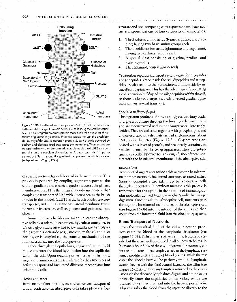
658 INTEGRATION OF PHYSIOLOGICAL S Y S T E M S . . . . . . . . . . . . . . . . . . . . . . . . . . . . . . . . . . . . . . . . .
Cells lining intestine
Figure 15-35 Facilitated transport proteins (GLUT5, GLUT2) are central to this model of sugartransport across the cells lining the small intestine. SGLTl is an integral membrane protein that couples the transport of Na+ to that of glucose or galactose. Fructose passes through the brush bor-
der by way of the GLUT5 transport protein. Sugar uptake is powered by
sodium and electrical gradients across the membrane. Then, sugars are transported down their concentration gradients via the GLUT2 transport
proteins on the basolateral membrane. A basolateral Na+/K+ pump pumps out Na+, creating the gradient that powers the whole process. [Adapted from Wright, 1993.1
of specific protein channels located in the membranes. This process is powered by coupling sugar transport to the sodium gradients and electrical gradients across the plasma membrane. SGLTl is the integral membrane protein that couples the transport of Na+ with glucose across the brush border. In this model, GLUT5 is the brush border fructose transporter, and GLUT2 is the basolateral membrane trans- porter for fructose as well as glucose and galactose (not shown).
Some monosaccharides are taken up into the absorp- tive cells by a related mechanism, hydrolase transport, in which a glycosidase attached to the membrane hydrolyzes the parent disaccharide (e.g., sucrose, maltose) and also acts as, or is coupled to, the transfer mechanism of the monosaccharide into the absorptive cell.
Once through the epithelium, sugar and amino acid molecules enter the blood by diffusion into the capillaries within the villi. Upon reaching other tissues of the body, sugars and amino acids are transferred by the same types of active transport and facilitated diffusion mechanisms into other body cells.
Active transport In the mammalian intestine, the sodium-driven transport of amino acids into the absorptive cells takes place via four
separate and non-competing cotransport systems. Each sys- tem transports just one of four categories of amino acids:
1. The 3 dibasic amino acids (lysine, arginine, and histi- dine) having two basic amino groups each
2. The diacidic amino acids (glutamate and aspartate), having two carboxyl groups each
3. A special class consisting of glycine, proline, and hydroxyproline
4. The remaining neutral amino acids
Yet another separate transport system exists for dipeptides and tripeptides. Once inside the cell, dipeptides and tripep- tides are cleaved into their constituent amino acids by in- tracellular peptidases. This has the advantage of preventing a concentration buildup of the oligopeptides within the cell, so there is always a large inwardly directed gradient pro- moting their inward transport.
Special handling of lipids The digestion products of fats, monoglycerides, fatty acids, and glycerol diffuse through the brush border membrane and are reconstructed within the absorptive cell into trigly- cerides. They are collected together with phospholipids and cholesterol into tiny droplets termed chylomicrons, about 150 pm in diameter (Figure 15-36). Chylomicrons are coated with a layer of protein, and are loosely contained in vesicles formed by the Golgi apparatus. They are subse- quently expelled by exocytosis through fusion of these vesi- cles with the basolateral membrane of the absorptive cell.
Endocytosis Transport of sugars and amino acids across the basolateral membranes occurs by facilitated transport, as noted earlier. Some oligopeptides are taken up by absorptive cells through endocytosis. In newborn mammals this process is responsible for the uptake in the intestine of immunoglob- ulin molecules derived from the mother's milk that escape digestion. Once inside the absorptive cell, nutrients pass through the basolateral membranes of the absorptive cell (see Figure 15-36) into the interior of the villus and then move from the interstitial fluid into the circulatory system.
Blood Transport of Nutrients From the interstitial fluid of the villus, digestion prod- ucts enter the blood or the lymphatic circulation (see Figure 15-36). Fishes have relatively simple lymphatic ves- sels, but these are well developed in all other vertebrates. In humans, about 80% of the chylomicrons, for example, en- ter the bloodstream via lymph carried in the lymphatic sys- tem, a modified ultrafiltrate of blood plasma, while the rest enter the blood directly. The pathway into the lymphatic system begins with the blind central lacteal of the villus (see Figure 15-21A). In humans lymph is returned to the circu- lation via the thoracic lymph duct. Sugars and amino acids primarily enter the capillaries of the villus, which are drained by venules that lead into the hepatic portal vein. This vein takes the blood from the intestine directly to the
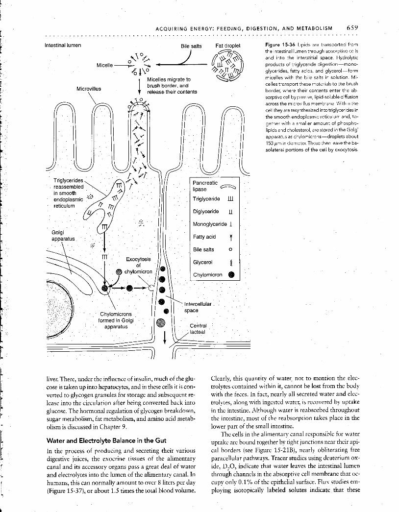
ACQUIRING ENERGY: FEEDING, D l . . . . . . . . . . . . . . . . . . . . . . . . . . . . . . . . . . . . . . . . . . . . . . . . . . . . Intestinal lumen Bile salts Fat droplet
Micelle -- 6 b0 +on rzr
1 Micelles migrate to <w Microvillus
brush border, and
\ release their contents
Figure 15-36 Lipids are transported from the Intestinal lumen through absorptive cells and into the interstitial space. Hydrolytic products of triglyceride digestion-mono- glycerides, fatty acids, and glycerol-form micelles with the blle salts in solution. Mi- celles transport these materials to the brush border, where their contents enter the ab- sorptive cell by passive, lipid-soluble diffusion across the m~crovillus membrane Wlthin the cell they are resynthesized into triglycerides in the smooth endoplasmic reticulum and, to- gether with a smaller amount of phospho- lipids and cholesterol, are stored In the Golgi apparatus as chylomicrons-droplets about 150 pm in diameter. These then leave the ba- solateral portions of the cell by exocytosis.
liver. There, under the influence of insulin, much of the glu- cose is taken up into hepatocytes, and in these cells it is con- verted to glycogen granules for storage and subsequent re- lease into the circulation after being converted back into glucose. The hormonal regulation of glycogen breakdown, sugar metabolism, fat metabolism, and amino acid metab- olism is discussed in Chapter 9.
Water and Electrolyte Balance in the Gut
In the process of producing and secreting their various digestive juices, the exocrine tissues of the alimentary canal and its accessory organs pass a great deal of water and electrolytes into the lumen of the alimentary canal. In humans, this can normally amount to over 8 liters per day (Figure 15-37), or about 1.5 times the total blood volume.
Clearly, this quantity of water, not to mention the elec- trolytes contained within it, cannot be lost from the body with the feces. In fact, nearly all secreted water and elec- trolytes, along with ingested water, is recovered by uptake in the intestine. Although water is reabsorbed throughout the intestine, most of the reabsorption takes place in the lower part of the small intestine.
The cells in the alimentary canal responsible for water uptake are bound together by tight junctions near their api- cal borders (see Figure 15-21B), nearly obliterating free paracellular pathways. Tracer studies using deuterium ox- ide, D,O, indicate that water leaves the intestinal lumen through channels in the absorptive cell membrane that oc- cupy only 0.1% of the epithelial surface. Flux studies em- ploying isotopically labeled solutes inlcate that these
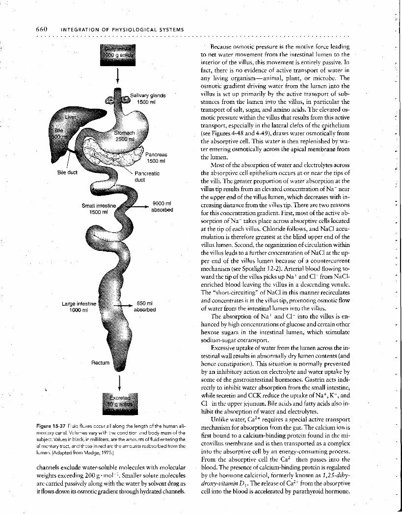
660 INTEGRATION OF PHYSIOLOGICAL SYSTEMS . . . . . . . . . . . . . . . . . . . . . . . . . . . . . . . . . . . . . . . . .
Figure 15-37 Fluid fluxes occur all along the length of the human ali- mentary canal. Volumes vary with the condition and body mass of the subject. Values in black, in milliliters, are the amounts of fluid entering the alimentary tract, and those in red are the amounts reabsorbed from the lumen. [Adapted from Madge, 1975.1
channels exclude water-soluble molecules with molecular weights exceeding 200 g . molk'. Smaller solute molecules are carried passively along with the water by solvent drag as it flows down its osmotic gradient through hydrated channels.
Because osmotic pressure is the motive force leading to net water movement from the intestinal lumen to the interior of the villus, this movement is entirely passive. In fact, there is no evidence of active transport of water in any living organism-animal, plant, or microbe. The osmotic gradient driving water from the lumen into the villus is set up primarily by the active transport of sub- stances from the lumen into the villus, in particular the transport of salt, sugar, and amino acids. The elevated 0s- motic pressure within the villus that results from this active transport, especially in the lateral clefts of the epithelium (see Figures 4-48 and 4-49), draws water osmotically from the absorptive cell. This water is then replenished by wa- ter entering osmotically across the apical membrane from the lumen.
Most of the absorption of water and electrolytes across the absorptive cell epithelium occurs at or near the tips of the villi. The greater proportion of water absorption at the villus tip results from an elevated concentration of Na+ near the upper end of the villus lumen, which decreases with in- creasing distance from the villus tip. There are two reasons for this concentration gradient. First, most of the active ab- sorption of Na+ takes place across absorptive cells located at the tip of each villus. Chloride follows, and NaCl accu- mulation is therefore greatest at the blind upper end of the villus lumen. Second, the organization of circulation within the villus leads to a further concentration of NaCl at the up- per end of the villus lumen because of a countercurrent mechanism (see Spotlight 12-2). Arterial blood flowing to- ward the tip of the villus picks up Na+ and C1- from NaC1- enriched blood leaving the villus in a descending venule. The "short-circuiting" of NaCl in this manner recirculates and concentrates it in the villus tip, promoting osmotic flow of water from the intestinal lumen into the villus.
The absorption of Na+ and C1- into the villus is en- hanced by high concentrations of glucose and certain other hexose sugars in the intestinal lumen, which stimulate sodium-sugar cotransport.
Excessive uptake of water from the lumen across the in- testinal wall results in abnormally dry lumen contents (and hence constipation). This situation is normally prevented by an inhibitory action on electrolyte and water uptake by some of the gastrointestinal hormones. Gastrin acts indi- rectly to inhibit water absorption from the small intestine, while secretin and CCK reduce the uptake of Na+, K+, and C 1 in the upper jejunum. Bile acids and fatty acids also in- hibit the absorption of water and electrolytes.
Unlike water, Ca2+ requires a special active transport mechanism for absorption from the gut. The calcium ion is first bound to a calcium-binding protein found in the mi- crovillus membrane and is then transported as a complex into the absorptive cell by an energy-consuming process. From the absorptive cell the Ca2+ then passes into the blood. The presence of calcium-binding protein is regulated by the hormone calcitriol, formerly known as 1,25-dihy- droxy-vitamin D3. The release of Ca2+ from the absorptive cell into the blood is accelerated by parathyroid hormone.
ACQUIRING ENERGY: FEEDING, DIGESTION, A N D METABOLISM 661 . . . . . . . . . . . . . . . . . . . . . . . . . . . . . . . . . . . . . . . . . . . . . . . . . . . . . . . . . . . . . . . . . . . . . . . . . . . . . . .
Vitamin B12, which has a molecular weight of 1357 g . molkl, is the largest water-soluble essential nutri- ent taken up intact across the intestinal lumen in the region of the distal ileum. This highly charged cobalt-containing compound is associated with food protein, to which it is bound as a coenzyme. In the process of absorption, B12 transfers from the dietary protein to a mucoprotein known as intrinsic factor (or hemopoietic factor) that is produced by the H+-secreting parietal cells of the stomach. Since B,, is essential for the synthesis and maturation of red blood corpuscles, pernicious anemia occurs when BIZ absorption is prevented by interference with its binding to intrinsic fac- tor. Some tapeworms "steal" B,, in the intestine of the host by producing a compound that removes it from intrinsic factor, making it unavailable to the host but available to the tapeworm.
NUTRITIONAL REQUIREMENTS
Whatever the form of food capture, ingestion, and diges- tion, all animals must acquire an appropriate variety and amount of nutritive substances, as we will now consider. Nutrients are substances that serve as sources of metabolic energy and as raw material for growth, repair of tissues, and production of gametes. Nutrients also include essential trace elements such as iodine, zinc, and other metals that may be required in extremely small quantities. There is wide variation between the nutritional needs of different species. Within a species, nutritional needs vary according to phenotypic differences in body size and composition and activity, and also with age, sex, and reproductive state. A gravid (egg-bearing) or pregnant female may require more nutrients than a male, while a male producing sperm may have greater nutritive needs than one that is not producing sperm. Regardless of reproductive state, a small animal re- quires more food for energy per gram of body weight than does a larger animal, because its metabolic rate per unit body weight is higher. Similarly, an animal with a high body temperature requires more food, to satisfy greater energy needs, than does an animal with lower body temperature. (The energetics of temperature, size, and other factors are discussed in Chapter 16).
Energy Balance
A balanced nutritional state exists when an animal has suf- ficient food intake of all nutrients necessary for long-term growth and maintenance. The nutritional requirements in- clude (1) sufficient sources of energy to power all body processes, (2) enough protein and amino acids to maintain a positive nitrogen balance (i.e., to avoid net loss of body proteins), (3) enough water and minerals to compensate for their loss or incorporation into body tissues, and (4) those essential amino acids and vitamins not synthesized within the body.
Energy balance requires that caloric intake over a given period of time equal the number of calories consumed for tissue maintenance and repair and for work (metabolic and
otherwise), plus the production of body heat in birds and mammals. Thus,
caloric intake = caloric output = calories consumed by tissues
+ heat produced
Insufficient intake of calories can be temporarily offset by using stores of fat and carbohydrates or proteins within tis- sues, but this produces a resultant loss of body weight. Conversely, caloric intake in excess of what is required for energy balance will result in increased storage of body fat, as in the large fat stores accumulated before long migra- tions in migratory birds or laid down by mammals before onset of hibernation.
Animals differ in their abilities to synthesize the sub- stances fundamental to maintenance and growth. Thus, for a given animal species, certain cofactors (Zn, I etc.) or building blocks (amino acids, etc.) essential for important biochemical reactions or for the production of tissue mole- cules may be required from food sources simply because those substances cannot be produced by the animal itself. Such items are known as essential nutrients.
Nutrient Molecules
A wide variety of molecules serve as nutrient molecules, in- cluding water, proteins and amino acids, carbohydrates, fats and lipids, nucleic acids, inorganic salts, and vitamins.
Water Of all the constituents of animal tissue, none is more per- vasively important to living tissue than water. This unique and marvelous substance can constitute 95% or more of the weight of some animal tissues. It is replenished in most animals by drinking (see Chapter 12) and by ingestion with food. Some marine and desert animals depend almost entirely on "metabolic water"-water produced during the oxidation of fats and carbohydrates-to replace wa- ter lost by evaporation, defecation, and urination (see Chapter 16).
Proteins and amino acids Proteins are used as structural components of tissues and as enzymes. They can also be utilized as energy sources if first broken down to amino acids (see Chapter 3). The proteins of animal tissues are composed of about 20 different amino acids. The ability to synthesize amino acids differs among species. Those amino acids that cannot be synthesized by an animal, but are required for synthesis of essential pro- teins, are the so-called essential amino acids for that animal. Recognition of this requirement has been of enormous eco- nomic significance in the poultry industry. The rate of growth of chickens at one time was limited by too small a proportion of a few essential amino acids in the grain diet they were provided. Supplementing the diet with these amino acids allowed full utilization of the other amino acids present in the feed, greatly increasing the rate of

protein synthesis and hence the rate of poultry growth and egg laying. Microbiologists artificially induce this limiting condition by genetically engineering microbes that require a specific amino acid (e.g., lysine) not normally found in their environment. Thus, the microbes will grow only in an environment enriched with the amino acid, serving as a safe- guard preventing their spread through normal populations.
and fats can be converted by most animals into carbohy- drates. The major sources of carbohydrate are the sugars, starches, and cellulose found in plants and the glycogen stored in animal tissues.
Lipids Lipid (fat) molecules are especially suitable as concentrated energy reserves. Each gram of fat provides over 2 times as much caloric energy as a gram of protein or carbohydrate. Consequently, lipids can store significantly more chemical energy per unit volume of tissue. Fat is commonly stored by animals for periods of caloric deficit, as during hibernation, when energy expenditure exceeds energy intake. Lipids are
Carbohydrates Carbohydrates are used primarily as immediate (glucose 6-phosphate) or stored (glycogen) sources of chemical en- ergy. However, they may also be converted to metabolic in- termediates or to fats (see Chapter 3). Conversely, proteins
TABLE 15-3 Some mammalian vitamins
Vltamln (des~gnat~on In Major d~etary Funct~on ~n Def~c~ency letter or name) sources, solublllty* Uptake, storage mammalst symptoms
Carotene (A) Egg yolk, green or yellow vegetables, fruits; FS
Absorbed from gut with Formation of visual pig- aid of bite; stored in liver ments; maintenance of
ep~thelial structures; im- portant in fetal develop- ment
Night blindness, skin lesions, birth defects
Calciferol (DJ Fish oils liver; FS Absorbed from gut; little Enhancement of calcium storage absorption from gut;
bone and tooth forma- tion
Rickets (defective bone forma- tion) In children, osteomalacia in adults
Tocopherol (E) Green leafy vegetables, meat, milk, eggs, butter; FS
Absorbed from gut; In humans, maintenance stored in adipose and of red cells; antioxidant. muscle tissues In other mammals, main-
tenance of pregnancy
Increased fragility of red blood cells, muscular dystrophies, abortion, muscular wastage
Napthoquinone (K) Synthesis by Intestinal flora, her, green leafy vegetables; FS
Absorbed from gut; little Enabling of prothrombin storage, excreted in synthesis by liver feces
Failure of coagulation
Thram~ne (B,) Bra~n, I~ver, k~dney, heart, whole grains, nuts, beans, potatoes
Absorbed from gut; Formation of cocar box- stored in liver, brain, ylase enzyme involved k~dney in decarboxylat~on
(Krebs cycle)
Stoppage of CHz0 metabolism at pyruvate, beriberi, neuritis, heart failure
Riboflavin (B,) Milk, eggs, lean meat, liver, whole grains; WS
Absorbed from gut; Flavoproteins in oxidative stored in kidney, liver, phosphorylation heart
Photophobia, fissuring of the skin
Niacin Lean meat, liver, whole grains; WS
Absorbed from gut; Coenzyme in hydrogen distributed to all tissues transport (NAD, NADP)
Pellagra, skin lesions, digestive disturbances, dementia
Cyanocobalamin Liver, kidney, brain, fish, ( B ~ d eggs, bacterial synthesis
in gut; WS
Absorbed from gut; Nucleoprotein synthesis; stored in liver, kidney, formation of erythrocytes brain
Pernicious anemia, malformed erythrocytes
Folic acid (folac~n, Meats; WS pteroylglutamic acid)
Absorbed from gut; Nucleoprotein synthesis; utilized as taken in formation of erythrocytes
Failure of erythrocytes to ma- ture, anemia
Pyridoxine (BJ Whole grains, traces in many foods; WS
Pantothenic acid Many foods; WS
Absorbed from gut; half Coenzyme for amino and appears in urine fatty acid metabolism
Dermatitis, nervous disorders
Absorbed from gut; Constituent of coenzyme stored in all tissues A (CoA)
Neuromotor, cardiovascular disorders
Biotin Egg yolk, tomatoes, I~ver, synthes~s by flora of GI tract; WS
Ascorbrc acid (C) Citrus; WS
Absorbed from gut Protein and fatty acid synthes~s; C0,fixation; transamination
Scaly dermatitis, muscle pains, weakness
Absorbed from gut; little Vital element for collagen storage and ground substance;
antioxidant
Scurvy (failure to form connec- tive tissue)
*FS = fat-soluble, WS = water-soluble
+Most vrtamrns have numerous funct~ons L~sted are a mere samplrng

ACQUIRING E N . . . . . . . . . . . . . . . . . . . . . . . . . . . . . . . . . . . . .
ERGY: FEEDING, DIGESTION, AND METABOLISM 663 . . . . . . . . . . . . . . . . . . . . . . . . . . . . . . . . . . . . . . . . .
G also important in certain tissue components such as plasma membranes and other membrane-based organelles of the cell and the myelin sheaths of axons. The fatty molecules or lipids include fatty acids, monoglycerides, triglycerides, sterols, and phospholipids.
Nucleic acids Although nucleic acids are essential for the genetic ma- chinery of the cell, all animal cells appear to be capable of synthesizing them from simple precursors. Thus, the intake of intact nucleic acids is not necessary from a nutritional perspective.
Inorganic salts Some chloride, sulfate, phosphate, and carbonate salts of the metals calcium, potassium, sodium, and magne- sium are important constituents of intra- and extracellu- lar fluids. Calcium phosphate occurs as hydroxyapatite [Ca,,(PO,),(OH),], a crystalline material that lends hard- ness and rigidity to the bones of vertebrates and the shells of mollusks. Iron, copper, and other metals are required for redox reactions (as cofactors) and for oxygen transport and binding (hemoglobin, myoglobin). Many enzymes require specific metal atoms to complete their catalytic functions. Animal tissues need moderate quantities of some ions (Ca, P, K, Na, Mg, S, and C1) and trace amounts of others (Mn, Fe, I, Co, Cu, Zn, and Se).
Vitamins Vitamins are a diverse and chemically unrelated group of organic substances that generally are required in small quantities primarily to act as cofactors for enzymes. Some vitamins important in human nutrition are listed in Table 15-3, along with their diverse functions. Detailed nutritional vitamin requirements are known primarily for domesticated animals grown for their meat, eggs, or other products. Very little is known about the vitamins involved in the metabolism of lower vertebrates and especially invertebrates.
The ability to synthesize different vitamins differs be- tween species, and those essential vitamins that an animal cannot produce itself must be obtained from other sources, primarily from plants but also from dietary animal flesh or from intestinal microbes. Ascorbic acid (vitamin C) is syn- thesized by many animals, but not by humans, who acquire it mainly from citrus fruits. Scurvy, a condition of ascorbic acid deficiency in humans, was common on board ships be- fore the British admiralty instituted the use of citrus fruit- especially limes-to supplement the diet of the crews. Their use of limes led to the general term limey to describe the English. Humans also are unable to produce vitamins K and B,, , which are produced by intestinal bacteria and then absorbed for distribution to the tissues. Fat-soluble vi- tamins such as A, D3, E, and K are stored in body fat de- posits. Water-soluble vitamins such as ascorbic acid are not stored in the body, however, and so must be ingested or produced continually to maintain adequate levels.
SUMMARY
All heterotrophic organisms acquire carbon compounds of moderate to high energy content from the tissues of other plants and animals. The chemical energy contained in these compounds originally was converted from radiant energy into chemical energy trapped in sugar molecules by photo- synthesizing autotrophs. Subsequent synthetic activity by au- totrophs and heterotrophs converts these simple carbon com- pounds into more complex carbohydrates, fats, and proteins.
Animals obtain food in many different ways, including absorption through the body surface in some aquatic or marine species, endocytosis in microorganisms, filter feed- ing, mucus trapping, sucking, biting, and chewing. Once in- gested, the food may be temporarily stored, as in a crop or rumen, or immediately subjected to digestion. Digestion consists of the enzymatic hydrolysis of large molecules into their monomeric building blocks. In multicellular animals, this takes place extracellularly in an alimentary canal. Di- gestive hydrolysis occurs only at low-energy bonds, most of the chemical energy of foodstuffs being conserved for in- tracellular energy metabolism once the products of diges- tion have been assimilated into the animal's tissues. Step- wise intracellular oxidations by coupled reactions then lead to a controlled release of chemical bond energy and mate- rial for cell growth and functioning.
Digestion in vertebrates begins in a region of low pH, the stomach, and proceeds to a region of higher pH, the small intestine. Proteolytic enzymes are released as proen- zymes, or zymogens, which are inactive until a portion of the peptide chain is removed by digestion. This procedure avoids the problem of proteolytic destruction of the enzyme-producing cells that store and secrete the zymogen granules containing the proenzyme. Other exocrine cells se- crete digestive enzymes (e.g., carbohydrases and lipases), mucin, or electrolytes such as HCl or NaHC03.
The motility of the vertebrate digestive tract depends on the coordinated activity of longitudinal and circular layers of smooth muscle. Peristalsis occurs when a ring of circular contraction proceeds along the gut preceded by a region in which the circular muscles are relaxed. The parasympa- thetic innervation stimulates motility, whereas the sympa- thetic innervation inhibits motility.
The motility of smooth muscle as well as the secretion of digestive juices is under fine neural and endocrine con- trol. All gastrointestinal hormones are peptides, and many of them also function as neuropeptides in the central ner- vous system, where they act as transmitters or short-range neurohormones. Both direct activation by food in the gut and neural activation stimulate the endocrine cells of the gastrointestinal mucosa that secrete peptide hormones. These hormones are active in either stimulating or inhibit- ing the activity of the various kinds of exocrine cells in the gut that produce digestive enzymes and juices.
Digestion products are taken up by the absorptive cells of the intestinal mucosa and transferred to the lymphatic and circulatory systems. The absorptive surface, in effect
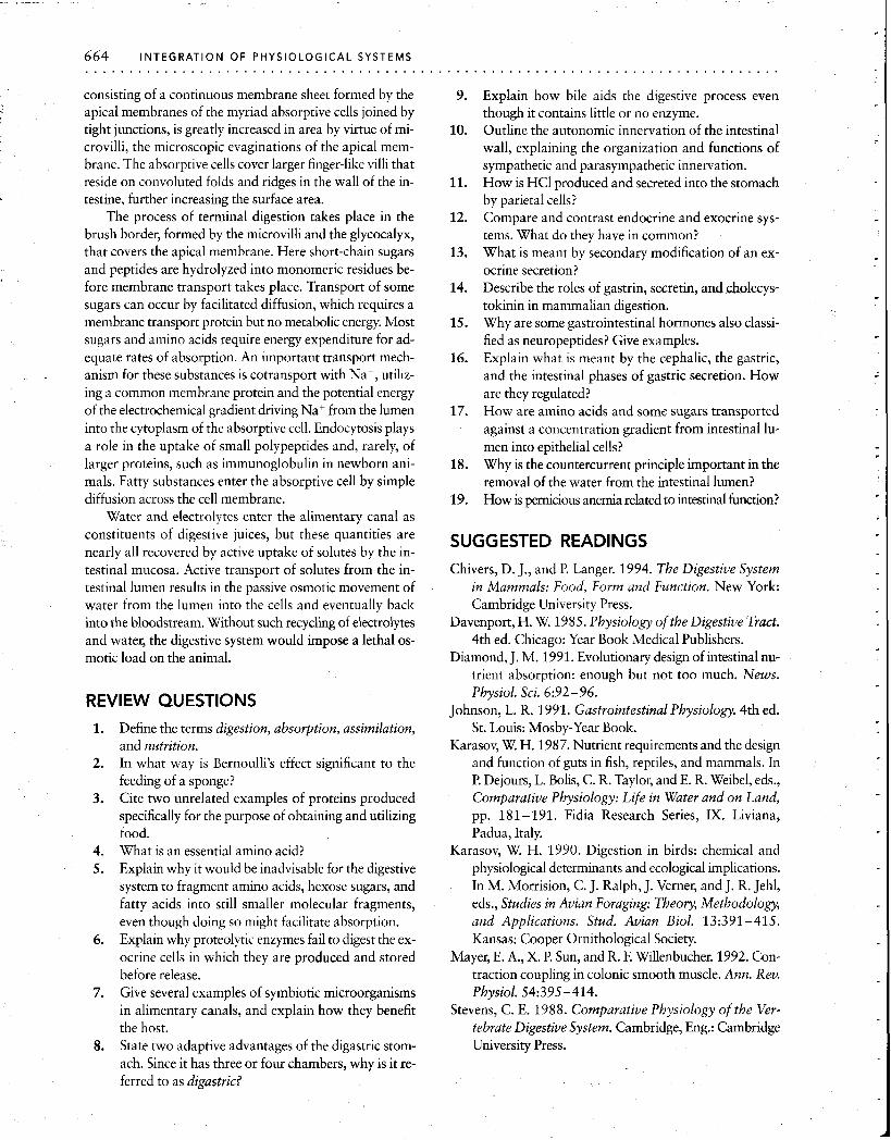
664 INTEGRATION O F PHYSIOLOGICAL SYSTEMS . . . . . . . . . . . . . . . . . . . . . . . . . . . . . . . . . . . . . . .
consisting of a continuous membrane sheet formed by the apical membranes of the myriad absorptive cells joined by tight junctions, is greatly increased in area by virtue of mi- crovilli, the microscopic evaginations of the apical mem- brane. The absorptive cells cover larger finger-like villi that reside on convoluted folds and ridges in the wall of the in- testine, further increasing the surface area.
The process of terminal digestion takes place in the brush border, formed by the microvilli and the glycocalyx, that covers the apical membrane. Here short-chain sugars and peptides are hydrolyzed into monomeric residues be- fore membrane transport takes place. Transport of some sugars can occur by facilitated diffusion, which requires a membrane transport protein but no metabolic energy. Most sugars and amino acids require energy expenditure for ad- equate rates of absorption. An important transport mech- anism for these substances is cotransport with Na+, utiliz- ing a common membrane protein and the potential energy of the electrochemical gradient driving Naf from the lumen into the cytoplasm of the absorptive cell. Endocytosis plays a role in the uptake of small polypeptides and, rarely, of larger proteins, such as immunoglobulin in newborn ani- mals. Fatty substances enter the absorptive cell by simple diffusion across the cell membrane.
Water and electrolytes enter the alimentary canal as constituents of digestive juices, but these quantities are nearly all recovered by active uptake of solutes by the in- testinal mucosa. Active transport of solutes from the in- testinal lumen results in the passive osmotic movement of water from the lumen into the cells and eventually back into the bloodstream. Without such recycling of electrolytes and water, the digestive system would impose a lethal os- motic load on the animal.
REVIEW QUESTIONS Define the terms digestion, absorption, assimilation, and nutrition. In what way is Bernoulli's effect significant to the feeding of a sponge? Cite two unrelated examples of proteins produced specifically for the purpose of obtaining and utilizing food. What is an essential amino acid? Explain why it would be inadvisable for the digestive system to fragment amino acids, hexose sugars, and fatty acids into still smaller molecular fragments, even though doing so might facilitate absorption. Explain why proteolytic enzymes fail to digest the ex- ocrine cells in which they are produced and stored before release. Give several examples of symbiotic microorganisms in alimentary canals, and explain how they benefit the host. State two adaptive advantages of the digastric stom- ach. Since it has three or four chambers, why is it re- ferred to as digastric?
Explain how bile aids the digestive process even though it contains little or no enzyme. Outline the autonomic innervation of the intestinal wall, explaining the organization and functions of sympathetic and parasympathetic innervation. How is HC1 produced and secreted into the stomach by parietal cells? Compare and contrast endocrine and exocrine sys- tems. What do they have in common? What is meant by secondary modification of an ex- ocrine secretion? Describe the roles of gastrin, secretin, and cholecys- tokinin in mammalian digestion. Why are some gastrointestinal hormones also classi- fied as neuropeptides? Give examples. Explain what is meant by the cephalic, the gastric, and the intestinal phases of gastric secretion. How are they regulated? How are amino acids and some sugars transported against a concentration gradient from intestinal lu- men into epithelial cells? Why is the countercurrent principle important in the removal of the water from the intestinal lumen? How is pernicious anemia related to intestinal function?
SUGGESTED READINGS Chivers, D. J., and P. Langer. 1994. The Digestive System
in Mammals: Food, Form and Function. New York: Cambridge University Press.
Davenport, H. W. 1985. Physiology of the Digestive Tract. 4th ed. Chicago: Year Book Medical Publishers.
Diamond, J. M. 1991. Evolutionary design of intestinal nu- trient absorption: enough but not too much. News. Physiol. Sci. 6:92-96.
Johnson, L. R. 1991. Gastrointestinal Physiology. 4th ed. St. Louis: Mosby-Year Book.
Karasov, W. H. 1987. Nutrient requirements and the design and function of guts in fish, reptiles, and mammals. In P. Dejours, L. Bolis, C. R. Taylor, and E. R. Weibel, eds., Comparative Physiology: Life in Water and on Land, pp. 18 1 - 191. Fidia Research Series, IX. Liviana, Padua, Italy.
Karasov, W. H. 1990. Digestion in birds: chemical and physiological determinants and ecological implications. In M. Morrision, C. J. Ralph, J. Verner, and J. R. Jehl, eds., Studies in Avian Foraging: Theory, Methodology, and Applications. Stud. Avian Biol. 13:391-415. Kansas: Cooper Ornithological Society.
Mayer, E. A., X. P. Sun, and R. F. Willenbucher. 1992. Con- traction coupling in colonic smooth muscle. Ann. Rev. Physiol. 54:395-414.
Stevens, C. E. 1988. Comparative Physiology of the Ver- tebrate Digestive System. Cambridge, Eng.: Cambridge University Press.