16 egypt. j. bot vol. 55, no, -...
TRANSCRIPT

Egypt. J. Bot., Vol. 55, No, 2. pp. 247-267 (2015)
16
E
Phenetic Analysis of Certain Taxa of Euphorbiaceae Grown in Egypt. M.M. Moawed, Salma Saaid, Zeinab Abdelsamie, M.
Tantawy*.
Botany Department, Faculty of Science, Ain Shams University,
Egypt.
uphorbiaceae is one of the major flowering plant families.
……...Macro- and micromorphological as well as vein architectural
characters of 34 taxa of Euphorbiaceae (14 genera, 29 species and
eight varieties) were investigated. The studied taxa were collected
from natural habitats and from different botanical gardens in Egypt.
The macro-, micromorphological characters and vein architectural
aspects were considered diagnostic characters which facilitate the
separation between the taxa under investigation. The sum of 346
attributes were numerically analyzed using NTsys-pc program
(version 2.02). The resulted phenogram is discussed showing two
outgroups and five major clusters. The present study recommends the
separation of Andrachne aspera and Putranjiva roxburghii in two
separate families; Phyllanthaceae and Putranjivaceae respectively
Keywords: Euphorbiaceae, Petiole, Vasculature, Stomata, Vein
architecture.
Euphorbiaceae comprise 322 genera, 8910 species (Thakur and Patil, 2011 a, b)
grouped in 52 tribes and five subfamilies Phyllanthoideae, Oldfieldioideae,
Acalyphoideae, Crotonoideae, and Euphorbioideae Webster (1975). To cite but
a few we can refer to the system of classification which deal with the position of
Euphorbiaceae e.g. Bentham and Hooker (1862-1883), Bessey (1915), Wettstein
(1935), Engler and Diels (1936), Benson (1957), Hutchinson (1959, 1969, 1973),
Melchior (1964), Cronquist (1968, 1981, 1988), Rendle (1969), Pax (1884),
Takhtajan (1969, 1980), Airy Shaw (1965, 1975, 1980), Webster (1975), APG
(2003, 2009) and Simpson, (2006).
Euphorbiaceae displays an extraordinary range of growth forms, perhaps
equaling or surpassing any other angiosperm family (Mahlberg and Sabharwal, 1968). The same authors also, stated that the laticifer development in Euphorbiaceae has concentrated in Euphorbia and closely related genera. The presence or absence of laticifers was used as characters in Webster (1975) classification of Euphorbiaceae. Latex is of very widespread occurrence among Crotonoideae and Euphorbioideae. (Spix & Martius, 1828; Schaeffer, 1971; Roosmalen, 1985; Mcpherson & Tirel, 1987, and Rudall, 1994). Lamina composition and anatomy has been considered to be important for the characterization of Euphorbiaceae for more than a century since the works of

M. M. MOAWED et al.
Egypt. J. Bot., Vol. 55, No. 2 (2015)
248
Solereder (1908) and Metcalfe & Chalk (1950). Anatomy of the family was investigated by different anatomists Metcalfe & Chalk, (1950) Scott et al, (1960); Mennega, (1987) Hayden, (1994) Hayden & Hayden, (1999) and Thakur & Patil, (2011 a & b). Stem, petiole and lamina anatomy show varied shape and vasculature (Dehay, 1935; Metcalfe & Chalk, (1950) and Scot et al., (1960). Stomata within the Euphorbiaceae show considerable variation as indicated by Raju and Rao (1977).
The value of leaf architecture in taxonomic studies was performed by many
authors to differentiate or clarify the relations between different species (Klucking, 1987; Yu & Chen, 1991 and Angélica et al., 2009). Concerning the leaf architecture of the family Euphorbiaceae, the following aspects were observed: Primary vein category: actinoromous or pinnate. Secondary vein: category: brochidodromous, craspedodromous (Euphorbia hirta, E. retusa, Ricinus communis) or semicraspedodromous (Acalypha wilkesiana ‘Hoffmanii’, A. wilkesiana var. macafeana, Hura crepitans and Putranjiva roxburghii). Spacing: decreasing toward base (Breynia disticha & Euphorbia tithymaloides), increasing toward base, irregular or uniform (Euphorbia milli var. splendens and Hura crepitans). Angle: decreasing toward base, increasing toward base, one pair acute basal secondaries or two pair acute basal secondaries (Euphorbia tithymaloides).Tertiary vein: category: alternate percurrent, mixed opposite/alternate percurrent or random reticulate. Course: admedially ramified or exmedially ramified. Quaternary and Quinary vein category: regular polygonal reticulate in all studied taxa. Freely ending ultimate veins: may be absent, once branched (Euphorbia tithymaloides and Putranjiva roxburghii) or two or more branched. Marginal ultimate looped in all studied taxa.
The ultimate goals of this study are to discuss whether Macro- and
micromorphological as well as vein architectural characters can provide a fundamental tool which help in explanation of the taxonomic trends within the family. The investigation of lamina abaxial and adaxial surface and stomata types is an attempt to reveal additional characteristics that might be useful for identification and assessment of taxonomic relationships among the species studied.
Material and Methods
The present study comprised 34 wild and ornamental taxa of Euphorbiaceae
(Table 1). The studied taxa were collected from botanical gardens, natural habitats in Egypt (wild species) and Herbarium of Faculty of Science, Ain Shams University. Identification of taxa under investigation was confirmed according to Täckholm (1974) and Boulos (2000). The macromorphological characters of the whole plant, habit, stem, leaf, inflorescence, and flowers were studied and described directly from the living and herbarium specimens. Stem, petiole parts and a portion of the middle lamina including the midrib were prepared according to Johanson (1940). Terminology of Eames (1929) and Metcalfe & Chalk (1950) was used to describe the anatomical features. Lamina epidermal samples were prepared from fresh materials and herbarium specimens for examination of epidermal characteristics, stomata and trichomes. Terminology of epidermal characteristics was based on Metcalfe and Chalk (1950), LAWG (1999) and

PHENETIC ANALYSIS OF CERTAIN TAXA OF …
Egypt. J. Bot., Vol. 55, No. 2 (2015)
249
Prabhakar (2004). For lamina vein architecture place leaf in a large beaker with ethanol (70% ethanol) and boil until become clear. The chlorophyll dissolves in the ethanol. After the leaves are clear place them in a warm (56 ˚C) solution of 5-10 % NaOH. They may then be removed, rinsed with water and studied. Leaf architectural terminology generally follows Hickey (1973, 1979) and LAWG (1999). Examination and photomicrographs were taken using (LM) and Digital Camera (Canon power-shot A720, 8.0 mega pixels). The magnification power was expressed by (x).
The data obtained from macro- and micromorphology as well as lamina vein
architecture of the investigated taxa were subjected to the numerical analysis. The cluster analysis was performed using NTsys program version 2.02 software (Rohlf, 1989) and the tree was constructed using the unweighted pair group method with arithmetic averages (UPGMA). The program can be used to find sets of similar objects (clusters) and to display low dimensional views (ordinations) of multivariate spaces. The grouping of operational taxonomic units (OTU’s) produced from the analysis were examined and compared with the previous and current taxonomic classifications of the Euphorbiaceae.
Results and discussion
The morphological criteria extracted from the taxa under investigation are
summarized in Table 2 as 0, 1. The phenogram showing the clustering of the studied taxa based on 346 macro and micro morphological character states (Fig. 2) revealed that; at the reference line of 1.17 level; two outgroups and five major clusters viz. A, B, C, D and E are separated. Outgroup 1 distinguished as a separate phenetic line at the level 1.23 includes Andrachne aspera while outgroup 2 separated at the level 1.21 includes Putranjiva roxburghii.
Andrachne aspera and Putranjiva roxburghii were classified in the
Euphorbiaceae by Webster (1975) and Engler & Prantl (1931) under subfamily Phyllanthoideae where Putranjiva roxburghii under tribe Drypeteae and Andrachne aspera under tribe Poranthereae and subtribe Poranthereae. APG II and III (2003, 2009) include Andrachne aspera and Putranjiva roxburghii in families Phyllanthaceae and Putranjivaceae consequently which are two of the five segregates of Euphorbiaceae sensu lato. APG II (2003) recognises five lineages of Euphorbiaceae sensu lato at family rank: Euphorbiaceae sensu stricto, Pandaceae, Phyllanthaceae, Picrodendraceae and Putranjivaceae all are members of Malpighiales in the eurosid I clade (Chase et al., 2002; Davis & Chase, 2004; Wurdack et al., 2004; Davis et al., 2005; Hoffmann et al., 2006; Simpson, 2006 and Tokuoka & Tobe, 2006).
The first comprehensive study of one of the segregate families
Phlyllanthaceae was published by Wurdack et al. (2004). It used molecular data of 52 genera including all tribes of Euphorbiaceae-Phyllanthoideae as well as selected outgroup taxa of the other subfamilies sensu Webster (1994 a, b) and Radcliffe-Smith (2001), representing all five euphorbiaceous families sensu APG II (2003).

M. M. MOAWED et al.
Egypt. J. Bot., Vol. 55, No. 2 (2015)
250
TABLE 1. The studied taxa and their sites of collection.
No. Taxa Locality or
Source
1 Acalpha wilkesiana ‘Hoffmanii’ A
2 A. wilkesiana var. macafeana W. Miller A
3 Aleurites moluccanus (L.) Willd. B
4 Andrachne aspera Spreng. D
5 Breynia disticha J. R. Forst. & G. Forst. A
6 Chrozophora brocchiana (Vis.) Schweinf. C
7 C. oblongifolia (Delile) A. Juss. Ex Spreng. C
8 C. plicata (Vahl) A. Juss. ex Spreng C
9 C. tinctoria (L.) A. Juss. C
10 Codiaeum variegatum a* A
11 C. variegatum b* A
12 C. variegatum c* A
13 C. variegatum d* A
14 Euphorbia cyathophora Murray A
15 E. dendroides L. E
16 E. helioscopia L. F
17 E. hirta L. F
18 E. milii var. splendens (Bojer ex Hooker) Ursch & Léandri A
19 E. milii var. tananarivae Léandri B
20 E. paralias L. I
21 E. peplis L. A
22 E. peplus L. F
23 E. pseudograntii Bruyns B
TABLE 1. Cont.
24 E. pulcherrima Willd. ex Klotzsch A
25 E. retusa Forssk. G
26 E. tithymaloides L. B
27 Hura crepitans L. B
28 Jatropha integerrima Jacq. B
29 J. multifida L. B
30 Joannesia princeps Vell. B
31 Mercurialis annua L. E
32 Putranjiva roxburghii Wall. B
33 Ricinus communis L. F
34 Triadica sebifera (L.) Small H
a*, b*, c* & d* are cultivars; a*: Codiaeum variegatum ‘Norma’ ; b*: C. variegatum
‘Aucubifolium’;
c*: C. variegatum ‘Ann Rutherford’ and ‘Picasso's Paintbrush’; d*: C. variegatum
‘Joseph's Coat’
A: Botanical Garden, Botany Department, Faculty of Science, Ain Shams University,
Alabbassia, Cairo. B:
Orman Botanical Garden, Giza. C: Herbarium, Botany Department, Faculty of Science,
Ain Shams University, Alabbassia, Cairo. D: Wadi Talaa, Saint Katherine, South Sinai. E:
Edku Lake, 30 Km East of Alexandria. F: Burg El-Arab, Alexandria. G: Cairo- El-Suez
Desert Road. H: Giza Zoo. I: Mersa Matrouh, El- Gharam Sea Shore.

PHENETIC ANALYSIS OF CERTAIN TAXA OF …
Egypt. J. Bot., Vol. 55, No. 2 (2015)
251
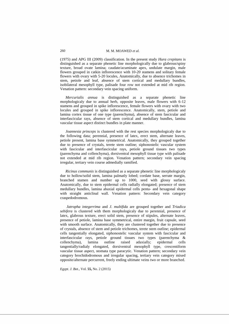
Fig. 1. Transverse section of Andrachne aspera stem x = 10; 2: Transverse section of Euphorbia
helioscopia stem x = 10; 3: Transverse section of Ricinus communis stem showing
medullary bundles x = 10; 4-8: Petiole microphotographs showing different vasculature
aspects; 4: Arc shape distinct bundles in Mercurialis annua x = 5; 5: Siphonostele
vasculature in Aleurites moluccanus x = 5; 6: kidney shape vasculature in Breynia
disticha x = 5; 7: Ring shape distinct bundles in Hura crepitans x = 5; 8: Horse shoe
shape vasculature in hura crepitans x = 5; 9: Lamina microphotographs showing Iso-
lateral mesophyll with extended palisade tissue in Andrachne aspera x = 10; 10:
Isobilateral mesophyll with palisade tissue not extended in Hura crepitans x = 10; 11:
Dorsiventral with palisade tissue not extended in Jatropha integrrima x = 10; 12:
Lamina abaxial surface of Acalypha wilkesiana var. macafeana showing paracytic
stomata x = 40; 13: Lamina abaxial surface of Euphorbia helioscopia showing
anomocytic stomata x = 20; 14: Lamina abaxial surface of Euphorbia retusa showing
anisocytic and anomotetracytic stomata x = 20; 15: Lamina architecture showing
brochidodromous 2º vein category in Aleurites moluccanus; 16: Lamina architecture
showing craspedodromous 2º vein category in Euphorbia hirta; 17: Lamina architecture
showing semi-craspedodromous 2º vein category and alternate percurrent 3º vein
categry in Acalypha wilkesiana var. macafeana; 18: Lamina architecture showing mixed
opposite/alternate percurrent 3º vein in Euphorbia pulcherimma.

M. M. MOAWED et al.
Egypt. J. Bot., Vol. 55, No. 2 (2015)
252
TABLE 2. Coding of 346 Characters States Representing Macro- and Micromorphological
Attributes (0) = Absent (1) = Present.
Macromorphological Characters (Whole Plant)
Duration Anuual (0) (1) Perennial (0) (1)
Habit Herb (0) (1) Shrub (0) (1) Succulent Shrub (0) (1) Tree (0) (1)
Latex Laticiferous (0) (1) Non-laticiferous (0) (1)
Texture
Glabrous (0) (1) Glabrous-Hairy (0) (1) Glabrous-Pubescent (0) (1)
Glabrous/Spiny (0) (1) Hairy (0) (1) Hairy/Pubescent (0) (1)
Scabrous (0) (1)
Stem Strenght Erect (0) (1) Erect/Prostrate (0) (1) Climbing (0) (1) Prostrate (0) (1)
Stem External
appear
Angular (0) (1) Terete (0) (1)
Stem internal appear Hollow (0) (1) Hollow/Solid (0) (1) Solid (0) (1)
Leaf Stipules Detection (0) (1)
Leaf Phyllotaxis Alternate (0) (1) Alternate/Opposite (0) (1) Opposite (0) (1) Spirally
(0) (1)
Leaf Petiole Detection (0) (1) Apical Gland Pair (0) (1)
Lamina Composition Compound Palmate (0) (1) Simple (0) (1) Simple/Lobed (0) (1)
Palmatly Lobed (0) (1)
Lamina Shape
Broad Ovate (0) (1) Elliptic (0) (1) Elliptic/Lanceolate (0) (1)
Elliptic/Oblong (0) (1) Linear (0) (1) Linear/Lanceolate (0) (1)
Oblong-triangular/Lanceolate (0) (1) Obovate (0) (1)
Obovate/Lanceolate (0) (1) Obovate/Oblanceolate (0) (1)
Obovate/Oblong-Spathulate (0) (1) Orbicular (0) (1) Ovate (0) (1)
Ovate/Elliptic (0) (1) Ovate/Lanceolate (0) (1) Ovate/Reniform
Curled (0) (1) Ovate/Rhombic (0) (1) Rhombic/Elliptical (0) (1)
Rhombic/Lanceolate (0) (1) Spathulate (0) (1) Sub-
orbicular/Reniform (0) (1)
Lamina Apex
Acuminate (0) (1) Acute (0) (1) Acute/Acuminate (0) (1) Caudate
(0) (1) Caudate/Acuminate (0) (1) Cuspidate/Acuminate (0) (1)
Mucronate (0) (1) Notched (0) (1) Obtuse (0) (1) Retuse/Blunt (0)
(1)
Lamina Base Shape Cordate (0) (1) Cuneate (0) (1) Cuneate/Cordate (0) (1)
Cuneate/Rounded (0) (1) Rounded (0) (1) Rounded/Cordate (0) (1)
Lamina Base
Symmetry
Asymmetrical (0) (1) Symmetrical (0) (1)
Lamina Margin
Crenate-Serrate (0) (1) Dentate (0) (1) Entire (0) (1) Entire/Dentate
(0) (1) Entire/Serrate Toward Apex (0) (1) Serrate (0) (1) Slightly
Toothed (0) (1) Undulate (0) (1)

PHENETIC ANALYSIS OF CERTAIN TAXA OF …
Egypt. J. Bot., Vol. 55, No. 2 (2015)
253
TABLE 2. Cont.
Lamina Colour
Bluish Green (0) (1) Bronze Green to Copper Red Mottled Red &
Purple (0) (1) Dark Green-Creamy Margin (0) (1) Dark Green on
Upper Surface, Pale Below (0) (1) Green (0) (1) Green in Shades
With Age and in Sunny Areas Becomes Variegated With Red & Pink,
to Pure White (0) (1) Green Variegated With Red or Pink (0) (1)
Green Spoted Yellow (0) (1) Green/Yellow (0) (1) Pale to Dark
Green With Purple Spots (0) (1) Rusty Whitish-Yellow (0) (1)
Whitish Above When Young/Green With Age (0) (1)
Flower Colour
Green (0) (1) Pink (0) (1) Red (0) (1) Scarlet Red (0) (1) White (0)
(1) Whitish Green (0) (1) Yellow (0) (1) Yellowish Green (0) (1)
Yellowish White (0) (1)
Staminate Grouping (0) (1)
Staminate Type
Catkin (0) (1) Cymose (0) (1) Solitary (0) (1) Raceme (0) (1)
Cyathium in Cymes (0) (1) Cyathium in Umbels (0) (1) Cyathium in
Clusters (0) (1) Cyathium Solitary (0) (1) Spike (0) (1)
Staminate Position Axillary (0) (1) Terminal (0) (1) Terminal-Axillary (0) (1)
Stamen Number 1 (0) (1) 2 (0) (1) 3 (0) (1) 4-12 (0) (1) 10-20 (0) (1) 15-32 (0) (1)
Up to 1000 (0) (1)
Staminate Branching (0) (1)
Pistillate Grouping (0) (1)
Pistillate Type
Catkin (0) (1) Cymose (0) (1) Solitary (0) (1) Raceme (0) (1)
Cyathium in Cymes (0) (1) Cyathium in Umbels (0) (1) Cyathium in
Clusters (0) (1) Cyathium Solitary (0) (1) Spike (0) (1)
Pistillate Position Axillary (0) (1) Terminal (0) (1) Terminal-Axillary (0) (1)
Locules Number 2-4 (0) (1) 5-20 (0) (1)
FruitType Capsule (0) (1) Drupe (0) (1)
Seed Shape
Broadly Ovoid (0) (1) Conical (0) (1) Depressed-Conical (0) (1)
Globose (0) (1) Ovoid (0) (1) Ovoid/Elliptic (0) (1)
Ovoid/Hexagonous (0) (1) Subglobose (0) (1) Subglobose/Ovoid (0)
(1) Trigonous (0) (1) Tetragonus (0) (1) Triangular/Ovoid (0) (1)
Seed Surface Glossy (0) (1) Reticulate (0) (1) Smooth (0) (1) Smooth/Minutely
Dotted (0) (1) Warty (0) (1) Wrinkled (0) (1)
Micromorphological Characters (Stem Anatomy)
Outline in T. S. Angled (0) (1) Compressed Ring (0) (1) Oval (0) (1) Terete (0) (1)
Terete/V- shape Furrows (0) (1)
Eglandular
Trichomes
Unicellular, Unbranched, Uniseriate (0) (1) Multicellular,
Unbranched, Uniseriate (0) (1) Stellate Unbranched, Multicellular,
Multiseriate (0) (1) Stellate (0) (1) Multicellular, Unicellular,
Unbranched, Uniseriate (0) (1)
Glandular Trichomes
Multicellular, Uni- & Biseriate Stalk with Multicellular Gland (0) (1)
Multicellular Sessile Gland (0) (1) Multicellular Gland with or
without Unicellular Stalk (0) (1) Unicellular, Uniseriate Stalk with
Unicellular Gland (0) (1)
Cuticle Thin (0) (1) Thick (0) (1)
Subepidermal Periderm (0) (1)

M. M. MOAWED et al.
Egypt. J. Bot., Vol. 55, No. 2 (2015)
254
TABLE 2. Cont.
Epidermal Cells Radially (0) (1) Tangentially (0) (1) Tangentially/Radially (0) (1)
Type of Cortical
Tissues
Parenchyma (0) (1) Parenchyma, Extra-xylary Fibers (0) (1) Parenchyma,
Collechyma (0) (1) Parenchyma, Chlorenchyma, Extra-xylary Fibers (0)
(1) Parenchyma, Collechyma, Extra-xylary Fibers (0) (1) Parenchyma,
Collechyma, Proto-phloem fibers (0) (1) Parenchyma, Palisade –Like,
Extra-xylary Fibers (0) (1) Parenchyma, Collechyma, Chlorenchyma,
Extra-xylary Fibers (0) (1)
Pith Width Wide (0) (1) Narrow (0) (1)
Pith Cell Type Thin Walled Parenchyma (0) (1) Thick Walled Parenchyma (0) (1)
Internal
Appearance
Hollow (0) (1) Solid (0) (1) Hollow-Solid (0) (1)
Vascular System
Aspect
Distinct (0) (1) Siphonostelic (0) (1)
Cambium Fascicular (0) (1) Fascicular + Interfascicular (0) (1)
Fascicular
Region
Vertical System Only (0) (1) Vertical + Horizontal (Uniseriate Rays) (0)
(1) Vertical + Horizontal (Uni- + Biseriate Rays) (0) (1)
Interfascicular
Region
Parenchyma (0) (1) Vertical system [Phloem (Sieve Tube, Companion,
Parenchyma cells) & Xylem (Vessels, Fibers, Parenchyma)] (0) (1)
Vertical system [Phloem (Parenchyma) & Xylem ( Fibers)] (0) (1)
Horizontal (Uniseriate Rays) (0) (1) Horizontal (Biseriate Rays) (0) (1)
Horizontal (Uni- + Biseriate Rays) (0) (1)
Cortical Bundles (0) (1)
Medullary
Bundles
(0) (1)
Crystals Druses (0) (1) Druses-Solitary (0) (1)
Micromorphological Characters (Petiole Anatomy)
Outline in T. S. Crescentiform (0) (1) Half-Circle (0) (1) Terete with Ridges and Furrows
(0) (1) Terete (0) (1)
Eglandular
Trichomes
Unicellular Unbranched, Uniseriate (0) (1) Multicellular, Unbranched,
Uniseriate (0) (1) Unicellular, Multicellular, Unbranched,Uniseriate (0)
(1) Stellate (0) (1) Stellate/Unbranched, Uniseriate, Multicellular (0) (1)
Multicellular Unbranched, Uniseriate & Multiseriate Hooked (0) (1)
Glandular
Trichomes
Unicellular Gland and Multicellular Uniseriate Stalk (0) (1) Short
Multicellular Stalk and Head (0) (1) Multicellular, Uni- and Biseriate
Stalk/Multicellular Gland (0) (1) Unicellular, Uniseriate Stalk/Unicellular
Gland (0) (1)
Cuticle Thin (0) (1) Thick (0) (1)
Epidermal Cells Radially (0) (1) Tangentially (0) (1) Radially-Tangentially (0) (1)
Type of Ground
Tissues
Parenchyma (0) (1) Parenchyma, Collechyma (0) (1) Parenchyma,
Palisade-Like (0) (1) Parenchyma, Collechyma, Extra-xylary Fibers (0) (1)
Parenchyma, Chlorenchyma, Collechyma, Extra-Xylary Fibers (0) (1)
Vascular System
Aspect
Arc Shape (0) (1) Horse Shoe (0) (1) Kidney Shape (0) (1) Ring (0) (1)
Siphonostele (0) (1)
No. of Vascular
Bundles
3 (0) (1) 4 (0) (1) 5 (0) (1) 6 (0) (1) 7 (0) (1) 8 (0) (1) 9 (0) (1) 10 (0)
(1) 11 (0) (1) 12 (0) (1) 15 (0) (1) 18 (0) (1) 20 (0) (1)
Crystals Druses (0) (1) Druses/Solitary (0) (1)
Micromorphological Characters (Lamina Anatomy)

PHENETIC ANALYSIS OF CERTAIN TAXA OF …
Egypt. J. Bot., Vol. 55, No. 2 (2015)
255
TABLE 2. Cont.
Outline in T.S. Flattened Adaxially (0) (1) Furrowed Adaxially (0) (1) Lobed
Adaxially (0) (1) Raised Adaxially (0) (1)
Eglandular
Trichomes
Multicellular,Unbranched, Uniseriate (0) (1) Multicellular,
Unicellular, Unbranched, Uniseriate (0) (1) Stellate (0) (1)
Glandular
Trichomes
Multicellular Uni-Biseriate Stalk-Multicellular Gland (0) (1)
Unicellular,Uniseriate Stalk-Unicellular Gland (0) (1)
Cuticle Thick (0) (1) Thin (0) (1)
Epidermal Cells Papillose (0) (1) Radially (0) (1) Tangentially (0) (1) Tangentially-
Radially (0) (1)
Mesophyll Tissue Dorsiventral (0) (1) Iso-bilateral (0) (1) Iso-lateral (0) (1)
No. of Palisade Rows 1(0) (1) 2 (0) (1) 4 (0) (1)
Palisade Extended at
Mid Rib Region
(0) (1)
Collenchyma Type Angular (0) (1) Angular/Annular (0) (1) Angular/Lamellar (0) (1)
Annular (0) (1) Lamellar (0) (1)
Ground Tissue Types Parenchyma (0) (1) Parenchyma, Chlorenchyma (0) (1)
Vascular System Aspect Centric Single (0) (1) Crescentiform (0) (1) Horse Shoe (0) (1) Plate
(0) (1) Ring (0) (1)
Vascular Bundles No. 1 (0) (1) 3 (0) (1) 4 (0) (1) 5 (0) (1) 6 (0) (1) 7 (0) (1) 11 (0) (1)
12 (0) (1) 15 (0) (1)
Crystals Druses (0) (1) Raphides (0) (1) Solitary/Druses (0) (1)
Micromorphological Characters (Lamina Epidermal Characteristics)
Abaxial Cell Shape
Tetragonal (0) (1) Tetragonal &Pentagonal (0) (1)
Tetragonal,Pentagonal & Hexagonal (0) (1) Trigonal, Tetragonal
&Pentagonal (0) (1) Pentagonal (0) (1) Pentagonal & Hexagonal (0)
(1)
Abaxial Anticlinal
Wall
Straight(0) (1)Wavy (0) (1)
Adaxial Cell Shape
Tetragonal (0) (1) Tetragonal, Pentagonal (0) (1)
Pentagonal,Hexagonal (0) (1) Trigonal, Tetragonal, Pentagonal (0)
(1) Tetragonal,Pentagonal, Hexagonal (0) (1)
Adaxial Anticlinal
Wall
Straight (0) (1) Wavy (0) (1)
Stomata Type
Anomocytic (0) (1) Brachyparacytic (0) (1)Paracytic (0) (1)
Anomocytic & Anisocytic (0) (1) Anomocytic & Anomotetracytic (0)
(1) Anomocytic & Brachyparacytic (0) (1) Paracytic & Anisocytic
(0) (1) Paracytic & Brachyparacytic (0) (1) Anomotetracytic,
Anomocytic, Paracytic & Brachyparacytic (0) (1)
Lamina Vein Architecture
1° Vein Category Actinoromous (0) (1) Pinnate (0) (1)
2° Vein Category Brochidodromous (0) (1) Craspedodromous (0) (1) 330
Semicraspedodromous (0) (1)
2° Vein Spacing Decreasing Toward Base (0) (1) Increasing Toward Base (0) (1)
Irregular (0) (1) Uniform (0) (1)
2° Vein Angle
Decreasing Toward Base (0) (1) Increasing Toward Base (0) (1) One
Pair Acute Basal Secondaries (0) (1) Two Pair Acute Basal
Secondaries (0) (1)
3° Vein Category Alternate Percurrent (0) (1) Mixed Opposite/Alternate Percurrent (0)
(1) Random Reticulate (0) (1)
3° Vein Course Admedially Ramified (0) (1) Exmedially Ramified (0) (1)

M. M. MOAWED et al.
Egypt. J. Bot., Vol. 55, No. 2 (2015)
256
Fig. 2. Phenogram Showing Clustering of the Studied Taxa Based on the 346
Macro/Micro Morphological Character States.
Andrachne aspera is separated as an outgroup in the present study on the
basis of morphologically characters viz. absence of latex, scabrous texture,
prostrate, suborbicular/reniform leaf shape; notched apex, solitary flowers;
stamens 5-6. Anatomically, stem epidermal cells tangentially/radially elongated;
palisade-like tissue in cortex of stem, palisade-like tissue in cortex of petiole,
petiole vascular tissue aspect kidney shape, brachparacytic and anomocytic
stomata. Putranjiva roxburghii is separated as outgroup which is
morphologically characterized by cymose inflorescence, stamens three, drupe
fruit. Anatomically it characterized by the presence of solitary/druses crystals,
stem trichomes eglandular multicellular unbranched uniseriate, anomotetracytic
and anomocytic stomata. These data confirmed those of Chase et al. (2002),
APG II (2003), Davis & Chase (2004), Wurdack et al. (2004), Davis et al.
(2005), Hoffmann et al. (2006), Tokuoka & Tobe (2006) and APG III (2009) in
regarding the placement of Andrachne aspera and Putranjiva roxburghii in the
families Phyllanthaceae and Putranjivaceae consequently which are two of the
five segregates of Euphorbiaceae sensu lato.
Cluster A
comprises two minor groups a1 & a2 that are grouped together at the
similarity level of 1.12. In the present study, all taxa of Euphorbia are clustered
together in the major cluster A. They are morphologically characterized by
presence of latex, absence of petiole glands, simple lamina composition,
cyathium inflorescence, ovary with three locules, capsule fruit. Anatomically,
they are characterized by absence of stem fascicular and interfascicular rays,
absence of glandular trichomes. The foregoing data were confirmed by Webster
(1975) and APG III (2009) classification systems in placing the taxa of

PHENETIC ANALYSIS OF CERTAIN TAXA OF …
Egypt. J. Bot., Vol. 55, No. 2 (2015)
257
Euphorbia at the same subfamily (Euphorbioideae), tribe (Euphorbieae) and
subtribe (Euphorbiinae). The subtribe includes the species with a true cyathium,
because of this unique structure; most species of Euphorbiinae have been treated
as members of the genus Euphorbia Park and Jansen (2007).
The first minor group a1
comprises eight species of Euphorbia viz. E .hirta, E. peplis, E. peplus, E.
helioscopia, E. tithymaloides, E. retusa, E. paralias and E. dendroides. Euphorbia
hirta is distinguished as a separate phenetic line at the level 1.12. E. peplus and E.
helioscopia are grouped together at the level of 0.885 and E. peplis is clustered
with them at the level of 0.895. E. tithymaloides is clustered with the rest three
species at the level of 0.986. E. paralias and E. dendroides are grouped together at
the level of 0.749 and E. retusa is clustered with them at the level of 0.836.
Euphorbia hirta is distinguished as a separate phenetic line on the basis of
the following morphological characters viz. erect/prostrate stem, opposite leaves,
rhombic/lanceolate lamina, serrate margin, terminal/axillary cyathium in clusters.
E. peplus and E. helioscopia are grouped together and E. peplis is clustered with
them. These taxa were morphologically clustered due to annual herbs, solid
stem, alternate leaves, cuneate lamina base, and green lamina colour.
Anatomically, they are clustered together due to terete stem outline, absence of
trichomes, absence of cortical and medullary bundles, absence of crystals,
flattened adaxial lamina outline, radially elongated lamina epidermal cells with
tetragonal/pentagonal wavy anticlinal walls, dorsiventral mesophyll tissue.
Venation pattern; brochidodromous secondary vein category with irregular
spacing, admedially ramified tertiary vein course.
In Webster (1975) classification he placed Pedilanthus as a genus in the
subtribe Euphorbiinae tribe Euphorbieae subfamily Euphorbioideae in the family
Euphorbiaceae. In Bentham & Hooker (1880 - 1883) and Engler & Prantl (1931)
they placed Pedilanthus in the tribe Euphorbieae. According to Webster (1994 a,
b), seven genera belong to the subtribe Euphorbiinae: Chamaesyce (± 300
species), Cubanthus (3 spp.), Endadenium (1 sp.), Euphorbia (± 2000 spp.),
Monadenium (± 70 spp.), Pedilanthus (14 spp.) and Synadenium (4 spp.) (Bruyns
et al., 2006). Steinmann (2003), Bruyns et al. (2006) and APG III (2009) works
which based on molecular data consider the genus Pedilanthus as a synonym of
Euphorbia and make a valid name Euphorbia tithymaloides.
In the present study, E. tithymaloides is clustered with the other Euphorbia
species due to presence of latex, absence of petiole glands, simple lamina
composition, cyathium inflorescence and capsule fruit. Anatomically, they are
characterized by absence of glandular trichomes. The present data confirmed
those of Steinmann (2003), Bruyns et al. (2006) and APG III (2009).
E. paralias and E. dendroides are grouped together and E. retusa is clustered
with them. These taxa were clustered on the basis of the following
morphological characters viz. perennial, glabrous texture, solid stem, alternate

M. M. MOAWED et al.
Egypt. J. Bot., Vol. 55, No. 2 (2015)
258
leaves, absence of stipules, absence of petiole, symmetrical lamina base,
cyathium grouped in umbels, smooth seed surface. Anatomically, they are
clustered together due to terete stem outline, absence of trichomes, siphonostelic
stem vascular aspect, absence of crystals, flattened adaxial lamina outline,
epidermal cells with tetragonal/pentagonal and hexagonal straight anticlinal
walls. Venation pattern; secondary vein category with irregular spacing and
angle decreasing toward base, tertiary vein category random reticulate and
admedially ramified course.
The second minor group a2
comprises Euphorbia milii var. splendens, E. milii var. tananarivae, E.
pseudograntii, E. pulcherrima and E. cyathophora. Euphorbia milii var. splendens
and E. milii var. tananarivae are grouped together at the 0.56 level. E. pulcherrima
and E. cyathophora grouped together at the level of 0.918 and clustered with E.
pseudograntii at the level of 0.996. Euphorbia milii var. splendens and E. milii var.
tananarivae are grouped together on the basis of the following morphological
characters viz. perennial succulent, glabrous/spiny texture, climbing angular solid
stem, presence of stipules, alternate leaves, presence of petiole, lamina simple;
obovate-oblong/spathulate, mucronate apex, cuneate symmetrical base, entire
margin, green colour, cyathium in axillary cymes, ovoid seed with warty surface.
Anatomically due to absence of crystals, absence of stem and leaf trichomes;
siphonostylic vascular tissue aspect, presence of cortical bundles, crescentiform
petiole outline; arc shape vascular tissue aspect, flattened adaxial lamina outline;
radially elongated epidermal cells, dorsiventral mesophyll type, palisade two rows
extended at mid rib region, crescentiform vascular tissue aspect, lamina epidermis
tetra-/pentagonal with wavy anticlinal walls abaxially and the same adaxially but
with straight anticlinal wall, stomata paracytic. Venation pattern: Pinnate primary
vein, secondary vein category brochidodromous.
E. pulcherrima and E. cyathophora grouped together and clustered with E.
pseudograntii. These taxa were morphologically clustered due to
glabrous/pubescent texture, erect hollow stem, petiole present, lamina base
cuneate; green colour, cyathium in cymes; stem vascular tissue siphonostele,
petiole vascular tissue aspect arc shape, lamina epidermal cells
tangentially/radially elongated; dorsiventral mesophyll type, lamina vascular
tissue aspect crescentiform. Venation pattern: primary vein pinnate, secondary
vein category brochidodromous. Tertiary vein category mixed opposite/alternate
percurrent, absence of freely ending ultimate veins.
E. pseudograntii in Webster (1975) classification was placed as Synadenium
genus in the subtribe Euphorbiinae tribe Euphorbieae subfamily Euphorbioideae
in the family Euphorbiaceae, and in Bentham & Hooker (1880 - 1883) and
Engler & Prantl (1931) place Synadenium under the tribe Euphorbieae.
As mentioned before according to Webster (1994 a, b), seven genera belong
to the subtribe Euphorbiinae: Chamaesyce (± 300 species), Cubanthus (3 spp.),
Endadenium (1 sp.), Euphorbia L. (± 2000 spp.), Monadenium (± 70 spp.),

PHENETIC ANALYSIS OF CERTAIN TAXA OF …
Egypt. J. Bot., Vol. 55, No. 2 (2015)
259
Pedilanthus (14 spp.) and Synadenium (4 spp.) (Bruyns et al., 2006). The genera
Endadenium, Monadenium and Synadenium are reduced to synonymy under
Euphorbia and the species are all transferred to Euphorbia. Consequently the
subtribe Euphorbiinae now consists of the single, very large, very widely
distributed and very diverse genus Euphorbia (Bruyns et. al., 2006). Steinmann
(2003), Bruyns et al. (2006) and APG III (2009) works which based on
molecular data reduce the genus Synadenium as a synonym of Euphorbia and
make a valid name Euphorbia pseudograntii. In the present study E.
pseudograntii grouped with other Euphorbia species on the basis of the
following morphological data viz. presence of latex, absence of petiole glands,
simple lamina composition, cyathium inflorescence, capsule fruit. Anatomically,
they are characterized by absence of stem fascicular and interfascicular rays,
absence of glandular trichomes. The forgoing data were confirmed by Steinmann
(2003), Bruyns et al. (2006) and APG III (2009).
Cluster B comprises four studied species of Chrozophora and grouped together at the
0.887. The genus placed under subfamily Acalyphoideae tribe Chrozophoreae according to Webster (1975) and APG III (2009). The species of Chrozophora share many characters and grouped together morphologicaly due to the following data; herb, absence of latex, hairy texture, solid stem, presence of stipules, alternate leaves, presence of petiole, presence of petiolar gland, lamina simple; symmetrical base, undulate margin, inflorescence simple raceme, ovary with three locules, fruit capsule. Anatomically, they are clustered together due to presence of crystals and stellate trichomes in both of stem, petiole and leaf. The stem and petiole epidermal cells radially elongated and cortex have two types of tissues, (parenchyma & collenchyma). Absence of stem cortical and medullary bundles, dorsiventral mesophyll type, palisade one row not extended at mid rib region, crescentiform vascular tissue aspect, stomata paracytic. Venation pattern of pinnate primary vein. Cluster C
comprises two minor groups c1 & c2 and clustered with cluster B at the 0. 173 level. The first minor group c1 is distinguished as a separate phenetic line including Hura crepitans at the level of 1.09. The second minor group c2 comprises Mercurialis annua, Joannesia princeps, Ricinus communis, Triadica sebifera, Jatropha integerrima, J. multifida and Aleurites moluccanus. Mercurialis annua is distinguished as a separate phenetic line at the level of 1.086. Joannesia princeps is clustered with the rest species at the level of 0.046. Ricinus communis is distinguished as a separate phenetic line at the level of 1.008. Jatropha integerrima and J. multifida are grouped together at the level of 0.715 and Triadica sebifera is clustered with them at the level of 0.935. Aleurites moluccanus is clustered with Triadica sebifera, Jatropha integerrima and J. multifida at the level 0.97.
The first minor group c1 is distinguished as a separate phenetic line including Hura crepitans which is
placed in tribe Hureae and subfamily Euphorbioideae according to Webster
M. M. MOAWED et al.
Egypt. J. Bot., Vol. 55, No. 2 (2015)
260
(1975) and APG III (2009) classification. In the present study Hura crepitans is distinguished as a separate phenetic line morphologically due to glabrous/spiny texture, broad ovate lamina; caudate/acuminate apex, undulate margin, male flowers grouped in catkin inflorescence with 10-20 stamens and solitary female flowers with ovary with 5-20 locules, Anatomically, due to absence trichomes in stem, petiole and leaf, absence of stem cortical and medullary bundles, isobilateral mesophyll type, palisade four row not extended at mid rib region. Venation pattern: secondary vein spacing uniform.
Mercurialis annua is distinguished as a separate phenetic line
morphologicaly due to annual herb, opposite leaves, male flowers with 6-12 stamens and grouped in spike inflorescence, female flowers with ovary with two locules and grouped in spike inflorescence. Anatomically, stem, petiole and lamina cortex tissue of one type (parenchyma), absence of stem fascicular and interfascicular rays, absence of stem cortical and medullary bundles, lamina vascular tissue aspect distinct bundles in plate manner.
Joannesia princeps is clustered with the rest species morphologicaly due to
the following data; perennial, presence of latex, erect stem, alternate leaves,
petiole present, lamina base symmetrical. Anatomically, they grouped together
due to presence of crystals, terete stem outline; siphonostelic vascular system
with fascicular and interfascicular rays, petiole ground tissues two types
(parenchyma and collenchyma), dorsiventral mesophyll tissue type with palisade
not extended at mid rib region. Venation pattern; secondary vein spacing
irregular, tertiary vein course admedially ramified.
Ricinus communis is distinguished as a separate phenetic line morphologicaly
due to hollow/solid stem, lamina palmatly lobed; cordate base, serrate margin,
branched stamen and number up to 1000, seed with glossy surface.
Anatomically, due to stem epidermal cells radially elongated; presence of stem
medullary bundles, lamina abaxial epidermal cells penta- and hexagonal shape
with straight anticlinal wall. Venation pattern: Secondary vein category
craspedodromous.
Jatropha integerrima and J. multifida are grouped together and Triadica
sebifera is clustered with them morphologicaly due to perennial, presence of
latex, glabrous texture, erect solid stem, presence of stipules, alternate leaves,
presence of petiole, lamina base symmetrical, entire margin, fruit capsule, seed
with smooth surface. Anatomically, they are clustered together due to presence
of crystals, absence of stem and petiole trichomes, terete stem outline; epidermal
cells tangentially elongated, siphonostelic vascular system with fascicular and
interfascicular rays, petiole ground tissues two types (parenchyma &
collenchyma), lamina outline raised adaxially; epidermal cells
tangentially/radialy elongated, dorsiventral mesophyll type, crescentiform
vascular tissue aspect, stomata type paracytic. Venation pattern; secondary vein
category brochidodromous and irregular spacing, tertiary vein category mixed
opposite/alternate percurrent, freely ending ultimate veins two or more branched.

PHENETIC ANALYSIS OF CERTAIN TAXA OF …
Egypt. J. Bot., Vol. 55, No. 2 (2015)
261
Aleurites moluccanus is clustered with Triadica sebifera, Jatropha
integerrima and J. multifida morphologicaly due to perennial, presence of latex,
erect solid stem, presence of stipules, alternate leaves, presence of petiole,
lamina base symmetrical, entire margin, and seed with smooth surface.
Anatomically, they are clustered together due to presence of crystals, absence of
stem trichomes, terete stem outline; epidermal cells tangentially elongated,
siphonostelic vascular system with fascicular and interfascicular rays, petiole
ground tissues two types (parenchyma and collenchyma), lamina outline raised
adaxially; epidermal cells tangentially/radialy elongated, dorsiventral mesophyll
type. Venation pattern; secondary vein category brochidodromous and irregular
spacing, freely ending ultimate veins two or more branched. The all recorded
criteria are in accordance with the different systems of Webster (1975) and APG
III (2009) that insure the occurance of these studied taxa in the subfamilies:
Acalyphoideae, Crotonoideae and Euphorbioideae.
Cluster D
comprises the four varieties of Codiaeum variegatum and grouped together at
the 0.73. The genus placed under subfamily Crotonoideae tribe Codiaeae
according to Webster (1975) and APG III (2009) and grouped together based on
the morphological characters viz. perennial shrub, presence of latex, glabrous
texture, erect solid stem, presence of stipules, alternate leaves, presence of
petiole, absence of petiolar gland, lamina simple/lobed; cuneate symmetrical
base, entire margin, inflorescence simple raceme, ovary with three locules, fruit
capsule, subglobose seed with smooth surface. Anatomically, they are clustered
together due to presence of crystals, terete stem outline, absence of stem cortical
and medullary bundles, petiole epidermal cells radially elongated, lamina outline
raised adaxially; lamina trichomes absence, epidermal cells tangentially/radialy
elongated, dorsiventral mesophyll type, palisade one row not extended at mid rib
region, ring vascular tissue aspect. Venation pattern; pinnate primary vein,
secondary vein category brochidodromous, tertiary vein category random
reticulate, absence of freely ending ultimate veins.
Cluster E
comprises two minor groups e1 and e2 and clustered with cluster D at the
1. 053 level. The first minor group e1 is distinguished as a separate phenetic line
including Breynia disticha at the level of 0.975. The second minor group e2
comprises Acalypha wilkesiana ‘Hoffmanii’ and A. wilkesiana var. macafeana
which are grouped together at the level 0.843.
The first minor group e1
is distinguished as a separate phenetic line including Breynia disticha.
The second minor group e2
comprises Acalypha wilkesiana ‘Hoffmanii’ and A. wilkesiana var.
macafeana which are grouped together. As mentioned before Webster (1975)
classified Euphorbiaceae into five subfamilies: Phyllanthoideae, Oldfieldioideae,
Acalyphoideae, Crotonoideae and Euphorbioideae. In the APG III (2009)

M. M. MOAWED et al.
Egypt. J. Bot., Vol. 55, No. 2 (2015)
262
classification, only the latter three subfamilies are included within
Euphorbiaceae, the Phyllanthoideae and Oldfieldioideae are elevated to family
rank, Phylanthaceae and Picrodendraceae, respectively in the Malpoghiales
(Simpson, 2006). This is in accordance with the recorded criteria in the present
study especially the data of the studied varieties of Acalypha.
Breynia disticha and Andrachne aspera was classified in the Euphorbiaceae
by Webster (1975) under the subfamily Phyllanthoideae. APG II and III (2003
&2009) includes Breynia disticha and Andrachne aspera in the family
Phyllanthaceae which is one of the five segregates of Euphorbiaceae sensu lato
recognised at family level.
APG II (2003) recognises five lineages of Euphorbiaceae sensu lato at family
rank: Euphorbiaceae sensu stricto, Pandaceae, Phyllanthaceae, Picrodendraceae
and Putranjivaceae all are members of Malpighiales in the eurosid I clade (Chase
et. al., 2002; Davis & Chase, 2004; Wurdack et. al., 2004; Davis et. al., 2005;
Hoffmann et. al., 2006 and Tokuoka & Tobe, 2006).
In the present study the closer relationships of Breynia disticha with Acalypha
wilkesiana ‘Hoffmanii’ and A. wilkesiana var. macafeana based on morphological
characters viz. perennial shrub, absence of latex, erect solid stem, presence of
stipules, alternate leaves, presence of petiole, absence of petiolar gland, lamina
simple; rounded symmetrical base, ovary with three locules, fruit capsule, smooth
seed surface. Anatomically, they are clustered together due to presence of crystals,
terete stem outline; epidermal cells tangentially elongated, siphonostelic vascular
tissue aspect, absence of stem cortical and medullary bundles, lamina outline raised
adaxially; dorsiventral mesophyll type, palisade one row not extended at mid rib
region. Venation pattern; tertiary vein course admedially ramified, absence of
freely ending ultimate veins. The distinction between Breynia disticha and
Acalypha wilkesiana ‘Hoffmanii’ and A. wilkesiana var. macafeana based
morphologicaly on texture, lamina shape, apex, margin and colour, inflorescence
type, seed shape, and anatomically based on presence of stem, petiole and lamina
trichomes in Acalypha and absence in Breynia, absence of stem rays in Breynia,
petiole outline and dermal, ground and vascular systems of Breynia differ from that
of Acalypha. Also they differ in vascular tissue of lamina, lamina epidermal
characters. Venation pattern differences; primary and secondary veins.
The closer relationships of Breynia disticha with Andrachne aspera based on
the following morphological criteria viz. absence of latex, solid stem, presence of
stipules, alternate leaves, presence of petiole, absence of petiolar gland, lamina
simple; symmetrical base, entire margin, ovary with three locules, fruit capsule.
Anatomically, they are clustered together due to presence of stem and lamina
crystals, terete stem outline; siphonostelic vascular tissue aspect with absence of
rays, absence of stem cortical and medullary bundles, petiole vascular tissue
aspect kidney shape. Venation pattern; secondary vein category
brochidodromous, absence of freely ending ultimate veins. The distinction
between Breynia disticha and Andrachne aspera based morphologicaly on habit,

PHENETIC ANALYSIS OF CERTAIN TAXA OF …
Egypt. J. Bot., Vol. 55, No. 2 (2015)
263
texture, stem strength, lamina shape, apex, base and colour, inflorescence type,
seed shape and surface. Anatomically the differences based on presence of stem,
petiole and leaf trichomes in Andrachne and absence in Breynia, differences in
stem dermal and ground systems, petiole outline and dermal & ground systems
of Breynia differ from that of Andrachne. Also they differ in lamina outline;
dermal, mesophyll and vascular tissues, lamina epidermal characters. Venation
pattern differences: primary vein, secondary vein spacing & angle, tertiary vein.
Conclusion
According to the recorded data in the present study Breynia disticha placed in
the family Euphorbiaceae and this is supported by the work of Webster (1975)
and these data conflicted with those of Chase et al. (2002), APG II (2003), Davis
and Chase (2004), Wurdack et al. (2004), Davis et al. (2005), Hoffmann et al.
(2006), Tokuoka & Tobe (2006) and APG III (2009) in which they placed
Breynia disticha with Andrachne aspera in the family Phyllanthaceae that is one
of the five segregates of Euphorbiaceae sensu lato recognized at family level.
We suggest keeping Breynia disticha in the family Euphorbiaceae and this is in
accordance with the recorded criteria in the present study. The results indicated
that macro- and micromorphological as well as vein architectural characters are
considered as valuable taxonomic criteria help in explanation of some taxonomic
trends within the family Euphorbiaceae.
References
Angélica Cervantes, Teresa Terrazas and Héctor M. Hernández (2009) Foliar
architecture and anatomy of bernardia and other genera of acalyphoideae
(Euphorbiaceae). Brittonia., 61(4): 375-391
A.P.G. (2003) An update of the angiosperm phylogeny group classification for the orders
and families of flowering plants: APG II. Bot. J. Linn. Soc., 141: 399-436.
A.P.G. (2009) An update of the angiosperm phylogeny group classification for the orders
and families of flowering plants: APG III. Bot. J. Linn. Soc., 161: 105-121.
Airy Shaw, A. K. (1965) On a new species of the genus Silvianthus hook. f., and on the
family Carlemanniaceae. Kew Bull., 19: 507-512.
Airy Shaw, A. K. (1975) The Euphorbiaceae from borneo. Kew Bull. 8: 1-245.
Airy Shaw, A. K. (1980) The Euphorbiaceae of new guinea. Kew Bull. 4: 1-253.
Benson, L. (1957) "Plant Classification". Oxford and IBH Publishing. New Delhi.
Bentham, G. and Hooker, J. D. (1862-1883) "Genera Plantarum". Reeve and Williams
and Nargate, London, England.
Bessey, C. E. (1915) The phylogenetic taxonomy of flowering plants. Ann. Mo. Bot.
Gard., 2: 109-164.

M. M. MOAWED et al.
Egypt. J. Bot., Vol. 55, No. 2 (2015)
264
Boulos, L. (2000) "Flora of Egypt", Vol. 2. Geraniaceae-Boraginaceae. Al Hadara
Publishing, Cairo, 352pp.
Bruyns, P.V., Mapaya, R.J. and Hedderson, T. (2006) A New Subgeneric classification
for Euphorbia (Euphorbiaceae) in Southern Africa based on ITS and psb A-trnH
sequence data. Taxon, 55 (2): 397-420.
Chase, M.W., Zmarzty, S., Lledo´, M. D., Wurdack, K. J., Swensen, S. M. and Fay,
M. F. (2002) When in doubt, put it in Flacourtiaceae: a molecular phylogenetic
analysis based on plastid rbcL DNA sequences. Kew Bull., 57:141-181.
Cronquist, A. (1968) "The Evolution and Classification of Flowering Plants". Thomas
Nelson and Sons, London, England.
Cronquist, A. (1981) "An Integrated System of Classification of Flowering Plants".
Columbia Univ. Press, New York, U.S.A.
Cronquist, A. (1988) "The Evolution and Classification of Flowering Plants". 2nd ed. The
New York Botanical Garden, New York. U.S.A. pp. 235.
Davis, C. C. and Chase, M. W. (2004) Elatinaceae are sister to Malpighiaceae;
peridiscaceae belong to saxifragales. Am. J. Bot., 91(2):262-273.
Davis, C. C., Webb, C. O., Wurdack, K. J., Jaramillo, C. A. and Donoghue, M. J.
(2005). Explosive radiation of (Malpighiales) supports a (Mid-Cretaceous) origin of
modern tropical rain forests. Am. Nat., 165 (3): 36-65.
Dehay, Ch. (1935) L’appareil libe´roligneux foliaire des Euphorbiace´es. Ann. Des. Sc.
Nat. Bot., 10:147-279. Eames, A. J. (1929) The role of floral anatomy in the determination of angiosperm
phylogeny. Proceedings of the International Congress of Plant Sciences, Ithaca,
1926: 423-427. Engler, A. and Prantl, K. (1931) "Die Naturalichen Pflanzenfamilien". Verlag von
Wilhelm Engelmann. Leipzig. Engler, A. and Diels, L. (1936) "Syllabus der Pflanzenfamillen" 2nd ed. Berlin.
Hayden, W. J. (1994) Systematic anatomy of Euphorbiaceae subfamily Oldfieldioideae-I
Overview. Ann. Missouri Bot. Gard., 81: 180-202.
Hayden, W. J. and Hayden, S. M. (1999) Stem development, medullary bundles and wood
anatomy of Croton grandulosus Var. Septentrionalis (Eupho.) IAWA, 15(1): 51 -53.
Hickey, L. J. (1973) Classification of the architecture of dicotyledonous leaves. Am. J.
Bot., 60: 17-33.
Hickey, L. J. (1979) A revised classification of the architecture of dicotyledonous leaves.
In: "Anatomy of the Dicotyledons" Metcalfe, C.R. and Chalk, L., (Ed)., 2nd ed. Vol.1,
Systematic anatomy of the leaf and stem. Clarendon Press, Oxford. pp. 25-39.

PHENETIC ANALYSIS OF CERTAIN TAXA OF …
Egypt. J. Bot., Vol. 55, No. 2 (2015)
265
Hoffmann, P., Wurdack, K. J. and Kathriarachchi, H. (2006) A Phylogenetic
classification of Phyllanthaceae (Malpighiales; Euphorbiaceae sensu lato). Kew Bull.,
61(1): 37-53. Hutchinson, J. (1959) "The families of flowering plants". vol. 1 Dicotyledons (2nd ed.)
claredon Press Oxford, England. Hutchinson, J. (1969) "Evolution and Phylogeny of Flowering Plants Dicotyledons:
Facts and Theory". Academic Press London and New York. press Oxford, England. Hutchinson, J. (1973) "The Families of Flowering Plants", 3rd ed. Oxford, the Clarendon
press. pp. 519-524. Johanson, D, A. (1940) "Plant Microtechnique". New York Book Company, p. 523. Klucking, E. P. (1987) "Leaf Venation Patterns" Vol. (2), Lauraceae, pp. 1-216.
Gebruder Borntraeger Verlagsbuchhandlung.
L. A. W. G. (Leaf Architecture Working Group), (1999) "Manual of Leaf Architecture.
Morphological Description and Categorization of Dicotyledonous and Net Veined
Monocotyledonous Angiosperms". Smithsonian Institution, Washington, D.C., USA.
Mahlberg, P. and Sabharwal, P. (1968) Origin and early development of nonarticulated
laticifers in embryos of Euphorbia niarginata. Am. J., Bot. 55:375-381.
Mcpherson, G. and C. Tirel. (1987) "Flore de la Nouvelle Caledonie et Dependences".
Euphorbiaceae I. Muséum National dʼHistoire Naturelle, Paris.
Melchior, H. (1964) "Engler's Syllabus Pflanzenfamilien" 12th ed. pp. 26-30; 221-240. Berlin.
Mennega, A. M. W. (1987) Anatomy of the Euphorbiaceae in particular of the subfamily
Phyllanthoideae. Bot. J. Linn. Soc., 94(1-2): 111-126.
Metcalfe, C. R. and Chalk, L. (1950) "Anatomy of Dicotyledons", vol 2. Clarendon
Press, Oxford, England.
Park, K. R. and Jansen, R. K. (2007) A phylogeny of Euphorbieae subtribe
Euphorbiinae (Euphorbiaceae) based on molecular data. Journal of Plant Biology,
50(6): 644-649.
Pax, F. (1884) Anatomie der Euphorbiaceen in ihrer Beziehung zum System derselben.
Bot. Jahrb. Syst., 5: 384-421.
Prabhakar, M. (2004) Structure, delimitation, nomenclature and classification of
Stomata. Acta Bot. Sinica, 46(2): 242–252.
Radcliffe-Smith, A. (2001) "Genera Euphorbiacearum". Royal Botanic Gardens, Kew. Raju, V. S. and Rao, P. N. (1977) Variation in the structure and development of foliar
stomata in the Euphorbiaceae. Bot. J. Linn. Soc., 75: 69-97. Rendle, A. B. (1969) "The Classification of Flowering Plants". Vol. 2, Dicotyledons.
Vikas publishing house Pvt. Ltd. Sahibabad (U. P.) India.

M. M. MOAWED et al.
Egypt. J. Bot., Vol. 55, No. 2 (2015)
266
Rohlf, P. J. (1989) "NTSYS-PC. Numerical Taxonomy and Multivariate Analysis
Systems". Exeter Publishing. New York.
Roosmalen, M. G. M. Van. (1985) "Fruits of the Guianan Flora". Institute of Systematic
Botany, Utrecht University, P. 482
Rudall, P. J. (1994) Laticifers in crotonoideae (Euphorbiaceae): Homology and
evolution. Ann. Missouri Bot. Gard., 81: 270-282.
Schaeffer, J. (1971) A revision of Endospermum benth. (Euphorbiaceae). Blumea, 19:
171-192.
Scott; Murray, F., Sjaholm, Virginia, Bowler and Edwin (1960) Light and Electron
microscope studies of the primary xylem of Ricinus communis. Am. J. Bot., 47: 162-173.
Simpson, M. G. (2006) "Plant Systematics". Elsevier/Academic Press. Amsterdam, Boston
Solereder, H. (1908) "Systematic Anatomy of the Dicotyledons", vol 2. Oxford University
Press, Oxford, pp. 739-763.
Spix, J. B. and Martius, C. F. P. (1828) "in Reise In Brasilien". M. Lindauer, Miinchen.
pp. 612, 726.
Steinmann, V. W. (2003) The Submersion of Pedilanthus into Euphorbia
(Euphorbiaceae). Acta Botanca Mexicana., 65: 45-50.
Täckholm, V. (1974) "Student’s Flora of Egypt". Cairo: Cairo University Press. Pp 888.
Takhtajan, A. (1969) "Flowering Plants: Origin and Dispersa"l. Trans. C. J. Jeffery.
Oliver and Boyd, Edinburgh.
Takhtajan, A. (1980) Outline of the classification of flowering plants (Magnoliophyta).
Bot. Rev., 46: 225-359.
Thakur, H. A. and Patil, D. A. (2011) Petiolar anatomy of some unstudied
Euphorbiaceae. J. Phytol., 2(12): 54-59.
Thakur, H. A. and Patil, D. A. (2011) The foliar epidermal studies in some hitherto
unstudied Euphorbiaceae Bot., 2(4): 22-30.
Tokuoka, T. and Tobe, H. (2006) Phylogenetic analyses of Malpighiales using plastid
and nuclear DNA sequences with particular reference to the embryology of
Euphorbiaceae s. s. J. Plant Res., 119: 599–616.
Webster, G. L. (1975) Conspectus of a new classification of the Euphorbiaceae. Taxon,
24: 593-601.
Webster, G. L. (1994 a) Classification of the Euphorbiaceae. Ann. Mo. Bot. Gard., 81(1):
3-33.
Webster, G. L. (1994 b) Synopsis of the genera and suprageneric taxa of Euphorbiaceae.
Ann. Mo. Bot. Gard., 81(1): 33-144.

PHENETIC ANALYSIS OF CERTAIN TAXA OF …
Egypt. J. Bot., Vol. 55, No. 2 (2015)
267
Wettstein, R. (1935) "Handbuch Der Systematischen Botanik". Franz Deuticke,
Leipzig/Wien.
Wurdack, K. J., Hoffmann, P., Samuel, R., Bruijn, A. de, van der Bank, M. and
Chase, M. W. (2004) Molecular phylogenetic analysis of phyllanthaceae
(Phyllanthoideae pro parte, Euphorbiaceae s. l.) using plastid rbcL DNA sequences.
Am. J. Bot., 91: 1882-1990.
Yu, C. and Chen, Z. (1991) "Leaf architecture of the woody dicotyledons from tropical
and subtropical China". International Academic publishes Beijing, p. 414.
(Received 18 /12/2014;
accepted 12 / 4 /2015)
التحليل التطوري لوحدات تصنيفية معينة من الفصيلة الفربيونية النامية
مصر ىف
محمد معوض، سلمى سعيد، ذينب عبد السميع، محمد طنطاوي
.مصر -جامعة عين شمس -كلية العلوم -قسم النبات
النباتيررة المرةرررا ال.بررر . رر ةرر تعتبررر العاةلررة الورييوويررة ما ررعا مررن العرراة ت
م ررعا تصررنيوية 34العراسررة تررم ةراسررة الصرروات المور ولوجيررة مالتدررر ية لعررعة
ووعا مثماوية أصناف. مقع تم جمع األصرناف 29جنسا، 14تايعة له العاةلة تدمل
قيع العراسرة مرن المرواان العبيعيرة مال رعاةب النباتيرة المفتلورة ر مصرر. عتبررت
صوات المور ولوجيرة مالتدرر ية مرن الصروات التدفيصرية التر سرهل الوصرل ال
مرن 346يين األوروا قيرع العراسرة، يره أ هررت وتراةل الت ليرل العرعةع لم مرو
( أن الو عات قيرع العراسرة 2.02)اإلصعار PC- NTsysالسمات ياستفعام يروامل
عرات رةيسرية ياإلفرا ة لر ممرس م مو phenogramقع ترم الوصرل يينهرا ر الر
كرررل يوصرررل أمصررر ةررر العراسرررة الم موعرررة . ملررر ل مرررار منهرررا إلثنرررين
رر عرراةلتين Putranjiva roxburghii م Andrachne asperaمررن
.عل التوال Putranjivaceaeم Phyllanthaceae ةما منوصلتين