16_pseudomonas aeruginosa in a neonatal intensive care unit molecular
TRANSCRIPT

8/7/2019 16_Pseudomonas Aeruginosa in a Neonatal Intensive Care Unit Molecular
http://slidepdf.com/reader/full/16pseudomonas-aeruginosa-in-a-neonatal-intensive-care-unit-molecular 1/24
This Provisional PDF corresponds to the article as it appeared upon acceptance. Fully formattedPDF and full text (HTML) versions will be made available soon.
Pseudomonas aeruginosa in a neonatal intensive care unit: molecularepidemiology and infection control measures
BMC Infectious Diseases 2009, 9:70 doi:10.1186/1471-2334-9-70
Valeria Crivaro ( [email protected] )Anna Di Popolo ( [email protected] )Alessandro Caprio ( [email protected] )
Antonietta Lambiase ( [email protected] )Mario Di Resta ( [email protected] )
Tonia Borriello ( [email protected] )Alda Scarcella ( [email protected] )
Maria Triassi ( [email protected] )Raffaele Zarrilli ( [email protected] )
ISSN 1471-2334
Article type Research article
Submission date 15 January 2009
Acceptance date 22 May 2009
Publication date 22 May 2009
Article URL http://www.biomedcentral.com/1471-2334/9/70
Like all articles in BMC journals, this peer-reviewed article was published immediately uponacceptance. It can be downloaded, printed and distributed freely for any purposes (see copyright
notice below).
Articles in BMC journals are listed in PubMed and archived at PubMed Central.
For information about publishing your research in BMC journals or any BioMed Central journal go to
BMC Infectious Diseases

8/7/2019 16_Pseudomonas Aeruginosa in a Neonatal Intensive Care Unit Molecular
http://slidepdf.com/reader/full/16pseudomonas-aeruginosa-in-a-neonatal-intensive-care-unit-molecular 2/24
Pseudomonas aeruginosa in a neonatal intensive care unit: molecular
epidemiology and infection control measures
Valeria Crivaro 1, Anna Di Popolo 1, Alessandro Caprio 1, Antonietta Lambiase 2, Mario
Di Resta 1, Tonia Borriello 1, Alda Scarcella 3, Maria Triassi 1, Raffaele Zarrilli *1
1Dipartimento di Scienze Mediche Preventive, Sezione di Igiene, Università Federico
II, Via S. Pansini 5, 80131 Napoli, Italy
2Dipartimento di Biologia e Patologia Cellulare e Molecolare, Università Federico II,
Via S. Pansini 5, 80131 Napoli, Italy
3Dipartimento di Pediatria, Università Federico II, Via S. Pansini 5, 80131 Napoli,
Italy
*Corresponding author
phone: 0039-081-7463026
fax: 0039-081-7463352
Email addresses:
ADP: [email protected]
MDR: [email protected]

8/7/2019 16_Pseudomonas Aeruginosa in a Neonatal Intensive Care Unit Molecular
http://slidepdf.com/reader/full/16pseudomonas-aeruginosa-in-a-neonatal-intensive-care-unit-molecular 3/24
Abstract
BackgroundPseudomonas aeruginosa , a non-fermentative, gram-negative rod, is responsible for a
wide variety of clinical syndromes in NICU patients, including sepsis, pneumonia,
meningitis, diarrhea, conjunctivitis and skin infections. An increased number of
infections and colonisations by P. aeruginosa has been observed in the neonatal
intensive care unit (NICU) of our university hospital between 2005 and 2007.
Methods
Hand disinfection compliance before and after an educational programme on hand
hygiene was evaluated. Identification of microrganisms was performed using
conventional methods. Antibiotic susceptibility was evaluated by MIC microdilution.
Genotyping was performed by PFGE analysis.
Results
The molecular epidemiology of Pseudomonas aeruginosa in the NICU of the
Federico II University hospital (Naples, Italy) and the infection control measures
adopted to stop the spreading of P. aeruginosa in the ward were described. From July
2005 to June 2007, P. aeruginosa was isolated from 135 neonates and caused severe
infections in 11 of them. Macrorestriction analysis of clinical isolates from 90
neonates identified 20 distinct genotypes, one major PFGE type (A) being isolated
from 48 patients and responsible for 4 infections in 4 of them, four other distinct
recurrent genotypes being isolated in 6 to 4 patients. Seven environmental strains
were isolated from the hand of a nurse and from three sinks on two occasions, two of

8/7/2019 16_Pseudomonas Aeruginosa in a Neonatal Intensive Care Unit Molecular
http://slidepdf.com/reader/full/16pseudomonas-aeruginosa-in-a-neonatal-intensive-care-unit-molecular 4/24
together with environmental microbiological sampling and an intense educational
programme on hand disinfection among the staff members.
Conclusions
P. aeruginosa infections in the NICU were caused by the cross-transmission of an
epidemic clone in 4 neonates, and by the selection of sporadic clones in 7 others. An
infection control programme that included active surveillance and strict adherence to
hand disinfection policies was effective in controlling NICU-acquired infections and
colonisations caused by P. aeruginosa .
BackgroundGram-negative bacteria have become relevant causes of healthcare-associated
infections in the neonatal intensive care unit (NICU) environment. [1,2]. Outbreaks by
Enterobacteriaceae have been widely described in this setting [3], and we recently
reported the increased circulation of ESBL-producing Klebsiella pneumoniae and
Serratia marcescens in the NICU of the Federico II University Hospital of Naples
between 2002 and 2004 [4,5].
Pseudomonas aeruginosa , a non-fermentative, gram-negative rod, is
responsible for a wide variety of clinical syndromes in NICU patients, including
sepsis, pneumonia, meningitis, diarrhea, conjunctivitis and skin infections [6].
Nevertheless, if compared to other gram-negative bacteria, outbreaks by P.
aeruginosa in NICU settings have been much less reported up-to-date and have been

8/7/2019 16_Pseudomonas Aeruginosa in a Neonatal Intensive Care Unit Molecular
http://slidepdf.com/reader/full/16pseudomonas-aeruginosa-in-a-neonatal-intensive-care-unit-molecular 5/24
aims of this study were: i) to analyze the molecular epidemiology and antimicrobial
susceptibility patterns of P. aeruginosa isolates; ii) to describe the infection control
measures undertaken to limit P. aeruginosa spread in the ward.
MethodsSetting and surveillance measures
The tertiary-level NICU of the University ‘Federico II’ hospital of Naples,
Italy serves approximately 350 admissions per year including both inborn and outborn
patients and consists of three rooms with a maximum capacity of eight neonates per
room. Sinks, chlorhexidine/alcohol hand disinfectants and gloves are available in each
room. Sinks, 4% chlorhexidine hand disinfectants, and gloves are available in each
room, together with hand disinfection instructions for staff members and visiting
parents. Healthcare-associated infections were defined using standard Centers for
Disease Control and Prevention definitions adapted to neonatal pathology [11].
Infections occurring after 48 hours of hospital stay were assumed to be hospital-
acquired, those resulting from passage through the birth canal or from transplacentar
transmission were excluded. For this study purposes, only severe infections (sepsis,
meningitis, arthritis, pneumonia, urinary tract infections) were considered.
Surveillance swabs from the nose/pharynx and rectum of each neonate admitted to the
ward were analysed weekly. Informed consent to participate in this study was
obtained from patients’ parents. The study protocol was reviewed and approved by
the local ethics committee P aeruginosa isolated from surveillance swabs and from

8/7/2019 16_Pseudomonas Aeruginosa in a Neonatal Intensive Care Unit Molecular
http://slidepdf.com/reader/full/16pseudomonas-aeruginosa-in-a-neonatal-intensive-care-unit-molecular 6/24
sites: air, room surfaces, sinks, hand disinfectants, baby incubators, monitors, and
staff hands. All three sinks present in the ward were sampled.
Hand disinfection educational programme
An educational programme on hand disinfection was repeatedly performed as
part of the ward’s plan for healthcare-associated infections control. It involved both
medical and nursing staffs, it was carried out each time for one week, and it consisted
of a 30 minutes review of the main topics on hand disinfection according to the US
Centers for Disease Control and Prevention (CDC) recommendations [12]. Hand
disinfection reminders were placed near each sink.
Hand disinfection compliance evaluation and statistical analysis
Hand disinfection compliance was defined as hand-washing with the
appropriate quantity of 4% chlorhexidine disinfectant and water for the recommended
time, before and after each patient contact and it was evaluated by carrying out an
observational study. Moreover, surveillance cultures from 30 randomly selected
healthcare workers’ hands were obtained by means of contact plates before and after
each educational programme.
A trained, disguised observer monitored staff’s (doctors and nurses)
compliance to hand disinfection for the two weeks preceding and the two weeks
following each series of educational meetings. Four patients were randomly selected
each day (Monday through Friday) and all healthcare workers (HCWs) who cared for
the target neonates were observed for 1 hour period during morning shifts and their
compliance to hand disinfection procedures before and after each patient contact was

8/7/2019 16_Pseudomonas Aeruginosa in a Neonatal Intensive Care Unit Molecular
http://slidepdf.com/reader/full/16pseudomonas-aeruginosa-in-a-neonatal-intensive-care-unit-molecular 7/24
the educational programme and after the first and the last series of educational
meetings. Pearson’s correlation coefficient between hand disinfection compliance in
the same periods and colonisation rates was also calculated. All results were
considered to be statistically significant at p < 0.05.
Microbiological methods
Isolates were identified using the API NE manual identification system
(bioMe´rieux, Marcy-L’Etoile, France) or the Phoenix automatic system (Becton
Dickinson Bioscience, Spark, MD, USA). The susceptibililty of isolates to 12 selected
antimicrobials was determined by the standard antimicrobial susceptibility methods
[13]. The antimicrobials were: amikacin, aztreonam, cefepime, cefotaxime,
ceftazidime, ciprofloxacin, gentamicin, imipenem, levofloxacin, meropenem,
piperacillin, and piperacillin/tazobactam.
Pulsed field gel electrophoresis
DNA macrorestriction with Xba I enzyme and pulsed-field gel electrophoresis
(PFGE) was performed on all P. aeruginosa infection isolates and on available
surveillance culture isolates as previously described [14].
ResultsEpidemiology and Microbiological Features of Pseudomonas aeruginosa
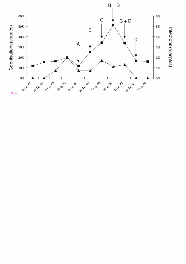
In the two years preceding the outbreak period P. aeruginosa has circulated in the
ward in an endemic fashion, being responsible for a mean colonisation rate of 10.6%
and for two ocular infections (data not shown) During the study period (July 2005 to

8/7/2019 16_Pseudomonas Aeruginosa in a Neonatal Intensive Care Unit Molecular
http://slidepdf.com/reader/full/16pseudomonas-aeruginosa-in-a-neonatal-intensive-care-unit-molecular 8/24
all the microbiologically surveilled) became colonised by P. aeruginosa during their
stay in the ward (Figure 1). Moreover, 72 patients developed 91 severe healthcare-
associated infections, with an infection rate of 14.8%, corresponding to 5.94 severe
infections/1000 patient-days. During the first year of the study (July 2005 to June
2006), 9.17 severe infections/1000 patient-days were registered, while such rate
decreased to 3.24 severe infections/1000 patient-days during the second year (July
2006 to June 2007). During the study period, P. aeruginosa proved to be the third
most common pathogen responsible for severe infections (12.1%), after Candida spp.
(21.8%) and Escherichia coli (16.8%). Moreover, no pathogen was identified in
18.8% of infants diagnosed as having an infection. P. aeruginosa was responsible for
11 severe infections in 11 neonates, with 3 and 4 of them having their birth-weights
below 1000 and 750 grams, respectively (Figure 1 and Table 1). All of the four
neonates weighing less than 750 grams died; three of them died within the first 24
hours from P. aeruginosa infection diagnosis and one died after 11 days. In addition
to isolation of P. aeruginosa from clinical samples (blood, respiratory secretions or
urines), all 11 infected patients had positive cultures for P. aeruginosa at
nasal/pharynx or rectum surveillance swabs .
Molecular typing of 90 non-repetitive isolates including 79 available colonisation
strains and all 11 infection strains (66.6% of all P. aeruginosa isolates) identified
twenty PFGE types, named A through T, which showed up to six fragments variation
in macrorestriction pattern (Figure 2 and data not shown). One predominant PFGE
profile (type A) was identified in 48 strains from 48 different patients (53.3% of all

8/7/2019 16_Pseudomonas Aeruginosa in a Neonatal Intensive Care Unit Molecular
http://slidepdf.com/reader/full/16pseudomonas-aeruginosa-in-a-neonatal-intensive-care-unit-molecular 9/24
patients, respectively, and caused no infections, with the exception of profile G (Table
1). Such PFGE profiles circulated in the ward in the following months: PFGE profile
Q was isolated from August to September 2005, profile R during January and
February 2007, profile J in December 2005, February 2006, January 2007 and May
2007, profile G during August, October and December 2005 and May 2007. The other
six infections were caused by sporadic PFGE types (B, F, K, L, O, and R) (Table 1
and Figure 2). In all 11 infected patients, surveillance cultures and clinical samples
showed identical PFGE profiles, thus excluding the possibility of multiclonal
infection in the same neonate (data not shown).
With the exception of G and O PFGE profiles, all isolates recovered from
infected neonates proved to be susceptible to aminopenicillins, ureidopenicillins,
monobactams, second-, third- and fourth-generation cephems, carbapenems,
fluoroquinolones, while resistant to trimethroprim-sulphometossazole,
cloramphenicols and tetracycline. PFGE types G and O showed resistance also to
imipenem and meropenem (MIC >8), gentamicin (MIC >8) and ciprofloxacin (MIC
>2); in addition, PFGE type G appeared to be resistant to piperacillin/tazobactam
(MIC >64/4). Moreover, such resistant profiles were responsible for two sepsis in two
neonates with extremely low birth-weight, both having a fatal outcome (Table 1).
Infection Control Interventions
After the sudden increase of infections caused by P. aeruginosa during the
fourth quarter of 2005, a combination of targeted infection control measures were
undertaken (Figure 1). A major concern of the infection control team was the

8/7/2019 16_Pseudomonas Aeruginosa in a Neonatal Intensive Care Unit Molecular
http://slidepdf.com/reader/full/16pseudomonas-aeruginosa-in-a-neonatal-intensive-care-unit-molecular 10/24
started and contact isolation for colonised and infected patients was reinforced.
Moreover, all P. aeruginosa strains isolated from clinical samples and available
strains isolated from surveillance swabs were collected for genotyping. After such
control measures were implemented, infection rate slightly decreased, while the rate
of colonised patients progressively increased.
Although no further isolations of multi-drug resistant P. aeruginosa were
made, tighter control interventions were then undertaken owing to the marked
increase of colonisations. Environmental microbiological sampling was performed
twice during the outbreak period (Figure 1). Reporting of PFGE analysis results was
followed by a 30 minutes daily educational programme on hand disinfection. Such
control measures were periodically repeated as shown in Figure 1. The daily
educational programme on hand disinfection was not specific for P. aeruginosa
containment as it was part of the ward’s plan for healthcare-associated infections
control. Staff’s overall attendance rate was high for all the educational programme’s
editions (> 90%).
Environmental microbiological sampling identified P. aeruginosa at the
following sites: three sinks on both occasions and a nurse’s hand on the second
sampling. Genotyping of such strains demonstrated that the isolate recovered from
one of the sinks on the first environmental sampling displayed an A profile and the
one from the nurse’s hand on the second environmental sampling displayed a G
profile. The other environmental isolates showed different profiles, not corresponding
to any of the profiles isolated from patients’ surveillance swabs or clinical specimens.
8/7/2019 16_Pseudomonas Aeruginosa in a Neonatal Intensive Care Unit Molecular
http://slidepdf.com/reader/full/16pseudomonas-aeruginosa-in-a-neonatal-intensive-care-unit-molecular 11/24
Hand disinfection compliance evaluation
P. aeruginosa of PFGE profile G was isolated only once from surveillance
cultures of a total of 90 randomly selected healthcare workers’ hands before and after
each educational programme, as described above. In addition, other 16 surveillance
cultures proved to be inappropriate (according to the local infection control committee
inappropriateness was defined as bacterial counts > 0.5 CFU/cm2
in high risk wards).
Hand disinfection compliance of HCWs before the first educational programme
proved to be 23.4% and 11.7% before and after each patient contact, respectively
(Table 2). Such rates significantly increased to 43.6% (p = .000) and 39.6% (p =
.000), respectively after the first intervention (Table 2). Further significant increases
were recorded after the last educational programme to 63.7% (p = .000) and to 57.1%
(p = .000), respectively (Table 2). Improvement in hand disinfection compliance
before patient contact proved to be very strongly, significantly, and inversely
correlated with rates of P. aeruginosa colonisation (r = -1, p = .004, data not shown).
Conversely, no significant correlation was found between the latter and improvement
in hand disinfection compliance after patient contact (r = -.992, p = .081, data not
shown). Finally, hand disinfection compliance of HCWs before and after patient
contact proved to be significantly different at all times of observation (Table 2 and
data not shown).
DiscussionThe overall incidence rate of P. aeruginosa infections in NICUs is reported to
be of approximately 10% [1 2 15] while infection attack rates during outbreak

8/7/2019 16_Pseudomonas Aeruginosa in a Neonatal Intensive Care Unit Molecular
http://slidepdf.com/reader/full/16pseudomonas-aeruginosa-in-a-neonatal-intensive-care-unit-molecular 12/24
developed 11 P. aeruginosa severe infections, with an infection attack rate of 1.8%,
and nearly 24% of the patients became colonised by P. aeruginosa , with an epidemic
peak of 50% at 18 months from onset. Ten of the 11 infected patients were pre-term
neonates (gestational age < 37 weeks) and 7 of them were of extremely low birth
weight (ELBW), i.e. below 1000 grams. ELBW neonates have actually been shown to
have a significantly increased risk of acquiring P. aeruginosa when compared to
higher birth-weight infants [9]. Moreover, all of the four infected neonates who died
weighed less than 1000 grams, therefore the crude mortality rate among ELBW
patients infected by P. aeruginosa was of 57%. Although no attributable mortality
rate was calculated, three of the four neonates died soon after (0-24 hours) the
infection was diagnosed, thus our data indirectly confirm other Authors’ findings
regarding the very high mortality rates related to P. aeruginosa infections [15],
especially in the lowest birth-weight categories’ infants [18]. In our setting, in
addition to the ELBW condition, two P. aeruginosa antibiotypes, both displaying
resistance to imipenem, meropenem, gentamicin and ciprofloxacin, have probably
affected the final outcome in two of the four fatal cases. We did not analyse
mechanisms of resistance as no further such phenotypes were identified. Nevertheless,
to our knowledge, this is one of the first accounts on two carbapenem-resistant P.
aeruginosa genetically unrelated strains which caused two sepsis in a NICU.
P. aeruginosa frequently causes multi-clone outbreaks, with the concurrent
isolation of genetically distinct strains among patients and healthcare workers
(HCWs) and in the environment. During a 15 months-long epidemic in a NICU,

8/7/2019 16_Pseudomonas Aeruginosa in a Neonatal Intensive Care Unit Molecular
http://slidepdf.com/reader/full/16pseudomonas-aeruginosa-in-a-neonatal-intensive-care-unit-molecular 13/24
two distinct genotypes, unrelated to any human isolate. Moreover, Foca et al. [17]
described the circulation of multiple P. aeruginosa PFGE profiles over a 33 months-
period in the NICU, showing the presence of a major clone, which was also isolated
from the hands of a nurse, of two other PFGE types, and of eight unique clones. No
environmental specimen proved to be positive for P. aeruginosa. Our study, covering
a 24 months time span, identified a predominant PFGE type which was responsible
for 36% of infections by P. aeruginosa and at least 35% of colonisations by the same
pathogen. Such PFGE profile was also found in one sink, but not on any nurse’s hand
and circulated in the ward together with less recurrent and with sporadic strains,
which caused the remaining infections and colonisations. Other five environmental
samples proved to be positive for distinct P. aeruginosa PFGE types, unrelated to the
ones colonising or infecting the patients. Transmission of P. aeruginosa from
environmental sources to patients and HCWs has been thoroughly described [19,20].
Our findings indicate the presence in our NICU of multiple and distinct P. aeruginosa
reservoirs, both environmental and human, and, owing to the long time period
between the appearance in neonates (July 2005) and the environmental isolation (2 nd
quarter 2006), we are not able to understand whether the only sink sample found to be
positive for P. aeruginosa PFGE profile A has been a result, rather than the origin, of
the pathogen’s circulation in the ward.
Healthcare workers’ (HCWs) hands have been frequently implicated in the
spreading of P. aeruginosa in the NICU setting [9,10,17]. At our institution one
HCW, with short to medium-length natural fingernails, had a positive hand culture for
8/7/2019 16_Pseudomonas Aeruginosa in a Neonatal Intensive Care Unit Molecular
http://slidepdf.com/reader/full/16pseudomonas-aeruginosa-in-a-neonatal-intensive-care-unit-molecular 14/24
sometimes identify the pathogen’s reservoir, thus enabling a successful and timely
outbreak containment [10]. Owing to organizational difficulties, the HCW found to
have a positive hand culture at our NICU could not be reassigned to non-clinical
activities. Moreover, no further analyses have been performed to establish her
contribution to the outbreak, therefore we can only hypothesize that she has been a
transient carrier of one of the less recurrent epidemic clones. Actually, patient
exposure within the first 14 days of NICU admission to a HCW with short natural
fingernails and with one positive hand culture for a P. aeruginosa epidemic clone has
not been recognized as an independent risk factor for acquiring P. aeruginosa
colonization or infection [9]. In turn, exposure to two HCWs with negative hand
cultures has been associated with an increased risk of colonisation by a P. aeruginosa
epidemic clone on multivariate analysis [17]. This finding suggests that transient
colonisations of HCWs’ hands by P. aeruginosa may be underestimated during
outbreaks investigations and that reinforcement of hand disinfection and of correct
gloves use should always be promptly initiated when an increased number of P.
aeruginosa isolations is detected. In agreement with previous data [21], our study
shows that, compared with all the traditional infection control interventions
undertaken to contain P. aeruginosa circulation of in the ward, the hand disinfection
educational programme was the most effective one. The programme started during the
fourth quarter of 2006, when the outbreak was at its peak value, and by the end of the
second quarter of 2007 the outbreak was over. Owing to the programme's success, the
meaning of different hand disinfection compliance rates before and after patient

8/7/2019 16_Pseudomonas Aeruginosa in a Neonatal Intensive Care Unit Molecular
http://slidepdf.com/reader/full/16pseudomonas-aeruginosa-in-a-neonatal-intensive-care-unit-molecular 15/24
aeruginosa outbreaks. Thus, the timely identification of increased isolation of this
pathogen, achieved by means of active surveillance, appears to be crucial to limit the
spreading of P. aeruginosa in NICU settings.
ConclusionsThis study suggests that an infection control programme based on active
surveillance and strict adherence to hand disinfection and gloves use policies and
supported by environmental sampling and molecular analysis is effective in
controlling NICU multi-clone P. aeruginosa outbreaks.
Competing interestsAll authors report no conflicts of interest relevant to this article.
Authors' contributionsVC, AS, AC and MDR carried out the active surveillance of healthcare-associated
infections and infection control interventions in the NICU, AL, ADP and TB isolated
the P. aeruginosa strains and carried out the antimicrobial susceptibility experiments,
ADP performed the PFGE experiments, VC, MT and RZ conceived the study
and
participated in its design and coordination, VC and RZ drafted the manuscript. All
authors read and approved the final manuscript.
Acknowledgements
8/7/2019 16_Pseudomonas Aeruginosa in a Neonatal Intensive Care Unit Molecular
http://slidepdf.com/reader/full/16pseudomonas-aeruginosa-in-a-neonatal-intensive-care-unit-molecular 16/24
References1. Nambiar S, Singh N: Change in epidemiology of health care-associated
infections in a neonatal intensive care unit. Pediatr Infect Dis J 2002, 9:
839-842
2. Couto RC, Carvalho EA, Pedrosa TM, Pedroso ER, Neto MC, Biscione FM: A
10-year prospective surveillance of nosocomial infections in neonatal
intensive care units. Am J Infect Control 2007, 35: 183-189
3. Gastmeier P, Loui A, Stamm-Balderjahn S, Hansen S, Zuschneid I, Shor D,
Behnke M, Obladen M, Vonberg RP, Ruden H.: Outbreaks in neonatal
intensive care units - they are not like others. Am J Infect Control 2007,
35: 172-176
4. Bagattini M, Crivaro V, Di Popolo A, Gentile F, Scarcella A, Triassi M, Villari
P, Zarrilli R: Molecular epidemiology of extended-spectrum ββββ -lactamase-
producing Klebsiella pneumoniae in a neonatal intensive care unit. J
Antimicrob Chemother 2006, 57: 979-982
5. Crivaro V, Bagattini M, Salza MF, Raimondi F, Rossano F, Triassi M, Zarrilli
R: Risk factors for extended-spectrum ββββ -lactamase-producing Serratia
marcescens and Klebsiella pneumoniae acquisition in a neonatal intensive
care unit. J Hosp Infect 2007, 67: 135-141
6. Gupta AK, Shashi S, Mohan M, Lamba IM, Gupta R: Epidemiology of
Pseudomonas aeruginosa infections in a neonatal intensive care unit. J
Trop Pediatr 1993, 39: 32-36.
8/7/2019 16_Pseudomonas Aeruginosa in a Neonatal Intensive Care Unit Molecular
http://slidepdf.com/reader/full/16pseudomonas-aeruginosa-in-a-neonatal-intensive-care-unit-molecular 17/24
with a water-bath used to thaw fresh frozen plasma. J Hosp Infect 1998,
39: 309-314
8. Gras-Le Guen C, Lepelletier D, Debillon T, Gournay V, Espaze E, Roze JC:.
Contamination of a milk bank pasteuriser causing a Pseudomonas
aeruginosa outbreak in a neonatal intensive care unit. Arch Dis Child Fetal
Neonatal Ed 2003, 88: F434-435
9. Moolenar RL, Crutcher JM, San Joaquin VH, Sewell LV, Hutwagner LC,
Carson LA, Robison DA, Smithee LM, Jarvis WR: A prolonged outbreak of
Pseudomonas aeruginosa in a neonatal intensive care unit: did staff
fingernails play a role in disease transmission. Infect Control Hosp
Epidemiol 2000, 21: 80-85
10. Zawacki A, O’Rourke E, Potter-Bynoe G, Macone A, Harbarth S, Goldmann
D: An outbreak of Pseudomonas aeruginosa pneumonia and bloodstream
infection associated with intermittent otitis externa in a healthcare
worker. Infect Control Hosp Epidemiol 2004, 25: 1083-1089
11. Garner JS, Jarvis WR, Emori TG, Horan TC, Hughes JM: CDC definitions
for nosocomial infections. In: Olmsted RN, editor. APIC infection control
and applied epidemiology: principle and practice. St Louis. Mosb; 1996. A1-
20.
12. Centers for Disease Control and Prevention: Guideline for hand hygiene in
health care settings. MMWR Morb Mortal Wkly Rep 2002, 51: 1-44.
13. Clinical and Laboratory Standards Institute: Performance standards for

8/7/2019 16_Pseudomonas Aeruginosa in a Neonatal Intensive Care Unit Molecular
http://slidepdf.com/reader/full/16pseudomonas-aeruginosa-in-a-neonatal-intensive-care-unit-molecular 18/24
14. Orsi GB, Villari P, Mondillo V, Fabiani M, Marzuillo C, Penni A, Venditti M:
A plasma expander-related Pseudomonas aeruginosa outbreak. Scand J
Infect Dis 2006, 38: 1085-1088
15. Gordon A, Isaacs D: Late onset neonatal gram-negative bacillary infection
in Australia and New Zealand. Pediatr Infect Dis J 2006, 25: 25-29
16. Gérardin P, Farny K, Simac C, Laurent AF, Grandbastien B, Robillard PY:
Pseudomonas aeruginosa infections in a neonatal care unit at Reunion
Island. Arch Pediatr 2006, 13: 1500-1506
17. Foca M, Jacob K, Whittier S, Della Latta P, Factor S, Rubenstein D, Saiman
L: Endemic Pseudomonas aeruginosa infection in a neonatal intensive care
unit . New Engl J Med 2000, 343: 695-700
18. Stoll BJ, Hansen N, Fanaroff AA, Wright LL, Carlo WA, Ehrenkranz RA,
Lemons JA, Donovan EF, Stark AR, Tyson JE, Oh W, Bauer CR, Korones
SB, Shankaran S, Laptook AR, Stevenson DK, Papile LA, Poole WK: Late-
onset sepsis in very low birth weight neonates: the experience of the
NICHD Neonatal research Network. Pediatrics 2002, 110: 285-291
19. Petignat C, Francioli P, Nahimana I, Wenger A, Bille J, Schaller MD, Revelly
JP, Zanetti G, Blanc DS: Exogenous sources of Pseudomonas aeruginosa in
intensive care unit patients: implementation of infection control measures
and follow-up with molecular typing. Infect Control Hosp Epidemiol 2006,
27: 953-957
20. Rogues A-M, Boulestreau H, Lashéras A, Boyer A, Gruson D, Merle C,
8/7/2019 16_Pseudomonas Aeruginosa in a Neonatal Intensive Care Unit Molecular
http://slidepdf.com/reader/full/16pseudomonas-aeruginosa-in-a-neonatal-intensive-care-unit-molecular 19/24
21. Lam BCC, Lee J, Lau YL: Hand Hygiene Practices in a Neonatal Intensive
Care Unit: A Multimodal Intervention and Impact on Nosocomial
Infection. Pediatrics 2004, 114 :e565-e571

8/7/2019 16_Pseudomonas Aeruginosa in a Neonatal Intensive Care Unit Molecular
http://slidepdf.com/reader/full/16pseudomonas-aeruginosa-in-a-neonatal-intensive-care-unit-molecular 20/24
Figure legends
Figure 1 -Timing of control measures and incidence of isolation of P. aeruginosa
in the NICU during the study period. Squares and triangles represent neonates
colonised or infected by P. aeruginosa , respectively. The following letters indicate
the infection control interventions performed: A, alert surveillance for P. aeruginosa ,
collection of strains isolated from clinical samples, and reinforcement of contact
isolation precautions; B, environmental microbiological sampling; C, reporting of
PFGE analyses to staff members; D, thirty minutes daily educational programme on
hand disinfection carried out in the ward for one week.
Figure 2 - Representative PFGE profiles of P. aeruginosa isolates from neonates
in the NICU. Capital letters on the top of the lanes indicate PFGE types identified; m,
phage lambda DNA molecular mass markers. Sizes of lambda DNA molecular mass
markers are shown on the right of the panel.

8/7/2019 16_Pseudomonas Aeruginosa in a Neonatal Intensive Care Unit Molecular
http://slidepdf.com/reader/full/16pseudomonas-aeruginosa-in-a-neonatal-intensive-care-unit-molecular 21/24
Tables
Table 1 - Clinical and microbiological features of P. aeruginosa isolates.
patientgestational
age(weeks)
birthweight(grams)
type of infection outcome resistance phenotype antibio
typePFGEtype
TZP IPM GEN CIP1 33,0 1950 sepsis discharge S S S S 1 B2 26,2 790 pneumonia discharge S S S S 1 L3 38,4 3160 pneumonia discharge S S S S 1 A4 24,4 700 sepsis death R R R R 3 G5 26,4 600 sepsis death S R R R 2 O6 28,0 960 pneumonia discharge S S S S 1 K7 36,0 2550 pneumonia discharge S S S S 1 F8 25,2 770 pneumonia discharge S S S S 1 A9 23,1 700 sepsis death S S S S 1 A
10 33,0 1400 U.T.I. discharge S S S S 1 R11 30,0 650 pneumonia death S S S S 1 A
NOTE. U.T.I., Urinary tract infection; S, susceptible; R, resistant.

8/7/2019 16_Pseudomonas Aeruginosa in a Neonatal Intensive Care Unit Molecular
http://slidepdf.com/reader/full/16pseudomonas-aeruginosa-in-a-neonatal-intensive-care-unit-molecular 22/24
B + D
8/7/2019 16_Pseudomonas Aeruginosa in a Neonatal Intensive Care Unit Molecular
http://slidepdf.com/reader/full/16pseudomonas-aeruginosa-in-a-neonatal-intensive-care-unit-molecular 23/24
0%
10%
20%
30%
40%
50%
60%
1 s t
q . 0 5
2 n d
q . 0 5
3 r d
q . 0 5
4 t h q .
0 5
1 s t
q . 0 6
2 n d
q . 0 6
3 r d
q . 0 6
4 t h q .
0 6
1 s t
q . 0 7
2 n d
q . 0 7
3 r d
q . 0 7
0%
1%
2%
3%
4%
5%
6%
A
B
C C + D
D
C o
l o n
i s a
t i o n s
( s q u a r e s
)I nf e
c t i o n s ( t r i a n g
l e s )
Figure 1
L

8/7/2019 16_Pseudomonas Aeruginosa in a Neonatal Intensive Care Unit Molecular
http://slidepdf.com/reader/full/16pseudomonas-aeruginosa-in-a-neonatal-intensive-care-unit-molecular 24/24
m B O L m A A B O K B G R F m
- 242.5 kb
- 194.0 kb
- 145.5 kb
- 97.0 kb
- 48.5 kb
Figure 2